
Prospects of Curcumin Nanoformulations in Cancer Management


1
Department of Pharmaceutics, School of Pharmacy, University of Health and Allied Sciences, Ho PMB 31, Ghana
2
Faculty of Pharmacy, Middle East University, Amman 11831, Jordan
3
Department of Pharmaceutical Sciences, University of Connecticut, Storrs, CT 06269, USA
4
Pharmaceutical Sciences Department, College of Pharmacy, QU Health, Qatar University, Doha P.O. Box 2713, Qatar
*
Author to whom correspondence should be addressed.
Molecules 2022, 27(2), 361; https://doi.org/10.3390/molecules27020361
Submission received: 16 November 2021
/
Revised: 27 December 2021
/
Accepted: 3 January 2022
/
Published: 7 January 2022
(This article belongs to the Special Issue Recent Progress in Health Benefits from Curcumin)
Abstract
:There is increasing interest in the use of natural compounds with beneficial pharmacological effects for managing diseases. Curcumin (CUR) is a phytochemical that is reportedly effective against some cancers through its ability to regulate signaling pathways and protein expression in cancer development and progression. Unfortunately, its use is limited due to its hydrophobicity, low bioavailability, chemical instability, photodegradation, and fast metabolism. Nanoparticles (NPs) are drug delivery systems that can increase the bioavailability of hydrophobic drugs and improve drug targeting to cancer cells via different mechanisms and formulation techniques. In this review, we have discussed various CUR-NPs that have been evaluated for their potential use in treating cancers. Formulations reviewed include lipid, gold, zinc oxide, magnetic, polymeric, and silica NPs, as well as micelles, dendrimers, nanogels, cyclodextrin complexes, and liposomes, with an emphasis on their formulation and characteristics. CUR incorporation into the NPs enhanced its pharmaceutical and therapeutic significance with respect to solubility, absorption, bioavailability, stability, plasma half-life, targeted delivery, and anticancer effect. Our review shows that several CUR-NPs have promising anticancer activity; however, clinical reports on them are limited. We believe that clinical trials must be conducted on CUR-NPs to ensure their effective translation into clinical applications.
1. Introduction
According to Global Cancer Incidence, Mortality and Prevalence (GLOBOCAN) 2020, over 19.3 million new cancer cases were diagnosed globally in 2020, with nearly 10.0 million deaths attributed to this statistic [1]. Specifically, lung cancer is the prominent cause of death from cancer in men, while breast and cervical cancers are the primary causes of cancer death in women. Cancer originates when cells in the body start to grow out of control and typically develop slowly over several years. Thus, cells in almost any part of the body can potentially develop into a cancer and can spread to other areas of the body.
The prognosis and treatment options for cancer mainly depend on the stage of the cancer, tumor recurrence, and the patient’s general health. The four prevalent cancer treatment procedures are surgery, chemotherapy, radiotherapy, and targeted therapy [2]. Surgery is the first therapeutic approach and primary procedure. Usually, surgery is combined with chemotherapy and/or radiation therapy to avoid residual tumor recurrence. In radiotherapy, energy and X-rays produced by a linear accelerator are employed to destroy cellular DNA and inhibit cell proliferation [3]. On the other hand, chemotherapy relies on cytotoxic drugs to inhibit the growth of cancer cells and kill them [4]. Chemotherapy is either administered before surgery to shrink tumors, or after surgery as adjuvant chemotherapy. Due to the limited efficacy of non-selective chemotherapeutic drugs, targeted therapy has evolved as a rational option. In targeted therapy, drugs are targeted to specific receptors present on cancer cells or in structures related to cancer growth, such as blood vessels. Additionally, cancer cell proliferation and metastasis are blocked via the inhibition of specific mediators such as epidermal growth factors (EGFs) [5]. However, the effectiveness of targeted therapy depends on the release of the therapeutic agent at the cancer target site, as well as minimizing off-target side effects to normal tissues [6].
Regardless of the advanced innovations in cancer therapy, treatment remains arduous. For instance, surgery is affiliated with detrimental harm to adjacent organs and tissues, discomfort, infections, and relapse [7,8,9]. While chemotherapy, the typical cancer treatment option, whether administered as a neoadjuvant, adjuvant, or sole therapy, demonstrates severe side effects, involving fatigue, sores in the mouth and throat, nausea, vomiting, and blood disorders [10,11,12]. Radiation therapy also manifests a wide range of side effects akin to skin changes, fatigue, and diarrhea, among others, that disturb the well-being of the patients. Even though targeted therapy has emerged as an approach to conquer the lack of specificity in conventional chemotherapy, there are potential risks and challenges associated with this novel strategy. For example, some cancer cell types develop resistance to drugs over the treatment course, thereby rendering the targeted therapy ineffective by driving the drug out of the cancer cells and decreasing intracellular drug concentration [13]. Therefore, in most cases, targeted therapy is used in combination with chemotherapy; however, this strategy does not reduce the toxicity experienced with chemotherapeutic drugs.
To overcome the aforementioned constraints, chemotherapeutic agents from plants are becoming serious contenders as chemotherapeutic alternatives due to their manifestation of reduced toxicity to adjoining cells [14] while still providing potent anticancer effects in some cases. One such chemotherapeutic agent is curcumin (CUR), a major chemical constituent in turmeric, which has received much attention in the past decades because of its use in Indian and Chinese traditional medicine for centuries to treat a variety of conditions including infections, inflammation, and depression. It is also used as a spice. Thus, CUR is widely accepted by the public as it is derived from natural sources, all of which gives the perception that it is safe to use and likely to manifest fewer side effects when used therapeutically [15]. However, the systemic bioavailability following the oral administration of free CUR is poor due to the low solubility and rapid metabolism as stated earlier [16]. Aptly, extensive research has elaborated on the therapeutic potential of CUR against a range of cancers [17], but only through formulation intervention.
2. Curcumin (CUR)
2.1. General Background Information
CUR is a hydrophobic, orange-yellow, crystalline phytochemical derived from the rhizomes of turmeric (Curcuma longa), a plant which grows in the Indian subcontinent and tropical countries in South East Asia [13]. From ancient times, turmeric was therapeutically used to treat various respiratory conditions, liver disorders, abdominal pain, and many other ailments [18,19].
Later in the 13th century, turmeric was introduced to Europe as Indian saffron by Arab merchants and used as a colorant in foods, cosmetics, and textiles [20].
The primary source of turmeric is India, where about 80% of global turmeric is consumed [21]. Epidemiological studies attribute the low incidence of colon cancer in the Indian subcontinent to the chemo-preventive properties of diets rich in CUR [22].
The presence of a cluster of sesquiterpenes, such as (S)-ar-turmerone, zingiberene, β-turmerone, and curlone, and a variety of other volatile compounds (e.g., monoterpenes) in turmeric, gives its fragrance when used as a food seasoning [23]. The active constituents in turmeric are known as curcuminoids, with the major curcuminoid being a bis-α,β-unsaturated diketone. Apart from CUR, also called diferuloylmethane, the other two bioactive analogs are demethoxycurcumin (DMC) and bisdemethoxycurcumin (BDMC). The chemical structures of the three curcuminoids are presented in Figure 1.
CUR is considered the principal curcuminoid that exhibits most of the therapeutic activities in turmeric [24]. Moreover, the characteristic yellow color of turmeric is attributable to CUR [25].
The bright yellow color of curcumin turns red and yellow in basic and acidic media, respectively [26]. It also fluoresces under ultraviolet light. In aprotic solvents (e.g., ethyl acetate and acetone), CUR has a noticeable fluorescent intensity variation from 494 to 538 nm. In solvents such as alcohols and dimethyl sulfoxide (DMSO), the fluorescence shifts to 535–560 nm, while the fluorescence spectrum of CUR in cyclohexane is notably distinct with two fluorescence maximas at 446 and 470 nm [27,28,29].
The IUPAC name of CUR is 1,7-bis (4-hydroxy-3-methoxy-phenyl) hepta-1, 6-diene-3, 5-dione, with a molecular weight of 368.37 Da, melting point of 183 °C, and three pKa values of 7.80, 7.35, and 9.0 [14]. It demonstrates keto-enol tautomerism, with the predominant keto- form in acidic and neutral solutions, and a stable -enol form in alkaline media [30].
CUR is lipophilic and is thus insoluble in water, but soluble in organic solvents such as DMSO, methanol, acetone, and ethanol [22]. Furthermore, CUR is a potent H-atom donor at pH 3.0–7.0 and an electron donor at an alkaline pH, at which it dissociates to form feruloyl methane, ferulic acid, and vanillin [27,31,32]. The bis-keto form of CUR dominates under acidic and neutral conditions, whereas the enol tautomer prevails at pH values over 8.0 [14,20]. The stability of CUR is considered pH-dependent, with the least stability in acidic media [32].
Shen and Ji [33] found that CUR degrades to dihydroferulic acid and ferulic acid in cell culture media with 10% fetal bovine serum and in in vivo studies. Even though the mechanism of degradation is still vague today, it is thought to be through its α,β-unsaturated β-diketo moiety [34]. Fortunately, this constraint can be diminished by encapsulating CUR into either liposomes, cyclodextrin, lipids, surfactants, or polymeric nanoparticles (NPs) [29].
The most compelling and key rationale for the therapeutic use of CUR as an anticancer alternative is its extremely superior safety profile; thus, it is declared as GRAS (generally recognized as safe) by the United States Food and Drug Administration [18]. However, all the preclinical and clinical data from the oral administration of CUR have revealed that it manifests poor systemic bioavailability with high susceptibility to metabolic degradation, whereby only about 2.30 µg/mL of CUR was registered in serum levels after an oral administration of 10 g of CUR [20]. This shows that CUR undergoes extensive metabolic degradation prior to absorption within the intestine and liver, which minimizes its usefulness following oral intake.
2.2. Pharmacological Properties of CUR
In the last few decades, there has been an increased interest by researchers in the therapeutic effects of CUR as a natural alternative to chemical drugs in the management of several ailments [34]. Indeed, CUR is reported to possess a variety of pharmacological activities, including antimalarial [35,36,37], antibacterial [38,39], antiviral [40,41,42], antifungal [43,44], antioxidant [45,46,47], anti-inflammatory [45,48,49], antidiabetic [50,51], anti-human immunodeficiency virus [52,53,54], and anticancer [38,39,55,56,57,58] activities.
2.3. Anticancer Properties of CUR
Generally speaking, normal cells have a restrained balance between growth upholding and growth opposing signals [59]. Thus, the proliferation and differentiation of cells transpires only when required. However, this balance is disturbed in tumor cells, which show continuous cell proliferation, loss of differentiation, and programmed cell death. Consequently, a hyper-proliferative state of cells is attained, which presents as cancer [60]. Other peculiarities recognized in tumor cells include metastasis, angiogenesis, and apoptosis [61].
CUR has been extensively studied as a potential anticancer remedy, as well as a chemopreventive and direct therapeutic agent. The anticancer properties of CUR have been proven in vitro, in vivo, and in clinical studies. It is reported that the anticancer properties of CUR are exhibited via the inhibition of cell proliferation, induction of apoptosis, and devaluation of tumor load.
2.3.1. Effects of CUR on Transcription Factors
Specific transcription factors that are reported to be involved in the anticancer effect of CUR include nuclear factor kappa B (NF-κB) [62], activator protein-1 [63], early growth response-1 [64], peroxisome proliferator-activated receptor-γ [65], signal transducer and activator of transcription, hypoxia inducible factor-1 [66], β-catenin [67], NF-E2-related factor 2 [68], electrophile response element [69], and androgen receptor [70]. CUR is believed to modulate various signaling pathways, thereby contributing to the activation of the aforementioned transcription factors [71]. Thus, CUR is able to regulate cell proliferation, inflammation, metastasis, angiogenesis, and invasion [66].
2.3.2. Effects of CUR on Growth Factors and Protein Kinase
CUR suppresses and downregulates the expression of several growth factors that contribute to the development of various cancers [72]. The activity of several tyrosine kinases increases due to mutations, which subsequently results in the malignant metamorphosis and metastasis of human cancers. CUR downregulates epidermal growth factor receptor (EGFR) activity and epidermal growth factor (EGF)-induced tyrosine phosphorylation of EGFR, eventually resulting in reduced protein kinase activity [73]. In addition, CUR suppresses the activities of protamine kinase, pp60c-src tyrosine kinase, autophosphorylation-activated protein kinase, and protein kinase C [74].
2.3.3. Effects of CUR on Inflammatory Cytokines
NF-κB is activated during the initial stages of inflammation. Consequently, the production of the multifunctional cytokine tumor necrosis factor (TNF) is upregulated, which, in turn, activates the production of interleukin (IL)-1 [75]. The activation of both TNF and IL-1 promotes the expression of several genes and proteins that engender acute and chronic inflammation [76]. It has been reported that chronic inflammation and activation of inflammatory cytokines mediate tumorigenesis [75,77,78].
CUR shows a synergistic apoptotic effect when combined with TNF-related apoptosis-inducing ligand. It also shows anti-inflammatory activity by blocking the phosphorylation of IκBα and inhibitors of NF-κB. This results in the inhibition of NF-κB activation and, subsequently, TNF. Moreover, CUR inhibits TNF expression [79] as well as phorbol-methyl-acetate-induced TNF-α levels in various cells [80].
2.3.4. Effects of CUR on Enzymes
CUR has regulatory effects on various enzymes associated with inflammation and cancer, including fatty acid synthase, ATP-citrate lyase (ACLY), stearoyl-CoA desaturase 1, and cholesterol O-acyl-transferase [78,81]. Additionally, CUR potently inhibits carbonyl reductase [82,83], downregulates the expression of other reductases in the aldo-keto reductase superfamily, and inhibits the transport of anthracyclines out of tumor cells [84].
Glutathione-S-transferase enzymes (GSTs) are reportedly involved in chemotherapy resistance in several cancer cell lines due to the methylation of GSTs [85]. CUR covalently binds to the catalytic thiolate of DNA methyltransferase 1, resulting in the blocking of DNA methylation of GSTs [86]. High levels of ACLY have been detected in various cancers such as breast, bladder, colorectal, lung, liver, prostate, and stomach tumors. ACLY activation is promoted by increased levels of glucose and insulin-like growth factors, which subsequently intervene in cancer progression [87]. However, CUR decreases the hepatic expression of ACLY [88].
Cyclooxygenase-2 (COX-2) expression is promoted by growth factors, inflammatory cytokines, oncogenes, carcinogens, and tumor promoters. However, COX-2 inhibitors are believed to aid in cancer prevention and treatment [85]. In a previous study, CUR was found to suppress COX-2 activity directly and selectively. Additionally, it inhibited bile acid and phorbol-ester-induced COX-2 expression and interferon-alpha-induced COX-2 activation [89]. These data clearly show that CUR has an anticancer effect.
3. Potential of Nanodrug Delivery Systems in Cancer Treatment
Nanodrug delivery has been proposed as the frontier for the effective delivery of anticancer agents and, hence, for cancer management. Oral nanodrug systems can be formulated to traverse the gastrointestinal epithelia effectively, and thus circumvent the metabolic constraints that the payload is subjected to within the gastrointestinal tract. According to the National Cancer Institute, nanotechnology has the potential of improving the current status of cancer detection, treatment, and prevention [90].
NPs have several characteristics ideal for the enhanced delivery of CUR in cancer management [91] (Figure 2). They exhibit a large surface area to volume ratio, high drug loading propensity, controlled drug release characteristics, and fairly good stability on storage [92]. NPs can be functionalized to deliver drugs specifically to cancer cells with minimal interaction with healthy tissue. Such targeting of NPs to cancer tissue is classified as either passive- or active-targeting [93]. The passive-targeting mechanism, also known as the EPR effect, occurs when NPs are in the size range of 10–100 nm, and, when in circulation, can selectively enter tumors through the surrounding leaky blood vessels and the interstitial space [94]. Owing to this EPR effect, NPs can also improve the safety and pharmacokinetic characteristics of active pharmaceutical ingredients [95]. Typically, NPs that employ the EPR effect to deliver drugs are intravenously administered since orally administered NPs have several hostile barriers to traverse within the gastrointestinal tract delivery in the systemic circulation. However, the active targeting of NPs to cancer cells entails specific ligand–receptor recognition and interaction on the cell surface [96].
Typically, cancer cells express higher levels of cell-surface receptors as compared to normal cells. This allows NPs conjugated with a targeting ligand to explicitly interact with them via receptor-mediated molecular recognition. There are several examples of CUR-NP delivery systems, which may be subdivided into organic and inorganic NPs [97]. Their particle size, surface charge (zeta potential, ZP), hydrophilicity/hydrophobicity, and composition, among other characteristics, can be tailored for a diverse array of applications [98]. However, the primary consideration when designing any drug delivery system is to control drug concentration within the therapeutic window and improve patient compliance in order to maintain effective treatment cycles with short recovery periods.
The following sections are a review of potentially useful CUR-containing NP preparations for the management of different cancers.
4. Prostate Cancer
4.1. Fibrinogen NPs
CUR-loaded fibrinogen NPs (CUR-FNPs) (size: 150–200 nm; ZP: −28 mV, encapsulation efficiency (EE): 90%) fabricated through chemical cross-linking with CaCl2 in a two-step coacervation process were found to be comparatively non-toxic to normal fibroblast L929 cells, but toxic to PC-3 prostate cancer cells, whereas free CUR showed no cytotoxicity [99]. The CUR-FNPs also showed a dose-dependent apoptotic effect on the cancer cells with significant internalization and retention within the cells.
4.2. Cyclodextrin (CD)-Based NPs
Yallapu and colleagues [100] fabricated β-CD-CUR-NPs (52.6 nm) and found that uptake of the NPs by PC-3 cells was higher than that of free CUR. Similarly, the apoptosis rate was higher with the NPs, indicating the potential of the β-CD-CUR-NPs for managing prostate cancer. In addition, Ndong Ntoutoume and colleagues [101] prepared CUR-CD/cellulose nanocrystal complexes (206.8 nm) by ionic interaction with improved CUR solubility. The NPs showed a higher antiproliferative effect against PC-3 and DU145 cancer cells compared to free CUR. The half-maximal inhibitory concentration (IC50) values for the NPs after 48 h of treatment were 7.5 and 5.5 µM against the PC-3 and DU145 cells, respectively, as opposed to 10 and 18 µM, respectively, for free CUR. These findings are promising; however, in vivo studies are needed to confirm the potential of these CD-based CUR-NPs for managing prostate cancer.
4.3. Magnetic and Dendrimer-Based NPs
In the study conducted by Yallapu and colleagues [100], CUR-loaded magnetic and dendrimer-based NPs (8.6 and 37.4 nm, respectively) were also evaluated in PC-3 cells with similar cellular uptake findings. However, uptake was higher for the dendrimers due to their attachment to the cells. The considerably small sizes of these NPs hold potential for cancer management, but further studies are required.
4.4. Polymeric NPs
Yallapu and colleagues [100] also found that the uptake of CUR-NPs formulated with hydroxypropyl methyl cellulose (HPMC-CUR-NPs) and poly(lactic-co-glycolic acid) (PLGA-CUR-NPs) (5.2 and 58.1 nm, respectively) was higher compared to free CUR in PC-3 cells. Unlike free CUR, the NPs also induced apoptosis, with superior effects from the HPMC-CUR-NPs. The HPMC-CUR-NPs had a greater antiproliferative effect and higher cytotoxicity via higher internalization, retention, and apoptosis compared to free CUR in C4-2, DU-145, PC-3, and LNCaP cells (IC50: 11.5 ± 4.2–37.4 ± 3.2 µM vs. 7.8 ± 1.78–30.1 ± 1.9 µM). Clearly, the HPMC-CUR-NPs could be potentially useful in prostate cancer treatment.
In a later study, PLGA-CUR-NPs prepared via nanoprecipitation caused large and extensive vacuoles in C4-2 and DU-145 cells, but fewer and smaller vacuoles in PC-3 cells, which is possibly indicative of lesser NP uptake. Conversely, this was not observed in cells treated with free CUR. The NPs also inhibited cell growth better in vitro (especially at 4 and 6 μM) and reduced tumor volume in mice with C4-2 xenograft tumors better than the free drug without causing systemic toxicity. Additionally, the conjugation of prostate-specific membrane antigen (PSMA) monoclonal antibody to the NPs resulted in improved NP uptake and targeted CUR delivery to PSMA-expressing cells [102].
Rao and colleagues [103] have also prepared thermally-responsive CUR-NPs (EE, 54.3–73.9%) using Pluronic F127 and chitosan via an emulsification–interfacial crosslinking–solvent evaporation–dialysis method. NP size was reduced as temperature was increased from 22 °C (~300 nm) to 37 °C (~22 nm). Mild hyperthermia (43 °C for 1–1.5 h) increased NP uptake, retention, and delivery to the nuclei of PC-3 cells. The IC50 of NPs plus hyperthermia was >7-fold lower than that of NPs only, suggesting that mild hyperthermia combined with the CUR-NPs may increase PC-3 cell destruction. However, no in vivo studies were conducted to support these results.
CUR-loaded, pH-sensitive, redox NPs (PR-CUR-NPs) (35 nm, 82% EE) have been formulated using poly(ethylene glycol)-b-poly [4-(2,2,6,6-tetramethylpiperidine-1-oxyl)aminomethylstyrene]) by the dialysis method [104]. The PR-CUR-NPs improved CUR solubility, suppressed its oxidative degradation, and were significantly more toxic to PC-3 cells compared to free CUR or empty NPs. The PR-CUR-NPs (10 mg/kg, intravenously) also significantly reduced tumor volume in tumor-bearing nude mice compared to free CUR by suppressing oxidative stress.
Polyvinyl alcohol (PVA)-CUR-NPs (46–67.3 nm, −34.6 ± 1.4 to −37 ± 4.3 mV, 71.6–90.3% EE) have been fabricated using the flash nanocomplexation technique via hydrogen bonding interactions [105] without an organic solvent. The cellular uptake and cytotoxicity of the NPs in PC-3 cells was negatively correlated to drug load. Interestingly, free CUR underwent a higher cellular uptake and was more cytotoxic than the NPs. Formulation optimization could possibly improve these findings.
4.5. Lipid NPs
In a recent study by Tanaudommongkon and colleagues [106], Miglyol 812 and d-alpha-tocopheryl PEG succinate 1000 (MT) were used to formulate CUR-NPs (138.7 ± 5.4 nm, −24.4 mV, 96.3 ± 6.0% EE) by the nanoemulsion method. The NPs were cytotoxic to docetaxel (DTX)-resistant castration-resistant prostate cancer (CRPC) cells. Compared to free CUR, the CUR-MT-NPs were 5-fold and about 2-fold more cytotoxic to PC-3 and DU145 cells, respectively. The CUR-MT-NPs were also equally cytotoxic to sensitive and resistant cells, similar to the observation for free CUR against PC-3 cells. The CUR-MT-NPs completely overcame the resistance to DTX in both PC-3 and DU145 cells.
5. Lung Cancer
5.1. Liposomes
Rahman and colleagues [107] fabricated liposomes (420 nm, EE > 66%) containing βCD-CUR complexes with better CUR water solubility than free CUR. The complexes and NPs were prepared by the methanol reflux and thin-film hydration methods, respectively. The median effective dose (EC50) values for the CUR-loaded liposomes, free CUR, βCD-CUR, and βCD-C-loaded liposomes were 0.90, 1.5, 2.4, and 2.9 µM, respectively, on A549 cells (p < 0.05), clearly showing that increasing the aqueous solubility of CUR may not necessarily correlate with improved cytotoxicity.
5.2. Lipid NPs
Wang and colleagues [108] used the sol–gel method to fabricate CUR-loaded solid lipid nanoparticles (SLNs) (20–80 nm, −11.6 mV, 75% EE) using stearic acid, lecithin, and polyoxyethylene (50) stearate. The IC50 of the CUR-SLNs (4 μM) against A549 cells was 20-fold lower than that of free CUR. The NPs (200 mg/kg daily, 5 days/week, 19 days) had no effect on body weight, but significantly reduced tumor volume by 65.3% in nude female mice xenografted with A549 cells, compared to 19.5% by free CUR. Additionally, the SLNs significantly increased CUR bioavailability (26.4-fold) in female BALB/c mice and mostly accumulated in the lung and tumor tissues after intraperitoneal administration [108].
CUR-loaded cationic lipid NPs (CUR-CLNs) (194.9 ± 7.4 nm, −28.15 ± 2.25 mV, ~98% EE) formulated by the emulsification evaporation-low temperature solidification method also showed better oral pharmacokinetic characteristics (higher bioavailability, higher plasma concentration, and lower clearance) compared to free CUR in rats. Following intravenous administration, the relative bioavailability of the CUR-CLNs to free CUR was 439.76%. The CUR-CLNs also had a better anticancer effect in vitro (Lewis lung cancer, LLC cells; IC50 20.25 μM vs. 39.70 μM) and in vivo (LLC-bearing C57BL/6J mice; tumor growth inhibition rate, ~66 vs. ~39%). The higher anticancer efficacy of the CUR-CLNs was attributed to the increased uptake and higher accumulation in the cells [109].
5.3. Gold NPs
Hoshikawa and colleagues [110] have developed PEGylated gold NPs with photothermal effects for CUR delivery. The NPs (<10 mV) were conjugated to CD (α-, β-, and γ-CDs) for CUR encapsulation, with β-CD producing the highest CUR EE%. The average size of the NPs, regardless of the CD used, was 25–35 nm (gold nanocore, ~5 nm). The CUR-CD-Au-NPs were significantly cytotoxic to A549 cells; however, their effect was similar to that of free CUR.
5.4. Polymeric NPs
Yin and colleagues [111] have fabricated CUR-NPs using three amphiphilic methoxy PEG (mPEG)–polycaprolactone (PCL) block copolymers via the nanoprecipitation method, with the mPEG10k–PCL30k giving the highest drug loading efficiency and the most sustained drug release profile. The particle size, zeta potential (ZP), and encapsulation efficiency ranges were 102.3 + 11.3 − 140.3 + 14.2 nm, −4.7 + 0.4 to −7.8 + 1.4 mV, and 75.2 + 6.3 − 83.1 + 5.8%, respectively. Reduction of A549 cell viability after 24–72 h of treatment was better with the NPs compared to free CUR, but similar at doses >80 µM. CUR uptake by the cells was also increased by the NPs [111].
CUR-coordinated ROS-responsive NPs (163.8 nm, −0.31 mV, 65% EE) have been fabricated using a biocompatible 4-(hydroxymethyl) phenylboronic acid-modified PEG-grafted poly(acrylic acid) polymer (PPH). The NPs improved CUR stability and were potent against A549 cancer cell proliferation in vitro. ROS inhibition with N-acetylcysteine resulted in the suppression of the cytotoxic effect of the NPs, which validates the selectivity of PPH-NPs for high-ROS cancer cells. Importantly, the findings showed that CUR release from the NPs was enhanced in the presence of ROS [112].
CUR-containing chitosan NPs (170–200 nm) have been prepared by ionic gelation and evaluated against human non-small cell lung carcinoma (H1299) cells [113]. Toxicity, bioavailability, and chemopreventive efficacy were evaluated in Swiss albino mice after the mice were administered the NPs or free CUR one week before treatment with benzo[a]pyrene (B[a]P), and then on alternative days for up to 4 months. CUR retention in the lungs of the mice was higher for the NPs than for free CUR. One fourth of the NP dose was also more potent in inhibiting B[a]P-induced lung carcinogenesis than free CUR was. Additionally, the CUR-loaded NPs were more effective in reducing nodule size, showing that the NPs can improve the chemopreventive efficacy of CUR against lung cancer.
6. Colorectal Cancer (CRC)
6.1. Liposomes
Pandelidou and colleagues [114] formulated and evaluated CUR-loaded liposomes (108.0 ± 8.9 nm, 85% EE) for anticancer effect against CRC cells. The liposomes were taken up by HCT116 cells to a greater extent than free CUR, and were subsequently more cytotoxic to HCT116, HCT15, and DLD-1 cells compared to free CUR (IC50: < 6 µM vs. 4.5–47.3 µM). The liposomes improved CUR activity against the cells, showing a higher potency against the HCT116 and HCT15 cells.
In another study, Chen and colleagues [115] formulated CaCO3-encapsulated liposomes containing CUR (LCC) (155.3 ± 3.8 nm, −14.2 ± 0.3 mV, 77.67 ± 1.82% EE) with pH-sensitive properties for targeted CUR release by W/O emulsion-mediated film dispersion. The LCC formulation was more cytotoxic to HCT116 cells compared to free CUR or CUR-only liposomes. Similarly, LCC caused a reduction in tumor volume in C57BL/6 mice with colon cancer better than CUR, attributable to enhanced CUR accumulation in the tumors.
In the aforementioned study by Rahman and colleagues [107], CUR-containing complexes and liposomes were also tested on SW-620 cancer cells, where the EC50 values for CUR-loaded liposomes, free CUR, CUR-loaded complexes, and βCD-C-loaded liposomes were 0.96, 1.9, 2.95, and 3.25 µM, respectively. This finding was similar to the trend observed for the lung cancer cells, with the βCD-C complex preparations having the lowest antiproliferative effects, possibly due to the higher aqueous solubility of CUR provided by the complex.
6.2. Micelles
Gou and colleagues [116] prepared CUR-containing monomethoxy PEG-PCL micelles (27.3 ± 1.3 nm, 99.16 ± 1.02% EE) by the single-step nanoprecipitation method. Although the micelles were less cytotoxic to C-26 cells than free CUR (IC50: 3.95 µg/mL vs. 5.78 µg/mL), the inhibition of angiogenesis was better in an alginate-encapsulated tumor cell assay. Importantly, the micelles (25 mg/kg CUR, intravenously) inhibited colon tumor growth in mice more than the free drug, showing their potential use in CRC management.
Another type of CUR-loaded micelles prepared with stearic acid-g-chitosan oligosaccharide (CSO-SA) (114.7 ± 16.9 nm, 18.5 ± 0.4 mV, 29.9 ± 2.9% EE) was found to protect CUR from biotransformation and hydrolysis, thereby improving CUR stability. The CUR-CSO-SA micelles showed higher in vitro uptake and 6-fold higher cytotoxicity in CRC cells than free CUR. After 14 days of treatment, the CUR-CSO-SA micelles reduced tumor size and CD44+/CD24+ cell subpopulation both in vitro and in nude mice [117].
Raveendran and colleagues [118] have used solvent dialysis to prepare a CUR-containing micelles using Pluronic/PCL amphiphilic block copolymer, with PCL, which is hydrophobic, forming the core of the micelles to improve CUR loading. The micelles (195.7 ± 7.3 nm, 17.6 ± 0.4 mV, 72.08 ± 4.29% EE) improved CUR uptake and cytotoxicity in Caco-2 cells.
In a later study, CUR-loaded micelles (27.6 ± 0.7 nm, 0.11 ± 0.34 mV, 96.08 ± 3.23% EE) were also fabricated using monomethyl PEG-PCL and trimethylene carbonate (TMC) via a single-step solid dispersion method. TMC stabilized the micelles by inhibiting PCL crystallization. The micelles increased CUR uptake and cytotoxicity in CT26 cells, and were more effective in suppressing tumor growth in female BALB/c mice (50 mg/kg CUR, intravenously) with fewer toxic effects [119].
Chang and colleagues [120] have also evaluated CUR micelles (34–80 nm, 58–63% EE) prepared using poly(PEG methyl ether methacrylate)-block-poly(styrene) block copolymer in WiDr human colon carcinoma cells. Interestingly, larger micelles were more rapidly endocytosed and exocytosed than the smaller ones were. However, CUR-loaded micelles were better internalized by the cells than unloaded micelles. Additionally, after 72 h of exposure, smaller CUR-loaded micelles remarkably reduced cell proliferation compared to free CUR [120].
6.3. Nanogel (NG)
One benefit of NGs is that they confer structural stability to drug delivery systems and protect encapsulated drugs from degradation. Madhusudana Rao and colleagues [121] have used gelatin and acrylamidoglycolic acid to fabricate CUR-loaded pH-sensitive NGs (100 nm, 42–48% EE) by a simple emulsion polymerization technique. The stabilization of NG networks was achieved with glutaraldehyde. The in vitro CUR release was higher at pH 7.4 than at 1.2. Compared to free CUR, the CUR-NGs showed better anticancer activity against HCT116 cells after 48 h of treatment.
Seok and colleagues [122] have also fabricated a hemocompatible NG using hyaluronic acid (HA) cross-linked zein for CUR delivery. The CUR-NG (200–250 nm, 20–40 mV, 94.15% EE) were more cytotoxic to CT26 cancer cells than normal NIH3T3 cells (IC50: 37 µg/mL vs. 94 µg/mL) via a higher apoptosis rate. The CUR-NG (3 mg/kg, intravenously) also showed a higher anticancer effect in BALB/c nude mice compared to free CUR [122].
Recently, Borah and colleagues [123] formulated CUR-loaded amylopectin-albumin core–shell NG functionalized with folic acid (FA) (90 nm, −24 mV, 100% EE) that induced early-stage apoptosis of human HT29 cells, whereas free CUR did not. The NG also increased CUR uptake and retention (by 60%) in FA receptor-positive HT29 cells. Additionally, it showed potential for oral delivery as it was resistant to degradation in simulated gastric and intestinal fluids.
6.4. CD-Based NPs
Ndong Ntoutoume and colleagues [101] have formulated CUR-loaded CD/cellulose nanocrystal complexes by ionic interaction (206.8 nm, −29.6 ± 2.7 mV) that improved CUR uptake by HT29 cells and exhibited an anticancer effect that was 3–4 times more effective than CUR alone. These NPs also increased the aqueous solubility of CUR.
In another study, CUR-NPs (169–338 nm, 17.1 mV, 53.0% EE) fabricated using chitosan, HA, and sulfobutyl-ether-β-CD by ionic gelation also significantly inhibited HT29 cell proliferation. The NPs were better internalized by the cancer cells compared to normal I407 cells. The aqueous solubility of CUR was also improved (3.72 μg/mL to 70 μg/mL) after encapsulation into NPs [124].
6.5. Lipid NPs
Chirio and colleagues [125] have fabricated CUR-SLNs (<300 nm, 22.13 ± 3.19 mV, 28–81% EE) by coacervation based on fatty acid precipitation. The highest EEs were obtained with palmitic acid or PVA 9000. Interestingly, the different SLNs prepared, whether loaded with CUR or not, had similar cytotoxic effects against HCT116 cells, which is attributable to low CUR EE and reduced CUR solubility in NPs with higher EEs.
6.6. Gold NPs
Positively charged CUR-Au-NPs (160 ± 20 nm, 18 ± 3 mV) with pH-, radiofrequency- and thermo-responsive properties have been formulated by Sanoj Rejinold and colleagues [126]. The NPs remained in tumors in mice bearing CT26 xenografts for up to 2 weeks. Additionally, the circulation time of CUR in the blood was much longer (up to a week) for the NPs compared to free CUR. Importantly, CUR accumulation was higher in tumors than in other organs, showing the potential of the NPs in targeted treatment of colon cancer.
Alibolandi and colleagues [127] have also formulated CUR-loaded hybrid dendrimer Au-NPs (<10 nm). PEG-Au-poly(amidoamine) (PAMAM) NPs were prepared first and then loaded with CUR. Mucin-1 conjugated aptamer (Apt) increased the uptake and cytotoxicity of the NPs in HT29 and C26 cells. The in vivo antitumor effects (survival rate and tumor growth inhibition) of the NPs (2 mg/kg CUR, intravenously, BALB/c mice) were better than those of free CUR, and were further improved with the Apt-conjugated NPs.
6.7. Polymeric NPs
Chuah and colleagues [128] have formulated CUR-loaded chitosan NPs (340 ± 4.5 nm, 43.7 ± 0.4 mV, 77.44 ± 0.2% EE) to enhance colonic CUR delivery through mucoadhesion. The CUR-NPs had better mucoadhesion properties than the empty NPs. Uptake of the CUR-NPs by HT29 cells was also higher than that of free CUR. Additionally, HT29 cell viability was better reduced after 72 h of treatment and when CUR was loaded into the NPs.
CUR-NPs have been fabricated with PLGA, soybean lecithin, and DSPE-PEG2000-COOH via nanoprecipitation, and functionalized with a ribonucleic acid Apt against epithelial cell adhesion molecule, which is overexpressed on colorectal adenocarcinoma cells, for targeted CUR delivery. The Apt-CUR-NPs (90 ± 1.9 nm, −36.3 ± 4.2 mV, 89.98 ± 3.8% EE) showed 64-fold higher binding and/or internalization by HT29 cells than CUR-NPs functionalized with negative control Apt. The Apt-CUR-NPs also had a higher antiproliferative activity against HT29 colon cells than free CUR and a significantly higher bioavailability and 6-fold longer half-life in male Sprague Dawley rats than the free drug, showing their potential benefit in CRC treatment [129].
CUR-loaded polymeric NPs (136 nm, 48 mV, 95% EE) have also been fabricated using chitosan and gum arabic by Udompornmongkol and Chiang [130] via emulsification-solvent diffusion. The NPs were stable in simulated gastrointestinal fluids. Their uptake by HCT116 and HT29 cells, as well as their antiproliferative effect against the cells, were better than those of free CUR, showing the anti-CRC potential of the NPs.
In another study, CUR-NPs prepared by emulsification-diffusion-evaporation using Eudragit E 100 (248.40 ± 3.89 nm, 65.77 ± 3.17% EE) had a 19-fold higher inhibition rate on the growth of Colon-26 cells, had a better oral bioavailability in Wistar rats, and a reduced tumor volume in Colon-26 tumor-bearing BALB/c mice (50 mg/kg CUR, orally, daily for 30 days) as compared to free CUR, showing the potential of the CUR-NPs in CRC management [131].
Silk fibroin (SF) has been used to fabricate CUR-NPs (<100 nm) via solution-enhanced dispersion by the supercritical CO2 technique. The IC50s of free CUR, CUR-SF-NPs, and fluorouracil against HCT116 cells were 5.339, 4.383, and 0.432 μg/mL, respectively; however, the CUR-SF-NPs had the most superior anticancer effect at CUR concentrations >10 μg/mL [132].
Polymeric self-emulsifying NPs (100–180 nm, 64.85 ± 0.12% EE) were formulated by Wadhwa and colleagues [133] via quasi-emulsion solvent diffusion using HPMC acetate succinate. The in vitro release studies showed that up to a 5 h lag time preceded CUR release from the NPs, which approximates the time for cecal arrival following oral intake. Furthermore, the optimized NPs were more cytotoxic to HT29 cells than free CUR (IC50: 20.32 µM vs. 28.56 µM), and CUR was successfully delivered to the colon in guinea pigs by the NPs, even though they were not well absorbed in this region.
Alkhader and colleagues [134] have prepared CUR-containing chitosan-pectinate NPs (CUR-CS-PEC-NPs) (206.0 ± 6.6 nm, 32.8 ± 0.5 mV, 64% EE) that are more mucoadhesive at an alkaline pH than at an acidic pH. A very high CUR release (>80%) was achieved in a pectinase-enriched medium (pH 6.4), indicating that the NPs are suited for colon-targeted CUR delivery. In a further study, Alkhader and colleagues [135] found that free CUR and CUR-CS-PEC-NPs inhibited HT29 cell proliferation in a dose- and time-dependent manner. Uptake of free CUR and CUR-CS-PEC-NPs by HT29 cells was comparable; however, the oral bioavailability of CUR in Sprague Dawley rats was significantly higher for the NPs.
Sabra and colleagues [136] have also formulated CUR-loaded modified citrus pectin-chitosan NPs (MCPC-CUR-NPs; 178 ± 0.896 nm, 35.7 ± 1.41 mV, 69.43% EE) that exhibit mucoadhesive properties using a one-step ionic gelation technique. Similar to the findings of Alkhader and colleagues [134], the MCPC-CUR-NPs exhibited a better mucoadhesive property at pHs of 7.0, 5.5, and 6.25 than at 1.2. Additionally, CUR release over 24 h from the NPs was higher in a 33% (w/v) cecal medium than in an acidic medium (pH 1.2) (68% versus 18%). In a further study, the MCPC-CUR-NPs showed more toxicity to HT29 and HCT-116 cells compared to unmodified NPs or free CUR, particularly after ≥48 h of treatment. Additionally, pectin modification significantly increased the cellular uptake of the NPs, especially at low CUR concentrations [137]. These results show that MCPC-CUR-NPs may be beneficial in the management of CRC.
7. Breast Cancer
7.1. Lipid NPs
CUR-loaded transferrin-mediated SLNs (Tf-CUR-SLNs; 206 ± 3.2 nm, 8.21 ± 0.89 mV, 77.27 ± 2.34% EE) prepared by homogenization have been shown to increase CUR photostability and inhibit MCF-7 cell proliferation. Cellular uptake of the Tf-CUR-SLNs was higher than that for free CUR or CUR-SLNs, possibly due to the targeting effect by Tf-CUR-SLNs on MCF-7 cells [138].
Sun and colleagues [139] fabricated CUR-loaded SLNs (152.8 ± 4.7 nm, 90% EE) by high-pressure homogenization, where cell viability was inhibited for a prolonged period, whereas cells treated with free CUR recovered viability after 72 h, confirming the superiority of the NPs. The cellular uptake of the NPs increased from 10 min to 3 h of incubation, then decreased after 3–6 h. On the other hand, uptake of free CUR was rapid in the first 10 min, but decreased sharply afterwards. Additionally, the bioavailability of CUR in Sprague Dawley rats after intravenous administration was 1.25-fold higher from the NPs compared to free CUR.
Wang and colleagues [140] have also prepared CUR-SLNs by low-temperature solid emulsification using stearic acid and lecithin as lipids. The CUR-SLNs (40 nm, −25.3 ± 1.3 mV, 72.47% EE) induced a higher level of cytotoxicity and apoptosis (IC50: 28.42 μM vs. 18.78 μM) and were better taken up by SK-BR-3 breast cancer cells than free CUR. Similar to several of the reported studies on CUR-loaded NPs, no in vivo anticancer evaluations were performed.
Minafra and colleagues [141] produced CUR-SLNs (302.5 nm, 41.4 ± 4.6 mV, 68.62% EE) with radiosensitizing ability by ethanolic precipitation followed by homogenization. Free CUR and the CUR-SLNs had a comparable cytotoxic effect on MDA-MB-231 breast cancer cells. Irradiation of cancer cells that had accumulated by CUR-SLNs with 2 Gy of photon beam resulted in the protection against radiation-induced oxidative stress and an antitumor effect through the activation of autophagy simultaneously. These findings necessitate further studies on the concomitant use of CUR-SLNs and radiotherapy for treating breast cancer.
7.2. NG
Wei and colleagues [142] have prepared a NG loaded with CUR conjugated to cholesteryl-HA (CHA) for targeted CUR delivery to CD44-expressing drug-resistant cancer cells. The NG particles (20 nm) were stable in simulated gastrointestinal fluids and had a 400-fold higher aqueous solubility than pure CUR. The NG was also more cytotoxic to 4T1 cells (IC50: 2 μg/mL vs. 5 μg/mL) and inhibited tumor growth in BALB/c mice better than free CUR did.
7.3. Silica NPs
Li and colleagues [143] found that CUR-loaded mesoporous silica NPs (MSNs) modified with HA or polyethyleneimine (PEI)-FA were more cytotoxic to MDA-MB-231 cells at 20–60 µg/mL than free CUR, CUR-MSNs, or CUR-PEI-MSNs were. This was attributed to the affinity of the CUR-PEI-FA-MSNs for FA and CD44 receptors, which are typically overexpressed on MDA-MB-231 cells. Cellular uptake was in the order: CUR-PEI-FA-MSNs > CUR-HA-MSNs > CUR-PEI-MSNs > CUR-MSNs > free CUR. The CUR-PEI-FA-MSNs (8 mg/kg CUR, every 3 days via the tail vein) showed the best antitumor effect in female BALB/c nude, pointing to the importance of suitably surface-modified NPs in cancer management.
7.4. ZnO NPs
CUR-loaded phenyl boronic acid (PBA)-conjugated and pH-responsive ZnO NPs (CUR-ZnO-PBA-NPs) (413.63 ± 9.5 nm, −16.4 ± 0.30 mV, 27% EE) have been fabricated by Kundu and colleagues [144]. The uptake of ZnO-PBA-NPs was higher than ZnO-NPs by MCF-7 cells. Furthermore, CUR-ZnO-PBA-NPs were more cytotoxic to MCF-7 cells, accumulated more in tumors, and reduced tumor volume better in Swiss albino mice (10 mg/kg CUR, intravenously on alternate days for 14 days) compared to free CUR. Intriguingly, the CUR-ZnO-PBA-NPs alleviated tumor-induced splenomegaly, which could be valuable in breast cancer patients.
7.5. Hybrid Magnetic-Polymeric NPs
CUR-loaded magnetic alginate/chitosan NPs (CUR-MAC-NPs) (172–199 nm, −28.7 to −34.2 mV, 67.5% EE) prepared via co-precipitation were internalized by MDA-MB-231 3–6 fold higher than free CUR. The CUR-MAC-NPs were also significantly less toxic to normal human dermal fibroblasts. However, in vivo studies are needed to clarify the potential of the CUR-MAC-NPs using an external magnetic field [145].
7.6. Human Serum Albumin (HSA) NPs
Saleh and colleagues [146] have formulated CUR-containing HSA-NPs decorated with human epidermal growth factor receptor 2 (HER2) Apt (Apt-CUR-HSA-NPs; 281.1 ± 11.1 nm, −33.3 ± 2.5 mV, 71.3% EE%) through desolvation. The NPs increased the aqueous solubility of CUR 400-fold. Additionally, the presence of Apt increased the cytoplasmic uptake and cytotoxicity of the NPs in HER2-overexpressing SK-BR-3 cells, indicating that the Apt-CUR-HSA-NPs may be a promising treatment for HER2-positive breast cancer. Recently, Hasanpoor and colleagues (2020) similarly fabricated CUR-HSA-NPs and functionalized the particles with programmed death ligand 1 (PDL1) binding peptide, with the peptide-CUR-HSA-NPs (246.5 nm, −24.5 ± 1.5 mV, 77.8% EE) showing a similar trend of uptake and cytotoxicity in high PDL1-expressing MDA-MB-231 cells. Matloubi and Hassan [147] have likewise fabricated CUR-HSA-NPs (220 nm, −7 mV, 70% EE), but without surface functionalization. The cytotoxicity of the NPs was lesser on peripheral blood mononuclear cells, but higher on MCF7 and SK-BR3 cells from 48 h of treatment compared to free CUR. These findings are promising, with regard to improved anticancer effects due to NP surface functionalization. However, further studies in animal models are needed to show their potential in breast cancer management.
7.7. Magnetic NPs
Ashkbar and colleagues [148] found that Fe3O4-SiO2-CUR-NPs (20–60 nm, −57.5 mV) combined with photodynamic therapy (PDT) and photothermal therapy (PTT) produced the highest reduction of 4T1 tumor volume in female BALB/c mice compared to the NPs only, free CUR plus PDT, NPs plus PTT, or PDT plus PTT. Importantly, tumor volume was decreased by 58% by the NPs alone compared to the untreated group. These show the benefit of the CUR-NPs and, even more significantly, the potential of the triple therapy in breast cancer management.
7.8. Polymeric NPs
Khan and colleagues [149] fabricated CUR-PLGA NPs (265–606 nm, −3.1 to −13.7 mV, >50% EE) by solvent evaporation and tested them on MDA-MB-231 cells, whereby a 10-fold increase in solubility and 3-fold increase in anticancer activity compared to CUR was observed. Sampath and colleagues [150] have investigated the use of tocopherol PEG 1000 succinate (TPGS, as emulsifier) with different capping agents (chitosan, dextran, and PEG) to fabricate CUR-PLGA-NPs by emulsion solvent evaporation (<200 nm, 7.94–40.47 mV, 82–89% EE). All NPs produced reduced MCF-7 cell proliferation better than free CUR did, with the highest cellular uptake observed when TPGS and dextran were used.
CUR-loaded poly-glycerol-malic acid-dodecanedioic acid NPs (110–218 nm, −17.5 to −18.9 mV, 75–81% EE) fabricated via nanoprecipitation have shown a higher antiproliferative effect against MCF-7 and MDA-MB 231 cells compared to free CUR. Interestingly, the cytotoxicity of free CUR decreased with increasing incubation periods, whereas the reverse was observed for the NPs after 7 days [151].
7.9. Phospholipid-Polymeric NPs
CUR-loaded phospholipid NPs conjugated with EGF have been formulated via thin-film hydration by Jung and colleagues [152]. The EGF-CUR-NPs (229.3 ± 6.0 nm, 63.3% EE) exhibited a dose-dependent suppression of MDA-MB-468 TNBC cell survival and were more cytotoxic than free CUR or CUR-only NPs. The EGF-CUR-NPs (10 mg/kg, intraperitoneally, three times weekly, total of 8 injections) also suppressed tumor growth in mice bearing MDA-MB-468 tumors better compared to empty or CUR-only NPs, showing that they may be beneficial for treating TNBCs with an overexpression of EGF receptors.
7.10. Polymer-Coated Gold NPs
FA-functionalized CUR-Au-PVP-NPs (358.7 nm, −12.5 mV) prepared by layer-by-layer assembly have been shown to be cytotoxic to MDA-MB-231, MCF-7, and 4T1 cancer cells, but not to normal L929 and MCF-10A cells at CUR concentrations of <100 µg/mL. The FA-CUR-Au-PVP NPs (10 mg/kg, intratumorally, 2 weeks) inhibited tumor growth in BALB/c mice bearing 4T1 tumors more than free CUR [153].
7.11. Radiolabeled NPs
Huang and colleagues [154] used membrane dialysis to fabricate self-assembled 99mTc-radiolabeled CUR-NPs with HA-cholesteryl hemisuccinate conjugates and D-a-TPGS. The NPs (144 nm, −21.25 ± 1.66 mV, 84.0 ± 5.0% EE) inhibited 4T1 cell growth by two-fold compared CUR (IC50 values: 38 μg/mL vs. 77 μg/mL). Tumor growth in tumor-bearing BALB/c mice was also inhibited by the NPs (50 mg/kg, every 2 days, total of 5 injections) without toxicities to major organs; however, the in vivo effect of free CUR was not determined.
7.12. NG
Setayesh and colleagues [155] grafted octadecylamine (ODA) to chondroitin sulfate (CS) to form a CS-ODA conjugate that was used to prepare CUR-loaded NG (311 ± 20.29 nm, −13.25 ± 0.35 mV, 79.56 ± 5.56% EE). Interestingly, the CS-ODA-NG and free CUR were equally cytotoxic to MCF-7 cells after ≥48 h of incubation; however, cellular uptake of the NG was higher.
8. Oral, Cervical, Ovarian, and Pancreatic Cancers
Reported studies on the evaluation of CUR nanoformulations for oral, cervical, ovarian, and pancreatic cancers have been summarized in Table 1.
9. Synergistic Effect of CUR-NPs with Other Anticancer Drugs
Combination drug treatment is a popular strategy that can be beneficial in overcoming drug resistance and dose-limiting toxicity, which are typical concerns associated with the management of cancer [179]. Some studies have shown that NPs containing CUR in addition to other drugs can have synergistic anticancer effects. For instance, NPs co-loaded with CUR and doxorubicin have been shown to be effective against lung cancer [180]. In other studies, NPs containing CUR and paclitaxel [181], methotrexate [182], GANT61 [183], or doxorubicin [184] have been shown to be effective against breast cancer. It has also been found that CUR and docetaxel [185] or CUR and cabazitaxel [186] have synergistic effects against prostate cancer. Finally, it has been revealed that CUR and oxaliplatin [187], 5-fluorouracil [188], camptothecin [189,190], chrysin [191], doxorubicin [192,193], or cetuximab [194] may synergistically inhibit the growth of colon cancer cells. From the foregoing, it is possible that the abovementioned cancers may be better managed with NPs loaded with both CUR and the other anticancer drugs. However, clinical trials are required to clarify the effectiveness of these proposed combination therapies.
10. Clinical Trials Conducted on CUR-NPs
Pharmaceutical formulations that show promise in preclinical studies must be evaluated in clinical trials, because preclinical findings may not be necessarily be reflected in humans.
There is only a handful of clinical trials (ongoing or completed) covering the therapeutic evaluation of CUR-NPs, with trials related to CUR-NPs for cancer management being even more scarce. A clinical trial has been conducted on a CUR-NP formulation called THERACURMIN, in which six healthy volunteers were orally administered two doses (150 and 210 mg) of the preparation. The trial showed a high CUR bioavailability following administration to human subjects. However, the subjects were not administered free CUR [195]. It was also found from a randomized, placebo-controlled, double-blind phase I dose escalation study performed in healthy participants (n = 50, male and female) that short-term administration of intravenous liposomal CUR (<0.2 µm; dose, 120 mg/m2) may be safe but can cause changes in red blood cell morphology [196].
A clinical trial on the use of plant exosomes (50–100 nm) to deliver CUR to colon tumors and normal colon tissue started in January 2011. Subjects are being administered CUR tablets, CUR conjugated with plant exosomes, or no treatment, with the primary outcome measure being CUR concentration in normal and cancerous tissue. The study is ongoing and therefore no outcomes have been published yet (NCT number: NCT01294072, 2011).
CUR-containing nanostructured lipid particles (100 mg p.o. bid daily) have been evaluated in an open-label phase II clinical trial (50 colorectal cancer patients with unresectable metastasis) in addition to standard chemotherapy treatment (Avastin/FOLFIRI [folinic acid, fluorouracil, irinotecan]). The primary end point was to evaluate progression-free survival (time frame, 2 years) (NCT number: NCT02439385, 2015).
Saadipoor and colleagues [197] have conducted a randomized controlled trial to assess the benefit of CUR-loaded nanomicelles (10 nm, ∼100% EE) in radiation-induced proctitis in prostate cancer patients undergoing radiotherapy. Unfortunately, the CUR formulation was not found to be efficacious in the patients.
From the foregoing, it is very evident that more evaluations of CUR nanoformulations in human subjects are required. This is important because several studies have shown promising anticancer benefits of some CUR-NPs in vitro and in animal models.
11. Conclusions
CUR has received extensive interest for its diverse health benefits in general and its anticancer effects in particular. It has been proposed as a natural and effective anticancer agent with mild to no side effects. A plethora of studies have shown multiple anticancer mechanisms of CUR and the synergistic effects of combining CUR with other anticancer agents. Since poor bioavailability is the main obstacle in the use of CUR for cancer treatment, research has been devoted towards fabricating advanced drug delivery systems to improve CUR stability and bioavailability. A good number of in vitro, in vivo, and clinical studies have shown that CUR has an anticancer effect, and that NPs improve the oral bioavailability, stability, and targeted delivery of CUR, which could make CUR-NPs useful in cancer management.
Author Contributions
Conceptualization, H.A., E.A. and R.S.; writing—original draft preparation, H.A., E.A. and R.S.; writing—review and editing, H.A., E.A., R.S. and N.B.; table and figure preparation, H.A., R.S. and N.B. All authors have read and agreed to the published version of the manuscript.
Funding
This research received no external funding.
Informed Consent Statement
Not applicable.
Conflicts of Interest
The authors declare no conflict of interest.
References
- Sung, H.; Ferlay, J.; Siegel, R.L.; Laversanne, M.; Soerjomataram, I.; Jemal, A.; Bray, F. Global Cancer Statistics 2020: GLOBOCAN Estimates of Incidence and Mortality Worldwide for 36 Cancers in 185 Countries. CA Cancer J. Clin. 2021, 71, 209–249. [Google Scholar] [CrossRef]
- Watanabe, T.; Muro, K.; Ajioka, Y.; Hashiguchi, Y.; Ito, Y.; Saito, Y.; Hamaguchi, T.; Ishida, H.; Ishiguro, M.; Ishihara, S. Japanese Society for Cancer of the Colon and Rectum (JSCCR) guidelines 2010 for the treatment of colorectal cancer. Int. J. Clin. Oncol. 2012, 17, 1–29. [Google Scholar] [CrossRef] [PubMed] [Green Version]
- Jong, K.H. Controversial issues in radiotherapy for rectal cancer: A systematic review. Radiat. Oncol. J. 2017, 35, 295–305. [Google Scholar] [CrossRef] [Green Version]
- Wu, X.; Zhang, J.; He, X.; Wang, C.; Lian, L.; Liu, H.; Wang, J.; Lan, P. Postoperative Adjuvant Chemotherapy for Stage II Colorectal Cancer: A Systematic Review of 12 Randomized Controlled Trials. J. Gastrointest. Surg. 2012, 16, 646–655. [Google Scholar] [CrossRef] [PubMed]
- Masoud, V.; Pagès, G. Targeted therapies in breast cancer: New challenges to fight against resistance. World J. Clin. Oncol. 2017, 8, 120–134. [Google Scholar] [CrossRef] [PubMed] [Green Version]
- Padma, V.V. An overview of targeted cancer therapy. BioMedicine 2015, 5, 1–6. [Google Scholar] [CrossRef] [PubMed]
- Cohen, L.; Hack, T.F.; De Moor, C.; Katz, J.; Goss, P.E. The Effects of Type of Surgery and Time on Psychological Adjustment in Women After Breast Cancer Treatment. Ann. Surg. Oncol. 2000, 7, 427–434. [Google Scholar] [CrossRef] [Green Version]
- Crawford, S.C.; Vasey, P.A.; Paul, J.; Hay, A.; Davis, J.A.; Kaye, S.B. Does aggressive surgery only benefit patients with less advanced ovarian cancer? Results from an international comparison within the SCOTROC-1 trial. J. Clin. Oncol. 2005, 23, 8802–8811. [Google Scholar] [CrossRef]
- Gamba, A.; Romano, M.; Grosso, L.M.; Tamburini, M.; Cantú, G.; Molinari, R.; Ventafridda, V. Psychosocial adjustment of patients surgically treated for head and neck cancer. Head Neck 1992, 14, 218–223. [Google Scholar] [CrossRef]
- Farrell, C.; Brearley, S.G.; Pilling, M.; Molassiotis, A. The impact of chemotherapy-related nausea on patients’ nutritional status, psychological distress and quality of life. Support. Care Cancer 2013, 21, 59–66. [Google Scholar] [CrossRef]
- Groopman, J.E.; Itri, L.M. Chemotherapy-induced anemia in adults: Incidence and treatment. J. Natl. Cancer Inst. 1999, 91, 1616–1634. [Google Scholar] [CrossRef] [PubMed] [Green Version]
- Molassiotis, A.; Russell, W.; Hughes, J.; Breckons, M.; Lloyd-Williams, M.; Richardson, J.; Hulme, C.; Brearley, S.G.; Campbell, M.; Garrow, A.; et al. The effectiveness of acupressure for the control and management of chemotherapy-related acute and delayed nausea: A randomized controlled trial. J. Pain Symptom Manag. 2014, 47, 12–25. [Google Scholar] [CrossRef] [PubMed]
- Moorthi, C.; Kathiresan, K. Curcumin–Piperine/Curcumin–Quercetin/Curcumin–Silibinin dual drug-loaded nanoparticulate combination therapy: A novel approach to target and treat multidrug-resistant cancers. J. Med. Hypotheses Ideas 2013, 7, 15–20. [Google Scholar] [CrossRef] [Green Version]
- Jagetia, G.; Aggarwal, B.B. Spicing Up of the Immune System by Curcumin. J. Clin. Immunol. 2007, 27, 19–35. [Google Scholar] [CrossRef] [PubMed]
- Hua, S. Orally administered liposomal formulations for colon targeted drug delivery. Front. Pharmacol. 2014, 5, 1–4. [Google Scholar] [CrossRef] [PubMed] [Green Version]
- Prasad, S.; Gupta, S.C.; Tyagi, A.K.; Aggarwal, B.B. Curcumin, a component of golden spice: From bedside to bench and back. Biotechnol. Adv. 2014, 32, 1053–1064. [Google Scholar] [CrossRef]
- Chuah, L.H.; Billa, N.; Roberts, C.J.; Burley, J.C.; Manickam, S. Curcumin-containing chitosan nanoparticles as a potential mucoadhesive delivery system to the colon. Pharm. Dev. Technol. 2013, 18, 591–599. [Google Scholar] [CrossRef]
- Hewlings, S.J.; Kalman, D.S. Curcumin: A Review of Its’ Effects on Human Health. Foods 2017, 6, 92. [Google Scholar] [CrossRef]
- Katona, B.W.; Weiss, J.M. Chemoprevention of Colorectal Cancer. Gastroenterology 2020, 158, 368–388. [Google Scholar] [CrossRef]
- Anand, P.; Kunnumakkara, A.B.; Newman, R.A.; Aggarwal, B.B. Bioavailability of curcumin: Problems and promises. Mol. Pharm. 2007, 4, 807–818. [Google Scholar] [CrossRef]
- Prasad, S.; Aggarwal, B.B. Turmeric, the Golden Spice: From Traditional Medicine to Modern Medicine; CRC Press: Boca Raton, FL, USA, 2011; pp. 263–288. [Google Scholar]
- Wilken, R.; Veena, M.S.; Wang, M.B.; Srivatsan, E.S. Curcumin: A review of anti-cancer properties and therapeutic activity in head and neck squamous cell carcinoma. Mol. Cancer 2011, 10, 1–19. [Google Scholar] [CrossRef] [Green Version]
- Ibáñez, M.D.; Blázquez, M.A. Curcuma longa L. Rhizome Essential Oil from Extraction to Its Agri-Food Applications. A Review. Plants 2021, 10, 44. [Google Scholar] [CrossRef] [PubMed]
- Nasery, M.M.; Abadi, B.; Poormoghadam, D.; Zarrabi, A.; Keyhanvar, P.; Khanbabaei, H.; Ashrafizadeh, M.; Mohammadinejad, R.; Tavakol, S.; Sethi, G. Curcumin Delivery Mediated by Bio-Based Nanoparticles: A Review. Molecules 2020, 25, 689. [Google Scholar] [CrossRef] [Green Version]
- Horev, L.; Ramot, Y.; Klapholz, L. Yellow Feet in a Patient with Breast and Thyroid Carcinoma, Due to Oral Intake of Turmeric. Drug Saf. Case Rep. 2015, 2, 4. [Google Scholar] [CrossRef] [Green Version]
- Bernabé-Pineda, M.; Ramírez-Silva, M.T.; Romero-Romo, M.; González-Vergara, E.; Rojas-Hernández, A. Determination of acidity constants of curcumin in aqueous solution and apparent rate constant of its decomposition. Spectrochim. Acta Part A Mol. Biomol. Spectrosc. 2004, 60, 1091–1097. [Google Scholar] [CrossRef]
- Nair, K.L.; Thulasidasan, A.K.T.; Deepa, G.; Anto, R.J.; Kumar, G.S.V. Purely aqueous PLGA nanoparticulate formulations of curcumin exhibit enhanced anticancer activity with dependence on the combination of the carrier. Int. J. Pharm. 2012, 425, 44–52. [Google Scholar] [CrossRef]
- Park, S.; Lee, S.Y. Significant enhancement of curcumin photoluminescence by a photosensitizing organogel: An optical sensor for pyrrole detection. Sensors Actuators B Chem. 2015, 220, 318–325. [Google Scholar] [CrossRef]
- Priyadarsini, K.I. Photophysics, photochemistry and photobiology of curcumin: Studies from organic solutions, bio-mimetics and living cells. J. Photochem. Photobiol. C Photochem. Rev. 2009, 10, 81–95. [Google Scholar] [CrossRef]
- Yallapu, M.M.; Nagesh, P.K.B.; Jaggi, M.; Chauhan, S.C. Therapeutic Applications of Curcumin Nanoformulations. AAPS J. 2015, 17, 1341–1356. [Google Scholar] [CrossRef] [Green Version]
- Barzegar, A. The role of electron-transfer and H-atom donation on the superb antioxidant activity and free radical reaction of curcumin. Food Chem. 2012, 135, 1369–1376. [Google Scholar] [CrossRef]
- Tønnesen, H.H.; Másson, M.; Loftsson, T. Studies of curcumin and curcuminoids. XXVII. Cyclodextrin complexation: Solubility, chemical and photochemical stability. Int. J. Pharm. 2002, 244, 127–135. [Google Scholar] [CrossRef]
- Shen, L.; Ji, H.F. The pharmacology of curcumin: Is it the degradation products? Trends Mol. Med. 2012, 18, 138–144. [Google Scholar] [CrossRef] [PubMed]
- Priyadarsini, K.I. The Chemistry of Curcumin: From Extraction to Therapeutic Agent. Molecules 2014, 19, 20091–20112. [Google Scholar] [CrossRef] [Green Version]
- Reddy, R.C.; Vatsala, P.G.; Keshamouni, V.G.; Padmanaban, G.; Rangarajan, P.N. Curcumin for malaria therapy. Biochem. Biophys. Res. Commun. 2005, 326, 472–474. [Google Scholar] [CrossRef] [PubMed]
- Manohar, S.; Khan, S.I.; Kandi, S.K.; Raj, K.; Sun, G.; Yang, X.; Calderon Molina, A.D.; Ni, N.; Wang, B.; Rawat, D.S. Synthesis, antimalarial activity and cytotoxic potential of new monocarbonyl analogues of curcumin. Bioorganic Med. Chem. Lett. 2013, 23, 112–116. [Google Scholar] [CrossRef] [PubMed]
- Ghosh, A.; Banerjee, T.; Bhandary, S.; Surolia, A. Formulation of nanotized curcumin and demonstration of its antimalarial efficacy. Int. J. Nanomed. 2014, 9, 5373–5387. [Google Scholar] [CrossRef] [Green Version]
- Ghaffari, S.B.; Sarrafzadeh, M.H.; Salami, M.; Khorramizadeh, M.R. A pH-sensitive delivery system based on N-succinyl chitosan-ZnO nanoparticles for improving antibacterial and anticancer activities of curcumin. Int. J. Biol. Macromol. 2020, 151, 428–440. [Google Scholar] [CrossRef]
- Cheng, Y.; Zhang, Y.; Deng, W.; Hu, J. Antibacterial and anticancer activities of asymmetric lollipop-like mesoporous silica nanoparticles loaded with curcumin and gentamicin sulfate. Colloids Surfaces B Biointerfaces 2020, 186, 110744. [Google Scholar] [CrossRef]
- Zandi, K.; Ramedani, E.; Khosro, M.; Tajbakhsh, S.; Dailami, I.; Rastian, Z.; Fouladvand, M.; Yousefi, F.; Farshadpour, F. Natural Product Communications Evaluation of Antiviral Activities of Curcumin Derivatives. Nat. Prod. Commun. 2010, 5, 8–11. [Google Scholar]
- Singh, R.K.; Rai, D.; Yadav, D.; Bhargava, A.; Balzarini, J.; De Clercq, E. Synthesis, antibacterial and antiviral properties of curcumin bioconjugates bearing dipeptide, fatty acids and folic acid. Eur. J. Med. Chem. 2010, 45, 1078–1086. [Google Scholar] [CrossRef]
- Mathew, D.; Hsu, W.L. Antiviral potential of curcumin. J. Funct. Foods 2018, 40, 692–699. [Google Scholar] [CrossRef]
- Zorofchian Moghadamtousi, S.; Abdul Kadir, H.; Hassandarvish, P.; Tajik, H.; Abubakar, S.; Zandi, K. A review on antibacterial, antiviral, and antifungal activity of curcumin. Biomed Res. Int. 2014, 2014, 186864. [Google Scholar] [CrossRef] [PubMed]
- Khalil, O.A.K.; De Faria Oliveira, O.M.M.; Vellosa, J.C.R.; De Quadros, A.U.; Dalposso, L.M.; Karam, T.K.; Mainardes, R.M.; Khalil, N.M. Curcumin antifungal and antioxidant activities are increased in the presence of ascorbic acid. Food Chem. 2012, 133, 1001–1005. [Google Scholar] [CrossRef]
- Somparn, P.; Phisalaphong, C.; Nakornchai, S.; Unchern, S.; Morales, N.P. Comparative antioxidant activities of curcumin and its demethoxy and hydrogenated derivatives. Biol. Pharm. Bull. 2007, 30, 74–78. [Google Scholar] [CrossRef] [PubMed] [Green Version]
- Jang, M.H.; Piao, X.L.; Kim, J.M.; Kwon, S.W.; Park, J.H. Inhibition of cholinesterase and amyloid-&bgr; aggregation by resveratrol oligomers from Vitis amurensis. Phyther. Res. 2008, 22, 544–549. [Google Scholar] [CrossRef]
- Sonkaew, P.; Sane, A.; Suppakul, P. Antioxidant activities of curcumin and ascorbyl dipalmitate nanoparticles and their activities after incorporation into cellulose-based packaging films. J. Agric. Food Chem. 2012, 60, 5388–5399. [Google Scholar] [CrossRef]
- Meng, B.; Li, J.; Cao, H. Antioxidant and Antiinflammatory Activities of Curcumin on Diabetes Mellitus and its Complications. Curr. Pharm. Des. 2013, 19, 2101–2113. [Google Scholar] [CrossRef] [Green Version]
- Yu, Y.; Shen, Q.; Lai, Y.; Park, S.Y.; Ou, X.; Lin, D.; Jin, M.; Zhang, W. Anti-inflammatory effects of curcumin in microglial cells. Front. Pharmacol. 2018, 9, 1–10. [Google Scholar] [CrossRef] [Green Version]
- Gutierres, V.O.; Campos, M.L.; Arcaro, C.A.; Assis, R.P.; Baldan-Cimatti, H.M.; Peccinini, R.G.; Paula-Gomes, S.; Kettelhut, I.C.; Baviera, A.M.; Brunetti, I.L. Curcumin Pharmacokinetic and Pharmacodynamic Evidences in Streptozotocin-Diabetic Rats Support the Antidiabetic Activity to Be via Metabolite(s). Evid.-Based Complement. Altern. Med. 2015, 2015, 678218. [Google Scholar] [CrossRef] [Green Version]
- Akolade, J.O.; Oloyede, H.O.B.; Onyenekwe, P.C. Encapsulation in chitosan-based polyelectrolyte complexes enhances antidiabetic activity of curcumin. J. Funct. Foods 2017, 35, 584–594. [Google Scholar] [CrossRef]
- Sharma, A.; Yadav, A.; Gupta, N.; Sharma, S.; Kakkar, R.; Cwiklinski, K.; Quaye, E.; Mahajan, S.D.; Schwartz, S.A.; Kumar Sharma, R. Multifunctional mesoporous curcumin encapsulated iron-phenanthroline nanocluster: A new Anti-HIV agent. Colloids Surf. B Biointerfaces 2019, 180, 289–297. [Google Scholar] [CrossRef]
- Praditya, D.; Kirchhoff, L.; Brüning, J.; Rachmawati, H.; Steinmann, J.; Steinmann, E. Anti-infective properties of the golden spice curcumin. Front. Microbiol. 2019, 10, 1–16. [Google Scholar] [CrossRef] [PubMed] [Green Version]
- Rai, M.; Ingle, A.P.; Pandit, R.; Paralikar, P.; Anasane, N.; Santos, C.A. Dos Curcumin and curcumin-loaded nanoparticles: Antipathogenic and antiparasitic activities. Expert Rev. Anti. Infect. Ther. 2020, 18, 367–379. [Google Scholar] [CrossRef] [PubMed]
- Fan, Y.; Yi, J.; Zhang, Y.; Yokoyama, W. Improved Chemical Stability and Antiproliferative Activities of Curcumin-Loaded Nanoparticles with a Chitosan Chlorogenic Acid Conjugate. J. Agric. Food Chem. 2017, 65, 10812–10819. [Google Scholar] [CrossRef] [PubMed]
- Chen, S.; Wu, J.; Tang, Q.; Xu, C.; Huang, Y.; Huang, D.; Luo, F.; Wu, Y.; Yan, F.; Weng, Z.; et al. Nano-micelles based on hydroxyethyl starch-curcumin conjugates for improved stability, antioxidant and anticancer activity of curcumin. Carbohydr. Polym. 2020, 228, 115398. [Google Scholar] [CrossRef]
- Bolat, Z.B.; Islek, Z.; Demir, B.N.; Yilmaz, E.N.; Sahin, F.; Ucisik, M.H. Curcumin- and Piperine-Loaded Emulsomes as Combinational Treatment Approach Enhance the Anticancer Activity of Curcumin on HCT116 Colorectal Cancer Model. Front. Bioeng. Biotechnol. 2020, 8, 1–21. [Google Scholar] [CrossRef] [PubMed] [Green Version]
- Allegra, A.; Innao, V.; Russo, S.; Gerace, D.; Alonci, A.; Musolino, C. Anticancer Activity of Curcumin and Its Analogues: Preclinical and Clinical Studies. Cancer Investig. 2017, 35, 1–22. [Google Scholar] [CrossRef]
- Jahanban-Esfahlan, R.; Seidi, K.; Manjili, M.H.; Jahanban-Esfahlan, A.; Javaheri, T.; Zare, P. Tumor Cell Dormancy: Threat or Opportunity in the Fight against Cancer. Cancers 2019, 11, 1207. [Google Scholar] [CrossRef] [Green Version]
- Igal, R.A. Stearoyl-coa desaturase-1: A novel key player in the mechanisms of cell proliferation, programmed cell death and transformation to cancer. Carcinogenesis 2010, 31, 1509–1515. [Google Scholar] [CrossRef] [Green Version]
- Kanikarla-Marie, P.; Lam, M.; Menter, D.G.; Kopetz, S. Platelets, circulating tumor cells, and the circulome. Cancer Metastasis Rev. 2017, 36, 235–248. [Google Scholar] [CrossRef]
- Singh, S.; Aggarwal, B.B. Activation of transcription factor NF-κB is suppressed by curcumin (diferulolylmethane). J. Biol. Chem. 1995, 270, 24995–25000. [Google Scholar] [CrossRef] [Green Version]
- Mishra, A.; Kumar, R.; Tyagi, A.; Kohaar, I.; Hedau, S.; Bharti, A.C.; Sarker, S.; Dey, D.; Saluja, D.; Das, B. Curcumin modulates cellular AP-1, NF-kB, and HPV16 E6 proteins in oral cancer. Ecancermedicalscience 2015, 9, 1–12. [Google Scholar] [CrossRef] [PubMed] [Green Version]
- Giri, R.K.; Rajagopal, V.; Kalra, V.K. Curcumin, the active constituent of turmeric, inhibits amyloid peptide-induced cytochemokine gene expression and CCR5-mediated chemotaxis of THP-1 monocytes by modulating early growth response-1 transcription factor. J. Neurochem. 2004, 91, 1199–1210. [Google Scholar] [CrossRef]
- Mazidi, M.; Karimi, E.; Meydani, M.; Ghayour-Mobarhan, M.; Ferns, G.A. Potential effects of curcumin on peroxisome proliferator-activated receptor-γ in vitro and in vivo. World J. Methodol. 2016, 6, 112. [Google Scholar] [CrossRef] [PubMed]
- Shehzad, A.; Lee, Y.S. Molecular mechanisms of curcumin action: Signal transduction. BioFactors 2013, 39, 27–36. [Google Scholar] [CrossRef] [PubMed]
- Prasad, C.P.; Rath, G.; Mathur, S.; Bhatnagar, D.; Ralhan, R. Potent growth suppressive activity of curcumin in human breast cancer cells: Modulation of Wnt/β-catenin signaling. Chem. Biol. Interact. 2009, 181, 263–271. [Google Scholar] [CrossRef]
- Peng, X.; Dai, C.; Liu, Q.; Li, J.; Qiu, J. Curcumin attenuates on carbon tetrachloride-induced acute liver injury in mice via modulation of the Nrf2/HO-1 and TGF-β1/Smad3 pathway. Molecules 2018, 23, 215. [Google Scholar] [CrossRef] [PubMed] [Green Version]
- Balstad, T.R.; Carlsen, H.; Myhrstad, M.C.W.; Kolberg, M.; Reiersen, H.; Gilen, L.; Ebihara, K.; Paur, I.; Blomhoff, R. Coffee, broccoli and spices are strong inducers of electrophile response element-dependent transcription in vitro and in vivo - Studies in electrophile response element transgenic mice. Mol. Nutr. Food Res. 2011, 55, 185–197. [Google Scholar] [CrossRef]
- Choi, H.Y.; Lim, J.E.; Hong, J.H. Curcumin interrupts the interaction between the androgen receptor and Wnt/β-catenin signaling pathway in LNCaP prostate cancer cells. Prostate Cancer Prostatic Dis. 2010, 13, 343–349. [Google Scholar] [CrossRef] [Green Version]
- Shishodia, S.; Singh, T.; Chaturvedi, M.M. Modulation of transcription factors by curcumin. Adv. Exp. Med. Biol. 2007, 595, 127–148. [Google Scholar] [CrossRef]
- Filippi, A.; Ciolac, O.A.; Ganea, C.; Mocanu, M.M. ERBB proteins as molecular target of dietary phytochemicals in malignant diseases. J. Oncol. 2017, 2017, 1532534. [Google Scholar] [CrossRef] [PubMed]
- Soung, Y.H.; Chung, J. Curcumin inhibition of the functional interaction between integrin α6β4 and the epidermal growth factor receptor. Mol. Cancer Ther. 2011, 10, 883–891. [Google Scholar] [CrossRef] [PubMed] [Green Version]
- Leu, T.H.; Maa, M.C. The molecular mechanisms for the antitumorigenic effect of curcumin. Curr. Med. Chem.-Anti-Cancer Agents 2002, 2, 357–370. [Google Scholar] [CrossRef]
- Elmarakby, A.A.; Sullivan, J.C. Relationship between oxidative stress and inflammatory cytokines in diabetic nephropathy. Cardiovasc. Ther. 2012, 30, 49–59. [Google Scholar] [CrossRef] [PubMed]
- Keyel, P.A. How is inflammation initiated? Individual influences of IL-1, IL-18 and HMGB1. Cytokine 2014, 69, 136–145. [Google Scholar] [CrossRef] [PubMed]
- Federico, A.; Morgillo, F.; Tuccillo, C.; Ciardiello, F.; Loguercio, C. Chronic inflammation and oxidative stress in human carcinogenesis. Int. J. Cancer 2007, 121, 2381–2386. [Google Scholar] [CrossRef]
- Fernandes, J.V.; Fernandes, T.A.A.D.M.; de Azevedo, J.C.V.; Cobucci, R.N.O.; de Carvalho, M.G.F.; Andrade, V.S.; De Araújo, J.M.G. Link between chronic inflammation and human papillomavirus-induced carcinogenesis (Review). Oncol. Lett. 2015, 9, 1015–1026. [Google Scholar] [CrossRef] [Green Version]
- Bhandarkar, S.S.; Arbiser, J.L. Curcumin as an inhibitor of angiogenesis. Mol. Targets Ther. Uses Curcumin Health Dis. 2007, 595, 185–195. [Google Scholar]
- Kunnumakkara, A.B.; Anand, P.; Aggarwal, B.B. Curcumin inhibits proliferation, invasion, angiogenesis and metastasis of different cancers through interaction with multiple cell signaling proteins. Cancer Lett. 2008, 269, 199–225. [Google Scholar] [CrossRef] [PubMed]
- Hubbard, A.K.; Rothlein, R. Intercellular adhesion molecule-1 (ICAM-1) expression and cell signaling cascades. Free Radic. Biol. Med. 2000, 28, 1379–1386. [Google Scholar] [CrossRef]
- Hintzpeter, J.; Hornung, J.; Ebert, B.; Martin, H.J.; Maser, E. Curcumin is a tight-binding inhibitor of the most efficient human daunorubicin reductase-Carbonyl reductase 1. Chem. Biol. Interact. 2015, 234, 162–168. [Google Scholar] [CrossRef]
- Menna, P.; Gonzalez Paz, O.; Chello, M.; Covino, E.; Salvatorelli, E.; Minotti, G. Anthracycline cardiotoxicity. Expert Opin. Drug Saf. 2012, 11, 21–36. [Google Scholar] [CrossRef] [PubMed]
- Boušová, I.; Skálová, L.; Souček, P.; Matoušková, P. The modulation of carbonyl reductase 1 by polyphenols. Drug Metab. Rev. 2015, 47, 520–533. [Google Scholar] [CrossRef] [PubMed]
- Duvoix, A.; Morceau, F.; Delhalle, S.; Schmitz, M.; Schnekenburger, M.; Galteau, M.M.; Dicato, M.; Diederich, M. Induction of apoptosis by curcumin: Mediation by glutathione S-transferase P1-1 inhibition. Biochem. Pharmacol. 2003, 66, 1475–1483. [Google Scholar] [CrossRef]
- Blasius, R.; Duvoix, A.; Morceau, F.; Schnekenburger, M.; Delhalle, S.; Henry, E.; Dicato, M.; Diederich, M. Curcumin stability and its effect on glutathione S-transferase P1-1 mRNA expression in K562 cells. Ann. N. Y. Acad. Sci. 2004, 1030, 442–448. [Google Scholar] [CrossRef] [PubMed]
- Naeini, M.B.; Momtazi, A.A.; Jaafari, M.R.; Johnston, T.P.; Barreto, G.; Banach, M.; Sahebkar, A. Antitumor effects of curcumin: A lipid perspective. J. Cell. Physiol. 2019, 234, 14743–14758. [Google Scholar] [CrossRef]
- Maithilikarpagaselvi, N.; Sridhar, M.G.; Swaminathan, R.P.; Sripradha, R.; Badhe, B. Curcumin inhibits hyperlipidemia and hepatic fat accumulation in high-fructose-fed male Wistar rats. Pharm. Biol. 2016, 54, 2857–2863. [Google Scholar] [CrossRef] [Green Version]
- Lee, J.; Im, Y.H.; Hae, H.J.; Joo, H.K.; Joon, O.P.; Kim, K.; Won, S.K.; Jin, S.A.; Chul, W.J.; Young, S.P.; et al. Curcumin inhibits interferon-α induced NF-κB and COX-2 in human A549 non-small cell lung cancer cells. Biochem. Biophys. Res. Commun. 2005, 334, 313–318. [Google Scholar] [CrossRef]
- Frank, A.; Eric, M.P.; Robert, L.; Omid, C.F. Nanoparticles Technologies for Cancer Therapy. Handb. Exp. Pharmacol. 2010, 197, 55–86. [Google Scholar]
- Wong, T.W.; Colombo, G.; Sonvico, F. Pectin matrix as oral drug delivery vehicle for colon cancer treatment. AAPS PharmSciTech 2011, 12, 201–214. [Google Scholar] [CrossRef] [Green Version]
- Jahan, S.T.; Sadat, S.M.A.; Walliser, M.; Haddadi, A. Targeted Therapeutic Nanoparticles: An Immense Promise to Fight against Cancer. J. Drug Deliv. 2017, 2017, 9090325. [Google Scholar] [CrossRef] [PubMed]
- Bertrand, N.; Wu, J.; Xu, X.; Kamaly, N.; Farokhzad, O.C. Cancer nanotechnology: The impact of passive and active targeting in the era of modern cancer biology. Adv. Drug Deliv. Rev. 2014, 66, 2–25. [Google Scholar] [CrossRef] [Green Version]
- Danhier, F.; Feron, O.; Préat, V. To exploit the tumor microenvironment: Passive and active tumor targeting of nanocarriers for anti-cancer drug delivery. J. Control. Release 2010, 148, 135–146. [Google Scholar] [CrossRef] [PubMed]
- Ventola, C.L. Progress in Nanomedicine: Approved and Investigational Nanodrugs. Pharm. Ther. 2017, 42, 742. [Google Scholar]
- Attia, M.F.; Anton, N.; Wallyn, J.; Omran, Z.; Vandamme, T.F. An overview of active and passive targeting strategies to improve the nanocarriers efficiency to tumour sites. J. Pharm. Pharmacol. 2019, 71, 1185–1198. [Google Scholar] [CrossRef] [PubMed] [Green Version]
- Romero, G.; Moya, S.E. Synthesis of organic nanoparticles. In Frontiers of Nanoscience; Elsevier Ltd.: Amsterdam, The Netherlands, 2012; Volume 4, pp. 115–141. ISBN 978-0-12-415769-9. [Google Scholar]
- Lacour, S. Emerging Questions for Emerging Technologies: Is There a Law for the Nano? In Nanomaterials: A Danger or a Promise?: A Chemical and Biological Perspective; Brayner, R., Coradin, T., Fiévet, F., Eds.; Springer: London, UK, 2013; pp. 357–378. ISBN 9781447142133. [Google Scholar]
- Sanoj Rejinold, N.; Muthunarayanan, M.; Chennazhi, K.P.; Nair, S.V.; Jayakumar, R. Curcumin loaded fibrinogen nanoparticles for cancer drug delivery. J. Biomed. Nanotechnol. 2011, 7, 521–534. [Google Scholar] [CrossRef] [PubMed]
- Mohan Yallapu, M.; Ray Dobberpuhl, M.; Michele Maher, D.; Jaggi, M.; Chand Chauhan, S. Design of Curcumin loaded Cellulose Nanoparticles for Prostate Cancer. Curr. Drug Metab. 2011, 13, 120–128. [Google Scholar] [CrossRef] [PubMed] [Green Version]
- Ndong Ntoutoume, G.M.A.; Granet, R.; Mbakidi, J.P.; Brégier, F.; Léger, D.Y.; Fidanzi-Dugas, C.; Lequart, V.; Joly, N.; Liagre, B.; Chaleix, V.; et al. Development of curcumin-cyclodextrin/cellulose nanocrystals complexes: New anticancer drug delivery systems. Bioorganic Med. Chem. Lett. 2016, 26, 941–945. [Google Scholar] [CrossRef]
- Yallapu, M.M.; Khan, S.; Maher, D.M.; Ebeling, M.C.; Sundram, V.; Chauhan, N.; Ganju, A.; Balakrishna, S.; Gupta, B.K.; Zafar, N.; et al. Anti-cancer activity of curcumin loaded nanoparticles in prostate cancer. Biomaterials 2014, 35, 8635–8648. [Google Scholar] [CrossRef] [Green Version]
- Rao, W.; Zhang, W.; Poventud-Fuentes, I.; Wang, Y.; Lei, Y.; Agarwal, P.; Weekes, B.; Li, C.; Lu, X.; Yu, J.; et al. Thermally responsive nanoparticle-encapsulated curcumin and its combination with mild hyperthermia for enhanced cancer cell destruction. Acta Biomater. 2014, 10, 831–842. [Google Scholar] [CrossRef] [Green Version]
- Thangavel, S.; Yoshitomi, T.; Sakharkar, M.K.; Nagasaki, Y. Redox nanoparticles inhibit curcumin oxidative degradation and enhance its therapeutic effect on prostate cancer. J. Control. Release 2015, 209, 110–119. [Google Scholar] [CrossRef]
- Ke, X.; Tang, H.; Mao, H.Q. Effective encapsulation of curcumin in nanoparticles enabled by hydrogen bonding using flash nanocomplexation. Int. J. Pharm. 2019, 564, 273–280. [Google Scholar] [CrossRef]
- Prostate, D.C.; Cells, C.; Tanaudommongkon, I.; Tanaudommongkon, A.; Prathipati, P. Curcumin nanoparticles and their cytotoxicity in docetaxel-resistant castration-resistant prostate cancer cells. Biomedicines 2020, 8, 253. [Google Scholar]
- Rahman, S.; Cao, S.; Steadman, K.J.; Wei, M.; Parekh, H.S. Native and ß-cyclodextrin-enclosed curcumin: Entrapment within liposomes and their in vitro cytotoxicity in lung and colon cancer. Drug Deliv. 2012, 19, 346–353. [Google Scholar] [CrossRef] [PubMed]
- Wang, P.; Zhang, L.; Peng, H.; Li, Y.; Xiong, J.; Xu, Z. The formulation and delivery of curcumin with solid lipid nanoparticles for the treatment of on non-small cell lung cancer both in vitro and in vivo. Mater. Sci. Eng. C 2013, 33, 4802–4808. [Google Scholar] [CrossRef] [PubMed]
- Li, S.; Fang, C.; Zhang, J.; Liu, B.; Wei, Z.; Fan, X.; Sui, Z.; Tan, Q. Catanionic lipid nanosystems improve pharmacokinetics and anti-lung cancer activity of curcumin. Nanomed. Nanotechnol. Biol. Med. 2016, 12, 1567–1579. [Google Scholar] [CrossRef]
- Hoshikawa, A.; Nagira, M.; Tane, M.; Fukushige, K.; Tagami, T.; Ozeki, T. Preparation of curcumin-containing α-, β-, and γ-cyclodextrin/ polyethyleneglycol-conjugated gold multifunctional nanoparticles and their in vitro cytotoxic effects on A549 cells. Biol. Pharm. Bull. 2018, 41, 908–914. [Google Scholar] [CrossRef] [PubMed] [Green Version]
- Yin, H.; Zhang, H.; Liu, B. Superior anticancer efficacy of curcumin-loaded nanoparticles against lung cancer. Acta Biochim. Biophys. Sin. 2013, 45, 634–640. [Google Scholar] [CrossRef] [PubMed] [Green Version]
- Luo, C.Q.; Xing, L.; Cui, P.F.; Qiao, J.B.; He, Y.J.; Chen, B.A.; Jin, L.; Jiang, H.L. Curcumin-coordinated nanoparticles with improved stability for reactive oxygen species-responsive drug delivery in lung cancer therapy. Int. J. Nanomed. 2017, 12, 855–869. [Google Scholar] [CrossRef] [Green Version]
- Vijayakurup, V.; Thulasidasan, A.T.; Shankar, M.G.; Retnakumari, A.P.; Nandan, C.D.; Somaraj, J.; Antony, J.; Alex, V.V.; Vinod, B.S.; Liju, V.B.; et al. Chitosan encapsulation enhances the bioavailability and tissue retention of curcumin and improves its efficacy in preventing b[a]p-induced lung carcinogenesis. Cancer Prev. Res. 2019, 12, 225–236. [Google Scholar] [CrossRef] [Green Version]
- Pandelidou, M.; Dimas, K.; Georgopoulos, A.; Hatziantoniou, S.; Demetzos, C. Preparation and characterization of lyophilised EGG PC liposomes incorporating curcumin and evaluation of its activity against colorectal cancer cell lines. J. Nanosci. Nanotechnol. 2011, 11, 1259–1266. [Google Scholar] [CrossRef]
- Chen, Y.; Du, Q.; Guo, Q.; Huang, J.; Liu, L.; Shen, X.; Peng, J. A W/O emulsion mediated film dispersion method for curcumin encapsulated pH-sensitive liposomes in the colon tumor treatment. Drug Dev. Ind. Pharm. 2019, 45, 282–291. [Google Scholar] [CrossRef]
- Gou, M.; Men, K.; Shi, H.; Xiang, M.; Zhang, J.; Song, J.; Long, J.; Wan, Y.; Luo, F.; Zhao, X.; et al. Curcumin-loaded biodegradable polymeric micelles for colon cancer therapy in vitro and in vivo. Nanoscale 2011, 3, 1558–1567. [Google Scholar] [CrossRef] [PubMed]
- Wang, K.; Zhang, T.; Liu, L.; Wang, X.; Wu, P.; Chen, Z.; Ni, C.; Zhang, J.; Hu, F.; Huang, J. Novel micelle formulation of curcumin for enhancing antitumor activity and inhibiting colorectal cancer stem cells. Int. J. Nanomed. 2012, 7, 4487–4497. [Google Scholar] [CrossRef] [Green Version]
- Raveendran, R.; Bhuvaneshwar, G.S.; Sharma, C.P. In vitro cytotoxicity and cellular uptake of curcumin-loaded Pluronic/Polycaprolactone micelles in colorectal adenocarcinoma cells. J. Biomater. Appl. 2013, 27, 811–827. [Google Scholar] [CrossRef] [PubMed]
- Yang, X.; Li, Z.; Wang, N.; Li, L.; Song, L.; He, T.; Sun, L.; Wang, Z.; Wu, Q.; Luo, N.; et al. Curcumin-encapsulated polymeric micelles suppress the development of colon cancer in vitro and in vivo. Sci. Rep. 2015, 5, 1–15. [Google Scholar] [CrossRef] [PubMed]
- Chang, T.; Trench, D.; Putnam, J.; Stenzel, M.H.; Lord, M.S. Curcumin-Loading-Dependent Stability of PEGMEMA-Based Micelles Affects Endocytosis and Exocytosis in Colon Carcinoma Cells. Mol. Pharm. 2016, 13, 924–932. [Google Scholar] [CrossRef]
- Madhusudana Rao, K.; Krishna Rao, K.S.V.; Ramanjaneyulu, G.; Ha, C.S. Curcumin encapsulated pH sensitive gelatin based interpenetrating polymeric network nanogels for anti cancer drug delivery. Int. J. Pharm. 2015, 478, 788–795. [Google Scholar] [CrossRef]
- Seok, H.Y.; Sanoj Rejinold, N.; Lekshmi, K.M.; Cherukula, K.; Park, I.K.; Kim, Y.C. CD44 targeting biocompatible and biodegradable hyaluronic acid cross-linked zein nanogels for curcumin delivery to cancer cells: In vitro and in vivo evaluation. J. Control. Release 2018, 280, 20–30. [Google Scholar] [CrossRef]
- Borah, P.K.; Das, A.S.; Mukhopadhyay, R.; Sarkar, A.; Duary, R.K. Macromolecular design of folic acid functionalized amylopectin–albumin core–shell nanogels for improved physiological stability and colon cancer cell targeted delivery of curcumin. J. Colloid Interface Sci. 2020, 580, 561–572. [Google Scholar] [CrossRef]
- Abruzzo, A.; Zuccheri, G.; Belluti, F.; Provenzano, S.; Verardi, L.; Bigucci, F.; Cerchiara, T.; Luppi, B.; Calonghi, N. Chitosan nanoparticles for lipophilic anticancer drug delivery: Development, characterization and in vitro studies on HT29 cancer cells. Colloids Surf. B Biointerfaces 2016, 145, 362–372. [Google Scholar] [CrossRef]
- Chirio, D.; Gallarate, M.; Peira, E.; Battaglia, L.; Serpe, L.; Trotta, M. Formulation of curcumin-loaded solid lipid nanoparticles produced by fatty acids coacervation technique. J. Microencapsul. 2011, 28, 537–548. [Google Scholar] [CrossRef]
- Sanoj Rejinold, N.; Thomas, R.G.; Muthiah, M.; Chennazhi, K.P.; Manzoor, K.; Park, I.K.; Jeong, Y.Y.; Jayakumar, R. Anti-cancer, pharmacokinetics and tumor localization studies of pH-, RF- and thermo-responsive nanoparticles. Int. J. Biol. Macromol. 2015, 74, 249–262. [Google Scholar] [CrossRef]
- Alibolandi, M.; Hoseini, F.; Mohammadi, M.; Ramezani, P.; Einafshar, E.; Taghdisi, S.M.; Ramezani, M.; Abnous, K. Curcumin-entrapped MUC-1 aptamer targeted dendrimer-gold hybrid nanostructure as a theranostic system for colon adenocarcinoma. Int. J. Pharm. 2018, 549, 67–75. [Google Scholar] [CrossRef]
- Chuah, L.H.; Roberts, C.J.; Billa, N.; Abdullah, S.; Rosli, R. Cellular uptake and anticancer effects of mucoadhesive curcumin-containing chitosan nanoparticles. Colloids Surf. B Biointerfaces 2014, 116, 228–236. [Google Scholar] [CrossRef]
- Li, L.; Xiang, D.; Shigdar, S.; Yang, W.; Li, Q.; Lin, J.; Liu, K.; Duan, W. Epithelial cell adhesion molecule aptamer functionalized PLGA-lecithin-curcumin-PEG nanoparticles for targeted drug delivery to human colorectal adenocarcinoma cells. Int. J. Nanomed. 2014, 9, 1083–1096. [Google Scholar] [CrossRef] [Green Version]
- Udompornmongkol, P.; Chiang, B.H. Curcumin-loaded polymeric nanoparticles for enhanced anti-colorectal cancer applications. J. Biomater. Appl. 2015, 30, 537–546. [Google Scholar] [CrossRef]
- Chaurasia, S.; Chaubey, P.; Patel, R.R.; Kumar, N.; Mishra, B. Curcumin-polymeric nanoparticles against colon-26 tumor-bearing mice: Cytotoxicity, pharmacokinetic and anticancer efficacy studies. Drug Dev. Ind. Pharm. 2016, 42, 694–700. [Google Scholar] [CrossRef] [PubMed]
- Xie, M.; Fan, D.; Li, Y.; He, X.; Chen, X.; Chen, Y.; Zhu, J.; Xu, G.; Wu, X.; Lan, P. Supercritical carbon dioxide-developed silk fibroin nanoplatform for smart colon cancer therapy. Int. J. Nanomed. 2017, 12, 7751–7761. [Google Scholar] [CrossRef] [PubMed] [Green Version]
- Wadhwa, J.; Asthana, A.; Gupta, S.; Shilkari Asthana, G.; Singh, R. Development and optimization of polymeric self-emulsifying nanocapsules for localized drug delivery: Design of experiment approach. Sci. World J. 2014, 2014, 516069. [Google Scholar] [CrossRef] [PubMed] [Green Version]
- Alkhader, E.; Billa, N.; Roberts, C.J. Mucoadhesive Chitosan-Pectinate Nanoparticles for the Delivery of Curcumin to the Colon. AAPS PharmSciTech 2017, 18, 1009–1018. [Google Scholar] [CrossRef]
- Alkhader, E.; Roberts, C.J.; Rosli, R.; Yuen, K.H.; Seow, E.K.; Lee, Y.Z.; Billa, N. Pharmacokinetic and anti-colon cancer properties of curcumin-containing chitosan-pectinate composite nanoparticles. J. Biomater. Sci. Polym. Ed. 2018, 29, 2281–2298. [Google Scholar] [CrossRef] [PubMed]
- Sabra, R.; Billa, N.; Roberts, C.J. An augmented delivery of the anticancer agent, curcumin, to the colon. React. Funct. Polym. 2018, 123, 54–60. [Google Scholar] [CrossRef]
- Sabra, R.; Roberts, C.J.; Billa, N. Courier properties of modified citrus pectinate-chitosan nanoparticles in colon delivery of curcumin. Colloid Interface Sci. Commun. 2019, 32, 100192. [Google Scholar] [CrossRef]
- Mulik, R.S.; Mönkkönen, J.; Juvonen, R.O.; Mahadik, K.R.; Paradkar, A.R. Transferrin mediated solid lipid nanoparticles containing curcumin: Enhanced in vitro anticancer activity by induction of apoptosis. Int. J. Pharm. 2010, 398, 190–203. [Google Scholar] [CrossRef]
- Sun, J.; Bi, C.; Chan, H.M.; Sun, S.; Zhang, Q.; Zheng, Y. Curcumin-loaded solid lipid nanoparticles have prolonged in vitro antitumour activity, cellular uptake and improved in vivo bioavailability. Colloids Surf. B Biointerfaces 2013, 111, 367–375. [Google Scholar] [CrossRef]
- Wang, W.; Chen, T.; Xu, H.; Ren, B.; Cheng, X.; Qi, R.; Liu, H.; Wang, Y.; Yan, L.; Chen, S.; et al. Curcumin-loaded solid lipid nanoparticles enhanced anticancer efficiency in breast cancer. Molecules 2018, 23, 1578. [Google Scholar] [CrossRef] [Green Version]
- Minafra, L.; Porcino, N.; Bravatà, V.; Gaglio, D.; Bonanomi, M.; Amore, E.; Cammarata, F.P.; Russo, G.; Militello, C.; Savoca, G.; et al. Radiosensitizing effect of curcumin-loaded lipid nanoparticles in breast cancer cells. Sci. Rep. 2019, 9, 1–16. [Google Scholar] [CrossRef] [Green Version]
- Wei, X.; Senanayake, T.H.; Bohling, A.; Vinogradov, S. V. Targeted nanogel conjugate for improved stability and cellular permeability of curcumin: Synthesis, pharmacokinetics, and tumor growth inhibition. Mol. Pharm. 2014, 11, 3112–3122. [Google Scholar] [CrossRef] [PubMed] [Green Version]
- Li, N.; Wang, Z.; Zhang, Y.; Zhang, K.; Xie, J.; Liu, Y.; Li, W.; Feng, N. Curcumin-loaded redox-responsive mesoporous silica nanoparticles for targeted breast cancer therapy. Artif. Cells Nanomed. Biotechnol. 2018, 46, 921–935. [Google Scholar] [CrossRef] [PubMed] [Green Version]
- Kundu, M.; Sadhukhan, P.; Ghosh, N.; Chatterjee, S.; Manna, P.; Das, J.; Sil, P.C. pH-responsive and targeted delivery of curcumin via phenylboronic acid-functionalized ZnO nanoparticles for breast cancer therapy. J. Adv. Res. 2019, 18, 161–172. [Google Scholar] [CrossRef]
- Song, W.; Su, X.; Gregory, D.A.; Li, W.; Cai, Z.; Zhao, X. Magnetic alginate/chitosan nanoparticles for targeted delivery of curcumin into human breast cancer cells. Nanomaterials 2018, 8, 907. [Google Scholar] [CrossRef] [Green Version]
- Saleh, T.; Soudi, T.; Shojaosadati, S.A. Aptamer functionalized curcumin-loaded human serum albumin (HSA) nanoparticles for targeted delivery to HER-2 positive breast cancer cells. Int. J. Biol. Macromol. 2019, 130, 109–116. [Google Scholar] [CrossRef] [PubMed]
- Matloubi, Z.; Hassan, Z. HSA-curcumin nanoparticles: A promising substitution for Curcumin as a Cancer chemoprevention and therapy. DARU J. Pharm. Sci. 2020, 28, 209–219. [Google Scholar] [CrossRef] [PubMed]
- Ashkbar, A.; Rezaei, F.; Attari, F.; Ashkevarian, S. Treatment of breast cancer in vivo by dual photodynamic and photothermal approaches with the aid of curcumin photosensitizer and magnetic nanoparticles. Sci. Rep. 2020, 10, 1–12. [Google Scholar] [CrossRef]
- Khan, M.N.; Haggag, Y.A.; Lane, M.E.; McCarron, P.A.; Tambuwala, M.M. Polymeric Nano-Encapsulation of Curcumin Enhances its Anti-Cancer Activity in Breast (MDA-MB231) and Lung (A549) Cancer Cells Through Reduction in Expression of HIF-1α and Nuclear p65 (Rel A). Curr. Drug Deliv. 2018, 15, 286–295. [Google Scholar] [CrossRef] [PubMed]
- Sampath, M.; Pichaimani, A.; Kumpati, P.; Sengottuvelan, B. The remarkable role of emulsifier and chitosan, dextran and PEG as capping agents in the enhanced delivery of curcumin by nanoparticles in breast cancer cells. Int. J. Biol. Macromol. 2020, 162, 748–761. [Google Scholar] [CrossRef]
- Kumari, M.; Sharma, N.; Manchanda, R.; Gupta, N.; Syed, A.; Bahkali, A.H.; Nimesh, S. PGMD/curcumin nanoparticles for the treatment of breast cancer. Sci. Rep. 2021, 11, 1–17. [Google Scholar] [CrossRef]
- Jung, K.H.; Lee, J.H.; Park, J.W.; Kim, D.H.; Moon, S.H.; Cho, Y.S.; Lee, K.H. Targeted therapy of triple negative MDA-MB-468 breast cancer with curcumin delivered by epidermal growth factor-conjugated phospholipid nanoparticles. Oncol. Lett. 2018, 15, 9093–9100. [Google Scholar] [CrossRef] [Green Version]
- Mahalunkar, S.; Yadav, A.S.; Gorain, M.; Pawar, V.; Braathen, R.; Weiss, S.; Bogen, B.; Gosavi, S.W.; Kundu, G.C. Functional design of pH-responsive folate-targeted polymer-coated gold nanoparticles for drug delivery and in vivo therapy in breast cancer. Int. J. Nanomed. 2019, 14, 8285–8302. [Google Scholar] [CrossRef] [Green Version]
- Huang, C.; Chen, F.; Zhang, L.; Yang, Y.; Yang, X.; Pan, W. 99mTc radiolabeled HA/TPGS-based curcumin-loaded nanoparticle for breast cancer synergistic theranostics: Design, in vitro and in vivo evaluation. Int. J. Nanomed. 2020, 15, 2987–2998. [Google Scholar] [CrossRef]
- Setayesh, A.; Bagheri, F.; Boddohi, S. Self-assembled formation of chondroitin sulfate-based micellar nanogel for curcumin delivery to breast cancer cells. Int. J. Biol. Macromol. 2020, 161, 771–778. [Google Scholar] [CrossRef] [PubMed]
- Yallapu, M.M.; Maher, D.M.; Sundram, V.; Bell, M.C.; Jaggi, M.; Chauhan, S.C. Curcumin induces chemo/radio-sensitization in ovarian cancer cells and curcumin nanoparticles inhibit ovarian cancer cell growth. J. Ovarian Res. 2010, 3, 1–12. [Google Scholar] [CrossRef] [Green Version]
- Bondì, M.L.; Emma, M.R.; Botto, C.; Augello, G.; Azzolina, A.; Di Gaudio, F.; Craparo, E.F.; Cavallaro, G.; Bachvarov, D.; Cervello, M. Biocompatible Lipid Nanoparticles as Carriers to Improve Curcumin Efficacy in Ovarian Cancer Treatment. J. Agric. Food Chem. 2017, 65, 1342–1352. [Google Scholar] [CrossRef]
- Duse, L.; Agel, M.R.; Pinnapireddy, S.R.; Schäfer, J.; Selo, M.A.; Ehrhardt, C.; Bakowsky, U. Photodynamic therapy of ovarian carcinoma cells with curcumin-loaded biodegradable polymeric nanoparticles. Pharmaceutics 2019, 11, 282. [Google Scholar] [CrossRef] [Green Version]
- Chang, P.Y.; Peng, S.F.; Lee, C.Y.; Lu, C.C.; Tsai, S.C.; Shieh, T.M.; Wu, T.S.; Tu, M.G.; Chen, M.Y.; Yang, J.S. Curcumin-loaded nanoparticles induce apoptotic cell death through regulation of the function of MDR1 and reactive oxygen species in cisplatin-resistant CAR human oral cancer cells. Int. J. Oncol. 2013, 43, 1141–1150. [Google Scholar] [CrossRef] [PubMed] [Green Version]
- Singh, S.P.; Sharma, M.; Gupta, P.K. Enhancement of phototoxicity of curcumin in human oral cancer cells using silica nanoparticles as delivery vehicle. Lasers Med. Sci. 2014, 29, 645–652. [Google Scholar] [CrossRef] [PubMed]
- Mazzarino, L.; Loch-Neckel, G.; Dos Santos Bubniak, L.; Mazzucco, S.; Santos-Silva, M.C.; Borsali, R.; Lemos-Senna, E. Curcumin-loaded chitosan-coated nanoparticles as a new approach for the local treatment of oral cavity cancer. J. Nanosci. Nanotechnol. 2015, 15, 781–791. [Google Scholar] [CrossRef]
- Pillai, G.R.; Srivastava, A.S.; Hassanein, T.I.; Chauhan, D.P.; Carrier, E. Induction of apoptosis in human lung cancer cells by curcumin. Cancer Lett. 2004, 208, 163–170. [Google Scholar] [CrossRef]
- Saengkrit, N.; Saesoo, S.; Srinuanchai, W.; Phunpee, S.; Ruktanonchai, U.R. Influence of curcumin-loaded cationic liposome on anticancer activity for cervical cancer therapy. Colloids Surf. B Biointerfaces 2014, 114, 349–356. [Google Scholar] [CrossRef]
- Sajomsang, W.; Gonil, P.; Saesoo, S.; Ruktanonchai, U.R.; Srinuanchai, W.; Puttipipatkhachorn, S. Synthesis and anticervical cancer activity of novel pH responsive micelles for oral curcumin delivery. Int. J. Pharm. 2014, 477, 261–272. [Google Scholar] [CrossRef]
- Khan, M.A.; Zafaryab, M.; Mehdi, S.H.; Ahmad, I.; Rizvi, M.M.A. Characterization and anti-proliferative activity of curcumin loaded chitosan nanoparticles in cervical cancer. Int. J. Biol. Macromol. 2016, 93, 242–253. [Google Scholar] [CrossRef] [PubMed]
- Zaman, M.S.; Chauhan, N.; Yallapu, M.M.; Gara, R.K.; Maher, D.M.; Kumari, S.; Sikander, M.; Khan, S.; Zafar, N.; Jaggi, M.; et al. Curcumin Nanoformulation for Cervical Cancer Treatment. Sci. Rep. 2016, 6, 1–14. [Google Scholar] [CrossRef] [Green Version]
- Ahmadi, F.; Ghasemi-Kasman, M.; Ghasemi, S.; Tabari, M.G.; Pourbagher, R.; Kazemi, S.; Alinejad-Mir, A. Induction of apoptosis in HeLa cancer cells by an ultrasonic-mediated synthesis of curcumin-loaded chitosan-alginate-STPP nanoparticles. Int. J. Nanomed. 2017, 12, 8545–8556. [Google Scholar] [CrossRef] [PubMed] [Green Version]
- Bai, F.; Diao, J.; Wang, Y.; Sun, S.; Zhang, H.; Liu, Y.; Wang, Y.; Cao, J. A New Water-Soluble Nanomicelle Formed through Self-Assembly of Pectin-Curcumin Conjugates: Preparation, Characterization, and Anticancer Activity Evaluation. J. Agric. Food Chem. 2017, 65, 6840–6847. [Google Scholar] [CrossRef] [PubMed]
- Malekmohammadi, S.; Hadadzadeh, H.; Rezakhani, S.; Amirghofran, Z. Design and Synthesis of Gatekeeper Coated Dendritic Silica/Titania Mesoporous Nanoparticles with Sustained and Controlled Drug Release Properties for Targeted Synergetic Chemo-Sonodynamic Therapy. ACS Biomater. Sci. Eng. 2019, 5, 4405–4415. [Google Scholar] [CrossRef]
- Wang, W.Y.; Cao, Y.X.; Zhou, X.; Wei, B. Delivery of folic acid-modified liposomal curcumin for targeted cervical carcinoma therapy. Drug Des. Devel. Ther. 2019, 13, 2205–2213. [Google Scholar] [CrossRef] [Green Version]
- You, L.; Liu, X.; Fang, Z.; Xu, Q.; Zhang, Q. Synthesis of multifunctional Fe3O4@PLGA-PEG nano-niosomes as a targeting carrier for treatment of cervical cancer. Mater. Sci. Eng. C 2019, 94, 291–302. [Google Scholar] [CrossRef]
- Dai, Y.; Jiang, Z.; Li, J.; Wang, M.; Liu, C.; Qi, W.; Su, R.; He, Z. Co-assembly of curcumin and a cystine bridged peptide to construct tumor-responsive nano-micelles for efficient chemotherapy. J. Mater. Chem. B 2020, 8, 1944–1951. [Google Scholar] [CrossRef]
- Mohanty, C.; Acharya, S.; Mohanty, A.K.; Dilnawaz, F.; Sahoo, S.K. Curcumin-encapsulated MePEG/PCL diblock copolymeric micelles: A novel controlled delivery vehicle for cancer therapy. Nanomedicine 2010, 5, 433–449. [Google Scholar] [CrossRef]
- Yallapu, M.M.; Ebeling, M.C.; Khan, S.; Sundram, V.; Chauhan, N.; Gupta, B.K.; Puumala, S.E.; Jaggi, M.; Chauhan, S.C. Novel curcumin-loaded magnetic nanoparticles for pancreatic cancer treatment. Mol. Cancer Ther. 2013, 12, 1471–1480. [Google Scholar] [CrossRef] [Green Version]
- Pan, K.; Luo, Y.; Gan, Y.; Baek, S.J.; Zhong, Q. Soft Matter assembled casein nanoparticles for enhanced dispersibility and bioactivity †. Soft Matter 2014, 10, 6820–6830. [Google Scholar] [CrossRef]
- Arya, G.; Das, M.; Sahoo, S.K. Biomedicine & Pharmacotherapy Evaluation of curcumin loaded chitosan / PEG blended PLGA nanoparticles for e ff ective treatment of pancreatic cancer. Biomed. Pharmacother. 2018, 102, 555–566. [Google Scholar] [CrossRef]
- Chirio, D.; Peira, E.; Sapino, S.; Dianzani, C.; Barge, A.; Muntoni, E.; Morel, S.; Gallarate, M. Stearoyl-Chitosan Coated Nanoparticles Obtained by Microemulsion Cold Dilution Technique. Int. J. Mol. Sci. 2018, 19, 3833. [Google Scholar] [CrossRef] [PubMed] [Green Version]
- Chirio, D.; Peira, E.; Dianzani, C.; Muntoni, E.; Gigliotti, C.L.; Ferrara, B.; Sapino, S.; Chindamo, G.; Gallarate, M. Development of Solid Lipid Nanoparticles by Cold Dilution of Microemulsions: Curcumin Loading. Preliminary In Vitro Studies, and Biodistribution. Nanomaterials 2019, 9, 230. [Google Scholar] [CrossRef] [Green Version]
- Wong, K.E.; Ngai, S.C.; Chan, K.; Lee, L.; Chan, K. Curcumin Nanoformulations for Colorectal Cancer: A Review. Front. Pharmacol. 2019, 10, 152. [Google Scholar] [CrossRef]
- Hong, Y.; Che, S.; Hui, B.; Yang, Y.; Wang, X.; Zhang, X.; Qiang, Y.; Ma, H. Lung cancer therapy using doxorubicin and curcumin combination: Targeted prodrug based, pH sensitive nanomedicine. Biomed. Pharmacother. 2019, 112, 108614. [Google Scholar] [CrossRef]
- Alemi, A.; Zavar Reza, J.; Haghiralsadat, F.; Zarei Jaliani, H.; Haghi Karamallah, M.; Hosseini, S.A.; Haghi Karamallah, S. Paclitaxel and curcumin coadministration in novel cationic PEGylated niosomal formulations exhibit enhanced synergistic antitumor efficacy. J. Nanobiotechnol. 2018, 16, 1–20. [Google Scholar] [CrossRef] [PubMed]
- Vakilinezhad, M.A.; Amini, A.; Dara, T.; Alipour, S. Methotrexate and Curcumin co-encapsulated PLGA nanoparticles as a potential breast cancer therapeutic system: In vitro and in vivo evaluation. Colloids Surf. B Biointerfaces 2019, 184, 110515. [Google Scholar] [CrossRef] [PubMed]
- Borah, A.; Pillai, S.C.; Rochani, A.K.; Palaninathan, V.; Nakajima, Y.; Maekawa, T.; Kumar, D.S. GANT61 and curcumin-loaded PLGA nanoparticles for GLI1 and PI3K/Akt-mediated inhibition in breast adenocarcinoma. Nanotechnology 2020, 31, 185102. [Google Scholar] [CrossRef] [PubMed]
- Abd-Ellatef, G.E.F.; Gazzano, E.; Chirio, D.; Hamed, A.R.; Belisario, D.C.; Zuddas, C.; Peira, E.; Rolando, B.; Kopecka, J.; Marie, M.A.S.; et al. Curcumin-loaded solid lipid nanoparticles bypass p-glycoprotein mediated doxorubicin resistance in triple negative breast cancer cells. Pharmaceutics 2020, 12, 96. [Google Scholar] [CrossRef] [Green Version]
- Yan, J.; Wang, Y.; Zhang, X.; Liu, S.; Tian, C.; Wang, H. Targeted nanomedicine for prostate cancer therapy: Docetaxel and curcumin co-encapsulated lipid–polymer hybrid nanoparticles for the enhanced anti-tumor activity in vitro and in vivo. Drug Deliv. 2016, 23, 1757–1762. [Google Scholar] [CrossRef]
- Chen, Y.; Deng, Y.; Zhu, C.; Xiang, C. Anti prostate cancer therapy: Aptamer-functionalized, curcumin and cabazitaxel co-delivered, tumor targeted lipid-polymer hybrid nanoparticles. Biomed. Pharmacother. 2020, 127, 110181. [Google Scholar] [CrossRef] [PubMed]
- Li, L.; Ahmed, B.; Mehta, K.; Kurzrock, R. Liposomal curcumin with and without oxaliplatin: Effects on cell growth, apoptosis, and angiogenesis in colorectal cancer. Mol. Cancer Ther. 2007, 6, 1276–1282. [Google Scholar] [CrossRef] [PubMed] [Green Version]
- Anitha, A.; Sreeranganathan, M.; Chennazhi, K.P.; Lakshmanan, V.K.; Jayakumar, R. In vitro combinatorial anticancer effects of 5-fluorouracil and curcumin loaded N,O-carboxymethyl chitosan nanoparticles toward colon cancer and in vivo pharmacokinetic studies. Eur. J. Pharm. Biopharm. 2014, 88, 238–251. [Google Scholar] [CrossRef]
- Xiao, B.; Si, X.; Han, M.K.; Viennois, E.; Zhang, M.; Merlin, D. cationic polymeric nanoparticles for synergistic colon cancer combination chemotherapy. J. Mater. Chem. B 2015. [Google Scholar] [CrossRef]
- Xiao, B.; Han, M.K.; Viennois, E.; Wang, L.; Zhang, M.; Si, X.; Merlin, D. Hyaluronic acid-functionalized polymeric nanoparticles for colon cancer-targeted combination chemotherapy. Nanoscale 2015, 7, 17745–17755. [Google Scholar] [CrossRef] [PubMed] [Green Version]
- Lotfi-Attari, J.; Pilehvar-Soltanahmadi, Y.; Dadashpour, M.; Alipour, S.; Farajzadeh, R.; Javidfar, S.; Zarghami, N. Co-Delivery of Curcumin and Chrysin by Polymeric Nanoparticles Inhibit Synergistically Growth and hTERT Gene Expression in Human Colorectal Cancer Cells. Nutr. Cancer 2017, 69, 1290–1299. [Google Scholar] [CrossRef] [PubMed]
- Tefas, L.R.; Sylvester, B.; Tomuta, I.; Sesarman, A.; Licarete, E.; Banciu, M.; Porfire, A. Development of antiproliferative long-circulating liposomes co-encapsulating doxorubicin and curcumin, through the use of a quality-by-design approach. Drug Des. Devel. Ther. 2017, 11, 1605–1621. [Google Scholar] [CrossRef] [Green Version]
- Sesarman, A.; Tefas, L.; Sylvester, B.; Licarete, E.; Rauca, V.; Luput, L.; Patras, L.; Banciu, M.; Porfire, A. Anti-angiogenic and anti-inflammatory effects of long-circulating liposomes co-encapsulating curcumin and doxorubicin on C26 murine colon cancer cells. Pharmacol. Rep. 2018, 70, 331–339. [Google Scholar] [CrossRef]
- Sabra, R.; Billa, N.; Roberts, C.J. Cetuximab-conjugated chitosan-pectinate (modified) composite nanoparticles for targeting colon cancer. Int. J. Pharm. 2019, 572, 118775. [Google Scholar] [CrossRef]
- Kanai, M.; Imaizumi, A.; Otsuka, Y.; Sasaki, H.; Hashiguchi, M.; Tsujiko, K.; Matsumoto, S.; Ishiguro, H.; Chiba, T. Dose-escalation and pharmacokinetic study of nanoparticle curcumin, a potential anticancer agent with improved bioavailability, in healthy human volunteers. Cancer Chemother. Pharmacol. 2012, 69, 65–70. [Google Scholar] [CrossRef] [PubMed] [Green Version]
- Storka, A.; Vcelar, B.; Klickovic, U.; Gouya, G.; Weisshaar, S.; Aschauer, S.; Bolger, G.; Helson, L.; Wolzt, M. Safety, tolerability and pharmacokinetics of liposomal curcumin in healthy humans. Int. J. Clin. Pharmacol. Ther. 2015, 53, 54–65. [Google Scholar] [CrossRef] [PubMed]
- Saadipoor, A.; Razzaghdoust, A.; Simforoosh, N.; Mahdavi, A.; Bakhshandeh, M.; Moghadam, M.; Abdollahi, H.; Mofid, B. Randomized, double-blind, placebo-controlled phase II trial of nanocurcumin in prostate cancer patients undergoing radiotherapy. Phyther. Res. 2019, 33, 370–378. [Google Scholar] [CrossRef] [PubMed] [Green Version]
Figure 1.
Chemical structures of curcuminoids.

Figure 2.
CUR-containing NP formulations with anticancer potential.


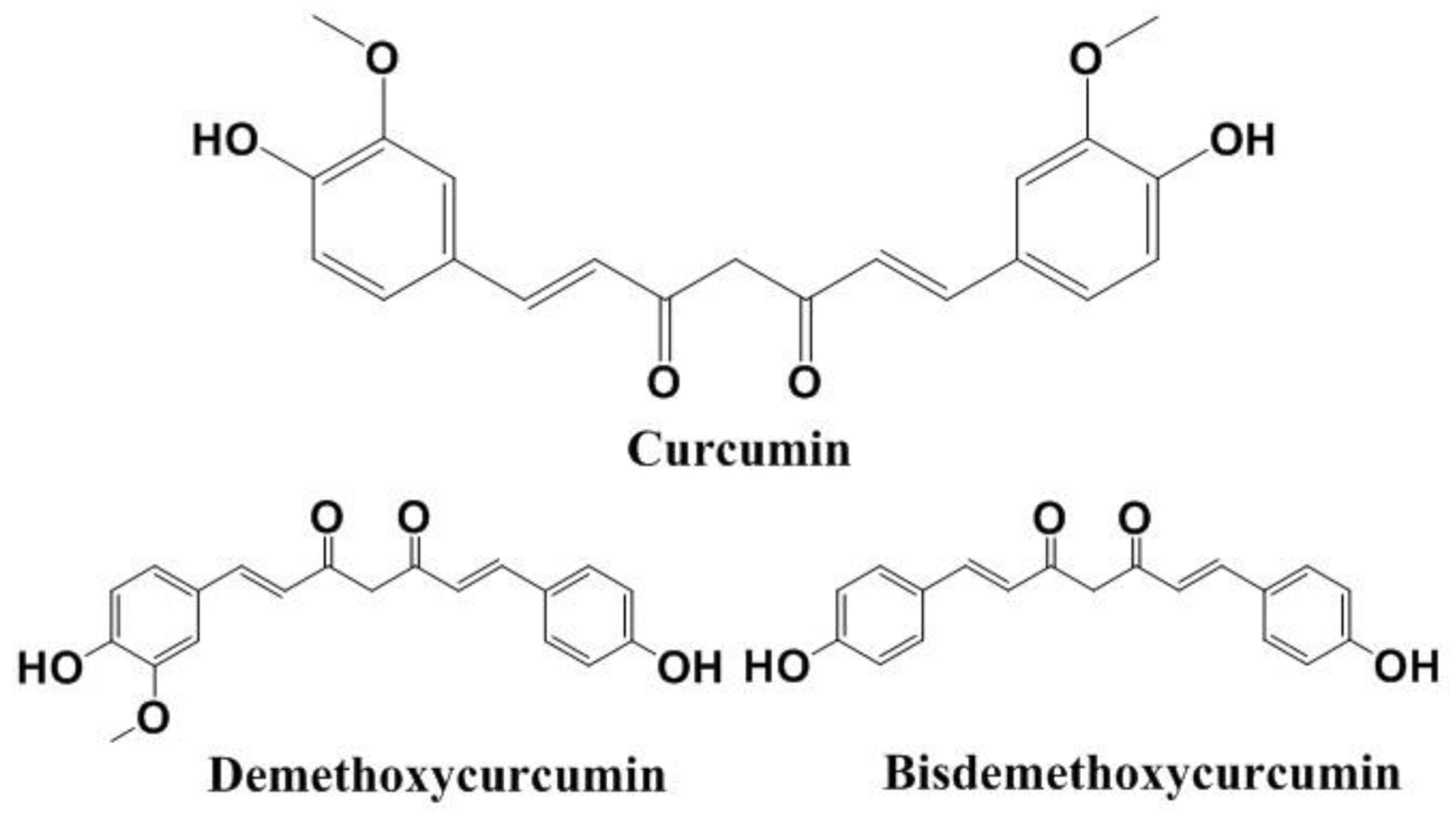
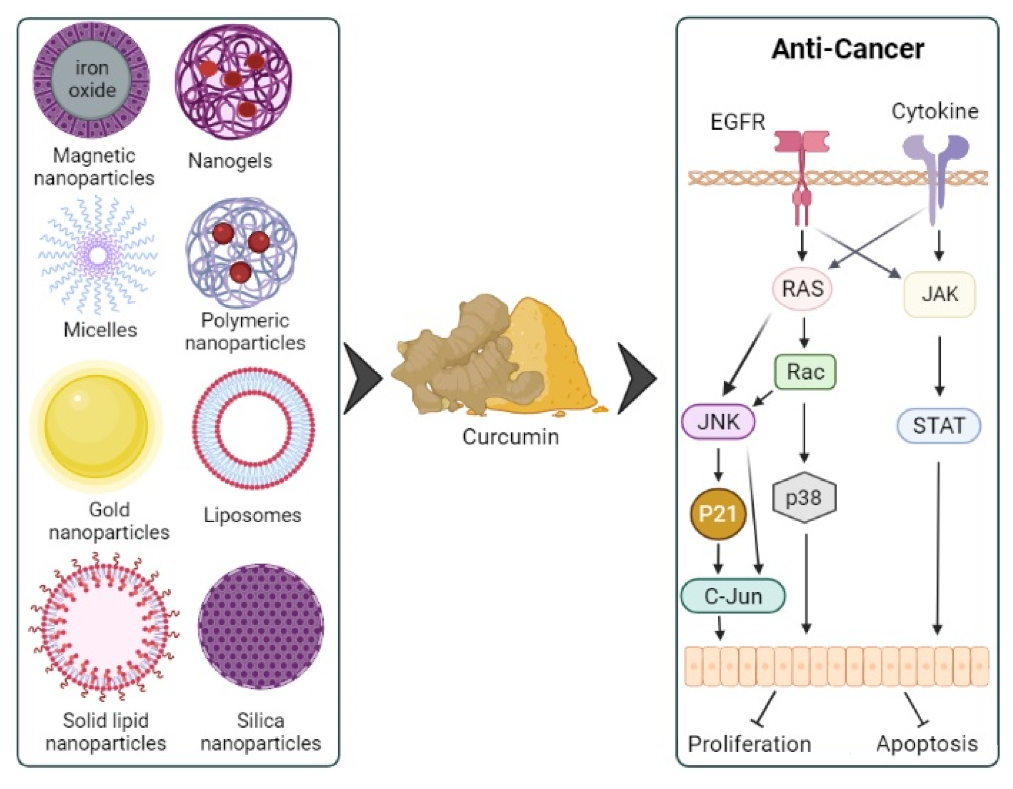
Table 1.
Summary of details from studies on CUR nanoformulations for ovarian, oral, cervical, and pancreatic cancers.
Table 1.
Summary of details from studies on CUR nanoformulations for ovarian, oral, cervical, and pancreatic cancers.
| Study Authors (Reference) | NP Type and Details | Cancer Cell Line/Cytotoxicity (IC50 of NPs vs. Free CUR) | Cellular Uptake of CUR-NPs vs. Free CUR | Animal Model | Treatment Regimen | In Vivo Antitumor Activity | Toxicity |
|---|---|---|---|---|---|---|---|
| Ovarian cancer | |||||||
| [156] | Polymeric NPs (PLGA, PVA, and poly-L-lysine) PM: Nanoprecipitation SZ: ~72 nm ZP: N/I EE: N/I | A2780CP (resistant to cisplatin) paired cells CUR-NPs inhibit cell proliferation compared to blank NPs IC50: N/I | N/D | N/D | N/D | N/D | N/D |
| [157] | Nanostructured lipid carriers (Compritol 888 ATO, Captex 355 EP/NF, Miglyol 812) PM: Precipitation SZ: 100–160 nm ZP: N/I EE: 40–100% | A2780S cells (cisplatin-sensitive) NPs: 21.2 ± 3.5 μM Free CUR: 22.2 ± 1.8 μM A2780CP cells (cisplatin- resistant) NPs: 19.0 ± 1.4 μM Free CUR: 20.2 ± 2.5 μM | Similar | N/D | N/D | N/D | N/D |
| [158] | Polymeric NPs (PLGA, PVA) PM: Emulsion–diffusion–evaporation Non-lyophilized NPs SZ: 203.6 ± 7.8 nm ZP: −5.24 ± 0.86 mV EE: 80.4 ± 10.6% Lyophilized NPs SZ: 201.8 ± 6.0 nm ZP: −5.43 ± 0.67 mV EE: N/I | SK-OV-3 cells ↑ for NPs (measured as amount of ROS generated) | ↑ for NPs (with or without irradiation from LED-based photodynamic therapy) | N/D | N/D | N/D | N/D |
| Oral cancer | |||||||
| [159] | Polymeric NPs (PLGA and PVA) PM: Single emulsion solvent evaporation SZ: 180 nm ZP: N/I EE: N/I | CAL27-cisplatin resistant cells IC50: N/I >80 µM for normal human gingival fibroblasts cells oral keratinocyte cells | N/D for free CUR | N/D | N/D | N/D | N/D |
| [160] | Silica NPs (Vinyltriethoxysilane, 3-aminopropyl-trimethoxysilane) PM: N/I SZ: ~32 nm ZP: −39 ± 1.0 mV EE: N/I | Human squamous cell carcinoma (4451) cells IC50: N/I | ↑ for NPs | N/D | N/D | N/D | N/D |
| [161] | Chitosan-coated PCL NPs PM: Nanoprecipitation SZ: 115.3–127.3 nm ZP: 23.5–40.1 mV EE: >99% | SCC-9 cells 24 h NPs: 271.5 ± 1.17 µM Free CUR: 93.40 ± 4.26 µM 48 h NPs: 260.3 ± 8.35 µM Free CUR: 75.21 ± 3.25 µM 72 h NPs: 92.04 ± 1.53 µM Free CUR: 17.46 ± 1.20 µM (p < 0.05 in each instance) Free CUR was more cytotoxic than the NPs | N/D | N/D | N/D | N/D | N/D |
| Cervical cancer | |||||||
| [27] | Polymeric NPs (PLGA; co-polymerization ratios 50:50 and 75:25, lactic/glycolic) PM: Single emulsion (solvent evaporation) SZ: 100–200 nm ZP: N/I EE: 74.73–90.03% | HeLa cells Comparable cytotoxicity | ↑ for NPs | ND | ND | ND | ND |
| [162] | NG (FA-conjugated cross-linked polymeric NPs) (acrylic acid, PEG diacrylate, FA) PM: Inverse emulsion polymerization SZ: 160–190 nm ZP: N/I EE: 61.2 ± 1.2% | HeLa cells ↑ for NPs IC50: N/I | ↑ for NPs | ND | ND | ND | ND |
| [163] | Liposomes (soybean lecithin and cholesterol, Montanov82®, and/or DDAB) PM: Thin film hydration method CUR-NPs (with Montanov82®) SZ: 161.5 ± 0.8 nm ZP: −1.4 ± 0.8 mV EE: 63.9 ± 3.8% CUR-NPs (with cholesterol) SZ: 161.8 ± 0.4 nm ZP: −0.1 ± 0.1 mV EE: 70.6 ± 0.5% CUR-NPs (with Montanov82® and DDAB) SZ: 252.4 ± 5.3 nm ZP: 28.8 ± 1.0 mV EE: 34.7 ± 0.3% CUR-NPs (with cholesterol and DDAB) SZ: 219.5 ± 9.3 nm ZP: 27.7 ± 0.9 mV EE: 68.9 ± 0.6% | HeLa cells ↑ for NPs than free CUR NPs: N/I Free CUR: 21 µM SiHa cells ↑ for NPs than free CUR NPs: N/I Free CUR: 16 µM For both cells, cytotoxicity ↑ for NPs containing DDAB | N/I | ND | ND | ND | ND |
| [164] | Micelles (N-benzyl-N,O-succinyl chitosan synthesized from chitosan by successive reductive N-benzylation, and N,O-succinylation) PM: Dialysis method SZ: 80 ± 4.0–97 ± 5.0 nm ZP: −27.1 ± 1.4 to −29.2 ± 1.4 mV EE: 7.57 ± 0.01–38.30 ± 5.70% | HeLa cells NPs: 4.34 ± 0.12 µM (4.7-fold ↓) Free CUR: 21.17 ± 1.80 µM SiHa cells NPs: 4.34 ± 0.12 µM (3.6-fold ↓) Free CUR: 16.28 ± 1.34 µM C33A cells NPs: 4.34 ± 0.12 µM (12.2-fold ↓) Free CUR: 54.29 ± 3.62 µM | Significantly ↑ for all NPs | - | - | - | - |
| [165] | Polymeric NPs (chitosan, sodium tripolyphosphate) PM: Ionic gelation SZ: 197 ± 16.8 nm ZP: 71 ± 6.4 mV EE: ~85% | SiHa cells NPs: 97.27 µg/mL HeLa cells NPs: 88.41 µg/mL CasKi cells NPs: 81.48 µg/mL C33A cells NPs: 95.46 µg/mL IC50: Values at 72 h Value N/I for free CUR | N/I Among cells, NP uptake over 25 h was CasKi > C33A > HeLa > SiHa | - | - | - | - |
| [166] | Polymeric NPs (PLGA, PVA, poly(l-lysine)) PM, SZ, ZP, EE: N/I | Caski cells and SiHa cells IC50: N/I ↑ cytotoxicity for NPs | ↑ for NPs | Female NOD scid gamma mice Caski cells (4 × 10⁶ cells, injection into cervix) TV: ~200 mm3 (maximum tumor burden allowed post treatment: 1100 mm3) | 100 μg intra-tumoral injection | Changes in TV NPs: 637 ± 68 mm3 Free CUR: 816 ± 94 mm3 | N/I |
| [167] | Polymeric NPs (Chitosan, alginate, sodium tripolyphosphate) PM: Ultrasonic-assisted method SZ: ~50 nm ZP: N/I EE: 70% | HeLa cells IC50: N/I ↑ cytotoxicity for NPs | N/I | ND | ND | ND | ND |
| [168] | Micelles (Pectin) PM: Self-assembly method SZ: 70–190 nm ZP: N/I EE: N/I | HeLa cells NPs: 14.1 ± 3.0 µM Free CUR: 40.9 ± 2.6 | N/I | ND | ND | ND | ND |
| [169] | Silica/titania mesoporous NPs (coated with polyethylenimine-FA) PM: Hydrolysis, condensation reactions, and surface functionalization. Drug loading (solvent deposition) SZ: 173 ± 15 nm ZP: N/I EE: 43.36 ± 0.32% | HeLa cells ↑ cytotoxicity for NPs (synergetic chemo-sonodynamic therapy observed) | N/I | ND | ND | ND | ND |
| [170] | Liposomes (DSPE, PEG2000, FA, SPC, cholesterol) PM: Thin-film hydration SZ: 112.3 ± 4.6 nm ZP: −15.3 ± 1.4 mV EE: 87.6% | HeLa cells NPs: 0.82 µg/mL Free CUR: 1.47 µg/mL | ↑ NPs-66.4 ± 6.2% Free CUR-5.7 ± 1.6% | Female BALB/c mice (~5 × 10⁶ cells in 100 μL PBS, lower right flank) TV: 100–150 mm3 | 25 mg/kg CUR, on alternate days for three weeks | Final TV NPs: 77.3 ± 56.5 mm3 Free CUR: 634.3 ± 67.4 mm3 | No obvious acute toxicity |
| [171] | Nano-niosomes (Fe3O4, PLGA, PEG, FA) PM: Double emulsion method (W/O/W) and vacuum drying SZ: 190.4 ± 5.3 nm ZP: N/I EE: 86.46% | HeLa229 cells IC50: N/I | ↑ | - | - | - | - |
| [172] | Micelles PM: Co-assembly of CUR and cystine/lysine-bridged peptide (CBP/LBP) SZ: ~250 nm ZP: N/I EE: 63.44% | HeLa cells ↑ cytotoxicity for NPs (but higher with the CBP) IC50: N/I | ↑ for NPs (but higher with the CBP) | Female BALB/c nude mice bearing HeLa cells Treatment was started on the 10th day when the tumor volume reached 100 mm3 | 2.5 mg/kg intravenous injection into tail, every three days for 14 days | TGI NPs (CBP): 69.12% NPs (LBP): 10.66% Free CUR: 36.14% | NPs: Minimal effects on healthy tissues Free CUR: Apoptosis in liver, kidney, spleen All formulations: No significant Effect on body weight |
| Pancreatic cancer | |||||||
| [173] | Polymeric micelles (methoxy(polyethylene glycol), PCL) PM: Modified dialysis SZ: 110 ± 6.4 nm ZP: −16 ± 2.77 mV EE: 57.6 ± 1.23% | PANC-1 cells NPs: 22.8 µM Free CUR: 24.75 µM MiaPaCa-2 cells NPs: 13.85 µM Free CUR: 14.96 µM | Test performed on PANC-1 cells only At 10 µM NP uptake 2.95-fold ↑ At 30 µM NP uptake 1.88-fold ↑ | - | - | - | - |
| [174] | Magnetic NPs (Fe(III) chloride hexahydrate (99%), Fe(II) chloride tetrahydrate, CD, Pluronic F-127) PM: N/I SZ: 109 nm ZP: −0.99 mV EE: N/I | HPAF-II and PANC-1 human pancreatic cancer cell lines IC50: N/I Similar cytotoxicity between NPs and free CUR to both cell lines | ↑ Similar uptake by both cell lines (54.06% vs. 53.86%) | Male athymic nude (nu/nu) mice Inoculated subcutaneously, left flank (5 × 10⁶ HPAF-II cells) | 13th day after inoculation Intratumoral administration, 20 μg CUR in 100 μL vehicle Animals sacrificed at the end of treatment or when TV = 1000 mm3 | TV ↓ by NPs more than by free CUR | N/D |
| [175] | Self-assembled casein (sodium caseinate) NPs PM: Self-assembly SZ: 104–213 nm ZP: −37.63 mV to −39.07 mV EE: 70% to ∼100% | BxPC3 cells NPs: 25.3 µg/mL Free CUR: 29.4 µg/mL | ↑ | - | - | - | - |
| [142] | NG (Cholesteryl-HA) PM: N/I SZ: 29.2 ± 5.4 nm ZP: −38.4 ± 3.9 mV EE: N/I | MiaPaCa-2 cells NPs: 9 μg/mL Free CUR: 18 μg/mL | N/I | Female nu-nu mice (5 × 10⁶ cells, subcutaneous injection, right flank) | 10th day after inoculation Intraperitoneal injection 6 mg/kg CUR twice every week | NG ↓ TV 5-fold vs free CUR by day 49 | No significant weight loss |
| [176] | Polymeric NPs (Chitosan, PEG, PLGA) PM: Emulsion solvent evaporation SZ: 264 nm ZP: 19.1 mV EE: 60% | PANC-1 cells NPs: 14.2 ± 4.6 μM Free CUR: 28 ± 4.1 μM MiaPaca-2 cells NPs: 6.1 ± 0.6 μM Free CUR: 20.3 ± 1.1 μM | PANC-1 cells 6.7-fold ↑ for NPs MiaPaca-2 cells 7.5-fold ↑ for NPs | - | - | - | - |
| [177] | Chitosan-coated lipid NPs (chitosan, stearoyl chloride, cholesterol) PM: Cold dilution of microemulsion SZ: 190.6 ± 1.5 nm ZP: 2.10 ± 0.51 mV EE: 73.4 ± 0.3% | PANC-1 cell lines IC50: N/I ↑ cytotoxicity of NPs at 5 and 10 µM CUR concentration | - | - | - | - | - |
| [178] | SLNs (trilaurin) PM: Cold dilution of microemulsion SZ: ∼200 nm ZP: −10.06 ± 2.66 mV EE: 75 ± 1.0% | CFPAC-1 and PANC-1 cells IC50: N/I ↑ cytotoxicity of NPs | - | - | - | - | - |
Abbreviations: CD, cyclodextrin; CUR, curcumin; EE, encapsulation efficiency; FA, folic acid; HA, hyaluronic acid; IC50: half-maximal inhibitory concentration; N/D, not determined; N/I, not indicated; NG, nanogel; NP, nanoparticle; PCL, polycaprolactone; PEG, polyethylene glycol; PM, preparation method; ROS, reactive oxygen species; SZ, size; TGI, tumor growth inhibition; TV, tumor volume; ZP, zeta potential.
Publisher’s Note: MDPI stays neutral with regard to jurisdictional claims in published maps and institutional affiliations. |
© 2022 by the authors. Licensee MDPI, Basel, Switzerland. This article is an open access article distributed under the terms and conditions of the Creative Commons Attribution (CC BY) license (https://creativecommons.org/licenses/by/4.0/).
Share and Cite
MDPI and ACS Style
Amekyeh, H.; Alkhader, E.; Sabra, R.; Billa, N. Prospects of Curcumin Nanoformulations in Cancer Management. Molecules 2022, 27, 361. https://doi.org/10.3390/molecules27020361
AMA Style
Amekyeh H, Alkhader E, Sabra R, Billa N. Prospects of Curcumin Nanoformulations in Cancer Management. Molecules. 2022; 27(2):361. https://doi.org/10.3390/molecules27020361
Chicago/Turabian StyleAmekyeh, Hilda, Enas Alkhader, Rayan Sabra, and Nashiru Billa. 2022. "Prospects of Curcumin Nanoformulations in Cancer Management" Molecules 27, no. 2: 361. https://doi.org/10.3390/molecules27020361