Article

The Influence of Light Wavelength on Resveratrol Content and Antioxidant Capacity in Arachis hypogaeas L.

1
Department of Crop Science, College of Sanghuh Life Science, Konkuk University, Seoul 05029, Korea
2
Peresbiotech Co., Ltd., Chuncheon 200-701, Korea
3
Interdisciplinary Program in Smart Science, Kangwon National University, Chuncheon 200-701, Korea
4
Food Science and Biotechnology, Kangwon National University, Chuncheon 24341, Korea
*
Author to whom correspondence should be addressed.
Agronomy 2021, 11(2), 305; https://doi.org/10.3390/agronomy11020305
Submission received: 18 December 2020 / Revised: 4 February 2021 / Accepted: 5 February 2021 / Published: 9 February 2021

Abstract

:
The quality and intensity of light can have a huge influence on plant growth and bioactive compound production. Compared to conventional lighting, very little is known about the influence of light emitting diodes (LED) irradiation on the antioxidant and antimicrobial properties and resveratrol content of peanut sprouts. This study was aimed at understanding the effects of LED light on the growth and antioxidant capacity of peanut sprouts. The resveratrol concentration was determined by liquid chromatography–tandem mass spectrometry. Peanut sprouts grown under blue LED light exhibited a higher total resveratrol content grown than those under fluorescent light and other LED light sources. The highest total phenolic content was recorded in the case of blue LED. The 1,1-diphenyl-2-picrylhydrazyl and 2,2-azino-bis 3-ethylbenzothiazoline-6-sulfonic acid radical scavenging values of blue LED‒treated and micro-electrodeless light-treated sprouts were significantly (p < 0.05) higher than that of sprouts grown under lights with other wavelengths. A Pearson correlation analysis revealed a strong association of the resveratrol, total phenolic, and flavonoid contents of peanut sprouts with 1,1-diphenyl-2-picrylhydrazyl (DPPH) and 2,2-azino-bis 3-ethylbenzothiazoline-6-sulfonic acid (ABTS), indicating its contribution to antioxidant activities. The anti-tyrosinase activity increased with an increase in the concentration of the tested sample. Blue LED-irradiated peanut extracts at a selected concentration range showed moderate cytotoxicity. Furthermore, the antimicrobial activity of peanut sprouts grown under blue LED was effective against Escherichia coli, Klebsiella pneumonia, and Vibrio litoralis. The present study revealed that the application of LEDs during the peanut sprouts growth improves the antioxidant activity, resveratrol concentration, and metabolite accumulation.

1. Introduction

Arachis hypogaeas L. is extensively cultivated as an oil source worldwide [1,2,3]. The seed of this plant is rich in resveratrol [4,5] and other phenolic compounds, such as flavonoids, isoflavones, p-coumaric acid, phytosterols, and phytic acid [6]. Resveratrol is associated with antioxidant activity, cardioprotective function [7], anticancer and neuroprotective effects [8,9,10], and antifungal activities [11,12,13]. It also reduces oxidative DNA damage [14] and prevents the development of cardiovascular diseases [15]. Moreover, phenolic compounds present in peanuts have been reported to play an important role in colon cancer chemoprevention [8].
Sprouting is a useful and inexpensive technique to increase the nutritional value and nutraceutical labels of bioactive compounds in plants [16,17]. Peanut sprouts, in particular, are usually consumed raw and provide a variety of bioactive compounds, such as amino acids, minerals, carbohydrates, and phenolic compounds, that can contribute to the prevention and treatment of certain diseases [18]. They have been receiving more attention lately because of the presence of resveratrol (3,4′,5 trihydroxystilbene) synthesized by resveratrol synthase [19]. Resveratrol is produced as a defense mechanism to protect plants from pathogen infection, mechanical stress, UV radiation, and reactive oxygen species (ROS) [20,21]. This natural polyphenolic compound has been reported to induce apoptosis [22,23], and prevent the development of diabetes, cancer, obesity, cardiovascular diseases, and inflammation [8,24,25,26]. Resveratrol synthesis is influenced by external stimuli, including light emitting diodes (LED) and ultrasonication (US).
A number of factors influence the growth, development, and phytochemical constituents of plant sprouts, among which the quality, intensity, and photoperiods of light can have a bigger influence than other factors [27,28]. Artificial light is widely used as a supplementary light source in greenhouses and growth chambers.
LEDs are being increasingly preferred as artificial light sources over conventional sources because of their low energy consumption and heat generation and long life expectancy [29,30]. Moreover, LEDs have the ability to control spectral composition and light intensity and offer a higher efficiency useful in in vitro tissue organogenesis and controlled environment agriculture [29,30]. Many previous studies have observed the regulatory effects of LEDs on the growth and development of plants, such as cucumbers [31,32], peas [33], buckwheat [34], and strawberries [35]. Apart from the regulatory effects, manipulation of the spectral quality and intensity of LEDs considerably influenced the antioxidant properties and composition of bioactive compounds in plants. Recently, red LEDs have been demonstrated to enhance β-carotene in pea plants [36] and fresh weights in Brassica napus L. [37]. The exposition of blue-LED led to the increment of anthocyanins, glucoraphanin content in the roots of Chinese kale sprouts [38], tocopherols, carotenoids, and xanthophyll pigments in the microgreens of mustard, beets, and parsley [39]. Previous studies have demonstrated a significant increase in the content of flavonoids in plant sprouts exposed to LEDs [30,40,41], along with an increase in vitamin C, β-carotene, tocopherol [38,42], and phenolic acid levels [30,40,43]. Increased antioxidant levels were observed in wheat and radishes [44].
Controlling the quality and intensity of LEDs not only increases the desired bioactive compounds but can also reduce undesirable chemical compounds in plants [45]. Red and blue LED treatments were proven to be effective in fortifying the levels of plant nutrients and bioactive compounds exhibiting better physiological functions, antioxidant properties, antimicrobial potential, and anticancer activities in plantlets grown in vitro. These compounds play a significant role in the scavenging, quenching, or degrading reactive oxygen species (ROS) produced in the metabolic processes [46]. The excessive production of ROS during metabolic processes can cause cell dysfunction and lead to various diseases. Foods rich in antioxidants can neutralize the radicals and, thus, balance the oxidant and antioxidant defense system [47]. However, the effects of different sources of light and ultrasonication on sprout growth, antioxidant activities, concentration, and the composition of phytochemicals such as resveratrol have not yet been reported.
In the present study, we exposed peanut sprouts to different intensities of light and investigated the total phenolic content and total flavonoid levels, antioxidant activities, and resveratrol concentrations. We also examined the impact of light-emitting diodes (LEDs) and ultrasonication on the cytotoxicity and antimicrobial properties of peanut sprouts. In addition, we attempted to elucidate the possible relationships among different antioxidant compounds by performing assays of the total phenolic content, flavonoid content, and resveratrol.

2. Materials and Methods

2.1. Chemicals

Methanol was purchased from J.T. Baker (Phillipsburg, NJ, USA). 1,1-Diphenyl-2-picrylhydrazyl (DPPH), 2,2-azino-bis 3-ethylbenzothiazoline-6-sulfonic (ABTS), butylated hydroxy toluene (BHT), Folin-Ciocalteau reagent, gallic acid, and quercetin were purchased from Sigma Chemical Co. (St. Louis, MO, USA). Standard resveratrol was obtained from Sigma Chemical Co. (St. Louis, MO, USA). All other chemicals and reagents used in the experiments were of analytical grade.

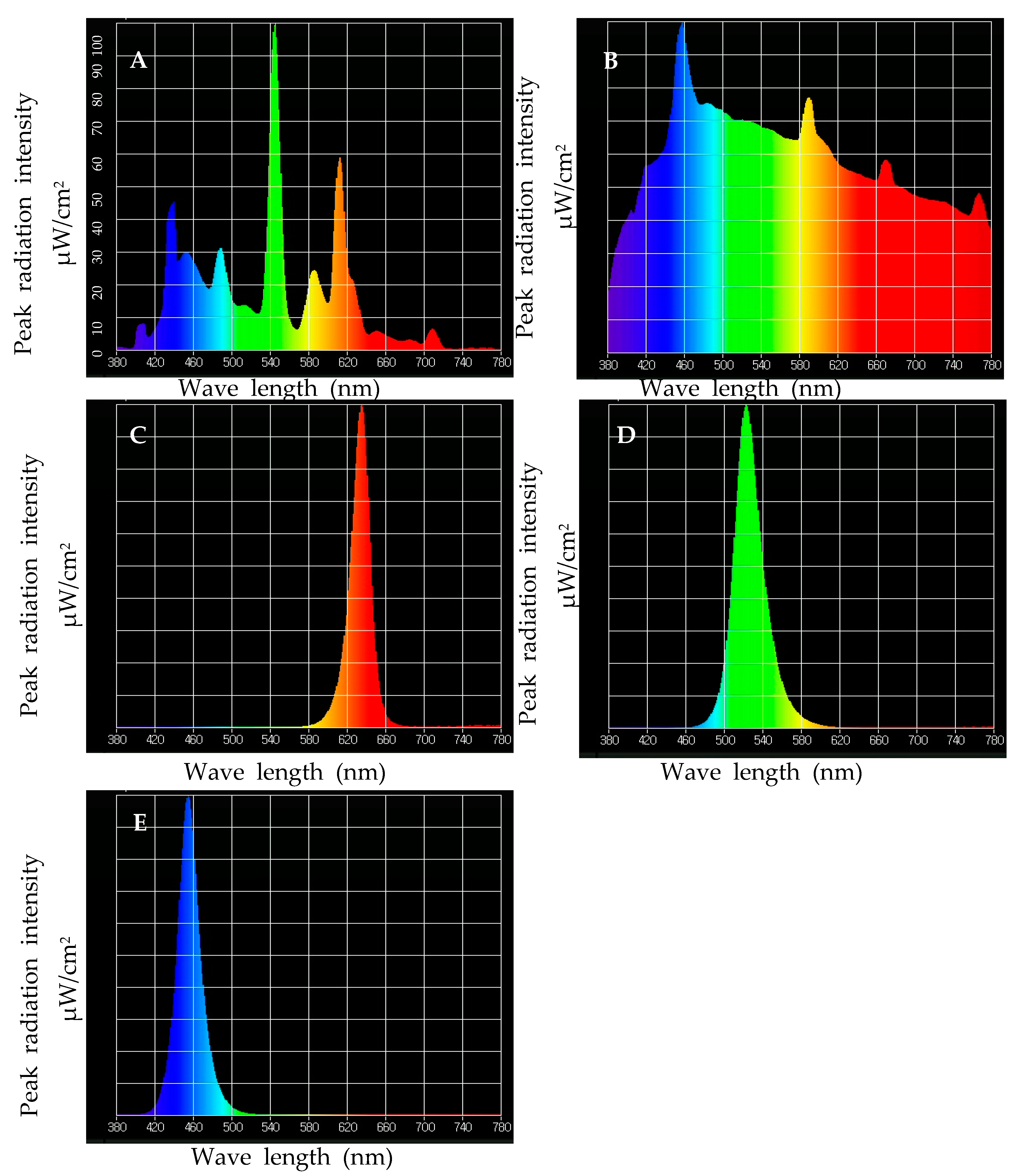
2.2. Plant Growth Conditions and Treatment

A. hypogaeas var. Heugttangkong seeds were provided by the Bioherb Research Center, Kangwon National University, South Korea. The experiment was performed in June 2020. Healthy seeds were selected, washed in distilled water, and subsequently, soaked in distilled water for 15 h at room temperature (25 °C). The soaked seeds were then uniformly distributed on a germination tray under dark conditions and allowed to germinate at a controlled temperature (25 °C) and 80% relative humidity in a growth chamber (Model HK-GC1000, Hankuk Scientific Technology, Kyounggi-do, South Korea). Germinating seeds were watered with 5 mL of distilled water every 12 h.
After three days, the emerged A. hypogaeas sprouts were grown under light sources (Figure 1). Three replicates were used to study the effects of the LEDs (48 × 48 cm, L × W) (Model: DYN-LC200-P12, Jeollabuk-do, Iksanosi, South Korea), and micro-electrodeless light (MEL) (Sunshine 400, Stray Light Optical Technologies, Inc., Scottsburg, IN, USA). Peanut sprouts grown under a fluorescent lamp (Model: TNFL-40/75, 10 Hz, Sigma lamps, C.H. lighting, Hangzhou, Zhejiang, China) were used as the control. Six growth chambers were independently constructed to provide lights using LEDs. Each light source was fixed separately on a steel ceiling. A portable spectrometer (UPRTek MK 350, Miaoli County 35059, Zhunan Township, Taiwan) was used to measure the spectral distribution of the different light sources. Each light source was located at an identical height, and the distance between the light source and the top of the sprout was maintained at 30 cm. The spectral distribution of fluorescent light (FL) ranged from 400–720 nm. The peak wavelengths of the green LEDs, red LEDs, and blue LEDs were 420–680 nm, 634 nm, and 454 nm, respectively, while the power of each LED treatment was 12.2 W, 11.5 W, 13.5 W, and 15.0 W, respectively. The spectral distribution of micro-electrodeless light (MEL) ranged from 380 to 780 nm. The photosynthetic photon flux density (PPFD) for light irradiation was maintained at 110 ± 10 µmol m−2 s−1. The sprouts were grown under a 12-h photoperiod for 15 days. Twenty healthy sprouts were used for each irradiation. The sprouts were sub-irrigated with 5 mL of distilled water every 12 h. The harvested A. hypogaeas sprouts samples were immediately washed with distilled water and dried in room temperature (25 °C). The dried sprouts were grounded and stored in an airtight polythene bag for chemical analysis.

2.3. Preparation of Extracts

Dried and grounded sample (1 g) of A. hypogaeas sprout treated with different light sources were mixed in 10 mL of 80% methanol for 24 h. The sample mixture was then filtered to remove debris using No. 1 Whatman filter paper (Schleicher & Schuell, Whatman International Ltd., Maidstone, UK). The solvents present in the mixture were evaporated at 42 °C in a rotary evaporator (Eyela, SB-1300, Shanghai Eyela Co. Ltd., Shanghai, China). The residual extracts were dissolved in 80% methanol (300 mL) and stored at 4 °C until further use.

2.3.1. Total Phenolic Content

The total phenolic content of the extracts was determined by spectrophotometry, using the Folin-Ciocalteu reagent according to the methodology described by Chung et al. [48]. In brief, 50 µL of peanut sprout extracts (1 mg mL−1) was mixed with an equal volume (50 µL) of Folin-Ciocalteu reagent in a 96-well plate (SPL Life Sciences Co., Gyeonggi-do, South Korea). Then, the mixture was incubated at room temperature (25 °C) in the dark for 5 min. After incubation, 100 µL of 20% (w/v) sodium carbonate (Na2CO3) was added to the solution, followed by 1 mL of distilled water. The 96-well plate containing the mixture was then inserted in a microplate reader (Multiskan GO, Thermo Fisher Scientific, Sanghai Instruments, Shanghai, China) for 30 min in the dark. Subsequently, the absorbance of each sample was recorded at a wavelength of 725 nm against a blank using a Shimadzu UV-VIS spectrophotometer (UV-1800, Shimadzu, Kyoto, Japan). A standard absorbance curve was prepared using 0-, 65.5-, 125-, and 250-mg L−1 solutions of gallic acid in a solvent comprising methanol:water (50:50, v/v). The results were expressed in gallic acid equivalent (GAE) of dry plant matter by weight (µg Qg−1 dry weight) using the following formula:
y = 0.002x + 0.0395
where x is the amount of gallic acid equivalent (μg mL−1), and y is the absorbance at 765 nm.

2.3.2. Total Flavonoid Content

Total flavonoid content of the A. hypogaeas sprout samples was measured using the method of Nieva Moreno et al. [49]. Initially, 500 µL of A. hypogaeas extract (1 mg mL−1) was mixed with 100 µL of 10% aluminum nitrate (Al(NO3)3) and 100 µL of 1-M potassium acetate (CH3COOK). The mixture was incubated for 5 min at room temperature and shaken well. Subsequently, 1 mL of 1-M sodium hydroxide (NaOH) was added, and the absorbance of each mixture was recorded against a blank at 510 nm using a Shimadju UV-VS spectrophotometer (UV-1800, Shimadzu, Kyoto, Japan). A standard curve was prepared using different concentrations (0.5, 10, and 100 mL−1) of quercetin. The total flavonoid content in the tested samples was expressed as a quercetin equivalent (Qu) per gram of dry plant matter. The following equation was used to obtain the total flavonoid content of the tested samples:
y = 0.0014x + 0.04
where x is the amount of quercetin equivalent (μg mL−1), and y is the absorbance at 510 nm.

2.4. Effect of Light Quality on Antioxidant Activity
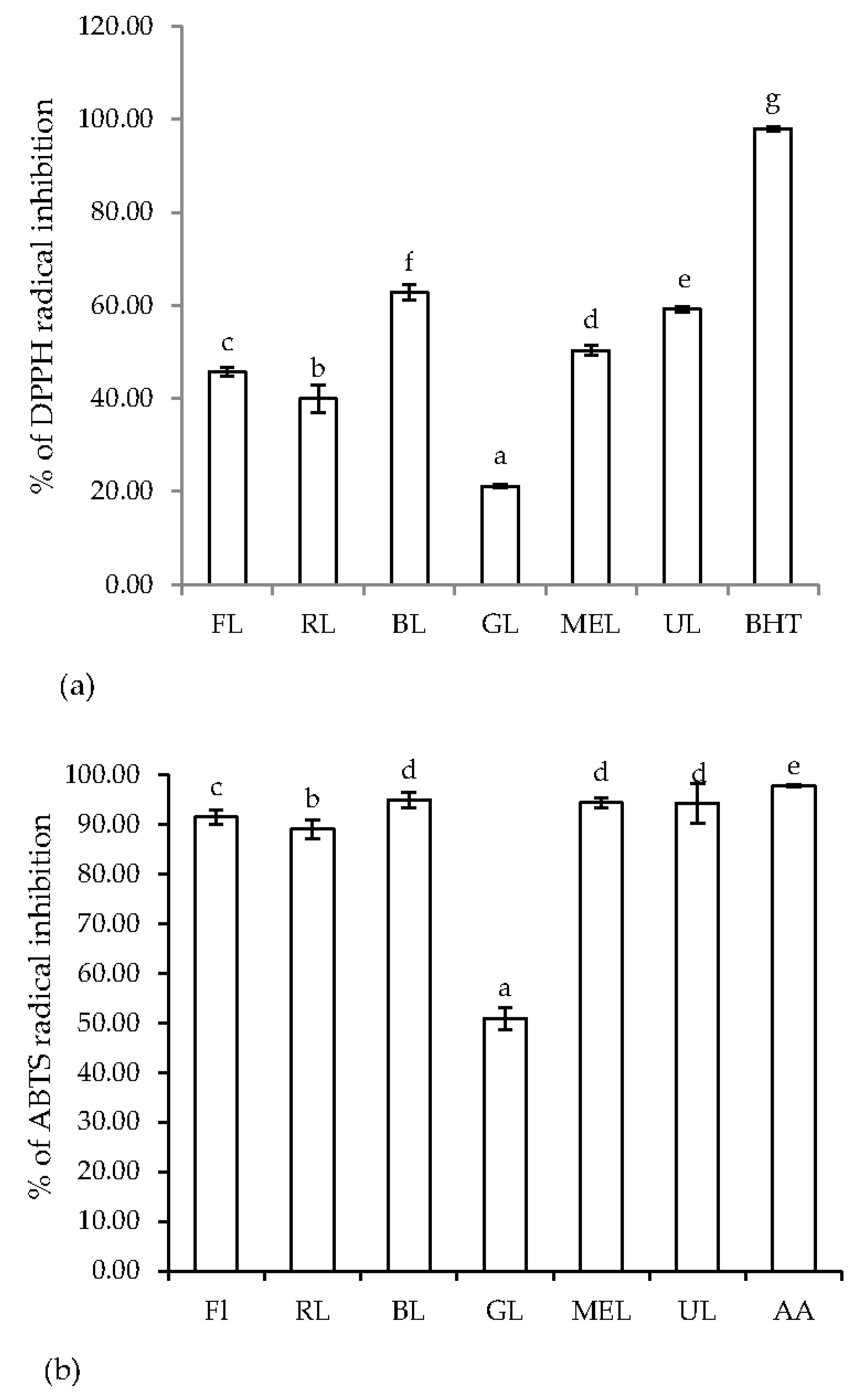
2.4.1. DPPH Method

Antioxidant activity was determined using the methodology described by Chung et al. [50]. Initially, 1 mL of extracted sample (0.1–1 mg mL−1) was mixed with 4 mL of 0.004% 1,1-diphenyl-2-picrylhydrazyl (DPPH) methanol solution. Then, the mixture was thoroughly stirred and, subsequently, incubated for 30 min at room temperature (25 °C) in the dark. The absorbance value of the mixtures was determined at 517 nm using a UV–VIS spectrophotometer (UV-1800, Shimadzu, Kyoto, Japan). Antioxidant activity was expressed as the percentage of oxidation inhibition and calculated using the following expression:
% inhibition = [(A0 − A1)/A0)] × 100
where A0 is the absorbance of the control reaction (containing all reagents except the test sample), and A1 is the absorbance of the test sample. The control was prepared without adding the sample or butylhydroxytoluene (BHT). The antioxidant activity of the sample was compared to that of BHT, which was determined under the same conditions as described above.

2.4.2. ABTS Assay

The antioxidant capacity of sprouts grown under light sources was measured using the ABTS scavenging test, as per the method described by Thaipong et al. [51], with some modifications. Initially, a radical solution was produced by the reaction of 7.4 mM of 2,2-azino-bis 3-ethylbenzothiazoline-6-sulfonic acid (ABTS) and 2.6-mM potassium persulfate (1:1, v/v). The ABTS+ solution was then stored in the dark for 12 h at room temperature (25 °C) before use. The mixture was then diluted with methanol to obtain an absorbance value of 0.700 at 734 nm using a spectrophotometer (Jasco V530 UV-VIS spectrophotometer, Hachioji, Tokyo, Japan). Then, 2.5 mL of the extract was mixed with an equal volume of ABTS+ solution. Then, the mixture was left to stand for 2 h, and the absorbance was recorded at 734 nm. Different concentrations (500–1000 µM) of Trolox were used to plot the standard curve. The ABTS capacity of each sample was calculated using the following equation:
ABTS radical scavenging activity (%) = [(A0 − A1)/A0)] × 100
where A0 is the absorbance of the control reaction (containing all reagents except the test sample), and A1 is the absorbance of the test sample.

2.5. Tyrosinase Inhibition Assay

The anti-tyrosinase activity of the peanut sprouts was recorded spectrophotometrically, as described by Kubo and Kinst-Hori [52], with some modifications. L-DOPA (3,4-dihydroxy-L-phenylalanine) was used as the substrate. Initially, 40 µL of sample was added to each well at various concentrations; 40 µL of mushroom (Agaricus bisporus) tyrosinase (Sigma-Aldrich Co. Ltd., St. Louis, MO, USA) was diluted with 67-mM potassium phosphate-buffered saline (PBS, pH 7.4; T&I Co. Ltd., Jongno-gu, Seoul, South Korea) and 120 µL of 10-mM L-DOPA (Sigma-Aldrich Co. Ltd.) and incubated at 37 °C in the dark for 30 min. Each sample was accompanied by a blank containing all the components except tyrosinase. Kojic acid was used instead of the sample as a positive control. The absorbance of all the tested samples was measured at 519 nm. The percentage inhibition was calculated using the following equation:
Percentage of tyrosinase inhibition (%) = [(A0 − A1)/A0)] × 100
where A0 is the absorbance of the control, and A1 is the absorbance of the test sample.

2.6. Liquid Chromatography/Tandem Mass Spectrometry (LC/MS) Analysis of Resveratrol

Resveratrol was extracted using a modified form of the method proposed by Langcake [53]. Briefly, 1 g of peanut seedling leaves was extracted with 3 mL of the methanol/water solvent. The resulting leaf extract was filtered through a 0.45-mm mesh membrane, and the solvents were evaporated at 41 °C using a rotary evaporator (Eyela, SB-1300, Shanghai Eyela Co. Ltd., Shanghai, China). Chromatographic separation of resveratrol was performed on a Waters ACQUITY H-Class Liquid Chromatograph equipped with SQD2 Mass Spectrometer (SQD2, Waters, Wilmslow, UK) mass detector using a Waters BEH C18 (2.1 mm × 100 mm, 1.7 μm) column. The samples were directly injected into the mass spectrometer at a concentration of 200 ppm to obtain the optimum atmospheric pressure, the ionization mode was ESI-negative, and the cone voltage for each analyte was 30 V. The injection volume and column temperature were maintained at 0.3 mL min−1 and 30 °C, respectively. The system ran two solvents, i.e., acetonitrile (ACN) as solvent A and water (10-mM ammonium acetate) as solvent B with the following gradient combinations: 0–10 min 30% B, 10–20 min 30–60% B, 20–25 min 60–90% B, and 25–30 min 90–60% B while maintaining the capillary voltage at 3.0 kV. All sample eluents were monitored at 254 nm. The purity of resveratrol was calculated by dividing each peak area by the total peak area. Chromatograms were detected using a UV detector at 306 nm. All experiments were conducted in triplicate.

2.7. Determination of Cytotoxicity

2.7.1. Cell Culture

The macrophage cell line RAW 264.7 was obtained from the Korean Cell Line Bank, Seoul, South Korea. A 5-mL aliquot of RAW cells was placed in a culture flask (SPL Life Science Co. Ltd., Pocheon-si, South Korea), to which 10% fetal bovine serum (FBS) (Hyclone Laboratories Inc., Logan, UT, USA) and 1% penicillin (Lonza Walkersville Inc., Walkersville, MD, USA) in dulbecco’s modified eagle’s medium (DMEM) were added; the cells were allowed to grow at 37 °C and 5% CO2 in an incubator. In order to count the dead and live cultured cells, 10 μL of Trypan blue was added to the culture, centrifuged at 1000 rpm for 3 min, and suspended in flasks. Subsequently, cultured RAW 264.7 cells were harvested and cultured in flasks. Over 90% viability was confirmed in each culture, and cells were used for the cytotoxicity assay.

2.7.2. MTT Assay

The cytotoxicity assay was performed using the MTT (3-(4,5-dimethylthiazol-2-yl)-2,5-diphenyltetrazolium bromide) kit (M6494) following the method described by Mosmann et al. [54]. The RAW 264.7 cells at a density of 15,000 cells/well were exposed to different concentrations of peanut sprout extracts (10, 50, and 100 ppm). The absorbance was measured at 620 nm using a microplate reader (Thermo Fisher Scientific Instrument Co. Ltd., Shanghai, China). The MTT assay was also performed with commercially available identified compounds, as described above. Tamoxifen (Sigma, St. Louis, MO, USA) was used as the positive control.
%   viable   cells   =   A 1 A 0   × 100
where A0 is the absorbance of the control, and A1 is the absorbance of the test sample.

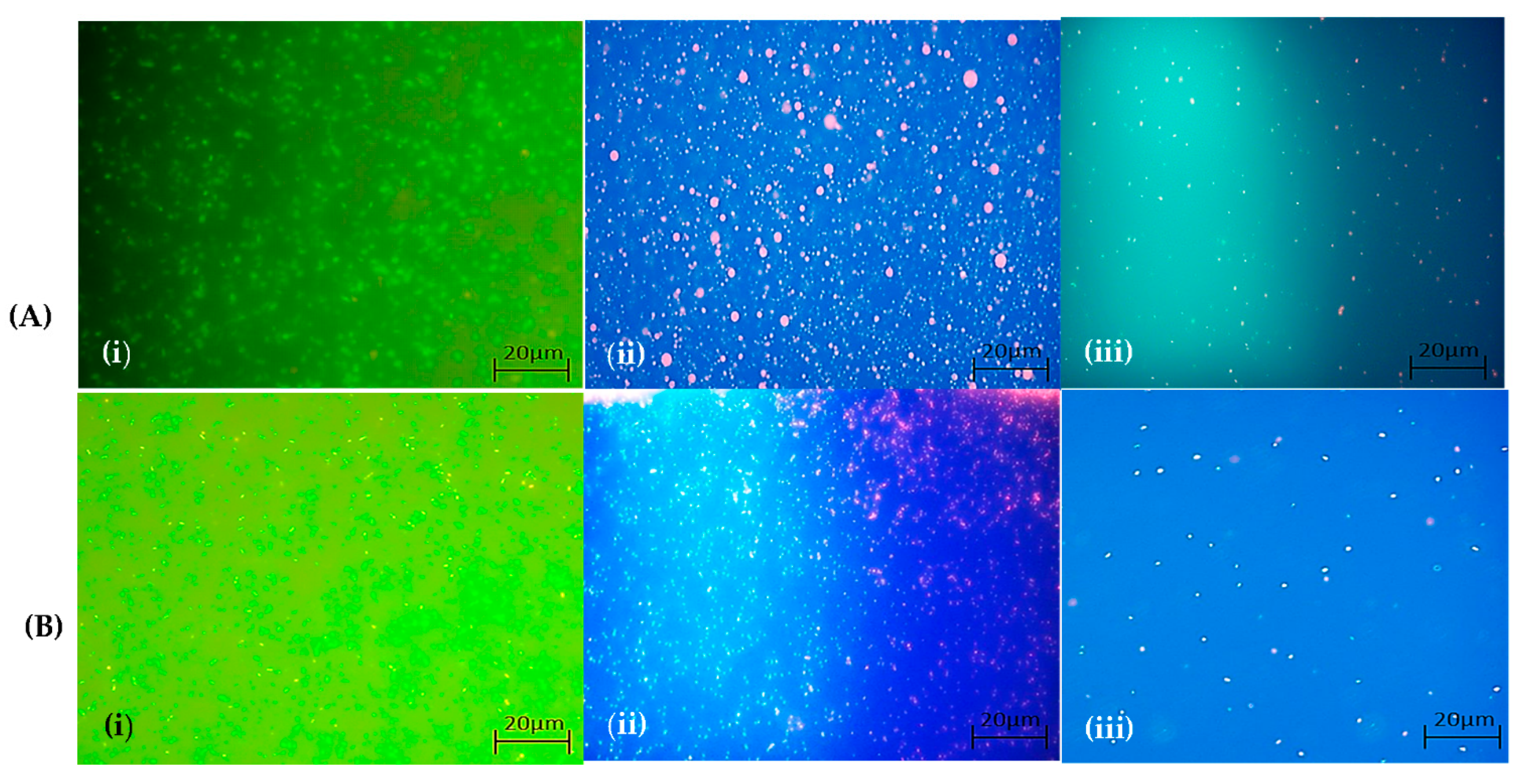
2.7.3. Qualitative Observation of Live and Dead Cell Staining Using Fluorescent Microscope

Preparation of the Cells

Initially, the adherent cells were cultured on sterile glass coverslips in confluent monolayers. Then, the cells were transferred to 35-mm disposable petri dishes (SPL Life Science Co. Ltd., Pocheon-City, Gyeonggi-Do, South Korea). The adherent cells were then washed with 500–1000 volume of Dulbecco’s phosphate-buffered saline (D-PBS). An aliquot of the cell suspension was transferred to a coverslip. Cells were allowed to settle to the surface of the glass coverslip at 37 °C in a covered 35-mm petri dish.

Microscopic Documentation of Dead and Live Cells

Initially, 100–150 µL of the combined LIVE/DEAD® assay reagents were added to the surface of a 22-mm square coverslip, and the cells were incubated for 30–45 min at room temperature. Following incubation, about 10 µL of the fresh LIVE/DEAD® reagent solution was added to a clean microscope slide. The slides were mounted on the microscope. To prevent evaporation, the glass slides were sealed with coverslips. The cells were observed under the fluorescence microscope (excitation 488 nm/540 nm and emission 530 nm/620 nm (fluorescein isothiocyanate (FITC)/ tetramethylrhodamine isothiocyanate (TRITC)filters). All representative images were acquired using a Nikon E200 inverted microscope equipped with a Nikon camera (DF-FI3, Osaka, Japan) and UV lamp (Nikon C-HGFI, Osaka, Japan) and analyzed using NIS-Elements-Microscope Imaging Software.

2.8. Antimicrobial Activity

2.8.1. Disc Diffusion Method

The antimicrobial activity of the peanut sprouts was evaluated using the disc diffusion method, as described by Chung et al. [50]. All the tested pathogenic bacterial strains were obtained from the Department of Food Science and Biotechnology, Kangwon National University, South Korea. The effects of different sprout extracts were investigated against Escherichia coli Castellani and Chalmers (ATCC35150), Vibrio litoralis (KCTC12320), Pseudomonas aeruginosa (ATCC25668), Klebsiella pneumoniae subsp. pneumoniae (Schroeter) Trevisan (ATCC 9621), Salmonella enteritidis serotype typhimurium Kauffmann and Edwards (ATCC14028), and Bacillus subtilis (KCCM 11316). Briefly, the tested bacterial strains were spread on a tryptic soy agar (TSA) medium uniformly at room temperature (25 °C). The sprout extract (50 µL) containing 1 mg mL−1 of dried plant sample was poured on filter paper discs (6 mm, Whatman, no. 3) and allowed to stand at room temperature (25 °C) for 10 min for the methanol to evaporate. The soaked filter paper was then placed on a tryptic soy agar (TSA) medium containing the tested microbial strains. The culture plates were incubated at 37 °C for 18–24 h.

2.8.2. LIVE/DEAD BacLight Bacterial Viability Test

The experiment was performed in a 24-well microtiter plate. Twenty-four-hour‒old bacterial culture in BHI (brain heart infusion broth) was harvested approximately at 108 colony forming unit (CFU) × mL−1. The sample was centrifuged (Eppendorf ® Centrifuge 5810R, Hamburg, Germany) at 4000 rpm for 20 min at 4 °C, and the bacterial pellet was washed twice with 1% PBS to remove the media components. After washing, the pellet was resuspended in PBS at 108 CFU × mL−1, and 500 µL of this suspension was dispensed into each well of a 24-well plate. The plant extract was dissolved in 0.5% dimethyl sulfoxide (DMSO) (2 mL/0.5 g), added to the bacterial samples, and incubated for 3 h at 37 °C. After the treatment, 2 µL of Sytox 9 was added to the control sample under dark conditions to observe the cell viability. Subsequently, 2 µL of propidium iodide was added to the plant extract-treated samples. The dye-treated samples were incubated at 37 °C for 20 min. Images were documented at a specific wavelength (excitation, 488 nm/540 nm/emission, 530 nm/620 nm) (SYTO® 9: 485⁄498 and propidium iodide (PI): 535/617) and light filter (FITC/TRITC filters) using a fluorescent microscope. All representative images (10× magnification) were acquired using an Olympus CKX53 inverted microscope equipped with DP74 camera (Olympus, Tokyo, Japan), and the wound widths were measured with CellSense Dimension 1.17 (Olympus, Tokyo, Japan).

2.9. Statistical Analysis

Statistical analysis was performed using analysis of variance (ANOVA). Significant differences between the parameters were determined by Duncan’s multiple comparison test at p < 0.05 and p < 0.01. All data were expressed as the mean ± standard deviation; experiments were performed in triplicate. The associations between the total phenolic content, total flavonoid content, and resveratrol with the antioxidant and antimicrobial levels and cytotoxicity were assessed based on Pearson’s correlation coefficient using SPSS version 20 (SPSS, IBM, New York, NY, USA, 2011).

3. Results

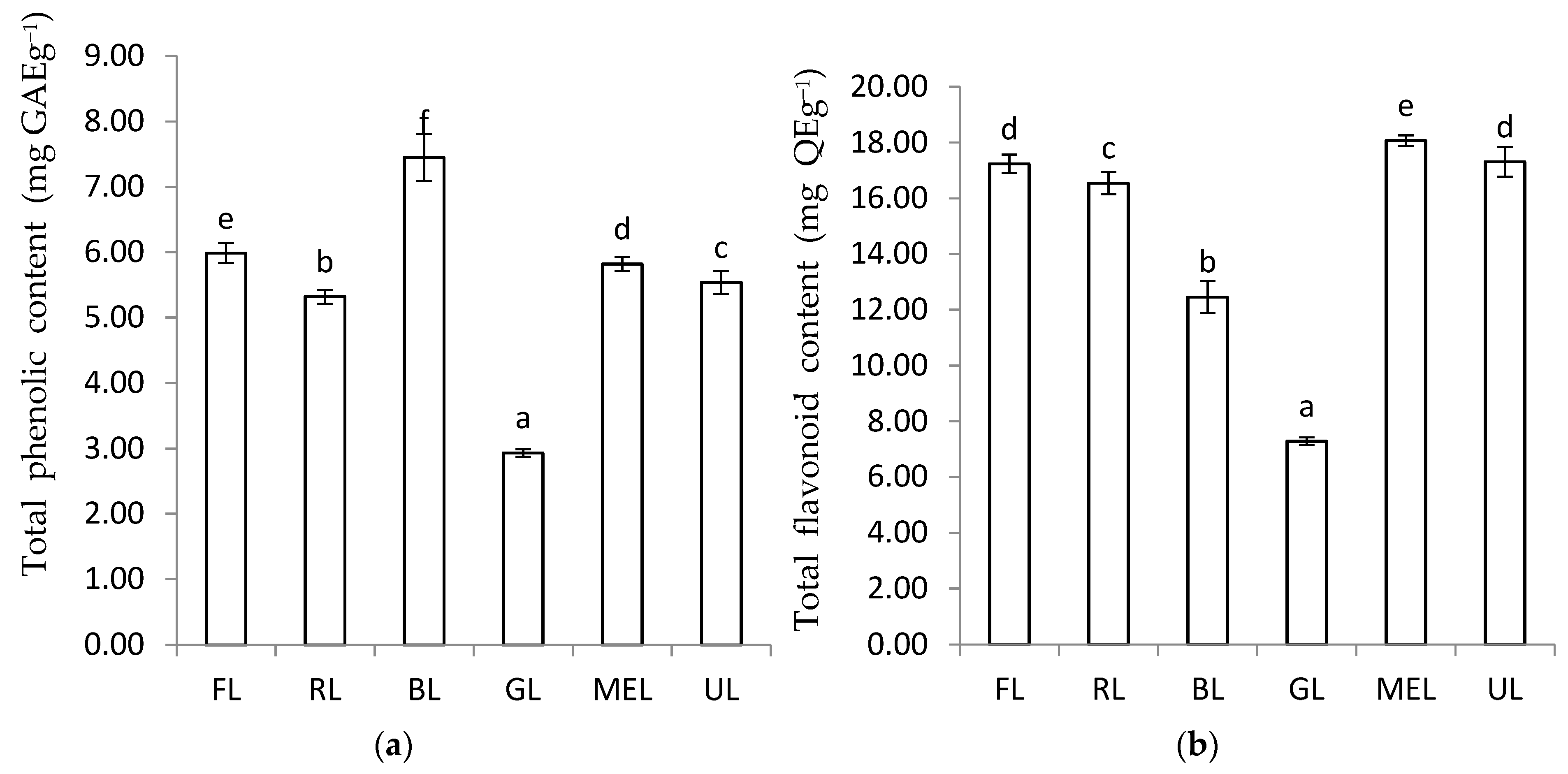
3.1. The Total Phenolic Content and Total Flavonoid Content of Peanut Sprout Grown under Different LED Types

The influence of different light spectra was clearly visible in the total phenolic content and total flavonoid levels of peanut sprouts (Figure 2a,b). The highest total phenolic content was recorded in the case of blue LED (7.45 ± 0.36 mg GAE g−1 extract). The irradiation of sprouts with green LED resulted in a significant decrease in the total phenolic concentration (2.93 ± 0.06 mg GAE g−1 extract). The total phenolic content levels followed the trend of blue LED > FL > MEL > ultrasound+ FL (UL) > red LED > green LED. The total flavonoid content was higher under MEL (18.07 ± 0.19 mg QE g−1 extract). The total flavonoid content followed the trend of MEL > UL > FL > red LED > green LED.

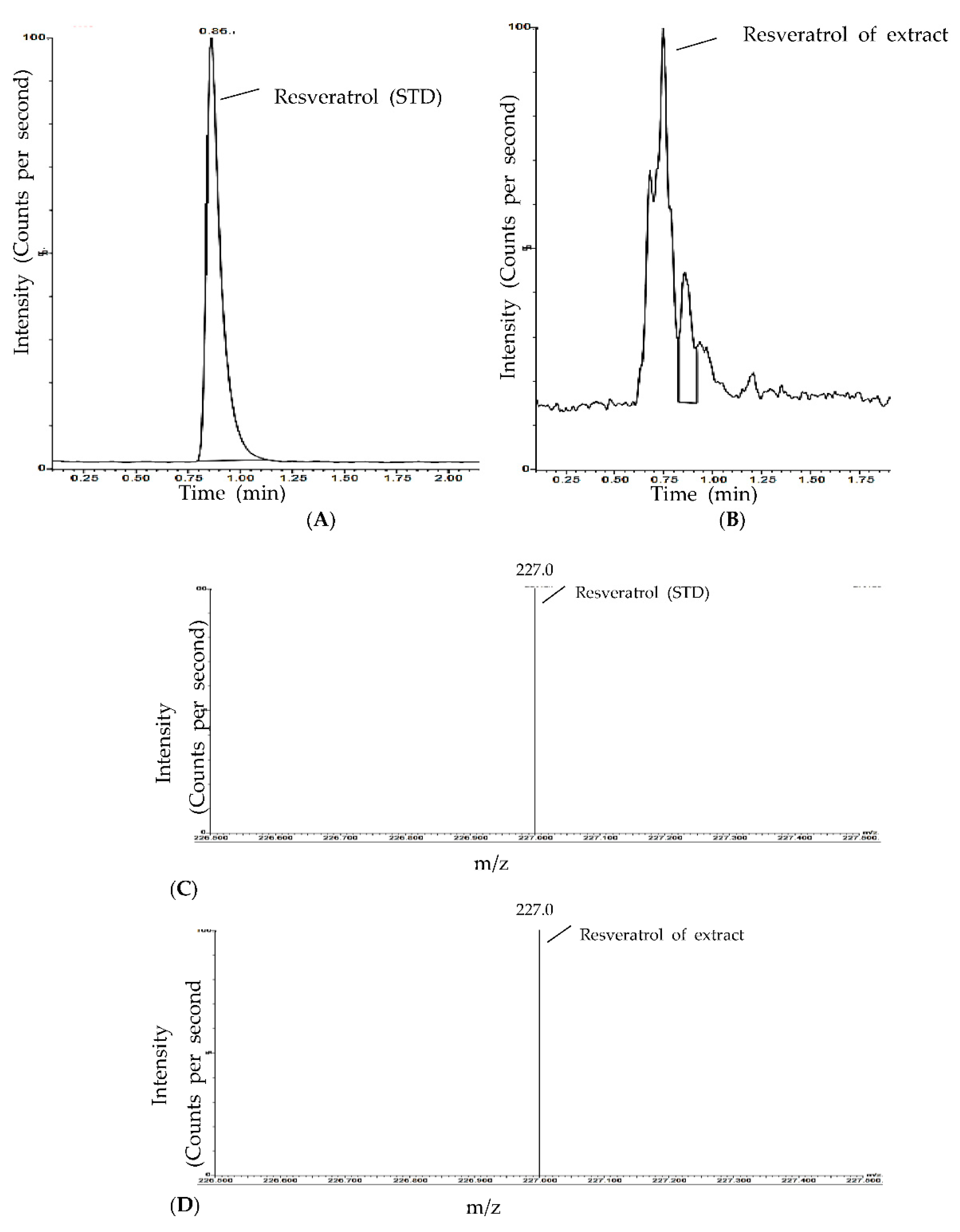
3.2. Resveratrol Content in Peanut Sprout Extracts Grown under Different LED Types

The influence of light quality on the resveratrol content in peanut sprouts is presented in Table 1. The resveratrol concentration in peanut sprouts grown under different light intensities was determined using a resveratrol standard. The LC/MS chromatogram (Figure 3a) clearly shows a major peak of the standard resveratrol (retention time = 25 s). When the mixture of plant extracts and standard resveratrol was examined under the same conditions using LC/MS, a similar peak value of resveratrol (retention time = 23 s) was observed (Figure 3b). The amount of resveratrol significantly varied in the different peanut samples, indicating that it is the major compound influenced by varying light intensities. Peanut sprouts grown under blue LED light exhibited a higher total resveratrol content than those under FL and other LED light sources. The total resveratrol content in the peanut sprouts followed the trend of blue LED > green LED > FL > MEL > red LED > UL.

3.3. Variation in the Antioxidant Properties of Peanut Sprouts Grown under Different LED Types

The antioxidant properties varied markedly among peanut sprouts grown under different light sources (Figure 4a,b). In this study, we observed that the DPPH value of blue LED-treated sprouts was significantly (p < 0.05) higher than that of sprouts grown under lights with other wavelengths. The influence of the light quality on the ABTS radical scavenging activity of the peanut sprouts is depicted in Figure 4b. The ABTS radical scavenging potential of peanut upon irradiation with blue LED was higher than that with any other treatment.

3.4. Tyrosinase Activity in Peanut Sprouts Grown under Different LED Types

At 1000 mg mL−1, the highest anti-tyrosinase activity of the peanut sprouts grown under FL light quality extracts was 45.46%. At the same concentration, peanut sprouts grown under MEL, red LED, UL, and blue LED light qualities exhibited moderate anti-tyrosinase activity, i.e., 33.24%, 32.38%, 29.69%, 27.69%, and 25.20%, respectively (Figure 5). A lower anti-tyrosinase activity was observed in sprouts grown under blue LED light at all tested concentrations. The anti-tyrosinase activity increased with an increase in the concentration of the tested sample. Kojic acid, used as a positive control in the study, showed a better anti-tyrosinase activity at 96.84%.

3.5. Cytotoxic Effect (MTT Assay)

The cell toxicities varied markedly among peanut sprouts grown under different light wavelengths. As shown in Figure 6, the methanol extracts of peanut sprouts at a concentration of 10 ppm showed no cytotoxicity. Blue LED-irradiated peanut extracts at a selected concentration range (200 ppm) showed moderate cytotoxicity. The degree of cytotoxicity increased with an increase in the sample concentration (10 ppm, 100 ppm, and 200 ppm). The exposition of sprouts with red LED and green LED showed cytotoxic activity at 89% and 88%, respectively, at a sample concentration of 200 ppm. As visualized under a phase-contrast microscope, a higher cytotoxicity was observed in RAW 264.7 cells incubated with the blue LED-irradiated peanut extracts compared to those exposed to FL-treated or other LED-treated extracts (Supplementary Materials Figure S1). Notably, the percentage of dead cells after treatment with the plant extracts did not reach 30% for all the tested extracts. Thus, the nontoxic nature of all the plant extracts corroborates well with the traditional use of peanut sprouts as supplementary food.

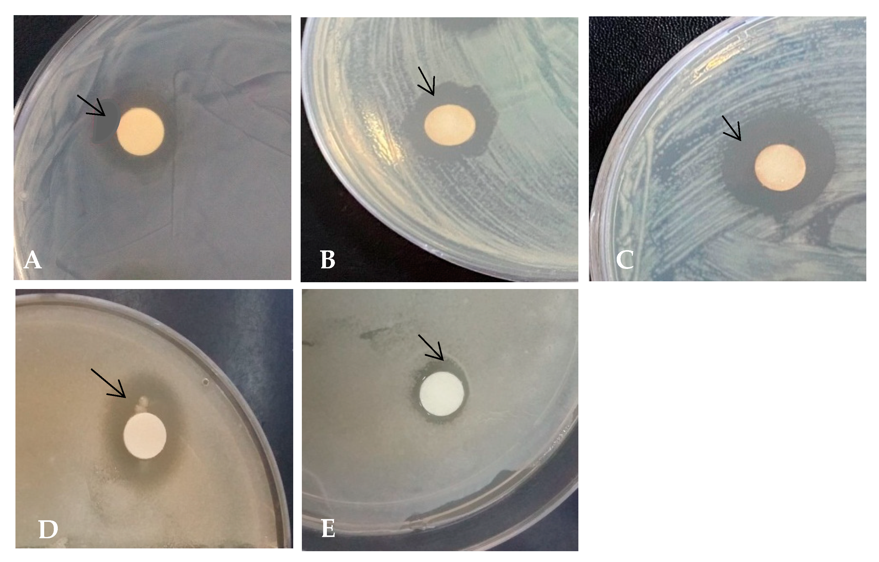
3.6. Antimicrobial Activity of Peanut Sprouts Measured Using the Zone of Inhibition Assay

The antimicrobial activity of peanut extracts exposed to LED light was tested against various Gram-positive and Gram-negative bacteria (Table 2). As depicted in Figure 7, the zone of inhibition varied in the different tested samples. The results showed that the antimicrobial activity of peanut sprouts grown under blue LED was effective against E. coli Castellani and Chalmers (ATCC35150), S. enteritidis serotype typhimurium Kauffmann and Edwards (ATCC14028), and V. litoralis (KCTC12320) at a concentration of 1000 ppm, with inhibitory zones of 9.50 ± 1.50 mm, 8.50 ± 0.20, and 8.35 ± 0.60, respectively. However, not all the extracts were sensitive to the tested bacterial strains. As shown in the results, B. subtilis (KCCM 11316) was less sensitive to all the LED-treated extracts. Red LED extracts inhibited the growth of P. aeruginosa (ATCC25668), whereas a higher concentration of green LED extracts was needed to inhibit the growth of S. enteritidis serotype typhimurium Kauffmann and Edwards (ATCC14028). As shown in Figure 8a,b, the presence of dead cells was evident after a 24-h incubation of E. coli Castellani and Chalmers (ATCC35150) and S. enteritidis serotype typhimurium Kauffmann and Edwards (ATCC14028) with peanut sprout extracts at concentrations of 0.25 mg mL−1 and 0.12 mg mL−1, respectively. The proportion of dead bacteria (stained by propidium iodide in red color) increased in a dose-dependent manner. However, the bacterial cells were predominantly viable (stained in green by Sytox 9) in the untreated sample or control. When compared with the fluorescent light-exposed extracts, the blue LED-exposed sprout extracts resulted in more dead cells.

4. Discussion

The concentration and composition of secondary metabolites in plants is influenced by the source of light and its wavelength [29]. In the present studies, blue LED exposition to the early growth period of peanut sprouts increased the total phenolic contents. This result is consistent with the results of Bian et al. [55], Demotes-Mainard et al. [56], Huché-Thélier et al. [57], and Samuolienė et al. [39], where blue light induced a higher total phenolic content in plants compared to other LED lights. Moreover, Chung et al. [50] reported an increased level of malonyldaidzin, malonyl genistin, p-coumaric acid salicylic acid, p-hydrobenzoic acid, and gentisic acid levels in Pachyrhizus erosus grown under blue LED irradiation. The effectiveness of blue LED in inducing a higher accumulation of gallic acid and quercetin was observed in wheat sprouts [58]. Additionally, an improvement in the accumulation of chlorogenic acid, p-hydrobenzoic acid, caffeic acid, p-coumeric acid, and ferulic acid was reported in blue LED grown pea sprouts [27]. Meanwhile, others have reported an increase in the accumulation of secondary metabolites such as cyanidin-monoglucosides in Gynura procumbens (Lour.) Merr. under blue LED light [59]. Phenolic-accumulating tissues are sensitive to UV radiation [60] and trigger the biosynthesis of phenolic compounds [61,62]. FL is characterized by a high UV emission (in contrast to blue LED). Therefore, it is possible that blue LED light could have induced a similar response in the growing peanut sprouts, resulting in a higher total phenolic content. According to the report of Amoozgar et al. [63], the expression of genes associated with the synthesis of phenolic compounds strongly depends on the quality of the light. In a similar study, plant seedlings grown under blue LED showed activation of the PAL gene, which encodes the enzyme that catalyzes the initial stages of phenylpropenoid and flavonoid biosynthesis in lettuces and tomatoes [43,64]. The highest total flavonoid content was observed in the MEL-treated sample, indicating that MEL is more effective than LEDs and FL for the accumulation of flavonoids in peanut sprouts. Thus, there is a possible relationship between the MEL light source and flavonoid biosynthesis pathway. However, the effect of the MEL light source on the mechanism of the increased flavonoid contents is not well-known yet.
Resveratrol is a major secondary metabolite in peanuts. It is associated with anticancer, anti-inflammatory, and neuroprotective effects; protective effects on the cardiovascular system; and prevention against degenerative diseases [65]. It is also considered an effective scavenger of reactive oxygen species (ROS) and reactive nitrogen species (RNS) produced during metabolic processes [66], which play a major role in maintaining the oxidation–reduction balance in cells expressing antioxidant enzymes, such as catalase (CAT), superoxide dismutase (SOD), and glutathione peroxidase [67,68]. Moreover, these compounds are sensitive to changes in the wavelength of light and the plant environment [69], which is consistent with the present results. A significant difference in the amount of resveratrol was observed upon exposure to different light sources, which is consistent with its antioxidant properties.
The spectral differences in light sources have a huge impact on the metabolic process of plants [70]. The secondary metabolites have antioxidant properties, and the concentration and composition of these compounds are influenced by the light quality [71,72]. Previous studies have demonstrated that exposing growing plant seedlings to different light spectra results in an increase in the levels of secondary metabolites [38]. This trend was also observed in the present study. Among the different light sources, the exposure of peanut sprouts to blue LEDs showed a higher DPPH radical scavenging activity than that in FL light-irradiated peanut sprouts. Our results were in-line with those observed by Chung et al. [50] in Pachyrhizus erosus. In addition, blue LED-treated peanut sprout extracts produced a higher ABTS value than under FL, red LED, and green LED lights but were not significant compared with the MEL- and UL-treated extracts. A higher ABTS radical scavenging activity was also observed with blue LED. This study, along with prior studies, indicate that blue LED is a more effective light source for increasing the antioxidant levels in peanut sprouts. In this study, a strong positive correlation was observed between the antioxidant levels and resveratrol content in the peanut sprouts. These correlations were higher in blue LED-exposed peanut sprouts compared with those observed in other light treatments. This could be attributed to higher levels of resveratrol production in the peanut sprouts irradiated with blue LED. However, the mechanism by which the amount of resveratrol was enhanced in the blue LED-irradiated peanut sprouts remains unclear. Several mechanisms that explain the variability in the antioxidant activity in plants grown under different LED lights have been considered. Many studies have shown that resveratrol possesses the ability to scavenge reactive oxygen species (ROS) such as OH radicals and O2 radicals regenerated during metabolic processes [73,74,75] and plays a key role in maintaining the levels of intracellular antioxidants that protect against the detrimental effects of ROS [74]. It was previously demonstrated that the antiradical properties of resveratrol are mediated through its reduction of respiratory chain complex III activity (electron transport chain), which reduces ROS regeneration [76]. In another study, resveratrol was shown to increase the levels of antioxidant enzymes, such as glutathione reductase, glutathione peroxidase, and glutathione s-transferase, which assist cells in neutralizing free radicals [77]. Although the exact mechanism has not been fully understood, our results indicate that the exposure to LED and MEL lights enhances the accumulation of phenolic compounds and resveratrol, which increases the antioxidant activity of peanut sprouts. This result was further supported by a strong positive correlation between the total flavonoid content and DPPH (r2 = 0.859 *, p < 0.05) and ABTS (r2 = 0.898 *, p < 0.05) values of peanut sprouts grown under blue LED illumination (Table S1). Previous studies on peanut seedlings have reported the presence of phenolic compounds, such as catechin, proartho cyanides, procyanidins [78], quininic acid, ferulic acid, [79], flavonoids, coumaric acid, caffeic acid, and chlorogenic acid [80]. Moreover, in the present study, the high correlation between the total flavonoid content and antioxidant activities indicates that antioxidant compounds other than resveratrol might be present in peanut extracts and produce a synergistic effect.
Concentrations equal or lower than 100 ppm showed no significant differences in the cell viability in all the treatments. At higher concentrations of peanut extracts treated with blue LEDs, the cytotoxicity of the peanut extracts was somewhat higher. The increased concentration of blue LED extracts to 200 ppm decreased the cell viability by a level close to 55% of RAW 264.7 cells. A number of studies indicated that a cell viability percentage between 50–70% was considered weakly toxic to cells [81]. The cytotoxic activity of polyphenols has been extensively studied on cell lines [82,83]. The phytochemicals such as flavonoids and flavones cause cytotoxicity by increasing the intracellular ROS levels [84]. However, the proper investigation and screening of phenolic compounds present in peanut sprouts grown under LED lights are needed to confirm this.
The antibacterial activity of plant extracts has been known for being influenced by light sources and attributed widely to the secondary metabolites of plants. Previous studies have also reported that resveratrol inhibits the growth of Gram-positive and Gram-negative bacteria [85,86,87,88]. This result is consistent with the published data of [84], which demonstrated that resveratrol effectively reduced the DNA content of pathogenic bacteria such as in Bacillus cereus, resulting in the suppression of cell division, thereby inhibiting bacterial growth [89]. Resveratrol was also reported to alter bacterial cell morphology and disrupt the intracellular machinery, thereby causing cell death [90]. In the present study, the peanut extracts showed a wide variation in the concentrations of resveratrol needed for inhibiting these microorganisms, which is consistent with previously published data [89,90,91,92,93,94,95], indicating the differential effect of resveratrol on bacteria. For example, according to Tegos et al. [88] and Paulo et al. [89], bacteria such as E. coli, Klebsiella pneumoniae, and Salmonella enterica serovar Typhimurium are less sensitive to resveratrol than other pathogenic bacteria. According to previous studies, resveratrol partially inhibits adenosine triphosphate (ATP) hydrolysis and ATP synthesis [96] and causes DNA fragmentation and septum formation in E. coli [97]. Another study reported a positive correlation between resveratrol and membrane damage and potassium leakage in pathogenic bacteria [98]. Furthermore, trans-resveratrol isolated from M. benthamianum showed higher antimicrobial activity against pathogenic bacterial strains, including E. coli, S. aureus, P. aeruginosa, and S. mutans. A similar inhibitory activity of resveratrol on pathogenic microorganisms was also observed by Docherty et al. [99] and Jung et al. [100]. Therefore, it is possible that higher resveratrol concentrations in blue LED-irradiated peanut sprouts can be responsible for the higher antibacterial activity of these extracts. In addition, resveratrol isolated from the seeds of Gnetum gnemon L exhibited potent antimicrobial activity against E. coli, B. subtilis, C. perfringens, S. cerevisiae [101], and Staphylococcus pneumoniae [102]. Gram-negative bacteria were less sensitive to resveratrol than Gram-positive bacteria [88,89]. The poor sensitivity of Gram-negative bacteria to resveratrol was previously attributed to the poor penetration of resveratrol into the outer membrane of bacteria. Resveratrol has been shown to exhibit antibiofilm properties against several Gram-negative [92] and Gram-positive bacteria [103,104] by inhibiting the expression of the genes CsgA and CSgB, which are responsible for biofilm production. Although the exact mechanism is not fully understood, our results indicate that the sensitivity of Gram-negative and Gram-positive bacteria to resveratrol varies. Numerous studies have linked the antimicrobial activity of plant extracts to their phytochemical compositions. In this study, a higher correlation between the total phenolic content/total flavonoid contents and antimicrobial activity indicates that secondary metabolites other than resveratrol present in the peanut sprouts could be the potential sources of antimicrobial agents. In another study, pea sprouts grown under blue LED exhibited a significant increase in the flavonoid, such as kaempherol and rutin, contents [27], indicating that blue LED may trigger the biosynthesis of flavonoids. Research into the molecular pathways of phenolic compounds has indicated that blue LED increases the expression of PAL and CRY genes, associated with the synthesis of phenylalanine ammonia lyase and chalcone enzymes, respectively, in Arabidopsis seedlings [105].

5. Conclusions

The present study demonstrated that blue LED is effective and suitable for enhancing the resveratrol content of peanut sprouts. Furthermore, the antioxidant capacity using DPPH and ABTS radicals and the total phenolic content increased considerably under the blue LED when compared to the green LED, red LED, micro-electrodeless light, and fluorescent light. Additionally, peanut extracts grown under blue LED illumination significantly inhibited the multiplication of the pathogenic bacterial strains. These observations indicate that blue LED is more effective and suitable for the growth of peanut sprouts. However, such studies on the influence of LEDs on peanut sprouts are still in their nascent stages. The phenolic compound components and their concentrations may vary in different peanut sprout samples that possess antioxidant properties and antimicrobial activities. To address this hypothesis, further studies are needed to identify potentially active bioactive compounds and biosynthesis mechanisms.

Supplementary Materials

The following are available online at https://www.mdpi.com/2073-4395/11/2/305/s1: Figure S1: Cytotoxic effect of peanut sprout on RAW 264.7 cells. (a) Fl light, (b) Red-LED, (c) Blue-LED, (d) Green-LED, (e) MEL, (f) Ul. Table S1: Correlation coefficient analysis between antioxidant activity, total phenolic content and total flavonoid content.

Author Contributions

B.K.G. designed the experiment and wrote the manuscript. C.Y.Y. supervised the experiment. R.C. and D.-H.O. performed the MTT assay and antimicrobial activities. C.L. performed the resveratrol analysis. M.H.H. performed the tyrosinase activity and antioxidant activity. H.-Y.C., S.-H.K., and I.-M.C. performed the phytochemical determination, analysis, and editing of the manuscript. All authors have read and agreed to the published version of the manuscript.

Funding

This study was supported by the KU Research Professor program. This work was funded by a BK21 FOUR (Fostering Outstanding Universities for Research, grant no. 4220201013822, team: Crop Genetic Resources Research Team for Future Human Resources Development in Sustainable Premium Agricultural Industry, Konkuk University), the National Research Foundation of Korea, Republic of Korea.

Institutional Review Board Statement

Not applicable.

Informed Consent Statement

Not applicable.

Data Availability Statement

Data sharing not applicable.

Conflicts of Interest

The authors declare that they have no conflicts of interest.

References

  1. Freitas, F.O.; Moretzsohn, M.C.; Valls, J.F.M. Genetic variability of Brazilian Indian landraces of Arachis hypogaea. L. Genet. Mol. Res. 2007, 6, 675–684. [Google Scholar]
  2. Zhao, X.; Chen, J.; Du, F. Potential use of peanut by-products in food processing: A review. J. Food Sci. Technol. 2012, 49, 521–529. [Google Scholar] [CrossRef]
  3. Wu, M.C.; Hou, C.Y.; Jiang, C.M.; Wang, Y.T.; Wang, C.Y.; Chen, H.H.; Chang, H.M. A novel approach of LED light radiation improves the antioxidant activity of pea seedlings. Food Chem. 2007, 101, 1753–1758. [Google Scholar] [CrossRef]
  4. Hasan, M.M.; Cha, M.; Bajpai, V.K.; Baek, K.H. Production of a major stilbene phytoalexin, resveratrol in peanut (Arachis hypogaea) and peanut products: A mini review. Rev. Environ. Sci. Bio/Technol. 2013, 12, 209–221. [Google Scholar] [CrossRef]
  5. Lopes, R.M.; Agostini-Costa, T.d.S.; Gimenes, M.A.; Silveira, D. Chemical composition and biological activities of Arachis species. J. Agric. Food Chem. 2011, 59, 4321–4330. [Google Scholar] [CrossRef] [PubMed]
  6. Sebei, K.; Gnouma, A.; Herchi, W.; Sakouhi, F.; Boukhchina, S. Lipids, proteins, phenolic composition, antioxidant and antibacterial activities of seeds of peanuts (Arachis hypogaea L.) cultivated in Tunisia. Biol. Res. 2013, 46, 257–263. [Google Scholar] [CrossRef] [Green Version]
  7. Ignatowicz, E.; Baer-Dubowska, W. Resveratrol, a natural chemopreventive agent against degenerative diseases. Pol. J. Pharmacol. 2001, 53, 557–569. [Google Scholar] [PubMed]
  8. Jang, M.; Cai, L.; Udeani, G.O.; Slowing, K.V.; Thomas, C.F.; Beecher, C.W.; Fong, H.H.; Farnsworth, N.R.; Kinghorn, A.D.; Mehta, R.G.; et al. Cancer chemopreventive activity of resveratrol, a natural product derived from grapes. Science 1997, 275, 218–220. [Google Scholar] [CrossRef] [Green Version]
  9. Cal, C.; Garban, H.; Jazirehi, A.; Yeh, C.; Mizutani, Y.; Bonavida, B. Resveratrol and cancer: Chemoprevention, apoptosis, and chemo-immunosensitizing activities. Curr. Med. Chem. Anti-Cancer Agents 2003, 3, 77–93. [Google Scholar] [CrossRef] [Green Version]
  10. Zhuang, H.; Kim, Y.S.; Koehler, R.C.; Dore, S. Potential mechanism by which resveratrol, a red wine constituent, protects neurons. Ann. N. Y. Acad. Sci. 2003, 993, 276–286. [Google Scholar] [CrossRef]
  11. Ingham, J.L. 3,5,4-Trihydroxystilbene as a phytoalexin from ground nuts (A. hypogaea). Phytochemistry 1976, 15, 1791–1793. [Google Scholar] [CrossRef]
  12. Hart, J.H. Role of phytostilbenes in decay and disease resistance. Annu. Rev. Phytopathol. 1981, 19, 437–458. [Google Scholar] [CrossRef]
  13. Jeandet, P.; Douillet-Breuil, A.C.; Bessis, R.; Debord, S.; Sbaghi, M.; Adrian, M. Phytoalexins from the Vitaceae: Biosynthesis, phytoalexin gene expression in transgenic plants, antifungal activity, and metabolism. J. Agric. Food Chem. 2002, 50, 2731–2741. [Google Scholar] [CrossRef] [PubMed]
  14. Cadenas, S.; Barja, G. Resveratrol, melatonin, vitamin E and PBN protect against oxidative DNA damage induced by the kidney carcinogen KBrO2, Free rad. Biol. Med. 1999, 26, 1531–1537. [Google Scholar]
  15. Frement, L. Biological effects of resveratrol. Life Sci. 2000, 66, 663–673. [Google Scholar] [CrossRef]
  16. Liu, H.K.; Cao, Y.; Huang, W.N.; Guo, Y.D.; Kang, Y.F. Effect of ethylene on total phenolics, antioxidant activity, and the activity of metabolic enzymes in mung bean sprouts. Eur. Food Res. Technol. 2013, 237, 755–764. [Google Scholar] [CrossRef]
  17. Swieca, M.; Gawlik-Dziki, U. Effects of sprouting and postharvest storage under cool temperature conditions on starch content and antioxidant capacity of green pea, lentil and young mung bean sprouts. Food Chem. 2015, 185, 99–105. [Google Scholar] [CrossRef]
  18. Li, Y.C.; Qian, H.; Sun, X.L.; Cui, Y.; Wang, H.Y.; Du, C.; Xia, X.H. The effects of germination on chemical composition of peanut seed. Food Sci. Technol. Res. 2014, 20, 883–889. [Google Scholar] [CrossRef] [Green Version]
  19. Limmongkon, A.; Janhom, P.; Amthong, A.; Kawpanuk, M.; Nopprang, P.; Poohadsuan, J.; Somboon, T.; Saijeen, S.; Surangkul, D.; Srikummool, M.; et al. Antioxidant activity, total phenolic, and resveratrol content in five cultivars of peanut Sprouts. Asian Pac. J. Trop. Biomed. 2017, 7, 332–338. [Google Scholar] [CrossRef]
  20. Chung, I.M.; Park, M.R.; Chun, J.C.; Yun, S.J. Resveratrol accumulation and resveratrol synthase gene expression in response to abiotic stresses and hormones in peanut plants. Plant Sci. 2003, 164, 103–109. [Google Scholar] [CrossRef]
  21. Martínez-M´arquez, A.; Morante-Carriel, J.A.; Ramírez-Estrada, K.; Cusid´o, R.M.; Palazon, J.; Bru-Martínez, R. Production of highly bioactive resveratrol analogues pterostilbene and piceatannol in metabolically engineered grapevine cell cultures. Plant Biotechnol. J. 2016, 14, 1813–1825. [Google Scholar] [CrossRef] [PubMed] [Green Version]
  22. Carbo, N.; Costelli, P.; Baccino, F.M.; Lopez-Soriano, F.J.; Argiles, J.M. Resveratrol, a natural product present in wine decreases tumour growth in a rat tumour model. Biochem. Biophys. Res. Commun. 1999, 254, 739–743. [Google Scholar] [CrossRef]
  23. Huang, C.; Ma, W.Y.; Goranson, A.; Dong, Z. Resveratrol suppresses cell transformation and induces apoptosis through a p53-dependent pathway. Carcinogenesis 1999, 20, 237–242. [Google Scholar] [CrossRef] [Green Version]
  24. Chang, C.C.; Lin, K.Y.; Peng, K.Y.; Day, Y.J.; Hung, L.M. Resveratrol exerts anti-obesity effects in high-fat diet obese mice and displays differential dosage effects on cytotoxicity, differentiation, and lipolysis in 3T3-L1 cells. Endocr. J. 2016, 63, 169–178. [Google Scholar] [CrossRef] [Green Version]
  25. Kimura, Y.; Okuda, H. Resveratrol isolated from Polygonum cuspidatum root prevents tumour growth and metastasis to lung and tumor-induced neovascularization in Lewis lung carcinoma-bearing mice. J. Nutr. 2001, 131, 1844–1849. [Google Scholar] [CrossRef]
  26. Wolters, D.A.; Washburn, M.P.; Yates, J.R. An automated multidimensional protein identification technology for shotgun proteomics. Anal. Chem. 2001, 73, 5683–5690. [Google Scholar]
  27. Liu, H.K.; Chen, Y.Y.; Hu, T.T.; Zhang, S.J.; Zhang, Y.H.; Zhao, T.Y.; Yu, H.E.; Kang, Y.F. The influence of light-emitting diodes on the phenolic compounds and antioxidant activities in pea sprouts. J. Funct. Foods 2016, 25, 459–465. [Google Scholar] [CrossRef]
  28. Ahn, S.Y.; Kim, S.A.; Yun, H.K. Inhibition of Botrytis cinerea and accumulation of stilbene compounds by light emitting diodes of grapevine leaves and differential expression of defense-related genes. Eur. J. Plant Pathol./Eur. Found. Plant Pathol. 2015, 143, 753–765. [Google Scholar] [CrossRef]
  29. Son, K.H.; Oh, M.M. Leaf Shape, Growth, and Antioxidant Phenolic Compounds of Two Lettuce Cultivars Grown under Various Combinations of Blue and Red Light-emitting Diodes. Hortiscience 2013, 48, 988–995. [Google Scholar] [CrossRef]
  30. Thwe, A.A.; Kim, Y.B.; Li, X.; Seo, J.M.; Kim, S.J.; Suzuki, T.; Chung, S.O.; Park, S.U. Effects of light-emitting diodes on expression of phenylpropanoid biosynthetic genes and accumulation of phenylpropanoids in Fagopyrum tataricum sprouts. J. Agric. Food Chem. 2014, 62, 4839–4845. [Google Scholar] [CrossRef] [PubMed]
  31. Hernández, R.; Kubota, C. Growth and morphological response of cucumber seedlings to supplemental red and blue photon flux ratios under varied solar daily light integrals. Sci. Hortic. 2014, 173, 92–99. [Google Scholar] [CrossRef]
  32. Hernández, R.; Kubota, C. Physiological responses of cucumber seedlings under different blue and red photon flux ratios using LEDs. Environ. Exp. Bot. 2016, 121, 66–74. [Google Scholar] [CrossRef]
  33. Solano, C.J.; Hernández, J.A.; Suardíaz, J.; Barba-Espín, G. Impacts of LEDs in the Red Spectrum on the Germination, Early Seedling Growth and Antioxidant Metabolism of Pea (Pisum sativum L.) and Melon (Cucumis melo L.). Agriculture 2020, 10, 204. [Google Scholar] [CrossRef]
  34. Zakir, H.M. Light emitting diodes increase phenolics of buckwheat (Fagopyrum esculentum) sprouts. J. Plant Interact. 2007, 2, 7178. [Google Scholar]
  35. Choi, H.; Moon, B.; Kang, N. Effects of LED light on the production of strawberry during cultivation in a plastic greenhouse and in a growth chamber. Sci. Hortic. 2015, 189, 22–31. [Google Scholar] [CrossRef]
  36. Peng, L.; Zou, L.; Su, Y.; Fan, Y.; Zhao, G. Effects of light on growth, levels of anthocyanin, concentration of metabolites in Fagopyrum tataricum sprout cultures. Int. J. Food Sci. Technol. 2015, 50, 1382–1389. [Google Scholar] [CrossRef]
  37. Park, C.H.; Kim, N.S.; Park, J.S.; Lee, S.Y.; Lee, J.W.; Park, S.U. Effects of light-emitting diodes on the accumulation of glucosinolates and phenolic compounds in sprouting canola (Brassica napus L.). Foods 2019, 8, 76. [Google Scholar] [CrossRef] [Green Version]
  38. Koga, R.; Meng, T.; Nakamura, E.; Miura, C.; Irino, N.; Devkota, H.P.; Yahara, S.; Kondo, R. The effect of photo-irradiation on the growth and ingredient composition of young green barley (Hordeum vulgare). Agric. Sci. 2013, 4, 185–194. [Google Scholar]
  39. Samuolienė, G.; Viršilė, A.; Brazaitytė, A.; Jankauskienė, J.; Sakalauskienė, S.; Vaštakaitė, V.; Novičkovas, A.; Viškelienė, A.; Sasnauskas, A.; Duchovskis, P. Blue light dosage affects carotenoids and tocopherols in microgreens. Food Chem. 2017, 228, 50–56. [Google Scholar] [CrossRef] [PubMed]
  40. Seo, J.M.; Arasu, M.V.; Kim, Y.B.; Park, S.U.; Kim, S.J. Phenylalanine and LED lights enhance phenolic compound production in tartary buckwheat sprouts. Food Chem. 2015, 177, 204–213. [Google Scholar] [CrossRef] [PubMed]
  41. Lee, S.W.; Seo, J.M.; Lee, M.K.; Chun, J.H.; Antonisamy, P.; Arasu, M.V.; Suzuki, T.; Al-Dhabi, N.A.; Kim, S.J. Influence of different LED lamps on the production of phenolic compounds in common and Tartary buckwheat sprouts. Ind. Crops Prod. 2014, 54, 320–326. [Google Scholar] [CrossRef]
  42. Ghimire, B.K.; Sacks, E.J.; Kim, S.H.; Yu, C.Y.; Chung, I.M. Profiling of phenolic compounds composition, morphological traits, and antioxidant activity of Miscanthus sacchariflorus L. accessions. Agronomy 2021, 11, 243. [Google Scholar] [CrossRef]
  43. Lee, M.J.; Son, J.E.; Oh, M.M. Growth and phenolic compounds of Lactuca sativa L. grown in a closed-type plant production system with UV-A, -B, or -C lamp. J. Sci. Food Agric. 2014, 942, 197–204. [Google Scholar] [CrossRef] [PubMed]
  44. Samuoliene, G.; Brazaityte, A.; Vaštakaite, V. Light-Emitting Diodes (LEDs) for Improved Nutritional Quality. In Light Emitting Diodes for Agriculture, 1st ed.; Dutta Gupta, S., Ed.; Springer: Singapore, 2018; pp. 149–190. [Google Scholar]
  45. Khattak, A.B.; Zeb, A.; Bibi, N. Impact of germination time and type of illumination on caroteinoid content, protein solubility and in-vitro protein digestibility of chickpea (Cicer arietinum L) sprouts. Food Chem. 2008, 109, 797–801. [Google Scholar] [CrossRef]
  46. Luczkiewicz, M.; Zarate, R.; Dembińska-Migas, W.; Migas, P.; Verpoorte, R. Production of pulchelin E in hairy roots, callus and suspension cultures of Rudbeckia hirta L. Plant Sci. 2002, 163, 91–100. [Google Scholar] [CrossRef]
  47. Kurutas, E.B. The importance of antioxidants which play the role in cellular response against oxidative/nitrosative stress: Current state. Nutr. J. 2016, 15, 1–22. [Google Scholar] [CrossRef] [Green Version]
  48. Chung, I.M.; Chelliah, R.; Oh, D.H.; Kim, S.H.; Yu, C.Y.; Ghimire, B.K. Tupistra nutans Wall. root extract, rich in phenolics, inhibits microbial growth and α-glucosidase activity, while demonstrating strong antioxidant potential. Braz. J. Bot. 2019, 42, 383–397. [Google Scholar] [CrossRef]
  49. Nieva Moreno, M.I.; Isla, M.I.; Sampietro, A.R.; Vattuone, M.A. Comparison of the free radicalscavenging activity of propolis from several regions of Argentina. J. Ethnopharmacol. 2000, 71, 109–114. [Google Scholar] [CrossRef]
  50. Chung, I.M.; Paudel, N.; Kim, S.H.; Yu, C.Y.; Ghimire, B.K. The influence of light wavelength on growth and antioxidant capacity in Pachyrhizus erosus (L.) Urban. J. Plant Growth Regul. 2019, 39, 296–312. [Google Scholar] [CrossRef]
  51. Thaipong, K.; Boonprakob, U.; Crosby, K.; Cisneros-Zevallos, L.; Byrne, D.H. Comparison of ABTS, DPPH, FRAP, and ORAC assays for estimating antioxidantactivity from guava fruit extracts. J. Food Compos. Anal. 2006, 19, 669–675. [Google Scholar] [CrossRef]
  52. Kubo, I.; Kinst-Hori, I. Tyrosinase inhibitors from anise oil. J. Agric. Food Chem. 1998, 46, 1268–1271. [Google Scholar] [CrossRef]
  53. Langcake, P.; Pryce, R. The production of resveratrol and the viniferins by grapevines in response to ultraviolet irradiation. Phytochem. 1977, 16, 1193–1196. [Google Scholar] [CrossRef]
  54. Mosmann, T. Rapid colorimetric assay for cellular growth and survival: Application to proliferation and cytotoxicity assays. J. Immunol. Methods 1983, 65, 55–63. [Google Scholar] [CrossRef]
  55. Bian, Z.H.; Yang, Q.C.; Liu, W.K. Effects of light quality on the accumulation of phytochemicals in vegetables produced in controlled environments: A review. J. Sci. Food Agric. 2015, 95, 869–877. [Google Scholar] [CrossRef] [PubMed]
  56. Demotes-Mainard, S.; Péron, T.; Corot, A.; Bertheloot, J.; Le Gourrierec, J.; Pelleschi-Travier, S.; Crespel, L.; Morel, P.; Huché-Thélier, L.; Boumaza, R.; et al. Plant responses to red and far-red lights, applications in horticulture. Environ. Exp. Bot. 2016, 121, 4–21. [Google Scholar] [CrossRef]
  57. Huché-Thélier, L.; Crespel, L.; Le Gourrierec, J.; Morel, P.; Sakr, S.; Leduc, N. Light signaling and plant responses to blue and UV radiations-perspectives for applications in horticulture. Environ. Exp. Bot. 2016, 121, 22–38. [Google Scholar] [CrossRef]
  58. Cuong, D.M.; Ha, T.W.; Park, C.H.; Kim, N.S.; Yeo, H.J.; Chun, S.W.; Kim, C.; Park, S.U. Effects of LED lights on Expression of Genes Involved in Phenylpropanoid Biosynthesis and Accumulation of Phenylpropanoids in Wheat Sprout. Agronomy 2019, 9, 307. [Google Scholar] [CrossRef] [Green Version]
  59. Lian, T.T.; Cha, S.Y.; Moe, M.M.; Kim, Y.J.; Bang, K.S. Effects of different colored LEDs on the enhancement of biologically active ingredients in callus cultures of Gynura procumbens (Lour.) Merr. Molecules 2019, 24, 4336. [Google Scholar] [CrossRef] [PubMed] [Green Version]
  60. Del Valle, J.C.; Buide, M.L.; Whittall, J.B.; Valladares, F.; Narbona, E. UV radiation increases phenolic compound protection but decreases reproduction in Silene littorea. PLoS ONE 2020, 15, e0231611. [Google Scholar] [CrossRef]
  61. Ebisawa, M.; Shoji, K.; Kato, M.; Shimomura, K.; Goto, F.; Yoshihara, T. Supplementary ultraviolet radiation B together with blue light at night increased qercetin content and flavonol systhase gene expression in leaf lettuce (Lactuca sativa L.). Environ. Control Biol. 2008, 46, 1–11. [Google Scholar] [CrossRef] [Green Version]
  62. Ryan, K.G.; Swinny, E.E.; Markham, K.R.; Winefield, C. Flavonoid gene expression and UV photoprotection in transgenic and mutant Petunia leaves. Phytochemistry 2002, 59, 23–32. [Google Scholar] [CrossRef]
  63. Amoozgar, A.; Mohammadi, A.; Sabzalian, M.R. Impact of light-emitting diode irradiation on photosynthesis, phytochemical composition and mineral element content of lettuce cv. Grizzly. Photosynthetica 2017, 55, 85–95. [Google Scholar] [CrossRef]
  64. Guo, J.; Wang, M.H. Ultraviolet A-specific induction of anthocyanin biosynthesis and PAL expression in tomato (Solanum lycopersicum L.). Plant Growth Regul. 2010, 621, 1–8. [Google Scholar] [CrossRef]
  65. Kopeć, A.; Piątkowska, E.; Leszczyńska, T.; Bieżanowska-Kopeć, R. Health-promoting affects of resveratrol. ŻYWNOŚĆ. Nauka. Technol. Jakość. 2011, 5, 5–15. (In Polish) [Google Scholar]
  66. Cordova-Gomez, M.; Galano, A.; Raul, J.; Alvarez-Idaboy, J.R. Piceatannol, a better peroxyl radical scavenger than resveratrol. RSC Adv. 2013, 3, 20209–20218. [Google Scholar] [CrossRef]
  67. Robb, E.L.; Page, M.M.; Wiens, B.E.; Stuart, J.A. Molecular mechanisms of oxidative stress resistance induced by resveratrol: Specific and progressive induction of MnSOD. Biochem. Biophys. Res. Commun. 2008, 367, 406–412. [Google Scholar] [CrossRef] [PubMed]
  68. Kavas, G.O.; Ayral, P.A.; Elhan, A.H. The Effects of Resveratrol on Oxidant/Antioxidant Systems and Their Cofactors in Rats. Adv. Clin. Exp. Med. 2013, 22, 151–155. [Google Scholar]
  69. Figueiras, T.S.; Neves-Petersen, M.T.; Petersen, S.B. Activation Energy of Light Induced Isomerization of Resveratrol. J. Fluoresc. 2011, 21, 1897–1906. [Google Scholar] [CrossRef] [PubMed]
  70. Zheng, L.; Van Labeke, M.C. Chrysanthemum morphology, photosynthetic efficiency and antioxidant capacity are differentially modified by light quality. J. Plant Physiol. 2017, 213, 66–74. [Google Scholar] [CrossRef]
  71. Li, Q.; Kubota, C. Effects of supplemental light quality on growth and phytochemicals of baby leaf lettuce. Environ. Exp. Bot. 2009, 67, 59–64. [Google Scholar] [CrossRef]
  72. Shahidi, F.; Ambigaipalan, P. Phenolics and polyphenolics in foods, beverages and spices: Antioxidant activity and health effects—A review J. Funct. Foods 2015, 18, 820–897. [Google Scholar] [CrossRef]
  73. Leonard, S.; Xia, C.; Jiang, B.H.; Stinefelt, B.; Klandorf, H.; Harris, G.K.; Shi, X. Resveratrol scavenges reactive oxygen species and effects radical-induced cellular responses. Biochem. Biophys. Res. Commun. 2003, 309, 1017–1026. [Google Scholar] [CrossRef]
  74. Losa, G.A. Resveratrol modulates apoptosis and oxidation in human blood mononuclear cells. Eur. J. Clin. Investig. 2003, 33, 818–823. [Google Scholar] [CrossRef] [PubMed]
  75. Martinez, J.; Moreno, J.J. Effect of resveratrol, a natural polyphenolic compound, on reactive oxygen species and prostaglandin production. Biochem. Pharm. 2000, 59, 865–870. [Google Scholar] [CrossRef]
  76. Zini, R.; Morin, C.; Bertelli, A.; Tillement, J.P. Effects of resveratrol on the rat brain respiratory chain. Drugs Experimen. Clin. Res. 1999, 25, 87. [Google Scholar]
  77. Yen, G.C.; Duh, P.D.; Lin, C.W. Effects of Resveratrol and 4-hexylresorcinol on Hydrogen Peroxide-induced Oxidative DNA Damage in Human Lymphocytes. Free Radic. Res. 2003, 37, 509–514. [Google Scholar] [CrossRef] [PubMed]
  78. Ma, Y.; Kosinska-Cagnazzo, A.; Kerr, W.L.; Amarowicz, R.; Swanson, R.B.; Pegg, R.B. Separation and characterization of phenolic compounds from dry-blanched peanut skins by liquid chromatography-electrospray ionization mass spectrometry. J. Chromatogr. A 2014, 1356, 64–81. [Google Scholar] [CrossRef]
  79. Francisco, M.L.D.; Resurreccion, A.V.A. Development of a reversed-phase high performance liquid chromatography (RPHPLC) procedure for the simultaneous determination of phenolic compounds in peanut skin extracts. Food Chem. 2009, 117, 356–363. [Google Scholar] [CrossRef]
  80. Attree, R.; Du, B.; Xu, B. Distribution of phenolic compounds in seed coat and cotyledon, and their contribution to antioxidant capacities of red and black seed coat peanuts (Arachis hypogaea L.). Ind. Crop. Prod. 2015, 67, 448–456. [Google Scholar] [CrossRef]
  81. Abdillahi, H.S.; Verschaeve, L.; Finnie, J.F.; Van Staden, J. Mutagenicity, antimutagenicity and cytotoxicity evaluation of South African Podocarpus species. J. Ethnopharmacol. 2012, 139, 728–738. [Google Scholar] [CrossRef]
  82. González-Sarrías, A.; Núñez-Sánchez, M.Á.; Tomás-Barberán, F.A.; Espín, J.C. Neuroprotective effects of bioavailable polyphenol-derived metabolites against oxidative stress-induced cytotoxicity in human neuroblastoma SH-SY5Y cells. J. Agric. Food Chem. 2017, 65, 752–758. [Google Scholar] [CrossRef] [Green Version]
  83. Smeriglio, A.; Cornara, L.; Denaro, M.; Barreca, D.; Burlando, B.; Xiao, J.; Trombetta, D. Antioxidant and cytoprotective activities of an ancient Mediterranean citrus (Citrus lumia Risso) albedo extract: Microscopic observations and polyphenol characterization. Food Chem. 2019, 279, 347–355. [Google Scholar] [CrossRef] [PubMed]
  84. Matsuo, M.; Sasaki, N.; Saga, K.; Kaneko, T. Cytotoxicity of Flavonoids toward Cultured Normal Human Cells. Biol. Pharm. Bull. 2005, 28, 253–259. [Google Scholar] [CrossRef] [PubMed] [Green Version]
  85. Mahady, G.B.; Pendland, S.L. Resveratrol inhibits the growth of Helicobacter pylori in vitro. Am. J. Gastroenterol. 2000, 95, 1849. [Google Scholar] [CrossRef]
  86. Docherty, J.J.; Fu, M.M.; Tsai, M. Resveratrol selectively inhibits Neisseria gonorrhoeae and Neisseria meningitidis. J. Antimicrob. Chemother. 2001, 47, 243–244. [Google Scholar] [CrossRef] [Green Version]
  87. Chan, M.M.Y. Antimicrobial effect of resveratrol on dermatophytes and bacterial pathogens of the skin. Biochem. Pharmacol. 2002, 63, 99–104. [Google Scholar] [CrossRef]
  88. Tegos, G.; Stermitz, F.R.; Lomovskaya, O.; Lewis, K. Multidrug pump inhibitors uncover remarkable activity of plant antimicrobials. Antimicrob. Agents Chemother. 2002, 46, 3133–3141. [Google Scholar] [CrossRef] [PubMed] [Green Version]
  89. Paulo, L.; Ferreira, S.; Gallardo, E.; Queiroz, J.A.; Domingues, F. Antimicrobial activity and effects of resveratrol on human pathogenic bacteria. World J. Microbiol. Biotechnol. 2010, 26, 1533–1538. [Google Scholar] [CrossRef]
  90. Emma, J.M.T.; Yang, Y.; Champer, J.; Kim, J. Resveratrol Demonstrates Antimicrobial Effects against Propionibacterium acnes In Vitro. Dermatol. Ther. 2014, 4, 249–257. [Google Scholar]
  91. Lechner, D.; Gibbons, S.; Bucar, F. Plant phenolic compounds as ethidium bromide efflux inhibitors in Mycobacterium smegmatis. J. Antimicrob. Chemother. 2008, 62, 345–348. [Google Scholar] [CrossRef] [Green Version]
  92. Augustine, N.; Goel, A.; Sivakumar, K.; Kumar, R.A.; Thomas, S. Resveratrol—A potential inhibitor of biofilm formation in Vibrio cholerae. Phytomedicine 2014, 21, 286–289. [Google Scholar] [CrossRef] [PubMed]
  93. Duarte, A.; Alves, A.C.; Ferreira, S.; Silva, F.; Domingues, F.C. Resveratrol inclusion complexes: Antibacterial and anti-biofilm activity against Campylobacter spp. and Arcobacter butzleri. Food Res. Int. 2015, 77, 244–250. [Google Scholar] [CrossRef]
  94. Sun, D.; Hurdle, J.G.; Lee, R.; Lee, R.; Cushman, M.; Pezzuto, J.M. Evaluation of flavonoid and resveratrol chemical libraries reveals abyssinone II as a promising antibacterial lead. ChemMedChem 2012, 7, 1541–1545. [Google Scholar] [CrossRef] [Green Version]
  95. Ferreira, S.; Silva, F.; Queiroz, J.A.; Oleastro, M.; Domingues, F.C. Resveratrol against Arcobacter butzleri and Arcobacter cryaerophilus: Activity and effect on cellular functions. Int. J. Food Microbiol. 2014, 180, 62–68. [Google Scholar] [CrossRef]
  96. Dadi, P.K.; Ahmad, M.; Ahmad, Z. Inhibition of ATPase activity of Escherichia coli ATP synthase by polyphenols. Int. J. Biol. Macromol. 2009, 45, 72–79. [Google Scholar] [CrossRef] [PubMed]
  97. Hwang, D.; Lim, Y.H. Resveratrol antibacterial activity against Escherichia coli is mediated by Z-ring formation inhibition via suppression of FtsZ expression. Sci. Rep. 2015, 5, 10029. [Google Scholar] [CrossRef] [PubMed]
  98. Subramanian, M.; Goswami, M.; Chakraborty, S.; Jawali, N. Resveratrol induced inhibition of Escherichia coli proceeds via membrane oxidation and independent of diffusible reactive oxygen species generation. Redox Biol. 2014, 2, 865–872. [Google Scholar] [CrossRef] [PubMed] [Green Version]
  99. Docherty, J.J.; McEwen, H.A.; Sweet, T.J.; Bailey, E.; Booth, T.D. Resveratrol inhibition of Propionibacterium acnes. J. Antimicrob. Chemother. 2007, 59, 1182–1184. [Google Scholar] [CrossRef] [PubMed] [Green Version]
  100. Jung, H.J.; Hwang, I.A.; Sung, W.S.; Kang, H.; Kang, B.S.; Seu, Y.B.; Lee, D.G. Fungicidal effect of resveratrol on human infectious fungi. Arch. Pharm. Res. 2005, 28, 557–560. [Google Scholar] [CrossRef]
  101. Kato, E.; Tokunaga, Y.; Sakan, F. Stilbenoids isolated from the seeds of Melinjo (Gnetum gnemon L.) and their biological activity. J. Agric. Food Chem. 2009, 57, 2544–2549. [Google Scholar] [CrossRef]
  102. Pettit, G.R.; Grealish, M.P.; Jung, M.K.; Hamel, E.; Pettit, R.K.; Chapuis, J.C.; Schmidt, J.M. Antineoplastic agents. 465. Structural modification of resveratrol: Sodium resverastatin phosphate. J. Med. Chem. 2002, 45, 2534–2542. [Google Scholar] [CrossRef]
  103. Coenye, T.; Brackman, G.; Rigole, P.; De Witte, E.; Honraet, K.; Rossel, B.; Nelis, H.J. Eradication of Propionibacterium acnes biofilms by plant extracts and putative identification of icariin, resveratrol and salidroside as active compounds. Phytomedicine 2012, 19, 409–412. [Google Scholar] [CrossRef] [PubMed]
  104. Lee, S.W.; Seo, J.M.; Lee, M.K.; Chun, J.H.; Antonisamy, P.; Arasu, M.V.; Suzuki, T.; Al Dhabi, N.A.; Lee, J.H.; Cho, H.S.; et al. Diverse plant extracts and trans-resveratrol inhibit biofilm formation and swarming of Escherichia coli O157: H7. Biofouling 2013, 29, 1189–1203. [Google Scholar] [CrossRef] [PubMed]
  105. Jenkins, G.I. UV-A and blue light signal transduction in Arabidopsis. Plant Cell Environ. 1997, 20, 773–778. [Google Scholar] [CrossRef] [PubMed] [Green Version]
Figure 1. Spectral distribution of light in the growth chamber. (A) FL, (B) MEL, (C) red LED, (D) green LED, and (E) blue LED. FL: Fluorescent light, RL: red LED, BL: blue LED, GL: green LED, MEL: micro-electrodeless light, and UL: Ultra sound+ FL.
Agronomy 11 00305 g001
Figure 2. Effect of the light source on (a) the total phenolic content and (b) total flavonoid content of peanut sprouts. FL: fluorescent light, RL: red LED, BL: blue LED, GL: green LED, MEL: micro-electrodeless light, and UL: ultrasound+ FL. Mean values within a column with the same lowercase letters were not significantly different (p < 0.05), according to Duncan’s multiple comparison test. Data are means ± standard deviation (n = 3). Means with different small letters, respectively, indicate a significant difference at p < 0.05. GAE: gallic acid equivalent.
Agronomy 11 00305 g002
Figure 3. Separation of resveratrol by a liquid chromatography-mass spectrometry (LC-MS) analysis. (A) Representative chromatogram of resveratrol standards. (B) Representative chromatogram of resveratrol containing an extract obtained from peanut sprouts grown under blue LED light. MS spectra of (C) standard resveratrol. (D) MS spectra of resveratrol from peanut extracts (blue LED). Optical density: UV detector at 306 nm.
Agronomy 11 00305 g003
Figure 4. Antioxidant capacity of peanut sprouts grown under different light qualities. (a) 1,1-Diphenyl-2-picrylhydrazyl (DPPH) radical scavenging activity. (b) 2,2-Azino-bis 3-ethylbenzothiazoline-6-sulfonic acid (ABTS) radical scavenging activity. FL: fluorescent light, RL: red LED, BL: blue LED, GL: green LED, MEL: micro-electrodeless light, UL: ultrasound+ FL, AA: ascorbic acid, and BHT: butylated hydroxytoluene. Mean values within a column with the same lowercase letters were not significantly different (p < 0.05), according to Duncan’s multiple comparison test. Data are means ± standard deviation (n = 3). Means with different small letters, respectively, indicate a significant difference at p < 0.05.
Agronomy 11 00305 g004
Figure 5. The effect of the peanut sprout concentration on the tyronase activity. FL: fluorescent light, RL: red LED, BL: blue LED, GL: green LED, MEL: micro-electrodeless light, UL: ultrasound+ FL, and KA: Kojic acid. Mean values within a column with the same lowercase letters were not significantly different (p < 0.05), according to Duncan’s multiple comparison test.
Agronomy 11 00305 g005
Figure 6. The effect of the peanut sprout concentration on the viability of RAW 264.7 cells. FL: fluorescence light, RL: red LED, BL: blue LED, GL: green LED, MEL: micro-electrodeless light, and UL: ultrasound+ FL. Mean values within a column with the same lowercase letters were not significantly different (p < 0.05), according to Duncan’s multiple comparison test.
Agronomy 11 00305 g006
Figure 7. The inhibition zone (indicated by arrowhead) of the methanol extract of peanut sprouts grown under different light spectra. (A) Vibrio litoralis (KCTC12320), (B) Klebsiella pneumoniae subsp. pneumoniae (Schroeter) Trevisan (ATCC 9621), (C) Escherichia coli Castellani and Chalmers (ATCC35150, (D) Salmonella enteritidis serotype typhimurium Kauffmann and Edwards (ATCC14028), and (E) Pseudomonas aeruginosa (ATCC25668).
Agronomy 11 00305 g007
Figure 8. Inverted microscopy-based images of cells. (A) E. coli Castellani and Chalmers (ATCC35150) (i) without treatment, (ii) treatment with peanut sprouts grown under blue LED, and (iii) treatment with peanut sprouts grown under FL light. (B) S. enteritidis serotype typhimurium Kauffmann and Edwards (ATCC14028) (i) without treatment, (ii) treatment with peanut sprouts grown under blue LED, and (iii) treatment with peanut sprouts grown under FL light. The LIVE/DEAD™ BacLight™ Bacterial Viability analysis showed the live cells in green and dead cells in red.
Agronomy 11 00305 g008
Table 1. Quantitative analyses of resveratrol contents in the peanut sprouts grown under the different light qualities.
LightResveratrol (μg g−1)
FL0.360 ± 0.020 c
RL0.160 ± 0.009 a
BL0.395 ± 0.010 d
GL0.390 ± 0.020 d
MEL0.200 ± 0.010 b
UL<LOQ 1
FL: fluorescent light, RL: red LED, BL: blue LED, GL: green LED, MEL: micro-electrodeless light, UL: ultrasound+ FL. Mean values within a column with the same lowercase letters were not significantly different (p < 0.05), according to Duncan’s multiple comparison test. 1 LOQ: limit of quantification.
Table 2. Antimicrobial activity of the peanut sprout extracts based on the zone of inhibition against the selected pathogens.
Microbial StrainsULFLRLBLGLMEL
-------------------------------------Zone of Inhibition (mm)-----------------------------
Vibrio litoralis (KCTC12320)3.34 ± 0.50 b5.50 ± 0.80 c2.50 ± 0.50 a8.35 ± 0.60 c3.56 ± 0.40 a5.15 ± 0.90 c
Klebsiella pneumoniae (ATCC 9621)6.00 ± 2.16 d6.11 ± 1.63 d6.50 ± 2.45 b7.67 ± 2.05 b5.67 ± 2.00 c5.00 ± 1.63 c
Bacillus subtilis (KCCM 11316)NDNDNDNDNDND
Escherichia coli (ATCC35150)2.00 ± 0.30 a5.10 ± 0.80 c6.20 ± 0.70 b9.50 ± 1.50 d4.50 ± 0.90 b2.10 ± 0.50 b
Salmonella enteritidis (ATCC14028)3.20 ± 0.30 b1.10 ± 0.20 a2.30 ± 0.50 a8.50 ± 0.20 c8.00 ± 0.50 d1.00 ± 0.10 a
Pseudomonas aeruginosa (ATCC25668)5.67 ± 1.89 c4.50 ± 1.63 b8.00 ± 2.45 c5.00 ± 0.81 a4.67 ± 1.69 b2.00 ± 0.81 b
Mean values within a column with the same lowercase letters were not significantly different (p < 0.05), according to Duncan’s multiple comparison test. FL: fluorescent light, RL: red LED, BL: blue LED, GL: green LED, MEL: micro-electrodeless light, and UL: ultrasound+ FL.
Publisher’s Note: MDPI stays neutral with regard to jurisdictional claims in published maps and institutional affiliations.

