Review

Laser Surface Texturing for Biomedical Applications: A Review

1
Department of Mechanical Engineering, Sikkim Manipal Institute of Technology, Sikkim Manipal University, Majhitar, East Sikkim 737136, India
2
Department of Mechanical Engineering, Aliah University, Kolkata 700160, India
3
VSB-Technical University Ostrava, Faculty of Mechanical Engineering, 17. listopadu 2172/15, 708 00 Ostrava, Czech Republic
*
Author to whom correspondence should be addressed.
Coatings 2021, 11(2), 124; https://doi.org/10.3390/coatings11020124
Submission received: 23 October 2020 / Revised: 17 January 2021 / Accepted: 18 January 2021 / Published: 22 January 2021
(This article belongs to the Special Issue Metal Surfaces)

Abstract

:
For generating a texture or pattern on a work surface, one of the emerging processes is laser surface texturing (LST). It is an effective method for producing texture on a work surface. Literature shows that various lasers have been applied to generate textures on the surface of work materials. Recently, LST has shown tremendous potential in the field of biomedical applications. Applying the LST process, the efficacy of the biomaterial has been drastically improved. This paper presents an in-depth review of laser surface texturing for biomedical applications. The effect of LST on important biomaterial has been thoroughly studied; it was found that LST has extreme potential for surface modification of biomaterial and can be utilized for biomedical applications.

1. Introduction

Surface texturing is a method of producing a defined pattern or texture on a work surface. It is an effective method of surface alteration for enhancing the tribological properties of the material, such as load capacity, wear resistance and coefficient of friction [1]. The same has been used in magnetic recording sliders [2], mechanical seals [3]. Various texturing processes such as LST, electric discharge texturing, focused ion beams, electrochemical machining, hot embossing, lithography, mechanical texturing has been employed by researchers to generate micro/nanopattern on the work surface. Among all, laser surface texturing (LST) has become a potential texturing method and has been widely used by researchers due to its high-efficiency, excellent controllability, environment friendly, and accuracy. In LST, melting and vaporization of material take place due to ablation when a high-energy beam of laser impinges the work surface. Laser ablation is divided into two types; (i) pyrolytic and (ii) photolytic process. The material absorbs the laser energy, and this energy is converted into heat, and the material starts to melt and vaporize in case of the pyrolytic process. In the photolytic process, absorption of photon induces chemical reaction and overcomes the binding energy of the material [4,5]. The microstructures can act as small traps for debris and behave as micro-reservoir for lubricant which reduces the friction [6]. LST has been successfully applied to various engineering materials like ceramic [7,8], metals [9,10], polymers [11]. This method has been successfully used in various engineering applications in the biomedical, tribology, coating domains. Various patterns/textures have been generated by LST to enhance the tribological behavior of the material. Not only that, but the process also enhances the coefficient of friction and wear resistance of the material. Some of the patterns include dimple and microgroove of various shapes and sizes [12,13]. The paper presents an in-depth review of LST on various applications related to biomedical. In addition, the important achievements obtained by various researchers are discussed in the paper with a critical review and microscopic view of machined surfaces.

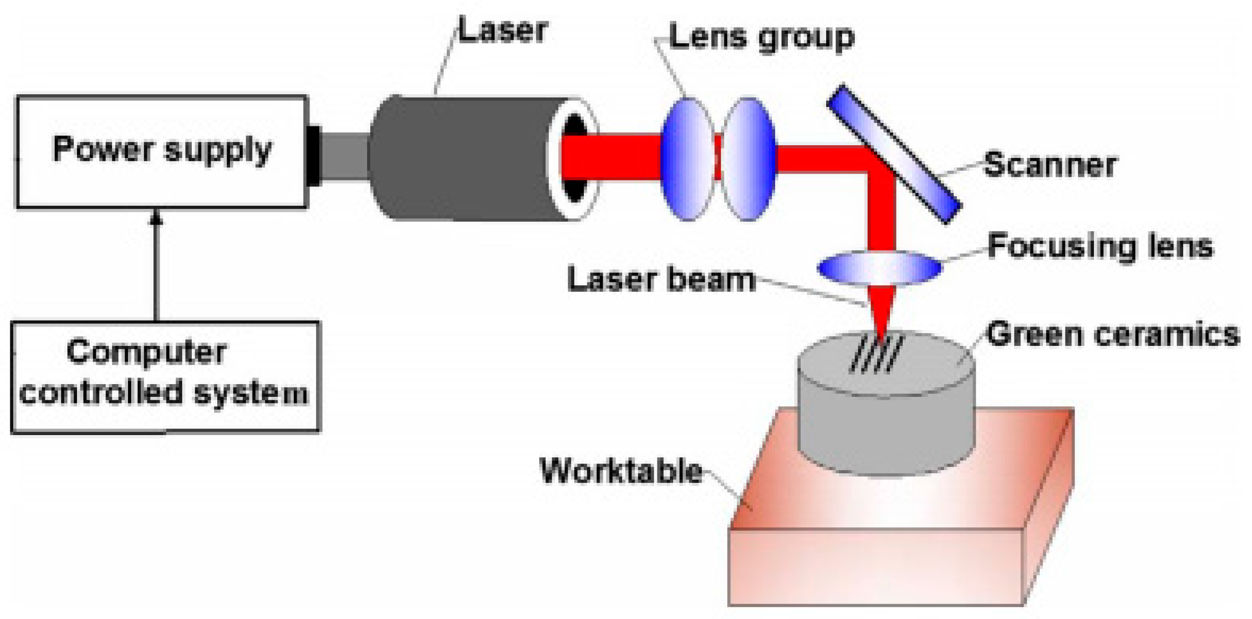
2. Methods of Laser Surface Texturing

The literature shows various methods for the generation of textures or patterns produced on the work surface. Among them, widely used methods are texturing using laser ablation and texturing using laser interference. This section presents an in-depth review of laser surface texturing using these methods.

2.1. Texturing Using Laser Ablation

During the laser ablation process, a focused beam of the laser is made to irradiate on the work surface, and it removes the material due to heating, which leads to melt and vaporization of the work material from the irradiated zone. Selective material is removed, and the surface topography is modified [11]. The laser ablation process can remove material in micron-level precision at a faster rate [14] and is an effective technique for developing textures [15]. LST using laser ablation phenomena has been successfully utilized by many researchers. Fabrication of superhydrophobic surface on the gold-coated aluminum plate was successfully generated using the fiber laser ablation process [16]. The kinematic model was also developed to forecast the texture achieved after PLA once the laser impinges to the surface and achieves its use to develop innovative beam paths for the generation of intricate geometrical surfaces [17]. Figure 1 depicts the schematic representation of the laser processing green ceramics. This Nd:YAG laser has a pulse duration of 10 ns and a wavelength of 1064 nm. Using a computer-controlled system, the laser power was controlled through the software interface. Using a scanner, the beam was bent perpendicularly and then; the beam was concentrated using a focusing lens. The laser system has a three-dimensional XYZ stage (precision of the XYZ stage is 10 pm), and for laser texturing, green ZrCb ceramics were placed on this stage. The pattern of laser texture was generated using the computer connected with this laser system.
The laser textured surface results in lower transmittance when compared to the untextured polycarbonate surface using laser ablation [18]. Dimples or pillars were successfully fabricated on the substrate, and diamond-like carbon deposition was done. This leads to improvement of the wear characteristics of the surface [19]. Xing et al. examined the impact of laser parameter on microchannel width, depth and material removal rate while fabricating in PCD using direct laser ablation and laser-induced plasma [20]. Liu et al. fabricated a microtextured groove (30–50 µm width and 15–50 µm depth) on ZrO2 ceramic. The influence of the laser variable was studied; it has been found that the laser variable has a substantial effect on texture quality [21].

2.2. Texturing Using Laser Interference

The patterns in this method are fabricated due to interference of two, three or four high-power pulsed laser beams, which permits periodic local heating of work surface by photothermal interaction among laser and work surface [22]. Direct laser interference patterning (DLIP) is a method for creating texture on the technical surfaces [23]. DLIP method are engaged in the development of optical biosensors based on Biophotonic Sensing Cells (BICELLs) [24]. Many studies have been carried out to fabricate a texture/pattern using DLIP [25,26]. Peter et al. developed a DLIP set up on the principle of four beam interference [27]. The group of researchers also shows that the ultrashort pulsed DLIP is a useful method for creating repeated surface structures on steel material and appropriate for retention and adjunct of bacteria [28].
Laser interference lithography has also been utilized for the fabrication of nanoscale patterns on silicon substrates. It has been observed that the consistent dot patterns are obtained in a region of 20 mm (W) and 20 mm (L) with a half-pitch size of nearby 190, 250, and 370 nm by enhancing the power to 0.600 mW/cm2 [29]. Surface treatment of aluminum has been carried out, and results have revealed that the laser texturing using the two-beam interference method is an effective method for the removal of contamination and surface oxide from the work surface [30]. Furlan et al. fabricated a pattern using DLIP on magnesium alloy and studied laser parameters to understand the relationship between fluence and quality of feature [31]. Morales et al. examined the impact of laser parameter on texture homogeneity during DLIP on Ti–6Al–4V, and the group of researchers found the enhancement of the texture homogeneity by 80–90% [32]. Cardoso et al. fabricated the hierarchical periodic surface on Al2024 alloy using direct laser writing (DLW) and DLIP and found the superhydrophobic surface after one week of fabrication process [33]. Sola et al. processed ophthalmic polydimethylsiloxane PDMS polymers using DLIP and observed damage in photo-based thermal and chemical in the laser-scanned area [34].

3. Laser Surface Texturing for Biomedical Applications

Biocompatibility of a material with host tissues is one of the most thriving areas of research in recent times. Materials with superior biocompatibility are appropriate in applications related to transplantation of tissue and bone [35]. The successful use of gold in a dental implant for centuries shows the use of material for biomedical applications, and the evolvement of biomaterial in the past centuries shows its extensive use in implants. The biomaterials are classified as metallic, ceramic, polymeric, and composite [36]. A texture on the implant can be obtained through grit blasting technique, acid etching, anodic oxidation and chemical vapor deposition. These methods are quick and simple, but the replicability of these methods is inferior. Compared with the above methods, LST is a quick, clean and precise method for implant modification and has been found to be a prospective technique for modification of implant [37,38]. Pereira et al. observed that laser ablation, blended thermal treatment, is a prospective method to produce a hydrophilic surface on ceramic implants by increasing the surface wettability [39]. Cunha et al. showed that the ultrafast LST technique on Ti–6Al–4V increases the surface wettability and regulates the performance of human mesenchymal stem cells (hMSCs) by altering the cytoskeleton shape, FAPs distribution and area, and proliferation [40]. Stango et al. performed coating of hydroxyapatite (HAP) on 316LSS and Ti–6Al–4V implant textured with laser and found that the laser textured surface provides more resistance to corrosion and shows that the surface is suitable for biomedical application [41]. Yu et al. created a microtexture on a titanium surface and concluded that the structured texture enhances the cell adhesion and performs an essential function in contact guidance [42].

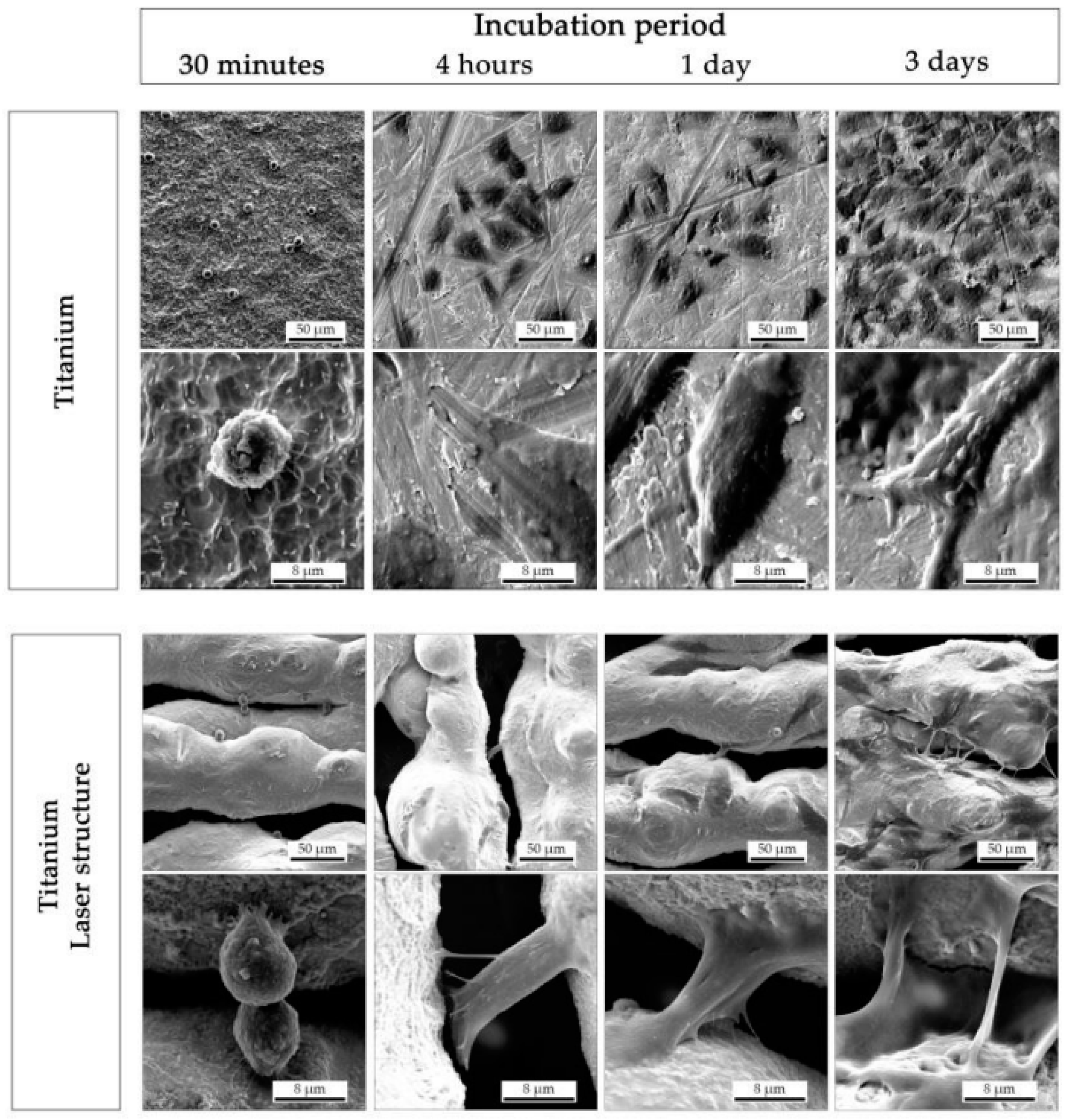
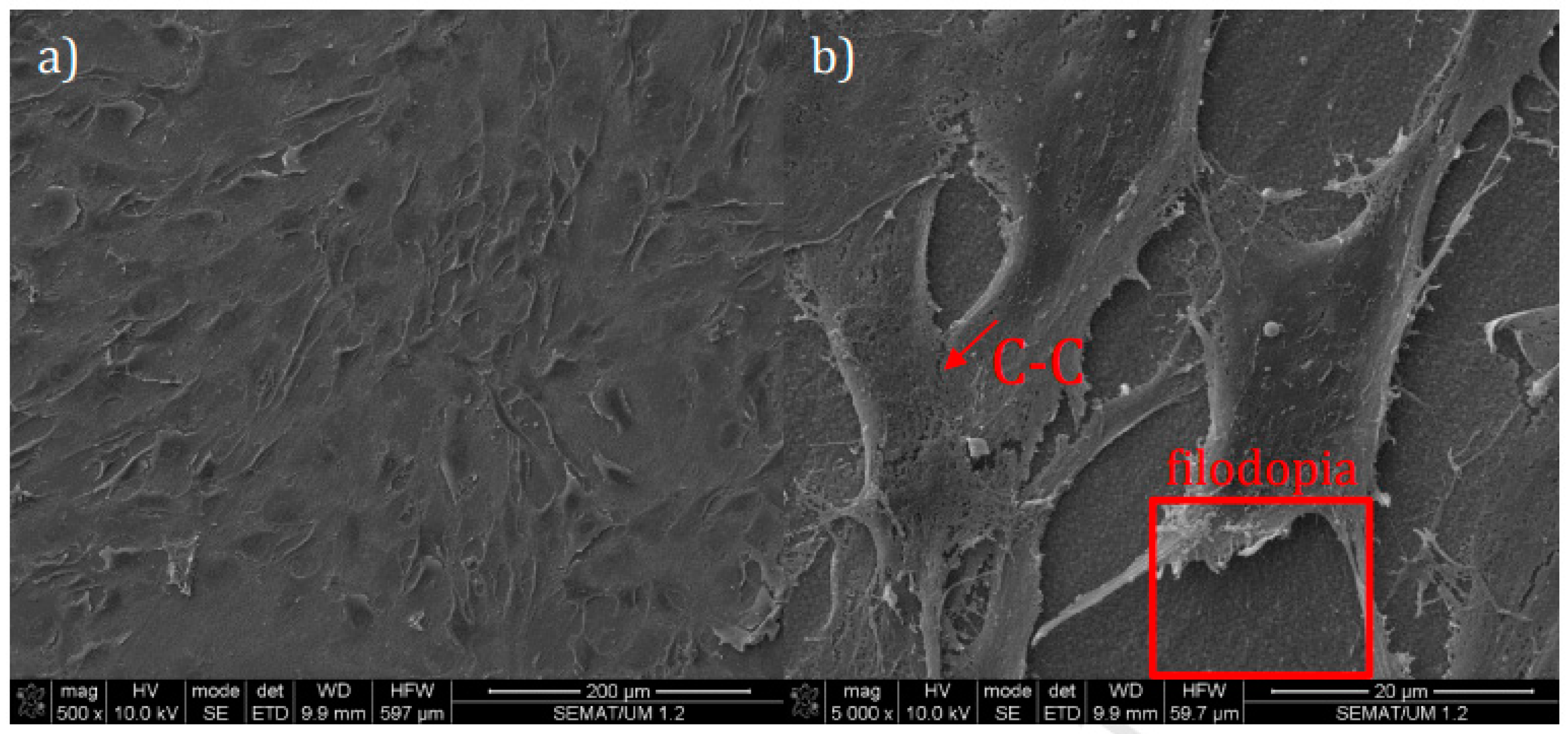
3.1. LST on Titanium and Its Alloys for Biomedical Applications

Materials like titanium (and Ti alloys) reveal a high specific strength, low electrical conductivity, corrosion resistance, makes it suitable for dental implantology and in biomedical applications and is used extensively since the early 1970s [43,44]. The reaction among the host tissue and titanium depends on the surface texture of the implant profile [45]. The biomedical applications of titanium alloys include bone plates, artificial hip joints and knee joints, screws used in fixing fractures, pacemakers, cardiac valve prostheses, and artificial hearts [46]. In addition to material, surface topography has a significant effect on the morphology of the cell its orientation, behavior and activity [47]. Though the material possesses outstanding thermo-physical properties and excellent biocompatible, owing to having a high friction coefficient and low wear resistance, the application area of such extraordinary material is limited. To surmount this, the surface treatment of the material is very much essential. Researchers across the globe have adopted many methods for the surface treatment of titanium and its alloys [48]. The laser micro/nanotexturing process modifies the surface properties related to osseointegration, namely biocompatibility, protein adsorption and cell/surface interactions [49]. Shot peening and acid etching and then laser treatment has been employed on Ti material (grade 2) and revealed the change of surface from hydrophobic to hydrophilic, and adsorption of proteins were mostly found in laser textured surface [50]. The researchers found that deposition of biphasic calcium phosphate occurs on textured Ti–6Al–4V materials, and this depicts enhancement in bioactivity, proliferation and cell adhesion and can be possibly utilized for dental and orthopedic related applications [51]. The significance of microgroove width on anti-corrosion and bio-tribological properties was studied during the coating of graphene oxide on Ti–6Al–4V and found improvement in anti-corrosion and antiwear properties [52]. Figure 2 shows the schematic representation of the ultraviolet (UV) solid laser. The wavelength of the UV laser is 355 nm, and the maximum power of the laser is 5 W. The laser is generated at the laser head, and the generated laser beam was passed through two galvanometric mirrors. Using an F-theta scanning lens, the laser beam then transferred to the top surface of the workpiece material. This F-theta lens has been engineered to provide the highest performance in laser scanning or engraving system. The distance between the F-theta lens and workpiece material surface measures the focusing condition during the laser texturing process.
The overlay structure generated on titanium alloy enhances the mechanical properties, and further, this process refines the grain of the alloy. Further, the hybrid approach of drenched treatment in m-SBF with textured formation is estimated to be an effective as well as environmentally compatible approach to extend the durability of bone screws and reduces the hurdles of slightly osteoporotic implants [53]. Figure 3 depicts SEM images of the un-textured and textured specimens: (a, a1) untextured surface, (b, b1) micro-bulge ring arrays (c, c1) micro-smooth ring arrays, and (d, d1) micro-smooth stacked ring. The laser microtexturing effectively reduces the bacteria attachment as compared to abrasive blasted surfaces during LST of Ti–6Al–4V [54]. A group of researchers has synthesized textured bioactive Ca–P coating having 100 and 200 µm placing on Ti–6Al–4V substrate. The authors demonstrated the better cytoskeleton association and propagation of the mouse MC3T3-E1 osteoblast-like cell. Further, improvement in vitro bioactivity and in vitro biocompatibility was achieved in their study [55]. The culture of cells shows that microgrooves on titanium alloy created by LST substantially increase the augmentation and distinction of MC3T3-E1 cells, which indicates the improvement in bioactivity [56].
Surface alteration by Nd-YAG laser on titanium alloy was also performed, and it was revealed that the smooth surface could release extra cell adhesion owing to its rise in surface tension and decrease in contact angle [57]. Further, the detachment of the enzyme test verified that maximum cells are affixed to the LTS at 140 J cm−2 and lesser to the untreated sample. The optimized parameter of Nd:YAG laser produces a suitable modification on Ti6Al4V alloy. Both SEM and contact angle, along with histopathological assessment, verify that Ti alloy having better chemical and physical properties is obtained and can be used for biomedical implementations [58]. Coathup et al. produced laser texture superhydrophilic Ti-6Al-4V surface having better surface chemistry and topography which significantly endorses osteoblast adhesion in culture. [59]. Textures fabricated with nanosecond pulsed laser and annealing in Ti–6Al–4V show lesser biofilm development on the textured superhydrophobic surface. The suggested approach improves in decreasing the risk of infection related to implants devoid of using cytotoxic bactericidal agents [60]. Borcherding et al. fabricated a texture on titanium alloy with titanium dioxide coating [61]. The experimental findings showed the improvement in cell feasibility on textured surfaces related to pure titanium, suggesting good cytocompatibility. Figure 4 illustrates the SEM image of human osteoblast-like cells, picturing the sticking on a time of three days at various surface conditions. Texturing like dimple structures was generated on the titanium alloy [62]. This textured surface exhibits substantial progress in bioactivity with respect to the Ca3(PO4)2 depositing rate in Hank’s solution. The cell proliferation (XTT) result shows analogous cell feasibility of textured Ti–6Al–4V compared to un-textured Ti-alloy. Laser texturing using femtosecond laser was carried out to reduce the establishment of Grade 2 titanium alloy surface by Staphylococcus aureus and the successive biofilm creation [63]. The process decreases bacterial adhesion and biofilm formation related to the polished surface. The authors also concluded that femtosecond LST is a capable way for bestowing orthopedic and dental implants with antibacterial characteristics and reducing implant-related infections. Ultrafast LST has been applied as a method for surface modification of Ti–6Al–4V dental and orthopedic implant to enhance osteoblastic assurance of human mesenchymal stem cells (hMSCs) [64]. The authors concluded that nanotextures involving laser-irradiated regular surface configurations and nano-sized pillars are capable of enhancing hMSC discrimination into an osteoblastic lineage. Three distinct topographies were processed with femtosecond laser at various laser parameters, and it was shown to enhance osteogenesis and hinder the adipogenesis of MSCs [65]. The formed nano-ripples only supported the osteoblastic commitment. Furthermore, the combination of nano-ripples and micro-patterns (pits) improved osteogenic ability. Raimbault et al. investigated the biological operation impacts of laser (femtosecond) formed structure on performance of cell and concluded that cells are responsive to the nanostructure excepting that the microstructure size is deeper than 5 μm and near to the cell size [66]. Cells are responsive to the microscale structure spreads with respect to these structures.

3.2. LST on Zirconia for Biomedical Application

Zirconia dental implants have appeared as an alternative to titanium implants due to their capability to osseointegrate, excellent mechanical properties, enhanced biocompatibility. They also possess other useful properties like its transparency and white color, which imitates the natural teeth [67,68]. Moura et al. presented LST on zirconia implant and shows that textured surface has greater chemical affinity among bone and 3Y-TZP disks, and thus, the process promotes better bone imprisonment on the surface [69]. The friction test of laser-treated and subsequent deposition of a HAp-coated zirconia reveals superb adhesion of HAp coating on textured zirconia surface. In addition, the bioactive coating reliability does not concede through implant insertion [70]. Figure 5 illustrates the SEM images of HAp-coated zirconia textured surface after the friction tests against bone and the subsequent EDS spectra of evident zones, Z1 and Z2.
The combined effect of laser texturing and cold-pressing methods on zirconia exhibits an effective method for fabrication of zirconia implants with tailored surface designs corresponding to the properties essential for specific application [71]. Guimarães et al. show that microtexture on zirconia surfaces coated by 45S5 BG and HAp glass via a dip coating. Subsequently, the biologically active coating remained laser sintered and revealed that square texture (groove width of 100 µm) functionality with a biologically active coating represents an increase in 90% of cell feasibility related to flat surfaces following incubation of 48 h [72]. Figure 6 illustrates the SEM images of the as-sintered (AS) and flat surface, indicating the common and detailed view of cells propagating. Madeira et al. fabricated Aunps and Agµps-functionalized zirconia produced by spray deposition, laser adhesion and laser texturing, additive laser strategies, respectively shows nice performance to friction test suggesting that the integrity is not influenced through implant attachment [73]. The laser processing method may be effectively used for the formation of specifically structured thin films on ceramic material and their biopolymer composites and bulk zirconia material, which improves mechanical and biological response in dental implants [74]. Utilizing Nd:YAG laser, the surface texture was formed to enhance the mechanical interlocking of hydroxyapatite (HAp) powder and subsequently enhances its adhesion to zirconia [75]. The outcomes depict that it is likely to create texture by altering energy density and atmosphere. Moreover, a good quantity of retained and sintered bioactive material was retrieved with higher laser power and lower scanning speed. Figure 7 depicts SEM images of the HAp sintered within the textured at various laser power and scan speeds.
Delgado-Ruı’z et al. studied the appropriateness of femtosecond laser for micro texturing cylindrical zirconia dental implants surface [76]. Sixty-six zirconia implants were utilized and split into three groups: as a surface with no laser treatment, micro-grooved texture and micro-grooved texture. The result shows that femtosecond laser micro texturing extends an attractive option to traditional surface treatments of zirconia implants due to its precision and lesser damage on surrounding regions. A study of the femtosecond laser ablation method was done to develop micro and nano-level structures to alter the surface topography of alumina-toughened zirconia (ATZ) [77]. The treated surface shows suggestively high expression of osteogenic transcription factors, genes. In addition, the development of a mineralized extracellular matrix was observed compared to the untreated surface. The linear microgroove arrays and superimposed crossline microgroove textured in zirconia substrates depicts that for steep surfaces, parallel micro line surfaces are capable of handling cell growth only, but the curved ones decrease the initial response and depict the least osteogenic response [78].

3.3. LST on Polymer

Polymers are natural or synthetic substances composed of very large molecules. These are developed by connecting a huge number of units known as monomers. In the area of biomedical applications, these polymers are extensively used [14]. The laser ablation of PTFE films of several thickness and densities has shown that laser treatment of polymer-tissue will be helpful in clinical tool [79]. The use of various laser wavelengths (1.064 µm, 532 nm and 355 nm) on the carbon-coated surface of polyethylene (UHMWPE) materials and the effect of laser parameters on material surface properties depicts that the 355 nm and 532 nm wavelength laser results in comparatively suitable for improving the surface conditions such as wettability and surface roughness of UHMWPE. The outcomes were revealed for maximizing surface conditions, and these also significantly affect the interaction process between cell and material [80]. CO2 laser texturing of poly (l-lactide) presents the capability to adjust the physical and structural characteristics of the material’s surface to the needs of the cells with substantial alterations in the mechanical properties of the treated polymer surface [81]. Study of laser variable on the wettability, roughness and hardness of polypropylene material was carried out using 1.064 µm, 355 nm and 532 nm lasers. The authors suggested that laser wavelengths are appropriate for increasing the surface roughness of polypropylene [82]. The Ra values are more than 1 µm, and this value is a minimum essential value to enhance the bonding of bone in the implant surface as per the literature. Mirzadeh et al. improved the hydrophilicity and biocompatibility of ethylene-propylene rubber N-vinylpyrrolidone (NVP) and 2-hydroxyethyl methacrylate (HEMA) by grafting to the polymer using pulsed CO2 laser at various laser power. Alveolar macrophages cultured on un-treated films shows the greater attachment of cell with good propagating and flattening while supports adhere to the treated EPR showed round with marginal cytoplasmic spreading and ruffling [83]. Using excimer lasers, Dinca P et al. carried out experimentation for single-step creation on natural composite substrates for roughness gradients and concluded that cell behavior of engineered polymer might participate a vital function for the progress of the next-generation of bioactive interfaces for perusing cell integration [84]. Koufaki et al. investigated the cell adhesion, and possibility on high rough polymeric surfaces with gradient roughness ratio and wettability formulated by laser micro/nano-textured Si surfaces shows that superior adhesion of both cell types was observed in microstructured surfaces linked to the unstructured surface. Furthermore, PC12 cells were noticed to adhere nicely to the patterned surface [85]. Direct laser writing on the biodegradable polymer to produce microchannels for attachment of C2C12 myoblast cells in the microchannels depicts a high degree of alignment after four days as cell proliferated into a merging patch inside the channels [86]. Waugh et al. presented the surface characteristics and properties of nylon 6,6 treated by CO2 laser and suggested that laser textured surfaces enhance the biomimetic properties of nylon 6,6 in terms of osteoblast cell response [87].

3.4. LST on Other Material for Biomedical Application

Magnesium (Mg) alloys have drawn great interest for use as implants. Hu et al. modified the Mg–6Gd–0.6Ca alloy with laser for investigating the cell behavior for the same and concluded that MC3T3-E1 exhibits better sticking to the laser-treated surface [88]. The LT method efficiently enhances the bactericidal properties of copper surfaces, and the LT-Cu kills bacteria selectively; no cytotoxicity was noted compared to mammalian cells [89]. Qin et al. have used laser surface texturing to create three types of texture on Co–Cr–Mo alloys surfaces and concluded that textured Co–Cr–Mo surface influence osteoblast proliferation, gene expression and morphology [90]. Purnama et al. fabricated a texture on SS316L and explored the impacts of the textured material on the proliferation of adhesion and endothelial cell arrangement. The finding shows that the textured surfaces supported these three processes of cells [91]. The texturing of AISI 304 steel at various laser fluences enables the fabrication of surface microfluidic appliances for chemical and biomedical applications [92].

4. Summary

The paper presents an in-depth review of laser surface texturing for biomedical applications. The researcher has used various types of lasers, such as carbon-dioxide laser, excimer laser, fiber laser, etc., to produce texture to explore the efficacy of the process and its impact on proliferation, osseointegration, cell adhesion, etc. The widely used biomaterial such as titanium and its alloy and zirconia has been reviewed thoroughly, indicating important contributions achieved and representation of suitable micrographs and plots. Apart from these materials, other material that has been used for biomedical applications also has been studied in-depth, mentioning the striking contributions of the research and development in the field of LST.
(i)
Though there are many studies on titanium and its alloys and zirconia for biomedical applications, very limited work has been carried out considering the effect of laser parameters on the laser texturing process. The parametric analysis of several surface properties (like roughness, wettability, hardness, etc.) is essential for the overall improvement of the process and commercial application in the emerging area;
(ii)
For other material, more research needs to be carried out for exploring the physics in LST, and clinical trials must be carried out to confirm the feasibility for its application in the biomedical domain. Further, it was observed that laser surface texturing has become a potential method for surface modification of biomaterial and may be successfully utilized for biomedical applications.

Author Contributions

Conceptualization, I.S., G.K.; methodology, B.B.P., R.C.; validation, A.S., I.S., R.C., I.S., G.K.; resources, G.K., I.S.; writing—I.S., G.K. All authors have read and agreed to the published version of the manuscript.

Funding

This research was funded by TMA Pai, Sikkim Manipal University.

Institutional Review Board Statement

Not applicable.

Informed Consent Statement

Not applicable.

Conflicts of Interest

The authors declare no conflict of interest.

References

  1. Bhaduria, D.; Batala, A.; Dimova, S.S.; Zhangb, Z.; Dongb, H.; Fallqvistc, M.; M’Saoubid, R. On design and tribological behaviour of laser textured surfaces. Procedia CIRP 2017, 60, 20–25. [Google Scholar] [CrossRef]
  2. Hausmann, U.P.; Joerges, P.; Heinzl, J.; Talke, F.E. Nano-texturing of magnetic recording sliders via laser ablation. Microsyst. Technol. 2009, 15, 1747–1751. [Google Scholar] [CrossRef] [Green Version]
  3. Etsion, I.; Halperin, G. A laser surface textured hydrostatic mechanical seal. Tribol. Trans. 2002, 45, 430–434. [Google Scholar] [CrossRef]
  4. Mao, B.; Arpith Siddaiah, Y.L.; Pradeep, L.M. Laser surface texturing and related techniques for enhancing tribological performance of engineering materials: A review. J. Manuf. Process. 2020, 53, 153–173. [Google Scholar] [CrossRef]
  5. Arslan, A.; Masjuki, H.H.; Kalam, M.A.; Varman, M.; Mufti, R.A.; Mosarof, M.H.; Khuong, L.S.; Quazi, M.M. Surface texture manufacturing techniques and tribological effect of surface texturing on cutting tool performance: A review. Crit. Rev. Solid State Mater. Sci. 2016, 41, 447–481. [Google Scholar] [CrossRef]
  6. Joshi, B.; Tripathi, K.; Gyawali, G.; Lee, S.W. The effect of laser surface texturing on the tribological performance of different Sialon ceramic phases. Prog. Nat. Sci. Mater. Int. 2016, 26, 415–421. [Google Scholar] [CrossRef] [Green Version]
  7. Li, D.; Chen, X.; Guo, C.; Tao, J.; Tian, C.; Deng, Y.; Zhang, W. Micro surface texturing of alumina ceramic with nanosecond laser. Procedia Eng. 2017, 174, 370–376. [Google Scholar] [CrossRef]
  8. Xing, Y.; Deng, J.; Feng, X.; Yu, S. Effect of laser surface texturing on Si3N4/TiC ceramic sliding against steel under dry friction. Mater. Design (1980–2015) 2013, 52, 34–45. [Google Scholar] [CrossRef]
  9. Šugár, P.; Šugárová, J.; Frnčík, M. Laser surface texturing of tool steel: Textured surfaces quality evaluation. Open Eng. 2016, 6, 90–97. [Google Scholar] [CrossRef]
  10. Tripathi, K.; Gyawali, G.; Joshi, B.; Amanov, A.; Wohn, S. Improved tribological behavior of grey cast Iron Under low and high viscous lubricants by laser surface texturing. Mater. Perform. Charact. 2017, 6, 24–41. [Google Scholar] [CrossRef]
  11. Riveiro, A.; Maçon, A.L.; del Val, J.; Comesaña, R.; Pou, J. Laser surface texturing of polymers for biomedical applications. Front. Phys. 2018, 6, 16. [Google Scholar] [CrossRef]
  12. Ezhilmaran, V.; Vasa, N.J.; Vijayaraghavan, L. Investigation on generation of laser assisted dimples on piston ring surface and influence of dimple parameters on friction. Surf. Coat. Technol. 2018, 335, 314–326. [Google Scholar] [CrossRef]
  13. Liang, L.; Yuan, J.; Li, X.; Yang, F.; Jiang, L. Wear behavior of the micro-grooved texture on WC-Ni3Al cermet prepared by laser surface texturing. Int. J. Refract. Met. Hard Mater. 2018, 72, 211–222. [Google Scholar] [CrossRef]
  14. White, N.; Eder, K.; Byrnes, J.; Cairney, J.M.; McCarroll, I.E. Laser ablation sample preparation for atom probe tomography and transmission electron microscopy. Ultramicroscopy 2020, 220, 113161. [Google Scholar] [CrossRef]
  15. Shum, P.W.; Zhou, Z.F.; Li, K.Y. To increase the hydrophobicity and wear resistance of diamond-like carbon coatings by surface texturing using laser ablation process. Thin Solid Film. 2013, 544, 472–476. [Google Scholar] [CrossRef]
  16. Eskandari, M.J.; Shafyei, A.; Karimzadeh, F. Investigation of wetting properties of gold nanolayer coated aluminum surfaces textured with continuous-wave fiber laser ablation. Thin Solid Film. 2020, 711, 138278. [Google Scholar] [CrossRef]
  17. Kong, M.C.; Miron, C.B.; Axinte, D.A.; Davies, S.; Kell, J. On the relationship between the dynamics of the power density and workpiece surface texture in pulsed laser ablation. CIRP Ann. 2012, 61, 203–206. [Google Scholar] [CrossRef]
  18. Yilbas, B.S.; Khaled, M.; Abu-Dheir, N.; Al-Aqeeli, N.; Said, S.A.; Ahmed, A.O.; Varanasi, K.K.; Toumi, Y.K. Wetting and other physical characteristics of polycarbonate surface textured using laser ablation. Appl. Surf. Sci. 2014, 320, 21–29. [Google Scholar] [CrossRef]
  19. Shum, P.W.; Zhou, Z.F.; Li, K.Y. To increase the hydrophobicity, non-stickiness and wear resistance of DLC surface by surface texturing using a laser ablation process. Tribol. Int. 2014, 78, 1–6. [Google Scholar] [CrossRef]
  20. Xing, Y.; Zhang, K.; Huang, P.; Liu, L.; Wu, Z. Assessment machining of micro-channel textures on PCD by laser-induced plasma and ultra-short pulsed laser ablation. Opt. Laser Technol. 2020, 125, 106057. [Google Scholar] [CrossRef]
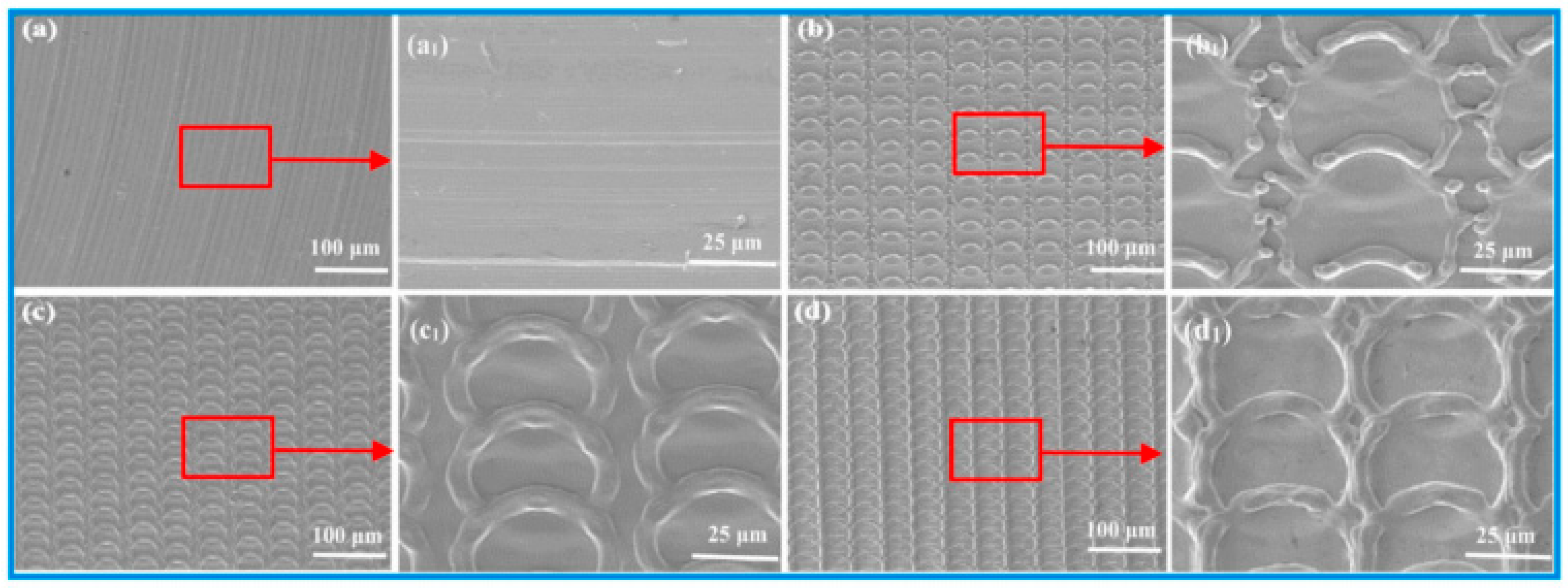
  21. Liu, Y.; Liu, L.; Deng, J.; Meng, R.; Zou, X.; Wu, F. Fabrication of micro-scale textured grooves on green ZrO2 ceramics by pulsed laser ablation. Ceram. Int. 2017, 43, 6519–6531. [Google Scholar] [CrossRef]
  22. Zabila, Y.; Perzanowski, M.; Dobrowolska, A.; Kac, M.; Polit, A.; Marszalek, M. Direct laser interference patterning: Theory and application. Acta Phys. Pol. Ser. A Gen. Phys. 2009, 115, 591. [Google Scholar] [CrossRef]
  23. Rosenkranz, A.; Hans, M.; Gachot, C.; Thome, A.; Bonk, S.; Mücklich, F. Direct laser interference patterning: Tailoring of contact area for frictional and antibacterial properties. Lubricants 2016, 4, 2. [Google Scholar] [CrossRef] [Green Version]
  24. Sanza, F.J.; Langheinrich, D.; Berger, J.; Hernandez, A.L.; Dani, S.; Casquel, R.; Lavin, A.; Otón, A.; Santamaría, B.; Laguna, M.; et al. Direct laser interference patterning (DLIP) technique applied to the development of optical biosensors based on biophotonic sensing cells (bicells). Int. Soc. Opt. Photonics 2015, 9351, 935114. [Google Scholar]
  25. El-Khoury, M.; Alamri, S.; Voisiat, B.; Kunze, T.; Lasagni, A.F. Fabrication of hierarchical surface textures using multi-pulse direct laser interference patterning with nanosecond pulses. Mater. Lett. 2020, 258, 126743. [Google Scholar] [CrossRef]
  26. Baumann, R.; Milles, S.; Leupolt, B.; Kleber, S.; Dahms, J.; Lasagni, A.F. Tailored wetting of copper using precise nanosecond direct laser interference patterning. Opt. Lasers Eng. 2020, 137, 106364. [Google Scholar] [CrossRef]
  27. Peter, A.; Onuseit, V.; Freitag, C.; Faas, S.; Graf, T. Flexible, compact and picosecond laser capable four-beam interference setup. In Proceedings of the Lasers in Manufacturing Conference, Munich, Germany, 18–22 June 2017; pp. 1–8. [Google Scholar]
  28. Peter, A.; Lutey, A.H.; Faas, S.; Romoli, L.; Onuseit, V.; Graf, T. Direct laser interference patterning of stainless steel by ultrashort pulses for antibacterial surfaces. Opt. Laser Technol. 2020, 123, 105954. [Google Scholar] [CrossRef]
  29. Choi, J.; Chung, M.H.; Dong, K.Y.; Park, E.M.; Ham, D.J.; Park, Y.; Song, I.S.; Pak, J.J.; Ju, B.K. Investigation on fabrication of nanoscale patterns using laser interference lithography. J. Nanosci. Nanotechnol. 2011, 11, 778–781. [Google Scholar] [CrossRef] [Green Version]
  30. Chen, J.; Sabau, A.S.; Jones, J.F.; Hackett, A.C.; Daniel, C.; Warren, D. Aluminum surface texturing by means of laser interference metallurgy. In Light Metals; Springer: Cham, Switzerland, 2015; pp. 427–429. [Google Scholar]
  31. Furlan, V.; Biondi, M.; Demir, A.G.; Pariani, G.; Bianco, A.; Previtali, B. Direct laser texturing using two-beam interference patterning on biodegradable magnesium alloy. In Proceedings of the International Congress on Applications of Lasers & Electro-Optics, San Diego, CA, USA, 16–20 October 2016; p. 182. [Google Scholar]
  32. Aguilar-Morales, A.I.; Alamri, S.; Kunze, T.; Lasagni, A.F. Influence of processing parameters on surface texture homogeneity using Direct Laser Interference Patterning. Opt. Laser Technol. 2018, 107, 216–227. [Google Scholar] [CrossRef]
  33. Cardoso, J.T.; Aguilar-Morales, A.I.; Alamri, S.; Huerta-Murillo, D.; Cordovilla, F.; Lasagni, A.F.; Ocaña, J.L. Superhydrophobicity on hierarchical periodic surface structures fabricated via direct laser writing and direct laser interference patterning on an aluminium alloy. Opt. Lasers Eng. 2018, 111, 193–200. [Google Scholar] [CrossRef]
  34. Sola, D.; Lavieja, C.; Orera, A.; Clemente, M.J. Direct laser interference patterning of ophthalmic polydimethylsiloxane (PDMS) polymers. Opt. Lasers Eng. 2018, 106, 139–146. [Google Scholar] [CrossRef]
  35. Radmanesh, M.; Kiani, A. Effects of laser pulse numbers on surface biocompatibility of titanium for implant fabrication. J. Biomater. Nanobiotechnol. 2015, 6, 168. [Google Scholar] [CrossRef] [Green Version]
  36. Kurella, A.; Dahotre, N.B. Surface modification for bioimplants: The role of laser surface engineering. J. Biomater. Appl. 2005, 20, 5–50. [Google Scholar] [CrossRef] [PubMed]
  37. Yu, Z.; Yin, S.; Zhang, W.; Jiang, X.; Hu, J. Picosecond laser texturing on titanium alloy for biomedical implants in cell proliferation and vascularization. J. Biomed. Mater. Res. Part B Appl. Biomater. 2020, 108, 1494–1504. [Google Scholar] [CrossRef]
  38. Fiorucci, M.P.; López, A.J.; Ramil, A. Surface modification of Ti6Al4V by nanosecond laser ablation for biomedical applications. J. Phys. Conf. Ser. 2015, 605, 012022. [Google Scholar] [CrossRef] [Green Version]
  39. Pereira, R.S.; Moura, C.G.; Henriques, B.; Chevalier, J.; Silva, F.S.; Fredel, M.C. Influence of laser texturing on surface features, mechanical properties and low-temperature degradation behavior of 3Y-TZP. Ceram. Int. 2020, 46, 3502–3512. [Google Scholar] [CrossRef]
  40. Cunha, A.; Oliveira, V.; Serro, A.P.; Zouani, O.E.; Almeida, A.; Durrieu, M.C.; Vilar, R. Ultrafast laser texturing of Ti-6Al-4V surfaces for biomedical applications. In Proceedings of the International Congress on Applications of Lasers & Electro-Optics, Miami, FL, USA, 6–10 October 2013; pp. 910–918. [Google Scholar]
  41. Stango, S.A.; Karthick, D.; Swaroop, S.; Mudali, U.K.; Vijayalakshmi, U. Development of hydroxyapatite coatings on laser textured 316 LSS and Ti-6Al-4V and its electrochemical behavior in SBF solution for orthopedic applications. Ceram. Int. 2018, 44, 3149–3160. [Google Scholar] [CrossRef]
  42. Yu, Z.; Yang, G.; Zhang, W.; Hu, J. Investigating the effect of picosecond laser texturing on microstructure and biofunctionalization of titanium alloy. J. Mater. Process. Technol. 2018, 255, 129–136. [Google Scholar] [CrossRef]
  43. Sidambe, A.T. Biocompatibility of advanced manufactured titanium implants—A review. Materials 2014, 7, 8168–8188. [Google Scholar] [CrossRef] [Green Version]
  44. Schiff, N.; Grosgogeat, B.; Lissac, M.; Dalard, F. Influence of fluoride content and pH on the corrosion resistance of titanium and its alloys. Biomaterials 2002, 23, 1995–2002. [Google Scholar] [CrossRef]
  45. Pou, P.; Riveiro, A.; del Val, J.; Comesaña, R.; Penide, J.; Arias-González, F.; Soto, R.; Lusquiños, F.; Pou, J. Laser surface texturing of Titanium for bioengineering applications. Procedia Manuf. 2017, 13, 694–701. [Google Scholar] [CrossRef]
  46. Elias, C.N.; Lima, J.H.; Valiev, R.; Meyers, M.A. Biomedical applications of titanium and its alloys. Jom 2008, 60, 46–49. [Google Scholar] [CrossRef]
  47. Tiainen, L.; Abreu, P.; Buciumeanu, M.; Silva, F.; Gasik, M.; Guerrero, R.S.; Carvalho, O. Novel laser surface texturing for improved primary stability of titanium implants. J. Mech. Behav. Biomed. Mater. 2019, 98, 26–39. [Google Scholar] [CrossRef]
  48. Ramskogler, C.; Warchomicka, F.; Mostofi, S.; Weinberg, A.; Sommitsch, C. Innovative surface modification of Ti6Al4V alloy by electron beam technique for biomedical application. Mater. Sci. Eng. C 2017, 78, 105–113. [Google Scholar] [CrossRef]
  49. Shukla, P.; Waugh, D.G.; Lawrence, J.; Vilar, R. Laser surface structuring of ceramics, metals and polymers for biomedical applications: A review. In Laser Surface Modification of Biomaterials; Woodhead Publishing: Cambridge, UK, 2016; pp. 281–299. [Google Scholar]
  50. Kuczyńska, D.; Kwaśniak, P.; Marczak, J.; Bonarski, J.; Smolik, J.; Garbacz, H. Laser surface treatment and the resultant hierarchical topography of Ti grade 2 for biomedical application. Appl. Surf. Sci. 2016, 390, 560–569. [Google Scholar] [CrossRef]
  51. Behera, R.R.; Das, A.; Hasan, A.; Pamu, D.; Pandey, L.M.; Sankar, M.R. Deposition of biphasic calcium phosphate film on laser surface textured Ti–6Al–4V and its effect on different biological properties for orthopedic applications. J. Alloy. Compd. 2020, 842, 155683. [Google Scholar] [CrossRef]
  52. Wang, C.; Li, Z.; Zhao, H.; Zhang, G.; Ren, T.; Zhang, Y. Enhanced anticorrosion and antiwear properties of Ti–6Al–4V alloys with laser texture and graphene oxide coatings. Tribol. Int. 2020, 152, 106475. [Google Scholar] [CrossRef]
  53. Xu, Y.; Liu, W.; Zhang, G.; Li, Z.; Hu, H.; Wang, C.; Zeng, X.; Zhao, S.; Zhang, Y.; Ren, T. Friction stability and cellular behaviors on laser textured Ti–6Al–4V alloy implants with bioinspired micro-overlapping structures. J. Mech. Behav. Biomed. Mater. 2020, 109, 103823. [Google Scholar] [CrossRef]
  54. Uhlmann, E.; Schweitzer, L.; Kieburg, H.; Spielvogel, A.; Huth-Herms, K. The Effects of Laser Microtexturing of Biomedical Grade 5 Ti-6Al-4V Dental Implants (Abutment) on Biofilm Formation. Procedia CIRP 2018, 68, 184–189. [Google Scholar] [CrossRef]
  55. Paital, S.R.; Bunce, N.; Nandwana, P.; Honrao, C.; Nag, S.; He, W.; Banerjee, R.; Dahotre, N.B. Laser surface modification for synthesis of textured bioactive and biocompatible Ca–P coatings on Ti–6Al–4V. J. Mater. Sci. Mater. Med. 2011, 22, 1393–1406. [Google Scholar] [CrossRef]
  56. Zheng, Q.; Mao, L.; Shi, Y.; Fu, W.; Hu, Y. Biocompatibility of Ti-6Al-4V titanium alloy implants with laser microgrooved surfaces. Mater. Technol. 2020, 1–10. [Google Scholar] [CrossRef]
  57. Khosroshahi, M.E.; Mahmoodi, M.; Saeedinasab, H.; Tahriri, M. Evaluation of mechanical and electrochemical properties of laser surface modified Ti–6Al–4V for biomedical applications: In vitro study. Surf. Eng. 2008, 24, 209–218. [Google Scholar] [CrossRef]
  58. Khosroshahi, M.E.; Tavakoli, J.; Mahmoodi, M. Analysis of bioadhesivity of osteoblast cells on titanium alloy surface modified by Nd: YAG laser. J. Adhes. 2007, 83, 151–172. [Google Scholar] [CrossRef]
  59. Coathup, M.J.; Blunn, G.W.; Mirhosseini, N.; Erskine, K.; Liu, Z.; Garrod, D.R.; Li, L. Controlled laser texturing of titanium results in reliable osteointegration. J. Orthop. Res. 2017, 35, 820. [Google Scholar] [CrossRef]
  60. Patil, D.; Aravindan, S.; Kaushal Wasson, M.; Rao, P.V. Fast fabrication of superhydrophobic titanium alloy as antibacterial surface using nanosecond laser texturing. J. Micro Nano-Manuf. 2018, 6, 011002. [Google Scholar] [CrossRef]
  61. Borcherding, K.; Marx, D.; Gätjen, L.; Specht, U.; Salz, D.; Thiel, K.; Wildemann, B.; Grunwald, I. Impact of Laser Structuring on Medical-Grade Titanium: Surface Characterization and In Vitro Evaluation of Osteoblast Attachment. Materials 2020, 13, 2000. [Google Scholar] [CrossRef]
  62. Kumari, R.; Scharnweber, T.; Pfleging, W.; Besser, H.; Majumdar, J.D. Laser Surface Textured Titanium Alloy (Ti-6Al-4V)-Part II-Studies on Bio-compatibity. Appl. Surf. Sci. 2015, 357, 750–758. [Google Scholar] [CrossRef]
  63. Cunha, A.; Elie, A.M.; Plawinski, L.; Serro, A.P.; do Rego, A.M.; Almeida, A.; Urdaci, M.C.; Durrieu, M.C.; Vilar, R. Femtosecond laser surface texturing of titanium as a method to reduce the adhesion of Staphylococcus aureus and biofilm formation. Appl. Surf. Sci. 2016, 360, 485–493. [Google Scholar] [CrossRef]
  64. Cunha, A.; Zouani, O.F.; Plawinski, L.; Botelho do Rego, A.M.; Almeida, A.; Vilar, R.; Durrieu, M.C. Human mesenchymal stem cell behavior on femtosecond laser-textured Ti-6Al-4V surfaces. Nanomedicine 2015, 10, 725–739. [Google Scholar] [CrossRef]
  65. Dumas, V.; Guignandon, A.; Vico, L.; Mauclair, C.; Zapata, X.; Linossier, M.T.; Bouleftour, W.; Granier, J.; Peyroche, S.; Dumas, J.C.; et al. Femtosecond laser nano/micro patterning of titanium influences mesenchymal stem cell adhesion and commitment. Biomed. Mater. 2015, 10, 055002. [Google Scholar] [CrossRef]
  66. Raimbault, O.; Benayoun, S.; Anselme, K.; Mauclair, C.; Bourgade, T.; Kietzig, A.M.; Girard-Lauriault, P.L.; Valette, S.; Donnet, C. The effects of femtosecond laser-textured Ti-6Al-4V on wettability and cell response. Mater. Sci. Eng. C 2016, 69, 311–320. [Google Scholar] [CrossRef]
  67. Apratim, A.; Eachempati, P.; Salian, K.K.; Singh, V.; Chhabra, S.; Shah, S. Zirconia in dental implantology: A review. J. Int. Soc. Prev. Community Dent. 2015, 5, 147. [Google Scholar] [CrossRef] [Green Version]
  68. Depprich, R.; Zipprich, H.; Ommerborn, M.; Naujoks, C.; Wiesmann, H.P.; Kiattavorncharoen, S.; Lauer, H.C.; Meyer, U.; Kübler, N.R.; Handschel, J. Osseointegration of zirconia implants compared with titanium: An in vivo study. Head Face Med. 2008, 4, 30. [Google Scholar] [CrossRef] [Green Version]
  69. Moura, C.G.; Pereira, R.; Buciumeanu, M.; Carvalho, O.; Bartolomeu, F.; Nascimento, R.; Silva, F.S. Effect of laser surface texturing on primary stability and surface properties of zirconia implants. Ceram. Int. 2017, 43, 15227–15236. [Google Scholar] [CrossRef]
  70. Faria, D.; Henriques, B.; Souza, A.C.; Silva, F.S.; Carvalho, O. Laser-assisted production of HAp-coated zirconia structured surfaces for biomedical applications. J. Mech. Behav. Biomed. Mater. 2020, 112, 104049. [Google Scholar] [CrossRef]
  71. Faria, D.; Madeira, S.; Buciumeanu, M.; Silva, F.S.; Carvalho, O. Novel laser textured surface designs for improved zirconia implants performance. Mater. Sci. Eng. C 2020, 108, 110390. [Google Scholar] [CrossRef]
  72. Mesquita-Guimarães, J.; Detsch, R.; Souza, A.C.; Henriques, B.; Silva, F.S.; Boccaccini, A.R.; Carvalho, O. Cell adhesion evaluation of laser-sintered HAp and 45S5 bioactive glass coatings on micro-textured zirconia surfaces using MC3T3-E1 osteoblast-like cells. Mater. Sci. Eng. C 2020, 1091, 10492. [Google Scholar] [CrossRef]
  73. Madeira, S.; Barbosa, A.; Moura, C.G.; Buciumeanu, M.; Silva, F.S.; Carvalho, O. Aunps and Agμps-functionalized zirconia surfaces by hybrid laser technology for dental implants. Ceram. Int. 2020, 46, 7109–7121. [Google Scholar] [CrossRef]
  74. Daskalova, A.; Angelova, L.; Carvalho, A.; Trifonov, A.; Nathala, C.; Monteiro, F.; Buchvarov, I. Effect of surface modification by femtosecond laser on zirconia based ceramics for screening of cell-surface interaction. Appl. Surf. Sci. 2020, 26, 145914. [Google Scholar] [CrossRef]
  75. Carvalho, O.; Sousa, F.; Madeira, S.; Silva, F.S.; Miranda, G. HAp-functionalized zirconia surfaces via hybrid laser process for dental applications. Opt. Laser Technol. 2018, 106, 157–167. [Google Scholar] [CrossRef]
  76. Delgado-Ruíz, R.A.; Calvo-Guirado, J.L.; Moreno, P.; Guardia, J.; Gomez-Moreno, G.; Mate-Sánchez, J.E.; Ramirez-Fernández, P.; Chiva, F. Femtosecond laser microstructuring of zirconia dental implants. J. Biomed. Mater. Res. Part B Appl. Biomater. 2011, 96, 91–100. [Google Scholar] [CrossRef] [PubMed]
  77. Carvalho, A.; Cangueiro, L.; Oliveira, V.; Vilar, R.; Fernandes, M.H.; Monteiro, F.J. Femtosecond laser microstructured Alumina toughened Zirconia: A new strategy to improve osteogenic differentiation of hMSCs. Appl. Surf. Sci. 2018, 435, 1237–1245. [Google Scholar] [CrossRef]
  78. Elena Sima, L.; Bonciu, A.; Baciu, M.; Anghel, I.; Dumitrescu, L.N.; Rusen, L.; Dinca, V. Bioinstructive Micro-Nanotextured Zirconia Ceramic Interfaces for Guiding and Stimulating an Osteogenic Response In Vitro. Nanomaterials 2020, 10, 2465. [Google Scholar] [CrossRef] [PubMed]
  79. Makropoulou, M.; Serafetinides, A.A.; Skordoulis, C.D. Ultra-violet and infra-red laser ablation studies of biocompatible polymers. Lasers Med Sci. 1995, 10, 201–206. [Google Scholar] [CrossRef]
  80. Riveiro, A.; Soto, R.; Del Val, J.; Comesaña, R.; Boutinguiza, M.; Quintero, F.; Lusquiños, F.; Pou, J. Laser surface modification of ultra-high-molecular-weight polyethylene (UHMWPE) for biomedical applications. Appl. Surf. Sci. 2014, 302, 236–242. [Google Scholar] [CrossRef]
  81. Tomanik, M.; Kobielarz, M.; Filipiak, J.; Szymonowicz, M.; Rusak, A.; Mroczkowska, K.; Antończak, A.; Pezowicz, C. Laser Texturing as a Way of Influencing the Micromechanical and Biological Properties of the Poly (l-Lactide) Surface. Materials 2020, 13, 3786. [Google Scholar] [CrossRef]
  82. Riveiro, A.; Soto, R.; Del Val, J.; Comesaña, R.; Boutinguiza, M.; Quintero, F.; Lusquiños, F.; Pou, J. Texturing of polypropylene (PP) with nanosecond lasers. Appl. Surf. Sci. 2016, 374, 379–386. [Google Scholar] [CrossRef]
  83. Mirzadeh, H.; Katbab, A.A.; Burford, R.P. CO2-laser graft copolymerization of HEMA and NVP onto ethylene-propylene rubber (EPR) as biomaterial-(III). Radiat. Phys. Chem. 1995, 46, 859–862. [Google Scholar] [CrossRef]
  84. Dinca, V.; Alloncle, P.; Delaporte, P.; Ion, V.; Rusen, L.; Filipescu, M.; Mustaciosu, C.; Luculescu, C.; Dinescu, M. Excimer laser texturing of natural composite polymer surfaces for studying cell-to-substrate specific response. Appl. Surf. Sci. 2015, 352, 82–90. [Google Scholar] [CrossRef]
  85. Koufaki, N.; Ranella, A.; Aifantis, K.E.; Barberoglou, M.; Psycharakis, S.; Fotakis, C.; Stratakis, E. Controlling cell adhesion via replication of laser micro/nano-textured surfaces on polymers. Biofabrication 2011, 3, 045004. [Google Scholar] [CrossRef]
  86. Yeong, W.Y.; Yu, H.; Lim, K.P.; Ng, K.L.; Boey, Y.C.; Subbu, V.S.; Tan, L.P. Multiscale topological guidance for cell alignment via direct laser writing on biodegradable polymer. Tissue Eng. Part C Methods 2010, 16, 1011–1021. [Google Scholar] [CrossRef] [PubMed]
  87. Waugh, D.; Lawrence, J. Laser surface processing of polymers for biomedical applications. In Laser-Assisted Fabrication of Materials; Springer: Berlin/Heidelberg, Germany, 2013; pp. 275–318. [Google Scholar]
  88. Hu, G.; Guan, K.; Lu, L.; Zhang, J.; Lu, N.; Guan, Y. Engineered functional surfaces by laser microprocessing for biomedical applications. Engineering 2018, 4, 822–830. [Google Scholar] [CrossRef]
  89. Selvamani, V.; Zareei, A.; Elkashif, A.; Maruthamuthu, M.K.; Chittiboyina, S.; Delisi, D.; Li, Z.; Cai, L.; Pol, V.G.; Seleem, M.N.; et al. Hierarchical micro/mesoporous copper structure with enhanced antimicrobial property via laser surface texturing. Adv. Mater. Interfaces 2020, 7, 1901890. [Google Scholar] [CrossRef]
  90. Qin, L.; Zeng, Q.; Wang, W.; Zhang, Y.; Dong, G. Response of MC3T3-E1 osteoblast cells to the microenvironment produced on Co–Cr–Mo alloy using laser surface texturing. J. Mater. Sci. 2014, 49, 2662–2671. [Google Scholar] [CrossRef]
  91. Purnama, A.; Furlan, V.; Dessi, D.; Demir, A.G.; Tolouei, R.; Paternoster, C.; Levesque, L.; Previtali, B.; Mantovani, D. Laser surface texturing of SS316L for enhanced adhesion of HUVECs. Surf. Eng. 2018, 1–10. [Google Scholar] [CrossRef]
  92. Krylach, I.V.; Kudryashov, S.I.; Olekhnovich, R.O.; Moskvin, M.K.; Uspenskaya, M.V. Tuning water wetting angle of a steel surface via nanosecond laser ablative nano/microtexturing for chemical and biomedical microfluidic applications. Laser Phys. Lett. 2019, 16, 105602. [Google Scholar] [CrossRef]
Figure 1. Representational illustration of laser surface texturing of green ceramic [22]. Reprinted from [22]. Copyright from Elsevier 2009.
Coatings 11 00124 g001
Figure 2. Schematic representation of laser processing of microgroove on Ti–6Al–4V [52]. Reprinted from [52]. Copyright from Elsevier 2020.
Coatings 11 00124 g002
Figure 3. SEM images of the un-textured and textured surfaces: (a,a1) untextured surface, (b,b1) micro-bulge ring arrays (c,c1) micro-smooth ring arrays, and (d,d1) micro smooth stacked ring [53]. Reprinted from [53]. Copyright from Elsevier 2020.
Coatings 11 00124 g003
Figure 4. SEM image of human osteoblast-like cells (MG-63), picturing the adhesion on a time of three days at various surface conditions [61].
Coatings 11 00124 g004aCoatings 11 00124 g004b
Figure 5. SEM images of HAp-coated zirconia textured surface, after the friction tests against bone and the subsequent EDS spectra of evident zones, Z1 and Z2 [70]. Reprinted from [70]. Copyright from Elsevier 2020.
Coatings 11 00124 g005
Figure 6. SEM images of the as-sintered (AS) and flat surface, indicating the (a) general and (b) detailed view of cells spreading [72]. Reprinted from [72]. Copyright from Elsevier 2020.
Coatings 11 00124 g006
Figure 7. SEM images of the hydroxyapatite (HAp) sintered within the textured at various laser power and scan speeds [75]. Reprinted from [75]. Copyright from Elsevier 2018.
Coatings 11 00124 g007
Publisher’s Note: MDPI stays neutral with regard to jurisdictional claims in published maps and institutional affiliations.

