Review

Endurance Exercise Mitigates Immunometabolic Adipose Tissue Disturbances in Cancer and Obesity
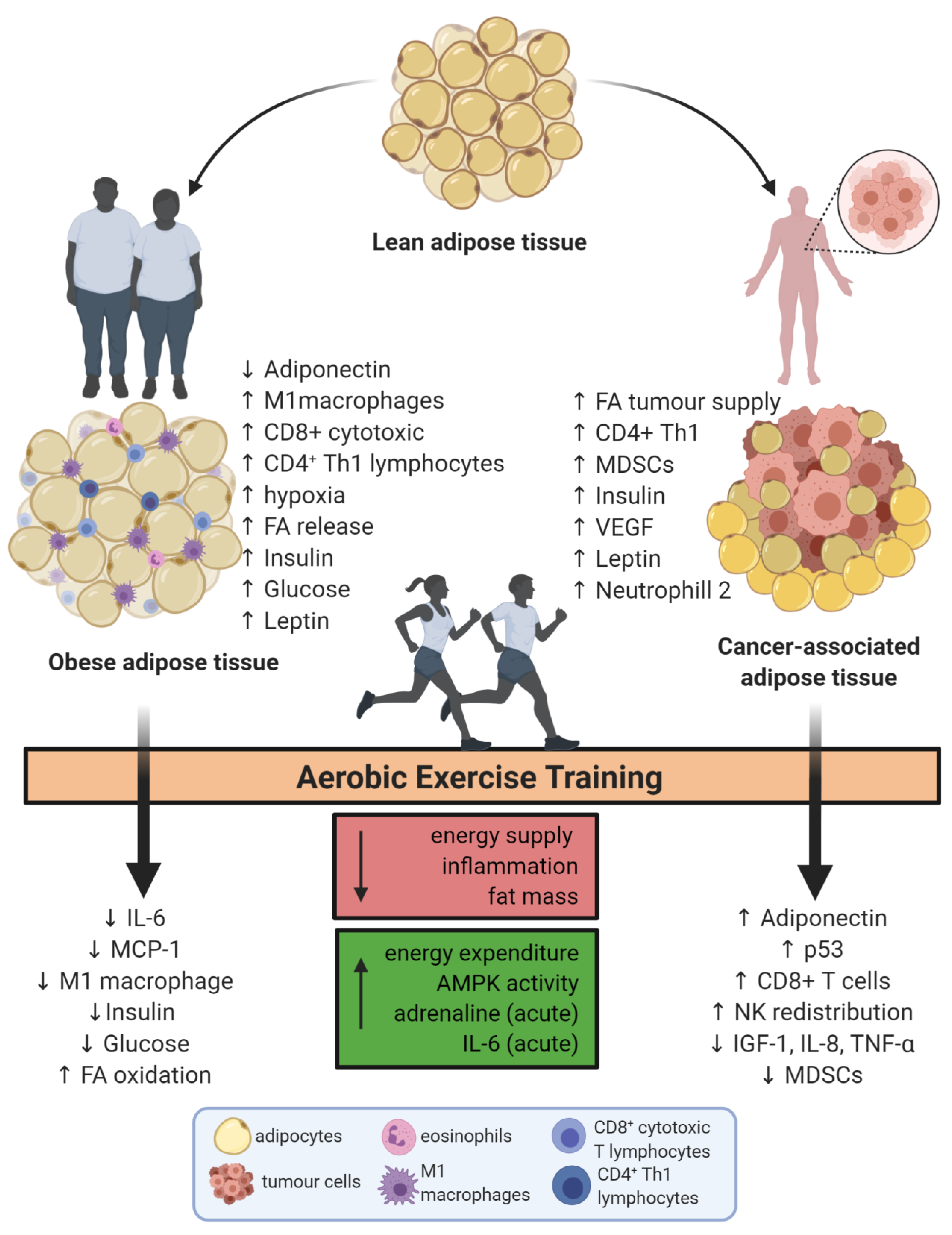
by
José Cesar Rosa-Neto
1,2,* and
Loreana Sanches Silveira
1
1
Immunometabolism Research Group, Department of Systems Biology, Institute of Biomedical Sciences 1, University of São Paulo (ICB1-USP), São Paulo 05508-000, Brazil
2
Laboratory of Experimental Surgery, Department of Surgery, Clinics Hospital of the Faculty of Medicine, University of São Paulo (HC-FMUSP), São Paulo 01246-903, Brazil
*
Author to whom correspondence should be addressed.
Int. J. Mol. Sci. 2020, 21(24), 9745; https://doi.org/10.3390/ijms21249745
Submission received: 22 September 2020 / Revised: 5 November 2020 / Accepted: 11 November 2020 / Published: 21 December 2020

Abstract

:
Adipose tissue is considered an endocrine organ whose complex biology can be explained by the diversity of cell types that compose this tissue. The immune cells found in the stromal portion of adipose tissue play an important role on the modulation of inflammation by adipocytokines secretion. The interactions between metabolic active tissues and immune cells, called immunometabolism, is an important field for discovering new pathways and approaches to treat immunometabolic diseases, such as obesity and cancer. Moreover, physical exercise is widely known as a tool for prevention and adjuvant treatment on metabolic diseases. More specifically, aerobic exercise training is able to increase the energy expenditure, reduce the nutrition overload and modify the profile of adipocytokines and myokines with paracrine and endocrine effects. Therefore, our aim in this review was to cover the effects of aerobic exercise training on the immunometabolism of adipose tissue in obesity and cancer, focusing on the exercise-related modification on adipose tissue or immune cells isolated as well as their interaction.

1. Introduction

Adipose tissue (AT) is a very complex organ which is able to expand or hypertrophy in overload of nutrients (stored as triacylglycerol) in adipocytes, or retract in periods of energy restriction by releasing fatty acids and glycerol in order to supply other body cells with energy. The AT is named according to its location in the body. The subcutaneous adipose tissue (SAT), found under the skin, and the visceral adipose tissue (VAT), that is distributed within the trunk along with the organs [1].
AT location is closely related to its function and consequently to its biology, thus a considerable difference between the different AT depots are found in animals and humans [2]. While the increase on the VAT depot is related with low chronic inflammation and insulin resistance, the SAT is associated with improvement on glucose homeostasis and it is not linked with chronic inflammation. In fact, the transplantation of SAT to VAT depots, in mice, improved the glucose homeostasis and decreased body fat mass [3]. On the same hand, AT distribution in human body is modulated by sexual dysmorphisms and aging, whereas the AT distribution in newborns is very different from elderly [3].
Noteworthy, many physiological functions are also fat depot-dependent. In 1991 Pond and Mattacks showed that both the ratio of stimulated-lipolysis by noradrenalin and the ability of insulin in blocking lipolysis are dependent on fat location [4]. The complex biology of AT can be explained by the diversity of cell types that compose this tissue. Adipocytes make up approximately 80–90% of AT, moreover adipocytes can be brown or white, while the stromal portion is formed by fibroblasts, pre-adipocytes and especially by resident and non-resident immune cells [5].
The white adipocyte is characterized by a big unilocular lipid droplet, few mitochondria and a higher ability to expand for triacylglycerol storage. On the other hand, the brown adipocyte has many small multilocular lipid droplets, great number of mitochondria and its main function is heat production by oxidative phosphorylation uncoupling [1]. In 2012 Wu et al. showed that in humans and rodents, white adipocytes that underwent a specific stimulation were able to modify their transcriptional pathways and function to a more like brown adipocytes, although they still had white adipocyte morphology [6]. Moreover, brown adipocytes can show similarities with white adipocytes such as lipid droplet enlargement and reduction on mitochondria numbers and thermogenic effects. This process is called “whitening” and is found in brown AT of mice fed with a high fat diet (HFD). An overexpression of induced carbohydrate element response-binding protein is also observed in this phenotype and can explain the role of higher insulin and glucose levels in obese animals as responsible for this change on brown adipocyte biology [7].
Most recently the importance of a third AT type—the so-called pink AT—was observed, especially in breast cancer. The adipocytes from pink AT are milk-secreting alveolar cells that suffer differentiation in WAT. Their morphology has robust cytoplasmatic lipid droplets, the apical region presents microvilli and the nucleus is located centrally within milk-containing granules [8]. The pink colour is observed during their transdifferentiation process in pregnancy and lactation [9].
With regard to the immune cells infiltrated in adipose tissue, it was observed that their type and proportion change under different conditions such as cancer and obesity [10]. In healthy adipose tissue the immune cell population is composed by a majority of cells with an anti-inflammatory profile, especially eosinophils, innate lymphoid cells (ILC2), CD4+ Th2 lymphocytes subset, T regulatory lymphocyte and M2 macrophage. On the other hand, this proportion is deeply modified in sterile inflammation diseases (obesity, cancer, diabetes and cardiovascular diseases) [11].
In obesity, the immune cell proportion is characterized by an increase in the number of M1 macrophages, CD4+ Th1 lymphocytes and CD8+ cytotoxic T lymphocytes (CTLs) [12]. In AT from HFD mice immune cell recruitment starts three days after the diet begins. The kinetics of immune cell infiltration are not fully clear. Talukdar and colleagues showed that the first immune cells recruited are neutrophils, which in turn, increase the chemoattraction of macrophages [13], Nishimura and colleagues showed that the first immune cells infiltrated in inflammation are CD8 T lymphocytes with a reduction of T regulatory CD4 lymphocytes and a posteriori recruitment of macrophages [14]. Indeed, with this great interaction between immune cells and adipocytes, the AT can be considered not only a metabolic but an immune organ as well [15]. Therefore, the balance between Th1, T regulatory and Th2 lymphocytes [16] and M1 and M2 macrophages [17] is essential to homeostasis of AT. An elegant study demonstrated that metabolic disturbances induced by elevated body mass index are associated with insulin resistance, inflammation and dyslipidemia. Whereas the M1:M2 ratio is not a predictor, howsoever the role of a subset of macrophages may be important to remodeling of AT [18]. In 2011, Mathis and Shoelson introduced the new emerging branch of biological science called immunometabolism [19]. Immunometabolism is a two way road, with interactions between metabolic tissues, such as adipose tissue, liver and skeletal muscle and immune cells, along with the importance of metabolic pathways in immune cell differentiation [20]. Therefore, the understanding of AT biology on health and diseases is essential to discovery new mechanisms and approaches to treat immunometabolic diseases, such as obesity, diabetes, cancer and cardiovascular disorders.
Physical exercise is an excellent tool for the prevention and adjuvant treatment of metabolic diseases [21]. On the other hand, a sedentary lifestyle is considered a disease, as the lack of physical activity induces visceral AT hypertrophy and triggers systemic inflammation which increases the risk of chronic diseases [22]. Physical exercise is able to modify metabolic pathways during and after sessions by inducing considerable immunometabolic changes [23,24].
Aerobic exercise training (AET)results in pleiotropic effects on adipose tissue and immune cells, with many variables as cited above and the results may vary among species (man, rat and mice) and type of adipose tissue (brown or white). Our aim in this review is therefore to show the effects of aerobic exercise training on the immunometabolism of adipose tissue in obesity and cancer.

2. The Role of Aerobic Exercise Training in Adipose Tissue Immunometabolism

White adipose tissue (WAT) shows many physiological functions, including the storage of lipids for fatty acid supply in a state of energy deprivation and/or during aerobic exercise. Aerobic exercise training can increase mitochondrial activity resulting in browning. Moreover, fatty acid and lipid composition are modified and there are also alterations on the profile of immune cells infiltrated in WAT and changes in the pattern of adipokines produced and released into circulation [25].
In mice, adaptations and modifications on subcutaneous WAT such as increase in mitochondrial gene expression and raise of oxygen consumption rate were observed on the eleventh day of wheel cage running [26,27]. It is not fully clear if AET is able to induce the browning on adipose tissue. In rodents the long-term AET (more than 8 weeks) consists of SAT browning [25]. Furthermore, three weeks of wheel voluntary running induces browning on inguinal SAT [28]. However, in humans, browning of SAT was not observed. In healthy humans 10 weeks of AET did not show SAT browning, although the trained subjects showed increased expression of insulin receptor, hexokinase II and succinate dehydrogenase, suggesting that the insulin sensitivity and oxidative metabolism are raised after this endurance training duration [29]. Moreover, well trained athletes from AET did not show an increase in SAT browning in comparison with sedentary subjects [30].
The AET adaptations on BAT are still controversial. Less current studies showed that AET (swimming or running) in rodents increased the mitochondrial activity in BAT [31,32]. However, more recent research showed BAT mass is reduced after 8 weeks of AET [33]. In the same direction, well trained athletes had decreased glucose uptake and cold-induced BAT activation compared with sedentary subjects [30].
The AET induces adjustments on adipocytokines released and the most studied in this context are adiponectin and leptin. AET reduces the mRNA and protein expression of both adipocytokines in SAT [34]. On the other hand, other studies showed that AET increased cytokines in animals. Lira et al. showed that IL-10 and TNF-α levels were raised in mesenteric adipose tissue (MEAT), and no difference was found in the retroperitoneal depot. Although both cytokines increased in MEAT, the elevation on IL-10 was higher than TNF-α, resulting in high IL-10/TNF-α ratio [35]. In the same direction, the epididimal and subcutaneous fat depots showed elevation of cytokines mRNA [34,36]. Recently, we observed that 8 weeks of moderate (40–60min, 5 times week at 55–65% of maximal velocity) treadmill running reduced TNF-α protein on subcutaneous WAT [37]. Finally, the training load is important for the inflammatory response in WAT. It is well established that moderate training induces a more pronounced anti-inflammatory profile. In this sense Lira et al. showed that overtrained rats presented increased TLR-4-NFkB pathways in WAT [38].
The role of AET over the immune cells infiltrated in WAT is still little studied. In general, it is observed that WAT of lean subjects show high concentrations of eosinophils, Treg lymphocytes, and Breg lymphocytes in humans and mice, furthermore lean adipose tissue from mice presents M2 macrophages [39]. Recently, it was demonstrated that lean trained mice possess a higher proportion of M2 macrophages (CD11c+; CD206+) than sedentary lean mice and that it is independent on peroxisome proliferator-activated receptor gamma (PPAR-γ) expression in myeloid cells [37]. PPARγ is a transcriptional factor that regulates immunometabolic pathways in adipose tissue. It is highly expressed on adipocytes and controls adipogenesis, de novo lipogenesis and insulin sensitivity. PPARγ is also expressed in immune cells, in special myeloid cells and its activation regulates the M2 polarization [40]. Nonetheless, as demonstrated the exercised-induced anti-inflammatory profile in SAT like the reduction in M2 polarization is PPARγ-dependent in myeloid cells [41].
So far, we have shown the effects of AET on WAT, therefore, in the next sections, we will focus on the role of AET in WAT in diseases like cancer and obesity that are characterized by low-grade inflammation, in which WAT participates in the genesis or maintenance.

3. Obese Adipose Tissue Remodeling by Aerobic Exercise Training

WAT is in constant remodeling, and depending on the excess or deprivation of energy it shows a high but not infinite ability to expand. Furthermore, this expansion can be by hypertrophy or hyperplasia. Hypertrophic process is a raise on the triacylglycerol storage into the lipid droplets with an increase on cell size, while hyperplasia is an increase in the number of adipocytes that are differentiated from pre-adipocytes [42].
In addition, the adipocyte molecular machinery is able to respond to acute modifications ofthe supply of nutrients. In this sense, adipocytes have a great number of hormone receptors, especially insulin and adrenergic receptors with robust sympathetic innervations [43], so the balance between insulin and adrenergic response regulates the storage or released of fatty acids. Recently, Fitzgibbons discussed the role of insulin and catecholamine in WAT. Both hormones increase the blood flow in WAT for different needs, while cathecolamine-induced vasodilatation triggers free fatty acids release, the insulin-induced increases blood flow to deliver the postprandial excess of glucose to the insulin-sensitive organs [44]. Thus, insulin resistance on adipocytes impairs the O2 delivery and can create a low pO2 environment. It is interesting that SAT vascularization is higher compared to visceral depots and it is correlated with better insulin sensitivity in humans [45].
Obese people have drastic modifications on the patterns of adipokines and cytokines production by WAT [43]. In this sense many cytokines and adipokines have interplaying functions between immunological/inflammatory and metabolic response. The best example is TNF-α, a cytokine usually found in high levels in WAT and blood of obese subjects. This cytokine induces to a pro-inflammatory response through disruption of adipogenic signals and insulin sensitivity due to PPARγ blocking and increased lipolysis [46]. Thus, an obesity-related immunometabolic disturbance is caused by nutrient overload (lipotoxicity and glucotoxicity) and by a deep perturbation of adipocytokines production by different WAT depots, which results in local and systemic implications [47].
In this sense, AET is the most suitable tool to mitigate these immunometabolic disturbances and reduce the risk to co-morbidities associated with obesity. AET increases the energy expenditure, reduces the nutrition overload and modifies the profile of adipocytokines and myokines with paracrine and systemic effects [48,49]. Moreover, AET is able to induce the remodeling of lipid species in SAT and BAT. Phospholipids and triacylglycerol were altered after three weeks of voluntary running. This interesting find showed that AET promotes a modification on the pattern of lipids and it is dependent on adipose tissue depot [50]. However, the physiological effects of these changes on lipid species should be better investigated to understand the function of each lipid class in the adipose tissue remodeling by AET. The effect of exercise over the immunometabolism depends on type, mode, duration and the practitioner’s fitness level.

4. Aerobic Exercise Training Causes Immunometabolic Adaptations That Mitigate the Disturbances Caused by Obesity

4.1. Findings in Rodent Models

Rodent models are necessary to understand the molecular mechanisms by which AET improves the immunometabolic disturbances caused by obesity. Studies with animal models easily isolate exercise training effects without caloric restriction, whereas in most human studies it is impossible to distinguish the isolated effects of exercise, diet and weight loss in metabolic and immunological improvement.
In obesity, the huge increase in fatty acids delivered together with the limited ability of adipocyte hyperplasia and consequently in restricted blood flow results in signaling of danger-associated molecular patterns (DAMPs) in WAT, concomitant with metabolic and inflammatory stress [23]. The mitochondrial stress caused by fatty acid oversupply releases many DAMPs such as mitochondrial DNA, ATP and cytochrome C [51]. The DAMPs’ activate pattern recognition receptor (PRR) pathways that incite the inflammasome machinery. Thus, the NLPR3 inflammasome pathway is activated and caspase-1 cleaves pro-IL-1β and pro-IL-18 in IL-1β and IL-18, respectively [52]. These pro-inflammatory cytokines together with monocyte chemoattractant protein (MCP-1) released into adipose tissue trigger the recruitment of many immune cells with stimulus to differentiation into pro-inflammatory subsets, thus, triggering low-grade inflammation [23].
Mice fed for four weeks with HFD were trained for 10 weeks on a treadmill at moderate intensity, five times per week. These mice were maintained on HFD and the AET was able to reduce the TNF-α and IL-18 protein expression on WAT, together with a reduction in weight gain [53]. Yamashita et al. showed that the anti-inflammatory effects of AET in obese rats were WAT depot-dependent. While caloric restriction (80% of daily caloric intake) reduced the cytokines content in retroperitoneal and MEAT, AET reduced the pro-inflammatory cytokines on MEAT only [54]. However, the improvement on glucose homeostasis was similar between caloric restriction and AET groups, highlighting the association between inflammation of MEAT and metabolic disturbance in obese rats. Similarly, mice fed with HFD (mainly trans-fatty acids) for 12 weeks and submitted to 8 weeks of AET showed a reduction on TNF-α and MCP-1 in SAT. Moreover, IL-6 and adiponectin protein expression were attenuated in isolated adipocytes but not in AT (stromal plus adipocytes). Thus, the contribution of adipocytokines in obesity and the AET effects seems to be different between WAT depots and isolated adipocytes [41]. In a study that fractionated adipocyte size and evaluated cytokines secretion, it was concluded that the very large adipocytes are the responsible for promoting inflammation [55], suggesting that VAT and SAT may play different roles in adipocytokines secretion and consequently in local inflammation. Additionally, chronic diseases and diagnosed pathologies are able to further reduce adiponectin levels and exercise does not seem to be an effective tool for restoring adiponectin levels and body composition changes mediated by exercise programs may be the most important outcome for adiponectin modulation [56].
The AET effects are not only in production of adipocytokines from adipose tissue, but it also induces pleiotropic changes in different systems and organs of the body. Many benefits found in adipose tissue are a consequence of the production of circulating factors released by skeletal muscle, liver, brain and other organs. Fibroblast growth factor 21 (FGF-21) is a strong candidate to be responsible for mediating the glucose whole body homeostasis by effects of glucose uptake in adipose tissue [57]. AET was able to reverse the FGF-21 resistance caused by obesity, specifically in AT, with subsequent improvement of glucose metabolism. Moreover, the knockout model for FGFR-1-KLB pathway on adipocytes showed impaired AET benefits [58,59].
As aforementioned the subset of immune cells infiltrated on WAT impacts tissue homeostasis. Macrophages are the majority of immune cells in the stromal portion of WAT (approximately 60%) [60]. Until the last decade it was believed that the main source of adipose tissue macrophages (ATM) was infiltration from circulating monocytes. However, macrophages from VAT are derived from primitive yolk-sack progenitors [61,62] together with the fact that there is a recruitment of monocytes to WAT in obesity [63].
AET is an excellent instrument to reduce obesity-induced macrophages infiltration in WAT. In 2010, Kawanish and colleagues showed that mice fed with HFD for 16 weeks showed increased macrophage infiltration and mRNA expression of M1 phenotype [64]. Recently, our group confirmed that 8 weeks of AET in obese mice caused a great reduction on M1 phenotype on SAT as analyzed by flow cytometry. Moreover, we showed that this reduction on M1 marker is PPAR-γ-dependent on myeloid cells (PPAR-γ Lys-CRE), since mice with this specific deletion sustained the proportion of M1 subsets on subcutaneous WAT even when trained [41].
An important point to be highlighted is that lipid metabolism is different between the macrophage subsets, while M1 synthesize lipids in order to increase prostaglandin production, for instance, M2 macrophages metabolize lipids as a fuel for oxidative metabolism [65]. Therefore, the transcription factors that regulate the lipid metabolism, in special the PPAR family, are essential for lipid-handling in macrophages and support the alternative phenotype [66]. On the other hand, the oversupply of lipids to ATM found in WAT of obese subjects induces lipotoxicity and induction of pro-inflammatory profile and AET plays an essential role in the reduction of this lipid overload.
Despite the division of macrophages subsets into classical M1 and alternative M2, new approaches have allowed us to understand that ATM in obese mice is more complex and it presents an intermediate phenotype. In a recent review Caslin et al. discuss the complex metabolism and expression of inflammatory mediators and metabolic routes in ATM of obese and lean mice [65]. Obesity causes recruitment of not only macrophages but other immune cells to WAT such as neutrophils and lymphocytes that are responsible for sustaining the inflammation too. Moreover, like in macrophages, 16 weeks of AET mitigates neutrophil and CD8 lymphocytes infiltration in WAT from obese mice [67,68].
In summary, it is well-established that AET induces an anti-inflammatory milieu associated with metabolic benefits in WAT of obese mice which is related to alternative activation of immune cells resident in WAT. However, the mechanisms that regulate the metabolic pathways and the association with modification on inflammatory activations in adipose tissue-associated immune cells, still remain unclear. The disclosure of these pathways would be helpful in the treatment of several diseases linked to low grade inflammation.

4.2. Studies in Humans

Data on the isolated effects of AET in humans is more scarce due to the existence of a variety of protocols for weight loss that are associated with deeper lifestyle changes such as caloric restriction combined with exercise. Moreover, many studies are based on combined exercise protocols (resistance and aerobic exercise).
Twelve weeks of combined training induced the modification of seven transcripts in subcutaneous WAT from normoglycaemic and eutrophic sedentary subjects, while the same protocol induced modificationsin the expression of 90 transcripts in SAT of dysglycaemic and overweight sedentary men [69]. Another study with the same period of intervention (12 weeks) of combined exercise showed that insulin sensitivity and cardiorespiratory fitness were improved by exercise training. Moreover, the lipid peroxidation was reduced and mRNA expression of antioxidant enzymes was increased in SAT from the gluteal region of obese black African women [70]. Interestingly, the difference on transcriptome of abdominal and gluteal SAT was impacted by the combined training in the same population. At baseline, the difference between the two depots of SAT was limited to 15 transcripts but rose to 315 after exercise intervention [71], indicating that exercise induces to a higher heterogeneity between the WAT depots.
In regarding to weight loss, the European Guidelines for Obesity Management in Adults support that the notion that long-term AET shows efficacy in reduction of body adipose mass only [72]. As aforementioned the maximal rate of fatty acid oxidation is found in aerobic exercise at 60–65% of VO2 max [73]. Therefore, lipolysis is an essential mechanism to maintain the energetic supply (fatty acids) for skeletal muscle contraction. Verboven et al. showed that the lipolysis of SAT is altered by obesity and insulin sensitivity after 12 weeks of exercise training. The contribution of adrenergic stimulated lipolysis is higher in insulin sensitive obese individuals compared with non-insulin sensitive obese subjects or insulin-sensitive lean subjects. Moreover, the authors showed that the participation of adrenergic lipolysis represents approximately 40% of the total lipolysis ratio in insulin-sensitive obese subjects and it is abrogated in insulin-resistant obese ones [74].
Many proteins are candidates for stimulating adrenaline-independent lipolysis. IL-6 is the most studied cytokine in the context of AET. This cytokine is a pleiotropic factor that regulates many immunometabolic tasks, from the acute phase of immune response [75] to glucose and lipid metabolism [76,77]. In the late 90sPedersen‘s group showed that aerobic exercise induces to a huge IL-6 serum concentration released mainly by skeletal muscle [78]. Recently two very elegant studies showed the role of IL-6 released into circulation during the AET in promoting lipolysis in visceral, pericardial and epicardial WAT [79,80]. Thus, obese humans submitted to12 weeks of AET with a group that received tocilizumab (IL-6 receptor antibody) to block the IL-6 signaling confirmed that visceral, pericardial and epicardial weight loss in obese subjects were dependent on IL-6 [79,80].
In summary the study of immunometabolism in obese humans after AET are scarcer than animal models. Thus, many molecular studies need to be done to expand our knowledge in this area. It is unquestionable that AET is the main intervention for reducing visceral adipose tissue, systemic inflammation, improves cardiorespiratory fitness, and insulin sensitivity compared to other long-term pharmacological interventions as reviewed in a recent meta-analysis [81].

5. Adipose Tissue and Cancer: Linked by Inflammation

After cardiovascular diseases cancer is the second leading cause of death worldwide. Moreover one third of these deaths could be prevented by modifying life habits such as elevated tobacco usage, alcohol consumption and higher body mass index which is closely related to healthy diet and physical inactivity [82]. Despite the fact cancer etiology is not exclusively lifestyle-dependant there are somatic mutations involved which are triggered by errors on DNA replication or on its repair machinery. These mutations are more susceptible in some types of cancer such as melanoma and lung cancer, which may be caused by ultraviolet light exposure and tobacco smoking, respectively [83]. Although, among other causes that have been associated to cancer, obesity is one of the most concerning ones. Studies have shown that approximately 20% of all cancers with especial focus on breast [84], ovarian [85], endometrial [86], prostate [87], colon [88], and pancreatic [89] are strongly related to an excessive body mass index [90].
Given the complex biology of cancer, a malignant tumour should not be seem as just an isolated group of cells withfast growth, resistance to death and tendency to evadeother tissues. Surrounding and inside a tumour there are a variety of cells coexisting and/or supporting it. The tumour microenvironment must not be neglected and understanding how it is formed or how cells are recruited is essential for developing therapies [91]. Tumour characteristics have been elucidated, however its metabolic reprogramming or its ability to modulate the immune system in order to prevent self-destruction or even orchestrate an inflammatory tumour-promoting environment are features that still need clarification [92].
The origin of the inflammatory cells present in tumour stroma is based not only on fully differentiated immune cells from bone marrow but also from immature myeloid progenitors, identified as myeloid-derived suppressor cells (MDSC) that are stimulated to a pro-tumour activity [93]. Furthermore, immune cells presenting anti-tumour activity are also found in the tumour microenvironment, resulting in an antagonistic network that produces and secretescytokines and growth factors. So, what makes the tumour overwhelm the immune system and attract excessive pro-tumour cells? This answer can be in the chronic low-grade inflammation, as previously mentioned, a state triggered by hypertrophy of VAT and lack of physical activity that has been associated with cancer initiation, promotion and progression [94].
Tumours takes advantage of the host not only by modulating the immune system to promote a suitable environment based on inflammation but also use adipose tissue as a nutrient source that provides substrate for their replication and development. It is known that the Warburg effect is based on the glycolitic metabolism as a benefic way to cell anabolism [95] however some cancers have showing sustained fatty acid uptake from adipose tissue nearby and increased FA and cholesterol synthesis by de novo FA synthesis [96].
Moreover, fatty acids metabolism in tumour cells can be rerouted to prostaglandin synthesis pathways. It is well established that the function of prostaglandin (PG) E2 (especially, but many other inflammatory lipid mediators too) are associated with sustained tumour growth [97]. PGE 2 is increased in breast cancer, glioblastoma, colorectal cancer, urothelial carcinoma, among others [98,99,100,101], confirming that PGE2 is essential for tumour proliferation signaling [97], and it is necessary to sustained alternative macrophage polarization [100].
Poor cancer prognosis is linked to obesity-related chronic inflammation, abnormal adipokine secretion and browning [102]. When a tumour grows, it occurs faster than non-cancer cells, so usually this cell mass is poor vascularized and a hypoxic microenvironment results, leading to extracellular matrix remodeling [103]. The hypoxia will force the cells to adapt to a more glycolytic metabolism [104,105] while the extra cellular matrix modifications will favour metastasis [103]. Unfortunately, tumour cells are not alone on this metabolic switch; macrophages are also affected by the increased production of lactate, nitric oxide, reactive oxygen species and prostaglandins thus they undergo to a more glycolitic metabolic reprogramming [106].
Tumour-associated macrophages (TAMs) represent the majority of the innate immune cell population in the tumour microenvironment and they are deeply involved in cancer-related inflammation so they are potential targets for cancer treatment [107]. The M1 subtype is related to the Th1 response (production of pro-inflammatory cytokines and reactive oxygen/nitrogen species) acting as a host defense and consequently favouring a good prognosis by acting against tumours [108]. On the contrary, M2 subtype polarization is known to be tumour-promoting due to its role in angiogenesis and matrix remodeling factors that facilitate tissue evasion and metastasis [109]. Despite this duality, the tumour is able to favour its own progression by stimulating pro-tumorigenic factors in order to recruit TAMs and secrete growth factors [110]. Studies showed that the MSDC were able to oblate T cell via arginase secretion [111] and the production of IL-10 and TGF-β [112] resulted in suppression of adaptive immunity.
Macrophages are not the only ones that may undergo polarization in a tumour microenvironment, thus neutrophills, which are traditionally involved in defense against infection, can present anti-tumorigenic (N1) and pro-tumorigenic (N2) subpopulations and the recruitment of N2 neutrophills have been related to a cancer-associated fibroblasts production of TGF-β [113]. Furthermore, NK cells, like CD8+ T cells, act against the tumour by inducing pro-apoptotic and pro-inflammatory factors such as TNF-α, IL-6, IFN-γ and granulocyte-macrophage colony-stimulating factor (GM-CSF) in an attempt to induce proliferation and differentiation of hematopoietic cells [114].
In this context, adipose tissue has a valuable role in cancer development and progression by collaborating in obesity-related insulin resistance, inflammation and adipokine production that feed the loop: immune cells and adipocytes. In the case of breast cancer more specifically, the mechanisms that have been purposed as key factors in this interaction include leptin, adipose tissue inflammation, insulin and insulin growth factor(IGF-1) and sex hormones [115].
Leptin is secreted by adipose tissue and its main function is to regulate energy expenditure and food intake by acting in the central nervous system and peripherally (liver, adipose tissue and skeletal muscle). Obese individuals, despite higher levels of leptin, present limitations in its transport and signalling [116]. The mechanism that may explain the leptin receptor inhibition is mediated by SOCS3 (suppressor of cytokine signalling 3) whose Tyr985/SOCS3 interaction prevents leptin binding to its receptor [117]. Leptin and leptin receptor are found in epithelial cancer cells and they are linked to tumour growth, cell death impairments and angiogenesis induction via increased FGF-2 and VEGF expression [118], moreover leptin has been shown to be related to chemotherapy responsiveness [119]. Studies have suggested that leptin levels can be a good predictor for breast cancer diagnosis [120], prognosis [121] and survival [122]. Additionally, leptin and leptin receptor-deficient animal models presented resistance to induced tumor development, corroborating the important role of leptin in tumorigenesis [123,124].
Adiponectin is another hormone secreted by adipose tissue which is important for insulin sensitivity [125] via AMPK activity enhancement [126]. AMPK contributes to insulin sensitivity due to an increase in glucose uptake in insulin sensitive organs, but, unlike leptin, adiponectin is reduced in obese subjects, thus its anti-inflammatory, anti-proliferative and pro-apoptotic effects are compromised in this population [127]. Solid tumours present adiponectin receptors [128] and studies with MCF-7 human breast cancer have indicated positive apoptotic response effects via p53 are modulated by AMPK phosphorylation induced by adiponectin [129,130] specially in ERα negative cancer [130,131,132]. Besides that, adiponectin is able to inhibit the activity of an enzyme responsible for estrogen production, aromatase, one of the major risk factors for breast cancer development in postmenopausal women [90].
IGF-1 also play an important role on cancer, especially in obesity, a condition in which IGF-1 concentration in serum is elevated in a long-term manner, thus its effects are more related to growth factors, such as anti-apoptotic and mitogenic properties [133]. Besides, evidence has shown a link between IGF receptor activation and the development of breast cancer together with the fact that this receptor overactivation is associated to resistance to radio therapies and tumor recurrence [134].
Adipose tissue plasticity is really important in this cancer crosstalk. It is clear that WAT is an energy supply, has angiogenic effects and collaborates in inflammation related to cancer [135]. Pink adipocytes, localized on mammary glands, may also impact on immune cells modulation and empower breast cancer development. Pink AT undergoes reversible transdifferentiation after lactation and PPARγ plays an important role in this process [136]. In colorectal cancer C/EBP-α, PGC-1α and NF-kappaB has been described as potential biomarkers [137]. Brown adipose tissue volume is associated with tumour recurrence/mortality, regardless of age, body mass index, gender or tumour type [138]. Cancer cachexia treatments consist of pharmacological and non-pharmacological strategies including the activation of PPARγ via improvements on insulin sensitivity and adipose tissue loss [139] and attenuation of inflammation by exercise, specially the aerobic training, which represents a safe and low cost intervention [140].
AET has been shown to be an important tool in cancer patient treatment based on controlling inflammation related to obesity and cancer cachexia [141], but also presenting positive effects on cancer-related fatigue [142,143], a subjective sense of physical, emotional, and/or cognitive tiredness extensively reported by cancer patients during and after treatments. The role of AET in cancer adipose tissue and inflammation will be further discussed below.

6. Therapeutic Effects of Exercise in Cancer

Exercised-related protective effects and benefits were firstly reported in cancer patients in the 80s [144,145]. Since then the number of studies in this field rose substantially and exercise started to be considered a tool for the treatment for cancer-related fatigue [146], for functional, psychological and emotional well-being [147] and for cancer cachexia [148]. Recently, it has been suggested that exercise is a worthy therapy in many cancer treatments at all stages [149], along with radiotherapy, chemotherapies and immunotherapies [150], including for cancer-related cognitive impairment [151].
Despite the lack of clinical trials considering different exercise types, intensity and frequency, many other variables have to be considered such as cancer types, stages, and therapies applied. Thus, identifying and advocating a general guideline for exercise training in cancer patients is an extremely difficult mission. Nonetheless there is no doubt that exercise is safe and efficient during and after cancer treatment [152]. In this sense and due to the higher prevalence of studies based on aerobic exercise, this review will focus only on this exercise model and its effects on adipose tissue and/or immune responses.

6.1. Studies in Animals

Firstly, it is interesting to highlight the benefits of exercise in cancer prevention in a lifelong manner, once the considerable impact that aging has especially on immune system and inflammation [153]. A recent study conducted in a naturally aging mouse model, systemic inflammation and cytokines associated with cancer progression were mitigated by lifelong lasting aerobic exercise. Furthermore, the presence of malignant tumours was only observed in sedentary mice [154]. Goh and collaborators also reported the effectiveness of exercise dose-response in cancer progression. Mice that had run longer distances prior to cancer injection (at 18 months old) developed smaller tumours compared to the shorter distance runners or sedentary animals [155]. Another interesting study reported that the protective effect of low-intensity exercise prior to cancer establishment in delaying growth and reducing breast tumor volume was mediated by a reduction of M2 macrophage infiltration [156].
In a spontaneous model of breast cancer, a vigorous exercise training protocol of 20 weeks (60 min/d, 20 m/min, 5% inclination and 6d/wk) was able to reduce the volume and number of tumours (metastasis) accompanied by decreased plasma concentration of MCP-1 and IL-6 [157]. Despite the high intensity/volume protocol, it may be explained by an accumulated protective effect of exercise and also by the importance of attenuated systemic inflammation for reducing cancer risk.
Reducing both systemic and tumour microenvironment inflammation are extremely important for lower cancer risk and development, although cell cycle arrest is another essential aspect that can be mediated by exercise. Yu and collaborators tested a 20 m/min, 60 min/day and 5 days/week for 10 weeks protocol in mouse skin cancer model and found that exercised mice showed reduced IGF-1 bioavailability via IGFBP-3 augmented expression and IGF-1 downregulation via PTEN (a tumour suppressor gene) overexpression [158].
Due to the elevated cancer prevalence in obese individuals, obese mice models are quite common in the literature. In a very elegant study Theriau et al. used serum-enriched culture media from high fatty diet sedentary and voluntary exercised mice in breast cancer cells. The authors concluded that exercise, in a dose-dependent manner, abolished the deleterious effects induced by obesity and also inferred that this positive outcome was modulated by the adipokine secretion (adiponectin and leptin) profile and adiponectin receptor 1 enhanced expression, that in turn, act on cell cycle inhibition [159]. Another study evaluated the cessation of a high fat diet, followed or not by exercise, on colon rectal cancer risk and the authors concluded that the positive effect of exercise was mediated by lowering inflammation to a similar level found on control animals that have never been obese before [160].
It was previously mentioned that tumour cells can increase production and recruitment of MDSCs from the bone marrow via granulocyte-macrophage colony-stimulating factor (GM-CSF). However, exercise was able to delay this recruitment and consequently enhance immunotherapies based on checkpoint inhibitors [161]. Additionally, exercise alone (by treadmill running at 18 m/min, 30 min per session, 5 days/week for 4 weeks) delayed triple negative breast cancer tumour growth and favored intratumoral leukocyte CD8+ T cells activation, demonstrated by elevating expression of CD69 and reducing frequency of MDSCs. Besides, when associated to radiotherapy and immunotherapy there was a tumoral mitigation of MDSCs accumulation which contributes to NK cell activation (anti-tumour activity) [162].
With regards to cytokines and growth factor concentration, an exercise dose-response was tested (6, 10 and 15 m/min) in a short-term protocol (20 days) and for this period the higher the intensity the more effective in delaying tumour growth. This outcome was explained by mobilization and redistribution of NK cells into the solid tumour via increased adrenaline and IL-6 in serum [163]. While epinephrine mobilizes NK cells into the circulation, muscle-derived IL-6 is responsible for NK cell redistribution [164]. These studies may explain the greatest efficacy of highest exercise intensity since hormone and IL-6 production are positively correlated to exercise intensity [165,166]. Paradoxally, lower concentrations on intra tumoral IL-6 showed a positive anti-tumour effect in association with VEGF reduction resulting in less tumour volume in groups that did exercise before and after malignancy [167]. It has been demonstrated that IL-6 is a potential biomarker in colorectal cancer patients and in cancer cell lines also associated with likelihood of cancer recurrence [168] and cachexia [169]. Other cytokines related to cancer-mediated inflammatory process such as IL-8, TNF-α and CRP were also reduced by exercise training (18 m/min for 12 weeks, at 30 min for 5 days per week) in breast cancer mice [170].
It is worth remembering that the group of cytokines regulated by exercise in tumour bearing mice is basically the same of those ones present in obese cancer patients. However, a weakness in the animal studies is the lack of adipose tissue in cancer models for analysis. Indeed, visceral obesity, considering the adipocytes and infiltrated immune cells, is particularly related to the increase concentration of this cytokines (TNF-α, IL-6, adiponectin, visfatin and so on), evidencing the important link between adipose tissue, inflammation and cancer.

6.2. Studies in Humans

It is a consensus that exercise can bring numerous benefits to cancer patients, whether they are already practicing activities before the tumour emergence or if started after diagnosis [171]. A review published in 2008 by the British Journal of Sports Medicine concluded that there is a 25% reduction in the risk of breast cancer in groups of more physically active women [172]. Some direct and indirect mechanisms by which exercise affects cancer are further discussed by Thomas, however we would like to explore those somehow connected to obesity such as energy metabolism and insulin resistance, leptin, IGF-1, chronic inflammation and obesity-related hormones and cytokines [173].
Growth hormones and IGF are mitogenic factors also produced in acute exercise sessions, in this sense, the hypothesis that various bouts of exercise could lead to tumour malignancy was tested by Rundqvist and colleagues in experiments that exposed prostate cancer cell lines to serum of athletes pre- and post-exercise session. Cells incubated with post-exercise serum presented 31% growth inhibition and further serum analysis showed that the inhibitory effect was possibly mediated by increased levels of IGFBP-1 [174]. In a human model of breast cancer survivors that performed exercise on cycle ergometers three times per week for 15 weeks, no differences in insulin, glucose or IGFBP-1 were observed, although IGF-1 was reduced and IGFBP-3 was elevated in the exercise group [175]. Both IGF binding proteins IGFBP-1 and 3 are inhibitors of insulin growth factor and are associated with lower risk of certain types of cancer such as prostate [176,177], colorectal [178], esophageal [179] and brain tumours [180]. Diet can affect IGF production thus it can be considered an important factor linking poor nutrition with cancer.
Regards to energy metabolism, the hyperglycaemia and hyperinsulinemia observed in insulin resistant patients are also associated to elevated risk of cancer and a widely known mechanism of increasing insulin sensitivity and glucose uptake is upregulating AMPK activity [181]. It was demonstrated that in MCF-7 breast cancer cultured with serum from human pre adipocytes led to cell proliferation yet AMPK and metformin treatments recovered the adipokine imbalance caused by LPS stimulation [129]. These findings reassure the importance of AMPK activation on obesity-related energy disturb, insulin resistance and cancer growth. Furthermore, AMPK downregulation is observed in chemotherapy with drugs such as doxorubicin, thus exercise may be considered an adjuvant therapy for restoring energy balance [182].
Inflammation, systemically or locally, is another factor that is strongly modulated by exercise, especially in the cancer context. Studies using a combination of aerobic and resistance exercise (16 weeks) in obese postmenopausal breast cancer survivors observed not only cardiometabolic and body composition progress but also anti-inflammatory effects. Post-exercise protocol, adipose tissue macrophages from the participants had lower M1 and higher M2 macrophages compared to their sedentary pairs. Additionally, IL-6 and TNF-α secretion also diminished, confirming the improvement on AT inflammation. Despite the well known inflammatory tumour microenvironment in cancer patients and tendency to M2 macrophage polarization, these results suggest the importance of physical activity in chronic inflammation attenuation and in minimizing cancer recurrence [183]. Another study performed on a similar population concluded that exercise training had positive effects on C-reactive protein, a widely known cancer marker [184].
Compared to resistance exercise, aerobic exercise has demonstrated to be more compliant in cancer survivors and had presented more efficiency on body composition management, as weight gain is commonly observed after cancer treatment. It was shown by a long term protocol (12 months) in which the aerobic group displayed significant improvements in aerobic capacity and muscle strength, including body composition benefits [185]. The engagement and exercise constancy during treatment is extremely important for cancer-related fatigue and sarcopenia, which are highly prevalence among cancer survivors due to chemotherapy toxicity [186]. It is important to note that the cancer type and stage of intervention, if conducted in patients at risk (prior to cancer emergence), during cancer treatment or survivors (after treatment), will deeply impact patients’ metabolic and physiological status. Thus, identifying and understanding each stage of the disease and the many possibilities of exercise as an intervention are indispensable for interpreting the studies outcomes or standardizing a protocol. Recently, Segal and colleagues published a guideline for adult cancer patient survivors or in treatment in which frequency, duration and intensity of exercise are discussed and some essential points are verified: (a) exercise is safe in moderate quantity; (b) the aims are improvements on muscular and aerobic fitness; (c) clinicians should suggest 150min/week of aerobic moderate-intensity exercise three to five times per week plus resistance training twice a week (two sets of 8–10 repetitions for 8–10 muscle groups), additionally each session must include warm-up and cool-down; (d) pre-existing comorbities must be considered; (e) group and supervised activities are preferred [187].
In a randomized clinical trial, women with increased risk of breast cancer were submitted to moderate intensity aerobic exercise for 5 months and they were subdivided into low-dose and high-dose (150 or 300 min/wk) groups. Not surprisingly, body composition was modulated and visceral adipose tissue was correlated with changes in cancer risk [188]. A dose-response effect of exercise on adiponectin and leptin was also demonstrated, nonetheless the dose response was dependent on changes in body fat [189]. Estrogen and hormonally-sensitive breast tissue decreased linearly in low and right doses of exercise, besides, no differences on estrogen levels was seem in the intervention groups [190]. The WISER Sister Study found augmented proinflammatory biomarkers after moderate to vigorous aerobic exercise intervention that lasted five menstrual cycles of healthy premenopausal women. However, the authors hypothesized that in this specific population the exercise-mediated mechanisms for cancer prevention may not be related to inflammation [191]. In the same direction, visceral adipose tissue of patients in colon cancer stage I to III submitted to an aerobic exercise protocol showed reductions in a dose-response manner, pointing to exercise as a cancer risk reducer for disease recurrence among survivors [192].
Exercise has a significant effect on serum leptin of obese individuals [193] although leptin levels in cancer patients seem to be controversial. In men with colorectal cancer submitted to 8 weeks moderate intensity walking in three 45-min sessions in each week body fat percentage was decreased but plasma leptin concentration did not change significantly [194]. On the other hand, in overweight/obese triple-negative breast cancer survivors, moderate-intensity aerobic exercise (150 min per week, for 12 weeks), despite no changes on serum cytokines and adipokines, had positive effects on body composition. Still, serum leptin and adiponectin and their ratio were significantly correlated with body mass index in the intervention group, confirming that changes in leptin and adiponectin may reflect the changes in adiposity with exercise intervention [195].
So far, it seems that the positive effects of exercise on obesity-related parameters of cancer are based on body composition changes. Still, the immune response modifications observed as consequence of aerobic training are more evident on tissue-specific rather than peripheral blood. Certainly, more human studies aiming immune changes are required to answer deeper questions in this complex cancer-related inflammation in exercise field. Figure 1 presents a summary of the alterations on adipose tissue in obesity and cancer before and after aerobic exercise training.

7. Conclusions

Exercise is an excellent tool to reduce the risk and severity of many chronic diseases. Immunometabolic modifications caused by aerobic exercise training are efficient in reducing low grade inflammation and increasing the immune surveillance. One of the chronic adaptations caused by exercise is mediated by energetic stress which induces beneficial molecular adaptations in adipose tissue and immune cells. Thus, the aerobic exercise training is responsible for immunometabolic modifications that are able to mitigate the inflammation-related metabolic disarrangement observed in obese populations. Moreover, the widely known strong association between obesity and cancer may be prevented by aerobic exercise training. Finally, the aerobic exercise is a low-cost tool with excellent results in the prevention and co-treatment of obesity and cancer. Many favorable outcomes are provided by the aerobic exercise training ability on managing body composition and adipose tissue remodeling.

Funding

This research is funded byFundação de Amparo à Pesquisa do Estado de São Paulo: FAPESP 2019/09679-2 and FAPESP 2019/09854-9.

Conflicts of Interest

The authors declare no conflict of interest. The funders had no role in the design of the study; in the collection, analyses, or interpretation of data; in the writing of the manuscript, or in the decision to publish the results.

References

  1. Cinti, S. The adipose organ at a glance. Dis. Model. Mech. 2012, 5, 588–594. [Google Scholar] [CrossRef] [PubMed] [Green Version]
  2. Pond, C.M.; Mattacks, C.A. The anatomy of adipose tissue in captive Macaca monkeys and its implications for human biology. Folia Primatol. 1987, 48, 164–185. [Google Scholar] [CrossRef] [PubMed]
  3. Schoettl, T.; Fischer, I.P.; Ussar, S. Heterogeneity of adipose tissue in development and metabolic function. J. Exp. Biol. 2018, 221, jeb162958. [Google Scholar] [CrossRef] [PubMed] [Green Version]
  4. Pond, C.M.; Mattacks, C.A. The effects of noradrenaline and insulin on lipolysis in adipocytes isolated from nine different adipose depots of guinea-pigs. Int. J. Obes. 1991, 15, 609–618. [Google Scholar] [PubMed]
  5. Khan, S.; Chan, Y.T.; Revelo, X.S.; Winer, D.A. The immune landscape of visceral adipose tissue during obesity and aging. Front. Endocrinol. 2020, 11, 267. [Google Scholar] [CrossRef]
  6. Wu, J.; Bostrom, P.; Sparks, L.M.; Ye, L.; Choi, J.H.; Giang, A.H.; Khandekar, M.; Virtanen, K.A.; Nuutila, P.; Schaart, G.; et al. Beige adipocytes are a distinct type of thermogenic fat cell in mouse and human. Cell 2012, 150, 366–376. [Google Scholar] [CrossRef] [Green Version]
  7. Wei, C.; Ma, X.; Su, K.; Qi, S.; Zhu, Y.; Lin, J.; Wang, C.; Yang, R.; Chen, X.; Wang, W.; et al. ChREBP-beta regulates thermogenesis in brown adipose tissue. J. Endocrinol. 2020, 245, 343–356. [Google Scholar] [CrossRef]
  8. Morroni, M.; Giordano, A.; Zingaretti, M.C.; Boiani, R.; De Matteis, R.; Kahn, B.B.; Nisoli, E.; Tonello, C.; Pisoschi, C.; Luchetti, M.M.; et al. Reversible transdifferentiation of secretory epithelial cells into adipocytes in the mammary gland. Proc. Natl. Acad. Sci. USA 2004, 101, 16801–16806. [Google Scholar] [CrossRef] [Green Version]
  9. De Matteis, R.; Zingaretti, M.C.; Murano, I.; Vitali, A.; Frontini, A.; Giannulis, I.; Barbatelli, G.; Marcucci, F.; Bordicchia, M.; Sarzani, R.; et al. In vivo physiological transdifferentiation of adult adipose cells. Stem Cells 2009, 27, 2761–2768. [Google Scholar] [CrossRef]
  10. Cozzo, A.J.; Fuller, A.M.; Makowski, L. Contribution of adipose tissue to development of cancer. Compr. Physiol. 2017, 8, 237–282. [Google Scholar] [CrossRef]
  11. Villarroya, F.; Cereijo, R.; Villarroya, J.; Gavaldà-Navarro, A.; Giralt, M. Toward an understanding of how immune cells control brown and beige adipobiology. Cell Metab. 2018, 27, 954–961. [Google Scholar] [CrossRef] [PubMed] [Green Version]
  12. Wang, Q.; Wu, H. T cells in adipose tissue: Critical players in immunometabolism. Front. Immunol. 2018, 9, 2509. [Google Scholar] [CrossRef] [PubMed] [Green Version]
  13. Talukdar, S.; Oh, D.Y.; Bandyopadhyay, G.; Li, D.; Xu, J.; McNelis, J.; Lu, M.; Li, P.; Yan, Q.; Zhu, Y.; et al. Neutrophils mediate insulin resistance in mice fed a high-fat diet through secreted elastase. Nat. Med. 2012, 18, 1407–1412. [Google Scholar] [CrossRef] [PubMed] [Green Version]
  14. Nishimura, S.; Manabe, I.; Nagasaki, M.; Eto, K.; Yamashita, H.; Ohsugi, M.; Otsu, M.; Hara, K.; Ueki, K.; Sugiura, S.; et al. CD8+ effector T cells contribute to macrophage recruitment and adipose tissue inflammation in obesity. Nat. Med. 2009, 15, 914–920. [Google Scholar] [CrossRef]
  15. Grant, R.W.; Dixit, V.D. Adipose tissue as an immunological organ. Obesity 2015, 23, 512–518. [Google Scholar] [CrossRef] [Green Version]
  16. Winer, S.; Chan, Y.; Paltser, G.; Truong, D.; Tsui, H.; Bahrami, J.; Dorfman, R.; Wang, Y.; Zielenski, J.; Mastronardi, F.; et al. Normalization of obesity-associated insulin resistance through immunotherapy. Nat. Med. 2009, 15, 921–929. [Google Scholar] [CrossRef]
  17. Lumeng, C.N.; Bodzin, J.L.; Saltiel, A.R. Obesity induces a phenotypic switch in adipose tissue macrophage polarization. J. Clin. Investig. 2007, 117, 175–184. [Google Scholar] [CrossRef] [Green Version]
  18. Jia, Q.; Morgan-Bathke, M.E.; Jensen, M.D. Adipose tissue macrophage burden, systemic inflammation, and insulin resistance. Am. J. Physiol. Endocrinol. Metab. 2020, 319, E254–E264. [Google Scholar] [CrossRef]
  19. Mathis, D.; Shoelson, S.E. Immunometabolism: An emerging frontier. Nat. Rev. Immunol. 2011, 11, 81. [Google Scholar] [CrossRef] [Green Version]
  20. O’Neill, L.A.; Kishton, R.J.; Rathmell, J. A guide to immunometabolism for immunologists. Nat. Rev. Immunol. 2016, 16, 553–565. [Google Scholar] [CrossRef] [Green Version]
  21. Simpson, R.J.; Lowder, T.W.; Spielmann, G.; Bigley, A.B.; LaVoy, E.C.; Kunz, H. Exercise and the aging immune system. Ageing Res. Rev. 2012, 11, 404–420. [Google Scholar] [CrossRef] [PubMed]
  22. Pedersen, B.K. The diseasome of physical inactivity-and the role of myokines in muscle-fat cross talk. J. Physiol. 2009, 587, 5559–5568. [Google Scholar] [CrossRef] [PubMed]
  23. Batatinha, H.A.P.; Biondo, L.A.; Lira, F.S.; Castell, L.M.; Rosa-Neto, J.C. Nutrients, immune system, and exercise: Where will it take us? Nutrition 2019, 61, 151–156. [Google Scholar] [CrossRef] [PubMed]
  24. Nieman, D.C.; Lila, M.A.; Gillitt, N.D. Immunometabolism: A multi-omics approach to interpreting the influence of exercise and diet on the immune system. Annu. Rev. Food Sci. Technol. 2019, 10, 341–363. [Google Scholar] [CrossRef] [PubMed]
  25. Lehnig, A.C.; Stanford, K.I. Exercise-induced adaptations to white and brown adipose tissue. J. Exp. Biol. 2018, 221, jeb161570. [Google Scholar] [CrossRef] [Green Version]
  26. Stanford, K.I.; Middelbeek, R.J.; Goodyear, L.J. Exercise effects on white adipose tissue: Beiging and metabolic adaptations. Diabetes 2015, 64, 2361–2368. [Google Scholar] [CrossRef] [Green Version]
  27. Stanford, K.I.; Middelbeek, R.J.; Townsend, K.L.; Lee, M.Y.; Takahashi, H.; So, K.; Hitchcox, K.M.; Markan, K.R.; Hellbach, K.; Hirshman, M.F.; et al. A novel role for subcutaneous adipose tissue in exercise-induced improvements in glucose homeostasis. Diabetes 2015, 64, 2002–2014. [Google Scholar] [CrossRef] [Green Version]
  28. Bostrom, P.; Wu, J.; Jedrychowski, M.P.; Korde, A.; Ye, L.; Lo, J.C.; Rasbach, K.A.; Bostrom, E.A.; Choi, J.H.; Long, J.Z.; et al. A PGC1-alpha-dependent myokine that drives brown-fat-like development of white fat and thermogenesis. Nature 2012, 481, 463–468. [Google Scholar] [CrossRef]
  29. Riis, S.; Christensen, B.; Nellemann, B.; Moller, A.B.; Husted, A.S.; Pedersen, S.B.; Schwartz, T.W.; Jorgensen, J.O.L.; Jessen, N. Molecular adaptations in human subcutaneous adipose tissue after ten weeks of endurance exercise training in healthy males. J. Appl. Physiol. 2019, 126, 569–577. [Google Scholar] [CrossRef] [Green Version]
  30. Vosselman, M.J.; Hoeks, J.; Brans, B.; Pallubinsky, H.; Nascimento, E.B.; van der Lans, A.A.; Broeders, E.P.; Mottaghy, F.M.; Schrauwen, P.; van Marken Lichtenbelt, W.D. Low brown adipose tissue activity in endurance-trained compared with lean sedentary men. Int. J. Obes. 2015, 39, 1696–1702. [Google Scholar] [CrossRef] [Green Version]
  31. Oh-ishi, S.; Kizaki, T.; Toshinai, K.; Haga, S.; Fukuda, K.; Nagata, N.; Ohno, H. Swimming training improves brown-adipose-tissue activity in young and old mice. Mech. Ageing Dev. 1996, 89, 67–78. [Google Scholar] [CrossRef]
  32. Yoshioka, K.; Yoshida, T.; Wakabayashi, Y.; Nishioka, H.; Kondo, M. Effects of exercise training on brown adipose tissue thermogenesis in ovariectomized obese rats. Endocrinol. Jpn. 1989, 36, 403–408. [Google Scholar] [CrossRef] [PubMed] [Green Version]
  33. Wu, M.V.; Bikopoulos, G.; Hung, S.; Ceddia, R.B. Thermogenic capacity is antagonistically regulated in classical brown and white subcutaneous fat depots by high fat diet and endurance training in rats: Impact on whole-body energy expenditure. J. Biol. Chem. 2014, 289, 34129–34140. [Google Scholar] [CrossRef] [PubMed] [Green Version]
  34. Gollisch, K.S.; Brandauer, J.; Jessen, N.; Toyoda, T.; Nayer, A.; Hirshman, M.F.; Goodyear, L.J. Effects of exercise training on subcutaneous and visceral adipose tissue in normal-and high-fat diet-fed rats. Am. J. Physiol. Endocrinol. Metab. 2009, 297, E495–E504. [Google Scholar] [CrossRef] [PubMed]
  35. Lira, F.S.; Rosa, J.C.; Yamashita, A.S.; Koyama, C.H.; Batista, M.L., Jr.; Seelaender, M. Endurance training induces depot-specific changes in IL-10/TNF-alpha ratio in rat adipose tissue. Cytokine 2009, 45, 80–85. [Google Scholar] [CrossRef] [Green Version]
  36. Castellani, L.; Root-Mccaig, J.; Frendo-Cumbo, S.; Beaudoin, M.S.; Wright, D.C. Exercise training protects against an acute inflammatory insult in mouse epididymal adipose tissue. J. Appl. Physiol. 2014, 116, 1272–1280. [Google Scholar] [CrossRef] [Green Version]
  37. Silveira, L.S.; Batatinha, H.A.P.; Castoldi, A.; Camara, N.O.S.; Festuccia, W.T.; Souza, C.O.; Rosa Neto, J.C.; Lira, F.S. Exercise rescues the immune response fine-tuned impaired by peroxisome proliferator-activated receptors gamma deletion in macrophages. J. Cell Physiol. 2019, 234, 5241–5251. [Google Scholar] [CrossRef]
  38. Lira, F.S.; Rosa, J.C.; Pimentel, G.D.; Tarini, V.A.; Arida, R.M.; Faloppa, F.; Alves, E.S.; do Nascimento, C.O.; Oyama, L.M.; Seelaender, M.; et al. Inflammation and adipose tissue: Effects of progressive load training in rats. Lipids Health Dis. 2010, 9, 109. [Google Scholar] [CrossRef] [Green Version]
  39. Dam, V.; Sikder, T.; Santosa, S. From neutrophils to macrophages: Differences in regional adipose tissue depots. Obes. Rev. 2016, 17, 1–17. [Google Scholar] [CrossRef]
  40. Qiao, N.; Lin, Y.; Wang, Z.; Chen, J.Y.; Ge, Y.Y.; Yao, S.L.; Gong, J. Maresin1 promotes M2 macrophage polarization through peroxisome proliferator-activated receptor-gamma activation to expedite resolution of acute lung injury. J. Surg. Res. 2020, 256, 584–594. [Google Scholar] [CrossRef]
  41. Silveira, L.S.; Biondo, L.A.; de Souza Teixeira, A.A.; de Lima Junior, E.A.; Castoldi, A.; Camara, N.O.S.; Festuccia, W.T.; Rosa-Neto, J.C.; Lira, F.S. Macrophage immunophenotype but not anti-inflammatory profile is modulated by peroxisome proliferator-activated receptor gamma (PPARgamma) in exercised obese mice. Exerc. Immunol. Rev. 2020, 26, 10–22. [Google Scholar]
  42. White, U.; Ravussin, E. Dynamics of adipose tissue turnover in human metabolic health and disease. Diabetologia 2019, 62, 17–23. [Google Scholar] [CrossRef] [PubMed] [Green Version]
  43. Sethi, J.K.; Vidal-Puig, A.J. Thematic review series: Adipocyte biology. Adipose tissue function and plasticity orchestrate nutritional adaptation. J. Lipid Res. 2007, 48, 1253–1262. [Google Scholar] [CrossRef] [PubMed] [Green Version]
  44. Fitzgibbons, T.P. Effect of weight gain on skeletal muscle and adipose tissue perfusion: Human fat goes with the flow. Arter. Thromb. Vasc. Biol. 2020, 40, 1617–1619. [Google Scholar] [CrossRef] [PubMed]
  45. Gealekman, O.; Guseva, N.; Hartigan, C.; Apotheker, S.; Gorgoglione, M.; Gurav, K.; Tran, K.V.; Straubhaar, J.; Nicoloro, S.; Czech, M.P.; et al. Depot-specific differences and insufficient subcutaneous adipose tissue angiogenesis in human obesity. Circulation 2011, 123, 186–194. [Google Scholar] [CrossRef] [PubMed]
  46. Cawthorn, W.P.; Sethi, J.K. TNF-alpha and adipocyte biology. FEBS Lett. 2008, 582, 117–131. [Google Scholar] [CrossRef] [Green Version]
  47. Zorena, K.; Jachimowicz-Duda, O.; Slezak, D.; Robakowska, M.; Mrugacz, M. Adipokines and obesity. Potential link to metabolic disorders and chronic complications. Int. J. Mol. Sci. 2020, 21, 3570. [Google Scholar] [CrossRef]
  48. Pedersen, B.K. Muscle as a secretory organ. Compr. Physiol. 2013, 3, 1337–1362. [Google Scholar] [CrossRef]
  49. Gonzalez-Gil, A.M.; Elizondo-Montemayor, L. The role of exercise in the interplay between myokines, hepatokines, osteokines, adipokines, and modulation of inflammation for energy substrate redistribution and fat mass loss: A review. Nutrients 2020, 12, 1899. [Google Scholar] [CrossRef]
  50. May, F.J.; Baer, L.A.; Lehnig, A.C.; So, K.; Chen, E.Y.; Gao, F.; Narain, N.R.; Gushchina, L.; Rose, A.; Doseff, A.I.; et al. Lipidomic adaptations in white and brown adipose tissue in response to exercise demonstrate molecular species-specific remodeling. Cell Rep. 2017, 18, 1558–1572. [Google Scholar] [CrossRef]
  51. Garcia-Martinez, I.; Shaker, M.E.; Mehal, W.Z. Therapeutic opportunities in damage-associated molecular pattern-driven metabolic diseases. Antioxid. Redox Signal. 2015, 23, 1305–1315. [Google Scholar] [CrossRef] [PubMed] [Green Version]
  52. Vandanmagsar, B.; Youm, Y.H.; Ravussin, A.; Galgani, J.E.; Stadler, K.; Mynatt, R.L.; Ravussin, E.; Stephens, J.M.; Dixit, V.D. The NLRP3 inflammasome instigates obesity-induced inflammation and insulin resistance. Nat. Med. 2011, 17, 179–188. [Google Scholar] [CrossRef] [PubMed]
  53. Mardare, C.; Kruger, K.; Liebisch, G.; Seimetz, M.; Couturier, A.; Ringseis, R.; Wilhelm, J.; Weissmann, N.; Eder, K.; Mooren, F.C. Endurance and resistance training affect high fat diet-induced increase of ceramides, inflammasome expression, and systemic inflammation in mice. J. Diabetes Res. 2016, 2016, 4536470. [Google Scholar] [CrossRef] [PubMed] [Green Version]
  54. Yamashita, A.S.; Lira, F.S.; Rosa, J.C.; Paulino, E.C.; Brum, P.C.; Negrao, C.E.; dos Santos, R.V.; Batista, M.L., Jr.; do Nascimento, C.O.; Oyama, L.M.; et al. Depot-specific modulation of adipokine levels in rat adipose tissue by diet-induced obesity: The effect of aerobic training and energy restriction. Cytokine 2010, 52, 168–174. [Google Scholar] [CrossRef]
  55. Skurk, T.; Alberti-Huber, C.; Herder, C.; Hauner, H. Relationship between adipocyte size and adipokine expression and secretion. J. Clin. Endocrinol. Metab. 2007, 92, 1023–1033. [Google Scholar] [CrossRef]
  56. Simpson, K.A.; Singh, M.A. Effects of exercise on adiponectin: A systematic review. Obesity 2008, 16, 241–256. [Google Scholar] [CrossRef]
  57. Coskun, T.; Bina, H.A.; Schneider, M.A.; Dunbar, J.D.; Hu, C.C.; Chen, Y.; Moller, D.E.; Kharitonenkov, A. Fibroblast growth factor 21 corrects obesity in mice. Endocrinology 2008, 149, 6018–6027. [Google Scholar] [CrossRef]
  58. Geng, L.; Liao, B.; Jin, L.; Huang, Z.; Triggle, C.R.; Ding, H.; Zhang, J.; Huang, Y.; Lin, Z.; Xu, A. Exercise alleviates obesity-induced metabolic dysfunction via enhancing FGF21 sensitivity in adipose tissues. Cell Rep. 2019, 26, 2738–2752.e4. [Google Scholar] [CrossRef] [Green Version]
  59. Yang, W.; Liu, L.; Wei, Y.; Fang, C.; Zhou, F.; Chen, J.; Han, Q.; Huang, M.; Tan, X.; Liu, Q.; et al. Exercise ameliorates the FGF21-adiponectin axis impairment in diet-induced obese mice. Endocr. Connect. 2019, 8, 596–604. [Google Scholar] [CrossRef] [Green Version]
  60. Lee, B.C.; Lee, J. Cellular and molecular players in adipose tissue inflammation in the development of obesity-induced insulin resistance. Biochim. Biophys. Acta 2014, 1842, 446–462. [Google Scholar] [CrossRef] [Green Version]
  61. Hassnain Waqas, S.F.; Noble, A.; Hoang, A.C.; Ampem, G.; Popp, M.; Strauss, S.; Guille, M.; Roszer, T. Adipose tissue macrophages develop from bone marrow-independent progenitors in Xenopus laevis and mouse. J. Leukoc. Biol. 2017, 102, 845–855. [Google Scholar] [CrossRef] [PubMed] [Green Version]
  62. Ivanov, S.; Merlin, J.; Lee, M.K.S.; Murphy, A.J.; Guinamard, R.R. Biology and function of adipose tissue macrophages, dendritic cells and B cells. Atherosclerosis 2018, 271, 102–110. [Google Scholar] [CrossRef] [PubMed]
  63. Wouters, K.; Gaens, K.; Bijnen, M.; Verboven, K.; Jocken, J.; Wetzels, S.; Wijnands, E.; Hansen, D.; van Greevenbroek, M.; Duijvestijn, A.; et al. Circulating classical monocytes are associated with CD11c(+) macrophages in human visceral adipose tissue. Sci. Rep. 2017, 7, 42665. [Google Scholar] [CrossRef] [PubMed]
  64. Kawanishi, N.; Yano, H.; Yokogawa, Y.; Suzuki, K. Exercise training inhibits inflammation in adipose tissue via both suppression of macrophage infiltration and acceleration of phenotypic switching from M1 to M2 macrophages in high-fat-diet-induced obese mice. Exerc. Immunol. Rev. 2010, 16, 105–118. [Google Scholar] [PubMed]
  65. Caslin, H.L.; Bhanot, M.; Bolus, W.R.; Hasty, A.H. Adipose tissue macrophages: Unique polarization and bioenergetics in obesity. Immunol. Rev. 2020, 295, 101–113. [Google Scholar] [CrossRef] [PubMed]
  66. Odegaard, J.I.; Ricardo-Gonzalez, R.R.; Goforth, M.H.; Morel, C.R.; Subramanian, V.; Mukundan, L.; Red Eagle, A.; Vats, D.; Brombacher, F.; Ferrante, A.W.; et al. Macrophage-specific PPARgamma controls alternative activation and improves insulin resistance. Nature 2007, 447, 1116–1120. [Google Scholar] [CrossRef] [Green Version]
  67. Kawanishi, N.; Mizokami, T.; Yano, H.; Suzuki, K. Exercise attenuates M1 macrophages and CD8+ T cells in the adipose tissue of obese mice. Med. Sci. Sports Exerc. 2013, 45, 1684–1693. [Google Scholar] [CrossRef]
  68. Kawanishi, N.; Niihara, H.; Mizokami, T.; Yada, K.; Suzuki, K. Exercise training attenuates neutrophil infiltration and elastase expression in adipose tissue of high-fat-diet-induced obese mice. Physiol. Rep. 2015, 3, e12534. [Google Scholar] [CrossRef]
  69. Lee, S.; Norheim, F.; Langleite, T.M.; Gulseth, H.L.; Birkeland, K.I.; Drevon, C.A. Effects of long-term exercise on plasma adipokine levels and inflammation-related gene expression in subcutaneous adipose tissue in sedentary dysglycaemic, overweight men and sedentary normoglycaemic men of healthy weight. Diabetologia 2019, 62, 1048–1064. [Google Scholar] [CrossRef] [Green Version]
  70. Nono Nankam, P.A.; Mendham, A.E.; De Smidt, M.F.; Keswell, D.; Olsson, T.; Bluher, M.; Goedecke, J.H. Changes in systemic and subcutaneous adipose tissue inflammation and oxidative stress in response to exercise training in obese black African women. J. Physiol. 2020, 598, 503–515. [Google Scholar] [CrossRef]
  71. Nono Nankam, P.A.; Bluher, M.; Kehr, S.; Kloting, N.; Krohn, K.; Adams, K.; Stadler, P.F.; Mendham, A.E.; Goedecke, J.H. Distinct abdominal and gluteal adipose tissue transcriptome signatures are altered by exercise training in African women with obesity. Sci. Rep. 2020, 10, 10240. [Google Scholar] [CrossRef] [PubMed]
  72. Yumuk, V.; Tsigos, C.; Fried, M.; Schindler, K.; Busetto, L.; Micic, D.; Toplak, H. European guidelines for obesity management in adults. Obes. Facts 2015, 8, 402–424. [Google Scholar] [CrossRef] [PubMed]
  73. Hargreaves, M.; Spriet, L.L. Exercise metabolism: Fuels for the fire. Cold Spring Harb. Perspect. Med. 2018, 8, a029744. [Google Scholar] [CrossRef] [PubMed]
  74. Verboven, K.; Stinkens, R.; Hansen, D.; Wens, I.; Frederix, I.; Eijnde, B.O.; Jocken, J.W.E.; Goossens, G.H.; Blaak, E.E. Adrenergically and non-adrenergically mediated human adipose tissue lipolysis during acute exercise and exercise training. Clin. Sci. 2018, 132, 1685–1698. [Google Scholar] [CrossRef] [PubMed]
  75. Ma, L.; Zhang, H.; Yin, Y.L.; Guo, W.Z.; Ma, Y.Q.; Wang, Y.B.; Shu, C.; Dong, L.Q. Role of interleukin-6 to differentiate sepsis from non-infectious systemic inflammatory response syndrome. Cytokine 2016, 88, 126–135. [Google Scholar] [CrossRef]
  76. Febbraio, M.A.; Hiscock, N.; Sacchetti, M.; Fischer, C.P.; Pedersen, B.K. Interleukin-6 is a novel factor mediating glucose homeostasis during skeletal muscle contraction. Diabetes 2004, 53, 1643–1648. [Google Scholar] [CrossRef] [Green Version]
  77. Petersen, E.W.; Carey, A.L.; Sacchetti, M.; Steinberg, G.R.; Macaulay, S.L.; Febbraio, M.A.; Pedersen, B.K. Acute IL-6 treatment increases fatty acid turnover in elderly humans in vivo and in tissue culture in vitro. Am. J. Physiol. Endocrinol. Metab. 2005, 288, E155–E162. [Google Scholar] [CrossRef]
  78. Ostrowski, K.; Rohde, T.; Zacho, M.; Asp, S.; Pedersen, B.K. Evidence that interleukin-6 is produced in human skeletal muscle during prolonged running. J. Physiol. 1998, 508, 949–953. [Google Scholar] [CrossRef]
  79. Christensen, R.H.; Lehrskov, L.L.; Wedell-Neergaard, A.S.; Legaard, G.E.; Ried-Larsen, M.; Karstoft, K.; Krogh-Madsen, R.; Pedersen, B.K.; Ellingsgaard, H.; Rosenmeier, J.B. Aerobic exercise induces cardiac fat loss and alters cardiac muscle mass through an interleukin-6 receptor-dependent mechanism: Cardiac analysis of a double-blind randomized controlled clinical trial in abdominally obese humans. Circulation 2019, 140, 1684–1686. [Google Scholar] [CrossRef]
  80. Wedell-Neergaard, A.S.; Lang Lehrskov, L.; Christensen, R.H.; Legaard, G.E.; Dorph, E.; Larsen, M.K.; Launbo, N.; Fagerlind, S.R.; Seide, S.K.; Nymand, S.; et al. Exercise-Induced changes in visceral adipose tissue mass are regulated by IL-6 signaling: A randomized controlled trial. Cell Metab. 2019, 29, 844–855.e3. [Google Scholar] [CrossRef]
  81. Rao, S.; Pandey, A.; Garg, S.; Park, B.; Mayo, H.; Despres, J.P.; Kumbhani, D.; de Lemos, J.A.; Neeland, I.J. Effect of exercise and pharmacological interventions on visceral adiposity: A systematic review and meta-analysis of long-term randomized controlled trials. Mayo Clin. Proc. 2019, 94, 211–224. [Google Scholar] [CrossRef] [PubMed]
  82. Anand, P.; Kunnumakkara, A.B.; Sundaram, C.; Harikumar, K.B.; Tharakan, S.T.; Lai, O.S.; Sung, B.; Aggarwal, B.B. Cancer is a preventable disease that requires major lifestyle changes. Pharm. Res. 2008, 25, 2097–2116. [Google Scholar] [CrossRef] [PubMed]
  83. Alexandrov, L.B.; Nik-Zainal, S.; Wedge, D.C.; Aparicio, S.A.; Behjati, S.; Biankin, A.V.; Bignell, G.R.; Bolli, N.; Borg, A.; Borresen-Dale, A.L.; et al. Signatures of mutational processes in human cancer. Nature 2013, 500, 415–421. [Google Scholar] [CrossRef] [PubMed] [Green Version]
  84. Fallone, F.; Deudon, R.; Muller, C.; Vaysse, C. Breast cancer, obesity and adipose tissue: A high-risk combination. Med. Sci. 2018, 34, 1079–1086. [Google Scholar] [CrossRef]
  85. Fairfield, K.M.; Willett, W.C.; Rosner, B.A.; Manson, J.E.; Speizer, F.E.; Hankinson, S.E. Obesity, weight gain, and ovarian cancer. Obs. Gynecol. 2002, 100, 288–296. [Google Scholar] [CrossRef]
  86. Berstein, L.M. Insulinemia, heterogeneity of obesity and the risk of different types of endometrial cancer: Existing evidence. Expert Rev. Endocrinol. Metab. 2016, 11, 51–64. [Google Scholar] [CrossRef]
  87. Ribeiro, R.; Monteiro, C.; Catalan, V.; Hu, P.; Cunha, V.; Rodriguez, A.; Gomez-Ambrosi, J.; Fraga, A.; Principe, P.; Lobato, C.; et al. Obesity and prostate cancer: Gene expression signature of human periprostatic adipose tissue. BMC Med. 2012, 10, 108. [Google Scholar] [CrossRef] [Green Version]
  88. Martinez-Useros, J.; Garcia-Foncillas, J. Obesity and colorectal cancer: Molecular features of adipose tissue. J. Transl. Med. 2016, 14, 21. [Google Scholar] [CrossRef] [Green Version]
  89. Murphy, N.; Jenab, M.; Gunter, M.J. Adiposity and gastrointestinal cancers: Epidemiology, mechanisms and future directions. Nat. Rev. Gastroenterol. Hepatol. 2018, 15, 659–670. [Google Scholar] [CrossRef]
  90. Tumminia, A.; Vinciguerra, F.; Parisi, M.; Graziano, M.; Sciacca, L.; Baratta, R.; Frittitta, L. Adipose tissue, obesity and adiponectin: Role in endocrine cancer risk. Int. J. Mol. Sci. 2019, 20, 2863. [Google Scholar] [CrossRef] [Green Version]
  91. Hanahan, D.; Weinberg, R.A. The hallmarks of cancer. Cell 2000, 100, 57–70. [Google Scholar] [CrossRef] [Green Version]
  92. Hanahan, D.; Weinberg, R.A. Hallmarks of cancer: The next generation. Cell 2011, 144, 646–674. [Google Scholar] [CrossRef] [Green Version]
  93. Murdoch, C.; Muthana, M.; Coffelt, S.B.; Lewis, C.E. The role of myeloid cells in the promotion of tumour angiogenesis. Nat. Rev. Cancer 2008, 8, 618–631. [Google Scholar] [CrossRef] [PubMed]
  94. Wagner, M.; Samdal Steinskog, E.S.; Wiig, H. Adipose tissue macrophages: The inflammatory link between obesity and cancer? Expert Opin. Targets 2015, 19, 527–538. [Google Scholar] [CrossRef] [PubMed]
  95. Vander Heiden, M.G.; Locasale, J.W.; Swanson, K.D.; Sharfi, H.; Heffron, G.J.; Amador-Noguez, D.; Christofk, H.R.; Wagner, G.; Rabinowitz, J.D.; Asara, J.M.; et al. Evidence for an alternative glycolytic pathway in rapidly proliferating cells. Science 2010, 329, 1492–1499. [Google Scholar] [CrossRef] [Green Version]
  96. Butler, L.; Perone, Y.; Dehairs, J.; Lupien, L.E.; de Laat, V.; Talebi, A.; Loda, M.; Kinlaw, W.B.; Swinnen, J.V. Lipids and cancer: Emerging roles in pathogenesis, diagnosis and therapeutic intervention. Adv. Drug Deliv. Rev. 2020. [Google Scholar] [CrossRef]
  97. Ching, M.M.; Reader, J.; Fulton, A.M. Eicosanoids in Cancer: Prostaglandin E2 receptor 4 in cancer therapeutics and immunotherapy. Front. Pharm. 2020, 11, 819. [Google Scholar] [CrossRef]
  98. Hidalgo-Estevez, A.M.; Stamatakis, K.; Jimenez-Martinez, M.; Lopez-Perez, R.; Fresno, M. Cyclooxygenase 2-regulated genes an alternative avenue to the development of new therapeutic drugs for colorectal cancer. Front. Pharm. 2020, 11, 533. [Google Scholar] [CrossRef]
  99. Nour Eldin, E.E.M.; Nour Eldein, M.M.; El-Readi, M.Z.; Mirza, A.A.; Fatani, S.H.; Al-Amodi, H.S.; Althubiti, M.A.; Al-Ezzi, E.M.; Eid, S.Y.; Kamel, H.F.M. Evaluation of the diagnostic and predicative values of 8-Iso-prostaglandin F2alpha as a biomarker of breast cancer. Oncol. Res. Treat. 2020, 43, 1–9. [Google Scholar] [CrossRef]
  100. Yin, J.; Kim, S.S.; Choi, E.; Oh, Y.T.; Lin, W.; Kim, T.H.; Sa, J.K.; Hong, J.H.; Park, S.H.; Kwon, H.J.; et al. ARS2/MAGL signaling in glioblastoma stem cells promotes self-renewal and M2-like polarization of tumor-associated macrophages. Nat. Commun. 2020, 11, 2978. [Google Scholar] [CrossRef]
  101. Yoshitake, R.; Saeki, K.; Eto, S.; Shinada, M.; Nakano, R.; Sugiya, H.; Endo, Y.; Fujita, N.; Nishimura, R.; Nakagawa, T. Aberrant expression of the COX2/PGE2 axis is induced by activation of the RAF/MEK/ERK pathway in BRAF(V595E) canine urothelial carcinoma. Sci Rep. 2020, 10, 7826. [Google Scholar] [CrossRef] [PubMed]
  102. Wang, Y.X.; Zhu, N.; Zhang, C.J.; Wang, Y.K.; Wu, H.T.; Li, Q.; Du, K.; Liao, D.F.; Qin, L. Friend or foe: Multiple roles of adipose tissue in cancer formation and progression. J. Cell Physiol. 2019, 234, 21436–21449. [Google Scholar] [CrossRef] [PubMed]
  103. Gilkes, D.M.; Semenza, G.L.; Wirtz, D. Hypoxia and the extracellular matrix: Drivers of tumour metastasis. Nat. Rev. Cancer 2014, 14, 430–439. [Google Scholar] [CrossRef] [PubMed] [Green Version]
  104. Kogure, A.; Naito, Y.; Yamamoto, Y.; Yashiro, M.; Kiyono, T.; Yanagihara, K.; Hirakawa, K.; Ochiya, T. Cancer cells with high-metastatic potential promote a glycolytic shift in activated fibroblasts. PLoS ONE 2020, 15, e0234613. [Google Scholar] [CrossRef] [PubMed]
  105. Sukumar, M.; Roychoudhuri, R.; Restifo, N.P. Nutrient competition: A new axis of tumor immunosuppression. Cell 2015, 162, 1206–1208. [Google Scholar] [CrossRef] [PubMed] [Green Version]
  106. Netea-Maier, R.T.; Smit, J.W.A.; Netea, M.G. Metabolic changes in tumor cells and tumor-associated macrophages: A mutual relationship. Cancer Lett. 2018, 413, 102–109. [Google Scholar] [CrossRef]
  107. Mantovani, A.; Marchesi, F.; Malesci, A.; Laghi, L.; Allavena, P. Tumour-associated macrophages as treatment targets in oncology. Nat. Rev. Clin. Oncol. 2017, 14, 399–416. [Google Scholar] [CrossRef]
  108. Murray, P.J.; Allen, J.E.; Biswas, S.K.; Fisher, E.A.; Gilroy, D.W.; Goerdt, S.; Gordon, S.; Hamilton, J.A.; Ivashkiv, L.B.; Lawrence, T.; et al. Macrophage activation and polarization: Nomenclature and experimental guidelines. Immunity 2014, 41, 14–20. [Google Scholar] [CrossRef] [Green Version]
  109. Chen, Y.; Song, Y.; Du, W.; Gong, L.; Chang, H.; Zou, Z. Tumor-associated macrophages: An accomplice in solid tumor progression. J. Biomed. Sci. 2019, 26, 78. [Google Scholar] [CrossRef]
  110. Sun, X.; Ingman, W.V. Cytokine networks that mediate epithelial cell-macrophage crosstalk in the mammary gland: Implications for development and cancer. J. Mammary Gland Biol. Neoplasia 2014, 19, 191–201. [Google Scholar] [CrossRef]
  111. Rodriguez, P.C.; Ochoa, A.C. Arginine regulation by myeloid derived suppressor cells and tolerance in cancer: Mechanisms and therapeutic perspectives. Immunol. Rev. 2008, 222, 180–191. [Google Scholar] [CrossRef] [PubMed]
  112. Rodriguez, P.C.; Hernandez, C.P.; Quiceno, D.; Dubinett, S.M.; Zabaleta, J.; Ochoa, J.B.; Gilbert, J.; Ochoa, A.C. Arginase I in myeloid suppressor cells is induced by COX-2 in lung carcinoma. J. Exp. Med. 2005, 202, 931–939. [Google Scholar] [CrossRef] [PubMed] [Green Version]
  113. Fridlender, Z.G.; Sun, J.; Kim, S.; Kapoor, V.; Cheng, G.; Ling, L.; Worthen, G.S.; Albelda, S.M. Polarization of tumor-associated neutrophil phenotype by TGF-beta: “N1” versus “N2” TAN. Cancer Cell 2009, 16, 183–194. [Google Scholar] [CrossRef] [PubMed] [Green Version]
  114. Guillerey, C.; Huntington, N.D.; Smyth, M.J. Targeting natural killer cells in cancer immunotherapy. Nat. Immunol. 2016, 17, 1025–1036. [Google Scholar] [CrossRef]
  115. Atoum, M.F.; Alzoughool, F.; Al-Hourani, H. Linkage between obesity leptin and breast cancer. Breast Cancer 2020, 14, 1–8. [Google Scholar] [CrossRef]
  116. Jequier, E. Leptin signaling, adiposity, and energy balance. Ann. N. Y. Acad. Sci. 2002, 967, 379–388. [Google Scholar] [CrossRef]
  117. Bjorbak, C.; Lavery, H.J.; Bates, S.H.; Olson, R.K.; Davis, S.M.; Flier, J.S.; Myers, M.G., Jr. SOCS3 mediates feedback inhibition of the leptin receptor via Tyr985. J. Biol. Chem. 2000, 275, 40649–40657. [Google Scholar] [CrossRef] [Green Version]
  118. Cao, R.; Brakenhielm, E.; Wahlestedt, C.; Thyberg, J.; Cao, Y. Leptin induces vascular permeability and synergistically stimulates angiogenesis with FGF-2 and VEGF. Proc. Natl. Acad. Sci. USA 2001, 98, 6390–6395. [Google Scholar] [CrossRef] [Green Version]
  119. Bain, G.H.; Collie-Duguid, E.; Murray, G.I.; Gilbert, F.J.; Denison, A.; McKiddie, F.; Ahearn, T.; Fleming, I.; Leeds, J.; Phull, P.; et al. Tumour expression of leptin is associated with chemotherapy resistance and therapy-independent prognosis in gastro-oesophageal adenocarcinomas. Br. J. Cancer 2014, 110, 1525–1534. [Google Scholar] [CrossRef]
  120. Niu, J.; Jiang, L.; Guo, W.; Shao, L.; Liu, Y.; Wang, L. The association between leptin level and breast cancer: A meta-analysis. PLoS ONE 2013, 8, e67349. [Google Scholar] [CrossRef] [Green Version]
  121. Miyoshi, Y.; Funahashi, T.; Tanaka, S.; Taguchi, T.; Tamaki, Y.; Shimomura, I.; Noguchi, S. High expression of leptin receptor mRNA in breast cancer tissue predicts poor prognosis for patients with high, but not low, serum leptin levels. Int. J. Cancer 2006, 118, 1414–1419. [Google Scholar] [CrossRef] [PubMed]
  122. Artac, M.; Bozcuk, H.; Kiyici, A.; Eren, O.O.; Boruban, M.C.; Ozdogan, M. Serum leptin level and waist-to-hip ratio (WHR) predict the overall survival of metastatic breast cancer (MBC) patients treated with aromatase inhibitors (AIs). Breast Cancer 2013, 20, 174–180. [Google Scholar] [CrossRef] [PubMed]
  123. Cleary, M.P.; Phillips, F.C.; Getzin, S.C.; Jacobson, T.L.; Jacobson, M.K.; Christensen, T.A.; Juneja, S.C.; Grande, J.P.; Maihle, N.J. Genetically obese MMTV-TGF-alpha/Lep(ob)Lep(ob) female mice do not develop mammary tumors. Breast Cancer Res. Treat. 2003, 77, 205–215. [Google Scholar] [CrossRef] [PubMed]
  124. Cleary, M.P.; Juneja, S.C.; Phillips, F.C.; Hu, X.; Grande, J.P.; Maihle, N.J. Leptin receptor-deficient MMTV-TGF-alpha/Lepr(db)Lepr(db) female mice do not develop oncogene-induced mammary tumors. Exp. Biol. Med. 2004, 229, 182–193. [Google Scholar] [CrossRef] [PubMed]
  125. Balsan, G.A.; Vieira, J.L.; Oliveira, A.M.; Portal, V.L. Relationship between adiponectin, obesity and insulin resistance. Rev. Assoc. Med. Bras. 2015, 61, 72–80. [Google Scholar] [CrossRef]
  126. Yamauchi, T.; Kamon, J.; Waki, H.; Terauchi, Y.; Kubota, N.; Hara, K.; Mori, Y.; Ide, T.; Murakami, K.; Tsuboyama-Kasaoka, N.; et al. The fat-derived hormone adiponectin reverses insulin resistance associated with both lipoatrophy and obesity. Nat. Med. 2001, 7, 941–946. [Google Scholar] [CrossRef]
  127. Arditi, J.D.; Venihaki, M.; Karalis, K.P.; Chrousos, G.P. Antiproliferative effect of adiponectin on MCF7 breast cancer cells: A potential hormonal link between obesity and cancer. Horm. Metab. Res. 2007, 39, 9–13. [Google Scholar] [CrossRef]
  128. Katira, A.; Tan, P.H. Adiponectin and its receptor signaling: An anti-cancer therapeutic target and its implications for anti-tumor immunity. Expert Opin. Targets 2015, 19, 1105–1125. [Google Scholar] [CrossRef]
  129. Grisouard, J.; Dembinski, K.; Mayer, D.; Keller, U.; Muller, B.; Christ-Crain, M. Targeting AMP-activated protein kinase in adipocytes to modulate obesity-related adipokine production associated with insulin resistance and breast cancer cell proliferation. Diabetol. Metab. Syndr. 2011, 3, 16. [Google Scholar] [CrossRef] [Green Version]
  130. Shrestha, A.; Nepal, S.; Kim, M.J.; Chang, J.H.; Kim, S.H.; Jeong, G.S.; Jeong, C.H.; Park, G.H.; Jung, S.; Lim, J.; et al. Critical role of AMPK/FoxO3A axis in globular adiponectin-induced cell cycle arrest and apoptosis in cancer cells. J. Cell Physiol. 2016, 231, 357–369. [Google Scholar] [CrossRef]
  131. Mauro, L.; Naimo, G.D.; Gelsomino, L.; Malivindi, R.; Bruno, L.; Pellegrino, M.; Tarallo, R.; Memoli, D.; Weisz, A.; Panno, M.L.; et al. Uncoupling effects of estrogen receptor alpha on LKB1/AMPK interaction upon adiponectin exposure in breast cancer. FASEB J. 2018, 32, 4343–4355. [Google Scholar] [CrossRef] [PubMed] [Green Version]
  132. Naimo, G.D.; Gelsomino, L.; Catalano, S.; Mauro, L.; Ando, S. Interfering role of ERalpha on adiponectin action in breast cancer. Front. Endocrinol. 2020, 11, 66. [Google Scholar] [CrossRef] [PubMed] [Green Version]
  133. AsghariHanjani, N.; Vafa, M. The role of IGF-1 in obesity, cardiovascular disease, and cancer. Med. J. Islam. Repub. Iran 2019, 33, 56. [Google Scholar] [CrossRef] [PubMed]
  134. Surmacz, E. Function of the IGF-I receptor in breast cancer. J. Mammary Gland Biol. Neoplasia 2000, 5, 95–105. [Google Scholar] [CrossRef] [PubMed]
  135. Correa, L.H.; Heyn, G.S.; Magalhaes, K.G. The Impact of the adipose organ plasticity on inflammation and cancer progression. Cells 2019, 8, 662. [Google Scholar] [CrossRef] [Green Version]
  136. Apostoli, A.J.; Skelhorne-Gross, G.E.; Rubino, R.E.; Peterson, N.T.; Di Lena, M.A.; Schneider, M.M.; SenGupta, S.K.; Nicol, C.J. Loss of PPARgamma expression in mammary secretory epithelial cells creates a pro-breast tumorigenic environment. Int. J. Cancer 2014, 134, 1055–1066. [Google Scholar] [CrossRef] [Green Version]
  137. Boughanem, H.; Cabrera-Mulero, A.; Hernandez-Alonso, P.; Bandera-Merchan, B.; Tinahones, A.; Tinahones, F.J.; Morcillo, S.; Macias-Gonzalez, M. The expression/methylation profile of adipogenic and inflammatory transcription factors in adipose tissue are linked to obesity-related colorectal cancer. Cancers 2019, 11, 1629. [Google Scholar] [CrossRef] [Green Version]
  138. Chu, K.; Bos, S.A.; Gill, C.M.; Torriani, M.; Bredella, M.A. Brown adipose tissue and cancer progression. Skelet. Radiol. 2020, 49, 635–639. [Google Scholar] [CrossRef]
  139. Beluzi, M.; Peres, S.B.; Henriques, F.S.; Sertie, R.A.; Franco, F.O.; Santos, K.B.; Knobl, P.; Andreotti, S.; Shida, C.S.; Neves, R.X.; et al. Pioglitazone treatment increases survival and prevents body weight loss in tumor-bearing animals: Possible anti-cachectic effect. PLoS ONE 2015, 10, e0122660. [Google Scholar] [CrossRef] [Green Version]
  140. Lira, F.S.; Neto, J.C.; Seelaender, M. Exercise training as treatment in cancer cachexia. Appl. Physiol. Nutr. Metab. 2014, 39, 679–686. [Google Scholar] [CrossRef]
  141. Teixeira, A.A.; Lira, F.S.; Pimentel, G.D.; Oliveira de Souza, C.; Batatinha, H.; Biondo, L.A.; Yamashita, A.S.; Junior, E.A.; Neto, J.C. Aerobic exercise modulates the free fatty acids and inflammatory response during obesity and cancer cachexia. Crit. Rev. Eukaryot. Gene Expr. 2016, 26, 187–198. [Google Scholar] [CrossRef] [PubMed]
  142. Meneses-Echavez, J.F.; Gonzalez-Jimenez, E.; Ramirez-Velez, R. Supervised exercise reduces cancer-related fatigue: A systematic review. J. Physiother. 2015, 61, 3–9. [Google Scholar] [CrossRef] [PubMed] [Green Version]
  143. Paramanandam, V.S.; Dunn, V. Exercise for the management of cancer-related fatigue in lung cancer: A systematic review. Eur. J. Cancer Care 2015, 24, 4–14. [Google Scholar] [CrossRef] [PubMed]
  144. Gauthier, M.M. Can exercise reduce the risk of cancer? Phys. Sportsmed. 1986, 14, 170–178. [Google Scholar] [CrossRef]
  145. Winningham, M.L.; MacVicar, M.G.; Burke, C.A. Exercise for cancer patients: Guidelines and precautions. Phys. Sportsmed. 1986, 14, 125–134. [Google Scholar] [CrossRef]
  146. Dimeo, F.; Rumberger, B.G.; Keul, J. Aerobic exercise as therapy for cancer fatigue. Med. Sci. Sports Exerc. 1998, 30, 475–478. [Google Scholar] [CrossRef]
  147. Courneya, K.S.; Friedenreich, C.M. Physical exercise and quality of life following cancer diagnosis: A literature review. Ann. Behav. Med. 1999, 21, 171–179. [Google Scholar] [CrossRef]
  148. Daneryd, P.L.; Hafstrom, L.R.; Karlberg, I.H. Effects of spontaneous physical exercise on experimental cancer anorexia and cachexia. Eur. J. Cancer 1990, 26, 1083–1088. [Google Scholar] [CrossRef]
  149. Cavalheri, V.; Granger, C.L. Exercise training as part of lung cancer therapy. Respirology 2020. [Google Scholar] [CrossRef]
  150. Ashcraft, K.A.; Warner, A.B.; Jones, L.W.; Dewhirst, M.W. Exercise as adjunct therapy in cancer. Semin. Radiat. Oncol. 2019, 29, 16–24. [Google Scholar] [CrossRef]
  151. Campbell, K.L.; Zadravec, K.; Bland, K.A.; Chesley, E.; Wolf, F.; Janelsins, M.C. The effect of exercise on cancer-related cognitive impairment and applications for physical therapy: Systematic review of randomized controlled trials. Phys. Ther. 2020, 100, 523–542. [Google Scholar] [CrossRef] [PubMed]
  152. Schmitz, K.H.; Courneya, K.S.; Matthews, C.; Demark-Wahnefried, W.; Galvao, D.A.; Pinto, B.M.; Irwin, M.L.; Wolin, K.Y.; Segal, R.J.; Lucia, A.; et al. American College of Sports Medicine roundtable on exercise guidelines for cancer survivors. Med. Sci. Sports Exerc. 2010, 42, 1409–1426. [Google Scholar] [CrossRef] [PubMed]
  153. Myers, C.E.; Mirza, N.N.; Lustgarten, J. Immunity, cancer and aging: Lessons from mouse models. Aging Dis. 2011, 2, 512–523. [Google Scholar] [PubMed]
  154. Nilsson, M.I.; Bourgeois, J.M.; Nederveen, J.P.; Leite, M.R.; Hettinga, B.P.; Bujak, A.L.; May, L.; Lin, E.; Crozier, M.; Rusiecki, D.R.; et al. Lifelong aerobic exercise protects against inflammaging and cancer. PLoS ONE 2019, 14, e0210863. [Google Scholar] [CrossRef] [PubMed]
  155. Goh, J.; Endicott, E.; Ladiges, W.C. Pre-tumor exercise decreases breast cancer in old mice in a distance-dependent manner. Am. J. Cancer Res. 2014, 4, 378–384. [Google Scholar] [PubMed]
  156. Kim, M.K.; Kim, Y.; Park, S.; Kim, E.; Kim, J.H. Effects of steady low-intensity exercise on high-fat diet stimulated breast cancer progression via the alteration of macrophage polarization. Integr. Cancer 2020, 19, 1–11. [Google Scholar] [CrossRef]
  157. Murphy, E.A.; Davis, J.M.; Barrilleaux, T.L.; McClellan, J.L.; Steiner, J.L.; Carmichael, M.D.; Pena, M.M.; Hebert, J.R.; Green, J.E. Benefits of exercise training on breast cancer progression and inflammation in C3(1)SV40Tag mice. Cytokine 2011, 55, 274–279. [Google Scholar] [CrossRef] [Green Version]
  158. Yu, M.; King, B.; Ewert, E.; Su, X.; Mardiyati, N.; Zhao, Z.; Wang, W. Exercise activates p53 and negatively regulates IGF-1 pathway in epidermis within a skin cancer model. PLoS ONE 2016, 11, e0160939. [Google Scholar] [CrossRef] [Green Version]
  159. Theriau, C.F.; Shpilberg, Y.; Riddell, M.C.; Connor, M.K. Voluntary physical activity abolishes the proliferative tumor growth microenvironment created by adipose tissue in animals fed a high fat diet. J. Appl. Physiol. 2016, 121, 139–153. [Google Scholar] [CrossRef] [Green Version]
  160. Emmons, R.; Xu, G.; Hernandez-Saavedra, D.; Kriska, A.; Pan, Y.X.; Chen, H.; De Lisio, M. Effects of obesity and exercise on colon cancer induction and hematopoiesis in mice. Am. J. Physiol. Endocrinol. Metab. 2019, 316, E210–E220. [Google Scholar] [CrossRef]
  161. Garritson, J.; Krynski, L.; Haverbeck, L.; Haughian, J.M.; Pullen, N.A.; Hayward, R. Physical activity delays accumulation of immunosuppressive myeloid-derived suppressor cells. PLoS ONE 2020, 15, e0234548. [Google Scholar] [CrossRef] [PubMed]
  162. Wennerberg, E.; Lhuillier, C.; Rybstein, M.D.; Dannenberg, K.; Rudqvist, N.P.; Koelwyn, G.J.; Jones, L.W.; Demaria, S. Exercise reduces immune suppression and breast cancer progression in a preclinical model. Oncotarget 2020, 11, 452–461. [Google Scholar] [CrossRef] [PubMed] [Green Version]
  163. Wang, B.; Xu, H.; Hu, X.; Ma, W.; Zhang, J.; Li, Y.; Yu, M.; Zhang, Y.; Li, X.; Ye, X. Synergetic inhibition of daidzein and regular exercise on breast cancer in bearing-4T1 mice by regulating NK cells and apoptosis pathway. Life Sci. 2020, 245, 117387. [Google Scholar] [CrossRef] [PubMed]
  164. Pedersen, L.; Idorn, M.; Olofsson, G.H.; Lauenborg, B.; Nookaew, I.; Hansen, R.H.; Johannesen, H.H.; Becker, J.C.; Pedersen, K.S.; Dethlefsen, C.; et al. Voluntary running suppresses tumor growth through epinephrine- and IL-6-dependent NK cell mobilization and redistribution. Cell Metab. 2016, 23, 554–562. [Google Scholar] [CrossRef] [PubMed] [Green Version]
  165. de Vries, W.R.; Bernards, N.T.; de Rooij, M.H.; Koppeschaar, H.P. Dynamic exercise discloses different time-related responses in stress hormones. Psychosom. Med. 2000, 62, 866–872. [Google Scholar] [CrossRef] [PubMed]
  166. Ostrowski, K.; Schjerling, P.; Pedersen, B.K. Physical activity and plasma interleukin-6 in humans–Effect of intensity of exercise. Eur. J. Appl. Physiol. 2000, 83, 512–515. [Google Scholar] [CrossRef]
  167. Shalamzari, S.A.; Agha-Alinejad, H.; Alizadeh, S.; Shahbazi, S.; Khatib, Z.K.; Kazemi, A.; Saei, M.A.; Minayi, N. The effect of exercise training on the level of tissue IL-6 and vascular endothelial growth factor in breast cancer bearing mice. Iran. J. Basic Med. Sci. 2014, 17, 231–258. [Google Scholar]
  168. Lu, C.C.; Kuo, H.C.; Wang, F.S.; Jou, M.H.; Lee, K.C.; Chuang, J.H. Upregulation of TLRs and IL-6 as a marker in human colorectal cancer. Int. J. Mol. Sci. 2014, 16, 159–177. [Google Scholar] [CrossRef] [Green Version]
  169. Carson, J.A.; Baltgalvis, K.A. Interleukin 6 as a key regulator of muscle mass during cachexia. Exerc. Sport Sci. Rev. 2010, 38, 168–176. [Google Scholar] [CrossRef]
  170. Lee, B.; Chung, W. Effects of aerobic exercise on cytokine expression in a breast cancer mouse model. Iran. J. Public Health 2020, 49, 14–20. [Google Scholar] [CrossRef]
  171. Kirkham, A.A.; Davis, M.K. Exercise prevention of cardiovascular disease in breast cancer survivors. J. Oncol. 2015, 2015, 917606. [Google Scholar] [CrossRef] [PubMed] [Green Version]
  172. Friedenreich, C.M.; Cust, A.E. Physical activity and breast cancer risk: Impact of timing, type and dose of activity and population subgroup effects. Br. J. Sports Med. 2008, 42, 636–647. [Google Scholar] [CrossRef] [PubMed]
  173. Thomas, R.J.; Kenfield, S.A.; Jimenez, A. Exercise-induced biochemical changes and their potential influence on cancer: A scientific review. Br. J. Sports Med. 2017, 51, 640–644. [Google Scholar] [CrossRef] [PubMed]
  174. Rundqvist, H.; Augsten, M.; Stromberg, A.; Rullman, E.; Mijwel, S.; Kharaziha, P.; Panaretakis, T.; Gustafsson, T.; Ostman, A. Effect of acute exercise on prostate cancer cell growth. PLoS ONE 2013, 8, e67579. [Google Scholar] [CrossRef]
  175. Fairey, A.S.; Courneya, K.S.; Field, C.J.; Bell, G.J.; Jones, L.W.; Mackey, J.R. Effects of exercise training on fasting insulin, insulin resistance, insulin-like growth factors, and insulin-like growth factor binding proteins in postmenopausal breast cancer survivors: A randomized controlled trial. Cancer Epidemiol. Biomark. Prev. 2003, 12, 721–727. [Google Scholar]
  176. Cao, Y.; Nimptsch, K.; Shui, I.M.; Platz, E.A.; Wu, K.; Pollak, M.N.; Kenfield, S.A.; Stampfer, M.J.; Giovannucci, E.L. Prediagnostic plasma IGFBP-1, IGF-1 and risk of prostate cancer. Int. J. Cancer 2015, 136, 2418–2426. [Google Scholar] [CrossRef] [Green Version]
  177. Hensley, P.J.; Cao, Z.; Pu, H.; Dicken, H.; He, D.; Zhou, Z.; Wang, C.; Koochekpour, S.; Kyprianou, N. Predictive and targeting value of IGFBP-3 in therapeutically resistant prostate cancer. Am. J. Clin. Exp. Urol. 2019, 7, 188–202. [Google Scholar]
  178. Hou, Y.L.; Luo, P.; Ji, G.Y.; Chen, H. Clinical significance of serum IGFBP-3 in colorectal cancer. J. Clin. Lab. Anal. 2019, 33, e22912. [Google Scholar] [CrossRef]
  179. Song, G.; Liu, K.; Zhu, X.; Yang, X.; Shen, Y.; Wang, W.; Shi, G.; Li, Q.; Duan, Y.; Zhao, Y.; et al. The low IGFBP-3 level is associated with esophageal cancer patients: A meta-analysis. World J. Surg. Oncol. 2016, 14, 307. [Google Scholar] [CrossRef] [Green Version]
  180. Rohrmann, S.; Linseisen, J.; Becker, S.; Allen, N.; Schlehofer, B.; Overvad, K.; Olsen, A.; Tjonneland, A.; Melin, B.S.; Lund, E.; et al. Concentrations of IGF-I and IGFBP-3 and brain tumor risk in the European prospective investigation into cancer and nutrition. Cancer Epidemiol. Biomark. Prev. 2011, 20, 2174–2182. [Google Scholar] [CrossRef] [Green Version]
  181. Miyamoto, L. AMPK as a metabolic intersection between diet and physical exercise. Yakugaku Zasshi 2018, 138, 1291–1296. [Google Scholar] [CrossRef] [PubMed]
  182. Biondo, L.A.; Silveira, L.S.; Teixeira, A.A.d.S.; Neto, J.C.R. White adipose tissue and cancer: Impacts of doxorubicin and potential co-therapies. Immunometabolism 2020, 2, e200030. [Google Scholar] [CrossRef]
  183. Dieli-Conwright, C.M.; Parmentier, J.H.; Sami, N.; Lee, K.; Spicer, D.; Mack, W.J.; Sattler, F.; Mittelman, S.D. Adipose tissue inflammation in breast cancer survivors: Effects of a 16-week combined aerobic and resistance exercise training intervention. Breast Cancer Res. Treat. 2018, 168, 147–157. [Google Scholar] [CrossRef] [PubMed]
  184. Fairey, A.S.; Courneya, K.S.; Field, C.J.; Bell, G.J.; Jones, L.W.; Martin, B.S.; Mackey, J.R. Effect of exercise training on C-reactive protein in postmenopausal breast cancer survivors: A randomized controlled trial. Brain Behav. Immun. 2005, 19, 381–388. [Google Scholar] [CrossRef]
  185. Schwartz, A.L.; Winters-Stone, K. Effects of a 12-month randomized controlled trial of aerobic or resistance exercise during and following cancer treatment in women. Phys. Sportsmed. 2009, 37, 62–67. [Google Scholar] [CrossRef]
  186. Cespedes Feliciano, E.; Chen, W.Y. Clinical implications of low skeletal muscle mass in early-stage breast and colorectal cancer. Proc. Nutr. Soc. 2018, 77, 382–387. [Google Scholar] [CrossRef]
  187. Segal, R.; Zwaal, C.; Green, E.; Tomasone, J.R.; Loblaw, A.; Petrella, T. Exercise for people with cancer: A clinical practice guideline. Curr. Oncol 2017, 24, 40–46. [Google Scholar] [CrossRef] [Green Version]
  188. Brown, J.C.; Kontos, D.; Schnall, M.D.; Wu, S.; Schmitz, K.H. The dose-response effects of aerobic exercise on body composition and breast tissue among women at high risk for breast cancer: A randomized trial. Cancer Prev. Res. 2016, 9, 581–588. [Google Scholar] [CrossRef] [Green Version]
  189. Sturgeon, K.; Digiovanni, L.; Good, J.; Salvatore, D.; Fenderson, D.; Domchek, S.; Stopfer, J.; Galantino, M.L.; Bryan, C.; Hwang, W.T.; et al. Exercise-Induced dose-response alterations in adiponectin and leptin levels are dependent on body fat changes in women at risk for breast cancer. Cancer Epidemiol. Biomark. Prev. 2016, 25, 1195–1200. [Google Scholar] [CrossRef] [Green Version]
  190. Schmitz, K.H.; Williams, N.I.; Kontos, D.; Domchek, S.; Morales, K.H.; Hwang, W.T.; Grant, L.L.; DiGiovanni, L.; Salvatore, D.; Fenderson, D.; et al. Dose-response effects of aerobic exercise on estrogen among women at high risk for breast cancer: A randomized controlled trial. Breast Cancer Res. Treat. 2015, 154, 309–318. [Google Scholar] [CrossRef]
  191. Haley, J.S.; Hibler, E.A.; Zhou, S.; Schmitz, K.H.; Sturgeon, K.M. Dose-dependent effect of aerobic exercise on inflammatory biomarkers in a randomized controlled trial of women at high risk of breast cancer. Cancer 2020, 126, 329–336. [Google Scholar] [CrossRef] [PubMed]
  192. Brown, J.C.; Rickels, M.R.; Troxel, A.B.; Zemel, B.S.; Damjanov, N.; Ky, B.; Rhim, A.D.; Rustgi, A.K.; Courneya, K.S.; Schmitz, K.H. Dose-response effects of exercise on insulin among colon cancer survivors. Endocr. Relat. Cancer 2018, 25, 11–19. [Google Scholar] [CrossRef] [PubMed]
  193. Yu, N.; Ruan, Y.; Gao, X.; Sun, J. Systematic review and meta-analysis of randomized, controlled trials on the effect of exercise on serum leptin and adiponectin in overweight and obese individuals. Horm. Metab. Res. 2017, 49, 164–173. [Google Scholar] [CrossRef] [PubMed] [Green Version]
  194. Nuri, R.; Moghaddasi, M.; Darvishi, H.; Izadpanah, A. Effect of aerobic exercise on leptin and ghrelin in patients with colorectal cancer. J. Cancer Res. 2016, 12, 169–174. [Google Scholar] [CrossRef]
  195. Swisher, A.K.; Abraham, J.; Bonner, D.; Gilleland, D.; Hobbs, G.; Kurian, S.; Yanosik, M.A.; Vona-Davis, L. Exercise and dietary advice intervention for survivors of triple-negative breast cancer: Effects on body fat, physical function, quality of life, and adipokine profile. Support. Care Cancer 2015, 23, 2995–3003. [Google Scholar] [CrossRef] [Green Version]
Figure 1. Aerobic exercise training effects on adipose tissue alterations related to obesity and cancer. IL-6 = interleukin 6; FA = fatty acids; MCP-1 = monocyte chemoattractant protein 1; NK = natural killer; AMPK = 5’ AMP-activated protein kinase; MDSCs = myeloid-derived suppressor cells; VEGF = vascular endothelial growth factor; IL-8 = Interleukin-8; TNF-α = tumour necrosis factor alpha. Created with BioRender.com.
Ijms 21 09745 g001
Publisher’s Note: MDPI stays neutral with regard to jurisdictional claims in published maps and institutional affiliations.

