Ecofriendly Synthesis of Silver Nanoparticles by Terrabacter humi sp. nov. and Their Antibacterial Application against Antibiotic-Resistant Pathogens


 ,
,  and
and 
Abstract
1. Introduction
2. Results and Discussion
2.1. Phenotypic and Biochemical Characteristics
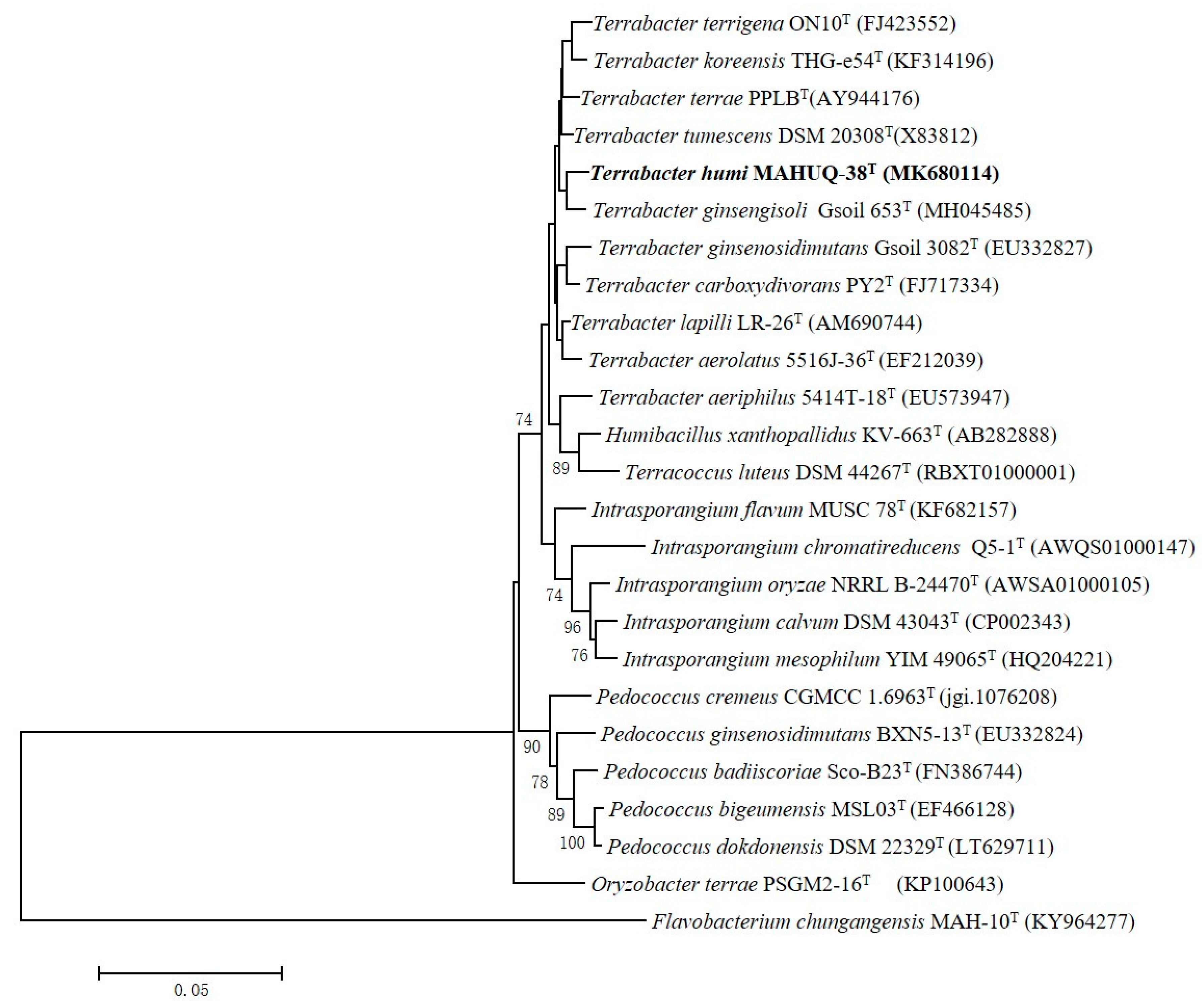
2.2. 16S rRNA Gene Sequence and Phylogenetic Analysis
2.3. Draft Genome and DNA G + C Content Analysis
2.4. DNA-DNA Hybridization
2.5. Cellular Fatty Acid, Respiratory Quinones and Polar Lipid Analysis
2.6. Taxonomic Conclusion
2.7. Description of Terrabacter humi sp. nov.
Terrabacter humi (hu’mi. L. gen. n. humi of/from soil)
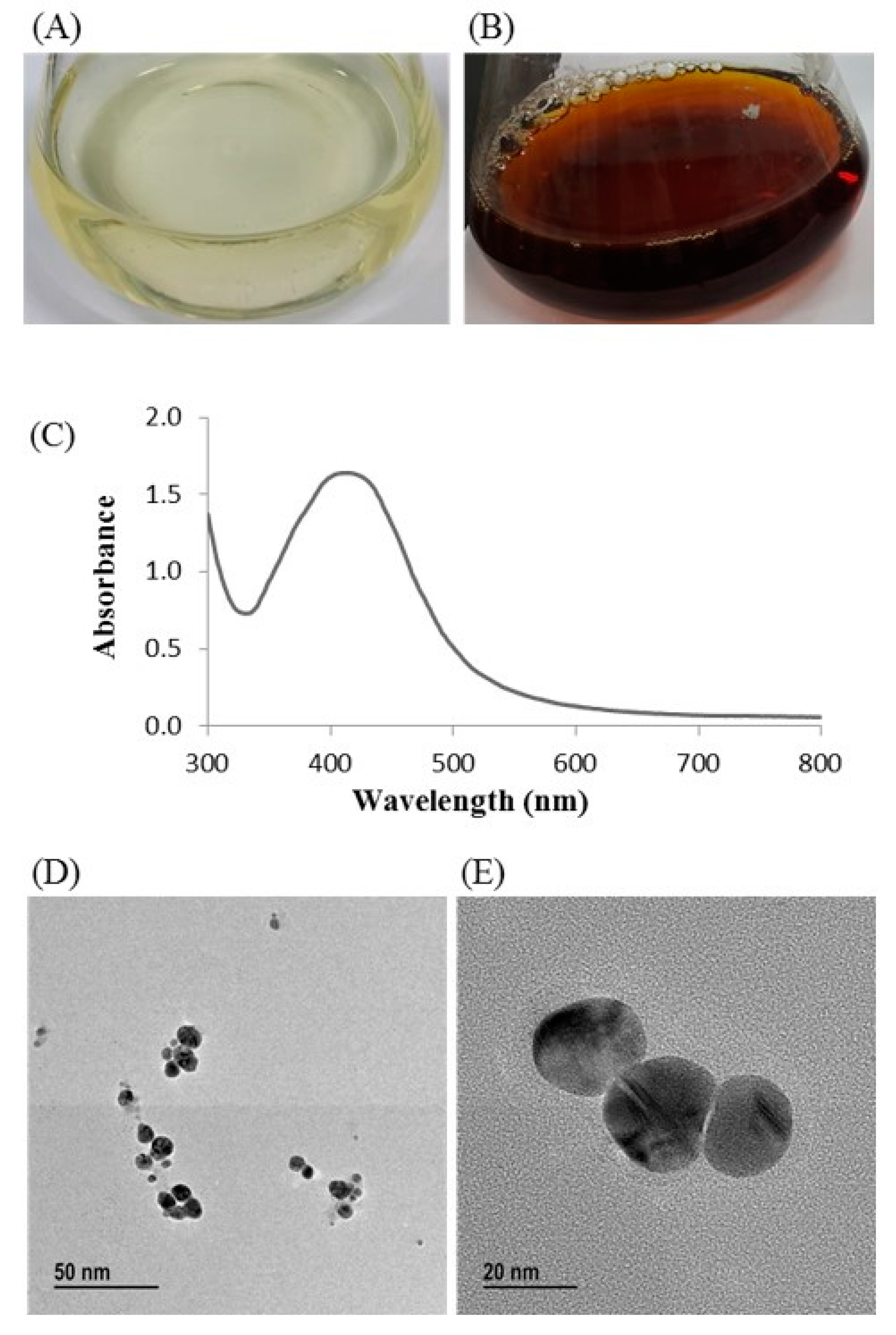
2.8. Ecofriendly Synthesis of AgNPs Using Terrabacter humi sp. nov.
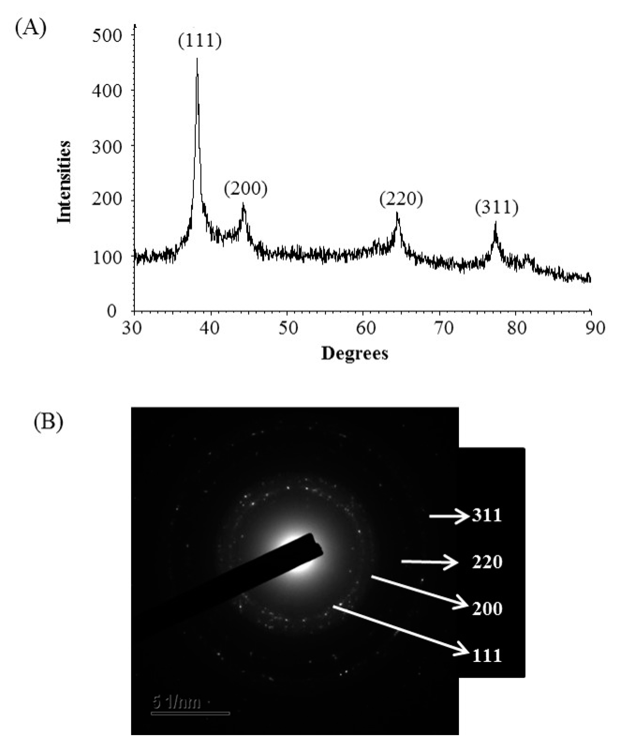
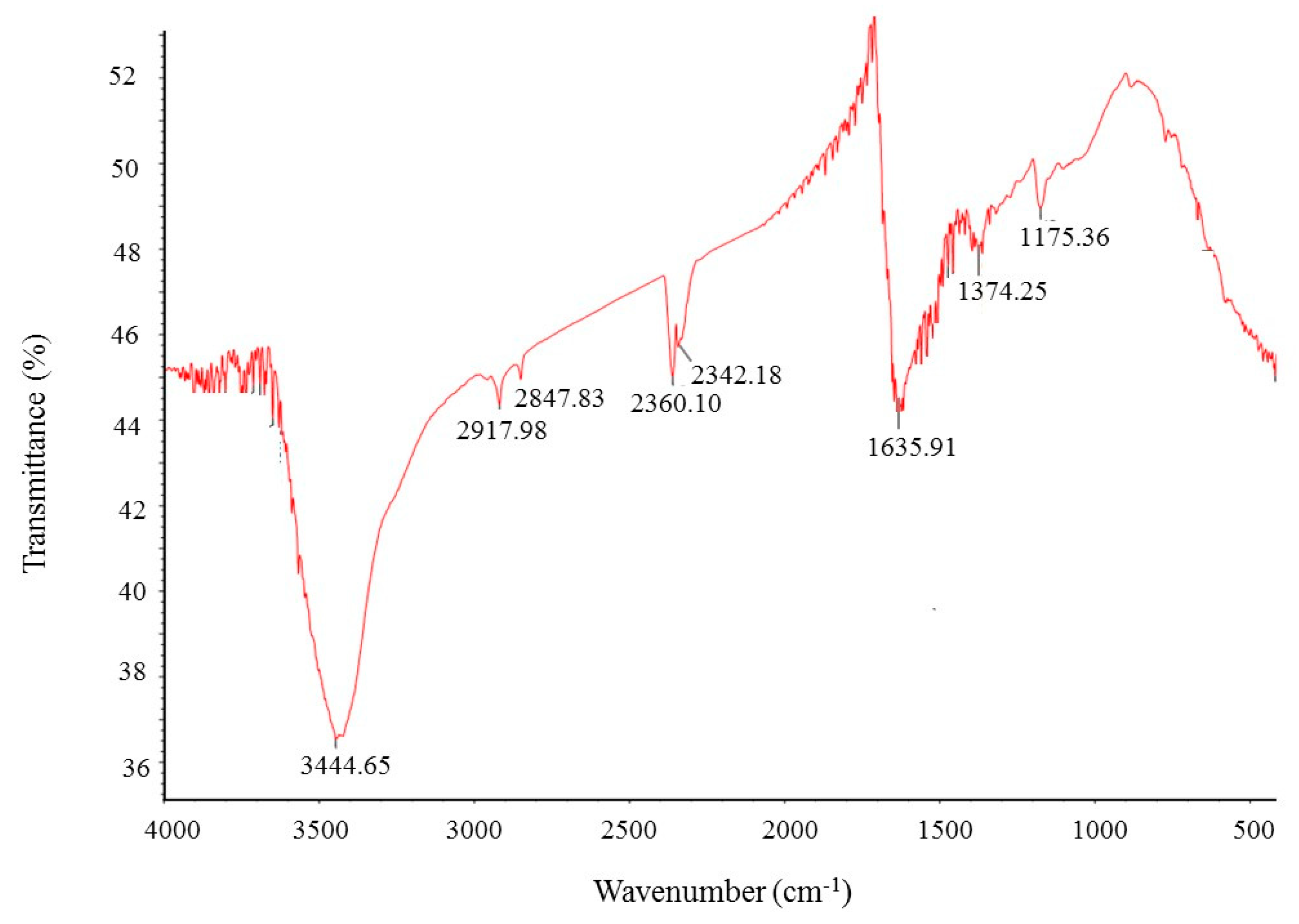
2.9. Characterization of Synthesized AgNPs
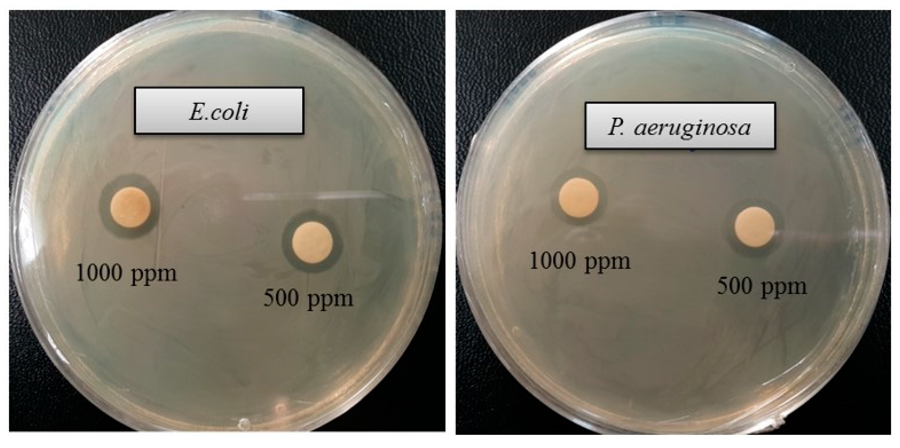
2.10. Antimicrobial Activity of Synthesized AgNPs
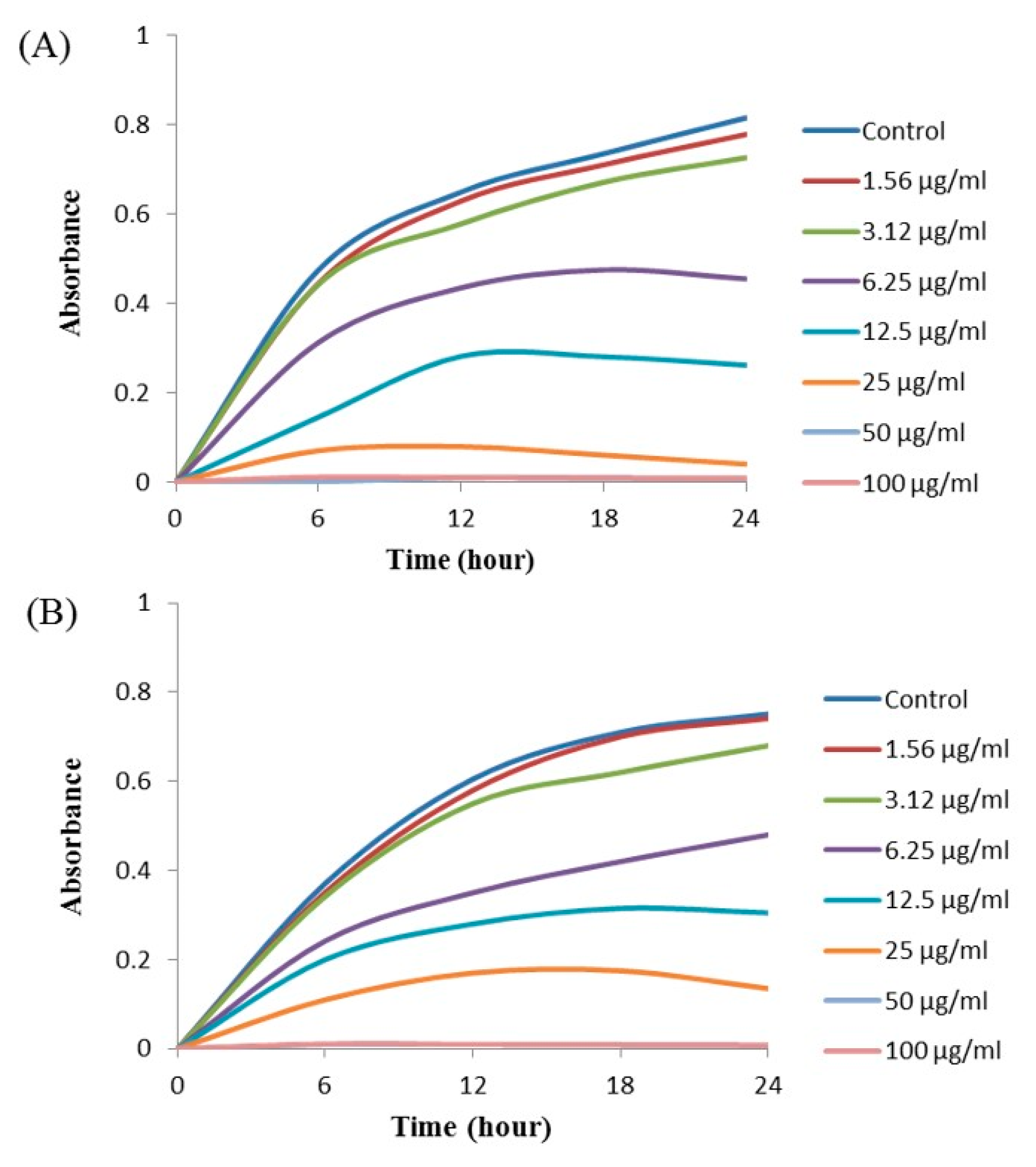
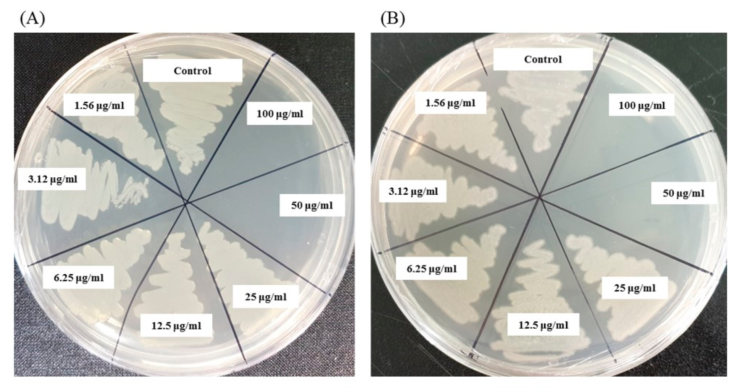
2.11. Minimal Inhibitory Concentration (MIC) and Minimal Bactericidal Concentration (MBC)
2.12. Investigation of Structural Changes by FE-SEM
3. Materials and Methods
3.1. Materials
3.2. Isolation of Bacteria
3.3. Phenotypic and Biochemical Characteristics
3.4. 16S rRNA Gene Sequencing and Phylogenetic Analysis
3.5. Genome Sequence Analysis
3.6. Cellular Fatty Acid, Respiratory Quinones and Polar Lipid Analysis
3.7. Ecofriendly Synthesis of AgNPs Using Terrabacter humi sp. nov.
3.8. Characterization of Synthesized AgNPs
3.9. Antimicrobial Activity of Synthesized AgNPs
3.10. Determination of Minimal Inhibitory Concentration and Minimal Bactericidal Concentration
3.11. Investigation of Structural Changes by FE-SEM
4. Conclusions
Supplementary Materials
Author Contributions
Funding
Acknowledgments
Conflicts of Interest
References
- Collins, M.D.; Dorsch, M.; Stackebrandt, E. Transfer of Pimelobacter tumescens to Terrabacter gen. nov. as Terrabacter tumescens comb. nov. and of Pimelobacter jensenii to Nocardioides as Nocardioides jensenii comb. nov. Int. J. Syst. Bacteriol. 1989, 39, 1–6. [Google Scholar] [CrossRef]
- Weon, H.Y.; Schumann, P.; Reiner, M.; Kroppenstedt, R.M.; Kim, B.Y.; Song, J.; Kwon, S.W.; Go, S.J.; Stackebrandt, E. Terrabacter aerolatus sp. nov., isolated from an air sample. Int. J. Syst. Evol. Microbiol. 2007, 55, 2491–2495. [Google Scholar] [CrossRef] [PubMed]
- Jin, M.F.; Quan, X.T.; Siddiqi, M.Z.; Liu, Q.Z.; Yu, H.S.; Im, W.T. Terrabacter ginsengihumi sp. nov., isolated from ginseng cultivating soil. J. Microbiol. 2018, 56, 331–336. [Google Scholar] [CrossRef] [PubMed]
- Montero-Barrientos, M.; Rivas, R.; Velazquez, E.; Monte, E.; Roig, M.G. Terrabacter terrae sp. nov., a novel actinomycete isolated from soil in Spain. Int. J. Syst. Evol. Microbiol. 2005, 55, 2491–2495. [Google Scholar] [CrossRef]
- Lee, J.E.; Seo, J.P.; Lee, D.W.; Ko, Y.H.; Lee, S.D. Terrabacter lapilli sp. nov., an actinomycete isolated from stone. Int. J. Syst. Evol. Microbiol. 2008, 58, 1084–1088. [Google Scholar] [CrossRef]
- Yoon, J.H.; Park, S.; Kang, S.J.; Jung, Y.T.; Kim, W. Terrabacter terrigena sp. nov., isolated from soil. Int. J. Syst. Evol. Microbiol. 2009, 59, 2798–2802. [Google Scholar] [CrossRef]
- Al-Dhabi, N.A.; Arasu, M.V. Environmentally-friendly green approach for the production of zinc oxide nanoparticles and their anti-fungal, ovicidal, and larvicidal properties. Nanomaterials 2018, 8, 500. [Google Scholar] [CrossRef]
- Ojo, S.A.; Lateef, A.; Azeez, M.A. Biomedical and catalytic applications of gold and silver-gold alloy nanoparticles biosynthesized using cell-free extract of Bacillus safensis LAU 13: Antifungal, dye degradation, anti-coagulant and thrombolytic activities. IEEE. Transon. Nanobiosci. 2016, 15, 433–442. [Google Scholar] [CrossRef]
- Taner, M.; Sayar, N.; Yulug, I.G. Synthesis, characterization and antibacterial investigation of silver–copper nanoalloys. J. Mater. Chem. 2011, 21, 13150–13154. [Google Scholar] [CrossRef]
- Wang, C.; Kim, Y.J.; Singh, P. Green synthesis of silver nanoparticles by Bacillus methylotrophicus, and their antimicrobial activity. Artific. Cells. Nanomed. Biotechnol. 2016, 44, 1127–1132. [Google Scholar]
- Dhandapani, P.; Maruthamuthu, S.; Rajagopal, G. Bio-mediated synthesis of TiO2 nanoparticles and its photocatalytic effect on aquatic biofilm. J. Photochem. Photobiol. B Biol. 2012, 110, 43–49. [Google Scholar] [CrossRef] [PubMed]
- Ali, A.A.; Asif, M.A.; Mashrai, A.M. Green synthesis of ZnO nanoparticles using Bacillus subtilis and their catalytic performance in the one-pot synthesis of steroidal thiophenes. Eur. Chem. Bull. 2014, 3, 939–945. [Google Scholar]
- Huq, M.A. Paenibacillus anseongense sp. nov. a Silver Nanoparticle Producing Bacterium Isolated from Rhizospheric Soil. Curr. Microbiol. 2020, 77, 2023–2030. [Google Scholar] [CrossRef] [PubMed]
- Singh, P.; Singh, H.; Kim, Y.J. Extracellular synthesis of silver and gold nanoparticles by Sporosarcina koreensis DC4 and their biological applications. Enzym. Microb. Technol. 2016, 86, 75–83. [Google Scholar] [CrossRef]
- Vigneshwaran, N.; Kathe, A.A.; Varadarajan, P.V. Silve-protein (core-shell) nanoparticle production using spent mushroom substrate. Langmuir 2007, 23, 7113–7117. [Google Scholar] [CrossRef]
- Castro-Aceituno, V.; Ahn, S.; Simu, S.Y.; Singh, P.; Mathiyalagan, R.; Lee, H.A.; Yang, D.C. Anticancer activity of silver nanoparticles from Panax ginseng fresh leaves in human cancer cells. Biomed. Pharmacother. 2016, 84, 158–165. [Google Scholar] [CrossRef]
- Huq, M.A. Microvirga rosea sp. nov.: A nanoparticle producing bacterium isolated from soil of rose garden. Arch. Microbiol. 2018, 200, 1439–1445. [Google Scholar] [CrossRef]
- Akter, S.; Huq, M.A. Biologically rapid synthesis of silver nanoparticles by Sphingobium sp. MAH-11 T and their antibacterial activity and mechanisms investigation against drug-resistant pathogenic microbes. Artif. Cells Nanomed. Biotechnol. 2020, 48, 672–682. [Google Scholar] [CrossRef]
- Chaloupka, K.; Malam, Y.; Seifalian, A.M. Nanosilver as a new generation of nanoproduct in biomedical applications. Trends Biotechnol. 2010, 28, 580–588. [Google Scholar] [CrossRef]
- Stackebrandt, E.; Goebel, B.M. Taxonomic note: A place for DNA-DNA reassociation and 16S rRNA sequence analysis in the present species definition in bacteriology. Int. J. Syst. Bacteriol. 1994, 44, 846–849. [Google Scholar] [CrossRef]
- Singh, N.; Khanna, P.K. In situ synthesis of silver nano-particles in polymethylmethacrylate. Mater. Chem. Phys. 2007, 104, 367–372. [Google Scholar] [CrossRef]
- Du, J.; Sing, H.; Yi, T.H. Biosynthesis of silver nanoparticles by Novosphingobium sp. THG-C3 and their antimicrobial potential. Artif. Cells Nanomed. Biotechnol. 2017, 45, 211–217. [Google Scholar] [CrossRef] [PubMed]
- Du, J.; Sing, H.; Yi, T.H. Antibacterial, anti-biofilm and anticancer potentials of green synthesized silver nanoparticles using benzoin gum (Styrax benzoin) extract. Bioprocess. Biosyst. Eng. 2016, 39, 1923–1931. [Google Scholar] [CrossRef] [PubMed]
- Singh, P.; Kim, Y.J.; Singh, H. Biosynthesis, characterization, and antimicrobial applications of silver nanoparticles. Int. J. Nanomed. 2015, 10, 2567–2577. [Google Scholar]
- Kalishwaralal, K.; BarathManiKanth, S.; Pandian, S.R. Silver nanoparticles impede the biofilm formation by Pseudomonas aeruginosa and Staphylococcus epidermidis. Colloids Surf. B Biointerfaces. 2010, 79, 340–344. [Google Scholar] [CrossRef] [PubMed]
- Cherian, T.; Ali, K.; Saquib, Q.; Faisal, M.; Wahab, R.; Musarrat, J. Cymbopogon Citratus Functionalized Green Synthesis of CuO-Nanoparticles: Novel Prospects as Antibacterial and Antibiofilm Agents. Biomolecules 2020, 10, 169. [Google Scholar] [CrossRef]
- Rehman, S.; Jermy, B.R.; Akhtar, S.; Borgio, J.F. Isolation and characterization of a novel thermophile; Bacillus haynesii, applied for the green synthesis of ZnO nanoparticles. Artif. Cells Nanomed. Biotechnol. 2019, 47, 2072–2082. [Google Scholar] [CrossRef]
- Bayroodi, E.; Jala, R. Modulation of antibiotic resistance in Pseudomonas aeruginosa by ZnO nanoparticles. Iran J. Microbiol. 2016, 8, 85–92. [Google Scholar]
- Ali, S.G.; Ansari, M.A.; Alzohairy, M.A.; Alomary, M.N.; Jalal, M.; AlYahya, S.; Asiri, S.M.M.; Khan, H.M. Effect of Biosynthesized ZnO Nanoparticles on Multi-Drug Resistant Pseudomonas Aeruginosa. Antibiotics 2020, 9, 260. [Google Scholar] [CrossRef]
- Shankar, S.; Rhim, J.W. Amino acid mediated synthesis of silver nanoparticles and preparation of antimicrobial agar/silver nanoparticles composite films. Carbohydr. Polym. 2015, 130, 353–363. [Google Scholar] [CrossRef]
- Kim, J.S.; Kuk, E.; Yu, K.N. Antimicrobial effects of silver nanoparticles. Nanomed. Nanotechnol. Biol. Med. 2007, 3, 95–101. [Google Scholar] [CrossRef] [PubMed]
- Sheu, S.Y.; Su, C.L.; Kwon, S.W.; Chen, W.M. Flavobacterium amniphilum sp. nov., isolated from a stream. Int. J. Syst. Evol. Microbiol. 2017, 67, 5179–5186. [Google Scholar] [CrossRef] [PubMed]
- Weisburg, W.G.; Barns, S.M.; Pelletier, D.A.; Lane, D.J. 16S ribosomal DNA amplification for phylogenetic study. J. Bacteriol. 1991, 173, 697–703. [Google Scholar] [CrossRef] [PubMed]
- Yoon, S.H.; Ha, S.M.; Kwon, S.; Lim, J.; Kim, Y. Introducing EzBioCloud: A taxonomically united database of 16S rRNA gene sequences and whole-genome assemblies. Int. J. Syst. Evol. Microbiol. 2017, 67, 1613–1617. [Google Scholar] [CrossRef] [PubMed]
- Thompson, J.D.; Gibson, T.J.; Plewniak, F.; Jeanmougin, F.; Higgins, D.G. The Clustal_X windows interface: Flexible strategies for multiple sequence alignment aided by quality analysis tools. Nucleic Acids Res. 1997, 24, 4876–4882. [Google Scholar] [CrossRef] [PubMed]
- Hall, T.A. BioEdit: A user-friendly biological sequence alignment editor and analysis program for Windows 95/98/NT. Nucleic Acids Symp. Ser. 1999, 41, 95–98. [Google Scholar]
- Kimura, M. The Neutral Theory of Molecular Evolution; Cambridge University Press: New York, NY, USA, 1983. [Google Scholar]
- Saitou, N.; Nei, M. The neighbor-joining method: A new method for reconstructing phylogenetic trees. Mol. Bio. Evol. 1987, 4, 406–425. [Google Scholar]
- Tamura, K.; Stecher, G.; Peterson, D.; Filipski, A.; Kumar, S. MEGA6: Molecular evolutionary genetics analysis version 6.0. Mol. Biol. Evol. 2013, 30, 2725–2729. [Google Scholar] [CrossRef]
- Felsenstein, J. Confidence limits on phylogenies: An approach using the bootstrap. Evolution 1985, 39, 783–791. [Google Scholar] [CrossRef]
- Meier-Kolthoff, J.P.; Auch, A.F.; Klenk, H.P.; Göker, M. Genome sequence-based species delimitation with confidence intervals and improved distance functions. BMC Bioinform. 2013, 14, 60. [Google Scholar] [CrossRef]
- Ezaki, T.; Hashimoto, Y.; Yabuuchi, E. Fluorometric deoxyribonucleic acid-deoxyribonucleic acid hybridization in microdilution wells as an alternative to membrane filter hybridization in which radioisotopes are used to determine genetic relatedness among bacterial strains. Int. J. Syst. Evol. Microbiol. 1989, 39, 224–229. [Google Scholar] [CrossRef]
- Huq, M.A.; Akter, S.; Lee, S.Y. Mucilaginibacter formosus sp. nov., a bacterium isolated from road-side soil. Antonie van Leeuwenhoek 2019, 112, 513–521. [Google Scholar] [CrossRef] [PubMed]
- Sasser, M. Identification of Bacteria by Gas Chromatography of Cellular Fatty Acids; MIDI Technical Note 101; MIDI Inc.: Newark, DE, USA, 1990. [Google Scholar]
- Collins, M.D. Isoprenoid quinones. In Chemical Methods in Prokaryotic Systematics; Goodfellow, M., O’Donnell, A.G., Eds.; Wilye: Chichester, UK, 1994; pp. 265–309. [Google Scholar]
- Akter, S.; Huq, M.A. Sphingomonas chungangi sp. nov., a bacterium isolated from soil sample of a garden. Int. J. Syst. Evol. Microbiol. 2020, 70, 4151–4157. [Google Scholar] [CrossRef] [PubMed]
- Ansari, M.A.; Baykal, A.; Asiri, S. Synthesis and characterization of antibacterial activity of spinel chromium-substituted copper ferrite nanoparticles for biomedical application. J. Inorg. Organomet. Polym. Mater. 2018, 28, 2316–2327. [Google Scholar] [CrossRef]


| Characteristics | 1 | 2 | 3 | 4 | 5 | 6 | 7 |
|---|---|---|---|---|---|---|---|
| Isolation source | Soil | Soil | Soil | Soil | Stone | Soil | Air |
| Cell morphology | Rod | Rod/coccus cycle | Long rods | Short rods/rods | Short rods | coccus-rod | Rod/coccoid |
| Colony color | Milky white | White/grey | Yellow | Greyish yellow | Bright yellow | yellowish | White |
| Motility | - | - | - | - | - | - | + |
| Reduction of nitrate (API 20 NE) | + | + | + | - | + | + | + |
| Arginine dihydrolase | - | - | + | - | - | - | - |
| Growth temperature (°C) | 10–40 | 10–35 a | 15–40 b | 10–37 c | 10–40 d | 18–37 e | 5–35 f |
| NaCl tolerance (%) | 0–5 | 0–5 a | 0–7 b | 0–3 c | 0–3 d | 0–5 e | 0–5 f |
| Hydrolysis of: | |||||||
| Casein | + | + | + | + | + | - | + |
| Starch | - | - | + | - | + | - | W |
| DNA | + | + | - | W | + | - | - |
| l-Tyrosine | + | - | - | - | - | W | + |
| Urea (API 20 NE) | - | - | + | - | - | - | - |
| Enzyme activity (API ZYM): | |||||||
| Esterase (C4) | + | + | + | + | + | - | + |
| Alkaline phosphatase | + | - | - | + | + | - | - |
| Esterase lipase (C8) | + | + | + | + | + | - | + |
| Lipase (C14) | W | - | + | + | + | + | + |
| Valine arylamidase | + | - | + | + | + | - | + |
| Cystine arylamidase | + | - | - | + | + | - | - |
| Trypsin | W | - | - | - | + | - | - |
| α-chymotrypsin | - | - | + | - | + | - | - |
| β-glucuronidase | - | - | + | - | + | + | - |
| α-mannosidase | - | - | + | + | - | - | W |
| a-glucosidase | + | - | + | + | + | + | + |
| a-galactosidase | + | - | + | + | + | + | + |
| β-glucosidase | W | - | + | + | + | + | W |
| β-galactosidase | + | - | + | + | + | + | + |
| Assimilation of (API 20 NE): | |||||||
| l-arabinose | W | + | - | - | - | - | - |
| N-acetyl-glucosamine | + | + | + | + | + | - | + |
| d-maltose | + | - | - | + | + | - | + |
| d-mannose | + | + | + | + | - | + | + |
| d-mannitol | - | + | - | + | - | + | + |
| DNA G + C content (mol%) | 70.8 | 69.2–72.4 a | 71.0 b | 71.6 c | 72.6 d | 70.5 e | 71.7 f |
| Features | Strain MAHUQ-38T |
|---|---|
| Accession No. | JACVCU000000000 |
| Biosample | SAMN15891656 |
| BioProject | PRJNA658849 |
| Total sequence length (nt) | 5,156,829 |
| Scaffold N50 | 910,522 |
| Scaffold L50 | 3 |
| Number of contigs | 19 |
| Sequencing method | de novo |
| Annotation pipeline | NCBI Prokaryotic Genome |
| DNA G + C content (mol%) | 70.8 |
| Total genes | 4664 |
| Genes (coding) | 4555 |
| Number of RNAs | 56 |
| tRNAs | 48 |
| rRNAs | 5 |
| Fatty Acid | 1 | 2 | 3 | 4 | 5 | 6 | 7 |
|---|---|---|---|---|---|---|---|
| C13:0 iso | - | - | 9.3 | 2.9 | 10.1 | - | Tr |
| C14:0 | - | 8.4 | 2.4 | 5.7 | - | Tr | Tr |
| C14:0 iso | 24.6 | 4.5 | 29.1 | 15.6 | 32.8 | 14.7 | 9.8 |
| C14:1 ω5c | 1.5 | 5.5 | - | - | - | - | - |
| C15:0 iso | 24.4 | 6.0 | - | 11.2 | - | 34.2 | 42.8 |
| C15:0 anteiso | 12.3 | 2.1 | Tr | - | - | 8.2 | 7.2 |
| C15:1 iso F | - | - | 23.9 | 3.3 | 19.6 | - | - |
| C15:1 ω6c | 1.4 | - | - | - | - | - | |
| C16:0 | - | 13.9 | 3.5 | 15.1 | 1.8 | Tr | 3.7 |
| C16:0 iso | 16.8 | 3.4 | 4.7 | -- | 3.1 | 21.3 | 15.1 |
| C16:0 3-OH | - | - | - | 7.1 | - | - | - |
| C16:1 2-OH | - | - | 4.8 | - | - | - | - |
| C16:1 iso H | 5.0 | - | - | 5.5 | 2.2 | 6.4 | 1.0 |
| C17:0 | - | 6.3 | 2.8 | - | 5.4 | - | 1.6 |
| C17:0 iso | - | - | - | - | - | Tr | 4.7 |
| C17:0 anteiso | 1.7 | - | - | - | Tr | 1.2 | 3.5 |
| C17:0 10-methyl | - | 2.3 | - | - | - | 1.0 | Tr |
| C17:0 cyclo | - | - | 3.1 | 4.7 | 1.6 | - | - |
| C17:1 ω8c | 4.6 | 11.9 | 2.6 | - | 3.2 | 1.9 | 1.2 |
| C17:1 anteiso ω9c | 1.3 | - | - | - | - | 1.1 | Tr |
| C17:1 anteiso A | - | - | 1.3 | 1.5 | 2.1 | - | - |
| C18:0 | - | 3.7 | 1.2 | 4.7 | Tr | - | 3.4 |
| C18:0 iso | - | - | 4.5 | 10.4 | 6.8 | - | 1.1 |
| C18:1 ω9c | - | 16.5 | - | 6.2 | Tr | 1.5 | 1.0 |
| C19:0 iso | - | - | 2.2 | - | 3.5 | - | - |
| Sum In Feature 3 | 1.3 | 6.5 | - | - | - | - | 1.4 |
| Sum In Feature 9 | 4.4 | 8.0 | - | - | - | - | 1.3 |
| Pathogenic Species | Zone of Inhibition (mm) | |
|---|---|---|
| 1000 ppm | 500 ppm | |
| Escherichia coli [ATCC 10798] | 15.6 ± 1.0 | 13.8 ± 0.7 |
| Pseudomonas aeruginosa [ATCC 10145] | 14.1 ± 1.2 | 13.4 ± 0.8 |
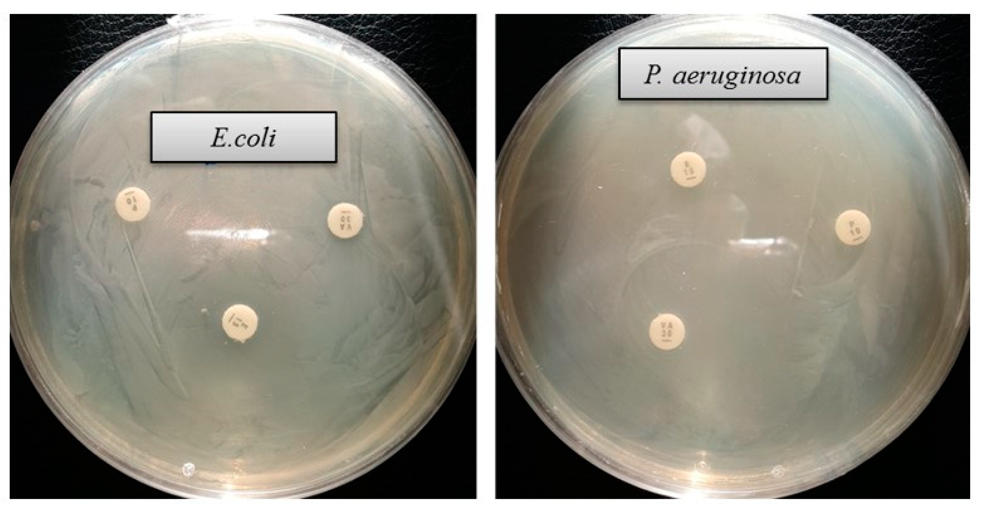
| Pathogenic Species | Antibiotic | Zone of Inhibition (mm) |
|---|---|---|
| Escherichia coli [ATCC 10798] | Erythromycin | - |
| Vancomycin | - | |
| Penicillin G | - | |
| Pseudomonas aeruginosa [ATCC 10145] | Erythromycin | - |
| Vancomycin | - | |
| Penicillin G | - |
Publisher’s Note: MDPI stays neutral with regard to jurisdictional claims in published maps and institutional affiliations. |
© 2020 by the authors. Licensee MDPI, Basel, Switzerland. This article is an open access article distributed under the terms and conditions of the Creative Commons Attribution (CC BY) license (http://creativecommons.org/licenses/by/4.0/).
Share and Cite
Akter, S.; Lee, S.-Y.; Siddiqi, M.Z.; Balusamy, S.R.; Ashrafudoulla, M.; Rupa, E.J.; Huq, M.A. Ecofriendly Synthesis of Silver Nanoparticles by Terrabacter humi sp. nov. and Their Antibacterial Application against Antibiotic-Resistant Pathogens. Int. J. Mol. Sci. 2020, 21, 9746. https://doi.org/10.3390/ijms21249746
Akter S, Lee S-Y, Siddiqi MZ, Balusamy SR, Ashrafudoulla M, Rupa EJ, Huq MA. Ecofriendly Synthesis of Silver Nanoparticles by Terrabacter humi sp. nov. and Their Antibacterial Application against Antibiotic-Resistant Pathogens. International Journal of Molecular Sciences. 2020; 21(24):9746. https://doi.org/10.3390/ijms21249746
Chicago/Turabian StyleAkter, Shahina, Sun-Young Lee, Muhammad Zubair Siddiqi, Sri Renukadevi Balusamy, Md. Ashrafudoulla, Esrat Jahan Rupa, and Md. Amdadul Huq. 2020. "Ecofriendly Synthesis of Silver Nanoparticles by Terrabacter humi sp. nov. and Their Antibacterial Application against Antibiotic-Resistant Pathogens" International Journal of Molecular Sciences 21, no. 24: 9746. https://doi.org/10.3390/ijms21249746
APA StyleAkter, S., Lee, S.-Y., Siddiqi, M. Z., Balusamy, S. R., Ashrafudoulla, M., Rupa, E. J., & Huq, M. A. (2020). Ecofriendly Synthesis of Silver Nanoparticles by Terrabacter humi sp. nov. and Their Antibacterial Application against Antibiotic-Resistant Pathogens. International Journal of Molecular Sciences, 21(24), 9746. https://doi.org/10.3390/ijms21249746