Review

Reading between the (Genetic) Lines: How Epigenetics is Unlocking Novel Therapies for Type 1 Diabetes

by
Ammira-Sarah AL-Shabeeb Akil
1,*,
Laila F. Jerman
1,
Esraa Yassin
1,
Sujitha S. Padmajeya
1,
Alya Al-Kurbi
1 and
Khalid A. Fakhro
1,2,3
1
Department of Human Genetics, Sidra Medicine, Doha P.O. Box 26999, Qatar
2
Department of Genetic Medicine, Weil Cornell Medical College, Doha P.O. Box 24144, Qatar
3
College of Health and Life Sciences, Hamad Bin Khalifa University, Doha P.O. Box 34110, Qatar
*
Author to whom correspondence should be addressed.
Cells 2020, 9(11), 2403; https://doi.org/10.3390/cells9112403
Submission received: 27 September 2020 / Revised: 25 October 2020 / Accepted: 27 October 2020 / Published: 3 November 2020
(This article belongs to the Special Issue Molecular Mechanisms in Metabolic Disease)

Abstract

:
Type 1 diabetes (T1D) is an autoimmune condition where the body’s immune cells destroy their insulin-producing pancreatic beta cells leading to dysregulated glycaemia. Individuals with T1D control their blood glucose through exogenous insulin replacement therapy, often using multiple daily injections or pumps. However, failure to accurately mimic intrinsic glucose regulation results in glucose fluctuations and long-term complications impacting key organs such as the heart, kidneys, and/or the eyes. It is well established that genetic and environmental factors contribute to the initiation and progression of T1D, but recent studies show that epigenetic modifications are also important. Here, we discuss key epigenetic modifications associated with T1D pathogenesis and discuss how recent research is finding ways to harness epigenetic mechanisms to prevent, reverse, or manage T1D.

1. Introduction

The hallmark of type 1 diabetes (T1D) is T cell-mediated autoimmune destruction of pancreatic beta cells, leading to insulin deficiency, elevated blood glucose concentrations, and life-long need for exogenous insulin therapy. Globally, the annual number of new cases of T1D is rising, and in the under-20 age group alone, numbers are fast approaching 100,000 (www.diabetesatlas.org). The incidence rate varies between countries and ranges from 6% in sub-Saharan Africa to 77% in some parts of Scandinavia [1]; altogether another five million people are expected to be diagnosed with T1D by 2050 [2], with profound implications for healthcare systems globally and the potential for a staggering socio-economic impact. Staying well with T1D requires normoglycemia to be achieved and maintained, but this goal is not currently achievable for many patients using existing treatment strategies [3]. Understanding the intrinsic and extrinsic factors underlying the development and progression of T1D is necessary for the development of novel intervention therapies that could delay or even prevent clinical progression in individuals susceptible to T1D or be used to treat/manage glycemic excursions in those worst affected.
Despite ongoing research, dissecting the etiology of T1D has proven a herculean task. Genetic studies have recognized more than 60 regions (loci) associated with or predisposing to T1D; the first and still strongest reported genetic association is with the human leukocyte antigen HLA region [4,5], while the second most significant T1D genetic association is with insulin gene promoter polymorphism [6]. Even so, only 5% or fewer of children bearing the high-risk HLA haplotypes and about 10% of those with insulin gene polymorphism go on to clinically develop T1D during the first 20 years of their lifetime [7,8]. Therefore, what are the important factors determining whether a genetically susceptible individual develops T1D, and how could these factors be used to help in the fight against this disease?
Epigenetic modification is a key mechanism affecting gene expression, effectively determining the extent and timing of expression of a given gene in response to internal and external stimuli. Multiple lines of evidence now implicate epigenetic factors in T1D incidence: firstly, although monozygotic (MZ) twins share 100% of their DNA, they exhibit a high discordance rate in the development of T1D, especially when disease onset occurs over the age of 15 [9]; furthermore, the rapid increase in T1D seen in recent years is unlikely to be a result of genetic changes across the population alone, due to the short timescale [10,11]. Alongside, other studies showed that in siblings sharing an HLA haplotype, it is rather the age at diabetes onset in their sibling that determines their risk of developing the condition, and not the shared HLA type itself [12]. Thus, it is not only the possession of high-risk genes that decide whether or not an individual progress to the clinical presentation of T1D but also whether, when, and to what extent those genes are expressed.
The growing interest in epigenetic changes in T1D and in factors driving them has led to investigations into epigenetic therapies to treat the condition. In this review, we discuss recent initiatives targeting epigenetic mechanisms involved in T1D, namely, DNA methylation, histone (post-translational) modifications, and non-coding RNA-mediated gene silencing (for a comprehensive discussion of these mechanisms see references [13,14]), as well as highlighting future avenues for productive therapeutic research.

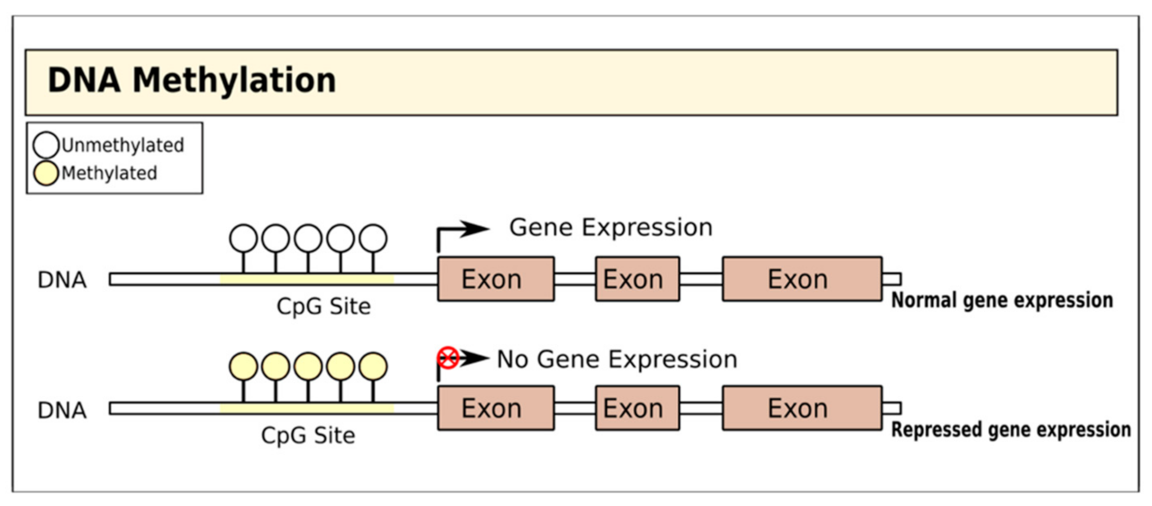
2. Therapies Targeting DNA Methylation in T1D

DNA methylation involves the addition of a methyl group to the fifth carbon of CpG dinucleotides (cytosine residues linked by a phosphate to a guanine nucleotide) by DNA methyltransferases, thereby blocking gene transcription [15] (Figure 1). In T1D, there is evidence that DNA methylation in both immune cells and pancreatic islet cells is important and may even substantially pre-date the diagnosis of the disease [16]. Studies of immune cells from the blood of adult MZ twins discordant for T1D have variably identified specific positions with distinct methylation patterns: one study on monocytes found that patterns of methylation at these sites were detectable many years before T1D diagnosis and were correlated with the presence of autoantibodies [17], while a genome-wide DNA methylation analysis identified 88 CpG methylation sites in B lymphocytes with significant modifications, including some affecting genes associated with T1D pathogenesis such as HLA, insulin (INS), interleukin 2 receptor subunit beta (IL-2RB), and CD226 [18]. A more recent MZ twin study did not identify any differences in DNA methylation in monocytes or B cells but found a single differentially methylated CpG position in T cells with genome-wide significance [19].
Genome-wide DNA methylation quantitative trait locus (mQTL) analysis of human pancreatic islets has revealed 383 significant CpG sites in known diabetes loci as potential methylation targets; importantly, some of the identified candidate genes, i.e., glutathione peroxidase 7 (GPX7), glutathione S-transferase theta 1 (GSTT1), and sorting nexin 19 (SNX19) directly affect key biological processes such as proliferation and apoptosis of pancreatic islet beta cells [20]. More recently, Ye et al. identified widespread genetic and epigenetic interactions at known T1D susceptibility loci; they showed (in their preprint) that DNA methylation at five loci, i.e., integrin subunit beta 3 binding protein (ITGB3BP), AF4/FMR2 family member 3 (AFF3), protein tyrosine phosphatase non-receptor type 2 (PTPN2), cathepsin H (CTSH), and cytotoxic T lymphocyte-associated protein 4 (CTLA4), is potentially causal to T1D [21]. Interestingly, it seems that some epigenetic changes may even be present years before T1D diagnosis: Johnson et al. identified 10 regions of the genome in which methylation differs between T1D cases and controls from as early as birth, in some cases multiple years before T1D diagnosis [22]. These findings raise the possibility of screening those with high-risk genetic phenotypes for concurrent high-risk epigenetic marks to identify those at greatest need of intensive monitoring and/or preventative intervention.
Although the significance of altered DNA methylation in T1D onset and pathogenesis is clear, there have yet to be large-scale trials of the use of DNA methyltransferase inhibitors in treating/preventing the condition. However, inhibitors of DNA methylation have been studied in vitro for their potential use in pancreatic cell reprogramming, with the future aim of regenerating patients’ beta cells as a novel T1D therapy. Lefebvre et al. found that treating pancreatic ductal cells with the DNA methylation inhibitor 5-azacytidine successfully promoted their conversion into pancreatic endocrine cells [23]. Similarly, in mice lacking DNA methyltransferase 1(DNMT1) and the alpha cell-maintaining factor Aristaless-related homeobox (Arx) specifically in pancreatic alpha cells, Chakravarthy et al. observed reprogramming of around half of alpha cells into biologically active beta cells in vivo [24]. Interestingly, these authors also found that a subset of T1D patients possess a small number of glucagon-expressing cells lacking both DNMT1 and Arx expression and instead ectopically express beta cell markers, including insulin [24]. While these results suggest that targeted DNMT1 inhibition might support the possibility of regeneration of lost beta cells in individuals with T1D, further studies addressing the longevity and functionality of these converted cells are warranted.
While not specifically assessing epigenetic modification, there are early indications that drugs as well as diets modulating DNA methylation could have some positive effect on delaying the progression to T1D. Long used in diabetes care, metformin has more recently been revealed to have multiple epigenetic modifying actions, including modulating the activity of DNA methyltransferases [25]. The REducing with MetfOrmin Vascular Adverse Lesions (REMOVAL trial) aimed to ameliorate long-term cardiovascular complications in diabetes patients using metformin [26]. In the REMOVAL trial, metformin treatment reduced the progression of atherosclerosis, weight gain, and low-density lipoprotein (LDL)–cholesterol levels [27,28], though the epigenetic effects of the treatment were not specifically measured. In type 2 diabetes, metformin treatment significantly decreased DNA methylation of metformin the transporter genes solute carrier family 22 member 1 (SLC22A1), solute carrier family 22 member 3 (SLC22A3), and solute carrier family 47 (multidrug and toxin extrusion), member 1 (MATE1) in the livers of patients; in the case of SLC22A1 and SLC22A3, reduced methylation was associated with less hyperglycemia and obesity [29]. Similarly, promising results were also seen in pre-clinical trials in adult rats with streptozotocin-induced diabetes treated with the DNMT inhibitor procainamide, which significantly reduced the activity of DNMT in the pancreas and significantly increased fasting insulin levels [30]. Thus, initial studies in both type 1 and 2 diabetes patients and models seem to indicate that DNMT inhibitors might be a promising therapeutic strategy. Several DNMT inhibitors are licensed for use in treating certain types of cancer [31] and could be worth exploring in diabetes.

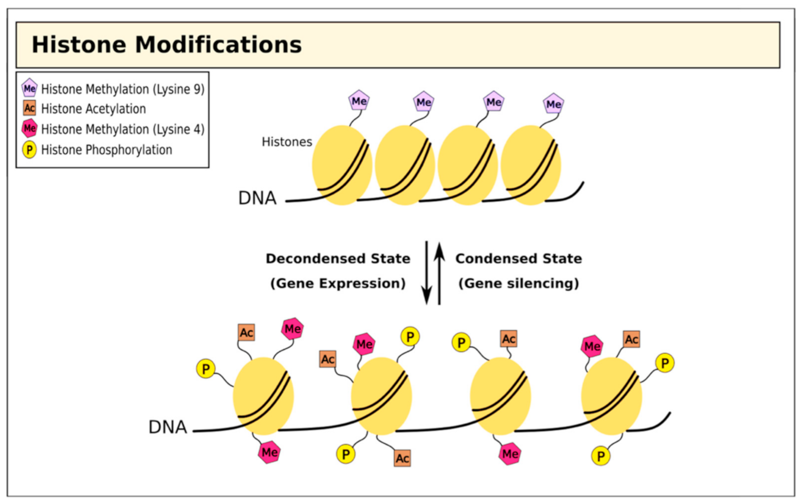
3. Therapies Targeting Histone Modifications in T1D

Alongside direct epigenetic modification of DNA, histones undergo post-transcriptional modifications to restructure chromatin in different ways [32,33,34,35,36]. Genomic DNA is bundled into chromatin, creating nucleosomes; the histone tails project from the nucleosome and are exposed to a wide range of covalent modifications including methylation, acetylation, ubiquitination, phosphorylation, sumoylation, and ADP ribosylation [37,38]. Collectively, these post-transcriptional modifications act to regulate chromatin structure, which has profound effects on several biological activities including gene transcription, DNA repair, and chromosome condensation [32] (Figure 2).
Histone modification is a significant feature of T1D [39], with studies showing aberrant histone modification as well as differential expression of the histone-modifying enzymes histone methyltransferase and histone deacetylase. A pivotal study by Miao et al. revealed increased methylation of lysine 9 in the H3 histone protein (H3K9me2) in lymphocytes from T1D patients: importantly, several of the genes affected had been linked with T1D pathology, such as CLTA4, a known T1D susceptibility gene, as well as genes affecting the signaling pathways of the immune mediators transforming growth factor-β (TGF-β), nuclear factor-ĸB (NF- ĸB), p38 mitogen-activated protein kinase (MAPKs), toll-like receptors (TLRs), and interleukin-6 (IL-6) [40]. The same group went on to show that acetylation of lysine 9 in the H3 histone protein (H3K9Ac) upstream of the major T1D susceptibility genes HLA-DRB1 and HLA-DQB1 was also significantly higher in monocytes from patients than in those from controls [41]. Furthermore, differential histone methylation and acetylation might be one mechanism explaining the T1D discordance seen in MZ twin studies. Following exposure to high glucose levels, in vitro skin fibroblasts showed significantly different expression of genes regulating epigenetic processes depending on whether they came from the affected or unaffected twin. While the expression of histone lysine methyltransferase (SET 7), H3K4 methyltransferase, and histone deacetylase (HDAC8) was lower in the cells from the type 1 diabetic twin, HDAC 4 was expressed at a higher level [42].
Histone modification inhibitors are a well-characterized class of epi-drugs with clear potential to be used therapeutically for T1D, either alone or in combination with DNA methyltransferase inhibitors. HDAC inhibitors (HDACi) were investigated by Lundh et al. after finding that pancreatic beta cells from children with newly diagnosed T1D exhibited relatively higher levels of HDAC1 and lower levels of HDACs-2 and -3 compared to controls; investigating this further in vitro, the authors showed that inhibition of HDAC1 and -3, but not HDAC2, protected primary and immortalized rat beta cells from apoptosis due to inflammatory cytokine exposure [43]. Building on this work, treating non-obese diabetic mice with the licensed lysine deacetylase inhibitors vorinostat and givinostat reversed diabetes [44], calling for future trials in human patients. Similarly encouraging data have come from trials of valproic acid, an HDAC class I and II inhibitor: in juvenile rats, valproic acid treatment positively influenced beta cell proliferation and functionality and increased insulin production, while simultaneously decreasing beta cell apoptosis and HBA1c and plasma glucose levels [45,46]. Studies in murine models also showed that the small molecule HDAC inhibitor trichostatin A [47] prevented T1D symptoms through increased acetylation of histone H3, which was associated with increased expression of the CD4+ T cell-derived lymphokine interferon gamma (INF-γ) and its transcription factor Tbet/Tbx21 [48], highlighting the potential role of chromatin remodeling agents in the protection against the development of T1D.
HDACi have also been used in studies of therapeutic cell reprogramming in the hope that this might be translated into a clinical application. Similarly to DNA methylation inhibitors, in vitro inhibition of the H3K27me3 methyltransferase Ezh2 enhanced neurogenin3-mediated production of beta-like cells from human embryonic stem cells [49], as well as the formation of insulin-producing cells from human induced pluripotent cells [50]. Combining the HDACi romidepsin with the DNA methyltransferase inhibitor 5-azacytidine also led to improved differentiation of adult human dermal fibroblasts into insulin- and glucagon-producing cells [51], but again the therapeutic translation of these findings is so far lacking. Recent studies from Marino and colleagues [52] demonstrate that feeding NOD mice a combination of acetate- and butyrate-rich diets offered protection against T1D progression. These studies indicate that acetate and butyrate might be working through distinct mechanisms and that a beneficial effect may be obtained through a combination of diets rich in these short-chain fatty acids. Acetate decreased the frequency of autoreactive T cells, whilst butyrate increased the number and function of regulatory T cells. Acetate- and butyrate-yielding diets enhanced gut integrity, reduced the circulating concentration of diabetogenic cytokines (such as IL-21), and demonstrated prevention in T1D onset. Clinical trials using such diets will provide intriguing results to understand the timing and potential of such diets in individuals at high risk of T1D.

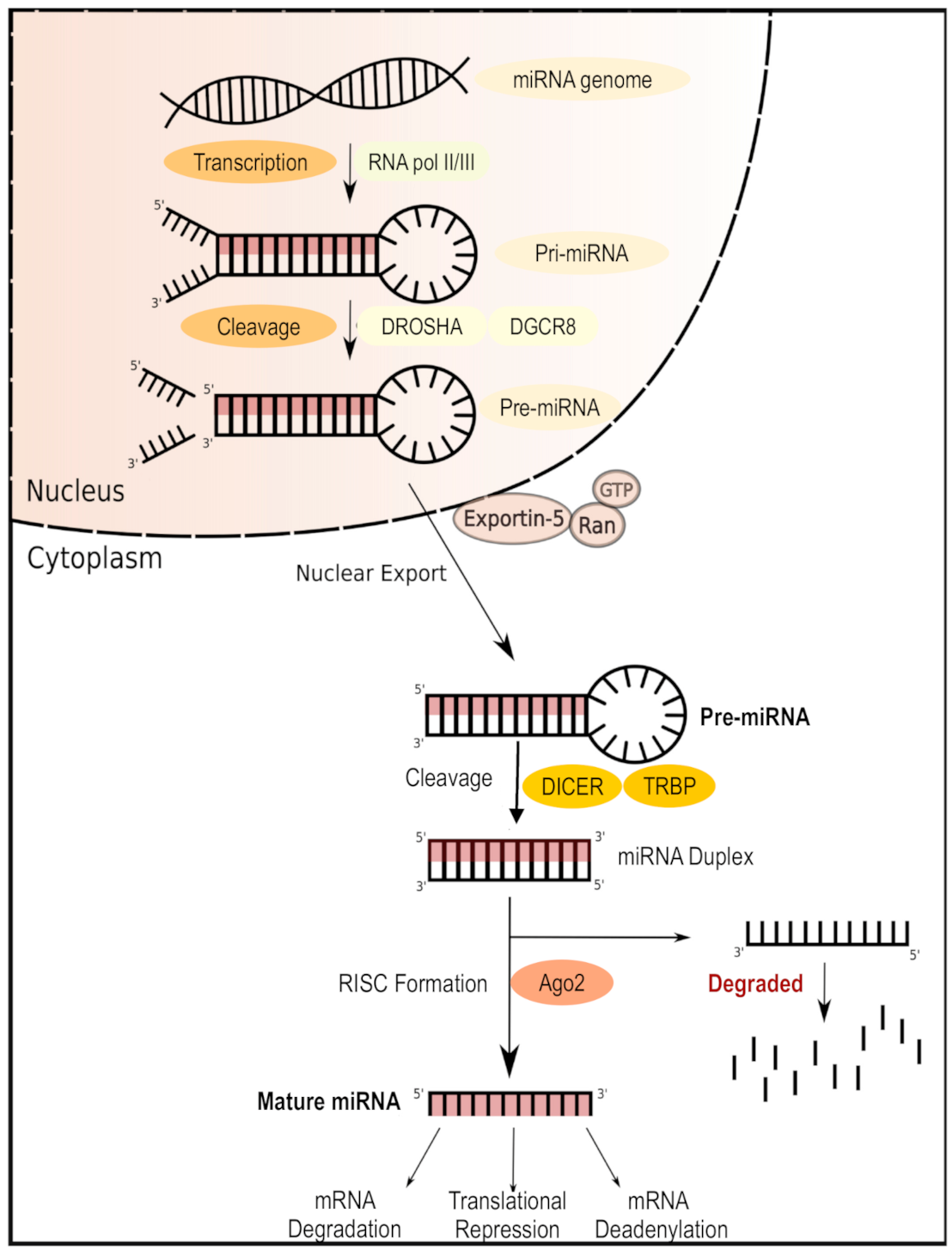
4. Therapies Targeting MicroRNAs in T1D

MicroRNAs (miRNAs) are short (around 22 nucleotides long) non-coding single-stranded RNA molecules [53] that modulate post-transcriptional gene expression, leading to target mRNA silencing (Figure 3). Multiple studies have demonstrated important associations between specific miRNAs and T1D (Table 1). One study on children with recent-onset T1D identified six miRNAs (miR-454-3p, miR-222-3p, miR-144-5p, miR-345-5p, miR-24-3p, and miR-140-5p) with high expression only in the early stages of diabetes; moreover, pathway analysis associated this pattern of differential miRNA expression with glycosaminoglycan biosynthesis as well as with PI3K/Akt, MAPK, and Wnt signaling pathways in early T1D [54]. Similarly, we also demonstrated that enteroviral infections disrupt the miRNA-directed suppression of proinflammatory factors within human islet beta cells, thereby resulting in an exacerbated antiviral immune response that promotes beta cell destruction and eventual T1D [55]. Further research on pancreatic cells from T1D patients linked the expression of miR-23a-3p, miR-23b-3p, and miR-149-5p with that of proapoptotic Bcl-2 proteins and consequent human β cell apoptosis [56], while autoreactive CD8+ T cells from patients with T1D showed evidence of repression of pro-apoptotic pathways via increased expression of miR-23b, miR-98, and miR-590-5p [57]. Thus, miRNAs are implicated at every stage of T1D, leading to intense research interest in targeting these molecules therapeutically.
To date, the majority of studies exploring the therapeutic potential of miRNA-targeted treatments for T1D have been carried out in vitro; still, some have yielded promising insights. El Ouaamari et al. showed that the application of 20-O-methyl-miR-375 antisense oligonucleotides increased the expression of the target gene PDK1 and reverted insulin release to normal in the rat beta cell line INS-1E [58]. Another study found that blocking miR-21, miR-34a, or miR-146a functions with antisense molecules preserved glucose responsiveness in the murine pancreatic beta cell line MIN6 under treatment with the inflammatory cytokine IL1β and also protected these cells from cytokine-induced apoptosis [59]. Moreover, Lovis et al. showed that blocking miR-34a or miR-146 activity partially protected palmitate-treated MIN6 cells from apoptosis but was insufficient to restore normal insulin secretion [60]. A major challenge of the clinical application of miRNA targeting is delivering the therapeutic agent to the target cells at an effective concentration. Pileggi et al. made important progress in this regard by establishing an islet transplantation model in rats where fluorescently labeled miRNA inhibitors were directly infused via an implanted catheter and effectively localized within target cells [61]. This study paves the way for in vivo trials of miRNA inhibitors aiming to preserve beta cell function or to increase the efficacy of transplantation of autologous islets or of those generated from induced pluripotent stem cells. Building on such work, a recent study conducted by Wang et al. demonstrated the potential of miR-216a to act as a therapeutic target in T1D through its negative regulation of phosphatase and tensin homolog (PTEN) expression levels; the administration of a miR-216a mimic in a murine model of T1D increased beta cell proliferation, decreased PTEN expression, and improved insulin secretion in vivo [62].
Despite these promising results and encouraging progress, many obstacles remain on the path to realizing the potential of miRNA-targeted therapies for T1D. For example, some miRNAs have multiple target genes, and occasionally, multiple miRNAs can regulate the same gene: this complex regulatory network can control several genes via one or a combination of miRNAs, which is further complicated by interactions with other epigenetic factors and linkers, such as DNA methylation and the histone code [63,64]. Future research will require to extend the studies on miRNA, currently carried out at the cellular level, to well-characterized T1D in vivo models to understand the direct role of miRNA-targeted genes and therapies in diabetes treatment.
Table 1. miRNAs that are differentially expressed between type 1 diabetes (T1D) patients and unaffected controls, targeting the drivers of epigenetic modification in T1D.
miRNATarget Gene/Pathway Related to T1DRegulation T1D Patients/ControlsTissue/CellReference
miR-101-3pc-MET proto-oncogene, receptor tyrosine kinase (c-Met-HGF), ephrin receptor, and signal transducer and activator of transcription 3 (STAT3) pathways linked to insulin secretion and β cells survivalUpregulated Serum [65]
miR-125b-5p and miR-365a-3p variation of hyperglycemia, measured (HbA1c) Upregulated Plasma [66]
miR-5190 and miR-770-5p variation of hyperglycemia, measured (HbA1c)DownregulatedPlasma [66]
miR-100-5pWnt signaling pathwayDownregulatedSerum [54]
miR-150-5ptranscription factor cMyb and IFN-rDownregulatedPBMCs [67]
miR-154-3pglycosaminoglycan biosynthesisDownregulatedSerum [54]
miR-24-3pMAPK and Wnt signaling pathwaysUpregulatedSerum [54]
miR-25-3psuppressor of cytokine Ssgnaling 5 (SOCS5), BTG anti-proliferation factor 2 (BTG2), PTEN/Akt pathway, and Notch signaling pathwayUpregulatedSerum [54]
miR-324-3pGLI family zinc finger 3 (GLI3), Wnt family member 2B (WNT2B), MAPK pathway, Wnt/β-catenin signaling pathwayUpregulatedSerum [54]
miR-377-3pLysine degradation and ubiquitin-mediated proteolysis pathways UpregulatedSerum [54]
miR-378eInsulin-like growth factor receptor and lipid metabolism pathways, Adiponectin expression pathwayDownregulatedPlasma-derived exosome [68]
miR-424-5pcaudal type homeobox 2 (CDX2) transcriptional factor pathwayDownregulatedPBMCs [67]
miR-454-3pnuclear factor of activated T cells 2 (NFATc2) and vitamin D-dependent calcium-binding protein calbindin 1 (CALB1), Wnt/β-catenin pathwaysUpregulatedSerum [54]
miR-490-5pCalcium-binding protein 5 (CABP5), phosphatidylinositol-4,5-bisphosphate 3-kinase catalytic subunit alpha (PIK3CA), nuclear factor of activated T cells 5 (NFAT5) and TGF-beta-signaling pathways DownregulatedSerum [54]
miR-574-3pMothers against decapentaplegic homolog 4(SMAD4) signaling pathwayDownregulatedPlasma-derived exosome [68]
miR-23a-3pDeath protein 5 (DP5), p53-upregulated modulator of apoptosis (PUMA), BCL2-associated X, apoptosis regulator (BAX), and BIM protein encoded by the BCL2L11 geneDownregulatedPancreatic cells [56]
miR-23b-3pDP5 and PUMADownregulatedPancreatic cells [56]
miR-149-5pDP5, PUMA, and BAXDownregulatedPancreatic cells [56]
miR-98TNF-related apoptosis-inducing ligand (TRAIL), TRAIL-receptor 2, FAS cell surface death receptor, and FASLG (Ligand)UpregulatedT cells [57]
miR-146aTNF receptor-associated factor 6 (TRAF6), NUMB endocytic adaptor protein, syntaxin 3 (STX3) and BAF chromatin-remodeling complex subunit(BCL11A)DownregulatedPBMCs [67]
miR-590-5pTRAIL, TRAIL-R2, FAS, and FASLGUpregulatedT cells [57]
miR-23bTRAIL, TRAIL-R2, FAS, and FASLGUpregulatedT cells [57]

5. Targeting the Drivers of Epigenetic Modification in T1D

While it has long been known that environmental factors can increase the risk of T1D in susceptible individuals, only recently have insights into cellular metabolism revealed epigenetic modifications as one of the mechanistic links between environmental stimuli and autoimmune disease [69]. This insight has created the field of “metaboloepigenetics”, a transformative framework that is connecting the effects of inflammation and nutritional status to metabolite concentration, which in turn influences the enzymes involved in epigenetic regulation, including DNA methyltransferases and HDACs [69]. Studies have now described several mechanisms that are involved in gene expression regulation and genome stability, such as chromatin remodeling, RNA splicing, nuclear RNA export, mRNA stabilization, and non-coding RNA expression, which are strongly influenced by dietary nutrients and related metabolites [70,71,72,73]. Methylation is one of the best described epigenetic modifications whose effects on genome function and relationship to nutrition and other environmental exposures are beginning to be understood [74]. Such knowledge raises the possibility of “metaboloepigenetic reprogramming”: eliminating specific metabolic substrates and managing the accumulation of harmful substances through nutritional and environmental modification is a promising new therapeutic approach to treating complex diseases [75,76,77,78,79]. As such, epigenetic information is a key factor to establish an individual’s inflammation-defense blueprint, whereby an autoimmune disease can be treated through a process of individual categorization, biomarker discovery, and personalized therapeutic intervention [72].

6. Conclusions and Future Directions

Diabetes is an expanding global health concern that places a considerable economic burden on individuals and societies and significantly affects patients’ quality of life. Refining the biomarkers of early disease or disease risk and identifying those populations that require intensive monitoring or additional treatment is becoming increasingly important. In conjunction with the advent of precision medicine, studies elucidating the relationship between individual genetic variants and specific epigenetic alterations/epigenetic patterns will provide a pathway for developing effective diagnostic approaches and innovative therapeutic strategies for T1D.
In the last decade, epigenetics has received great attention as the maestro of the T1D orchestra. In this short review, we have described some of the latest discoveries in the field of epigenetics of T1D: how interactions between epigenetic modifications and genetic variants can induce T1D pathogenesis, how altering metabolite concentrations could erase or generate various epigenetic markers and vice versa, how metabolism and chromatin modifications are interconnected, how these processes could significantly influence disease etiology and progression, and how skewing epigenetics can be the key for T1D therapy.
Since T1D is characterized by altered DNA methylation, histone modifications, and expression of several miRNAs, the treatment of this condition based on the restoration of a physiological epigenetic framework is highly conceivable. However, significant challenges remain. Further future study deigns such as nested case–control studies must be done with people with a known risk factor for T1D, as a better understanding of early disease onset will provide insight into the temporal nature of epigenetic alterations and will assess the directivity of their effect, which is currently lacking. Another important aspect is increasing the sample size to provide adequate power, with consideration of ethnicity and population differences. On the other hand, integration of different omics data will reveal new aspects of the disease, for example, allowing investigating the relationship between DNA methylation, alternative splicing, and metabolism in T1D patients. Alongside, some practical barriers need to be overcome, for example, how to deliver epigenetic modifiers to the specific cells and tissues of interest, how to retain them there at therapeutic doses, as well as how to assess the long-term risks, such as off-target effects, of these treatments. Overcoming these challenges will require well-designed and ambitious studies that can link in vitro molecular data with in vivo observations in animal models of T1D and, finally, with clinical studies in diabetes patients.

Author Contributions

Original draft preparation, A.-S.A.-S.A.; L.F.J., and E.Y.; review and editing, A.S.A and K.A.F.; visualization, A.A.-K.; supervision, A.S.A. and K.A.F.; project administration, A.S.A. and S.S.P. All authors have read and agreed to the published version of the manuscript.

Funding

This research was funded by Sidra Medicine through its Precision Medicine Program Grant—SDR#400149, Doha, Qatar.

Conflicts of Interest

The authors declare no conflict of interest.

References

  1. Patterson, C.C.; Karuranga, S.; Salpea, P.; Saeedi, P.; Dahlquist, G.; Soltesz, G.; Ogle, G.D. Worldwide estimates of incidence prevalence mortality of type 1 diabetes in children adolescents: Results from the International Diabetes Federation Diabetes Atlas 9th edition. Diabetes Res. Clin. Pract. 2019, 157, 107842. [Google Scholar] [CrossRef] [PubMed] [Green Version]
  2. Pociot, F. Type 1 diabetes genome-wide association studies: Not to be lost in translation. Clin. Transl. Immunol. 2017, 6, e162. [Google Scholar] [CrossRef] [PubMed] [Green Version]
  3. Atkinson, M.A.; Eisenbarth, G.S.; Michels, A.W. Type 1 diabetes. Lancet 2014, 383, 69–82. [Google Scholar] [CrossRef] [Green Version]
  4. Cudworth, A.G.; Woodrow, J.C. HL-A antigens and diabetes mellitus. Lancet 1974, 304, 1153. [Google Scholar] [CrossRef]
  5. Rojas, J.; Bermudez, V.; Palmar, J.; Martinez, M.S.; Olivar, L.C.; Nava, M.; Tomey, D.; Rojas, M.; Salazar, J.; Garicano, C.; et al. Pancreatic Beta Cell Death: Novel Potential Mechanisms in Diabetes Therapy. J. Diabetes Res. 2018, 2018, 9601801. [Google Scholar] [CrossRef] [PubMed] [Green Version]
  6. Pociot, F.; Akolkar, B.; Concannon, P.; Erlich, H.A.; Julier, C.; Morahan, G.; Nierras, C.R.; Todd, J.A.; Rich, S.S.; Nerup, J. Genetics of Type 1 Diabetes: What’s next? Diabetes 2010, 59, 1561–1571. [Google Scholar] [CrossRef] [PubMed] [Green Version]
  7. Ilonen, J.; Reijonen, H.; Herva, E.; Sjoroos, M.; Iitia, A.; Lovgren, T.; Veijola, R.; Knip, M.; Akerblom, H.K. Rapid HLA-DQB1 genotyping for four alleles in the assessment of risk for IDDM in the Finnish population. Diabetes Care 1996, 19, 795–800. [Google Scholar] [CrossRef]
  8. Steck, A.K.; Rewers, M.J. Genetics of type 1 diabetes. Clin. Chem. 2011, 57, 176–185. [Google Scholar] [CrossRef]
  9. Castillo-Fernandez, J.E.; Spector, T.D.; Bell, J.T. Epigenetics of discordant monozygotic twins: Implications for disease. Genome med. 2014, 6, 60. [Google Scholar] [CrossRef]
  10. Egro, F.M. Why is type 1 diabetes increasing? J. Mol. Endocrinol. 2013, 51, R1–R13. [Google Scholar] [CrossRef]
  11. Vehik, K.; Dabelea, D. The changing epidemiology of type 1 diabetes: Why is it going through the roof? Diabetes Metab. Res. Rev. 2011, 27, 3–13. [Google Scholar] [CrossRef] [PubMed]
  12. Gillespie, K.M.; Aitken, R.J.; Wilson, I.; Williams, A.J.; Bingley, P.J. Early onset of diabetes in the proband is the major determinant of risk in HLA DR3-DQ2/DR4-DQ8 siblings. Diabetes 2014, 63, 1041–1047. [Google Scholar] [CrossRef] [PubMed] [Green Version]
  13. Cerna, M. Epigenetic Regulation in Etiology of Type 1 Diabetes Mellitus. Int. J. Mol. Sci 2019, 21, 36. [Google Scholar] [CrossRef] [PubMed] [Green Version]
  14. Jerram, S.T.; Dang, M.N.; Leslie, R.D. The Role of Epigenetics in Type 1 Diabetes. Curr. Diab. Rep. 2017, 17, 89. [Google Scholar] [CrossRef]
  15. Jeltsch, A.; Jurkowska, R.Z. New concepts in DNA methylation. Trends Biochem. Sci. 2014, 39, 310–318. [Google Scholar] [CrossRef] [PubMed]
  16. Bansal, A.; Pinney, S.E. DNA methylation and its role in the pathogenesis of diabetes. Pediatr. Diabetes 2017, 18, 167–177. [Google Scholar] [CrossRef] [PubMed] [Green Version]
  17. Rakyan, V.K.; Beyan, H.; Down, T.A.; Hawa, M.I.; Maslau, S.; Aden, D.; Daunay, A.; Busato, F.; Mein, C.A.; Manfras, B.; et al. Identification of type 1 diabetes-associated DNA methylation variable positions that precede disease diagnosis. PLoS Genet. 2011, 7, e1002300. [Google Scholar] [CrossRef]
  18. Stefan, M.; Zhang, W.; Concepcion, E.; Yi, Z.; Tomer, Y. DNA methylation profiles in type 1 diabetes twins point to strong epigenetic effects on etiology. J. Autoimmun. 2014, 50, 33–37. [Google Scholar] [CrossRef] [Green Version]
  19. Paul, D.S.; Teschendorff, A.E.; Dang, M.A.N.; Lowe, R.; Hawa, M.I.; Ecker, S.; Beyan, H.; Cunningham, S.; Fouts, A.R.; Ramelius, A.; et al. Increased DNA methylation variability in type 1 diabetes across three immune effector cell types. Nat. Commun. 2016, 7, 13555. [Google Scholar] [CrossRef] [PubMed]
  20. Olsson, A.H.; Volkov, P.; Bacos, K.; Dayeh, T.; Hall, E.; Nilsson, E.A.; Ladenvall, C.; Rönn, T.; Ling, C. Genome-Wide Associations between Genetic and Epigenetic Variation Influence mRNA Expression and Insulin Secretion in Human Pancreatic Islets. PLoS Genet. 2014, 10, e1004735. [Google Scholar] [CrossRef]
  21. Ye, J.; Richardson, T.G.; McArdle, W.L.; Relton, C.L.; Gillespie, K.M.; Suderman, M.; Hemani, G. Identification of loci where DNA methylation potentially mediates genetic risk of type 1 diabetes. J. Autoimmun. 2018, 93, 66–75. [Google Scholar] [CrossRef] [Green Version]
  22. Johnson, R.K.; Vanderlinden, L.A.; Dong, F.; Carry, P.M.; Seifert, J.; Waugh, K.; Shorrosh, H.; Fingerlin, T.; Frohnert, B.I.; Yang, I.V.; et al. Longitudinal DNA methylation differences precede type 1 diabetes. Sci. Rep. 2020, 10, 3721. [Google Scholar] [CrossRef] [PubMed] [Green Version]
  23. Lefebvre, B.; Belaich, S.; Longue, J.; Vandewalle, B.; Oberholzer, J.; Gmyr, V.; Pattou, F.; Kerr-Conte, J. 5′-AZA induces Ngn3 expression and endocrine differentiation in the PANC-1 human ductal cell line. Biochem. Biophys. Res. Commun. 2010, 391, 305–309. [Google Scholar] [CrossRef]
  24. Chakravarthy, H.; Gu, X.; Enge, M.; Dai, X.; Wang, Y.; Damond, N.; Downie, C.; Liu, K.; Wang, J.; Xing, Y.; et al. Converting Adult Pancreatic Islet alpha Cells into beta Cells by Targeting Both Dnmt1 and Arx. Cell Metab. 2017, 25, 622–634. [Google Scholar] [CrossRef] [Green Version]
  25. Bridgeman, S.C.; Ellison, G.C.; Melton, P.E.; Newsholme, P.; Mamotte, C.D.S. Epigenetic effects of metformin: From molecular mechanisms to clinical implications. Diabetes Obes. Metab. 2018, 20, 1553–1562. [Google Scholar] [CrossRef] [PubMed] [Green Version]
  26. Petrie, J.R.; Chaturvedi, N.; Ford, I.; Hramiak, I.; Hughes, A.D.; Jenkins, A.J.; Klein, B.E.; Klein, R.; Ooi, T.C.; Rossing, P.; et al. Metformin in adults with type 1 diabetes: Design and methods of REducing with MetfOrmin Vascular Adverse Lesions (REMOVAL): An. international multicentre trial. Diabetes Obes. Metab. 2017, 19, 509–516. [Google Scholar] [CrossRef] [PubMed] [Green Version]
  27. Luo, F.; Das, A.; Chen, J.; Wu, P.; Li, X.; Fang, Z. Metformin in patients with and without diabetes: A paradigm shift in cardiovascular disease management. Cardiovasc. Diabetol. 2019, 18, 54. [Google Scholar] [CrossRef] [Green Version]
  28. Livingstone, R.; Boyle, J.G.; Petrie, J.R. & REMOVAL Study Team. A new perspective on metformin therapy in type 1 diabetes. Diabetologia 2017, 60, 1594–1600. [Google Scholar]
  29. García-Calzón, S.; Perfilyev, A.; Männistö, V.; de Mello, V.D.; Nilsson, E.; Pihlajamäki, J.; Ling, C. Diabetes medication associates with DNA methylation of metformin transporter genes in the human liver. Clin. Epigenetics 2017, 9, 102. [Google Scholar] [CrossRef] [Green Version]
  30. El-Hadidy, W.F.; Mohamed, A.R.; Mannaa, H.F. Possible protective effect of procainamide as an epigenetic modifying agent in experimentally induced type 2 diabetes mellitus in rats. Alexandria J. Med. 2015, 51, 65–71. [Google Scholar] [CrossRef] [Green Version]
  31. Sato, T.; Issa, J.-P.J.; Kropf, P. DNA Hypomethylating Drugs in Cancer Therapy. Cold Spring Harb. Perspect. Med. 2017, 7, a026948. [Google Scholar] [CrossRef] [PubMed] [Green Version]
  32. Handy, D.E.; Castro, R.; Loscalzo, J. Epigenetic modifications: Basic mechanisms and role in cardiovascular disease. Circulation 2011, 123, 2145–2156. [Google Scholar] [CrossRef] [Green Version]
  33. Kouzarides, T. Chromatin modifications and their function. Cell 2007, 128, 693–705. [Google Scholar] [CrossRef] [PubMed] [Green Version]
  34. Miao, F.; Chen, Z.; Genuth, S.; Paterson, A.; Zhang, L.; Wu, X.; Li, S.M.; Cleary, P.; Riggs, A.; Harlan, D.M.; et al. Evaluating the role of epigenetic histone modifications in the metabolic memory of type 1 diabetes. Diabetes 2014, 63, 1748–1762. [Google Scholar] [CrossRef] [PubMed] [Green Version]
  35. Puthanmadhom Narayanan, S.; Lee, J.-H.; Bhagwate, A.; Kuwelker, S.; Yan, H.; Ordog, T.; Bharucha, A.E. Epigenetic Alterations Are Associated with Gastric Emptying Disturbances in Diabetes Mellitus. Clin. Transl. Gastroenterol. 2020, 11, e00136. [Google Scholar] [CrossRef] [PubMed] [Green Version]
  36. Martin, C.; Zhang, Y. The diverse functions of histone lysine methylation. Nat. Rev. Mol. Cell Biol. 2005, 6, 838–849. [Google Scholar] [CrossRef]
  37. Strahl, B.D.; Allis, C.D. The language of covalent histone modifications. Nature 2000, 403, 41–45. [Google Scholar] [CrossRef]
  38. Cao, J.; Yan, Q. Histone ubiquitination and deubiquitination in transcription, DNA damage response, and cancer. Front. Oncol. 2012, 2, 26. [Google Scholar] [CrossRef] [Green Version]
  39. Wang, Z.; Xie, Z.; Lu, Q.; Chang, C.; Zhou, Z. Beyond Genetics: What Causes Type 1 Diabetes. Clin. Rev. Allergy Immunol. 2017, 52, 273–286. [Google Scholar] [CrossRef]
  40. Miao, F.; Smith, D.D.; Zhang, L.; Min, A.; Feng, W.; Natarajan, R. Lymphocytes from patients with type 1 diabetes display a distinct profile of chromatin histone H3 lysine 9 dimethylation: An. epigenetic study in diabetes. Diabetes 2008, 57, 3189–3198. [Google Scholar] [CrossRef] [Green Version]
  41. Miao, F.; Chen, Z.; Zhang, L.; Liu, Z.; Wu, X.; Yuan, Y.C.; Natarajan, R. Profiles of epigenetic histone post-translational modifications at type 1 diabetes susceptible genes. J. Biol. Chem. 2012, 287, 16335–16345. [Google Scholar] [CrossRef] [PubMed] [Green Version]
  42. Caramori, M.L.; Kim, Y.; Moore, J.H.; Rich, S.S.; Mychaleckyj, J.C.; Kikyo, N.; Mauer, M. Gene expression differences in skin fibroblasts in identical twins discordant for type 1 diabetes. Diabetes 2012, 61, 739–744. [Google Scholar] [CrossRef] [PubMed] [Green Version]
  43. Lundh, M.; Christensen, D.P.; Nielsen, M.D.; Richardson, S.J.; Dahllof, M.S.; Skovgaard, T.; Berthelsen, J.; Dinarello, C.A.; Stevenazzi, A.; Mascagni, P.; et al. Histone deacetylases 1 and 3 but not 2 mediate cytokine-induced beta cell apoptosis in INS-1 cells and dispersed primary islets from rats and are differentially regulated in the islets of type 1 diabetic children. Diabetologia 2012, 55, 2421–2431. [Google Scholar] [CrossRef] [PubMed] [Green Version]
  44. Christensen, D.P.; Gysemans, C.; Lundh, M.; Dahllöf, M.S.; Noesgaard, D.; Schmidt, S.F.; Mandrup, S.; Birkbak, N.; Workman, C.T.; Piemonti, L.; et al. Lysine deacetylase inhibition prevents diabetes by chromatin-independent immunoregulation and β-cell protection. Proc. Natl. Acad. Sci. USA 2014, 111, 1055–1059. [Google Scholar] [CrossRef] [Green Version]
  45. Khan, S.; Jena, G.B. Protective role of sodium butyrate, a HDAC inhibitor on beta-cell proliferation, function and glucose homeostasis through modulation of p38/ERK MAPK and apoptotic pathways: Study in juvenile diabetic rat. Chem. Biol. Interact. 2014, 213, 1–12. [Google Scholar] [CrossRef]
  46. Khan, S.; Jena, G. Valproic Acid Improves Glucose Homeostasis by Increasing Beta-Cell Proliferation, Function, and Reducing its Apoptosis through HDAC Inhibition in Juvenile Diabetic Rat. J. Biochem. Mol. Toxicol. 2016, 30, 438–446. [Google Scholar] [CrossRef]
  47. Grammatiki, M.; Rapti, E.; Karras, S.; Ajjan, R.A.; Kotsa, K. Vitamin D and diabetes mellitus: Causal or casual association? Rev. Endocr. Metab. Disord. 2017, 18, 227–241. [Google Scholar] [CrossRef]
  48. Martín-Subero, J.I.; Esteller, M. Profiling Epigenetic Alterations in Disease, in Epigenetic Contributions in Autoimmune Disease. In Advances in Experimental Medicine and Biology; Ballestar, E., Ed.; Springer US: Boston, MA, USA, 2011; pp. 162–177. [Google Scholar]
  49. Xu, C.-R.; Li, L.-C.; Donahue, G.; Ying, L.; Zhang, Y.-W.; Gadue, P.; Zaret, K.S. Dynamics of genomic H3K27me3 domains and role of EZH2 during pancreatic endocrine specification. EMBO J. 2014, 33, 2157–2170. [Google Scholar] [CrossRef] [Green Version]
  50. Fontcuberta-PiSunyer, M.; Cervantes, S.; Miquel, E.; Mora-Castilla, S.; Laurent, L.C.; Raya, A.; Gomis, R.; Gasa, R. Modulation of the endocrine transcriptional program by targeting histone modifiers of the H3K27me3 mark. Biochim. Biophys. Acta Gene Regul. Mech. 2018, 1861, 473–480. [Google Scholar] [CrossRef] [Green Version]
  51. Katz, L.S.; Geras-Raaka, E.; Gershengorn, M.C. Reprogramming adult human dermal fibroblasts to islet-like cells by epigenetic modification coupled to transcription factor modulation. Stem Cells Dev. 2013, 22, 2551–2560. [Google Scholar] [CrossRef] [PubMed] [Green Version]
  52. Marino, E.; Richards, J.L.; McLeod, K.H.; Stanley, D.; Yap, Y.A.; Knight, J.; McKenzie, C.; Kranich, J.; Oliveira, A.C.; Rossello, F.J.; et al. Gut microbial metabolites limit the frequency of autoimmune T cells and protect against type 1 diabetes. Nat. Immunol. 2017, 18, 552–562. [Google Scholar] [CrossRef] [PubMed]
  53. Joglekar, M.V.; Parekh, V.S.; Hardikar, A.A. New pancreas from old: Microregulators of pancreas regeneration. Trends Endocrinol. Metab. 2007, 18, 393–400. [Google Scholar] [CrossRef]
  54. Erener, S.; Marwaha, A.; Tan, R.; Panagiotopoulos, C.; Kieffer, T.J. Profiling of circulating microRNAs in children with recent onset of type 1 diabetes. JCI Insight 2017, 2, e89656. [Google Scholar] [CrossRef] [Green Version]
  55. Kim, K.W.; Ho, A.; Alshabee-Akil, A.; Hardikar, A.A.; Kay, T.W.; Rawlinson, W.D.; Craig, M.E. Coxsackievirus B5 Infection Induces Dysregulation of microRNAs Predicted to Target. Known Type 1 Diabetes Risk Genes in Human Pancreatic Islets. Diabetes 2016, 65, 996–1003. [Google Scholar] [CrossRef] [PubMed] [Green Version]
  56. Grieco, F.A.; Sebastiani, G.; Juan-Mateu, J.; Villate, O.; Marroqui, L.; Ladriere, L.; Tugay, K.; Regazzi, R.; Bugliani, M.; Marchetti, P.; et al. MicroRNAs miR-23a-3p, miR-23b-3p, and miR-149–5p Regulate the Expression of Proapoptotic BH3-Only Proteins DP5 and PUMA in Human Pancreatic beta-Cells. Diabetes 2017, 66, 100–112. [Google Scholar] [CrossRef] [Green Version]
  57. de Jong, V.M.; van der Slik, A.R.; Laban, S.; Slot, R.v.; Koeleman, B.P.; Zaldumbide, A.; Roep, B.O. Survival of autoreactive T lymphocytes by microRNA-mediated regulation of apoptosis through TRAIL and Fas in type 1 diabetes. Genes Immun. 2016, 17, 342–348. [Google Scholar] [CrossRef] [PubMed]
  58. Osmai, M.; Osmai, Y.; Bang-Berthelsen, C.H.; Pallesen, E.M.H.; Vestergaard, A.L.; Novotny, G.W.; Pociot, F.; Mandrup-Poulsen, T. MicroRNAs as regulators of beta-cell function and dysfunction. Diabetes Metab. Res. Rev. 2016, 32, 334–349. [Google Scholar] [CrossRef] [PubMed] [Green Version]
  59. Roggli, E.; Britan, A.; Gattesco, S.; Lin-Marq, N.; Abderrahmani, A.; Meda, P.; Regazzi, R. Involvement of microRNAs in the cytotoxic effects exerted by proinflammatory cytokines on pancreatic beta-cells. Diabetes 2010, 59, 978–986. [Google Scholar] [CrossRef] [Green Version]
  60. Lovis, P.; Roggli, E.; Laybutt, D.R.; Gattesco, S.; Yang, J.Y.; Widmann, C.; Abderrahmani, A.; Regazzi, R. Alterations in microRNA expression contribute to fatty acid-induced pancreatic beta-cell dysfunction. Diabetes 2008, 57, 2728–2736. [Google Scholar] [CrossRef] [Green Version]
  61. Pileggi, A.; Klein, D.; Fotino, C.; Bravo-Egana, V.; Rosero, S.; Doni, M.; Podetta, M.; Ricordi, C.; Molano, R.D.; Pastori, R.L. MicroRNAs in islet immunobiology and transplantation. Immunol. Res. 2013, 57, 185–196. [Google Scholar] [CrossRef]
  62. Wang, P.; Liu, Q.; Zhao, H.; Bishop, J.O.; Zhou, G.; Olson, L.K.; Moore, A. miR-216a-targeting theranostic nanoparticles promote proliferation of insulin-secreting cells in type 1 diabetes animal model. Sci. Rep. 2020, 10, 5302. [Google Scholar] [CrossRef] [Green Version]
  63. Chen, H.; Lan, H.-Y.; Roukos, D.H.; Cho, W.C. Application of microRNAs in diabetes mellitus. J. Endocrinol. 2014, 222, R1–R10. [Google Scholar] [CrossRef] [PubMed]
  64. Lin, J.; Chuang, C.-C.; Zuo, L. Potential roles of microRNAs and ROS in colorectal cancer: Diagnostic biomarkers and therapeutic targets. Oncotarget 2017, 8, 17328–17346. [Google Scholar] [CrossRef] [PubMed] [Green Version]
  65. Santos, A.S.; Neto, E.C.; Fukui, R.T.; Ferreira, L.R.P.; Silva, M.E.R. Increased Expression of Circulating microRNA 101–3p in Type 1 Diabetes Patients: New Insights into miRNA-Regulated Pathophysiological Pathways for Type 1 Diabetes. Front. Immunol. 2019, 10, 1637. [Google Scholar] [CrossRef] [Green Version]
  66. Satake, E.; Pezzolesi, M.G.; Dom, Z.I.M.; Smiles, A.M.; Niewczas, M.A.; Krolewski, A.S. Circulating miRNA Profiles Associated with Hyperglycemia in Patients with Type 1 Diabetes. Diabetes 2018, 67, 1013–1023. [Google Scholar] [CrossRef] [Green Version]
  67. Wang, G.; Gu, Y.; Xu, N.; Zhang, M.; Yang, T. Decreased expression of miR-150, miR146a and miR424 in type 1 diabetic patients: Association with ongoing islet autoimmunity. Biochem. Biophys. Res. Commun. 2018, 498, 382–387. [Google Scholar] [CrossRef] [PubMed]
  68. Garcia-Contreras, M.; Shah, S.H.; Tamayo, A.; Robbins, P.D.; Golberg, R.B.; Mendez, A.J.; Ricordi, C. Plasma-derived exosome characterization reveals a distinct microRNA signature in long duration Type 1 diabetes. Sci. Rep. 2017, 7, 5998. [Google Scholar] [CrossRef] [PubMed]
  69. Wang, Z.; Long, H.; Chang, C.; Zhao, M.; Lu, Q. Crosstalk between metabolism and epigenetic modifications in autoimmune diseases: A comprehensive overview. Cell Mol. Life Sci. 2018, 75, 3353–3369. [Google Scholar] [CrossRef] [PubMed]
  70. Van Rooij, E.; Bushell, M.; Zhang, C.-Y.; Dashwood, R.; James, W.; Harris, C.; Baltimore, D. The role of microRNA in nutritional control. J. Intern. Med. 2015, 278, 99–109. [Google Scholar]
  71. Guay, C.; Regazzi, R. Circulating microRNAs as novel biomarkers for diabetes mellitus. Nat. Rev. Endocrinol. 2013, 9, 513. [Google Scholar] [CrossRef] [Green Version]
  72. Kilberg, M.S.; Pan, Y.X.; Chen, H.; Leung-Pineda, V. Nutritional control of gene expression: How mammalian cells respond to amino acid limitation. Annu. Rev. Nutr. 2005, 25, 59–85. [Google Scholar] [CrossRef] [PubMed] [Green Version]
  73. Mazzone, R.; Zwergel, C.; Artico, M.; Taurone, S.; Ralli, M.; Greco, A.; Mai, A. The emerging role of epigenetics in human autoimmune disorders. Clin. Epigenetics 2019, 11, 34. [Google Scholar] [CrossRef] [PubMed] [Green Version]
  74. Stover, P.J.; James, W.P.T.; Krook, A.; Garza, C. Emerging concepts on the role of epigenetics in the relationships between nutrition and health. J. Intern. Med. 2018, 284, 37–49. [Google Scholar] [CrossRef] [PubMed] [Green Version]
  75. Guma, M.; Tiziani, S.; Firestein, G.S. Metabolomics in rheumatic diseases: Desperately seeking biomarkers. Nat. Rev. Rheumatol. 2016, 12, 269–281. [Google Scholar] [CrossRef] [PubMed]
  76. Galluzzi, L.; Kepp, O.; Heiden, M.G.V.; Kroemer, G. Metabolic targets for cancer therapy. Nat. Rev. Drug Discov. 2013, 12, 829–846. [Google Scholar] [CrossRef] [PubMed]
  77. Kinnaird, A.; Zhao, S.; Wellen, K.E.; Michelakis, E.D. Metabolic control of epigenetics in cancer. Nat. Rev. Cancer 2016, 16, 694–707. [Google Scholar] [CrossRef] [PubMed]
  78. Miranda-Gonçalves, V.; Lameirinhas, A.; Henrique, R.; Jerónimo, C. Metabolism and Epigenetic Interplay in Cancer: Regulation and Putative Therapeutic Targets. Front. Genet. 2018, 9, 427. [Google Scholar] [CrossRef] [Green Version]
  79. Wang, Y.-P.; Lei, Q.-Y. Metabolic recoding of epigenetics in cancer. Cancer Commu. 2018, 38, 25. [Google Scholar] [CrossRef] [Green Version]
Figure 1. Gene regulation by DNA methylation. DNA is methylated by the covalent addition of a methyl group to CpG dinucleotides by DNA methyltransferases. This process is generally associated with gene silencing.
Cells 09 02403 g001
Figure 2. Histone modifications and chromatin structure. Histone methylation at lysine residues is associated with both gene expression and silencing, while acetylation is associated with repression.
Cells 09 02403 g002
Figure 3. MicroRNA (miRNA) biogenesis and mechanism of action. miRNAs are transcribed in the nucleus as primary miRNA (pri-miRNA) and then cleaved into precursor miRNA (pre-miRNA) by the DORSHA–DGCR8 microprocessor complex. Pre-miRNA is then processed in the cytoplasm by the DICER enzyme, forming a bioactive miRNA duplex which can be bound by the RNA-induced silencing complex ready to target the complementary mRNA for silencing.
Cells 09 02403 g003
Publisher’s Note: MDPI stays neutral with regard to jurisdictional claims in published maps and institutional affiliations.

