Opinion

From Anonymous to Public Enemy: How Does a Mosquito Become a Feared Arbovirus Vector?

by
Didier Fontenille
1,* and
Jeffrey R. Powell
2
1
MIVEGEC unit, Université de Montpellier, Institut de Recherche pour le Développement (IRD), CNRS, BP 64501, 34394 Montpellier, France
2
Department of Ecology and Evolutionary Biology, Yale University, 21 Sachem Street, New Haven, CT 06511-8934, USA
*
Author to whom correspondence should be addressed.
Pathogens 2020, 9(4), 265; https://doi.org/10.3390/pathogens9040265
Submission received: 6 March 2020 / Revised: 1 April 2020 / Accepted: 2 April 2020 / Published: 5 April 2020
(This article belongs to the Special Issue Untargeted Alternative Routes of Arbovirus Transmission)

Abstract

:
The past few decades have seen the emergence of several worldwide arbovirus epidemics (chikungunya, Zika), the expansion or recrudescence of historical arboviruses (dengue, yellow fever), and the modification of the distribution area of major vector mosquitoes such as Aedes aegypti and Ae. albopictus, raising questions about the risk of appearance of new vectors and new epidemics. In this opinion piece, we review the factors that led to the emergence of yellow fever in the Americas, define the conditions for a mosquito to become a vector, analyse the recent example of the new status of Aedes albopictus from neglected mosquito to major vector, and propose some scenarios for the future.

1. Introduction

In a time of major social, climatic and environmental changes, several old concepts are back in fashion: “health is one” (one health approach), “the microbe is nothing, the context is everything” (Antoine Béchamp, Louis Pasteur), “diseases will always continue to emerge” [1]. All these old, but still very relevant views require a holistic approach, taking into account the complexity of interactions between diseases, microbes, hosts, vectors, environment, and their evolution as described by Mirko Grmek [2], under the term pathocenosis. Emergence of SARS-Cov2 viruses (Covid-19 disease) in 2019, dramatically confirms these predictions.
It is generally accepted that, other than our own species, the “most dangerous animal in the world” for humankind is the mosquito. This figure of speech, however forceful it may be, is misleading. Yes, there are mosquitoes responsible for the transmission of major human pathologies (malaria, filariasis, yellow fever, dengue, Zika, chikungunya, haemorrhagic fevers, encephalitis, etc.), and it is likely that most blood-sucking mosquitoes transmit some pathogens to some vertebrates. However, given that only a small minority of the thousands of mosquito species account for the vast majority of human diseases, “the mosquito” should be afforded the principal of “habeas corpus”, innocent until proven guilty.
In this opinion piece, we discuss the factors that lead to the emergence of a vector in the human environment causing the transmission of viruses pathogenic to humans. A related, but distinct phenomenon which we do not discuss, is the establishment of newly introduced pathogenic viruses in sylvatic vectors, leading to new cycles of enzootic transmission.

2. Vector or not yet Vector: Guilty or Presumed Innocent

Two species of mosquitoes of particular relevance to human public health, Aedes aegypti and Aedes albopictus, are good models for understanding, estimating and, if possible, anticipating and limiting the risk of emergence, establishment and transmission of pathogens to humans by vectors [3,4]. Aedes aegypti was suspected to be a vector of the yellow fever virus by Beauperthuy and later Finlay since the middle of the 19th century [5,6], and experimentally demonstrated as a vector in 1900 by Reed et al. [7], and therefore an enemy to be killed [8,9]. In contrast, Ae. albopictus became widely known only over the last 40 years when it was gradually detected on all continents (except Antarctica) outside its native region of Southeast Asia and implicated in the recent chikungunya pandemic [10,11]. These two species (aegypti and albopictus) of genus Aedes and subgenus Stegomyia are responsible for virtually all major outbreaks of diseases caused by the major primate arboviruses: dengue, Zika, chikungunya, and yellow fever. Both mosquitoes are invasive species, spreading thanks to man-made means of transport, and are extremely well adapted to human environments.
Transmission of viruses to humans by mosquitoes seems to be the rule for many public health officers. Actually, it is an exception. The vast majority of mosquitoes do not transmit human diseases. Of the 3585 species of mosquitoes currently formally described, less than 30 transmit the yellow fever virus, either naturally or in the laboratory. In other words, more than 3500 species of mosquitoes do not transmit the yellow fever virus. They are not guilty. There are many reasons for this: (1) the virus is not present in the geographical area of the vector mosquito species; either by “luck” because it was never introduced, or due to the absence of vertebrate amplifying hosts allowing the maintenance of cycles (primates in the case of yellow fever); (2) the mosquito is not competent to transmit (absence of required receptors or innate immunity controlling the pathogen); (3) the biology of the mosquito is not compatible with transmission (life span too short, does not take blood meals from primates, etc.).
However, our present understanding should not be taken as dogma. We must keep an open mind and explore what factors can make a non-vector mosquito become a vector, or what can cause a so-called “secondary” vector to become a major vector for humans. To continue our metaphor, what turns an “innocent” mosquito into a “guilty” menace; then into an effective vector to humans? In theory, it is very simple. Only two conditions are necessary: (1) the mosquito is physiologically capable of sustaining a virus infection that spreads to saliva and takes multiple blood meals from primates, i.e., was always a potential vector, (2) an event or several events occur in its environment that allow it to express this vector potential. Subsequently, adaptive mechanisms, under selection pressure, may cause this mosquito to become responsible for severe outbreaks. However, while this seems simple, in reality, a large number of factors are necessary for the conditions to be met that allow the emergence of a new vector.

3. Short History of Yellow Fever: Once Upon a Time in America

The yellow fever virus circulates in African forests from monkey to monkey, transmitted by forest Aedes (Ae. africanus, Ae. luteocephalus, Ae. furcifer, Ae. simpsoni s.l., Ae. opok, etc.), with incursions into villages where domestic anthropophilic Ae. aegypti are established, and able to replicate and transmit YF virus [12]. These domestic Ae. aegypti are themselves the result of a slow adaptation of forest ancestors to human habitats and blood [13]. The virus, once in humans, following cross species transmission, can be introduced through the movement of humans into African cities, where it finds Ae. aegypti populations adapted to urbanisation and able to generate outbreaks.
This village or urban yellow fever virus, already adapted to human and Ae. aegypti, was introduced into tropical America, probably via viremic people and/or Ae. aegypti transported on ships, during the triangular slave trade, which started in the 16th century [14,15]. Upon arrival to the Central and South American rainforest, not only did the virus find susceptible New World monkeys but also new competent endemic mosquitoes from two genera, Sabethes sp. and especially Haemagogus sp. Yellow fever virus thus had a second phase of cross species transmission (African monkeys to American monkeys, African Aedes mosquitoes to American mosquitoes).
The evolutionary history of Haemagogus genus mosquitoes, which are only found in the New World, is not well known. Phylogenetically, they seem to be quite close to the Aedes (Figure 1), which may explain their ability to transmit, more or less, the same viruses as Aedes aegypti: yellow fever, dengue [16], Zika [17], and chikungunya [18], all introduced into South America. New cycles of jungle yellow fever have thus developed, involving South American primates and tree-dwelling Haemagogus mosquitoes. These cycles have spread throughout South and Central America, where they are still active, as shown by the 2016–2018 yellow fever epidemic in Brazil, with more than 700 deaths [19]. This combination of both a sylvan and an urban cycle for yellow fever in the Americas regularly fuelled epidemics that greatly affected the history of colonization of the New World [20]. As Ae. aegypti adapted to American cities, even non-tropical locations were subject to yellow fever epidemics, such as the well-known Philadelphia epidemic in 1793 (5000 deaths). A largely successful eradication program against Ae. aegypti began initially in the 1930s and was started by the Rockefeller Foundation [21], and this continued with DDT after World War II. At the same time, wide vaccination coverage was undertaken. This led to almost no outbreaks of yellow fever due to Ae. aegypti in the Americas, since about 1970, although Ae. aegypti is again abundant [22], and able to transmit dengue fever virus and the recently introduced Zika virus. It is thought that the Brazil 2016-18 yellow fever outbreak was almost entirely due to spill over from the sylvan cycle and involved Haemagogus and Sabethes mosquitoes [23,24].
Interestingly, endemic yellow fever has never been reported in Asia, Madagascar or other Indian Ocean islands, for reasons that are still poorly understood. Primates (monkeys and humans) from these regions are susceptible, and Ae. aegypti and other experimentally competent Aedes mosquitoes are present there [26,27,28]. Importantly, Haemagogus sp. and Sabethes sp. are absent from Africa, the Indian Ocean and Asia.

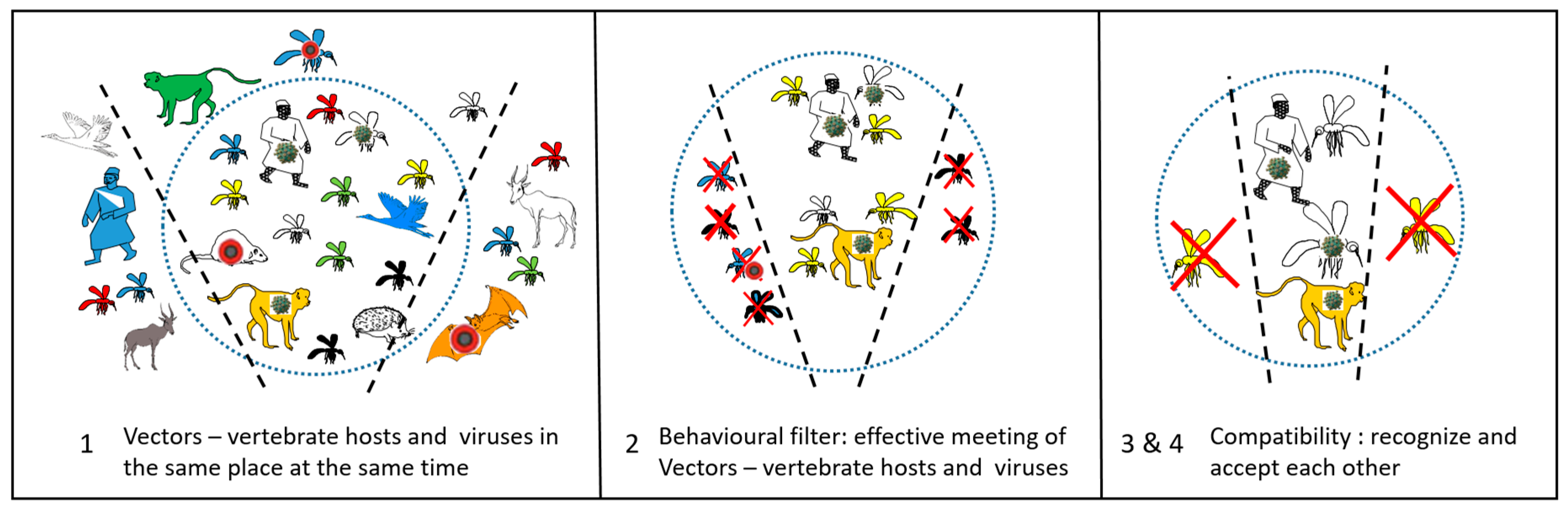
4. The Necessary Conditions

The history of yellow fever leads us to the consideration of the factors necessary, but not always sufficient, for a mosquito species that was initially of little interest to humans, to become a public health problem and an enemy to fight.
First of all, the mosquito, or to be precise, a given population of a given species of mosquito, must be biologically able to transmit the virus. This ability is called vectorial capacity, which includes vector competence (i.e., the ability of a mosquito to become infected after ingestion of an infected blood meal and later transmit the virus via its saliva). Both terms, vectorial capacity and vector competence, have been formalized since MacDonald [29], summarized by Cohuet and coll. [30], and include the following parameters:
  • The vector–host ratio (i.e. the vector density in relation to vertebrate host): m (the mosquito abundance);
  • The human feeding rate: the number of human bites per mosquito, per day: a (mosquito - human contact);
  • The daily survival rate (i.e. the probability of a mosquito surviving each day): p (mosquito longevity);
  • The extrinsic development time, the time necessary for viruses to complete development from ingestion in midgut to the saliva: n;
  • The infectiousness of the mosquito to the vertebrate host: b (largely dependent on virus titre in saliva)
  • The susceptibility of the vertebrate host to the virus (e.g., immune state, age, health, etc): c;
  • The vertebrate host infectious period: 1/r (how long the virus titre in the vertebrate remains at a level needed to infect a mosquito);
Knowing the values of these parameters makes it possible to calculate the basic reproductive rate of the virus, R0.
R0 = (ma2 × pn / − ln p) × bc × 1/r
R0 is the total number of cases derived from one infective case that the mosquito population would distribute to vertebrate hosts. R0 must equal at least 1 for the disease to persist or spread. For values less than 1, the disease will go extinct.
However, while we can write the simple equation above, in reality the parameters (m, a, p, n, b, c, r) are themselves complex, being dependent on many other factors. For just the mosquito, a non-exhaustive list includes mosquito genotype, mosquito microbiome including viruses, integrated RNA virus sequences in mosquito genomes, predation, competition at larval and adult stage, previous exposure to related viruses, etc. [31,32]. Moreover, it is not yet known which receptors in the cells of the stomach and salivary glands of mosquitoes are involved in the entry and exit of flaviviruses, like yellow fever virus [33].
Just this intrinsic biology of the mosquito is not sufficient to make it a vector. Its environment may, or may not, allow this vector potential to manifest itself. According to Euzet and Combes [33], the specificity of the vector–pathogen interaction passes through four stages, which they named encounter and compatibility filters (Figure 2). These four stages are: (1) to co-occur in space and time, (2) to meet each other (behavior), (3) to recognize each other (receptors), and (4) to accept each other (immunity).
All these conditions are rarely met, and it is therefore understandable why being a vector is an exception. However, considering the very large number of mosquito species and large number of viruses, while we are presently facing only a limited number of dangerous cycles, this must be a very small fraction of thousands, if not millions, of other potential cycles that have failed. The understanding of the current efficient cycles allows us to conceive possible future cycles.
We can hypothesize that only a few species in the genera Aedes, Haemagogus, and Sabethes have the capacity to replicate and transmit yellow fever virus to primates, i.e. (1) possess the receptors for cellular penetration of the virus, (2) have insufficient innate immunity to suppress virus replication, (3) live in the same environments as viremic vertebrates (monkeys or humans), (4) take blood from these vertebrates, and (5) survive long enough to retransmit the virus.

5. Aedes albopictus: From Local to Global Concern

Aedes albopictus became widely known only over the last 50 years, when it was gradually discovered on all continents outside its native Asia and caused a chikungunya pandemic [10,11,35,36]. A total of more than 40 viruses can be transmitted naturally or experimentally by Ae. albopictus [36]. Experimentally, it will take blood from many vertebrate species [37]. It also lives longer than Aedes aegypti [38]. Aedes albopictus therefore perfectly fulfils the necessary conditions in terms of biology and events to be an excellent vector of virus to humans.
Aedes albopictus is considered to be phylogeographically native to Southeast Asia [10]. This assumption is based on the fact that this mosquito is present everywhere in the forested areas of this region and that many species close to the Albopictus subgroup, member of the Scutellaris group, are present in Southeast Asia. However, the notoriety of Ae. albopictus, compared to relatives, comes from the fact that it has moved out of its area of origin, becoming worldwide in 50 years, adapting perfectly to urbanization, temperate climates, and international transport, as well as being involved in several dengue and chikungunya epidemics.
In tropical Southeast Asia, the five morphologically closely related species of the Albopictus subgroup live in forests (Aedes novalbopictus, Aedes patriciae, Aedes seatoi, Aedes subalbopictus and Aedes pseudalbopictus) and lay their eggs in tree holes and bamboo stumps [39,40]. These species are likely to be vectors of arboviruses to vertebrates from which they take blood. These forest-confined viruses are largely unknown, but may emerge through increased contact with humans or domestic animals (see below). They could then be transmitted by domestic vectors, such as Ae. aegypti and Ae. albopictus, in a pattern similar to the emergence of the yellow fever virus in Africa from forests to villages [12].
In Laos, Ae. albopictus is reported from deep natural forest [41], rubber forest and secondary forest [42], as well as in towns and villages. In contrast, in neighbouring countries, such as in Cambodia, this mosquito was found along hundreds of meters of forest edge, whereas Ae. pseudalbopictus, is found in deeper natural forests. Aedes albopictus is frequently found in cities such as the capital, Phnom Penh (Boyer pers. com.). Similarly, in Malaysia, Ae. albopictus is rare in forests [43], and in Yunnan Province, China, Ae. albopictus is sometimes more abundant than Ae. pseudalbopictus in bamboo forests, but is often absent from deep forests [40].
Outside Asia, Ae. albopictus populations display highly variable success in invading the deep forest. In Brazil, where Ae. albopictus was observed for the first time in the 1980s, Pereira dos Santos et al. found that Ae. albopictus was able to enter degraded forest in the Manaus region, up to 750 metres from the edge [44]. In Gabon, where Ae. albopictus was first reported in 2007, it was captured 12 years later in the Lopé natural forest, several kilometres from villages or clearings (Paupy pers. com.).
In both its native range Asia and recently invaded South America and Africa, Ae. albopictus has retained its ancestral capacity to colonize forest environments, laying eggs in natural pools of water and taking blood from nondomestic vertebrates. It is logical to think that under these conditions, Ae. albopictus populations would take blood from vertebrates carrying as yet unknown viruses, such as from monkeys, terrestrial mammals, birds or reptiles. If these forest-breeding Ae. albopictus are able to replicate these viruses, and then retransmit them to humans, they would then be excellent bridge vectors, allowing the emergence of forest viruses hitherto confined to sylvatic Aedes-vertebrates cycles. We can speculate that this is most likely to occur in Asia. In Africa, Ae. aegypti has already brought many, perhaps most, human adapted viruses from the forest to human habitats, such as yellow fever, dengue, chikungunya, and Zika viruses [13].
Mogi et al. [40], suggest that the spreading of Ae. albopictus from its original tropical forest region was possible following evolution from an ancestral wild species, due to adaptation to man-made habitats and then migration with humans to temperate climate regions, where Ae. albopictus developed a winter diapause. In most introduced localities, it probably encountered only limited competition from native mosquitoes and when it did, Ae. albopictus proved to be a robust competitor, e.g., with Ae. aegypti [45]. It is likely that the ancestral wild species was already a vector of forest vertebrate arboviruses, for example, dengue-like flaviviruses, or chikungunya-like alphaviruses, which are monkey viruses [46].
From the foregoing, it appears that Ae. albopictus, while being ancestrally a forest-breeding mosquito like its close relatives, among these relatives it is the most closely adapted to forest margins (ecotone), the transition from forest to degraded or secondary forests, open grasslands or scrub. Aedes aegypti in Africa is similar [47]. This may have pre-adapted these two Aedes, among all their congeners in Asia and Africa, to come into contact with human settlements and thus to adapt to this new niche, human settlements.
From regions recently colonized by Ae. albopictus, “modern” populations, highly adapted to the urban environment and able to transmit viruses such as chikungunya, Zika and dengue, spread to all continents. It is likely that invasive populations re-invaded regions where ancestral populations already existed, such as in Asia and the Indian Ocean. These ancestral populations then found themselves in unfavourable competition and modern Ae. albopictus replaced them [48].
It is not too difficult to predict the epidemiological future of Ae. albopictus. Its geographical distribution will increase, especially in temperate regions, its control by insecticides will be more difficult due to the emergence of resistance, and human pathogenic viruses will adapt to this new vector, increasing its efficiency to transmit. Ae. albopictus-vectored epidemics are almost certain to increase. It could be responsible for the epidemics of yellow fever, or of currently unknown viruses transmitted by forest edge Aedes mosquitoes of South American, South East Asia or Central Africa. A major unknown for Ae. albopictus’ impact in temperate regions is whether arbovirus replication at lower temperatures is selected to be rapid enough to reach saliva before the female dies.

6. Scenarios for the Future: The Worst Doesn’t Always Happen

To quote Charles Nicolle [1], it is certain that new epidemics will appear. Mosquitoes and viruses are poised to ambush humans following changes in vector ecosystems [49]. The process of establishment may be abrupt and rapid, as was the recent Zika pandemic with Ae. aegypti as vector, or more gradual, going through adapting mechanisms, as was the case with the emergence of the West Nile virus in several temperate cities, particularly in the USA [50].
The optimistic view, but not very realistic, is that the worst is already behind us. Urban environments, where the majority of the humans now live, are already colonized by Ae. aegypti, Ae albopictus, and/or Culex pipiens, which transmit or can transmit known viruses of tropical or equatorial origin: dengue, chikungunya, Zika, yellow fever, West Nile, as well as, at least potentially, many other tropical viruses like Japanese encephalitis and Rift Valley fever, with variable success depending on species and environments [13]. However, this optimistic view needs to be tempered. With global changes (environment, climate, demography, movements), the distribution areas of some of these human adapted vectors and viruses will continue to expand and we will see a steady increase of epidemics of viruses, including in temperate regions. While we cannot predict details of time and place, we know it will happen, and we can prepare for it.
A more pessimistic view is that the reservoir of unknown forest viruses, not presently affecting humans, but potentially transmissible to domestic animals and humans, is immense in Asia, Central and South America, and Africa. More than 500 arboviruses have so far been identified and described, but this is only the tip of the iceberg. From about 1930–1980, the Rockefeller Foundation sponsored a repository for viruses from field-collected arthropods, primarily mosquitoes, that reached more than 4000 isolates of insect-specific viruses or arboviruses, the vast majority of which are undescribed [51], and even this collection is far from complete. Consequently, several non-exclusive scenarios for the emergence of new human viral epidemics are possible:
(1)
Via primate-biting bridge vectors such as Ae. albopictus from forest edges in South America, Africa and Asia, forest Aedes of the Albopictus group in South East Asia, Haemagogus in South America, Stegomyia from forest galleries in Africa. In South America, Haemagogus and Sabethes are likely to transmit any new potential human viruses among primates and, as displayed by yellow fever, if these viruses are capable of transitioning to transmission in human habitats, it is likely to have already occurred.
(2)
By misfortune, as happened with the establishment of a sylvan cycle of yellow fever in Central and South America 500 years ago, new viruses may appear in transmission cycles that are heretofore unknown. The increase in trade and travel, and the establishment of invasive species (such as Ae. albopictus, Ae. koreicus, and Ae. japonicus, and even Ae. aegypti in Europe) suggest that this risk should not be overlooked.
(3)
Via increased human contacts with wild cycles due to deforestation and irrational forest exploitation. These ecological modifications favour the emergence of viruses from forest edges, and then to the human environment.
(4)
Via zoonotic cycles. For example, many viruses (West Nile, Japanese encephalitis, St Louis encephalitis virus, Murray valley, usutu) which are bird viruses, can be transmitted from birds (or mammals such as pigs) to humans, via vectors taking blood meals from both birds and humans, such as Cx. tritaeniorhynchus or Cx. pipiens, Cx. quinquefasciatus. It is very likely that some of these viruses, presently confined to wild cycles, will emerge in the coming years somewhere in the world, as a result of socio-ecological changes. Their shift from endemic to epidemic will be facilitated by close contact between humans and vertebrate hosts (urban commensal birds or rodents, farm animals).
(5)
By opening an ecological niche in urbanized areas. It is conceivable that local effective vector control by way of elimination of Ae. aegypti or Ae. albopictus would open their ecological niche in some areas, allowing a new species, such as Ae. malayensis in South-East Asia, already recognized as a vector of dengue and chikungunya viruses, to begin to colonize even closer to human populations. This scenario has not happened yet. Aedes albopictus may have replaced Ae. aegypti or vice versa, but for the moment no third species, with high vectorial capacity, has occupied their niches.
(6)
Via the evolution of viruses already known, but which have not yet found the conditions for emerging and spreading. Genetic changes in virus strains could lead to better adaptation to new vectors and a better transmissibility, as happened with the chikungunya virus and Ae. albopictus [52]. Viruses may also evolve resistance to drugs, when any are used, or human immune defences. Given the short generation time, large population size, and high mutation rate of RNA viruses (like yellow fever, dengue, and most pathogenic arboviruses), virus adaptation to efficient transmission by a human-preferring mosquito is rapid. That is, the virus more readily adapts to the mosquito (and vertebrate host), not the mosquito to the virus [53].
Among the hundreds of wild animal viruses, several viruses are regularly cited as worthy of surveillance. This is the case of the Mayaro alphavirus, closely related to the chickungunya virus. It is a South American virus of mammals, including monkeys, mainly transmitted by Haemagogus mosquitoes, and which could adapt to urban Ae. aegypti or Ae. albopictus, or to some other anthrophilic Culicidae. However, these other mosquitoes are considered poor vectors [54,55]. In Africa, the Spondweni virus is genetically close to the Zika virus, which places it on the list of viruses to keep an eye on. While it is indeed pathogenic to humans, very few human cases have been detected; it has no known domestic cycle. It is poorly transmitted experimentally by Aedes (Stegomyia), being efficiently transmitted by zoophilic mosquitoes (Aedes circumluteolus, Mansonia africana), but this situation could change [56]. Indeed, this virus has already been found in Haiti [57] in Cx. quinquefasciatus, a human-biting mosquito recorded in all tropical regions.

7. Conclusions

On a final note of optimism, it is expected that new outbreaks can be detected more quickly, thanks to surveillance and warning systems that have improved greatly in recent decades and thanks to the development of efficient, rapid and less costly diagnostic techniques. Moreover, not all emergences will find favourable environments, including encounter and compatibility filters, and are likely to quickly die out. For public health authorities to succeed in limiting new outbreaks, The International Health Regulations, published in 2005 by WHO [58], provide a framework and obligations for member states, and give hope that new epidemics could be brought under control through early detection, efficient vector control, vaccination when available, and other public health measures. While worst case scenarios should be planned for, they seldom actually arise. However, they happen regularly and the recent Covid-19 pandemic, which was not caused by an arbovirus, reminds us that, although we are aware that this can happen, we are not always properly prepared.

Author Contributions

Both authors, D.F. and J.R.P. wrote the paper. All authors have read and agree to the published version of the manuscript.

Funding

Publication fees were covered by Institut de Recherche pour le Développement (IRD).

Acknowledgments

We thank Poliana da Silva Lemos, for permission to use Figure 1 on phylogeny of Haemagogus janthinomys, Christophe Paupy for useful discussion and non-published data, and Sébastien Boyer for non-published data from Cambodia.

Conflicts of Interest

The authors declare no conflict of interest.

References

  1. Nicolle, C. Destin des Maladies Infectieuses; Librairie Félix Alcan: Paris, France, 1933; p. 216. [Google Scholar]
  2. Grmek, M. Les Maladies à L’aube de la Civilisation Occidentale; PAYOT: Paris, France, 1983; p. 527. [Google Scholar]
  3. Weaver, S.C. Prediction and prevention of urban arbovirus epidemics: A challenge for the global virology community. Antivir. Res. 2018, 156, 80–84. [Google Scholar] [CrossRef]
  4. Brady, O.J.; Hay, S.I. The Global Expansion of Dengue: How Aedes aegypti Mosquitoes Enabled the First Pandemic Arbovirus. Ann. Rev. Entomol. 2020, 65, 191–208. [Google Scholar] [CrossRef] [Green Version]
  5. Agramonte, A. An account of Dr. Louis-Daniel Beauperthuy, a pioneer in yellow fever research. Boston Med. Surg. J. 1908, 158, 927–930. [Google Scholar] [CrossRef] [Green Version]
  6. Finlay, C.J. The mosquito hypothetically considered as the agent of transmission of yellow fever. Presented at the Real Academia de Ciencias Medicas, Fısicas y Naturales de La Habana, Havana, Cuba, 14 August 1881. [Google Scholar]
  7. Reed, W.; Carroll, J.; Agramonte, A.; Lazear, J.W. The etiology of yellow fever: A preliminary note. Philad. Med. J. 1900, 6, 790–796. [Google Scholar]
  8. Christophers, S.R. Aedes aegypti (L.), the Yellow Fever Mosquito. In Its Life History, Bionomics, and Structure; Cambridge University Press: New York, NY, USA, 1960. [Google Scholar]
  9. Souza-Neto, J.A.; Powell, J.R.; Bonizzoni, M. Aedes aegypti vector competence studies: A review. Infect. Genet. Evol. 2019, 67, 191–209. [Google Scholar] [CrossRef]
  10. Hawley, A.H. The biology of Aedes albopictus. J. Am. Mosq. Control Assoc. 1988, 1, 1–39. [Google Scholar]
  11. Kotsakiozi, P.; Richardson, J.B.; Pichler, V.; Favia, G.; Martins, A.J.; Urbanelli, S.; Armbruster, P.A.; Caccone, A. Population genomics of the Asian tiger mosquito, Aedes albopictus: Insights into the recent worldwide invasion. Ecol. Evol. 2017, 7, 10143–10157. [Google Scholar] [CrossRef] [Green Version]
  12. WHO. Prevention and Control of Yellow Fever in Africa; World Health Organization: Geneva, Switzerland, 1986; p. 96. [Google Scholar]
  13. Powell, J.R. Mosquito-Borne Human Viral Diseases: Why Aedes aegypti? Am. J. Trop. Med. Hyg. 2018, 98, 1563–1565. [Google Scholar] [CrossRef]
  14. Powell, J.R.; Gloria-Soria, A.; Kotsakiozi, P. Recent history of Aedes aegypti: Vector genomics and epidemiology records. Bioscience 2018, 68, 854–860. [Google Scholar] [CrossRef] [Green Version]
  15. Chippaux, J.; Chippaux, A. Yellow fever in Africa and the Americas: A historical and epidemiological perspective. J. Venom. Anim. Toxins Incl. Trop. Dis. 2018, 24, 20. [Google Scholar] [CrossRef] [Green Version]
  16. de Figueiredo, M.L.; de C Gomes, A.; Amarilla, A.A.; de S Leandro, A.; de S Orrico, A.; de Araujo, R.F.; do S M Castro, J.; Durigon, E.L.; Aquino, V.H.; Figueiredo, L.T. Mosquitoes infected with dengue viruses in Brazil. Virol. J. 2010, 7, 152. [Google Scholar] [CrossRef] [Green Version]
  17. Fernandes, R.S.; Bersot, M.I.; Castro, M.G.; Telleria, E.L.; Ferreira-de-Brito, A.; Raphael, L.M.; Bonaldo, M.C.; Lourenço-de-Oliveira, R. Low vector competence in sylvatic mosquitoes limits Zika virus to initiate an enzootic cycle in South America. Sci. Rep. 2019, 9, 20151. [Google Scholar] [CrossRef] [Green Version]
  18. Figueiredo, L.T.M. Human Urban Arboviruses Can Infect Wild Animals and Jump to Sylvatic Maintenance Cycles in South America. Front. Cell. Infect. Microbiol. 2019, 9, 259. [Google Scholar] [CrossRef] [Green Version]
  19. Jácome, R.; Carrasco-Hernández, R.; Campillo-Balderas, J.A.; López-Vidal, Y.; Lazcano, A.; Wenzel, R.P.; Ponce de León, S. A yellow flag on the horizon: The looming threat of yellow fever to North America. Int. J. Infect. Dis. 2019, 87, 143–150. [Google Scholar] [CrossRef] [Green Version]
  20. McNeill, J.R. Mosquito Empires. In Ecology and War in the Greater Caribbean, 1620–1914; Cambridge University Press: New York, NY, USA, 2010. [Google Scholar]
  21. Soper, F.L. The elimination of urban yellow fever in the Americas through eradication of Aedes aegypti. Am. J. Publ. Health 1963, 53, 7–16. [Google Scholar] [CrossRef]
  22. Webb, J.L., Jr. Aedes aegypti suppression in the Americas: Historical perspectives. Lancet 2016, 388, 556–557. [Google Scholar] [CrossRef]
  23. Couto-Lima, D.; Madec, Y.; Bersot, M.I.; Compos, S.S.; de Albuquerque Motta, M.; Barreto dos Santos, F.; Vazeille, M.; Vasconcelos, P.F.; Lourenco-de-Oliveira, R.; Failloux, A.B. Potential risk of re-emergence of urban transmission of yellow fever virus in Brazil facilitated by competent Aedes populations. Sci. Rep. 2017, 7, 4848. [Google Scholar] [CrossRef]
  24. Moreira-Soto, A.; Torres, M.C.; Lima de Mendonça, M.C.; Mares-Guia, M.A.; Dos Santos Rodrigues, C.D.; Fabri, A.A.; Dos Santos, C.C.; Machado Araújo, E.S.; Fischer, C.; Ribeiro Nogueira, R.M.; et al. Evidence for multiple sylvatic transmission cycles during the 2016–2017 yellow fever virus outbreak, Brazil. Clin. Microbiol. Infect. 2018, 24, 1019. [Google Scholar] [CrossRef] [Green Version]
  25. Lemos, P.S.; Monteiro, H.A.O.; Castro, F.C.; Lima, C.P.S.; Silva, D.E.A.; Vasconcelos, J.M.; Oliveira, L.F.; Silva, S.P.; Cardoso, J.F.; Vianez-Júnior, J.L.S.G.; et al. Caracterização do genoma mitocondrial de Haemagogus janthinomys (Diptera: Culicidae). DNA Mitocondrial 2017, 28, 50–51. [Google Scholar] [CrossRef]
  26. Tabachnick, W.J.; Wallis, G.P.; Aitken, T.H.G.; Miller, B.R.; Amato, G.D.; Lorenz, L.; Powell, J.R.; Beaty, B.R. Oral infection of Aedes aegypti with yellow fever virus: Geographical variation and genetic considerations. Am. J. Trop. Med. Hyg. 1985, 34, 1219–1224. [Google Scholar] [CrossRef]
  27. Wasserman, S.; Tambyah, P.A.; Lim, P.L. Yellow fever cases in Asia: Primed for an epidemic. Int. J. Infect. Dis. 2016, 48, 98–103. [Google Scholar] [CrossRef] [Green Version]
  28. Brey, P.T.; Fontenille, D.; Tang, H. Re-evaluate yellow fever risk in Asia-Pacific region. Nature 2018, 554, 31. [Google Scholar] [CrossRef]
  29. Macdonald, G. The Epidemiology and Control of Malaria; Oxford University Press: Oxford, UK, 1957. [Google Scholar]
  30. Cohuet, A.; Harris, C.; Robert, V.; Fontenille, D. Evolutionary forces on Anopheles: What makes a malaria vector? Trends Parasitol. 2010, 26, 130–136. [Google Scholar] [CrossRef]
  31. Azar, S.R.; Weaver, S.C. Vector competence: What has Zika virus taught us. Viruses 2019, 11, 867. [Google Scholar] [CrossRef] [Green Version]
  32. Houé, V.; Gabiane, G.; Dauga, C.; Suez, M.; Madec, Y.; Mousson, L.; Marconcini, M.; Yen, P.S.; de Lamballerie, X.; Bonizzoni, M.; et al. Evolution and biological significance of flaviviral elements in the genome of the arboviral vector Aedes albopictus. Emerg. Microbes Infect. 2019, 8, 1265–1279. [Google Scholar] [CrossRef] [Green Version]
  33. Smith, D.R. An update on mosquito cell expressed dengue virus receptor proteins. Insect Mol. Biol. 2012, 21, 1–7. [Google Scholar] [CrossRef]
  34. Combes, C. Parasitism: The Ecology and Evolution of Intimate Interactions; University of Chicago Press: Chicago, IL, USA, 2001; p. 728. [Google Scholar]
  35. Paupy, C.; Delatte, H.; Bagny, L.; Corbel, V.; Fontenille, D. Aedes albopictus, an arbovirus vector: From the darkness to the light. Microbes Infect. 2009, 11, 1177–1185. [Google Scholar] [CrossRef]
  36. Reiter, P.; Fontenille, D.; Paupy, C. Aedes albopictusas an epidemic vector of Chikungunya virus: Another emerging problem? Lancet Inf. Dis. 2006, 6, 463–464. [Google Scholar] [CrossRef]
  37. Delatte, H.; Desvars, A.; Bouetard, A.; Bord, S.; Gimonneau, G.; Vourc’h, G.; Fontenille, D. Blood-feeding behavior of Aedes albopictus, vector of chikungunya on La Reunion. Vector Borne Zoonotic Dis. 2008, 8, 25–34. [Google Scholar] [CrossRef] [Green Version]
  38. Brady, O.J.; Johansson, M.A.; Guerra, C.A.; Bhatt, S.; Golding, N.; Pigott, D.M.; Delatte, H.; Grech, M.G.; Leisnham, P.T.; Maciel-de-Freitas, R.; et al. Modelling adult Aedes aegypti and Aedes albopictus survival at different temperatures in laboratory and field settings. Parasites Vectors 2013, 6, 351. [Google Scholar] [CrossRef] [Green Version]
  39. Huang, Y.M. Contributions to the mosquito fauna of Southeast Asia. XIV. The subgenus Stegomyia of Aedes in Southeast Asia. I. The scuterallis group of species. Contrib. Am. Entomol. Inst. 1972, 9, 1–109. [Google Scholar]
  40. Mogi, M.; Armbruster, P.A.; Tuno, N.; Aranda, C.; Yong, H.S. The climate range expansion of Aedes albopictus (Diptera: Culicidae) in Asia inferred from the distribution of Albopictus subgroup species of Aedes (Stegomyia). J. Med. Entomol. 2017, 54, 1615–1625. [Google Scholar] [CrossRef] [PubMed]
  41. Miot, E. Potential of the mosquito Aedes malayensis as an arbovirus vector in South East Asia. Ph.D. Thesis, Sorbonne University, Paris, France, 20 December 2019. [Google Scholar]
  42. Tangena, J.A.; Thammavong, P.; Malaithong, N.; Inthavong, T.; Ouanesamon, P.; Brey, P.T.; Lindsay, S.W. Diversity of mosquitoes (Diptera: Culicidae) attracted to human subjects in rubber plantations, secondary forests, and villages in Luang Prabang Province, northern Lao PDR. J. Med. Entomol. 2017, 54, 1589–1604. [Google Scholar] [CrossRef] [PubMed]
  43. Lee, J.M.; Wasserman, R.J.; Gan, J.Y.; Wilson, R.F.; Rahman, F.; Yek, S.H. Human activities attract harmful mosquitoes in a tropical urban landscape. EcoHealth 2020, 17, 52–63. [Google Scholar] [CrossRef]
  44. Pereira Dos Santos, T.; Roiz, D.; Santos de Abreu, F.V.; Luz, S.L.B.; Santalucia, M.; Jiolle, D.; Santos Neves, M.S.A.; Simard, F.; Lourenço-de-Oliveira, R.; Paupy, C. Potential of Aedes albopictus as a bridge vector for enzootic pathogens at the urban-forest interface in Brazil. Emerg. Microbes Infect. 2018, 7, 191. [Google Scholar] [CrossRef] [Green Version]
  45. Lounibos, L.P.H. Invasions by Insect Vectors of Human Disease. Ann. Rev. Entomol. 2002, 47, 233–266. [Google Scholar] [CrossRef]
  46. Pandit, P.S.; Doyle, M.M.; Smart, K.M.; Young, C.C.W.; Drape, G.W.; Johnson, C.K. Predicting wildlife reservoirs and global vulnerability to zoonotic Flaviviruses. Nat. Commun. 2018, 9, 5425. [Google Scholar] [CrossRef]
  47. Lounibos, L.P. Habitat segregation among African treehole mosquitoes. Ecol. Entomol. 1981, 6, 129–154. [Google Scholar] [CrossRef]
  48. Delatte, H.; Bagny, L.; Brengue, C.; Bouetard, A.; Paupy, C.; Fontenille, D. The invaders: Phylogeography of dengue and chikungunya viruses Aedes vectors, on the South West islands of the Indian Ocean. Infect. Genet. Evol. 2011, 11, 1769–1781. [Google Scholar] [CrossRef]
  49. Gould, E.; Pettersson, J.; Higgs, S.; Charrel, R.; de Lamballerie, X. Emerging arboviruses: Why today? One Health 2017, 4, 1–13. [Google Scholar] [CrossRef]
  50. Hadfield, J.; Brito, A.F.; Swetnam, D.M.; Vogels, C.B.F.; Tokarz, R.E.; Andersen, K.G.; Smith, R.C.; Bedford, T.; Grubaugh, N.D. Twenty years of West Nile virus spread and evolution in the Americas visualized by Nextstrain. PLoS Pathog. 2019, 15, e1008042. [Google Scholar] [CrossRef] [PubMed] [Green Version]
  51. Murphy, F.A.; Calisher, C.H.; Tesh, R.B.; Walker, D.H. In memoriam: Robert Ellis Shope (1929–2004). Emerg. Infect. Dis. 2004, 10, 762–765. [Google Scholar] [CrossRef]
  52. Schuffenecker, I.; Iteman, I.; Michault, A.; Murri, S.; Frangeul, L.; Vaney, M.C.; Lavenir, R.; Pardigon, N.; Reynes, J.M.; Pettinelli, F.; et al. Genome Microevolution of Chikungunya Viruses Causing the Indian Ocean Outbreak. PLoS Med. 2006, 3, e263. [Google Scholar] [CrossRef] [Green Version]
  53. Powell, J.R. An evolutionary perspective on vector-borne diseases. Front. Gent. 2019, 10, 1266. [Google Scholar] [CrossRef] [PubMed]
  54. Abad-Franch, F.; Grimmer, G.H.; de Paula, V.S.; Figueiredo, L.T.; Braga, W.S.M.; Luz, S.L.B. Mayaro Virus Infection in Amazonia: A Multimodel Inference Approach to Risk Factor Assessment. PLoS Negl. Trop. Dis. 2012, 6, e1846. [Google Scholar] [CrossRef] [PubMed]
  55. Pezzi, L.; A Diallo, M.; Rosa-Freitas, M.G.; Vega-Rua, A.; Ng, L.F.P.; Boyer, S.; Drexler, J.F.; Vasilakis, N.; Lourenço-de-Oliveira, R.; Weaver, S.C.; et al. GloPID-R report on chikungunya, o’nyong-nyong and Mayaro virus, part 5: Entomological aspects. Antivir. Res. 2020, 174, 104670. [Google Scholar] [CrossRef] [PubMed]
  56. Haddow, A.D.; Nasar, F.; Guzman, H.; Ponlawat, A.; Jarman, R.G.; Tesh, R.B.; Weaver, S.C. Genetic Characterization of Spondweni and Zika Viruses and Susceptibility of Geographically Distinct Strains of Aedes aegypti, Aedes albopictus and Culex quinquefasciatus (Diptera: Culicidae) to Spondweni Virus. PLoS Negl. Trop. Dis. 2016, 10, e0005083. [Google Scholar] [CrossRef] [Green Version]
  57. White, S.K.; Lednicky, J.A.; Okech, B.A.; Morris, J.G., Jr.; Dunford, J.C. Spondweni Virus in Field-Caught Culex quinquefasciatus Mosquitoes, Haiti, 2016. Emerg. Infect. Dis. 2018, 24, 1765–1767. [Google Scholar] [CrossRef] [Green Version]
  58. WHO. International Health Regulations, 3rd ed.; World Health Organization: Geneva, Switzerland, 2005; p. 74. [Google Scholar]
Figure 1. Phylogenetic tree built by the method of maximumlLikelihood, from sequences of mitochondrial genes COX1, COX2, NAD4, NAD5 and CYOB of Haemagogus janthinomys and other species of Diptera, from da Silva Lemos et al. 2017 [25]. The bootstrap values are represented in each node.
Pathogens 09 00265 g001
Figure 2. Encounter and compatibility filters, from Euzet and Combes [34].
Pathogens 09 00265 g002

