Article

Comprehensive Genome Analysis on the Novel Species Sphingomonas panacis DCY99T Reveals Insights into Iron Tolerance of Ginseng

1
College of Life Science, Kyung Hee University, Yongin 16710, Korea
2
School of Energy and Chemical Engineering, Ulsan National Institute of Science and Technology (UNIST), Ulsan 44919, Korea
3
Department of Food Science and Biotechnology, Sejong University, Seoul 05006, Korea
4
School of Biological Sciences, Ulsan National Institute of Science and Technology (UNIST), Ulsan 44919, Korea
5
Korean Genomics Industrialization and Commercialization Center, Ulsan National Institute of Science and Technology (UNIST), Ulsan 44919, Korea
*
Authors to whom correspondence should be addressed.
These authors contributed equally to this work.
Int. J. Mol. Sci. 2020, 21(6), 2019; https://doi.org/10.3390/ijms21062019
Submission received: 3 February 2020 / Revised: 11 March 2020 / Accepted: 13 March 2020 / Published: 16 March 2020
(This article belongs to the Special Issue Microbial Systems and Synthetic Biology)

Abstract

:
Plant growth-promoting rhizobacteria play vital roles not only in plant growth, but also in reducing biotic/abiotic stress. Sphingomonas panacis DCY99T is isolated from soil and root of Panax ginseng with rusty root disease, characterized by raised reddish-brown root and this is seriously affects ginseng cultivation. To investigate the relationship between 159 sequenced Sphingomonas strains, pan-genome analysis was carried out, which suggested genomic diversity of the Sphingomonas genus. Comparative analysis of S. panacis DCY99T with Sphingomonas sp. LK11 revealed plant growth-promoting potential of S. panacis DCY99T through indole acetic acid production, phosphate solubilizing, and antifungal abilities. Detailed genomic analysis has shown that S. panacis DCY99T contain various heavy metals resistance genes in its genome and the plasmid. Functional analysis with Sphingomonas paucimobilis EPA505 predicted that S. panacis DCY99T possess genes for degradation of polyaromatic hydrocarbon and phenolic compounds in rusty-ginseng root. Interestingly, when primed ginseng with S. panacis DCY99T during high concentration of iron exposure, iron stress of ginseng was suppressed. In order to detect S. panacis DCY99T in soil, biomarker was designed using spt gene. This study brings new insights into the role of S. panacis DCY99T as a microbial inoculant to protect ginseng plants against rusty root disease.

1. Introduction

Heavy metal exposure is a major threat to plant and environmental resources worldwide. The growth of industrialization has increased the accumulation of heavy metals in the environment resulting in adverse effects to the soil and crop productivity [1]. Although iron (Fe) is considered as an essential micronutrient for plant growth, metabolism, and development [2], optimum concentration of soils ranges from 0.2% to 55% (20,000 to 550,000 mg/kg) respectively [3]. Thus high concentrations of iron in soil can be detrimental to the yield and growth of plant [4,5]. P. ginseng has been widely used in Asian countries as a traditional medicine for thousands of years. Ginseng is a perennial plant, which grows for more than 6 years in humus-enriched soil at a slightly acidic pH (pH 5.0-6.0). Under this acidic environment, heavy metals are readily accessible to ginseng roots [6]. The healthy ginseng root is well documented as possessing various beneficial pharmacological properties including anti-cancer, anti-emetic, anti-oxidative, and anti-angiogenic effects [7,8,9,10]. The pharmaceutical properties of ginseng are attributed to its increased accumulation of secondary metabolites, including ginsenosides. Not only prolonging ginseng cultivation increase ginsenoside accumulation but also cause ginseng rusty root disease [11].
Although pathological mechanism of rusty root disease has not been clearly elucidated, some studies suggest that rusty root corresponds to the occurrence of physiological stress. Whereas some studies have linked the amassment of phenolic compounds on the ginseng root surface as a cause of rusty root [11,12,13], another study demonstrated that deposition of iron compounds may be the cause of the disease [5]. However, few studies have examined the soil characteristics linked to the rusty root disease, which is one of the key factors known to affect plant growth. Numerous studies indicates a possible link between rusty root and ginseng soil properties [14], where it was demonstrated that ferrous iron may be one of the foremost factors that cause rusty root disease. Another study showed that active reducing organic substances released under high moisture conditions can activate iron oxides leading to the accumulation of divalent iron. This evidence indicates that the deposition of Fe (II) iron in the ginseng root epidermis might induce the rusty root condition [14,15]. A number of studies also revealed that ginseng root quality is affected by pathogenic bacterial and fungal infection, resulting in reddish-brown to orange-brown discolored regions of the plant root [16]. Therefore, a better understanding of rhizosphere microbial properties in rusty root conditions can contribute to an increased ginseng yield by preventing rusty root conditions under iron stress [17].
In plant cultivation, microbial population is known to play a vital role in increasing or reducing yield [18,19] where specific bacterial populations have been chosen as fertilizers and pesticides to promote plant growth and yield. Microbes that help plant growth are known as plant growth-promoting bacteria. Plant growth-promoting bacteria can stimulate plant tolerance against heavy metal stress by upregulating expression of heavy metal stress-related genes [20]. In addition, the application of plant growth-promoting bacteria in bioremediation has played a significant role in the large scale removal of heavy metal pollutants from soil [21]. Several reports confirmed that certain types of plant growth-promoting bacteria exhibited metal tolerance properties, including Variovorax, Methylobacterium, Burkholderia, Okibacterium, Rhodococcus, Microbacterium, Sphingomonas, Curtobacterium, Serratia, Pseudomonas, Ralstonia, Staphylococcus, Bacillus, Arthrobacter, Paenibacillus, Chryseobacterium, and Leifsonia [22,23,24,25,26,27,28]. Among these, little is known about their iron-resistance and functional role in ginseng cultivation against iron toxicity.
In our research, newly isolated rhizobacterium from soil and root of rusty-ginseng is presented, as S. panacis DCY99T. Sphingomonas species are Gram-negative lipopolysaccharide (LPS)-free bacteria that possess glycosphingolipids instead of LPS. These bacteria inhabit not only the soil but diverse environment. In previous studies, various Sphingomonas species have been studied to exhibit traits for promoting plant growth. For example, S. sp. LK11 and Sphingomonas paucimobilis ZJSH1 are known to produce phytohormones such as gibberellins and indole acetic acid and to solubilize inorganic phosphate in soil [29,30]. S. paucimobilis EPA505 is known to degrade poly aromatic hydrocarbons [31,32]. Herein, the bacterium S. panacis DCY99T will be phenotypically and genotypically characterized. Through genome-scale analysis of Sphingomonas strains, we investigate plant growth promoting property of S. panacis during abiotic/biotic stress of ginseng plants.

2. Results

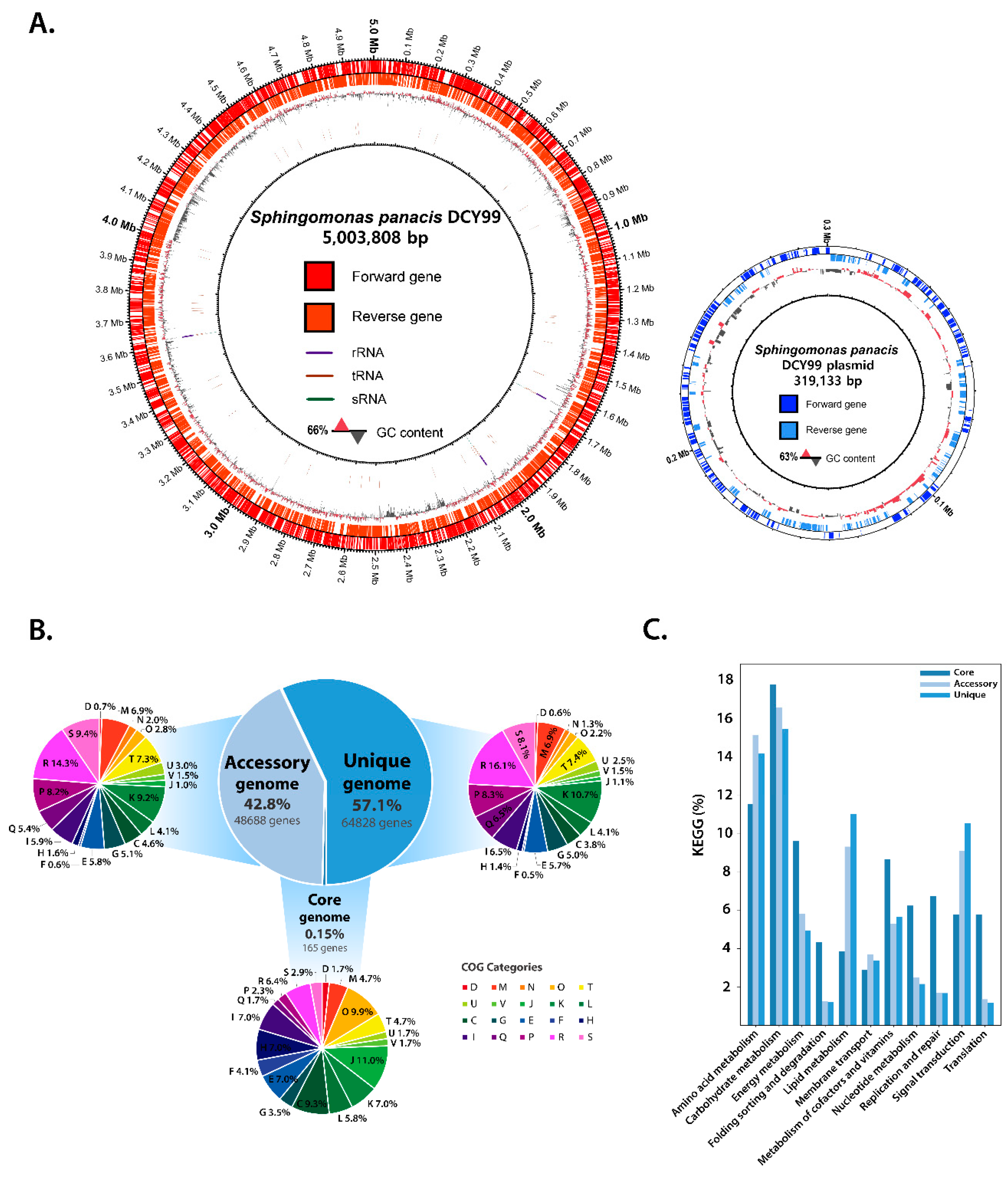
2.1. Reconstruction of the S. panacis DCY99T Genome

The completed genome sequence of S. panacis DCY99T was reconstructed using PacBio SMRT sequencing technology as described in the previous report [33]. The genome consists of a single circular chromosome of 5,003,808 bp with a GC content of 65.66% (Figure 1A), which is characteristic of most Sphingomonas strains (60% to 68%). A total of 4,572 genes was predicted, including 62 RNA genes (tRNA:50, rRNA:9, ncRNA:3). There is a single circular plasmid of 319,133 bp with a GC content of 62.71% with a predicted 300 CDSs.

2.2. Pan-Genome and Functional Genome Analysis with the 159 Sphingomonas Genomes

In order to better understand physiological properties of Sphingomonas for heavy metals resistance and growth-promoting effect on ginseng, pan-genome analysis was performed with the newly sequenced genome of S. panacis DCY99T and other public genomic sequences of Sphingomonas. A total of 159 Sphingomonas genomes including the one for strain DCY99T were analyzed. The resulting pan-genome was further divided by functional characterization of the core and accessory/unique genes (Figure 1B). Unlike the pan-genome of the other bacteria [34], the core-genome, which all of Sphingomonas strains share, has only 0.15% of the pan-genome (165 genes), suggesting the genomic diversity of this genus. Functional analysis showed that the core-genome of Sphingomonas include genes for translation (J: 11%), post-translational modification/chaperones and protein turnover (O: 9.9%), and energy production/and conversion (C: 9.3%). These results indicate that gene functions for protein synthesis and modification are highly conserved in Sphingomonas, suggesting the functional importance of genes in the core-genome. These genes include: rpl genes (rplA, rplI, rplK, rplL, rplM) and rpsA, dnaK, clpP, sucA that encode ribosomal proteins, molecular chaperone, ATP- dependent protease, succinyl-CoA synthetase respectively.
The accessory-genome, which covers 42.8% of the pan-genome (48,688 genes) include genes for transcription (K: 9.2%), inorganic ion transport metabolism (P: 8.2%), signal transduction mechanisms (T: 7.4%), and cell wall/membrane biogenesis (M: 6.9%). Genes encode for transcription and signal transduction mechanisms are commonly found in the core- or accessory-genomes of other bacteria. However, cell wall/membrane biogenesis ranking higher in the accessory-genome is of note for the Sphingomonas pan-genome. This is because Sphingomonas has been known for its unique type of lipid, sphingolipid [35]. Thus, higher conservation of genes for membrane biogenesis requires further investigation. In addition, two functional groups, general function prediction (R: 14.3%) and function unknown (S: 9.4%), had relatively higher occupancy in the accessory-genome, indicating yet a larger fraction of Sphingomonas pan-genome is still elusive for its function. The unique-genome which is not shared by more than two Sphingomonas strains, was the largest in the pan-genome. It accounted for 57.1% of the pan-genome (64,828 genes). Similar to the accessory-genome, transcription (K: 10.7%) and inorganic ion transport metabolism (P: 8.3%) were the most relevant functional groups for the unique-genome. In the transcription category of unique-genome, various transcriptional regulators of COG were found, multidrug efflux regulator (AcrR family) [36], regulator related to virulence, metabolism, quorum sensing and motility (LysR family) [37], L-arabinose operons regulator (AraC family) [38], sigma factor for extracytoplasmic function protein family such as metal resistance (RpoE) [39], multiple antibiotic resistance regulator (MarR family) [40,41], and regulator of biofilm formation (CsgD family) [42]. Subsequently, in the inorganic ion transport metabolism category, outer membrane receptor proteins related iron transport, arylsulfatase A and cation diffusion facilitator proteins were found (Supplementary Table S3). Iron transport and cation diffusion facilitator proteins, which show large number of matching genes, suggest the properties of Sphingomonas species found in various soil that contain different concentrations of heavy metals. Additionally, arylsulfatase A is involved in the metabolism of organic sulfur and sulfate. Sulfonated polysaccharides is abundant in the marine environment and function as a structural component in marine plants such as Gelidium and Gracilaria [43,44]. Some of Sphingomonas strains are found in the seawater and encode arylsulfatase to utilize these polysaccharides [45]. The diversity of these transcriptional regulators and membrane transport proteins is a possible explanation for the distinct lifestyles of the Sphingomonas strains. Consequently, these results suggests that the 159 Sphingomonas species can live and adapt to diverse environment niches such as bacterial growth plates [46], seawater [47,48,49], alpine soil [50], spacecraft [51], and arctic-lichen [52]. The differences in these environmental conditions support a rationale as to why the core-genome is so small among these strains. As such, the number of genomes in the core-pan plot increased. While the pan-genome gradually increased, the core-genome remained at a very low fraction (Supplementary Figure S2).
Subsequent analysis of the corresponding core-genome with the KEGG database revealed that enzymatic genes in the core carbon metabolic pathways such as citric acid cycle (TCA cycle) and glycolysis/gluconeogenesis are highly conserved for all Sphingomonas strains. For the accessory- and unique-genomes, genes encoding two-component signal transduction systems were frequently found, presumably for the ability to adapt and evolve in various or changing environmental stimuli. In bacteria with several dozen two-component systems, such as Escherichia coli, it is clear that the majority of the systems arose by gene duplication from one or more ancestral systems and evolved to acquire new environmental input signals and output specificities [53]. Similarly, genes involved in both fatty acid biosynthesis and degradation pathways were found in the accessory- and unique-genomes. These genes are important for Sphingomonas species to produce sphingolipids as a defining characteristic of this particular genus. Indeed, different Sphingomonas species produce different lipid compositions including major and minor lipids, which come from different environments and the use of different carbon sources [54]. Therefore, it might be concluded that Sphingomonas species use diverse enzymes in lipid metabolism based on the specific niche environment of each strain (Figure 1C). To visualize the evolutionary relationship between the strains of Sphingomonas, the pan-phylogenetic tree is constructed (Supplementary Figure S3).

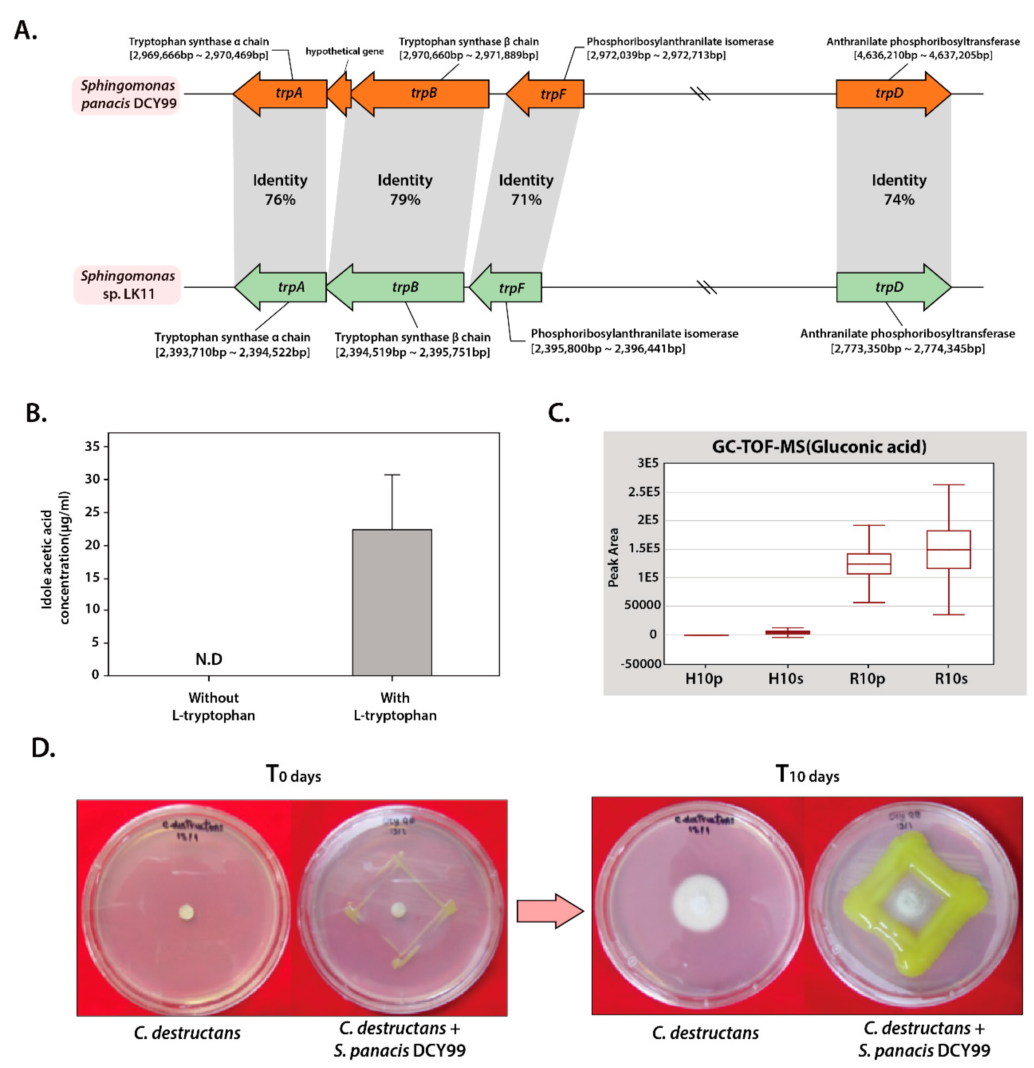
2.3. Plant Growth-Promoting Potential of S. panacis DCY99T

From the genomic sequence of S. panacis DCY99T isolated from the roots of P. ginseng Meyer was analyzed about enhancing plant growth, production of indole acetic acid, solubilization of phosphate, and antifungal activity. Previous studies have shown that only a few Sphingomonas strains are known to promote plant growth through production of phytohormones [55]. Indole acetic acid is the most common phytohormone produced by bacteria and plants. It is one of the auxin class derived from indole which induces cell elongation, cell division and also participates in various gene regulations [56].
S. panacis DCY99T was expected that it would show the ability to produce indole acetic acid because several bacteria that colonize the rhizosphere and plant roots can synthesize indole acetic acid from L-tryptophan [57]. However, complete indole acetic acid biosynthesis pathway was not found in strain DCY99T genomic sequence annotation data. Subsequently, comparative analysis using genome data of S. sp. LK11, which is known to produce phytohormones such as gibberellins (GAs) and indole acetic acid, was performed [29]. Likewise, complete genes for indole acetic acid biosynthesis were not found in strain LK11. However, the presence of tryptophan biosynthesis gene cluster (trpA, trpB, and trpD), phosphoribosylanthranilate isomerase (trpF; locus AWL63_13430), indole pyruvate ferredoxin oxidoreductase (locus AWL63_18305), and indole-3-glycerol phosphate synthase (locus AWL63_21155) which involve in branchpoint of indole acetic acid biosynthesis pathway shows the potential for indole acetic acid production [58]. Previous studies have indicated that the presence of tryptophan-related genes in rhizobacteria is associated with the production of indole acetic acid [59,60]. Thus, the comparative analysis of the tryptophan biosynthesis gene cluster of strain LK11 and DCY99T was carried out. The trpA,B,D,F genes from S. panacis DCY99T were found to have over 70% sequence identity when compared to S. sp. LK11 (Figure 2A). Based on the presence of indole acetic acid-related genes with high sequence identity, it is possible that S. panacis DCY99T might produce indole acetic acid to promote plant growth, which is similar to strain LK11. Experimental validation of indole acetic acid producing ability of strain DCY99T was performed by growing S. panacis DCY99T culture in media with and without additional L-tryptophan. In media with L-tryptophan, S. panacis DCY99T produced 22.4 ± 8.37 µg/mL of indole acetic acid whereas in media without L-tryptophan, presence of indole acetic acid was not determined (Figure 2B). As a result, our analysis is confirmed that S. panacis DCY99T produces indole acetic acid in a tryptophan-dependent manner.
Phosphorus (P) is one of the major essential macronutrients for biological growth and development of plants. Inorganic phosphate is solubilized by plant growth-promoting rhizobacteria, allowing an easy uptake by plant roots [61]. It has been reported that the major mechanism of inorganic phosphate solubilization is the action of organic acids such as gluconic acid and 2-ketogluconic acid synthesized by soil microorganisms [61,62,63,64]. Detailed genomic analysis of S. panacis DCY99T was revealed the presence of genes encoding for a complete gluconic acid synthesis pathway, which consists of pyrroloquinoline quinone-dependent glucose dehydrogenase (locus AWL63_22775) and gluconolactonase (locus AWL63_16415) (Supplementary Figure S4A). Interestingly, GC-TOF-MS result of P. ginseng Meyer is indicated that gluconic acid concentration is found much higher in rusty- ginseng epithelium and root where S. panacis DCY99T was isolated (Figure 3C). Also, when compared with other organic acids, gluconic acid is present at high concentrations in rusty ginseng. In addition, this strain is found to encode the conserved pst (phosphate-specific transport) operon that is shown to be responsible for the uptake inorganic phosphate in E. coli, and two-component signal transduction system consisting of phoB/phoR for phosphate transport [29] (Table 1). Phosphate-solubilizing ability of strain DCY99T was tested on Pikovskaya medium. In S. panacis DCY99T, clear halo regions around colonies which indicate its ability in solubilizing inorganic phosphate was observed.
In our previous study, S. panacis DCY99T was reported to exhibit a great antibacterial effect on a rice pathogenic bacteria Xoo PXO99Az [33]. The antifungal activity of S. panacis DCY99T against Cylindrocarpon destructans that cause rusty symptom and root-rot disease of American and Korean ginseng was evaluated [13]. Interestingly, S. panacis DCY99T is showed that effectively inhibited the growth of C. destructans, which highlights its potential may use for inducing antifungal activity against pathogenic C. destructans in ginseng (Figure 2D).

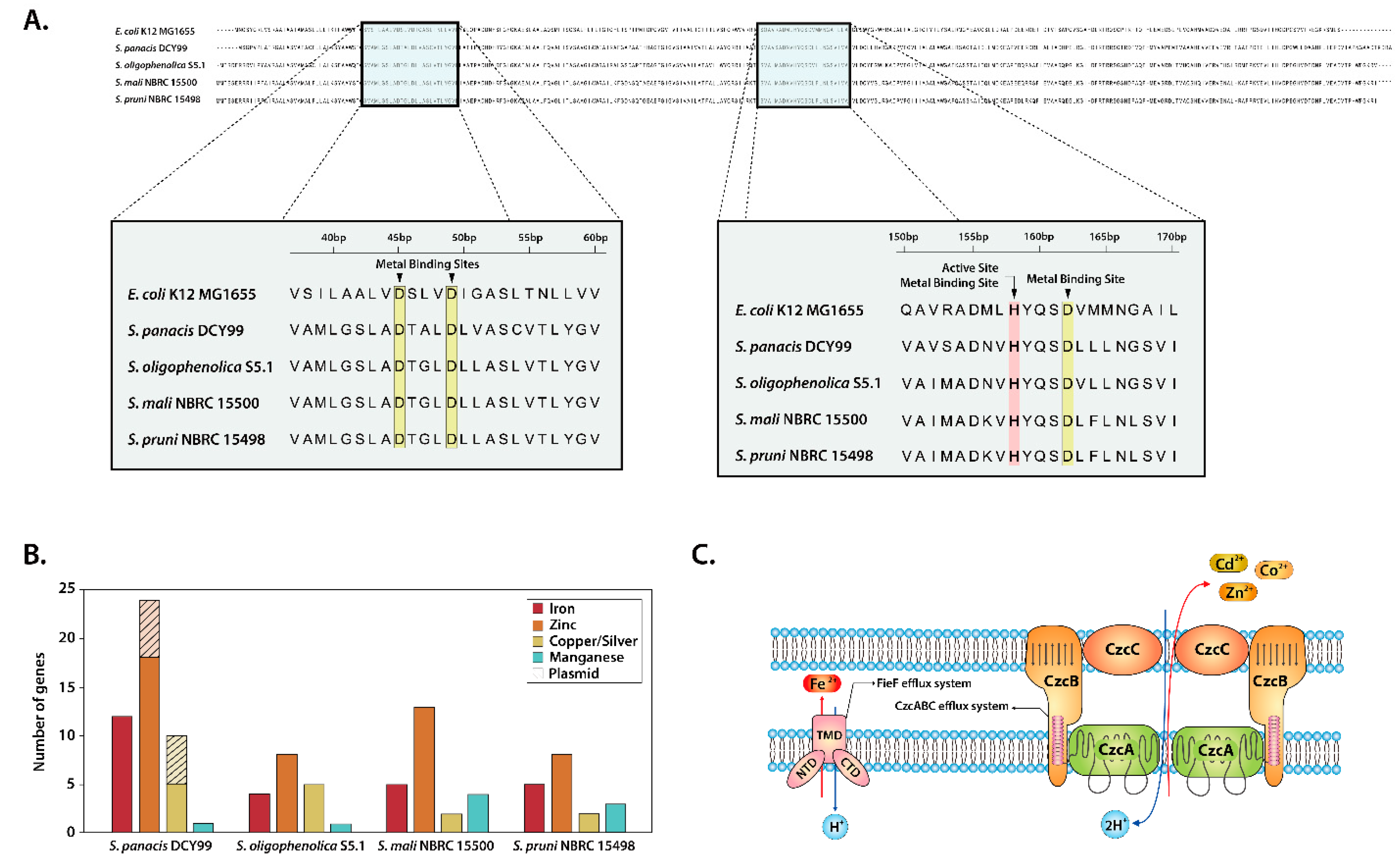
2.4. Heavy Metal Resistance of S. panacis DCY99T

Metal ions such as zinc, iron, cobalt and manganese are essential for almost all aspects of microbial metabolism. However, excess amount of heavy metals can be toxic to bacteria. Thus, many bacteria have developed efflux mechanisms or resistance to heavy metals [65,66]. Our comparative analysis of S. panacis DCY99T with three closely related strains of Sphingomonas was revealed the presence of heavy metal related genes and compared with our previous experiment [67]. Despite being one of essential metal, excess iron may affect growth, morphology and metabolism, thus microbes have four groups of different efflux systems [68]. Gene encodes for cation diffusion facilitator fieF (also named as yiiP) in E. coli which survive at high concentration of iron, was also identified in these four strains of Sphingomonas. Evidence suggests that fieF might be associated with the iron tolerance and full resistance to iron intoxication (Figure 3C) [69,70,71]. fieF genes from four strains of Sphingomonas and E. coli K-12 MG1655 were aligned. The analysis of alignment showed three aspartate residues and one histidine residue being highly conserved in the active site and metal-binding sites (Figure 3A). The conservation of significant amino acid residues in the fieF transport protein substantiates our previous analysis that the ability of Sphingomonas to grow under high iron conditions (~2 mM) (Table 2). Additionally, the czc efflux system was found in strain DCY99T genome and plasmid, consists of czcA, czcB, czcC and czcD. The czc operon was reported to confer resistance to cobalt, zinc and cadmium. While there are several czc efflux system models for different bacteria, S. panacis DCY99T was shown the highest identity with the model of S. sp. LK11 [29,72,73]. This czc model exists as a dimmer and efflux three type of heavy metals (Figure 3C).
In addition, copper resistance genes were identified in S. panacis DCY99T genome. Multi-copper oxidase encoded in S. panacis DCY99T is an important gene for copper resistance in Gram-negative bacteria such as E. coli, thus multi-copper oxidase is considered as a marker gene for copper-resistant bacteria [29,74,75]. Moreover, copper-transporting P-type ATPase was identified in the genome analysis, which especially found in bacteria that are resistant to copper [76,77]. Altogether, the number of genes involved in the uptake and resistance of each heavy metal in four strains of Sphingomonas genomes was quantified (Supplementary Table S2). Except genes related to manganese, S. panacis DCY99T showed the largest number of heavy metal related genes (Figure 3B). Interestingly, strain DCY99T genome is revealed to encode various genes including iron uptake regulator fur, iron transporter feoB, and several types of ferrichrome-iron receptors, ferric siderophore transporters, compared to other three strains. Furthermore, its plasmid encodes several heavy metal resistance genes such as related zinc, copper and silver, which was different from the other three strains of Sphingomonas. In particular, complete cus operon (cusC; locus AWL63_23365, cusB; locus AWL63_23370, cusA; locus AWL63_23375, cusF; locus AWL63_23380) was found in strain S. panacis DCY99T plasmid. Copper/silver resistant Gram-negative bacteria have cus operon which are expressed under high concentration of copper/silver. In E. coli, the cusCBA efflux complex confers resistance to toxic copper and silver [78,79,80]. The presence of several heavy metals related genes implies that S. panacis DCY99T may survive in the polluted soil with high concentration of iron, which associates to rusty root disease in ginseng.

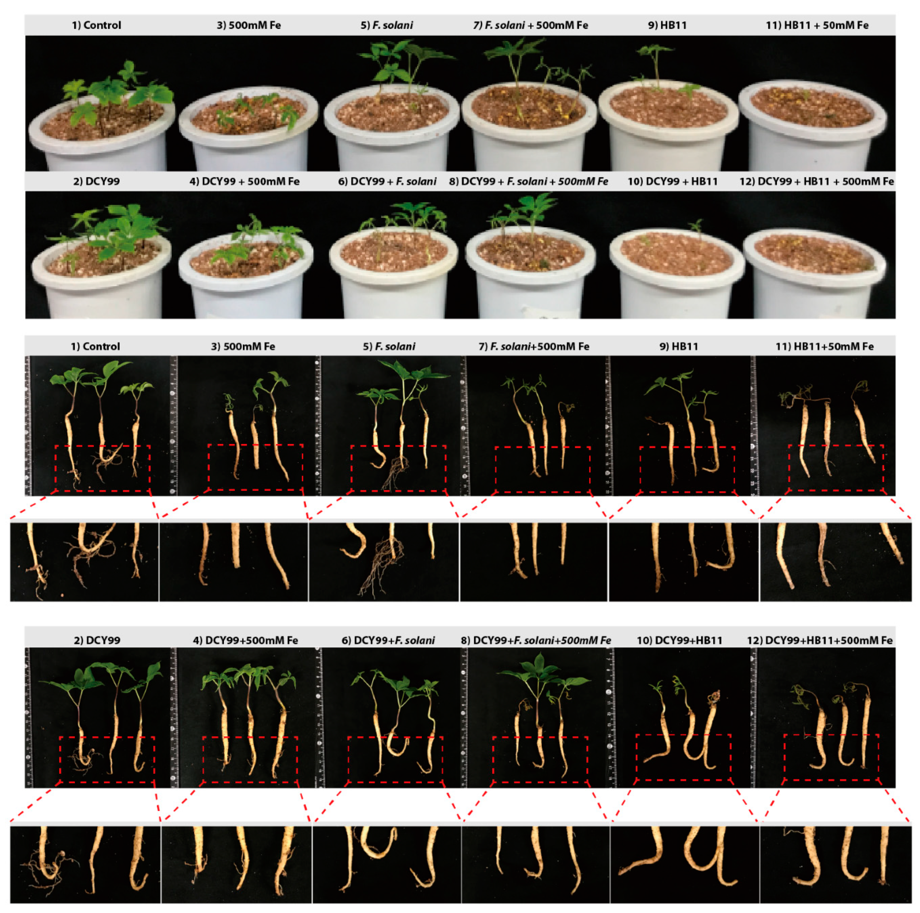
2.5. Improvement of P. ginseng Meyer Growth with S. panacis DCY99T under Biotic/Abiotic Stress

Rusty root of ginseng is related to the ecological conditions of the soil and metabolism of the plant caused by excessive iron in the soil, which are mainly composed of organic complex iron species [14]. The compatibility of S. panacis DCY99T with P. ginseng Meyer under abiotic/biotic stress was tested to determine the effect of strain DCY99T in high Fe (II) concentration conditions and fungal infection. Prior to the abiotic/biotic stress experiment, the pathogenicity of S. panacis DCY99T in ginseng was confirmed. P. ginseng was dipped in S. panacis DCY99T suspensions to inoculate at the root surface of ginseng. Then, ginseng seedlings were cultivated on sterilized artificial soils. The compatibility of strain DCY99T with P. ginseng using a pot assay was observed by morphological alterations after 7 days of bacterial inoculation. As a result, S. panacis DCY99T showed symbiosis with no significant effect on the growth of P. ginseng during the pot assay (Figure 4). Accordingly, the experiment was performed to confirm S. panacis DCY99T can affect ginseng growth against iron stress and fungi infection using a pot assay. During the high concentrations of iron exposure (500 mM), the aerial parts of ginseng plants were visibly stressed, and root color changed. P. ginseng suffered morphological and physiological changes that were noted in previous studies [5,11]. Interestingly, the morphology of seedlings inoculated with S. panacis DCY99T was drastically different. S. panacis DCY99T showed significantly suppressed iron stress of ginseng, leading to increased root, shoot and leaf development when compared with seedlings without S. panacis DCY99T. The compatibility of Fusarium solani with P. ginseng was assessed by investigating morphological alterations in fungal infection. F. solani is known to cause root rot or rusty root disease in ginseng [81]. No disease symptoms were observed in seedling with F. solani. However, S. panacis DCY99T showed to suppress iron stress of ginseng even if inoculated with F. solani. To further confirm the antifungal activity of S. panacis DCY99T in ginseng, highly aggressive Ilyonectria mors-panacis HB11 which is known to cause aggressive root rot was used in biotic stress [82]. Seedlings with I. mors-panacis HB11 were inoculated, after 7 days their root morphologies were slightly altered. In particular, brown discoloration was observed at the tip of the tap root and the foliar growth of plants was significantly affected. Seedling with I. mors-panacis HB11 and S. panacis DCY99T showed a slightly more stressed to aerial parts of ginseng. Furthermore, growth of ginseng under iron stress with I. mors-panacis HB11 was fully inhibited as those seedlings displayed discoloration on their stems and leaves. In addition, their roots were softened with brown discoloration. For seedlings with I. mors-panacis HB11 and S. panacis DCY99T treatment group were morphologically similar to seedlings with I. mors-panacis HB11. However, the morphology of roots inoculated with S. panacis DCY99T was not affected by stresses as much when compared with the I. mors-panacis HB11 infected seedlings. Consequently, S. panacis DCY99T was confirmed to protect seedlings against iron toxicity, thereby preventing developmental inhibition of ginseng under abiotic stressful environment. However, S. panacis DCY99T did not fully protect seedlings from fungal infection.

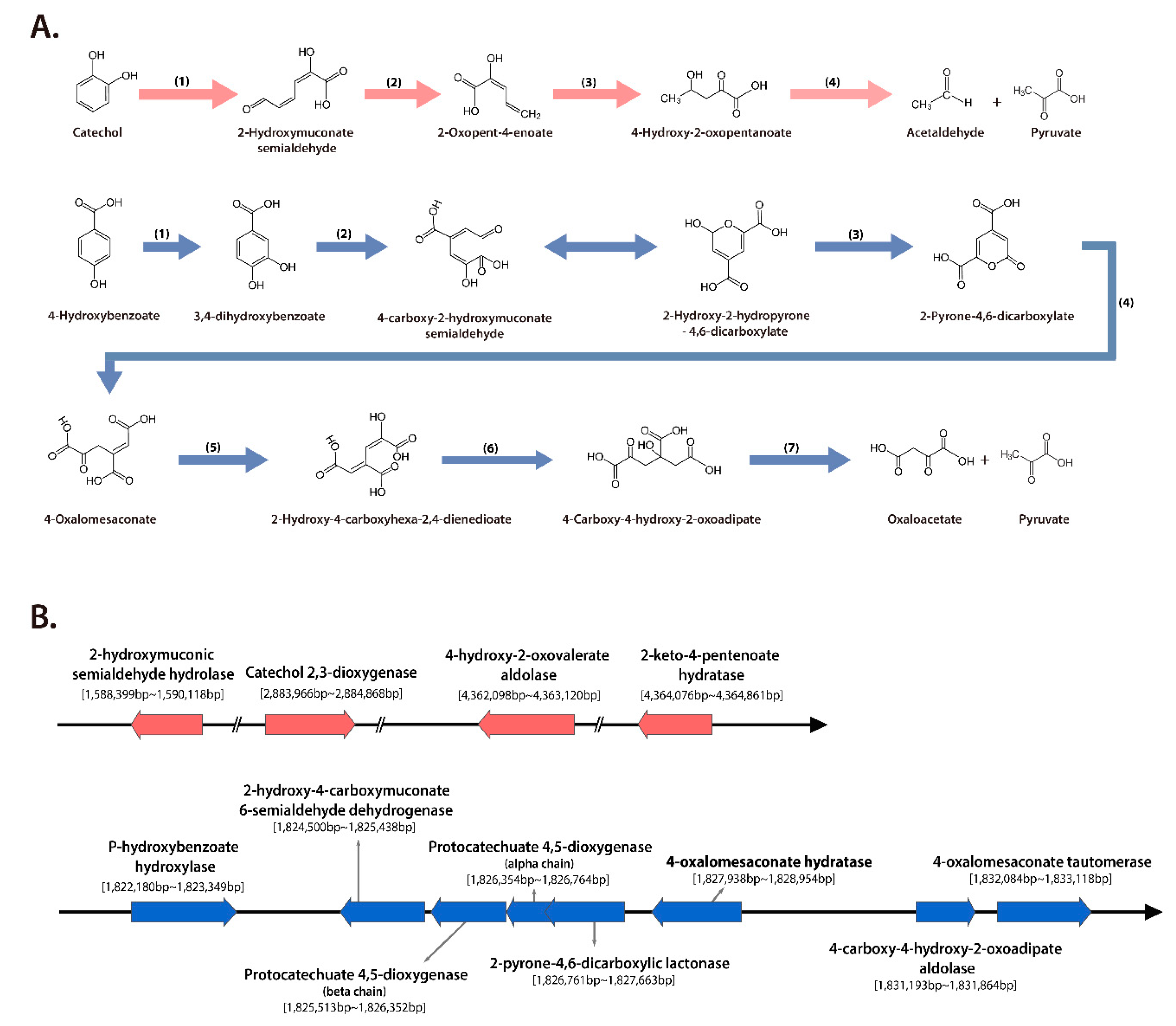
2.6. Phenolic Compounds and 3-hydroxybutanoic Acid Degradation by S. panacis DCY99T

S. panacis DCY99T was found in contaminated soil containing such as benzene, phenolic compounds, and polyaromatic hydrocarbons (PAHs). This strain may have a phenolic compounds-degrading ability when grown on minimal medium containing phenolic compounds. When cultivating ginseng, phenolic compounds such as 4-hydroxybenzoate, vanillin, syringic acid, vanillic acid, coumaric acid, ferulic acid, cinnamic acid, salicylic acid, and benzoic acid were accumulated in the surrounding soil or on the surface of ginseng [83]. The total phenolic content of rusty-ginseng was significantly higher (53%) than that of healthy ginseng roots [84]. The increase of phenolic compounds is consistent with the assumption that phenolics are a part of the cause of rusty-ginseng [84]. Some of Sphingomonas strains have been shown to grow on polyaromatic compounds, also strains from different environments have shown the ability to degrade low and high molecular weight polycyclic aromatic hydrocarbons [85,86,87,88,89].
Functional analysis based on protein homologies was performed using S. panacis DCY99T with S. paucimobilis EPA505. Strain EPA505 was reported to have degraded various polyaromatic hydrocarbons (PAHs) and phenolic compounds [31,32,89]. Sphingomonas species use catechol meta cleavage pathway that is one of the significant pathways for PAHs degradation (Figure 5A). S. paucimobilis EPA505 encodes catechol meta cleavage pathway and 4-hydroxybenzene degradation genes (Table 3) [90]. Genes of degradation pathways of strain EPA505 were used to confirm the presence of protein homologies in the strain DCY99T. S. panacis DCY99T was confirmed to have complete catechol cleavage pathway genes (locus tag AWL63_07130, AWL63_13025, AWL63_19815, AWL63_19825) in the genome (Figure 5B). Additionally, conserved pathway of degrading 4-hydroxybenzoate was observed. These eight genes are clustered around 1,822,180 bp to 1,833,118 bp on the chromosome (locus tag AWL63_08150, AWL63_08160, AWL63_08165, AWL63_08170, AWL63_08175, AWL63_08180, AWL63_08195, AWL63_08200) and may confer degradation ability from 4-hydroxybenzoate to pyruvate and oxaloacetate. It should be noted that these phenolic compounds degradation genes can be used by S. panacis DCY99T to survive on rusty-ginseng surfaces where phenolic compounds are typically present in high concentrations.
Additionally, GC-TOF-MS results of 3-hydroxybutanoic acid was found to be 10 times higher in rusty-ginseng than in healthy ginseng soil (Supplementary Figure S4B). The pathway for degrading 3-hydroxybutanoic acid to two acetyl CoA was found in strain DCY99T genome. This pathway consists of three genes and various Acetyl-CoA acetyltransferase (locus tag AWL63_01810, AWL63_07945, AWL63_07945) (Supplementary Figure S4C). S. panacis DCY99T was found in soils that caused ginseng damage due to the phenolic compounds that accumulated in the soil when the medicinal root crops such as ginseng or bellflower were grown for a long time [67]. These results suggest that S. panacis DCY99T can survive in the presence of phenolic compounds and will act as a basic indicator for measuring cultivated soil conditions to prevent ginseng root disease.

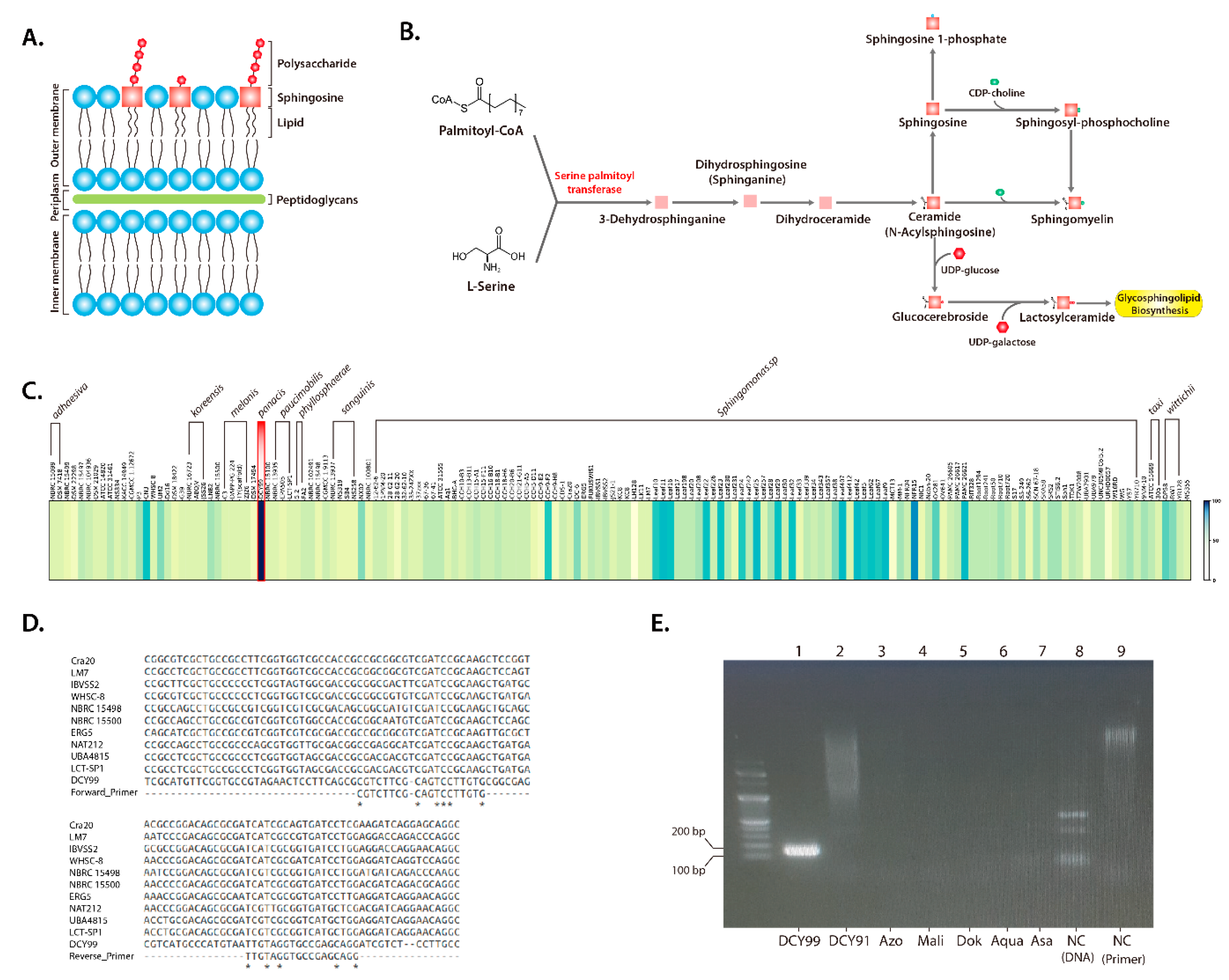
2.7. Design of a S. panacis DCY99T Biomarker

Most microorganisms do not produce sphingolipids [91]; however, Sphingomonas species are capable of producing sphingolipids in lieu of LPS. As the name implies, Sphingomonas species are gram-negative lipopolysaccharide (LPS)-free bacteria that utilize glycosphingolipids instead of LPS. Glycosphingolipids have remarkable structural similarities with LPS and its hydrophobic characteristics [35] (Figure 6A). This is one of reasons why Sphingomonas species are specialized in sphingolipid metabolism [92]. The major sphingolipid biosynthesis pathway, summarized from the KEGG database, is illustrated (Figure 6B). This pathway synthesizes sphingolipid and glycosphingolipid from Palmitoyl-CoA and L-Serine. The heat map is represented the conservation level of spt gene (EC:2.3.1.50), the initial step of sphingolipid synthesis is the condensation reaction from cytosolic serine and palmitoyl-CoA to 3-dehydrosphinganine. Result of conservation analysis based on DNA sequences, the spt gene was observed with >80% identity (Figure 6C). Additionally, the spt gene was verified that it is found in almost Sphingomonas strains. Therefore, the spt gene was first considered as a key gene for the sphingolipid pathway to design a RT-PCR primer for a S. panacis DCY99T biomarker.
A set of spt detection RT-PCR primers was designed from sequence alignment with the spt sequences of closely related Sphingomonas strains and S. panacis DCY99T (Table 4), resulting in primer sequences specific only to strain DCY99T (Figure 6D). The result of RT-PCR using the spt primer for 7 Sphingomonas strains including S. panacis DCY99T confirmed the specificity of strain DCY99T spt detection primers (Figure 6E). The strain-specific detection marker using the spt gene was verified by the 149 bp PCR product that distinguishes S. panacis DCY99T from other closely related Sphingomonas strains.

3. Discussion

The purpose of this study was to determine whether S. panacis DCY99T can promote ginseng growth under high concentrations of iron. In general Sphingomonas are gram-negative bacteria with more than 103 species [90]. Morphologically, they can be identified as a yellow-pigmented, non-motile, and non-fermentative rod [93]. The outer membrane of all Sphingomonas species utilize glycosphingolipids instead of lipopolysaccharides [35]. They are known to confer abiotic stress tolerance in plants [29] and biodegradation of polyaromatic hydrocarbon contaminants [86,87,88,89,94]. Plant growth-promoting rhizobacteria play an essential role in the development and growth of plants [95], in which Sphingomonas is often the microbe of interest for their plant growth-promoting activity in P. ginseng. Herein, S. panacis DCY99T isolated from rusty root ginseng and soil, showed significant growth-promoting effects on P. ginseng under high concentrations of iron stress.
The growth-promoting strain DCY99T was phenotypically and phylotypically identified as Sphingomonas panacis. Furthermore, strain DCY99T is the most closely related to S. oligophenolica, followed by S. asaccharolytica, S. mali, S. cynarae, S. pruni, and S. glacialis [67]. For a better understanding of rhizobacteria, it is crucial to analyze their genome properties and related genes responsible for adaptation to specific conditions. The genome of strain DCY99T consists of 5.0 Mb chromosomal genome and 315 kb plasmid with 4,810 genes predicted. Of which, 1,725 (36%) genes had their functions classified as hypothetical proteins which have their existence predicted, but there is a lack of experimental characterization [96]. As compared to other bacteria such as E. coli and Salmonella (<10%), this is a high number of hypothetical proteins. These obstacles make genomic analysis difficult but suggest the potential of discovering novel proteins with function of interest [97,98,99].
To clarify the relationship between Sphingomonas strains, the pan-genome analysis using BPGA (Bacterial Pan Genome Analysis) was carried out. BPGA performs pre-processing step to prepare sequence data for clustering. BPGA then runs USEARCH for the fastest clustering using 50% sequence identity cut-off [100]. The USEARCH algorithm is performed in these order: (i) A total of 755,014 genes were assembled from 159 species of Sphingomonas; (ii) Out of 755,014 genes, 113,682 genes were clustered in which each cluster is defined by one sequence, known as the centroid or representative sequence; (iii) The USEARCH algorithm used clusters to match one or more sequences from the total 755,014 genes, every sequence in the cluster must have similarity above a given identity threshold (>50%) with the centroid; (iv) Finally, the pan-genome analysis compiled a set of core genes that are clusters containing 159 sequences, accessory genes present in at least two or more sequences, and unique genes only found in a single sequences. The total number of core genes was 165 (0.15%), which was obviously lower as compared to other studies [34,101]. This difference may be due to the higher number of strains analyzed in this study and there was a rich diversity among these strains, including geographical and environmental diversity. Furthermore, the main concern was the quality of genome data of Sphingomonas downloaded from NCBI might be the reason for lower number of core genes. This is because if just one of the 187 Sphingomonas strains does not have a target gene, the core gene is considered to be an accessory gene. Of the 187 Sphingomonas genomes, there were only 15 complete genomes including strain DCY99T while the rest consist of whole-genome shotgun (WGS) sequencing data that contain several contigs. To confirm this problem, the pan-genome analysis using complete genomes of 15 strains was performed. There was a total of 25,745 centroids defined. Of which, 952 (3.69%) core genes, 15,148 (37.46%) accessory genes and 9,645 (59%) unique genes were identified. The number of core genes for 15 complete genomes increased considerably compared to the result of 159 genomes. As the number of analyzed genomes decreases, the number of core genes increases as expected. However, the number of core genes from 15 Sphingomonas complete genomes was still significantly less than that of other bacteria (Supplementary Table S3). In addition, the distribution of unique genes was similar to the results that used 159 strains. Therefore, 159 genomic data were used since our purpose in the pan-genome analysis was to focus on the diversity of the Sphingomonas genus. In short, these results supported that Sphingomonas can constantly obtain foreign genes in order to adapt to various environments [102].
The growth-promoting mechanism of strain DCY99T was determined to be due to a combination of indole acetic acid production, phosphate solubilization and antifungal activity. Indole acetic acid, the physiologically most active phytohormone in plants, acts as an important signaling molecule in the regulation of plant development [103]. Three main biosynthetic routes involving indole-3-pyruvic acid (IPyA), tryptamine (TAM), and indole-3-acetonitrile (IAN) have been studied in plant-associated bacteria [104]. Genes encoding for indole acetic acid biosynthetic pathways from Pseudomonas savastanoi, Enterobacter cloacae, Azospirillum brasilense were analyzed by USEARCH program to identify homologs in S. panacis DCY99T. However, no indole acetic acid biosynthetic pathways were observed. Therefore, comparative analysis of S. panacis DCY99T and S. sp. LK11 which is known to be a plant growth-promoting bacteria was performed [29]. This analysis supported that S. panacis DCY99T is a strain with great potential in promoting plant growth by producing indole acetic acid from L-tryptophan and solubilizing inorganic phosphate using organic acid such as gluconic acid. Several studies have revealed that ginseng root quality is affected by pathogenic bacteria and fungus [16,105]. Therefore, the ability to compete against pathogenic bacteria and fungus that harm ginseng plants supports a high possibility of plant growth-promoting rhizobacteria traits. Previous study has reported that strain DCY99T shows antibacterial effect on Xoo PXO99Az also known as rice pathogenic bacteria [33]. This study also suggested that S. panacis DCY99T has great antifungal ability against C. destructans that cause rusty symptom and root-rot disease.
Various studies have reported the heavy metal stress tolerance ability of plant growth-promoting bacteria belonging to different genera [29,106,107]. During exposure to heavy metal stress, these bacteria not only sustain their growth but also exhibit certain plant growth-promoting traits like production of phytohormones, suppression of abiotic/biotic stress [29,106,107]. In the presence of high concentrations of iron, the growth of strain DCY99T was found to be sustained, compared to 3 closely related strains based on their 16S rRNA gene sequences [67]. Comprehensive genome analysis of S. panacis DCY99T suggests that this strain has cation diffusion facilitator fieF similar to E. coli K-12 MG1655 and various heavy metal tolerance genes in genome and plasmid.
The assessment of the abiotic/biotic stress of 2-year-old P. ginseng with S. panacis DCY99T showed that the iron stress in ginseng with strain DCY99T was significantly suppressed as compared to ginseng growing under sole iron stress treatment. This was verified by the gradual increase in levels of iron concentration before the inoculation of strain DCY99T. Seedlings were stressed with different concentrations of iron (0, 250, 500, and 1000 mM) based on previous research [15,108]. The growth of ginseng seedlings was completely disturbed when they were exposed to 1000 mM Fe (Supplementary Figure S5A). Symptoms appeared to be consistent with those seen in the plants suffering from iron toxicity where root and shoot growth was inhibited and the plant biomass decreased [11,109,110]. At 500 mM Fe, ginseng seedlings gradually developed yellowing on the leaves to eventually a complete wilting of the foliage. Biomass reduced after iron stress (500 mM). Fresh weight of root and shoot decreased by 37% and 81% whereas dry weight of root and shoot decreased by 35% and 48% respectively (Supplementary Figure S5B). It was established that at 250 mM Fe, no significant difference was observed when compared to the control [108]. Therefore, 500 mM iron was used as the abiotic stress in this study. When the seedlings were primed with strain DCY99T under high concentration of iron, ginseng was conferred resistant of iron from S. panacis DCY99T. However, the mechanism of this phenomenon is unclear. One hypothesis is proposed about S. panacis DCY99T which survives in iron stress condition, produces gluconic acid - a powerful chelating agent to remove heavy metals [111]. It is established that soil microorganisms produce large amounts of gluconic acid when inorganic phosphates are solubilized [112,113]. Therefore, the gluconic acid produced by strain DCY99T may facilitate the growth of ginseng under high iron stress. In our studies, S. panacis DCT99T was reported to have antibacterial and antifungal activity. The antifungal activity of S. panacis DCY99T against I. mors-panacis HB11 and F. solani was validated using in vitro antagonistic test. Although strain DCY99T did not completely protect ginseng seedlings from fungal infection in pot assay, it reduced the morphological change of root. Therefore, S. panacis DCT99T exhibited a good protection of ginseng from high levels of iron, and a partial protection of ginseng against fungal infection.
S. panacis DCY99T can be found from ginseng root to contaminated soil containing benzene, phenolic compounds and polyaromatic hydrocarbons. The analysis of based on protein sequence homologies was proved the presence of PAHs and phenolic compounds degradation pathway in strain S. panacis DCY99T as well as strain S. paucimobilis EPA505. It was reported that strain EPA505 could grow on various PAHs and partially degrade high molecular weight PAHs [31,32,89]. Moreover, it has well-conserved pathways that degrade PAHs and phenolic compounds. Except for catechol 2,3-dioxygenase (EC 1.13.11.2), which has 30% identity, catechol meta cleavage pathway genes showed more than 50% identity. S. panacis DCY99T has shown to have higher identity to the 4-hydroxybenzene degradation pathway than the catechol meta cleavage pathway compared to S. paucimobilis EPA505 (Table 3). These results suggest that S. panacis DCY99T also has a high potentiality of surviving even in the presence of these compounds, also, S. panacis DCY99T is predicted more likely to grow in 4-hydroxybenzene than PAHs.
In our previous study of Sphingomonas species, the phylogenetic analysis through 16S rRNA sequence was conducted [67]. However, S. panacis DCY99T shared the highest 16S rRNA gene sequence identity with S. oligophenolica JCM 12082T (97.32%), followed by S. asaccharolytica KCTC 2825T (96.90%), S. mali KCTC 2826T (96.82%), S. cynarae JCM 17498T (96.76%), S. pruni KCTC 2824T (96.75%), and S. glacialis DSM 22294T (96.45%). This high identity can cause the reliability of the biomarker to be lowered. For these reasons, biomarker for strain DCY99T was constructed using spt gene that key enzyme of sphingolipid biosynthesis. The spt gene is found in all Sphingomonas strains and has more variation between species because identity over species is less than 80%. Our RT-PCR result supports that biomarker using spt gene can be used to detect strain DCY99T with high reliability. Consequently, S. panacis DCY99T will be made an eco-friendly strategy of ginseng cultivation through follow-up studies.

4. Materials and Methods

4.1. Sphingomonas Strains and Genomic Analysis

A total of 187 Sphingomonas genomes were obtained from the NCBI database and used to estimate the genome size/number of DNA coding sequences (CDS) in S. panacis DCY99T. The trend line y = 0.0008x + 737.57 ( = 0.7996) was used to exclude genomes with genes with more than +/− 10% difference (Supplementary Figure S1). The Sphingomonas genome was compared with E. coli K-12 MG1655 rpo genes using USEARCH and we excluded Sphingomonas genome data with 0% coverage of the rpoB and rpoD genes. As a result, only 159 strains were selected for analysis.

4.2. Plant Material and Culture Conditions

Two-year-old P. ginseng Meyer was obtained from the Ginseng Resource Bank, Kyung Hee University. To grow ginseng plants, artificial soil was prepared by mixing vermiculite, perlite, and peat moss at a 3:1:1 volume ratio. The mixed soil was autoclaved at 121 °C for 1 h and then air-dried. The sterilization step was repeated twice on different days. Before culturing the roots, tap water was sterilized to prepare the soil mixture at a 25% v/v ratio that was used to fill trays or pots for ginseng growth. Each tray or pot was placed in a 60 cm × 100 cm (0.6 m2) open cabinet inside a closed controlled chamber. We adjusted the photoperiod to a light:dark (16:8h) cycle using lamps (Philips TLD-RS-FLR32SSEX-D 865K) equal to 9500 lux for each covered area. The temperature was controlled at 25 ± 2 °C, and the moisture level was maintained at 60 ± 5%. Sterilized tap water was sprayed onto the soil surface daily, and watering was conducted once a week using sterilized tap water from a plate beneath the tray/pot.

4.3. S. panacis DCY99T Genome Analysis

The S. panacis DCY99T genome was deposited as SAMN04417200 in BioSample and as CP014168 for the genome and CP014169 for the plasmid in GenBank. Gene annotation revealed 4601 coding sequences (CDSs) using the RAST (Rapid Annotation using Subsystem Technology) server (Supplementary Table S1). The chromosome (CP014168.1) and plasmid (CP014169.1) were separated from the RAST output file and visualized using CIRCOS (Circular Genome Data Visualization).

4.4. Clusters of Orthologous Groups (COG) Analysis

A total of 159 Sphingomonas strains including S. panacis DCY99T were annotated by the RAST (Rapid Annotation using Subsystem Technology) server. The 159 annotated amino acid sequence FASTA files were used for BPGA-1.3 (Bacterial Pan Genome Analysis) analysis. For orthologous clustering of functional genes, the USEARCH clustering algorithm. A 50% sequence identity was used as the cut-off value to generate a Core-Pan plot. For pan-genome functional analysis, COG and KEGG pathway distribution were used. The COG and KEGG IDs were assigned to all representative protein sequences from each orthologous protein cluster based on protein BLAST against reference COG and KEGG databases. Subsequently, the percent frequencies of COG and KEGG categories were calculated for core genes, accessory genes, and unique genes [100]. Then, pan-phylogenetic tree was visualized using dendroscope [114].

4.5. Genome Comparisons

USEARCH program was used for detailed comparative analysis of S. panacis DCY99T with S. sp. LK11, S. paucimobilis EPA505, P. savastanoi, E. cloacae, and A. brasilense. S. sp. LK11 (CP013916.1) and S. paucimobilis EPA505 (NZ_BBJS00000000.1) genome were downloaded from NCBI database. Indole acetic acid pathway genes of P. savastanoi (P06617; Tryptophan 2-monooxygenase, P06618; Indoleacetamide hydrolase), E. cloacae (P23234; Indole-3-pyruvate decarboxylase) and A. brasilense (P51852; Indole-3-pyruvate decarboxylase) were downloaded from Uniport. Genes responsible for plant growth-promoting traits such as PAHs degradation and phenolic compound degradation were constructed to the USEARCH database. Comparative genome alignments were based on results of USEARCH algorithm with constructed database, identification of conserved gene order and construction of gene clusters.

4.6. In Vitro Plant Growth-Promotion and Product Assays

To assess indole acetic acid production, we followed the method as previously described [115], with some modifications for in vitro indole acetic acid production. At first, King B broth was used with and without additional L-tryptophan (3 g/L) [116]. After 6 days of incubation, indole acetic acid production was measured using a colorimetric method (Salkowski reagent). The concentration of indole acetic acid was also prepared to make the standard curve, the absorbance of bacterial supernatant was measured at 540nm for indole acetic acid production, the more different metabolites of indole acetic acid should be analyzed by LC with standards in further study. Qualitative testing of phosphate-solubilizing ability was checked by plate screening methods using a formulated medium [117]. A clear halo region around colonies in the opaque Pikovskaya medium indicated positive results for phosphate solubilization.

4.7. S. panacis DCY99T and P. ginseng Meyer Compatibility under Biotic /Abiotic Stress

Surface-disinfected 2-year-old ginseng roots were used for ginseng pot assays. A ginseng pot assay was started by dipping the ginseng roots in S. panacis DCY99T suspensions at various ODs (indicating variations in CFUs/mL) for 10 min, followed by cultivation in sterilized artificial soil consisting of vermiculite: perlite: peat moss at a 3:1:1 ratio with additional sterilized tap water [25% (v/v)] in pots (11 cm high and 11 cm diameter). We performed for the compatibility of strain DCY99T in 2 years ginseng roots and analyzed priming effect against iron stress. Two-year-old P. ginseng roots were rinsed with tap water and twice rinsed with sterilized DW. Ginseng roots were treated with 500 mM FeCl36H2O infected with F. solani and I. mors-panacis HB11 (KACC 44660) at 25 °C, respectively. We observed symptom development for 7 days after inoculation and then created a symptom severity scale. Each pot contained 5 roots. Each treatment was replicated in three pots.

4.8. GC-TOF-MS Analysis

Freeze-dried ginseng roots were prepared as powder. Then, 10 mg powder was extracted with 1 mL 80% methanol followed by homogenization using a mixer mill at 30 Hz/s for 10 min (Retsch MM400, Haan, Germany). The mixture was centrifuged at 13,000 rpm for 5 min, and the supernatant (100 μL) was vacuum dried using a speed vacuum concentrator (BioTron, Seoul, Korea). The extract was dissolved in 80% methanol (final concentration, 0.2 mg/mL), filtered using a 0.2-μm PTFE filter, and then measured by GC-TOF-MS (Supplementary Table S5). For each root, 10 biological and 3 analytical replicates were analyzed for organic acids including 3-hydroxybutanoic acid.

4.9. Constructing the Heat Map

The sphingolipid pathway genes of alpha-proteobacteria was downloaded from the KEGG and NCBI databases. Homologs for the serine palmitoyl transferase (EC:2.3.1.50) from S. panacis DCY99T was found in the NCBI database. USEARCH was used to compare strain DCY99T with the other 158 Sphingomonas strains and to select orthologs with the highest identity. As a result, Conserved genes were visualized using a heat map made with Python matplotlib.

4.10. Primer Design

For the first screening, 300 bp of the spt gene were collected from NCBI and analyzed for primer possibilities. Two sets of primers were designed and evaluated for species-specific markers (Table 4). After optimization of the annealing temperature range from 50–65 °C, the best PCR conditions were as follows: initial denaturation at 95 °C for 3 min, 30 cycles of 95 °C for 30 s, 50–65 °C for 30 s, and 72 °C for 1 min followed by a final elongation at 72 °C for 5 min. PCR was conducted using 100 ng of S. panacis DCY99T genomic DNA in a 25-μL total reaction volume of Genotech® 2× Green PreMix (Genotech, Daejeon, South Korea). The PCR mix included 1 µL DNA template (100 ng/µL), 1 µL each primer pair, 10 µL Green PreMix, and 7 µL sterile DW for a 20 µL total reaction mix. We verified species-specific primers for S. panacis DCY99T compared to other strains including DNA templates from S. panaciterrae DCY91T, S. mucosissima, S. dokdonensis, S. xinjiangensis, S. faeni, S. aurantiaca, and S. aerolata. Template DNA only and primer only PCR reactions were used for controls.

4.11. Primer Alignment

Nucleotide FASTA files of 159 Sphingomonas strains were aligned using ClustalW based on the spt primer product. The 10 Sphingomonas strains (Sphingomonas sp. Cra20, Sphingomonas sp. LM7, Sphingomonas sp. IBVSS2, Sphingomonas sp. ERG5, Sphingomonas sp. NAT212, Sphingomonas sp. UBA4815, Sphingomonas hengshuiensis WHSC-8, Sphingomonas pruni NBRC 15498, Sphingomonas pruni NBRC 15500, Sphingomonas paucimobilis LCT-SP1), which has notably different sequences, were used.

4.12. Quantification of S. panacis DCY99T Gene Expression

We used cell suspensions with different OD values as templates (1 μL) in a 15-μL total reaction volume of SYBR® Green SensiMix Plus Master Mix (Watford, England) and performed quantitative real-time PCR (qRT-PCR). Amplification, detection, and data analysis were carried out using a CFX 96/Connect Real-Time PCR system (Bio-Rad, Seoul, South Korea). We used the following thermal cycler conditions: 2 min at 95 °C followed by 40 cycles of 95 °C for 30 s, 64 °C for 50 s, and 72 °C for 1 min. The threshold cycle (Ct) was recorded, and the correlation between OD and Ct values is shown as an exponential regression.

Supplementary Materials

The following are available online at https://www.mdpi.com/1422-0067/21/6/2019/s1, Supplementary Figure S1. Scatter blot of genome size between number of coding sequence (CDS) from 159 Sphingomonas genomes. Supplementary Figure S2. Core-pan genome plot of the 159 Sphingomonas strains, Supplementary Figure S3. Phylogenetic tree of the 159 Sphingomonas strains, Supplementary Figure S4. D-gluconic acid biosynthesis and 3-hydroxybutanoic acid degradation pathway from S. panacis DCY99T, Supplementary Figure S5. Pot assessment of iron tolerance in P. ginseng, Supplementary Table S1. Detailed genome annotation of S. panacis DCY99T using RAST, Supplementary Table S2. Detailed heavy metals related genes of four Sphingomonas strains, Supplementary Table S3. Result of BPGA from 159 Sphingomonas strains, Supplementary Table S4. Result of BPGA from 15 Sphingomonas strains, Supplementary Table S5. Result of GC-TOF-MS from healthy and rusty P. ginseng.

Author Contributions

Y.-J.K. and J.Y.P. carried out the experimental procedures, performed a complete analysis, and prepared the original draft of the manuscript. S.R.B. and Y.H. carried out experimental procedures. H.T.L. participated in the visualization. L.K.N. and D.C.Y. carried out review and editing. D.K. coordinated the study and finalized the manuscript. All authors have read and agreed to the published version of the manuscript.

Funding

This research was funded by the “Cooperative Research Program for Agriculture Science and Technology Development (Project No. PJ0128132017)” Rural Development Administration grant, and also supported by a grant from the Basic Science Research Program through the National Research Foundation of Korea funded by the Ministry of Education (2019R1A2C1010428), Republic of Korea.

Conflicts of Interest

The authors declare no conflict of interest.

References

  1. Shahid, M.; Khalid, S.; Abbas, G.; Shahid, N.; Nadeem, M.; Sabir, M.; Aslam, M.; Dumat, C. Heavy metal stress and crop productivity. In Crop Production and Global Environmental Issues; Springer: Berlin/Heidelberg, Germany, 2015; pp. 1–25. [Google Scholar]
  2. Hell, R.; Stephan, U.W. Iron uptake, trafficking and homeostasis in plants. Planta 2003, 216, 541–551. [Google Scholar] [CrossRef]
  3. Bodek, I. Environmental Inorganic Chemistry: Properties, Processes, and Estimation Methods; Pergamon Press: Pergamon, Turkey, 1988. [Google Scholar]
  4. De Dorlodot, S.; Lutts, S.; Bertin, P. Effects of ferrous iron toxicity on the growth and mineral composition of an interspecific rice. J. Plant Nutr. 2005, 28, 1–20. [Google Scholar] [CrossRef]
  5. Zhang, Y.; Wang, Q.; Xu, C.; Sun, H.; Wang, J.; Li, L. Iron (Fe2+)-induced toxicity produces morphological and physiological changes in roots in Panax ginseng grown in hydroponics. Toxicol. Environ. Chem. 2016, 98, 630–637. [Google Scholar] [CrossRef]
  6. Balusamy, S.R.D.; Kim, Y.-J.; Rahimi, S.; Senthil, K.S.; Lee, O.R.; Lee, S.; Yang, D.-C. Transcript pattern of cytochrome P450, antioxidant and ginsenoside biosynthetic pathway genes under heavy metal stress in Panax ginseng Meyer. Bull. Environ. Contam. Toxicol. 2013, 90, 194–202. [Google Scholar] [CrossRef]
  7. Kang, K.S.; Yokozawa, T.; Kim, H.Y.; Park, J.H. Study on the nitric oxide scavenging effects of ginseng and its compounds. J. Agric. Food Chem. 2006, 54, 2558–2562. [Google Scholar] [CrossRef] [PubMed]
  8. Koo, H.-N.; Jeong, H.-J.; Choi, I.-Y.; An, H.-J.; Moon, P.-D.; Kim, S.-J.; Jee, S.-Y.; Um, J.-Y.; Hong, S.-H.; Shin, S.-S. Mountain grown ginseng induces apoptosis in HL-60 cells and its mechanism have little relation with TNF-α production. Am. J. Chin. Med. 2007, 35, 169–182. [Google Scholar] [CrossRef] [PubMed]
  9. Sagar, S.M.; Yance, D.; Wong, R. Natural health products that inhibit angiogenesis: A potential source for investigational new agents to treat cancer—Part 1. Curr. Oncol. 2006, 13, 14. [Google Scholar] [PubMed]
  10. Shin, B.-K.; Kwon, S.W.; Park, J.H. Chemical diversity of ginseng saponins from Panax ginseng. J. Ginseng Res. 2015, 39, 287–298. [Google Scholar] [CrossRef] [Green Version]
  11. Rahman, M.; Punja, Z.K. Biochemistry of ginseng root tissues affected by rusty root symptoms. Plant Physiol. Biochem. 2005, 43, 1103–1114. [Google Scholar] [CrossRef]
  12. Zhou, Y.; Yang, Z.; Gao, L.; Liu, W.; Liu, R.; Zhao, J.; You, J. Changes in element accumulation, phenolic metabolism, and antioxidative enzyme activities in the red-skin roots of Panax ginseng. J. Ginseng Res. 2017, 41, 307–315. [Google Scholar] [CrossRef] [Green Version]
  13. Farh, M.E.-A.; Kim, Y.-J.; Kim, Y.-J.; Yang, D.-C. Cylindrocarpon destructans/Ilyonectria radicicola-species complex: Causative agent of ginseng root-rot disease and rusty symptoms. J. Ginseng Res. 2018, 42, 9–15. [Google Scholar] [CrossRef] [PubMed]
  14. Wang, Y.; Li, Z.; Sun, Y.; Guo, S.; Tian, S.; Liu, Z. Studies on the genesis of ginseng rust spots. J. Ginseng Res. 1997, 21, 69–77. [Google Scholar]
  15. Wang, Q.; Xu, C.; Sun, H.; Ma, L.; Li, L.; Zhang, D.; Zhang, Y. Analysis of the relationship between rusty root incidences and soil properties in Panax ginseng. In Proceedings of IOP Conference Series: Earth and Environmental Science; IOP Publishing: Bristol, UK, 2016; Volume 41, p. 012001. [Google Scholar]
  16. Punja, Z.; Wan, A.; Goswami, R.; Verma, N.; Rahman, M.; Barasubiye, T.; Seifert, K.; Lévesque, C. Diversity of Fusarium species associated with discolored ginseng roots in British Columbia. Can. J. Plant Pathol. 2007, 29, 340–353. [Google Scholar] [CrossRef]
  17. Majeed, A.; Abbasi, M.K.; Hameed, S.; Imran, A.; Rahim, N. Isolation and characterization of plant growth-promoting rhizobacteria from wheat rhizosphere and their effect on plant growth promotion. Front. Microbiol. 2015, 6, 198. [Google Scholar] [CrossRef] [Green Version]
  18. Raza, W.; Ling, N.; Yang, L.; Huang, Q.; Shen, Q. Response of tomato wilt pathogen Ralstonia solanacearum to the volatile organic compounds produced by a biocontrol strain Bacillus amyloliquefaciens SQR-9. Sci. Rep. 2016, 6, 24856. [Google Scholar] [CrossRef] [Green Version]
  19. Raza, W.; Yousaf, S.; Rajer, F.U. Plant growth promoting activity of volatile organic compounds produced by biocontrol strains. Sci. Lett. 2016, 4, 40–43. [Google Scholar]
  20. Sukweenadhi, J.; Kim, Y.-J.; Choi, E.-S.; Koh, S.-C.; Lee, S.-W.; Kim, Y.-J.; Yang, D.C. Paenibacillus yonginensis DCY84T induces changes in Arabidopsis thaliana gene expression against aluminum, drought, and salt stress. Microbiol. Res. 2015, 172, 7–15. [Google Scholar] [CrossRef]
  21. Gouda, S.; Kerry, R.G.; Das, G.; Paramithiotis, S.; Shin, H.-S.; Patra, J.K. Revitalization of plant growth promoting rhizobacteria for sustainable development in agriculture. Microbiol. Res. 2018, 206, 131–140. [Google Scholar] [CrossRef]
  22. Barzanti, R.; Ozino, F.; Bazzicalupo, M.; Gabbrielli, R.; Galardi, F.; Gonnelli, C.; Mengoni, A. Isolation and characterization of endophytic bacteria from the nickel hyperaccumulator plant Alyssum bertolonii. Microb. Ecol. 2007, 53, 306–316. [Google Scholar] [CrossRef]
  23. Belimov, A.; Hontzeas, N.; Safronova, V.; Demchinskaya, S.; Piluzza, G.; Bullitta, S.; Glick, B. Cadmium-tolerant plant growth-promoting bacteria associated with the roots of Indian mustard (Brassica juncea L. Czern.). Soil Biol. Biochem. 2005, 37, 241–250. [Google Scholar] [CrossRef]
  24. Benmalek, Y.; Halouane, A.; Hacene, H.; Fardeau, M.-L. Resistance to heavy metals and bioaccumulation of lead and zinc by Chryseobacterium solincola strain 1YB-R12T isolated from soil. Int. J. Environ. Eng. 2014, 6, 68–77. [Google Scholar] [CrossRef]
  25. Dell’Amico, E.; Cavalca, L.; Andreoni, V. Analysis of rhizobacterial communities in perennial Graminaceae from polluted water meadow soil, and screening of metal-resistant, potentially plant growth-promoting bacteria. FEMS Microbiol. Ecol. 2005, 52, 153–162. [Google Scholar] [CrossRef] [PubMed] [Green Version]
  26. Idris, R.; Trifonova, R.; Puschenreiter, M.; Wenzel, W.W.; Sessitsch, A. Bacterial communities associated with flowering plants of the Ni hyperaccumulator Thlaspi goesingense. Appl. Environ. Microbiol. 2004, 70, 2667–2677. [Google Scholar] [CrossRef] [Green Version]
  27. Jiang, C.-Y.; Sheng, X.-F.; Qian, M.; Wang, Q.-Y. Isolation and characterization of a heavy metal-resistant Burkholderia sp. from heavy metal-contaminated paddy field soil and its potential in promoting plant growth and heavy metal accumulation in metal-polluted soil. Chemosphere 2008, 72, 157–164. [Google Scholar] [CrossRef] [PubMed]
  28. Kuffner, M.; De Maria, S.; Puschenreiter, M.; Fallmann, K.; Wieshammer, G.; Gorfer, M.; Strauss, J.; Rivelli, A.; Sessitsch, A. Culturable bacteria from Zn-and Cd-accumulating Salix caprea with differential effects on plant growth and heavy metal availability. J. Appl. Microbiol. 2010, 108, 1471–1484. [Google Scholar] [CrossRef] [PubMed]
  29. Asaf, S.; Khan, A.L.; Khan, M.A.; Al-Harrasi, A.; Lee, I.-J. Complete genome sequencing and analysis of endophytic Sphingomonas sp. LK11 and its potential in plant growth. 3 Biotech 2018, 8, 389. [Google Scholar] [CrossRef] [Green Version]
  30. Yang, S.; Zhang, X.; Cao, Z.; Zhao, K.; Wang, S.; Chen, M.; Hu, X. Growth-promoting Sphingomonas paucimobilis ZJSH1 associated with D endrobium officinale through phytohormone production and nitrogen fixation. Microb. Biotechnol. 2014, 7, 611–620. [Google Scholar] [CrossRef]
  31. Story, S.; Kline, E.; Hughes, T.; Riley, M.; Hayasaka, S.S. Degradation of aromatic hydrocarbons by Sphingomonas paucimobilis strain EPA505. Arch. Environ. Contam. Toxicol. 2004, 47, 168–176. [Google Scholar] [CrossRef]
  32. Desai, A.M.; Autenrieth, R.L.; Dimitriou-Christidis, P.; McDonald, T.J. Biodegradation kinetics of select polycyclic aromatic hydrocarbon (PAH) mixtures by Sphingomonas paucimobilis EPA505. Biodegradation 2008, 19, 223–233. [Google Scholar] [CrossRef]
  33. Kim, Y.-J.; Lim, J.; Sukweenadhi, J.; Seok, J.W.; Lee, S.-W.; Park, J.C.; Taizhanova, A.; Kim, D.; Yang, D.C. Genomic Characterization of a Newly Isolated Rhizobacteria Sphingomonas panacis Reveals Plant Growth Promoting Effect to Rice. Biotechnol. Bioprocess Eng. 2019. [Google Scholar] [CrossRef]
  34. Jacobsen, A.; Hendriksen, R.S.; Aaresturp, F.M.; Ussery, D.W.; Friis, C. The Salmonella enterica Pan-genome. Microb. Ecol. 2011, 62, 487. [Google Scholar] [CrossRef] [PubMed] [Green Version]
  35. Krziwon, C.; Zähringer, U.; Kawahara, K.; Weidemann, B.; Kusumoto, S.; Rietschel, E.T.; Flad, H.-D.; Ulmer, A.J. Glycosphingolipids from Sphingomonas paucimobilis induce monokine production in human mononuclear cells. Infect. Immun. 1995, 63, 2899–2905. [Google Scholar] [CrossRef] [PubMed] [Green Version]
  36. Su, C.-C.; Rutherford, D.J.; Edward, W.Y. Characterization of the multidrug efflux regulator AcrR from Escherichia coli. Biochem. Biophys. Res. Commun. 2007, 361, 85–90. [Google Scholar] [CrossRef] [PubMed] [Green Version]
  37. Maddocks, S.E.; Oyston, P.C. Structure and function of the LysR-type transcriptional regulator (LTTR) family proteins. Microbiology 2008, 154, 3609–3623. [Google Scholar] [CrossRef] [PubMed] [Green Version]
  38. Bustos, S.A.; Schleif, R.F. Functional domains of the AraC protein. Proc. Natl. Acad. Sci. USA 1993, 90, 5638–5642. [Google Scholar] [CrossRef] [PubMed] [Green Version]
  39. Egler, M.; Grosse, C.; Grass, G.; Nies, D.H. Role of the extracytoplasmic function protein family sigma factor RpoE in metal resistance of Escherichia coli. J. Bacteriol. 2005, 187, 2297–2307. [Google Scholar] [CrossRef] [PubMed] [Green Version]
  40. Grove, A. MarR family transcription factors. Curr. Biol. 2013, 23, R142–R143. [Google Scholar] [CrossRef] [Green Version]
  41. Grove, A. Regulation of metabolic pathways by MarR family transcription factors. Comput. Struct. Biotechnol. J. 2017, 15, 366–371. [Google Scholar] [CrossRef]
  42. Ogasawara, H.; Kori, A.; Yamada, K.; Yamamoto, K.; Ishihama, A. Regulation of the E. coli csgD gene encoding the master regulator of biofilm formation: Interplay between multiple transcription factors. In Proceedings of the 2009 International Symposium on Micro-NanoMechatronics and Human Science, Nagoya, Japan, 8–11 November 2009; pp. 262–266. [Google Scholar]
  43. JANG, Y.-H.; KIM, H.-R.; CHOI, T.J.; NAM, S.-W.; KIM, Y.T. Overexpression of Arylsulfatase in E. coli and Its Application to Desulfatation of Agar. J. Microbiol. Biotechnol. 2004, 14, 777–782. [Google Scholar]
  44. Melo, M.; Feitosa, J.; Freitas, A.; De Paula, R. Isolation and characterization of soluble sulfated polysaccharide from the red seaweed Gracilaria cornea. Carbohydr. Polym. 2002, 49, 491–498. [Google Scholar] [CrossRef]
  45. Romanenko, L.A.; Uchino, M.; Frolova, G.M.; Tanaka, N.; Kalinovskaya, N.I.; Latyshev, N.; Mikhailov, V.V. Sphingomonas molluscorum sp. nov., a novel marine isolate with antimicrobial activity. Int. J. Syst. Evol. Microbiol. 2007, 57, 358–363. [Google Scholar] [CrossRef]
  46. Shin, S.C.; Kim, S.J.; Ahn, D.H.; Lee, J.K.; Park, H. Draft genome sequence of Sphingomonas echinoides ATCC 14820. J. Bacteriol. 2012, 194, 1843. [Google Scholar] [CrossRef] [PubMed] [Green Version]
  47. Yoon, J.-H.; Lee, C.-H.; Yeo, S.-H.; Oh, T.-K. Sphingopyxis baekryungensis sp. nov., an orange-pigmented bacterium isolated from sea water of the Yellow Sea in Korea. Int. J. Syst. Evol. Microbiol. 2005, 55, 1223–1227. [Google Scholar] [CrossRef] [PubMed] [Green Version]
  48. Gu, J.-G.; Han, B.; Duan, S.; Zhao, Z.; Wang, Y. Degradation of the endocrine-disrupting dimethyl phthalate carboxylic ester by Sphingomonas yanoikuyae DOS01 isolated from the South China Sea and the biochemical pathway. Int. Biodeterior. Biodegrad. 2009, 63, 450–455. [Google Scholar] [CrossRef]
  49. Eguchi, M.; Ostrowski, M.; Fegatella, F.; Bowman, J.; Nichols, D.; Nishino, T.; Cavicchioli, R. Sphingomonas alaskensis strain AFO1, an abundant oligotrophic ultramicrobacterium from the North Pacific. Appl. Environ. Microbiol. 2001, 67, 4945–4954. [Google Scholar] [CrossRef] [PubMed] [Green Version]
  50. Margesin, R.; Zhang, D.-C.; Busse, H.-J. Sphingomonas alpina sp. nov., a psychrophilic bacterium isolated from alpine soil. Int. J. Syst. Evol. Microbiol. 2012, 62, 1558–1563. [Google Scholar] [CrossRef] [PubMed]
  51. Pan, L.; Zhou, H.; Li, J.; Huang, B.; Guo, J.; Zhang, X.-L.; Gao, L.-C.; Xu, C.; Liu, C.-T. Draft genome sequence of Sphingomonas paucimobilis strain LCT-SP1 isolated from the Shenzhou X spacecraft of China. Stand. Genom. Sci. 2016, 11, 18. [Google Scholar] [CrossRef] [Green Version]
  52. Lee, H.; Shin, S.C.; Lee, J.; Kim, S.J.; Kim, B.-K.; Hong, S.G.; Kim, E.H.; Park, H. Genome sequence of Sphingomonas sp. strain PAMC 26621, an Arctic-lichen-associated bacterium isolated from a Cetraria sp. J. Bacteriol. 2012, 194, 3030. [Google Scholar] [CrossRef] [Green Version]
  53. Hoch, J.A. Two-component and phosphorelay signal transduction. Curr. Opin. Microbiol. 2000, 3, 165–170. [Google Scholar] [CrossRef]
  54. Kinjo, Y.; Pei, B.; Bufali, S.; Raju, R.; Richardson, S.K.; Imamura, M.; Fujio, M.; Wu, D.; Khurana, A.; Kawahara, K. Natural Sphingomonas glycolipids vary greatly in their ability to activate natural killer T cells. Chem. Biol. 2008, 15, 654–664. [Google Scholar] [CrossRef] [Green Version]
  55. Dodd, I.; Zinovkina, N.; Safronova, V.; Belimov, A. Rhizobacterial mediation of plant hormone status. Ann. Appl. Biol. 2010, 157, 361–379. [Google Scholar] [CrossRef]
  56. Tank, J.G.; Pandya, R.V.; Thaker, V.S. IAA and zeatin controls cell division and endoreduplication process in quiescent center cells of Allium cepa root. Indian J. Plant Physiol. 2015, 20, 124–129. [Google Scholar] [CrossRef]
  57. Mohite, B. Isolation and characterization of indole acetic acid (IAA) producing bacteria from rhizospheric soil and its effect on plant growth. J. Soil Sci. Plant Nutr. 2013, 13, 638–649. [Google Scholar] [CrossRef]
  58. Ouyang, J.; Shao, X.; Li, J. Indole-3-glycerol phosphate, a branchpoint of indole-3-acetic acid biosynthesis from the tryptophan biosynthetic pathway in Arabidopsis thaliana. Plant J. 2000, 24, 327–334. [Google Scholar] [CrossRef] [PubMed]
  59. Tadra-Sfeir, M.; Souza, E.; Faoro, H.; Müller-Santos, M.; Baura, V.; Tuleski, T.; Rigo, L.; Yates, M.; Wassem, R.; Pedrosa, F. Naringenin regulates expression of genes involved in cell wall synthesis in Herbaspirillum seropedicae. Appl. Environ. Microbiol. 2011, 77, 2180–2183. [Google Scholar] [CrossRef] [PubMed] [Green Version]
  60. Gupta, A.; Gopal, M.; Thomas, G.V.; Manikandan, V.; Gajewski, J.; Thomas, G.; Seshagiri, S.; Schuster, S.C.; Rajesh, P.; Gupta, R. Whole genome sequencing and analysis of plant growth promoting bacteria isolated from the rhizosphere of plantation crops coconut, cocoa and arecanut. PLoS ONE 2014, 9, e104259. [Google Scholar] [CrossRef] [Green Version]
  61. Rodríguez, H.; Fraga, R. Phosphate solubilizing bacteria and their role in plant growth promotion. Biotechnol. Adv. 1999, 17, 319–339. [Google Scholar] [CrossRef]
  62. Halder, A.; Mishra, A.; Bhattacharyya, P.; Chakrabartty, P. Solubilization of rock phosphate by Rhizobium and Bradyrhizobium. J. Gen. Appl. Microbiol. 1990, 36, 81–92. [Google Scholar] [CrossRef]
  63. Sundararao, W. Phosphate dissolving organisms in the soil and rhizosphere. Indian J. Agric. Sci. 1963, 33, 272–278. [Google Scholar]
  64. Craven, P.A.; Hayasaka, S.S. Inorganic phosphate solubilization by rhizosphere bacteria in a Zostera marina community. Can. J. Microbiol. 1982, 28, 605–610. [Google Scholar] [CrossRef]
  65. Chandrangsu, P.; Rensing, C.; Helmann, J.D. Metal homeostasis and resistance in bacteria. Nat. Rev. Microbiol. 2017, 15, 338. [Google Scholar] [CrossRef] [PubMed]
  66. Bååth, E. Effects of heavy metals in soil on microbial processes and populations (a review). Water Air Soil Pollut. 1989, 47, 335–379. [Google Scholar] [CrossRef]
  67. Singh, P.; Kim, Y.-J.; Hoang, V.-A.; Farh, M.E.-A.; Yang, D.-C. Sphingomonas panacis sp. nov., isolated from rhizosphere of rusty ginseng. Antonie Van Leeuwenhoek 2015, 108, 711–720. [Google Scholar] [CrossRef] [PubMed]
  68. Giller, K.E.; Witter, E.; Mcgrath, S.P. Toxicity of heavy metals to microorganisms and microbial processes in agricultural soils: A review. Soil Biol. Biochem. 1998, 30, 1389–1414. [Google Scholar] [CrossRef]
  69. Pi, H.; Helmann, J.D. Ferrous iron efflux systems in bacteria. Metallomics 2017, 9, 840–851. [Google Scholar] [CrossRef]
  70. Brocklehurst, K.R.; Morby, A.P. Metal-ion tolerance in Escherichia coli: Analysis of transcriptional profiles by gene-array technology. Microbiology 2000, 146, 2277–2282. [Google Scholar] [CrossRef] [Green Version]
  71. Grass, G.; Otto, M.; Fricke, B.; Haney, C.J.; Rensing, C.; Nies, D.H.; Munkelt, D. FieF (YiiP) from Escherichia coli mediates decreased cellular accumulation of iron and relieves iron stress. Arch. Microbiol. 2005, 183, 9–18. [Google Scholar] [CrossRef]
  72. Kunito, T.; Kusano, T.; Oyaizu, H.; Senoo, K.; Kanazawa, S.; Matsumoto, S. Cloning and sequence analysis of czc genes in Alcaligenes sp. strain CT14. Biosci. Biotechnol. Biochem. 1996, 60, 699–704. [Google Scholar] [CrossRef] [Green Version]
  73. Nies, D.H. The cobalt, zinc, and cadmium efflux system CzcABC from Alcaligenes eutrophus functions as a cation-proton antiporter in Escherichia coli. J. Bacteriol. 1995, 177, 2707–2712. [Google Scholar] [CrossRef] [Green Version]
  74. Voloudakis, A.E.; Reignier, T.M.; Cooksey, D.A. Regulation of resistance to copper in Xanthomonas axonopodis pv. vesicatoria. Appl. Environ. Microbiol. 2005, 71, 782–789. [Google Scholar] [CrossRef] [Green Version]
  75. Grass, G.; Rensing, C. CueO is a multi-copper oxidase that confers copper tolerance in Escherichia coli. Biochem. Biophys. Res. Commun. 2001, 286, 902–908. [Google Scholar] [CrossRef] [PubMed]
  76. Weissman, Z.; Berdicevsky, I.; Cavari, B.-Z.; Kornitzer, D. The high copper tolerance of Candida albicans is mediated by a P-type ATPase. Proc. Natl. Acad. Sci. USA 2000, 97, 3520–3525. [Google Scholar] [CrossRef] [PubMed] [Green Version]
  77. Samanovic, M.I.; Ding, C.; Thiele, D.J.; Darwin, K.H. Copper in microbial pathogenesis: Meddling with the metal. Cell Host Microbe 2012, 11, 106–115. [Google Scholar] [CrossRef] [PubMed] [Green Version]
  78. Kershaw, C.J.; Brown, N.L.; Constantinidou, C.; Patel, M.D.; Hobman, J.L. The expression profile of Escherichia coli K-12 in response to minimal, optimal and excess copper concentrations. Microbiology 2005, 151, 1187–1198. [Google Scholar] [CrossRef] [PubMed] [Green Version]
  79. Delmar, J.A.; Su, C.C.; Yu, E.W. CusC Heavy Metal Efflux Channel of Escherichia Coli. Encycl. Inorg. Bioinorg. Chem. 2011, 1, 1–14. [Google Scholar]
  80. Gudipaty, S.A.; Larsen, A.S.; Rensing, C.; McEvoy, M.M. Regulation of Cu (I)/Ag (I) efflux genes in Escherichia coli by the sensor kinase CusS. FEMS Microbiol. Lett. 2012, 330, 30–37. [Google Scholar] [CrossRef] [Green Version]
  81. Jiao, X.; Lu, X.; Chen, A.J.; Luo, Y.; Hao, J.J.; Gao, W.J.M. Effects of Fusarium solani and F. oxysporum Infection on the Metabolism of Ginsenosides in American Ginseng Roots. Molecules 2015, 20, 10535–10552. [Google Scholar] [CrossRef] [Green Version]
  82. Farh, M.E.-A.; Kim, Y.-J.; Abbai, R.; Singh, P.; Jung, K.-H.; Kim, Y.-J.; Yang, D.-C. Pathogenesis strategies and regulation of ginsenosides by two species of Ilyonectria in Panax ginseng: Power of speciation. J. Ginseng Res. 2019, 44, 332–340. [Google Scholar] [CrossRef]
  83. Bi, X.; Yang, J.; Gao, W. Autotoxicity of phenolic compounds from the soil of American ginseng (Panax quinquefolium L.). Allelopath. J. 2010, 25, 115–121. [Google Scholar]
  84. Campeau, C.; Proctor, J.T.; Jackson, C.-J.C.; Rupasinghe, H.V. Rust-spotted North American ginseng roots: Phenolic, antioxidant, ginsenoside, and mineral nutrient content. HortScience 2003, 38, 179–182. [Google Scholar] [CrossRef] [Green Version]
  85. Fredrickson, J.; Balkwill, D.; Drake, G.; Romine, M.; Ringelberg, D.; White, D. Aromatic-degrading Sphingomonas isolates from the deep subsurface. Appl. Environ. Microbiol. 1995, 61, 1917–1922. [Google Scholar] [CrossRef] [PubMed] [Green Version]
  86. Daugulis, A.J.; McCracken, C.M. Microbial degradation of high and low molecular weight polyaromatic hydrocarbons in a two-phase partitioning bioreactor by two strains of Sphingomonas sp. Biotechnol. Lett. 2003, 25, 1441–1444. [Google Scholar] [CrossRef] [PubMed]
  87. Leys, N.M.; Bastiaens, L.; Verstraete, W.; Springael, D. Influence of the carbon/nitrogen/phosphorus ratio on polycyclic aromatic hydrocarbon degradation by Mycobacterium and Sphingomonas in soil. Appl. Microbiol. Biotechnol. 2005, 66, 726–736. [Google Scholar] [CrossRef] [PubMed]
  88. Leys, N.M.; Ryngaert, A.; Bastiaens, L.; Verstraete, W.; Top, E.M.; Springael, D. Occurrence and phylogenetic diversity of Sphingomonas strains in soils contaminated with polycyclic aromatic hydrocarbons. Appl. Environ. Microbiol. 2004, 70, 1944–1955. [Google Scholar] [CrossRef] [Green Version]
  89. Leys, N.; Ryngaert, A.; Bastiaens, L.; Top, E.; Verstraete, W.; Springael, D. Culture independent detection of Sphingomonas sp. EPA 505 related strains in soils contaminated with polycyclic aromatic hydrocarbons (PAHs). Microb. Ecol. 2005, 49, 443–450. [Google Scholar] [CrossRef]
  90. White, D.C.; Sutton, S.D.; Ringelberg, D.B. The genus Sphingomonas: Physiology and ecology. Curr. Opin. Biotechnol. 1996, 7, 301–306. [Google Scholar] [CrossRef]
  91. Heung, L.J.; Luberto, C.; Del Poeta, M. Role of sphingolipids in microbial pathogenesis. Infect. Immun. 2006, 74, 28–39. [Google Scholar] [CrossRef] [Green Version]
  92. Olsen, I.; Jantzen, E. Sphingolipids in bacteria and fungi. Anaerobe 2001, 7, 103–112. [Google Scholar] [CrossRef]
  93. Asaf, S.; Numan, M.; Khan, A.L.; Al-Harrasi, A. Sphingomonas: From diversity and genomics to functional role in environmental remediation and plant growth. Crit. Rev. Biotechnol. 2020, 40, 1–15. [Google Scholar] [CrossRef]
  94. Chen, J.; Wong, M.H.; Wong, Y.S.; Tam, N.F. Multi-factors on biodegradation kinetics of polycyclic aromatic hydrocarbons (PAHs) by Sphingomonas sp. a bacterial strain isolated from mangrove sediment. Mar. Pollut. Bull. 2008, 57, 695–702. [Google Scholar] [CrossRef]
  95. Lugtenberg, B.; Kamilova, F. Plant-growth-promoting rhizobacteria. Annu. Rev. Microbiol. 2009, 63, 541–556. [Google Scholar] [CrossRef] [PubMed] [Green Version]
  96. Galperin, M.Y. Conserved ‘hypothetical’proteins: New hints and new puzzles. Int. J. Genom. 2001, 2, 14–18. [Google Scholar]
  97. Garg, R.; Tripathi, D.; Kant, S.; Chandra, H.; Bhatnagar, R.; Banerjee, N. The conserved hypothetical protein Rv0574c is required for cell wall integrity, stress tolerance, and virulence of Mycobacterium tuberculosis. Infect. Immun. 2015, 83, 120–129. [Google Scholar] [CrossRef] [PubMed] [Green Version]
  98. Nan, J.; Brostromer, E.; Liu, X.-Y.; Kristensen, O.; Su, X.-D. Bioinformatics and structural characterization of a hypothetical protein from Streptococcus mutans: Implication of antibiotic resistance. PLoS ONE 2009, 4, e7245. [Google Scholar] [CrossRef]
  99. Brown, T.A.; Ahn, S.-J.; Frank, R.N.; Chen, Y.-Y.M.; Lemos, J.A.; Burne, R.A. A hypothetical protein of Streptococcus mutans is critical for biofilm formation. Infect. Immun. 2005, 73, 3147–3151. [Google Scholar] [CrossRef] [Green Version]
  100. Chaudhari, N.M.; Gupta, V.K.; Dutta, C. BPGA-an ultra-fast pan-genome analysis pipeline. Sci. Rep. 2016, 6, 24373. [Google Scholar] [CrossRef] [Green Version]
  101. Lukjancenko, O.; Wassenaar, T.M.; Ussery, D.W. Comparison of 61 sequenced Escherichia coli genomes. Microb. Ecol. 2010, 60, 708–720. [Google Scholar] [CrossRef] [Green Version]
  102. Yang, Z.-K.; Luo, H.; Zhang, Y.; Wang, B.; Gao, F. Pan-genomic analysis provides novel insights into the association of E. coli with human host and its minimal genome. Bioinformatics 2019, 35, 1987–1991. [Google Scholar] [CrossRef]
  103. Ryu, R.J.; Patten, C.L. Aromatic amino acid-dependent expression of indole-3-pyruvate decarboxylase is regulated by TyrR in Enterobacter cloacae UW5. J. Bacteriol. 2008, 190, 7200–7208. [Google Scholar] [CrossRef] [Green Version]
  104. Kochar, M.; Vaishnavi, A.; Upadhyay, A.; Srivastava, S. Bacterial biosynthesis of indole-3-acetic acid: Signal messenger service. Mol. Microb. Ecol. Rhizosphere 2013, 1, 309–326. [Google Scholar]
  105. Punja, Z.K. Fungal pathogens of American ginseng (Panax quinquefolium) in British Columbia. Can. J. Plant Pathol. 1997, 19, 301–306. [Google Scholar] [CrossRef]
  106. Burd, G.I.; Dixon, D.G.; Glick, B.R. Plant growth-promoting bacteria that decrease heavy metal toxicity in plants. Can. J. Microbiol. 2000, 46, 237–245. [Google Scholar] [CrossRef] [PubMed]
  107. El-Meihy, R.M.; Abou-Aly, H.E.; Youssef, A.M.; Tewfike, T.A.; El-Alkshar, E.A. Efficiency of heavy metals-tolerant plant growth promoting bacteria for alleviating heavy metals toxicity on sorghum. Environ. Exp. Bot. 2019, 162, 295–301. [Google Scholar] [CrossRef]
  108. Huo, Y.; Kang, J.P.; Ahn, J.C.; Kim, Y.J.; Piao, C.H.; Yang, D.U.; Yang, D.C. Siderophore-producing rhizobacteria reduce heavy metal-induced oxidative stress in Panax ginseng Meyer. J. Ginseng Res. 2020. [Google Scholar] [CrossRef]
  109. Chatterjee, C.; Gopal, R.; Dube, B. Impact of iron stress on biomass, yield, metabolism and quality of potato (Solanum tuberosum L.). Sci. Hortic. 2006, 108, 1–6. [Google Scholar] [CrossRef]
  110. Ghani, A. Toxic effects of heavy metals on plant growth and metal accumulation in maize (Zea mays L.). Chemistry 2010, 3, 325–334. [Google Scholar]
  111. Fischer, K.; Bipp, H.-P. Removal of heavy metals from soil components and soils by natural chelating agents. Part II. Soil extraction by sugar acids. Water Air Soil Pollut. 2002, 138, 271–288. [Google Scholar] [CrossRef]
  112. Rodriguez, H.; Gonzalez, T.; Goire, I.; Bashan, Y. Gluconic acid production and phosphate solubilization by the plant growth-promoting bacterium Azospirillum spp. Naturwissenschaften 2004, 91, 552–555. [Google Scholar] [CrossRef]
  113. Hwangbo, H.; Park, R.D.; Kim, Y.W.; Rim, Y.S.; Park, K.H.; Kim, T.H.; Suh, J.S.; Kim, K.Y. 2-Ketogluconic acid production and phosphate solubilization by Enterobacter intermedium. Curr. Microbiol. 2003, 47, 0087–0092. [Google Scholar]
  114. Huson, D.H.; Richter, D.C.; Rausch, C.; Dezulian, T.; Franz, M.; Rupp, R. Dendroscope: An interactive viewer for large phylogenetic trees. BMC Bioinform. 2007, 8, 460. [Google Scholar] [CrossRef] [Green Version]
  115. Glickmann, E.; Dessaux, Y. A critical examination of the specificity of the salkowski reagent for indolic compounds produced by phytopathogenic bacteria. Appl. Environ. Microbiol. 1995, 61, 793–796. [Google Scholar] [CrossRef] [PubMed] [Green Version]
  116. Shokri, D.; Emtiazi, G. Indole-3-acetic acid (IAA) production in symbiotic and non-symbiotic nitrogen-fixing bacteria and its optimization by Taguchi design. Curr. Microbiol. 2010, 61, 217–225. [Google Scholar] [CrossRef] [PubMed]
  117. Pikovskaya, R. Mobilization of phosphorus in soil in connection with vital activity of some microbial species. Mikrobiologiya 1948, 17, 362–370. [Google Scholar]
Figure 1. (A) Visualization of the circular genome of S. panacis DCY99T. The genome is shown with a base pair (bp) ruler on the outer ring. The S. panacis DCY99T main chromosome is 5,003,808 bp in size; the plasmid is 319,133 bp in size. The chromosome is arranged clockwise. The two outer circles represent S. panacis DCY99T CDSs on the forward and reverse strands, respectively. The next circle indicates GC% content and next three circles indicate rRNA, tRNA, and sRNA, respectively. The S. panacis DCY99T plasmid does not contain the three RNAs. (B) Sphingomonas pan-genome statistics. The Sphingomonas pan-genome can be subdivided into three categories: (i) the core-genome (the set of genes shared by all genomes), (ii) the accessory-genome (the set of genes present in some but not all genomes), and (iii) the unique-genome (genes that are unique to a single genome). The function of each gene in a group was classified using COGs. COG categories are as follows. For cellular processes and signaling, D: cell cycle control, cell division, and chromosome partitioning; M: cell wall/membrane/envelope biogenesis, N: cell motility; O: posttranslational modification, chaperones and protein turnover, T: signal transduction mechanisms, U: intracellular trafficking, secretion, and vesicular transport, V: defense mechanisms. For information storage and processing, J: translation, ribosomal structure, and biogenesis, K: transcription, L: replication, recombination, and repair. For metabolism, C: energy production and conversion, G: carbohydrate transport and metabolism, E: amino acid transport and metabolism, F: nucleotide transport and metabolism, H: coenzyme transport and metabolism, I: lipid transport and metabolism, P: inorganic ion transport and metabolism, Q: secondary metabolites biosynthesis, transport, and catabolism, R: general function prediction only, and S: function unknown. (C) KEGG pathway distribution of the 159 Sphingomonas strains.
Ijms 21 02019 g001
Figure 2. (A) Identity of trp operon between S. sp. LK11 and S. panacis DCY99T. (B) Comparison of indole acetic acid production by the strain DCY99T without L-tryptophan and with L-tryptophan, 22.4 ± 8.37 µg/mL of indole acetic acid is produced from L-tryptophan. (C) Results of GC-TOF-MS for D-gluconic acid from epithelium and roots of healthy and rusty 10-year-old P. ginseng (Hp, Healthy ginseng epithelium; Hs, Healthy ginseng root; Rp, Rusty-ginseng epithelium; Rs, Rusty-ginseng root). (D) In vitro antagonistic test against C. destructans. C. destructans cultured without and with S. panacis DCY99T.
Ijms 21 02019 g002
Figure 3. (A) Alignment of fieF genes from E. coli K-12 MG1655 and S. panacis DCY99T; Sphingomonas oligophenolica S5.1; Sphingomonas mail NBRC 15500; Sphingomonas pruni NBRC 15498. (B) Number of heavy metals related genes in four stains of Sphingomonas. (C) Proposed model for the czc efflux system in S. panacis DCY99T as suggested S. sp. LK11. The czc efflux system consist of cell wall “outer” membrane protein (CzcC); “inner” plasma membrane transport protein (CzcA); membrane fusion protein that extends through both membranes (CzcB) [29].
Ijms 21 02019 g003
Figure 4. Pot assessment of iron tolerance in P. ginseng and resistance against the fungal infection. Morphological appearance of P. ginseng in response to iron stress and fungal infection. Pot assay was observed by morphological alterations after 7 days of bacterial inoculation, each pot included five seedlings. (1) Control; (2) P. ginseng seedlings inoculated with S. panacis DCY99T; (3) Control under 500 mM iron stress; (4) P. ginseng seedlings inoculated with S. panacis DCY99T under 500 mM iron stress; (5) P. ginseng seedlings inoculated with F. solani; (6) P. ginseng seedlings inoculated with F. solani and S. panacis DCY99T; (7) P. ginseng seedlings inoculated with F. solani under 500 mM iron stress; (8) P. ginseng seedlings inoculated with F. solani and S. panacis DCY99T under 500 mM iron stress; (9) P. ginseng seedlings inoculated with I. mors-panacis HB11; (10) P. ginseng seedlings inoculated with I. mors-panacis HB11 and S. panacis DCY99T; (11) P. ginseng seedlings inoculated with I. mors-panacis HB11 under 500 mM iron stress; (12) P. ginseng seedlings inoculated with I. mors-panacis HB11 and S. panacis DCY99T under 500 mM iron stress. During iron exposure, the aerial parts and roots of ginseng plants were visibly stressed, however, when the seedlings were primed with S. panacis DCY99T at the time of iron exposure, iron tolerance was exhibited. But, S. panacis DCY99T did not fully confer antifungal effect to seedlings.
Ijms 21 02019 g004
Figure 5. (A) Catechol meta cleavage and 4-hydroxybenzoate degradation pathways by Sphingomonas species. Catechol meta cleavage pathway consist of four genes: (1) Catechol 2,3-dioxygenase; (2) 2-hydroxymuconic semialdehyde hydrolase; (3) 2-keto-4-pentenoate hydratase; (4) 4-hydroxy-2-oxovalerate aldolase. 4-hydroxybenzoate degradation pathways contain seven genes: (1) P-hydroxybenzoate hydroxylase; (2) Protocatechuate 4,5-dioxygenase; (3) 4-carboxy-2-hydroxymuconate-6-semialdehyde dehydrogenase; (4) 2-pyrone-4,6-dicarboxylic acid hydrolase; (5) 4-oxalomesaconate tautomerase; (6) 4-oxalomesaconate hydratase; (7) 4-carboxy-4-hydroxy-2-oxoadipate aldolase. (B) Position of genes that encode catechol meta cleavage and 4-hydroxybenzoate metabolism from S. panacis DCY99T genome.
Ijms 21 02019 g005
Figure 6. (A) LPS-free gram-negative bacterial cell wall structure of Sphingomonas species. (B) Sphingolipid pathway data from the KEGG database. (C) Heat map based on spt DNA sequences of the 159 Sphingomonas strains for gene of serine palmitoyltransferase from strain DCY99T. The sequences of spt gene are from the KEGG database. (D) Sequence alignment of the spt primer for S. panacis DCY99T and 10 different Sphingomonas strains. (E) Quantitative real-time PCR (RT-PCR) analysis of the spt primer with 8 Sphingomonas strains. Lanes-1 and 2, S. panacis DCY99T and Sphingomonas panaciterrae DCY91; Lane-3, Sphingomonas azotifigens; Lane-4, S. mali; Lane-5, Sphingomonas dokdonensis; Lane-6, Sphingomonas aquatilis; Lane-8, negative control with template DNA, and Lane-9, negative control with primer.
Ijms 21 02019 g006
Table 1. Phosphate metabolism of S. panacis DCY99T.
GeneLocusProduct
phoBAWL63_12720Phosphate regulon transcriptional regulatory protein PhoB
phoRAWL63_12750Phosphate regulon sensor protein PhoR
phoUAWL63_12725Phosphate transport system regulatory protein PhoU
pstBAWL63_12730Phosphate ABC transporter, ATP-binding protein PstB
pstAAWL63_12735Phosphate ABC transporter, permease protein PstA
pstCAWL63_12740Phosphate ABC transporter, permease protein PstC
pstSAWL63_12745Phosphate ABC transporter, substrate-binding protein PstS
Table 2. Metal tolerance of four Sphingomonas strains.
MetalsDCY99TS. oligophenolicaS. maliS. pruni
No metals++++++++++++
Iron++++++++
Zinc++++++w
Cupper+w+++
Silverww-w
Manganese++++++++
Table 3. PAHs and 4-hydroxybenzene degradation pathway of S. paucimobilis EPA505.
Locus TagCatechol Meta Cleavage PathwayIdentity
BV96_03589Catechol 2,3-dioxygenase30%
BV96_021732-hydroxymuconic semialdehyde hydrolase61%
BV96_035862-keto-4-pentenoate hydratase50%
BV96_002454-hydroxy-2-oxovalerate aldolase82%
Locus tag4-hydroxybenzene Degradation PathwayIdentity
BV96_00588P-hydroxybenzoate hydroxylase84%
BV96_00594Protocatechuate 4,5-dioxygenase alpha chain80%
BV96_00593Protocatechuate 4,5-dioxygenase beta chain82%
BV96_005924-carboxy-2-hydroxymuconate-6-semialdehyde dehydrogenase85%
BV96_006002-pyrone-4,6-dicarboxylic acid hydrolase57%
BV96_005994-oxalomesaconate tautomerase79%
BV96_005954-oxalomesaconate hydratase82%
BV96_005984-carboxy-4-hydroxy-2-oxoadipate aldolase84%
Table 4. Sequence information for probe and primers in this study.
Sequence DefinitionSequence Length Probe SequenceGenomic PositionLengthTmGC%
1261TCACATCGGCGTCGAACGTCATGCCCA10062778.859.3
Sequence DefinitionProduct Length Sense/Anti-sense PrimerGenomic PositionLengthTmGC%
149CGTCTTCGCAGTCCTTGTG9071967.457.9
CCTGCTCGGCACCTACAA10551868.461.1

