Article

Endothelial Dysfunction and Advanced Glycation End Products in Patients with Newly Diagnosed Versus Established Diabetes: From the CORDIOPREV Study

by
Silvia de la Cruz-Ares
1,2,†,
Magdalena P. Cardelo
1,2,†,
Francisco M. Gutiérrez-Mariscal
1,2,
José D. Torres-Peña
1,2,
Antonio García-Rios
1,2,
Niki Katsiki
3,
María M. Malagón
2,4,
José López-Miranda
1,2,
Pablo Pérez-Martínez
1,2,† and
Elena M. Yubero-Serrano
1,2,*,†
1
Lipids and Atherosclerosis Unit, Instituto Maimónides de Investigación Biomédica de Córdoba (IMIBIC)/Reina Sofia University Hospital/University of Córdoba, 14004 Córdoba, Spain
2
CIBER Fisiopatología Obesidad y Nutrición (CIBEROBN), Instituto de Salud Carlos III, 28029 Madrid, Spain
3
Department of Cell Biology, Physiology and Immunology, University of Cordoba, 14004 Córdoba, Spain
4
First Department of Internal Medicine, Division of Endocrinology-Metabolism, Diabetes Center, AHEPA University Hospital, 546 21 Thessaloniki, Greece
*
Author to whom correspondence should be addressed.
Both authors contributed equally to this work.
Nutrients 2020, 12(1), 238; https://doi.org/10.3390/nu12010238
Submission received: 16 December 2019 / Revised: 7 January 2020 / Accepted: 14 January 2020 / Published: 16 January 2020
(This article belongs to the Special Issue Nutrition on Endothelial Function)

Abstract

:
Endothelial dysfunction and intima-media thickness of common carotid arteries (IMT-CC) are considered subclinical markers of atherosclerotic cardiovascular disease (ASCVD). Advanced glycation end products (AGEs) are increased in type 2 diabetes mellitus (T2DM) patients, compared with non-diabetics, being implicated in micro- and macrovascular complications. Our aim was to compare serum AGEs levels and subclinical atherosclerotic markers between patients with established and newly diagnosed T2DM. Among 540 patients with T2DM and coronary heart disease from the CORDIOPREV study, 350 patients had established T2DM and 190 patients had newly diagnosed T2DM. Serum levels of AGEs (methylglyoxal (MG) and N-carboxymethyl lysine (CML)) and subclinical atherosclerotic markers (brachial flow-mediated vasodilation (FMD) and IMT-CC) were measured. AGEs levels (all p < 0.001) and IMT-CC (p = 0.025) were higher in patients with established vs. newly diagnosed T2DM, whereas FMD did not differ between the two groups. Patients with established T2DM and severe endothelial dysfunction (i.e., FMD < 2%) had higher serum MG levels, IMT-CC, HOMA-IR and fasting insulin levels than those with newly diagnosed T2DM and non-severe endothelial dysfunction (i.e., FMD ≥ 2%) (all p < 0.05). Serum CML levels were greater in patients with established vs. newly diagnosed T2DM, regardless of endothelial dysfunction severity. Serum AGEs levels and IMT-CC were significantly higher in patients with established vs. newly diagnosed T2DM, highlighting the progressively increased risk of ASCVD in the course of T2DM. Establishing therapeutic strategies to reduce AGEs production and delay the onset of cardiovascular complications in newly diagnosed T2DM patients or minimize ASCVD risk in established T2DM patients is needed.

Graphical Abstract

1. Introduction

Type 2 diabetes mellitus (T2DM) is associated with an increased risk of atherosclerotic cardiovascular disease (ASCVD) which is fostered by chronic exposure to hyperglycemia, as well as other factors such as hypertension, dyslipidemia or genetic predisposition [1]. Indeed, T2DM patients are up to four times more likely to suffer cardiovascular (CV) events than patients without the disease [2].
Endothelial dysfunction, a condition that contributes to the pathogenesis of vascular disease in T2DM, is considered a reliable marker of subclinical ASCVD, as it appears before the development of atherosclerotic lesions or the occurrence of clinical events [3,4]. Flow-mediated vasodilation (FMD) of the brachial artery is the most widely used non-invasive marker of endothelial dysfunction in the clinical setting [5]. However, in a more advanced stage of the disease, changes in the intima-media thickness of common carotid arteries (IMT-CC) can be seen as structural modifications within the vessel wall occur. In this context, increased IMT-CC is used as a surrogate marker of elevated CV risk [6]. As endothelial dysfunction represents a key early step in the development of atherosclerosis, establishing strategies to prevent or improve endothelial dysfunction may minimize the risk of developing atherosclerotic diseases.
Advanced glycation end products (AGEs), a complex group of oxidant compounds synthesized from slowly occurring non-enzymatic glycation between reducing sugars, such as glucose or fructose, and proteins or lipids, play an important role in the pathogenesis of diabetic vascular disease [7,8,9]. Methylglyoxal (MG), a highly reactive carbonyl species formed as a by-product of glycolysis, is a strong precursor of AGEs [10], whereas N-carboxymethyl lysine (CML) is a well-characterized AGE present in long-lived proteins that has been used as an indicator of oxidative stress in biological systems [11]. Small amounts of AGEs are generated in vivo as a normal consequence of metabolism, and they gradually accumulate during aging, especially in the context of associated chronic diseases [12]. In T2DM, AGEs production is enhanced by chronic hyperglycemia and, at the same time, their clearance is reduced. Their accumulation triggers oxidative stress, inflammation and apoptosis [13,14,15,16]. It is well established that AGEs can reduce nitric oxide (NO) production and endothelial NO synthase activity under high glucose concentrations [17]. These data, combined with the findings that AGEs are increased in human atherosclerotic tissues, support the idea that AGEs may contribute to vascular diabetic disease through endothelial dysfunction [18,19,20]. To date, no studies have evaluated AGEs levels in patients with established T2DM compared with those with newly diagnosed T2DM, thus consequently discriminating diabetic subpopulations at increased CV risk.
Considering all the above, the aim of the present study was to compare serum levels of AGEs and subclinical atherosclerotic markers (i.e., endothelial dysfunction and IMT-CC) between patients with established T2DM and those with newly diagnosed T2DM from the CORonary Diet Intervention with Olive oil and cardiovascular PREVention (CORDIOPREV) study.

2. Patients and Methods

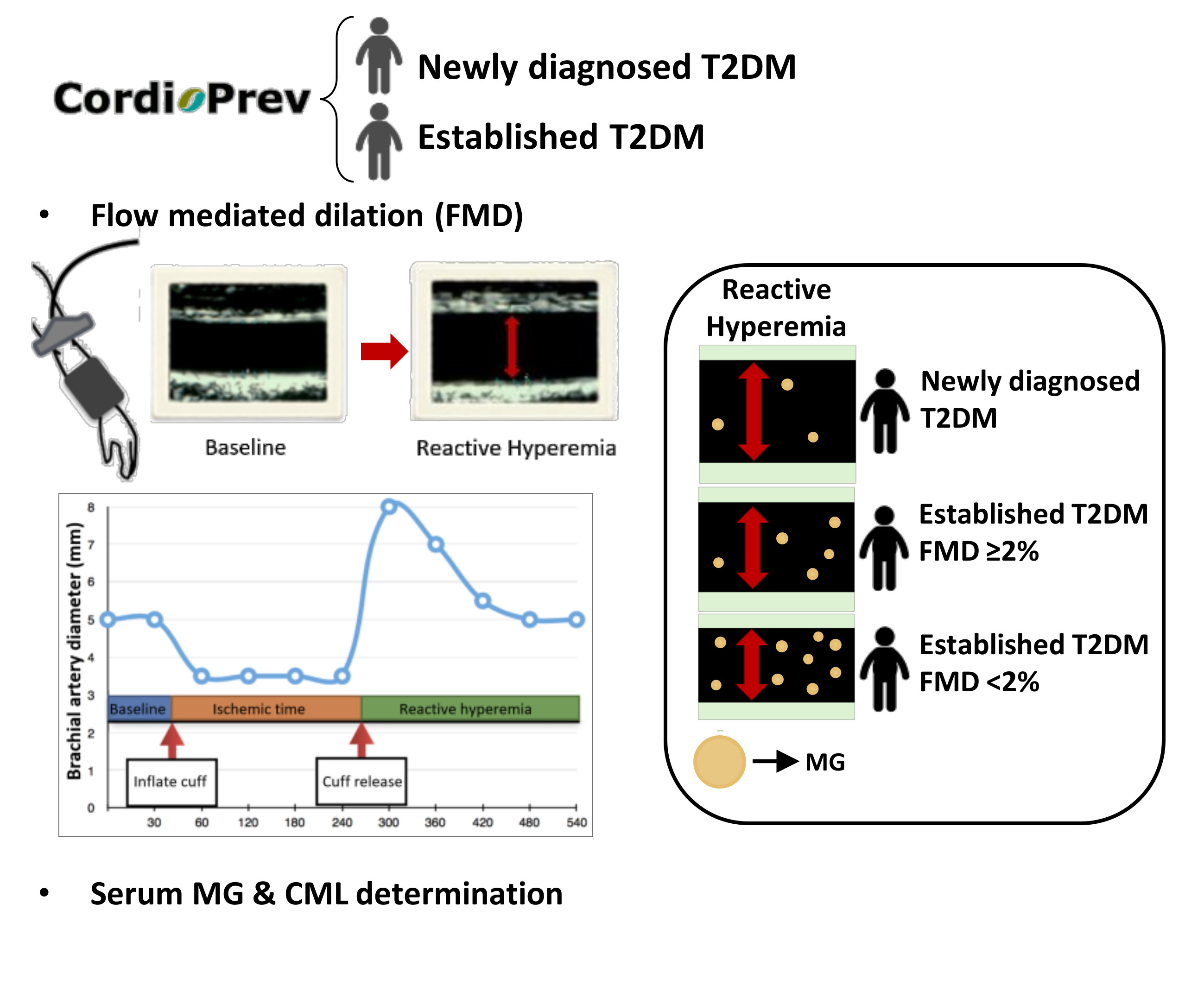
2.1. Patient Population

This work was carried out within the framework of the CORDIOPREV study (Clinicaltrials.gov number NCT00924937), which is a randomized, single-blind, controlled trial that includes 1002 patients with coronary heart disease (CHD). The rationale, methodology and baseline characteristics of the participants in the CORDIOPREV study have been published elsewhere [21]. Prior to recruitment and initiation of the study protocol, written consent was obtained from all participants. All the amendments follow the Helsinki Declaration and good clinical practices and were approved by the Ethics Committee of the Hospital Reina Sofía (Cordoba, Spain).
In the present cross-sectional study, we included those T2DM patients (n = 540) who met, at baseline, the criteria for diabetes diagnosis proposed by the American Diabetes Association [22]. T2DM patients were then categorized in 2 groups: (a) patients with established T2DM (n = 350), i.e., those with a prior medical history of T2DM before entering the study that were receiving treatment (medication or diet), and (b) patients with newly diagnosed T2DM (n = 190), who had no previous history of T2DM, thus being diagnosed during the recruitment period of the CORDIOPREV study.

2.2. Anthropometric Measurements and Laboratory Tests

Patients were given an appointment at 8.00 a.m., following a 12 h fast, and were admitted to the laboratory for anthropometric and biochemical tests. Anthropometric parameters were measured by trained dietitians using calibrated scales (BF511 Body Composition Analyzer/Scale, OMROM, Kyoto, Japan) and a wall-mounted stadiometer (Seca 242, HealthCheck Systems, Brooklyn, NY, USA). Waist circumference was measured midway between the lowest rib and the iliac crest. Body mass index (BMI) was then calculated as weight per square meter (kg/m2). Smoking status, alcohol intake and drug therapy were also recorded for each participant. Systolic and diastolic blood pressure were measured with a validated digital automated blood pressure monitor.
Venous blood samples were collected from the antecubital vein in Vacutainer tubes containing EDTA or no anticoagulant. Serum glucose, HbA1c, total cholesterol, high-density lipoprotein cholesterol (HDL-C) and triglyceride (TG) levels were measured by spectrophotometry using an Architect c-16000 analyzer (Abbot®, Chicago, IL, USA). Low-density lipoprotein cholesterol (LDL-C) was calculated using the Friedewald formula (provided serum TG levels were <400 mg/dL) [23]. Apolipoprotein (ApoA1 and ApoB) concentrations were determined by immunoturbidimetry and plasma insulin by chemiluminescent microparticle immunoassay using an Architect i-2000 analyzer (Abbott®). Insulin resistance was defined by the homeostasis model assessment of insulin resistance (HOMA-IR) index, calculated as fasting insulin (mU/L) × fasting plasma glucose (mmoL/L)/22.5 [24].

2.3. Evaluation of Endothelial Function

Ultrasonography of the brachial artery was performed to measure endothelial-dependent FMD following the guidelines established by the International Brachial Artery Reactivity Task Force [25]. Briefly, tests were performed during the morning, after overnight fast, under stable temperature conditions, refraining patients from doing any physical activity in the previous 48 h, and without stopping medication. Patients remained in supine position for at least 10 min before the test, and until the last image was recorded. The patients’ right arm was immobilized with a stereotactic device, and a high resolution 12 mHz transducer connected to an ultrasound scanner (EnVisor HD, Philips®, Bothell, WA, USA) was placed on the antecubital fossa, in a location where anterior and posterior intimal layers of a non-tortuous longitudinal segment of the brachial artery could be located, to acquire a baseline rest image. Subsequently, the pressure cuff was inflated to 300 mmHg and maintained for 4.5 min. Next, the pressure cuff was deflated, reactive hyperemia was measured and, 1 min after cuff deflation, the new artery diameter and blood-flow velocity were recorded. The whole procedure (from 30 s before deflating the cuff until 2 min after its deflation) was recorded on video.
FMD was defined as the percentage of change between the diameter of the brachial artery after cuff deflation (D2) and the basal diameter pre-occlusion (D1), calculated as [(D2 − D1)/D1] × 100. Participants were electrocardiogram-monitored throughout the whole procedure, and arterial diameter was measured coinciding with the R wave of the electrocardiogram. Intra-observer and inter-observer variabilities were 8.85% and 8.70%, respectively.

2.4. Estimation of Severity of Endothelial Dysfunction

Patients with available FMD measurements were classified according to the FMD cutoff value of 2%, which serves as a benchmark to predict cardiovascular events in high risk populations [26]. Among patients with severe endothelial dysfunction (i.e., FMD < 2%) (n = 172), 116 had established T2DM and 56 had newly diagnosed T2DM. Among patients with non-severe endothelial dysfunction (i.e., FMD ≥ 2%) (n = 320), 202 had established T2DM and 118 had newly diagnosed T2DM.

2.5. Ultrasound Measurement of IMT-CC

Carotid arteries were examined using a Doppler ultrasound high-resolution B-mode (Envisor C Ultrasound System, Philips, Eindhoven, The Netherlands), following the recommendations of the American Society of Echocardiography Carotid Intima-Media Thickness Task Force [27]. Measurements were registered using semi-automatic software (QLAB Advance Ultrasound Quantification Software, v5.0, Phillips, Eindhoven, The Netherlands). Measures were performed in triplicate, obtaining the general mean of the IMT of both common carotid arteries (IMT-CC) for each patient.

2.6. Determination of Serum Levels of AGEs

Methylglyoxal (MG) and N-carboxymethyl lysine (CML) were measured in the serum using competitive ELISA kits (OxiSelect™ Methylglyoxal Competitive ELISA Kit and OxiSelect™ N-epsilon-(Carboxymethyl) Lysine Competitive ELISA Kit, Cell Biolabs, Inc., San Diego, CA, USA), following the manufacturer’s instructions.

2.7. Statistical Analysis
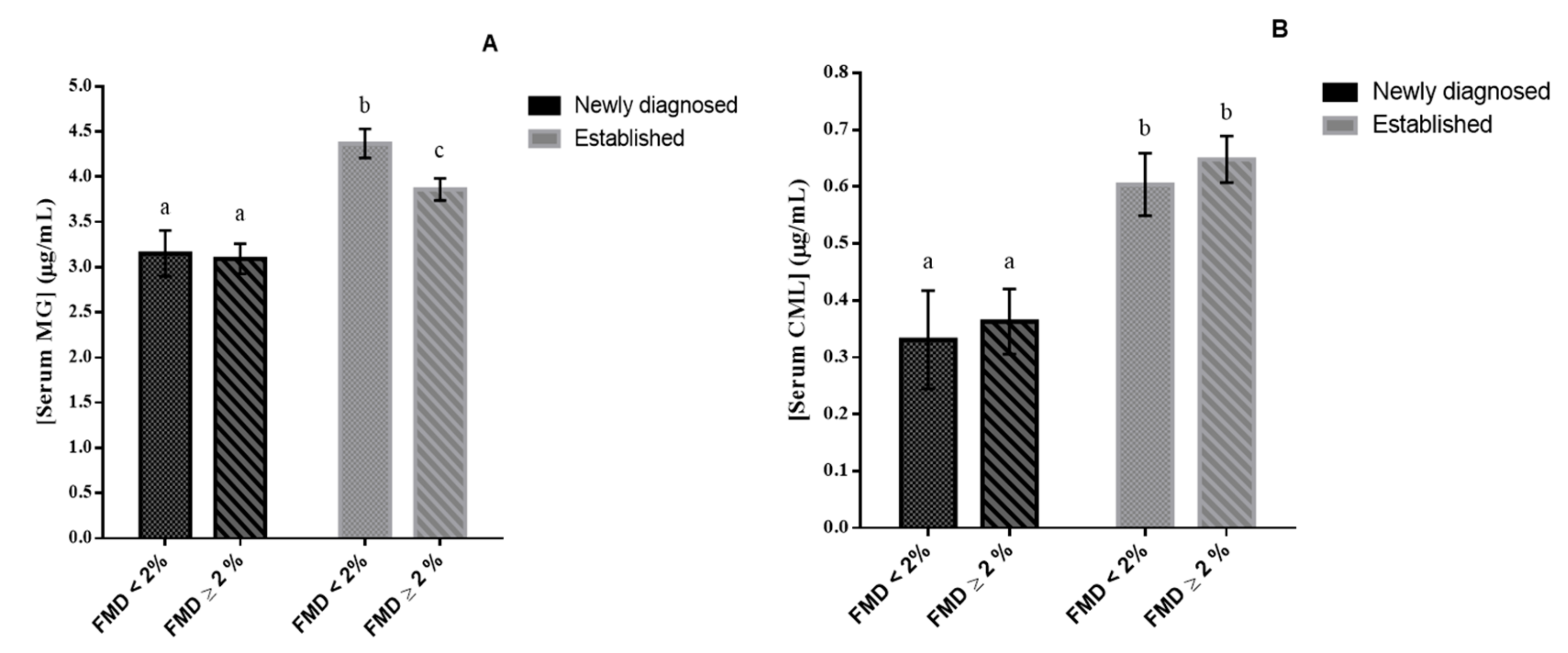
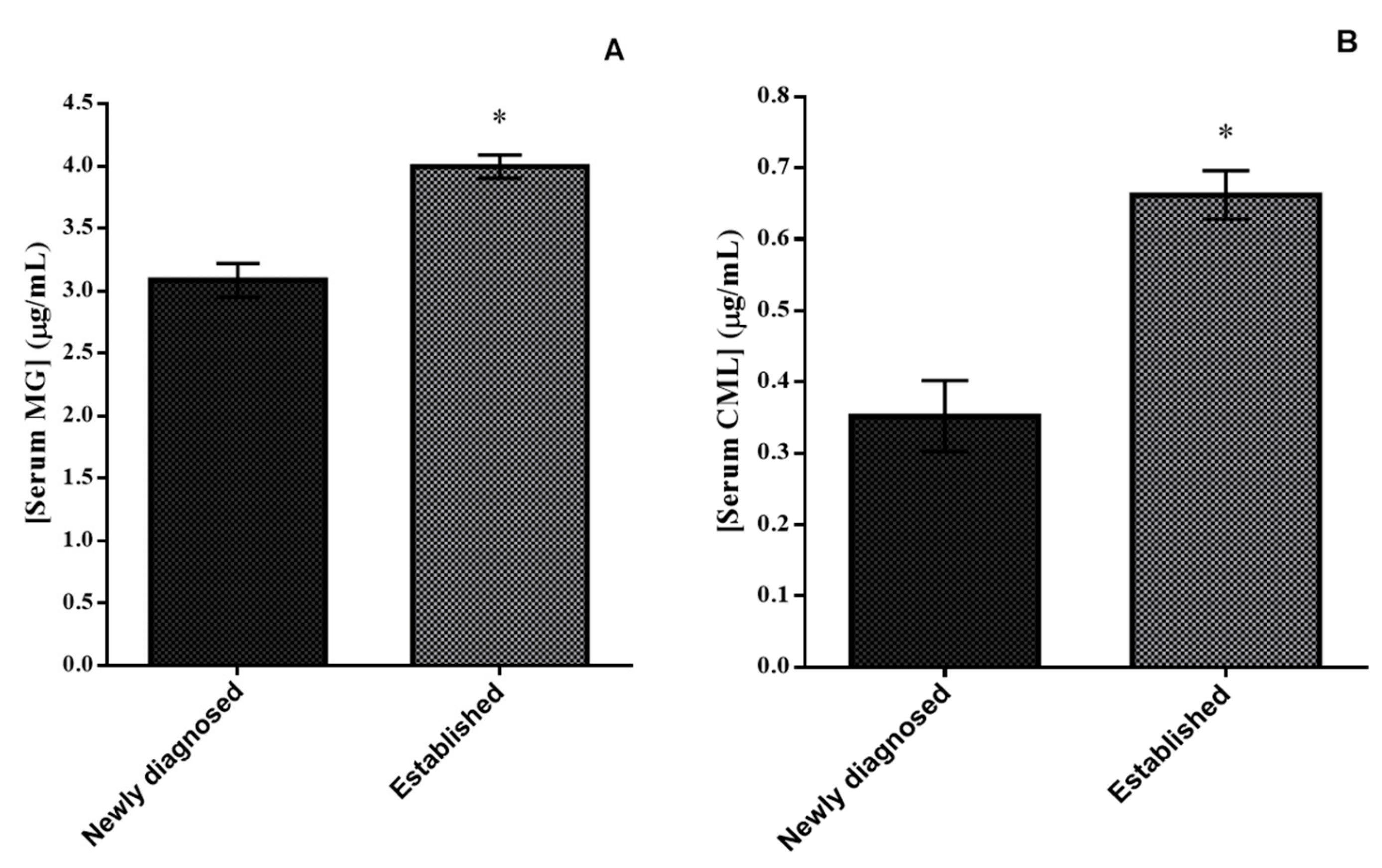
Statistical analyses were carried out using SPSS 23.0 for Windows (SPSS Inc., Chicago, IL, USA). The Kolmogorov–Smirnov normality test was performed for the evaluation of the distribution of the quantitative variables, and continuous variables that deviated significantly from the assumption of normality were transformed. Continuous variables were compared using t-test when comparing between two groups, or one-way analysis of variance (ANOVA) and post-hoc multiple comparisons analysis using the LSD test (homogeneous variances) or Tamhane’s statistic (non-homogeneous variances) when comparing more than two groups. Pearson chi-squared test was employed to compare categorical characteristics. One-way ANCOVA was conducted to compare MG and CML serum levels to control for age, sex and body mass index as covariates.

3. Results

Anthropometric and biochemical characteristics for the patients with established T2DM (n = 350) and those with newly diagnosed T2DM (n = 190) are shown in Table 1. Patients with established T2DM were older, had higher systolic blood pressure, fasting glucose, fasting insulin, HbAc1 and HOMA-IR values, as well as lower levels of HDL-C, LDL-C and ApoB than those with newly diagnosed T2DM (p < 0.05 for all comparisons). Furthermore, IMT-CC was higher in patients with established vs. newly diagnosed T2DM (p = 0.025), with all average values higher than 0.7 mm, which is the suggested cutoff value to determine the presence or absence of carotid atherosclerotic disease [28]. In contrast, FMD did not differ among the two study groups.
Significantly more patients with established T2DM were taking calcium channel blockers, angiotensin converting enzyme inhibitors or angiotensin II receptor blockers and fibrates than those with newly diagnosed T2DM (p < 0.05 for all comparisons) (Table 1). Obviously, patients with established T2DM were on antidiabetic therapy, whereas those with newly diagnosed T2DM were not.
Serum levels of both MG and CML were significantly higher in patients with established T2DM vs. those with newly diagnosed T2DM (4.00 ± 0.09 vs. 3.09 ± 0.13 for MG and 0.66 ± 0.03 vs. 0.35 ± 0.05 for CML (all p < 0.001) (Figure 1A,B, respectively).
Patients with established T2DM and severe endothelial dysfunction had the highest serum MG levels (4.37 ± 0.16) (Figure 2A). These patients also had higher IMT-CC, HOMA-IR, and fasting insulin levels than those with newly diagnosed T2DM and non-severe endothelial dysfunction (Table 2) (p < 0.05 for all comparisons). Serum CML levels were higher in patients with established vs. newly diagnosed T2DM, regardless of the severity of endothelial dysfunction (Figure 2B). Moreover, patients with established T2DM, with severe or non-severe endothelial dysfunction, had higher systolic blood pressure compared with patients with newly diagnosed T2DM with non-severe endothelial dysfunction (Table 2) (p < 0.05 for all comparisons).

4. Discussion

Most of the studies published to date have reported increased levels of AGEs in T2DM patients compared with healthy populations [14,15,16,17]. However, to the best of our knowledge, this is the first study that assessed differences in AGEs concentrations between patients with CHD and established T2DM vs. those with CHD and newly diagnosed T2DM. In the present study, patients with established T2DM had higher serum AGEs levels (both MG and CML) than those with newly diagnosed T2DM.
Although FMD (an early subclinical atherosclerosis marker) did not differ between these two study groups, IMT-CC (a subclinical atherosclerotic marker related to vascular damage) was increased in patients with established vs. newly diagnosed T2DM. Moreover, when the presence of severe endothelial dysfunction was considered, patients with established T2DM exhibited the highest serum MG levels among all groups.
Experimental and clinical studies have suggested that the increased formation of AGEs is one of the causes of endothelial dysfunction in T2DM [18]. AGEs enhance vasoconstriction (by increasing endothelin-1 levels), reduce vasodilation (by decreasing NO levels) and stimulate AGE-modification of extracellular matrix to accelerate the progression of atherosclerosis [17]. In this context, Ninomiya et al. [19] reported an inverse association between FMD and accumulated fluorescent AGEs in the skin of T2DM patients. This is in line with our findings in patients with established T2DM, who exhibited the highest MG levels when severe endothelial dysfunction was present.
The harmful effects of AGEs are mainly due to their ability to cross-link proteins, thus affecting their conformation, altering their enzymatic activity and reducing their degradation capacity and clearance [8]. In T2DM patients, AGEs accumulate as their production is enhanced by chronic hyperglycemia, thus worsening AGE-induced deleterious effects. This could explain our results as a greater dysregulation of glucose homeostasis seen in patients with established T2DM (who had higher fasting glucose, insulin and HbA1c levels compared with patients with newly diagnosed T2DM) could lead to higher serum levels of AGEs.
AGEs could also contribute to T2DM progression, not only due their oxidant properties per se, but also by triggering the cascade of oxidative damage and inflammatory response through the action of receptors such as nuclear factor kappa B (NFkB), nuclear factor erythroid 2-related factor 2 (Nrf2) and more specifically the receptor for AGEs (RAGE) [29]. Therefore, AGEs can lead to vascular inflammation and pathological angiogenesis, contributing to the long-term vascular complications of T2DM [30,31]. This might also explain, at least partly, our finding that patients with established T2DM and severe endothelial dysfunction had, in addition to the highest levels of serum MG, higher IMT-CC than patients with newly diagnosed T2DM and non-severe endothelial dysfunction. In this context, it has been shown that accumulation of AGEs in the vessel wall can be responsible for the formation of a rigid fibrin network and collagen stiffness that contribute to increase IMT-CC in T2DM patients [7].
Endogenous AGEs formation represents a minor component of the total body load of AGEs; dietary AGEs are one of the most important exogenous sources of AGEs that depends on nutrient composition and food processing methods applied [32,33]. The intake of meals rich in AGEs can acutely impair endothelial function in patients with and without T2DM [34]. Conversely, low-AGE diets were shown to reverse insulin resistance and chronic inflammation, inhibit the progression of atherosclerosis and prevent experimental diabetic complications [35,36]. In this context, we have recently published that, in both elderly adults and patients with the metabolic syndrome, a Mediterranean diet could be a beneficial dietary model in terms of AGEs reduction as it has a low content of dietary AGEs, thus consequently reducing their circulating levels and the degree of oxidative stress and inflammation [37,38].
The present study has some limitations. As it is a cross-sectional study, associations between serum levels of AGEs, FMD and IMT-CC values cannot be inferred. Furthermore, we could not assess the effects of drug therapy on AGEs, FMD and IMT-CC. Moreover, the results are limited to a population of T2DM patients with CHD and may not be suitable for extrapolation to healthy populations. Finally, as this is an ancillary study, information about the time of diagnosis of T2DM in established diabetes patients was not available for all the cases.

5. Conclusions

To the best of our knowledge, this is the first study to show differences in AGEs levels between patients with established T2DM and those with newly diagnosed disease. Furthermore, although FMD did not differ between the two groups, IMT-CC was higher in patients with established vs. newly diagnosed T2DM. The presence of severe endothelial dysfunction was associated with increased IMT-CC and MG levels. As AGEs accumulation is the result of endogenous formation, oral intake and renal clearance, they can be lowered by changes in dietary habits and pharmacological treatment. The present findings support the need for establishing strategies to prevent or reduce AGEs in order to delay the onset of CV complications in newly diagnosed T2DM patients and to minimize CV risk in patients with established T2DM.

Author Contributions

Conceptualization, P.P.-M. and E.M.Y.-S.; Data curation, F.M.G.-M.; Formal analysis, S.d.l.C.-A., M.P.C. and F.M.G.-M.; Funding acquisition, J.L.-M.; Investigation, S.d.l.C.-A. and M.P.C.; Methodology, J.D.T.-P.; Resources, J.L.-M. and P.P.-M.; Supervision, J.L.-M. and P.P.-M.; Validation, A.G.-R., M.M.M. and E.M.Y.-S.; Visualization, J.D.T.-P., A.G.-R., N.K. and M.M.M.; Writing—original draft, S.d.l.C.-A.; Writing—review & editing, N.K., P.P.-M. and E.M.Y.-S. All authors have read and agreed to the published version of the manuscript.

Funding

This research was funded by Consejería de salud (PI-0283/2017) and Instituto de Salud Carlos III FIS (PI18/01822) to E.M.Y.-S.

Acknowledgments

The CORDIOPREV study is supported by Fundación Patrimonio Comunal Olivarero. CITOLIVA and CEAS, Consejería de Economía, Innovación y Ciencia (CVI-7450); Ministerio de Economía (AGL2015-67896-P, PIE14/00005, PIE14/00031); Instituto de Salud Carlos III (ISCIII) (FIS 14/00872; FIS 17/01024), co-funded with FEDER (European Regional Development Fund). S.d.l.C.-A. is supported by Juan de la Cierva-Formación Programme (FJCI-2017-31634, MICIU, Spain) and M.P.C. is supported by an ISCIII’s PFIS fellowship (FI17/00081, MICIU, Spain). CIBEROBN is an initiative of Instituto de Salud Carlos III.

Conflicts of Interest

N.K. has given talks, attended conferences and participated in trials sponsored by Astra Zeneca, Bausch Health, Boehringer Ingelheim, Elpen, Mylan, NovoNordisk, Sanofi, Servier and Vianex. The other authors declare that they have no current or potential conflict of interest.

References

  1. Zheng, Y.; Ley, S.H.; Hu, F.B. Global aetiology and epidemiology of type 2 diabetes mellitus and its complications. Nat. Rev. Endocrinol. 2018, 14, 88–98. [Google Scholar] [CrossRef] [PubMed]
  2. Low Wang Cecilia, C.; Hess Connie, N.; Hiatt William, R.; Goldfine Allison, B. Clinical Update: Cardiovascular disease in diabetes mellitus. Circulation 2016, 133, 2459–2502. [Google Scholar] [CrossRef] [PubMed]
  3. Halcox, J.P.J.; Schenke, W.H.; Zalos, G.; Mincemoyer, R.; Prasad, A.; Waclawiw, M.A.; Nour, K.R.A.; Quyyumi, A.A. Prognostic value of coronary vascular endothelial dysfunction. Circulation 2002, 106, 653–658. [Google Scholar] [CrossRef] [PubMed]
  4. Bertoluci, M.C.; Cé, G.V.; da Silva, A.M.; Wainstein, M.V.; Boff, W.; Puñales, M. Endothelial dysfunction as a predictor of cardiovascular disease in type 1 diabetes. World J. Diabetes 2015, 6, 679–692. [Google Scholar] [CrossRef]
  5. Celermajer, D.S.; Sorensen, K.E.; Gooch, V.M.; Spiegelhalter, D.J.; Miller, O.I.; Sullivan, I.D.; Lloyd, J.K.; Deanfield, J.E. Non-invasive detection of endothelial dysfunction in children and adults at risk of atherosclerosis. Lancet 1992, 340, 1111–1115. [Google Scholar] [CrossRef]
  6. Touboul, P.; Hennerici, M.; Meairs, S.; Adams, H.; Amarenco, P.; Bornstein, N.; Csiba, L.; Desvarieux, M.; Ebrahim, S.; Hernandez, R.H.; et al. Mannheim Carotid Intima-Media Thickness and Plaque Consensus (2004–2006–2011): An Update on Behalf of the Advisory Board of the 3rd and 4th Watching the Risk Symposium 13th and 15th European Stroke Conferences, Mannheim, Germany, 2004, and Brussels, Belgium, 2006. Cerebrovasc. Dis. 2012, 34, 290–296. [Google Scholar]
  7. Fournet, M.; Bonté, F.; Desmoulière, A. Glycation damage: A possible hub for major pathophysiological disorders and aging. Aging Dis. 2018, 9, 880. [Google Scholar] [CrossRef] [Green Version]
  8. Stirban, A.; Gawlowski, T.; Roden, M. Vascular effects of advanced glycation endproducts: Clinical effects and molecular mechanisms. Mol. Metab. 2014, 3, 94–108. [Google Scholar] [CrossRef]
  9. Goh, S.-Y.; Cooper, M.E. The role of advanced glycation end products in progression and complications of diabetes. J. Clin. Endocrinol. Metab. 2008, 93, 1143–1152. [Google Scholar] [CrossRef] [Green Version]
  10. Kold-Christensen, R.; Johannsen, M. Methylglyoxal metabolism and aging-related disease: Moving from correlation toward causation. Trends Endocrinol. Metab. 2019. [Google Scholar] [CrossRef]
  11. Thorpe, S.R.; Baynes, J.W. CML: A brief history. Int. Congr. Ser. 2002, 1245, 91–99. [Google Scholar] [CrossRef]
  12. Semba, R.D.; Nicklett, E.J.; Ferrucci, L. Does accumulation of advanced glycation end products contribute to the aging phenotype? J. Gerontol. A. Biol. Sci. Med. Sci. 2010, 65, 963–975. [Google Scholar] [CrossRef] [PubMed] [Green Version]
  13. Aso, Y.; Inukai, T.; Tayama, K.; Takemura, Y. Serum concentrations of advanced glycation endproducts are associated with the development of atherosclerosis as well as diabetic microangiopathy in patients with type 2 diabetes. Acta Diabetol. 2000, 37, 87–92. [Google Scholar] [CrossRef] [PubMed]
  14. Kajikawa, M.; Nakashima, A.; Fujimura, N.; Maruhashi, T.; Iwamoto, Y.; Iwamoto, A.; Matsumoto, T.; Oda, N.; Hidaka, T.; Kihara, Y.; et al. Ratio of serum levels of AGEs to soluble form of RAGE is a predictor of endothelial function. Diabetes Care 2015, 38, 119–125. [Google Scholar] [CrossRef] [Green Version]
  15. Kalousová, M.; Skrha, J.; Zima, T. Advanced glycation end-products and advanced oxidation protein products in patients with diabetes mellitus. Physiol. Res. 2002, 51, 597–604. [Google Scholar]
  16. Kilhovd, B.K.; Berg, T.J.; Birkeland, K.I.; Thorsby, P.; Hanssen, K.F. Serum levels of advanced glycation end products are increased in patients with type 2 diabetes and coronary heart disease. Diabetes Care 1999, 22, 1543–1548. [Google Scholar] [CrossRef]
  17. Soro-Paavonen, A.; Zhang, W.-Z.; Venardos, K.; Coughlan, M.T.; Harris, E.; Tong, D.C.K.; Brasacchio, D.; Paavonen, K.; Chin-Dusting, J.; Cooper, M.E.; et al. Advanced glycation end-products induce vascular dysfunction via resistance to nitric oxide and suppression of endothelial nitric oxide synthase. J. Hypertens. 2010, 28, 780–788. [Google Scholar] [CrossRef]
  18. Tan, K.C.B.; Chow, W.-S.; Ai, V.H.G.; Metz, C.; Bucala, R.; Lam, K.S.L. Advanced glycation end products and endothelial dysfunction in type 2 diabetes. Diabetes Care 2002, 25, 1055–1059. [Google Scholar] [CrossRef] [Green Version]
  19. Ninomiya, H.; Katakami, N.; Sato, I.; Osawa, S.; Yamamoto, Y.; Takahara, M.; Kawamori, D.; Matsuoka, T.; Shimomura, I. Association between subclinical atherosclerosis markers and the level of accumulated advanced glycation end-products in the skin of patients with diabetes. J. Atheroscler. Thromb. 2018, 25, 1274–1284. [Google Scholar] [CrossRef] [Green Version]
  20. Ren, X.; Ren, L.; Wei, Q.; Shao, H.; Chen, L.; Liu, N. Advanced glycation end-products decreases expression of endothelial nitric oxide synthase through oxidative stress in human coronary artery endothelial cells. Cardiovasc. Diabetol. 2017, 16, 52. [Google Scholar] [CrossRef] [Green Version]
  21. Delgado-Lista, J.; Perez-Martinez, P.; Garcia-Rios, A.; Alcala-Diaz, J.F.; Perez-Caballero, A.I.; Gomez-Delgado, F.; Fuentes, F.; Quintana-Navarro, G.; Lopez-Segura, F.; Ortiz-Morales, A.M.; et al. Coronary Diet Intervention with Olive Oil and Cardiovascular Prevention Study (The Cordioprev Study): Rationale, Methods, and Baseline Characteristics. Am. Heart J. 2016, 177, 1–25. [Google Scholar] [CrossRef] [PubMed]
  22. ADA 2. Classification and Diagnosis of Diabetes: Standards of Medical Care in Diabetes—2018. Diabetes Care 2018, 41, S13–S27. [Google Scholar] [CrossRef] [PubMed] [Green Version]
  23. Friedewald, W.T.; Levy, R.I.; Fredrickson, D.S. Estimation of the concentration of low-density lipoprotein cholesterol in plasma, without use of the preparative ultracentrifuge. Clin. Chem. 1972, 18, 499–502. [Google Scholar] [PubMed]
  24. Song, Y.; Manson, J.E.; Tinker, L.; Howard, B.V.; Kuller, L.H.; Nathan, L.; Rifai, N.; Liu, S. Insulin sensitivity and insulin secretion determined by homeostasis model assessment and risk of diabetes in a multiethnic cohort of women: The Women’s Health Initiative Observational Study. Diabetes Care 2007, 30, 1747–1752. [Google Scholar] [CrossRef] [Green Version]
  25. Corretti, M.C.; Anderson, T.J.; Benjamin, E.J.; Celermajer, D.; Charbonneau, F.; Creager, M.A.; Deanfield, J.; Drexler, H.; Gerhard-Herman, M.; Herrington, D.; et al. Guidelines for the ultrasound assessment of endothelial-dependent flow-mediated vasodilation of the brachial artery: A report of the International Brachial Artery Reactivity Task Force. J. Am. Coll. Cardiol. 2002, 39, 257–265. [Google Scholar] [CrossRef] [Green Version]
  26. Fathi, R.; Haluska, B.; Isbel, N.; Short, L.; Marwick, T.H. The relative importance of vascular structure and function in predicting cardiovascular events. J. Am. Coll. Cardiol. 2004, 43, 616–623. [Google Scholar] [CrossRef] [Green Version]
  27. Stein, J.H.; Korcarz, C.E.; Hurst, R.T.; Lonn, E.; Kendall, C.B.; Mohler, E.R.; Najjar, S.S.; Rembold, C.M.; Post, W.S.; American Society of Echocardiography Carotid Intima-Media Thickness Task Force. Use of carotid ultrasound to identify subclinical vascular disease and evaluate cardiovascular disease risk: A consensus statement from the American Society of Echocardiography Carotid Intima-Media Thickness Task Force. Endorsed by the Society for Vascular Medicine. J. Am. Soc. Echocardiogr. Off. Publ. Am. Soc. Echocardiogr. 2008, 21, 93–111. [Google Scholar]
  28. Rohde, L.E.; Lee, R.T.; Rivero, J.; Jamacochian, M.; Arroyo, L.H.; Briggs, W.; Rifai, N.; Libby, P.; Creager, M.A.; Ridker, P.M. Circulating cell adhesion molecules are correlated with ultrasound-based assessment of carotid atherosclerosis. Arterioscler. Thromb. Vasc. Biol. 1998, 18, 1765–1770. [Google Scholar] [CrossRef] [Green Version]
  29. Vlassara, H.; Uribarri, J. Advanced glycation end products (AGE) and diabetes: Cause, effect, or both? Curr. Diab. Rep. 2014, 14, 453. [Google Scholar] [CrossRef] [Green Version]
  30. Yamagishi, S.; Ueda, S.; Okuda, S. Food-derived advanced glycation end products (AGEs): A novel therapeutic target for various disorders. Curr. Pharm. Des. 2007, 13, 2832–2836. [Google Scholar] [CrossRef]
  31. Saremi, A.; Howell, S.; Schwenke, D.C.; Bahn, G.; Beisswenger, P.J.; Reaven, P.D. Advanced glycation end products, oxidation products, and the extent of atherosclerosis during the VA Diabetes Trial and Follow-up Study. Diabetes Care 2017, 40, 591–598. [Google Scholar] [CrossRef] [PubMed] [Green Version]
  32. Uribarri, J.; Cai, W.; Sandu, O.; Peppa, M.; Goldberg, T.; Vlassara, H. Diet-derived advanced glycation end products are major contributors to the body’s AGE pool and induce inflammation in healthy subjects. Ann. N. Y. Acad. Sci. 2005, 1043, 461–466. [Google Scholar] [CrossRef] [PubMed]
  33. Uribarri, J.; Woodruff, S.; Goodman, S.; Cai, W.; Chen, X.; Pyzik, R.; Yong, A.; Striker, G.E.; Vlassara, H. Advanced glycation end products in foods and a practical guide to their reduction in the diet. J. Am. Diet. Assoc. 2010, 110, 911–916.e12. [Google Scholar] [CrossRef] [PubMed] [Green Version]
  34. Uribarri, J.; Stirban, A.; Sander, D.; Cai, W.; Negrean, M.; Buenting, C.E.; Koschinsky, T.; Vlassara, H. Single oral challenge by advanced glycation end products acutely impairs endothelial function in diabetic and nondiabetic subjects. Diabetes Care 2007, 30, 2579–2582. [Google Scholar] [CrossRef] [PubMed] [Green Version]
  35. Tan, K.C.B.; Shiu, S.W.M.; Wong, Y.; Tam, X. Serum advanced glycation end products (AGEs) are associated with insulin resistance. Diabetes Metab. Res. Rev. 2011, 27, 488–492. [Google Scholar] [CrossRef]
  36. Uribarri, J.; Cai, W.; Ramdas, M.; Goodman, S.; Pyzik, R.; Chen, X.; Zhu, L.; Striker, G.E.; Vlassara, H. Restriction of advanced glycation end products improves insulin resistance in human type 2 diabetes: Potential role of AGER1 and SIRT1. Diabetes Care 2011, 34, 1610–1616. [Google Scholar] [CrossRef] [Green Version]
  37. López-Moreno, J.; Quintana-Navarro, G.M.; Camargo, A.; Jiménez-Lucena, R.; Delgado-Lista, J.; Marín, C.; Tinahones, F.J.; Striker, G.E.; Roche, H.M.; Pérez-Martínez, P.; et al. Dietary fat quantity and quality modifies advanced glycation end products metabolism in patients with metabolic syndrome. Mol. Nutr. Food Res. 2017, 61, 1601029. [Google Scholar] [CrossRef]
  38. López-Moreno, J.; Quintana-Navarro, G.M.; Delgado-Lista, J.; García-Ríos, A.; Delgado-Casado, N.; Camargo, A.; Pérez-Martínez, P.; Striker, G.E.; Tinahones, F.J.; Pérez-Jiménez, F.; et al. Mediterranean Diet reduces serum advanced glycation end products and increases antioxidant defenses in elderly adults: A randomized controlled trial. J. Am. Geriatr. Soc. 2016, 64, 901–904. [Google Scholar] [CrossRef] [Green Version]
Figure 1. Serum levels of methylglyoxal (μg/mL) (A) and carboxy-methyl-lysine (μg/mL) (B) in T2DM patients studied. One-way ANCOVA results (adjusted mean ± SEM) controlling for age, sex and BMI. * p < 0.001 indicates significant differences.
Nutrients 12 00238 g001
Figure 2. (A) Serum levels of methylglyoxal (μg/mL) and (B) carboxy-methyl-lysine (μg/mL) according to FMD classification (2% cutoff) in T2DM patients studied. One-way ANCOVA results (adjusted mean ± SEM) controlling for age, sex and BMI. Bars with different superscript letters (a, b, c) are significantly different (p < 0.05).
Nutrients 12 00238 g002
Table 1. Characteristics of the study groups 1.
Patients with Newly Diagnosed Diabetes
(n = 190)
Patients with Established Diabetes
(n = 350)
p Value
Age (years)60.0 ± 0.661.9 ± 0.40.015
Sex (men/women)158/32285/640.724
Weight (kg)86.7 ± 1.085.0 ± 0.80.191
BMI (kg/m2)31.4 ± 0.331.1 ± 0.30.431
Waist circumference (cm)106 ± 0.8105 ± 0.60.294
DBP (mmHg)76.9 ± 0.976.1 ± 0.60.448
SBP (mmHg)137 ± 1.5143 ± 1.10.001
HDL-cholesterol (mg/dL)41.5 ± 0.739.6 ± 0.50.023
LDL-cholesterol (mg/dL)92.4 ± 1.982.5 ± 1.4<0.001
Triglycerides (mg/dL)146 ± 6.0150 ± 4.10.604
ApoA-1 (mg/dL)127± 1.4125 ± 1.20.294
ApoB (mg/dL)77.3 ± 1.472.4 ± 1.00.005
Fasting glucose (mg/dL)120 ± 1.7157 ± 3.0<0.001
Fasting insulin (mU/L)10.8 ± 0.513.6 ± 0.90.024
HOMA-IR4.27 ± 0.256.50 ± 0.43< 0.001
HbAc1 (%)6.67 ± 0.067.66 ± 0.07<0.001
FMD (%)4.11 ± 0.453.46 ± 0.350.256
IMT-CC (mm)0.72 ± 0.010.75 ± 0.010.025
Alcohol intake (>16 g/day) (%)19.923.10.865
Current tobacco use (%)12.29.80.380
Antihypertensive drugs (%)
Angiotensin converting enzyme inhibitors or angiotensin II receptor blockers38.955.6<0.001
Calcium channel blockers16.824.90.039
Beta-blockers62.663.60.852
Nitrates10101.000
Diuretics42.146.70.320
Lipid lowering drugs (%)
Statins86.885.70.795
Fibrates02.90.017
Oral hypoglycemic agents (%)072.8<0.001
Insulin (%)025.8<0.001
1 Values are presented as mean ± SEM (standard error of the mean). Numerical variables were analysed using independent t-test (95% confidence interval) for mean difference, whereas categorical variables were analysed using χ2 test. Values in bold were significantly different (p < 0.05). n, sample size; BMI, body mass index; DBP, diastolic blood pressure; systolic blood pressure; HDL, high-density lipoprotein; LDL, low-density lipoprotein; Apo, apolipoprotein; HOMA-IR, homeostatic model assessment for insulin resistance; FMD, flow-mediated dilation; IMT-CC, intima-media thickness of both common carotid arteries; HbAc1, glycated haemoglobin.
Table 2. Characteristics of the study groups considering the severity of endothelial dysfunction 1.
Patients with Newly Diagnosed DiabetesPatients with Established Diabetes
Severe Endothelial Dysfunction (n = 56)Non-Severe Endothelial Dysfunction (n = 118)Severe Endothelial Dysfunction (n = 116)Non-Severe Endothelial Dysfunction (n = 202)p Value
Age (years)60.4 ± 1.259.8 ± 0.862.3 ± 0.761.4 ± 0.60.112
Sex (men/women)46/10 ab98/20 ab87/29 b175/27 a0.074
Weight (kg)84.7 ± 1.588.1 ± 1.384.6 ± 1.485.2 ± 1.10.229
BMI (kg/m2)31.5 ± 0.531.6 ± 0.431.2 ± 0.431.2 ± 0.40.877
Waist circumference (cm)104 ± 1.3107 ± 1.1104 ± 1.1105 ± 0.80.204
DBP (mmHg)77.6 ± 1.676.7 ± 1.074.2 ± 0.977.4 ± 0.70.062
SBP (mmHg)141 ± 2.7 ab136 ± 1.8 a145 ± 2.0 b143 ± 1.4 b0.018
HDL-cholesterol (mg/dL)41.0 ± 1.441.6 ± 0.840.0 ± 1.039.1 ± 0.60.128
LDL-cholesterol (mg/dL)93.5 ± 3.8 a92.6 ± 2.5 a85.8 ±2.7 ab81.7 ± 1.7 b0.001
Triglycerides (mg/dL)151 ± 12.2142 ± 7.0161 ± 7.2146 ± 5.40.253
ApoA-1 (mg/dL)125 ± 2.6128 ± 1.7127 ± 2.2124 ± 1.40.221
ApoB (mg/dL)81.5 ± 3.0 a75.8 ± 1.8 ab74.9 ± 1.8 ab71.8 ± 1.3 b0.007
Fasting glucose (mg/dL)122 ± 3.4 a118 ± 2.1 a155 ± 6.0 b155 ± 3.4 b<0.001
Fasting insulin (mU/L)11.2 ± 1.0 ab10.4 ± 0.7 b15.2 ± 1.7 a12.9 ± 1.0 ab0.052
HOMA-IR4.27 ± 0.52 ab4.18 ± 0.31 b7.06 ± 0.9 a6.09 ± 0.45 ab0.002
HbAc1 (%)6.81 ± 0.11 a6.63 ± 0.08 a7.57 ± 0.13 b7.72 ± 0.09 b<0.001
FMD (%)−1.78 ± 0.70 a6.91 ± 0.34 b−2.19 ± 0.45 a6.70 ± 0.30 b<0.001
IMT-CC (mm)0.74 ± 0.02 ab0.72 ± 0.01 a0.76 ± 0.02 b0.75 ± 0.01 ab0.162
Alcohol intake (>16 g/day) (%)21.819.122.423.10.682
Current tobacco use (%)17.9 a7.8 b9.9 ab8.7 ab0.176
Antihypertensive drugs (%)
Angiotensin converting enzyme inhibitors or angiotensin II receptor blockers28.6 a43.2 ab61.2 c51.5 b,c<0.001
Calcium channel blockers12.519.521.624.80.236
Beta-blockers62.562.765.562.40.950
Nitrates8.911.06.011.40.442
Diuretics41.1 abc40.7 c55.2 b43.1 ac0.094
Lipid lowering drugs (%)
Statins87.584.783.687.10.805
Fibrates0 ab0 b3.4 a3.0 ab0.129
Oral hypoglycemic agents (%)0 a0 a73.3 b72.8 b<0.001
Insulin (%)0 a0 a26.7 b24.8 b<0.001
1 Values are presented as mean ± SEM (standard error of the mean). Numerical variables were analysed using one-way ANOVA, whereas categorical variables were analysed using χ2 test. Values in bold were significantly different (p < 0.05). Values in the same row with different superscript letters (a, b, c) are significantly different. BMI, body mass index; DBP, diastolic blood pressure; systolic blood pressure; HDL, high-density lipoprotein; LDL, low-density lipoprotein; Apo, apolipoprotein; HOMA-IR, homeostatic model assessment for insulin resistance; HbAc1, glycated haemoglobin; FMD, flow-mediated dilation; IMT-CC, intima-media thickness of both common carotid arteries.

