Review

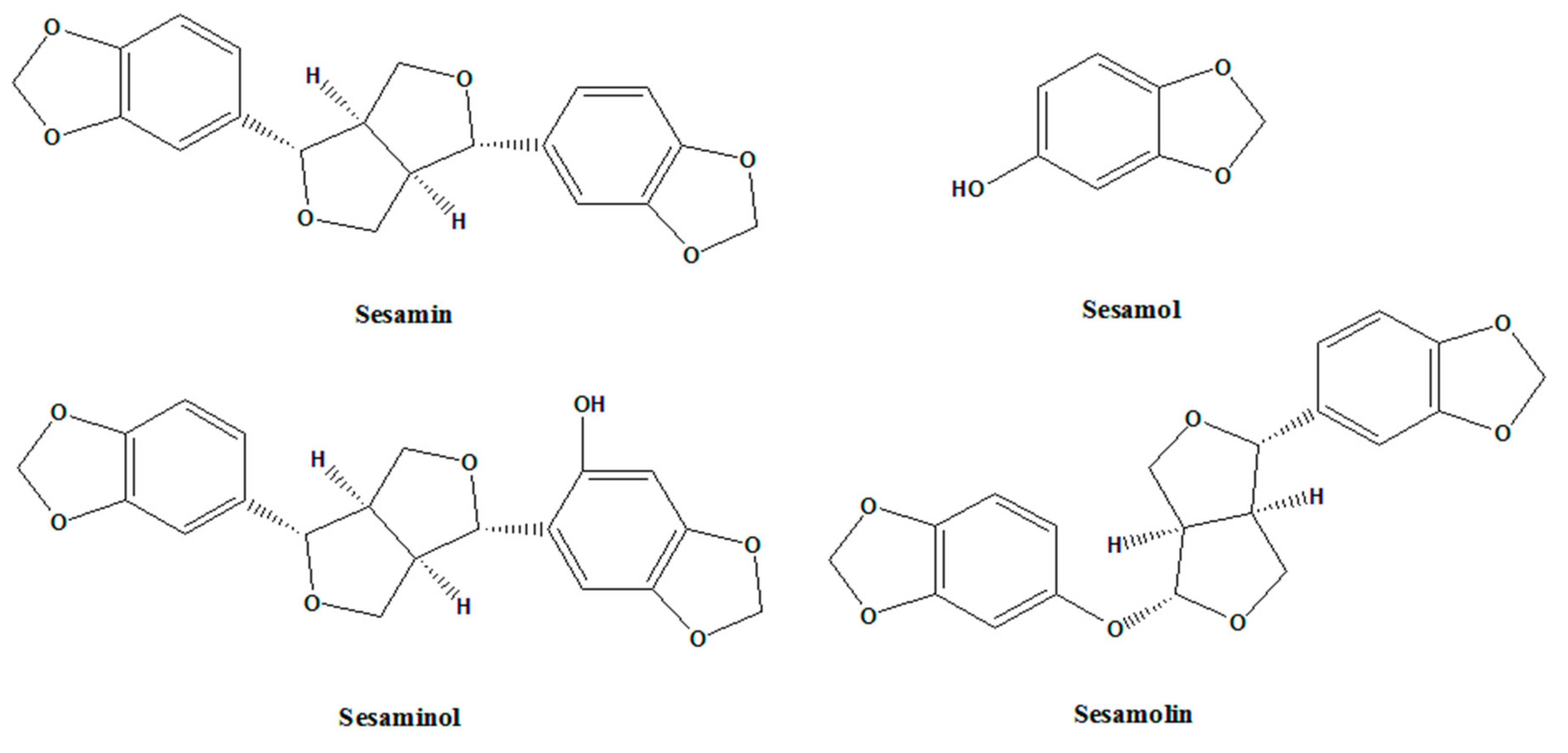
Anti-Inflammatory and Anticancer Properties of Bioactive Compounds from Sesamum indicum L.—A Review

by
Ming-Shun Wu
1,2,3,
Levent Bless B. Aquino
4,
Marjette Ylreb U. Barbaza
4,
Chieh-Lun Hsieh
5,
Kathlia A. De Castro-Cruz
4,
Ling-Ling Yang
6,7 and
Po-Wei Tsai
8,*
1
Division of Gastroenterology, Department of Internal Medicine, Wan Fang Hospital, Taipei Medical University, Taipei 116, Taiwan
2
School of Medicine, College of Medicine, Taipei Medical University, Taipei 110, Taiwan
3
Integrative Therapy Center for Gastroenterologic Cancers, Wan Fang Hospital, Taipei Medical University, Taipei 116, Taiwan
4
School of Chemical, Biological, Materials Engineering and Sciences, Mapúa University, Manila 1002, Metro Manila, Philippines
5
Department of Athletics Sports, College of Humanities and Social Sciences, Chang Jung Christian University, Tainan 711, Taiwan
6
School of Pharmacy, College of Pharmacy, Taipei Medical University, Taipei 110, Taiwan
7
College of Acupuncture and Oriental Medicine, Houston, TX 77063, USA
8
Department of Medical Sciences Industry, College of Health Sciences, Chang Jung Christian University, Tainan 711, Taiwan
*
Author to whom correspondence should be addressed.
Molecules 2019, 24(24), 4426; https://doi.org/10.3390/molecules24244426
Submission received: 9 November 2019 / Revised: 28 November 2019 / Accepted: 3 December 2019 / Published: 4 December 2019
(This article belongs to the Special Issue Food and Drug Analysis)

Abstract

:
The use of foodstuff as natural medicines has already been established through studies demonstrating the pharmacological activities that they exhibit. Knowing the nutritional and pharmacological significance of foods enables the understanding of their role against several diseases. Among the foods that can potentially be considered as medicine, is sesame or Sesamum indicum L., which is part of the Pedaliaceae family and is composed of its lignans such as sesamin, sesamol, sesaminol and sesamolin. Its lignans have been widely studied and are known to possess antiaging, anticancer, antidiabetes, anti-inflammatory and antioxidant properties. Modern chronic diseases, which can transform into clinical diseases, are potential targets of these lignans. The prime example of chronic diseases is rheumatic inflammatory diseases, which affect the support structures and the organs of the body and can also develop into malignancies. In line with this, studies emphasizing the anti-inflammatory and anticancer activities of sesame have been discussed in this review.

1. Introduction

Numerous studies have already been able to prove that different kinds of food are capable for acting against several types of diseases due to their various medicinal properties. Sesamum indicum L., more commonly referred to as sesame, is one of the foods that are known to exhibit pharmacological applications. Sesame, from the Pedaliaceae family [1], has been known way back several decades ago (1600 BC), and is originated from Indonesia according to the Hindu legends. Sesame is considered as one of the oldest condiments in the human history [2,3]. In line with this, sesame is one of the first crops to produce oil [4]. Its herbal medicinal property was first discovered in China and India during the 8th century BC, and is believed to relieve toothaches, to give energy, to prevent aging, to sooth the mind and body and to treat bites of insects [5,6]. The largest area of sesame cultivation is found in India and provides 27.9% of the world’s sesame [7]. Some countries also cultivate sesame especially in the tropical countries of Africa and Asia [8].
Majority of the sesame lignans are found in the sesame seeds. Sesame seeds comprise of 50% oil and 25% protein and the rest are sugars, moisture, fibers and minerals [9,10]. There are four commonly known sesame lignans namely, sesamin, sesamol, sesaminol and sesamolin. These sesame lignans (Figure 1) are known for its various biological activities and applications. Sesamin, a furfuran lignan, plays a role in scavenging free radicals and lipid and glucose metabolism. Sesamol possess high antioxidant activity, which involves membrane protection from peroxidation of lipids [11,12]. Sesaminol exhibits inhibitory property against membrane lipid peroxidation and improves the tocopherols availability of vitamin E through enhancement of concentrations of liver and plasma [13,14]. Sesamolin upregulates the rate of peroxisomal fatty acid oxidation and hepatic mitochondrial and has a synergistic effect on pyrethrum insecticides [15,16,17]. Not only is sesame oil abundant in sesame seeds, but sesame oil also has excellent quality nutrition and stability [18]. Among other vegetable oils, sesame oil has many uses and has high therapeutic values, which make it interesting to study. The presence of sesame lignans such as sesamin, sesamol, sesamolin and other acylglycerols (oleic acid, linoleic acid, palmitic acid, stearic acid and arachidic acid) in sesame oil contributes to its distinct properties [19,20]. This paper presents an in-depth review on the anti-inflammatory and anti-cancer properties of sesame.

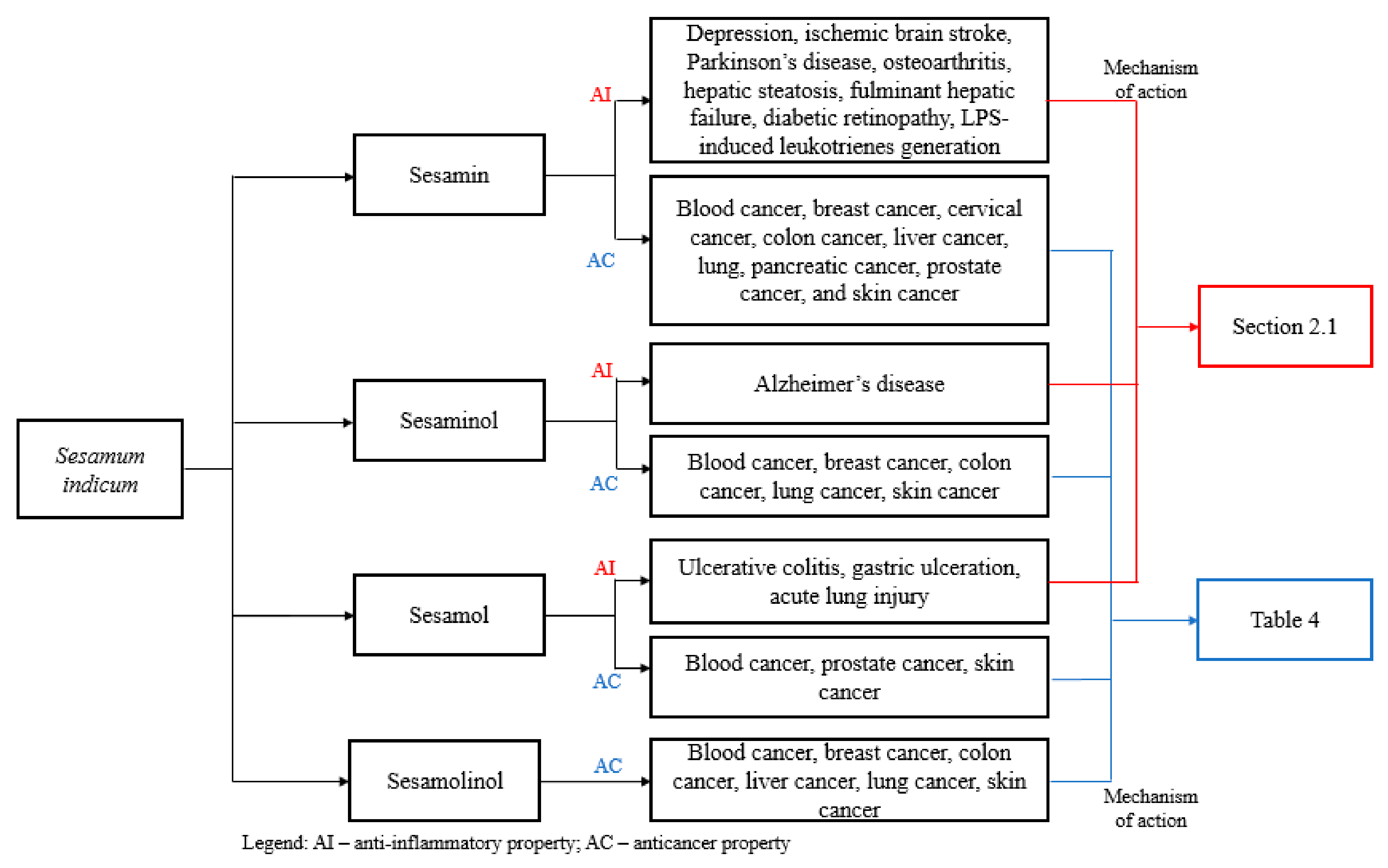
2. Pharmacological Applications of Sesame

2.1. Anti-Inflammatory Activity

Inflammation has been recorded way back several centuries ago (1500 BCE–600 CE) in the ancient Indian medicine called Ayurveda [21]. In the modern era, the term ‘inflammation’ comes from the Latin word inflammare that has a meaning of “to set on fire” [22]. Redness, heat, pain and swelling are the characterizations of inflammation and are induced by several factors such as frostbite, infection by pathogens, burns, chemical irritants, physical injuries, oxidative stress, ischemia, toxins and hypersensitivity [23,24]. Inflammation is part of the body’s defense mechanism against pathogens or pathogen-associated molecular patterns [25]. Restoring, regenerating and repairing of the damaged tissues or organ hemeostasis through cellular network and signaling pathways are involved in the inflammation process. Conversely, severe acute inflammation or prolongation of inflammation may lead to pathology, organ failure, chronic inflammatory diseases, autoimmunity and death. The active cellular components that are responsible for the processing of acute and chronic inflammation are monocytes/macrophages, endothelial cells, neutrophils, innate lymphoid cells, mucosal-associated invariant T cells, mast cells, natural killer cells, dendritic cells and other lower forms of T cells [26,27,28,29]. Table 1, Table 2 and Table 3 depicts the in vitro/in vivo models for the anti-inflammatory properties of sesame.

2.1.1. Neurodegenerative Disease

Dysregulation of microglia, a principal cell type of brain, is pivotal to recruitment of cell inflammation and expression of pro-inflammatory factors, which later results to neurodegeneration [30]. Such neurodegenerative diseases are Parkinson’s disease, Alzheimer’s disease and other multiple sclerosis [31]. The inflammation begins when the pattern recognition receptors (PRRs) and pathogen associated molecular patters (PAMPs) interacts [32]. A common type of PPRs that detects the lipopolysaccharide (LPS) is Toll-like receptor 4 (TLR4), which is expressed highly on the surface of the microglia when activated. Once activated, the pro-inflammatory mediator is released via mitogen-activated protein kinase (MAPK) and nuclear factor kappa-light-chain-enhancer of activated B cells (NF-кB) pathways [33] and further generation of other pro-inflammatory mediators begins (e.g., interleukin 1 beta (IL-1β), interleukin-6 (IL-6), tumor necrosis factor alpha (TNF-α), reactive oxygen species (ROS) and nitric oxide (NO)). Other pro-inflammatory molecules like prostaglandin E2 (PGE2) are also initiated simultaneously through phosphorylation of its subunits. There are several NF-кB subunits such as p-IKKα/β, p-IκBα and p-p65, and several MAPK subunits such as c-Jun N-terminal kinase (JNK), p38 and extracellular-signal-regulated kinase (ERK).
In the literature study [34] showed that wherein the BV-2 microglial cell line was used, sesamin shows dose-dependent decrease in expression of TLR4 in LPS-stimuli. Notably, at 50 μM concentration of sesamin, there is a significant decrease in the expression of TLR4. Furthermore, sesamin suppresses the phosphorylation of p-IkBα and p-p65 over a moderate period. In addition, sesamin decreases the phosphorylation of JNK, however, the phosphorylation of p38 is slightly reduced. Interestingly, sesamin with 50 μM indomethacin dose-dependently reduced IL-1β and IL-6 mRNA gene expression and moderately reduced TNF-α before LPS exposure. Similarly, sesamin decreases the levels of cyclooxygenase-2 (COX-2) gene expressions and hinders the production of PGE2. Moreover, sesamin reduced the expression of inducible nitric oxide synthase (iNOS) gene and as well as reduced production of NO in dose-dependent manner. Sesamin exhibits a reduced effect for neurotoxicity of LPS-mediated microglia activation, which eventually increases the viability of neuronal cells.
In another study [35], in which same microglial cell line was used, further investigation on the p38 MAPK signaling pathway was commenced to support its role in cytokine production. It has been found that 50 μM concentration of sesamin suppresses the p38 MAPK activation (40–75%) induced by LPS. The inhibitory effect of sesamin is similar to that of SB203580 (p38 MAPK inhibitor), which inhibits the production of IL-6 mRNA and protein production specifically. Similarly, sesamolin has been reported to reduce the activation of p38 MAPK induced by LPS, however it has not yet been fully studied.
One of the mental disorders associated with neuroinflammation is depression. The downregulation of norepinephrine (NE), and serotonin (5-hydroxytryptamine, 5-HT) levels and the decrease of synaptic content cause depression [36,37]. Brain derived neurotrophic factor (BDNF) controls the development of neuronal function and becomes ineffective in neurodegenerative disorders such as depression [38]. The ionized calcium binding adaptor molecule 1 (IBA-1) is responsible for the microglia activation in hippocampus and cortex [39]. Several studies already used chronic unpredictable mild stress (CUMS) for the physiological pathway elucidation of depression [40].
CUMS-induced depression in mice [41] shows relatively positive results toward forced swimming, tail suspension, elevated plus maze, Morris water maze and Y-maze tests when sesamin is administered (50 ppm/d) for 6 weeks. Furthermore, sesamin upregulates the levels of 5-HT and NE in striatum only, suggesting its beneficial effects on depressive like behaviors. Neurotrophin-3 (NT3) and BDNF shows increase expression in hippocampus when treated with sesamin. Comparatively, sesamin decreases the expression of IBA-1 expression and as a result, the production of inflammatory cytokines ceases.
Another neurodegenerative disorder is the ischemic brain stroke. Shutting off the flow of cerebral blood to thrombi results to ischemic brain stroke due to loss of oxygen and energy supply to crucial tissues of the brain. In middle cerebral artery occlusion (MCAO)-treated mice [42], sesamin (30 ppm) mitigates brain injury by suppressing the production of inflammatory mediators. Interestingly, sesamin reduces the expression levels of p-ERK1/2 together with p-p38 of ischemic brain tissue in MCAO-induced brain damage.
Sesamin at dosage 20 ppm has shown to suppress 6-hydroxydopamine (6-OHDA) that induces Parkinson’s disease in rats [43] via decrease in inflammatory mediator levels in the brain. Sesamin has the capability to alleviate astrogliosis based on the lowering effect of glial fibrillary acidic protein (GFAP) immunoreactivity. Similarly, the inhibitory effect of sesamin against inflammatory agents (MAPK and COX-2) results to stabilizing the oxidative stress and mortality in kainic acid-induced status epilepticus [44]. This study, however, is not fully studied as the degree of inflammatory markers was not discussed.
Degradation of heme to iron, carbon monoxide and biliverdin is caused by a phase II antioxidant enzyme called heme oxygenase (HO). There are other isoforms of HO such as inducible HO-1 and constitutive HO-2 and HO-3 [45]. Biliverdin reductase transform biliverdin to bilirubin and bilirubin is believed to possess anti-oxidative properties [46,47].
At 100 μM concentration of sesamin, there is an increase in the (HO-1) protein level in RAW 264.7 macrophage cells. The HO-1 mRNA expression, however, is not affected by sesamin. Several studies have claimed that sesamin activates Nf-кB or MAPK signaling pathway, not to mention, sesamin affects the p65 and p38 MAPK effectively in RAW 264.7 cells. The ZnPP IX, a HO-1 inhibitor, reduced the inhibitory effect of sesamin on the release of NO. The degradation of HO-1 protein through the ubiquitination pathway is partially suppressed by sesamin. The proteasome activity, however, is not affected by sesamin. Hence, the ubiquitination mechanism inhibition by sesamin is still unclear [48].
In rat pheochromocytoma PC12 cells study [49], episesamin and sesamin metabolites are investigated. Based from the luciferase reporter assays, episesamin and sesamin metabolites has the capability to activate the signaling cells nuclear factor E2-related factor 2/antioxidant response element (Nrf2/ARE), which further upgrades to phase II detoxification enzyme expression. Moreover, sesamin metabolites induced the expression of detoxification enzymes such as HO-1, y-GCSc and NQO-1 in a dose-dependent manner. Different signal transduction pathways are also affected by the sesamin metabolites through phosphorylation. In addition, sesamin metabolites increase the expression of HO-1 mRNA and protein, which comes before the nuclear translocation of Nrf2.
In the 16-week-old senescence-accelerated mouse-prone 8 (SAMP8) study [50], sesaminol exhibit a reducing effect on the inflammatory cytokines namely IL-6, IL-1β and TNF-α via real-time polymerase chain reactor (PCR) assay in the brain of the modeled mouse. This further contributes in the mitigation of the Alzheimer’s disease.

2.1.2. Osteoarthritis

The degradation and the tearing down of cartilage matrix are characterized in a chronic articular disease called osteoarthritis (OA) [51,52]. Chondrocyte has become the context of pathogenesis of OA, which has an outcome of imbalance between degradation and synthesis of cartilage extracellular matrix (CEM). Inflammatory cytokine network could stimulate matrix metalloproteinases (MMPs) generation together with PGE2 and NO in chondrocytes [53,54]. During the progression of OA, MMPs (e.g., collagenase-3/MMP-13) cleaves and denatures the type II collagen and proteoglycan at the surface of the cartilage. The degradation of the collagen and proteoglycan results to the loss of tensile strength in the matrix of cartilage due to the increase of water content [55,56]. Interleukin-1 (IL-1) and TNF-α can prompt the expression of MMPs expression via c-fos signaling pathway.
In papain-induced OA rat study [57], sesamin has been reported to exhibit chondroprotective effects for OA. Sesamin has no effect on the activity of aggrecanase, the main proteoglycan in the cartilage tissue, but has reversible effect in the expression of MMP-1, -3 and -13 in the human articular chondrocyte (HAC) culture. Not to mention, sesamin suppresses the IL-1β induced MMP expression at both protein and mRNA levels. Moreover, sesamin has been reported to reverse the synergistic effect of combined IL-1β and oncostatin M (OSM) and stops the degradation of type II collagen and proteoglycan. Having said this, sesamin can slow down the destruction of cartilage prompting to the development of OA.
Another study [58], wherein the articular cartilage of the 12 patients undergoing knee replacement surgery were acquired, suggests that sesamin activates Nrf2 signaling pathway and up-regulates HO-1 protein expression and further inhibits the inflammatory gene expressions in the OA chondrocytes. Coupled with, sesamin inhibits the activation of NF-кB during the process of Nrf2 activation.
Sesamol exhibits an inhibitory effect on MMPs expression that triggers OA [59]. The specific MMPs are MMP-1, -9 and -13 and the expression of these MMPs initiate the destruction of cartilage. The cell line used was the human chondrosarcoma cell line (SW1353) and was put into different concentrations of sesamol (5–20 μM). The study claimed that activation of MMP-9 and its expression is prompted concentration-dependently by TNF-α. MMP-1 and MMP-13, however, is not improved by TNF-α but still these are inhibited by sesamol in response to PMA (phorbol 12-myristate 13-acetate). Sesamol, moreover, has been reported to inhibit the degradation of IкB-α, which also considers the inhibition of NF-αB activation, which then propagates the TNF-α signaling. In restoring the chondrocytes via IL-1β signaling pathway, there would be production of MMP-9. The IL-Iβ induced p38 MAPK activation promotes generation of MMP-9 by chondrocytes. Having said this, sesamol has been found to alleviate the phosphorylation of p38 MAPK induced by IL-1β. Moreover, sesamol has been shown to attenuate the expression of MMP-1/-9 in MIA-induced OA in rats though the effect of sesamol in the destruction of cartilage was not studied.
One study [60] discussed the effectivity of encapsulating sesamol with micelles. Encapsulated sesamol in phosphatidyl choline micelles have no cytotoxic activity in the cells and improves its bioavailability against inflammatory. As compared to free sesamol, encapsulated sesamol has a higher percentage in decreasing the production of ROS induced by LPS intracellular. Free sesamol decreases the generation of ROS by 42.6%, whereas encapsulated sesamol decreases the generation of the same species by 74.8%. Investigation on the extent of encapsulated sesamol on other inflammatory cytokines is not yet discussed.
In a clinical trial study [61], a randomized double arm, double-blind active-controlled was designed in 104 male and female participants ages 30–70 years old. Half of the participants are the control group and half are the intervention group. The intervention and took 1.5 mL of sesame oil thrice a day for a span of 4 weeks, while the control group took diclofenac gel in the same span of time. The clinical results show that sesame oil reduces knee OA pain and other body systems related to knee OA more effectively than the control group. On the other hand, the control group was non-inferior as compared to sesame oil when it comes to knee joint stiffness. The study claimed to be the first clinical trial to conduct efficacy of sesame oil in OA patients. All things considered, the study needs extensive and deep exploration regarding other external and internal factors of OA and its response to sesame.

2.1.3. Liver Disease

Nonalcoholic fatty liver disease (NAFLD) occurs when there is generation of fat in the liver in the absence of alcohol. NAFLD is characterized by lobular hepatitis, hepatic steatosis, and liver cell injury [62]. One of the mechanisms involved in the pathogenesis of NAFLD is the inflammation in hepatocytes [63]. Nuclear receptor such as Liver X receptor α (LXR-α) and peroxisome proliferator-activated receptor α (PPAR-α) are responsible for the regulation of lipid homeostasis and are crucial [64].
In the high-fat diet (HFD) Sprague-Dawley rat model [65], sesamin administered at dose 160 ppm by weight exhibit a reduced level of steatosis through inhibition of gene tags: TC and TAG accumulation. Equally, sesamin reversed the effect of HFD to the levels of inflammatory cytokines such as IL-6 and TNF-α. In parallel, sesamin downregulates the LXR-α prompted by HFD, while sesamin upregulates the expression of PPAR-α, which stimulates attenuation in the hepatic fat accumulation.
The life threatening of fulminant hepatic failure (FHF) is described as the immense hepatocyte necrosis and sudden decrease in the liver function. This results to multiple organ disfunction syndromes and hepatic encephalopathy [66,67]. In lipopolysaccharide/d-galactosamine (LPS/d-GalN)-induced FHF mice model [68], 100 ppm of sesamin shows improved mortality and promotes serum aminotransferases activity for 6 h. Moreover, sesamin possess hepatoprotection by reducing the regulation of expression of hepatic TNF-α mRNA and protein as well as intercellular adhesion molecule-1 (ICAM-1) and endothelial cell adhesion molecule-1 (ECAM-1). Similarly, sesamin downregulates the expression of TLR4 hindering the MAPK and NF-кB signaling pathway. In one small study, sesamin alleviates the synergistic effect of combined lead (Pb) and LPS causing acute hepatic injury effectively. The suppressive effect of sesamin positively affects several inflammatory signaling pathways such as COX-2, iNOS, JNK and p38 MAPKs, CHOP and GADD45b [69].
The conversion of sesamin into catechol derivatives and converted further into glucuronides or sulfates in the liver [70,71,72,73] has been reported to have anti-inflammatory effect on macrophage cell line (J774.1 cells). The said sesamin metabolites have been reported to have strong inhibitory effects on the LPS-induced NO production [74]. Through methylation, the catechol group of the sesamin metabolites was found to cause this inhibition. Though the level of the inhibitory effect of the sesamin metabolites differs, its inhibitory strength is relative to one another. One of the noticeable interactions is the deconjugation of (7α,7′α,8α,8′α)-3,4-dihydroxy-3′,4′-methylenedioxy-7,9′:7′,9-diepoxylignane (SC1) glucuronide to (7α,7′α,8α,8′α)-3-methoxy-4-hydroxy-3′,4′-methylenedioxy-7,9′:7′,9-diepoxylignane (SC1m) in macrophages. With this, the time-dependent deconjugation enhances the inhibitory effect against the LPS-induced NO production in macrophages. Correspondingly, the inhibitory effects of SC1 glucuronide are controlled by the β-glucuronidase activity and catechol-O-methyltransferase (COMT) activity in macrophages. β-glucuronidase activity hinders the accumulation of SC1m in macrophages and hence, withdraws its inhibitory effect. On the other hand, the COMT activity helps the inhibitory effects of SC1 glucuronide in macrophages. The sulfates of the sesamin metabolites, however, possess weak or non-inhibitory effects towards the LPS-stimulated macrophages. The conversion of sesamin to SC1 during the metabolism of macrophage is therefore the prominent form for the anti-inflammatory effects [75].
Sesame oil, at dosage 1 and 2 ppm, possess protective effect against nutritional steatohepatitis in the methionine-choline deficient (MCD) mice model [76]. Sesame oil with concentration of 4 ppm has been reported to mitigate hepatic injury, α-SMA, fibrosis and reduces the activity of MMP-2, and -9. However, at the same concentration of sesame oil, it elevates the expression of PPAR-γ and tissue inhibitor matrix metalloproteinase 1 (TIMP-1) [77]. It was mentioned, consequently, that consumption of a large dosage of sesame oil might cause steatohepatitis due to suppression of its antioxidative effect [78]. PPAR-γ, when activated, is responsible for suppressing the expression of collagen and inhibition of hepatic fibrosis by blocking the profibrogenic transforming growth factor beta (TGF-β)/Smad pathway which then down-regulates the expression of α-SMA expression [79,80,81]. MMPs cleave the fibrillar extracellular matrix (ECM) and aid in apoptosis, specifically MMP-2 and -9. As mentioned above, sesame oil inhibits MMP-2 and -9 as it regulates the expression of TIMP-1. This results to alleviating the injury, hepatic stellate cell (HSC) activation and degradation of ECM and utmost is reversing the fibrosis [82]. Conversely, the US Food and Drug Administration’s approval for its medication for the treatment of non-alcoholic steatohepatitis (NASH) has not yet been made.
In a different rat model [83], sesame oil has been found to possess preventive expression of proteins in HFD-induced endoplasmic reticulum (ER) stress and to prevent the initiation of apoptosis. Furthermore, sesame oil has been reported to decrease the expression levels of lipogenic transcription factors (by 44.58%) and enzymes in the liver, which also increases the expression of fatty acid oxidation-related gene. Similarly, sesame oil suppresses the regulation of sterol regulatory element-binding protein 1 (SREBP-1) by 22.91% and the expression of FAS by 50.38%, however, it promotes the expression levels of PPAR-α and carnitine palmitoyl transferase 1 (CPT-1). In view of these regulations, this increases the hepatic lipid oxidation and hence prevents hepatic fat accumulation.

2.1.4. Diabetic Eye Disease

Diabetic retinopathy is a microvascular complication that affects the neurovascular of the retina as a result of neurodegeneration, neuro-inflammation, eventual fibrosis and other diabetic-related damage [84]. In general theory, pathological conditions related to inflammatory activates microglia in the retina and further initiate neuro-inflammation [85]. This eventually led to tissue ischemia, vascular occlusion and cell death causing blindness [86].
In streptozotocin (STZ)-induced diabetic retinopathy mice model, administered 30 ppm of sesamin alleviates the retinal inflammation. The treatment decreases the mRNA levels of TNF-α and ICAM-1 I unlike in the diabetic group. This further elaborates the possibility of reducing the production of inflammatory cytokines in diabetic retina. Sesamin suppresses the induced diabetic retinal injury by inhibition of TNF-α and microglia Iba-1 [87].

2.1.5. Inflammatory Bowel Disease

Ulcerative colitis is an inflammatory bowel disease (IBD) that involves damaging of mucosal tissue via dysregulation of the inflammatory system. In the dinitrochlorobenzene (DNCB)-induced IBD albino rat model [88], sesamol decreases the activity of myeloperoxidase (MPO), which is considered to manifest the anti-inflammatory activities. Although sesamol has incapability to decrease the IL-6 and TNF-α cytokine levels induced by DNCB, sesamol can undergo through ROS pathway.
Aspirin, when ingested, can kill different inflammatory diseases however this can possibly cause acute gastroduodenal injury, which considers the bleeding of ulcers [89]. This happens when there is lipid peroxidation in gastric mucosal caused by ROS, which then regulates the inflammatory cytokines [90,91]. In an aspirin-induced gastric mucosal rat model [92], sesamol has been reported to have suppression effect on neutrophil activation and infiltration when aspirin induces gastric inflammation. The activation of neutrophil initiates the expression of proinflammatory mediators and eventually upregulates the production of nitric oxide, which causes cell damage and lipid peroxidation. The inhibition of sesamol on the neutrophil activation, moreover, is reported to have no effect on the physiological aspect of the aspirin-treated system.
In one study [93], sesaminol triglucoside can be metabolized to enterolignans through the walls of the large intestines such as ST-1, ST-2 and ST-3. Correspondingly, these sesaminol triglucoside metabolites are transformed further into hydroxylate metabolites when absorbed to the intestines and is excreted in urine. ST-1 and ST-2 possess catechol moiety, which considers its antioxidant activity. ST-2 has showed a remarkable inhibitory effect against the LPS stimulated TNF-α and IL-6 production in RAW 264.7 cells. STG blocks the generation of NO, which is induced by LPS and inhibits cytosolic phospholipase A2 (cPLA2), COX-2 and iNOS expression [94].
In one small study of adhesive small bowel obstruction (SBO) [95], a clinical trial wherein sixty-four patients (control: 33 patients; intervention: 31 patients) were administered, is conducted in a span of three hours with 150 mL sesame oil. The results show more effectivity of sesame oil in SBO as compared to the control group. Only a few patients were required to undergo surgery in the intervention group as compared to the control group and the observed duration of stay in the hospital was shorter than the control group.

2.1.6. Cardiovascular Disease

Endothelial dysfunction ignites the start of the chronic inflammatory process called atherosclerosis. This pathogen pathway is enhanced by transforming low-density lipoprotein (LDL) to oxidized low-density lipoprotein (oxLDL) as a consequence of ROS and oxidative stress. Exposure of endothelial cells to oxLDL promotes the expression of proinflammatory cytokines [96].
In the human umbilical vein endothelial cells (HUVECs) model [97], sesamin at a dose of 100 μM has nearly 100% inhibitory effect on the activation of NF-кB induced by oxLDL. Similarly, sesamin also suppresses the discharge of interleukin IL-8, and endothelin ET-1, prompted by oxLDL. The expression of adhesion molecules on the surface has been attenuated by sesamin by half of the initial dose.
Cardiac hypertrophy is the unusual enlargement of heart muscles that leads to the changes in the extracellular matrix of the heart. Renin-angiotensin system (RAS) is crucial in the development of cardiac hypertrophy especially in the left ventricle. MAPK stimulates the hypertrophic response of Angiotensin II (Ang II), the bioactive peptide component of RAS [98,99,100,101]. In the study of DOCA/salt-induced left ventricular hypertrophy (LVH) rat model, sesame oil exhibited a down-regulation of p-p38 and p-JNK levels. However, sesame oil had no observed effect on the ERK activation in the LVH rat [102].

2.1.7. Lung Disease

Acute lung injury is caused by lung inflammation as a consequence of endotoxemia [103]. Promotion of pulmonary inflammatory cell sequestration and enhanced production of pro-inflammatory mediators affects the alveolar space resulting to lung dysfunction [104,105,106,107]. In the study of systematic endotoxin-induced acute pulmonary injury in rats [108], administered sesamol at 1–3 ppm blocks the inflammatory cells induced by LPS in infiltrating the alveolar space, suppresses the protein leakage and expression of inflammatory cytokines in bronchoalveolar lavage fluid (BALF). The NF-кB activation in alveolar macrophage is inhibited by sesamol. Sesamin at dosages 1 and 3 ppm, moreover, suppresses the generation of nitric oxide related to the alveolar macrophage.
Leukotrienes are mediators of lipid that involves the pathogenesis in asthma. The enzyme 5-lipoxygenase (5-LOX) draw in the production of inflammatory mediators through metabolism of arachidonic acid into leukotriene B4 (LTB4) and leukotriene C4 (LTC4), potent inflammatory mediators, by the presence of five lipoxygenase activating protein (FLAP). LTC4 is claimed to possess powerful inflammatory eicosanoid, which enhances vascular permeability [109]. Sesamin has been reported to reduce the levels of LTB4 more effectively than sesamol. On the contrary, sesamin and sesamol downregulates the serum level of LTC4. The cumulative effects of the combined sesamin and sesamol reduces the said inflammatory mediators, although its reducible strength is equivalent to that of the individual effects of sesamol and sesamin suggesting that the synergistic interactions are absent. For the most part, sesamin and sesamol exhibits anti-leukotriene effects, which downregulates the receptors and key enzymes of leukotriene pathway and further diminishing the pro-inflammatory leukotrienes production [110].
Non-heme iron-containing enzyme called lipoxygenases (LOX) is characterized by its catalysis activity in incorporating molecular oxygen into polyunsaturated fatty acids [111]. The conversion of arachidonic acid to hydroxyeicosatetraenoic acid (HETE) via metabolism with LOX promotes expression of leukotrienes and regulates the inflammation pathway [112].
In the study of kinetic inhibition [113], sesamol competitively inhibits LOX with IC50 value of 51.84 μM and an inhibitory constant (Ki) of 4.9 μM. The competitive inhibition is happening in either the active substrate site or the active metal ion site. Hence, in the ferric reducing ability power (FRAP) assay of the same study, 55.35 μM of sesamol reduces half of the Fe3+-LOX into Fe2+-LOX indicating the partial interaction of sesamol to the active metal ion site.
Table 1. In vivo models for the anti-inflammatory effects of sesame lignans.
CompoundInflammatory Disease/DisorderRat ModelReferences
SesaminDepressionChronic unpredictable mild stress (CUMS) rat model[41]
Ischemic brain strokeMiddle cerebral artery occlusion (MCAO) rat model[42]
Parkinson’s disease6-hydroxydopamine (6-OHDA) rat model[43]
OsteoarthritisPapain-induced osteoarthritis rat model[57]
Hepatic steatosisHigh-fat diet rat model[65]
Fulminant hepatic failured-galactosamine (d-GalN)-sensitized rat model[66]
Acute hepatic injuryLead-induced acute hepatic injury rat model[69]
Diabetic RetinopathyStreptozotocin (STZ) induced rat model[87]
LPS-induced leukotrienes generationad libitum semi-synthetic diet rat model[110]
SesamolUlcerative colitisDinitrochlorobenzene (DNCB)—induced rat model[88]
Gastric ulcerationAspirin-induced gastric mucosal rat model[92]
Acute lung injuryEndotoxin-induced acute pulmonary inflammation rat model[108]
SesaminolAlzheimer’s diseasesenescence-accelerated mouse-prone 8 (SAMP8) model[50]
Sesame OilNonalcoholic steatohepatitisMethionine-choline deficient (MCD) diet rat model[76]
Hepatic steatosisHigh-fat diet-fed rat model[83]
Cardiac hypertrophyDeoxycorticosterone/salt (DOCA/salt)-induced hypertension uninephrectomized rat model[102]
Table 2. In vitro models for the anti-inflammatory effects of sesame lignans.
CompoundMechanism of ActionCell LineReferences
SesaminInhibition of LPS-induced TLR4 expressionBV-2 microglial cell[34]
Inhibition of LPS-induced IL-6 mRNA and proteinBV-2 microglial cell[35]
Inhibition of HO-1 protein ubiquitinationRAW 264.7 murine macrophage cells[48]
Activation of Nrf2/AREPC12 rat pheochromocytoma cells.[49]
Inhibition of IL-1β-stimulated human osteoarthritis chondrocytes.Primary chondrocytes[58]
Inhibition of oxidized low-density lipoprotein (oxLDL)-induced endothelial dysfunctionHuman umbilical vein endothelial cells (HUVECs)[97]
Episesamin and Sesamin metabolitesActivation of Nrf2/AREPC12 rat pheochromocytoma cells.[49]
Sesamin Catechol GlucuronidesInhibition of LPS-induced NO productionJ774.1 mouse macrophage-like cells[75]
SesamolInhibition of MMPs expressionSW1353 human chondrosarcoma cells[59]
Inhibition of inflammatory LOXSoy LOX-1 enzyme model[113]
Sesaminol TriglucosideInhibition of IL-6 and TNF-αRAW 264.7 murine macrophage cells[93]
SesamolinReduce the activation of p38 MAPKBV-2 microglial cell[35]

2.1.8. Others

The extent of the effect of other sesame extracts on the inflammatory cytokines and mediators has also been investigated thoroughly. One study involves the aqueous extract of sesame oil (SOAE) [114], which has twenty-eight identified molecules that ranges from moderate to polar in nature. The SOAE-methoxyphenol derivatives (SOAE-8; i.e., vanillyl alcohol, p-hydroxyphenylacetic acid, vanillic acid, coniferyl alcohol, p-coumaric acid, ferulic acid, sinapic acid and syringic acid) are the key components for its anti-inflammatory property. The absence of the sesame lignans in SOAE paves way for the study to be engrossing. Monocyte derived macrophages (MDMs) and RAW 264.7 macrophage cells were used, and the results are positive with slight distinction. SOAE-8 successfully reduced dose-dependently the mRNA levels of the inflammatory markers (IL-6, IL-1β and TNF-α) in MDMs. Meanwhile, in RAW264.7 cells, only TNF-α mRNA level was not reduced dose-dependently by SOAE-8.
The ethanol extract in black sesame seeds (BSSEE) attenuates liver inflammatory response in fructose-induced NAFLD rat model [115]. Three major lignans, which are sesamin (16.33%), sesaminol (1.92%) and sesamolin (13.06%), were found in BSSEE. Three major lignans, which are sesamin (16.33%), sesaminol (1.92%) and sesamolin (13.06%), were found in BSSEE. Inflammatory cytokines are dose-dependently reduced by BSSEE when administered at 0.5–2 ppm. Infiltration of inflammatory cells is also hindered in the presence of BSSEE. Correspondingly, BSSEE with concentrations of 1 ppm and 2 ppm promotes the activation of Nrf2 and improves the levels of MAPKs and NF-кB. In another study of BSSEE, Freund’s complete adjuvant (FCA)-induced arthritis rat model was used [116]. The study examined the effect of BSSEE on the inflammatory cytokines (IL-6 and TNF-α). It has been reported that at dosage 800 ppm of BSSEE, the levels of inflammatory cytokines induced by rheumatoid arthritis are reduced in the span of 28 days. Comparatively, extracts of black sesame seeds via CO2 supercritical fluid extraction (SFE) exhibits neuroprotective activity against ischemia [117]. While its composition is mainly made up of fatty acids (caprylic acid, capric acid, lauric acid, myristic acid, palmitoleic acid, margaric acid, linolenic acid, arachidic acid, behenic acid, palmitic acid, stearic acid, linoleic acid and oleic acid) and phytosterol (cholesterol, brassicasterol, stigmasterol, ∆-5, 24 stigmastadienol, ∆-7 stigmastanol, ∆-7 avenasterol, eritrodiol, campesterol + campestanol + 24 methylene cholesterol, clerosterol + ∆-5, 23 stigmastadienol, ∆-5 avenasterol and β-Sitosterol + sitostanol), its synergistic interactions play a vital role. The infiltration of leukocyte reduces when treated with CO2 SFE extracts of black sesame seeds. On the contrary, the study suggests further evaluation of its mediating-effect on the neuronal disorders.
The ethanol extract in the sesame coat (EESC) also manifests anti-inflammatory property [118]. It was reported that EESC contains sesamin, sesamolin, phenolic compounds and tetranortriterpenoids [119]. Using RAW 264.7 macrophage cell line, EESC (0.08 ppm) exhibited lowering effect on the levels of NO production as well as PGE2 production. EESC, at the same dosage, inhibits both the iNOS and COX-2 protein expressions by 94% and 53%. Inhibitory effect on the LPS-induced degradation of IкB-protein has been observed when EESC was applied.
Table 3. In vitro/in vivo applications of different extracts from different sesame components.
Sesame ComponentMode of Extraction/SolventIn vitro/In vivoReferences
Sesame OilAqueous extractRAW 264.7 macrophage cell line[114]
Black Sesame SeedsEthanol extractFructose-induced NAFLD rat model[115]
Black Sesame SeedsEthanol extractFreund’s complete adjuvant (FCA)-induced arthritis rat model[116]
Black Sesame SeedsCO2 supercritical fluid extractionEndothelin-1-induced focal ischemia rat model[117]
Sesame CoatEthanol extractRAW 264.7 macrophage cell line[118]

2.2. Anti-Cancer Activity

During inflammation, the immune system of the body is tasked to the release of reactive oxygen and nitrogen species (RONS) to fight against pathogens and to protect the body. RONS are also responsible for tissue repair and regeneration [120]. However, these chemicals are able to obstruct DNA repair mechanisms, which can potentially lead to DNA damage. With damaged DNA, the chance of mutations significantly rises, promoting tumorigenesis. It was reported that chronic inflammation is part of the 15% precedencies of recorded cancer cases [121]. Millions of cancer cases are recorded every year and millions of people succumb to different types of cancer. In 2018, 9.6 million deaths in the world were caused by cancer [122]. Due to this, efforts in finding a cure for and attempts to understand cancer continue to rise [123]. Cancer is commonly defined as the abnormal growth of the cells, but there are indications that mark the development of cancer in the human body. It was suggested that six essential alterations in cell physiology are mainly responsible for the uncontrolled growth of the cells. Self-sufficient in growth signals, insensitive to growth-inhibitory signals, evasive of apoptosis, unlimited replication potential, maintenance of angiogenesis and invasive to tissues are the six characteristics of cells during tumor development [124,125]. However, in a study more than a decade later after these indications were identified, two other hallmarks of cancer have been discovered. It was reported that tumor cells are also able to reprogram energy metabolism and are evasive of immune destruction, with genome instability and inflammation as the primary causes [126]. Knowing these hallmarks allow the creation of defensive strategies against cancer and also understanding the fundamentals of these indications opens the opportunity for a better comprehension as to how neoplastic diseases emerge.

2.2.1. Lung Cancer

Oil extracted from sesame stood out among other vegetable oils because of its exceptional nutritional characteristics [2]. Sesame lignans have demonstrated several pharmacological applications [127], including anti-proliferative activity, which made them subjects of anticancer studies. Lung cancer, which is the most diagnosed cancer in 2018 [122], has been studied by Harikumar and colleagues by treating human lung adenocarcinoma cell line H1299 with sesamin solution. Sesamin was able to significantly inhibit the proliferation of H1299 cells, with a 50% inhibitory dose of 40.1 µmol/L [128]. Sesaminol has also been proven to exhibit anticancer property against lung carcinoma A549 cells at a concentration of 50 µM and 6 h of treatment [129]. The mechanism of action of sesaminol on A549 cells is tackled in the following section (2.2.2 Breast Cancer).
Another lignan of sesame, sesamol, has also been explored for its anticancer properties [130]. It has been studied for its apoptotic effect in lung adenocarcinoma SK-LU-1 cell line. After 48 h of treatment, the lignan showed selective antiproliferation effect with an IC50 of 2.7 mM on SK-LU-1 cells and 7.6 mM on Vero cells. The apoptotic effect of sesamol was also found to be both time and dose-dependent. The higher the concentration, the higher number of cell deaths was recorded. At 2.7 mM sesamol, evident necrosis occurred in a time-dependent manner, while at 5.4 mM sesamol, early stage apoptosis was affected [131,132]. A 24-h sesamol treatment of SK-LU-1 cells showed an increase in the activities of caspase 3/7, which largely participate in the propagation of death signals. Specifically, the increase and activation of caspase 3 causes the cleavage of a DNA repair protein known as PARP. Consequently, DNA damage occurs and cell deaths are achieved. The loss of mitochondrial membrane potential (Δψm) was also investigated as one of the factors of apoptosis and a 48-h treatment revealed that longer exposure to sesamol leads to a greater decrease in Δψm. This proposes that mitochondria are part of the apoptotic pathway that occurs in SK-LU-1 cells [131,132].
Another study revealed that sesamin also has a protective action against the down-regulation of the PI3K-Akt signaling pathway in a nickel-induced apoptosis in mouse liver. PI3K-Akt pathway is responsible for the restriction of apoptosis and for the promotion of cell survival [133,134]. This action of sesamin showed that the lignan is capable of preventing DNA damage, hence, it becomes a potential anticancer agent. In line with this, it was also reported that the ability of sesamin to reduce COX-2 gene expressions in A549 cell line occurs through PI3K-Akt pathway [135] and inhibition of this pathway inactivates inflammatory response, which reduces restenosis [136].

2.2.2. Breast Cancer

Another common type of cancer is the female breast cancer, which accounts for 11.6% of the total cancer deaths [122]. For that reason, breast cancer is also widely studied. In 2007, sesamin was examined for its effect on the proliferation of human breast cancer cell line MCF-7 for 24 h. Results showed that the inhibition is done through G1 phase growth arrest and is dose-dependent, with a cytostatic effect at 100 µM sesamin. The lignan was also used to assess the effect of down-regulation of cyclin D1 protein expression in different types of human tumor cells including the breast cancer cell line T-47D, the lung cancer cell line A549, the transformed renal cell line 293T, the immortalized keratinocyte cell line HaCaT, the melanoma cell line UACC-62 and the osteosarcoma cell line MG63, which proved that suppression of sesamin generally occurs in the tumor cells. In line with this, the down-regulation of cyclin D1 protein expression was examined as one of the main factors that cause the growth inhibitory effect of sesamin against cyclin D1-depleted MCF-7 breast cancer cells. Results showed that the inhibitory effect of sesamin is largely dependent on the presence of cyclin D1 as cyclin D1-depleted MCF-7 cells were almost insensitive to the sesamin treatment [137]. In the study of Harikumar, the same antiproliferation activity of sesamin was also observed against breast cancer cells MDA-MB-231 with an IC50 value of 51.1 µmol/L [128].
The anticancer function of sesamin also manifests even with a different mechanism of action. Sesamin was also able to impede the proangiogenic activity of MCF-7 cells. A 24-h pretreatment of MCF-7 cells and macrophages were carried out with 50 µM of sesamin, which resulted to the suppression of angiogenesis upon observation of the endothelial capillary tube assay and the network formation of the cells. Observation done on MCF-7 cells alone, however, revealed that the sesamin treatment was not significantly cytotoxic to the breast cancer cells as it did not decrease the cells’ viability even at sesamin concentration of 100 µM within a 72-h treatment period. MФCM-treated MCF-7 cells were also examined for the same activity and sesamin showed a more promising inhibition performance than when MCF-7 cells were treated with the control medium. The same suppressive effect of sesamin was also exhibited against a more malignant cell line, MDA-MB-231 cells, treated with MФCM. To properly investigate the mechanism of action of sesamin on the tumor cells, vascular endothelial growth factor (VEGF) and matrix metallopeptidase 9 (MMP-9), which are essential factors of angiogenesis, were evaluated. MФCM treatment was found to induce VEGF and MMP-9 mRNA expression in MCF-7 cells, but treatment with sesamin drastically hindered this. The same results were obtained for MDA-MB-231 cells [138].
A combined treatment with γ-tocotrienol also prevents the proliferation of MCF-7 and MDA-MB-231 cells. It was suspected that sesamin could increase the antiproliferative activity of γ-tocotrienol by inhibiting its metabolic degradation but the study revealed that the synergistic inhibiting effect of the two compounds is the result of the induction of G1 cell cycle arrest and the reduction of protein expression levels involved in the cell cycle. The synergistic inhibitory performance of sesamin and γ-tocotrienol is also effective against the murine +SA mammary epithelial cell line of a mouse [139]. An in vivo study on the proliferation of MCF-7 cells in athymic mice presented a comparison of the apoptotic activity of sesamin and of a lignan of flaxseed, secoisolariciresinol diglucoside (SDG), in which sesamin showed a more promising activity [140]. Tumor cell proliferation declined by 38% with sesamin treatment and cell apoptosis rose by 91% as opposed to the 37% reduction of proliferation with SDG treatment without exhibition apoptotic property. Their ability to down-regulate growth factor receptors known as EGFR and HER2 explains the reduced tumor growth and sesamin’s ability to reduce pMAPK expression causes it to be more effective than SDG.
Just like with sesamin, sesamol was also used against the human breast cancer MCF-7 cells. Endothelial cell line EA.hy 926 was used to observe morphological changes that are caused by the exposure to sesamol. Endothelial cells were treated with sesamol for 72 h with a concentration range of 0.051.0 mM. It was found that lower doses of sesamol caused the cells to swell, while a concentration of 1 mM caused apparent cell death. Fragmented nuclei were present in the cells that were treated with 1 mM sesamol, indicating apoptosis. To check whether sesamol is capable of inhibiting the growth of MCF-7 cells, the cells were subjected to a 3-day treatment at 0.10 mM. This revealed that cell numbers are indeed lower than the controls. Combining PI labeling with a TUNEL assay led to the information that cell deaths occur in both S and G2/M phases [141]. Whether the inhibitory effect of sesamol on MCF-7 cells was solely caused by the apoptosis was disclosed in the study. No other pathway of inhibition was presented.
Aside from this, sesaminol has also been utilized against breast cancer cell lines MDA-MB-231 and MCF-7, along with lung carcinoma cell line A549 stated in the previous section. As mentioned, restriction of cyclin D1 expression plays a major role in some of the inhibitory pathways induced by sesame lignans. It was reported that the same statement applies to sesaminol, which was verified when a 6-h treatment at 50 µM sesaminol reduced cyclin D1 expression in MCF-7 cells. Although the same conditions did not produce the same result in MDA-MB-231 cells, it was stated that 100 µM sesaminol with 24 h to 48 h of sesaminol treatment achieved the desired result. To investigate how sesaminol manifests the same activity, sesaminol-immobilized FG beads were incubated with MCF-7 cells to identify, which proteins would bind to the beads. Among the proteins was an inner mitochondrial membrane protein known as adenine nucleotide translocase 2 or ANT2. This protein is said to be overexpressed in different malignant tumors and is recognized as an oncoprotein. To confirm whether ANT2 is involved in the anticancer activity of sesaminol, a knockdown of ANT2 in MCF-7 cells was performed. This resulted to growth inhibition and accumulation of cells in the G1 phase [129].

2.2.3. Prostate Cancer

Following breast cancer is the prostate cancer, which is responsible for 7.1% of the total cancer deaths. Similar with the findings on γ-tocotrienol, γ-tocopherol was also found to have antiproliferation effect on the human prostate cancer cell line PC-3 [142] and that intake of sesamin, sesamolin or sesame seed oil in general [143] has a significant impact on the plasma levels of tocopherols in humans [144]. The presence of γ-tocopherol becomes more evident with sesamin intake and at low levels of α-tocopherol [145]. It was reported that sesamin prevents the tocopherol from being metabolized by inhibiting CYP3A-dependent carboxychroman production to which tocopherol is metabolized to [146]. The same lignan was also found effective against the solid tumor cell line DU145 and the 50% inhibitory sesamin dose was 60.2 µmol/L [128].

2.2.4. Colon Cancer

Colorectal cancer, which is next to prostate cancer as one of the leading causes of cancer deaths [122], was investigated to determine whether sesame lignans can act against its cell lines. Sesamin was capable of suppressing the growth of colon cancer cells HCT116 with an IC50 value of 57.2 µmol/L [128] and in the study of Watanabe, colon adenocarcinoma RKO cells were also used to observe the effectivity of sesaminol against the proliferation of these cancer cells and the same results were observed with 6-h treatment of 50 µM sesaminol [129]. Five compounds found in sesame seeds, including sesamol, were tested for their ability to suppress the transcriptional activity of COX-2 because its excessive production of prostaglandin is an essential factor of colorectal cancer. The human colon cancer cell line DLD-1 was used to measure the activity of COX-2 up to 100 µM of the test compounds. At 100 µM, the inhibiting effect of sesamol was 50%, while ferulic acid, sesamin, sesamolin and syringic acid did not manifest any favorable results. Due to this, sesamol was further examined in terms of its capability to hinder intestinal polyp formation in Min mice. The effects of administering 500 ppm sesamol to Min mice for eight weeks were observed. It was stated that sesamol administration did not affect the mice in any aspect abnormally and that it successfully lessened the number of polyps in the small intestine and colon by 75% of the untreated group [147].
Human colorectal carcinoma cell line HCT116 was also evaluated as the target of sesamol, to which it caused cell death with an IC50 value of 2.59 mM. Concentration range 0.5–5 mM showed exceptional inhibition of the survival of HCT116, but not on the viability of the normal Vero cells. The cell cycle arrest ability of sesamol was then compared to cisplatin with an exposure of 48 h. Sesamol was able to induce G0/G1 cell cycle arrest at a low concentration (0.05 mM) as opposed to 100 µM of cisplatin. At both low and high concentrations, sesamol was also able to arrest the S phase, with the highest cell cycle arrest peaking at 1 mM [148]. Treatment beyond this concentration revealed a relationship with the S phase arrest that is inversely proportional. Sesamol increases the cells in the S phase and decreases the number in the G0/G1 phase. Analyzing this led to the understanding that subG1 phase is also suppressed by both sesamol and cisplatin, leading to DNA fragmentation and cell death. Data gathered translate to the fact that the mitochondria are indeed a factor in the apoptotic pathway induced by sesamol as it enhanced Δψm. Colon cancer induced by 1,2-dimethylhydrazine (DMH) in Wistar rats was observed to whole sesame paste (WSP) and resistant starch type 2 (RS2) as anticancer agents. It was reported that both WSP and RS2 have restrictive actions against the initiation of DMH-induced colorectal cancer and they are capable of reducing the number of mucin depleted foci [149]. This result is similar to the study on azoxymethane-induced colon carcinogenesis, which proved that sesame could act against the said cancer [150].

2.2.5. Liver Cancer

The induction of cell cycle arrest was also tested on the human hepatocellular carcinoma cell line HepG2. The MTT assay was used to observe the viability of HepG2 cells under the influence of sesamin and the data indicated that the cells were inhibited after 48 h with an IC50 value of 98 µM, but sesamin was less cytotoxic to L02 cells. Unlike with the growth inhibition of the breast cancer cells, the antiproliferation activity on HepG2 cell was caused by the suppression of the STAT3 signaling pathway, which controls genes that participate both in cell cycle and apoptosis. This induces G2/M phase arrest and a dose-dependent early apoptosis resulting to reduced proliferation [151]. Parallel observations were made when sesamol was used against HepG2 cells, in which the antiproliferative activity of 1 mM sesamol was over 90%. Just like with other inhibitory effects of sesame lignans, this was also reported as concentration-dependent. To understand which cell death patterns occur in HepG2 cells, the deaths were observed at different concentrations. Chromosomal DNA fragmentation was noticeable at as low as 50 µM sesamol. At this concentration, characteristics of apoptosis such as nuclear shrinkage and membrane blebbing were recorded. At higher doses like 200 and 1000 µM, necrosis was observed [152,153].
The location of sesamol in cells was identified to further analyze its apoptotic effect. It was shown that sesamol undergoes nuclear localization in HepG2 cells and this phenomenon is related to sesamol’s cytotoxicity because this means that sesamol can travel to and accumulate in the nuclei of cancer cells. The transportation of sesamol into the nucleus, however, was not discussed in the study. In a more recent study of the same group of researchers, it was revealed that the intrinsic apoptosis pathway was achieved by the reduction of the Bcl-2 expression, although there were no changes observed on the expression of Bax. Sesamol also enhanced cytochrome c release, which activates caspase-3. As discussed previously, caspase-3 activation leads to the cleavage of PARP, which is responsible for DNA repair. In contrast, the extrinsic pathway happened through the amplified protein expression of Fas/FasL and through the activation of tBid and caspase-8, caused by sesamol. Further experimentations also showed that sesamol suppressed both autophagy and mitophagy in the liver cancer cells via reduced LC3 expression, an indicator of autophagy, and via triggering the loss of Δψm [152,153]. Phenolic extract from black and white sesame seeds have also been used to study the same activity and have been proven to exhibit significant antiproliferative property [154].

2.2.6. Cervical Cancer

Cervical cancer, ranked fourth as the leading cause of cancer death among females [125], was studied by observing the effect of sesamin on the viability and the migration of the HeLa human cervical cancer cell line. The CCK-8 assay was used to determine the cells’ viability, while scratch wound assays were used for the migration test. Similar with other inhibitory actions of sesamin, its effect on the HeLa cells was also found to be dose-dependent. The apoptosis rate of HeLa cells also increased with 50 µM sesamin treatment within 48 h as compared to the HeLa cells without sesamin treatment and this occurrence was caused by the increased ratio of Bax, a pro-apoptotic protein, to Bcl-2, an anti-apoptotic protein [155]. It was also revealed that sesamin treatment increased the injured endoplasmic reticulum leading to programmed cell deaths. There was also up-regulation of the levels of p-IRE1α and p-JNK in HeLa cells, which were reported to be the pathway responsible for the ER-stress mediated apoptosis.

2.2.7. Blood Cancer

Sesamin can also suppress the proliferation of human leukemic cell lines, KBM-5 and K562, and of a multiple myeloma cell line, U266. The IC50 values of sesamin for these cells are 42.7, 48.3 and 51.7 µmol/L, respectively. While investigating the biological pathway of sesamin in these cells, it was discovered that pretreatment with the lignan allows the restriction of cyclooxygenase-2 (COX-2) and cyclin D1 expressions induced by tumor necrosis factor (TNF), which is a cell signaling protein. These expressions are known to play essential roles in the propagation of cancer. Aside from this, TNF can also induce expression of gene products involved in angiogenesis and sesamin exhibited the same inhibitory action against these expressions. It was revealed that sesamin is capable of hindering the growth of the cells and its inhibition ability depends on both its concentration and the duration of the treatment. The inhibitory action of sesamin was made possible with the induction of TNF to NF-κB activation, which is responsible for the involved cellular responses. Sesamin was able to augment the apoptotic activity of TNF by downregulating the expression of gene products [131].
Aside from the study of Harikumar on leukemic cells using sesamin treatment, study utilizing sesamol against blood cancer cells have also been carried out. Interaction of sesamol with human lymphoid leukemia Molt 4B cells resulted to growth inhibition and induced apoptosis in a concentration-dependent manner. Morphological change indicating apoptosis and DNA fragmentation were observed in the cancer cells and the fragments of DNA increased the longer the contact time. These changes were not observed in sesamol-treated normal lymphocytes, leading to the conclusion that sesamol could induce cell death to restrict the growth of the leukemic cells [156]. The study did not present a detailed mechanism of action of the DNA fragmentation nor the apoptotic pathway.
A study in 2010, both in vivo and in vitro, presented the cytotoxic activity of two oxidation products of sesamol, a trimer and a tetramer. Sesamol and its oxidation products were used to treat rat thymocytes to know how the compounds will change the lethality of the cells. FeCl3 was reacted with sesamol to undergo oxidation and yield the trimer and the tetramer. A 24-h incubation with 30 µM sesamol did not affect the population of the cells exerting propidium fluorescence, which is used as an indicator for dead cells. On the other hand, trimer at the same concentration resulted to a slight growth in population, while tetramer amplified it. The biological pathway of the apoptotic effect of tetramer was similar to that of sesamol against the SK-LU-1 cell line. Tetramer managed to elevate the activity of caspases, which in turn increased DNA damage. When tetramer was tested on K562 cells as an antiproliferative agent, the results showed that K562 cells were inhibited depending on the concentration of tetramer, which ranged from 3 to 30 µM. Only the concentration above 10 µM was able to manifest significant inhibitory performance. It was also discovered that at 30 µM, tetramer already exhibits a significant increase in lethality. Unfortunately, at these specific concentrations, the cytotoxicity of tetramer on normal cells was found to be greater than the cancer cells, which makes it difficult to consider the compound as a possible anticancer agent [157]. To be able to utilize tetramer against K562 cells, a mechanism of action that protects the normal cells from its cytotoxic action must first be developed. Acute myeloid leukemia cells, HL-60 and Molt-4, were also examined and only HL-60 cells suffered from DNA fragmentation due to exposure to sesamol [158].
The idea of sesaminol acting against human lymphoid leukemia Molt 4B cell line was also investigated by the same research team that explored sesamol’s effect on Molt 4B cells and parallel data were acquired as with the study on sesamol. The inhibition was also concentration-dependent and morphological changes indicating apoptosis were also reported. Apoptotic bodies were observed after three days of treatment with 45 µM sesaminol and it was noted that the growth inhibition of sesaminol is better than other sesame lignans. Specific mechanism of action of sesaminol on its induced apoptosis in Molt 4B cells was also not presented in this study [159,160].
On the other hand, along with sesamin, sesamolin was studied for its inhibitory effect against Burkitt’s lymphoma cells, Raji. The study aimed to utilize the sesame lignans to improve NK cell lysis activity so identification of a cancer cell line that has low cytotoxicity against NK cells was first carried out. Human leukemia cell line K562, T cell leukemia cell line Jurkat, and human Burkitt’s lymphoma cell line Raji were the three tumor cell lines used with LHD assay. Both K562 and Jurkat cells were highly sensitive to the cytolysis activity of the NK cells in contrast with its effect on Raji cells. Due to this, Raji cell line was used as the subject of study. It was shown that sesamolin decreased the cells’ viability by 31% compared to the untreated cells at a concentration of 80 µg/mL. Meanwhile, sesamin showed an even greater cytotoxic activity with concentrations higher than 2.5 µg/mL. Since the goal of the study was to use the lignans as boosting agents, concentrations were set at a level that is not toxic to Raji cells. Sesamolin-treated Raji cells were proven to be more sensitive to NK cell lysis than the untreated ones, confirming that sesamol enhanced the lysis activity of the NK cells. On the other hand, no significant results were observed with the tests involving sesamin. Since NKG2D ligands are deemed as one of the key role players in the activity of NK cells against cancer cells, it was assessed whether sesamolin treatment has an impact on its expression. Below toxic concentration of sesamolin, it was confirmed that the expression indeed increased. The same amplification of expression was observed with other key role players namely ULBP-1, ULPB-2 and MIC-A/B. The latter part of the study reported that sesamolin increased the phosphorylation of the ERK pathway, which is one of the pathways responsible for NKG2D ligand expression in Raji cells [161]. Although sesamin did not affect the NK cell lysis activity, it is important to note that it exhibits a much greater cytotoxicity to Raji cells even at low concentrations. This presents the possibility of sesame acting against the growth of Raji cells in a different pathway.

2.2.8. Skin Cancer

The antimelanogenesis property of sesamin, was evaluated in comparison to recognized depigmenting agents, kojic acid and β-arbutin. Its ability to function as a sunscreen was investigated through the measurement of UV absorption. Its effects against tyrosinase activity through mushroom and cellular tyrosinase were also observed. The results showed that sesamin was able to absorb ultraviolet in the UVB range with an absorbance intensity of approximately 1.3 at 290 nm, as opposed to the absorbances of kojic acid and β-arbutin equal to 0.2 at 300 nm. Sesamin also exhibited a slight inhibitory effect against tyrosinase. In the same study above, similar antimelanogenesis findings were observed for sesamol. The lignan was also able to absorb ultraviolet in the UVB range with an absorbance intensity of 0.8 at 300 nm, four folds higher than that of kojic acid and β-arbutin. Its inhibition of mushroom tyrosinase activity in vitro was shown to be concentration-dependent just like with kojic acid. The IC50 values for sesamol and kojic acids were 0.33 µg/mL and 6.15 µg/mL, respectively. On the other hand, β-arbutin did not exhibit an inhibitory activity, leading to the conclusion that sesamol has the greatest effect against tyrosinase enzyme [162].
To further assess the ability of sesamol, its cytotoxicity on Vero and SK-MEL2 cell lines were examined. Cytotoxicity of sesamol to Vero cell line was 22.8% after 48 h of treatment, while at a concentration range of 600–800 µg/mL, its cytotoxicity to SK-MEL2 cell line was between 42–97%, with an IC50 value of 608.93 µg/mL. Sesamolin, another lignan, was also tested for its antimelanogenesis activity. Similar with the results in the inhibition of mushroom tyrosinase activity, the sesame lignans and kojic acid obstructed the activity of cellular tyrosinase, while β-arbutin was negative. In line with this, sesamolin was also found to manifest the highest inhibition equal to 50% compared to the other two sesame compounds with only 23% of inhibition. Inhibition of melanin pigment was also investigated among the test compounds and sesamolin showed the most favorable inhibition performance at 25 µg/mL. Other proteins involved in melanogenesis, TRP-1 and TRP-2, were also exposed to the sesame compounds and their levels in SK-MEL2 cells were analyzed using Western blot analysis. It was illustrated that sesamolin lowered TRP-1 and TRP-2 protein levels by 36% and 15%, respectively. Sesame oil was also tested against SK-MEL cells [162], while sesaminol was tested against the SK-MEL-28 cell line, which was found to reduce cyclin D1 expression at a concentration of 100 µM within one to two days of incubation period [132].
Studies have shown that essential polyunsaturated fatty acids such as linoleic acid are capable of exhibiting antiproliferative activity against malignant cell lines. It was also reported that sesame oil is made up of 96% triglycerides and around 90% of its esterified fatty acids are oleic and linoleic acids in an approximately equal proportion. In line with this, sesame oil was proven to manifest greater growth inhibitory effect against melanoma cells than compared to its effect on normal melanocytes. An incubation period of five days showed that the average growth rate of melanoma cells and normal cells were 2.6 and 2.2, respectively [163]. Further investigation on the pathway of inhibition, however, was not executed. In a similar study, the effect of sesamin, sesamol and sesamolin on the synthesis of melanin in mouse melanoma B16F10 cells was observed and only sesamol showed a significant inhibitory effect, which was approximately 63% of the synthesis at 100 µg/mL. The production of melanin was halted by sesamol by inhibiting the specific activities of mushroom tyrosinase, monophenolase and diphenolase. At the same concentration, sesamol was also able to disrupt the viability of the cells by 60% as it was found to exhibit an apoptotic effect [164].
The chemopreventive capability of sesamol, along with sesame oil and two other products, against two-stage skin carcinogenesis of mice was also inspected in vivo. The two-stage skin carcinogenesis initiated by 7,12-dimethylbenz(a)anthracene (DMBA) and promoted by the tumor promoter 12-O-tetradecanoylphorbol-13-acetate (TPA) was exposed to resveratrol, sesamol, sesame oil and sunflower oil. Ten weeks of treatment and prior to TPA promotion, the mice showed gross tumor incidence of 20%, 20%, 30% and 20%, respectively, against the 100% control group. After 20 weeks of treatment, only resveratrol and sesamol manifested more than 30% and 10% inhibitory potential, respectively. The ability of the compounds to cancel TPA tumor promotion and impede tumor latency further demonstrated their chemopreventive effects [165]. In-depth mechanisms of action of the products involved against the proliferation of skin papillomas in the mice have not been provided. To test sesamol’s impact on the process of neoplastic development, an ex vivo research on the permeation of sesamol to the LACA mice skin was assessed by using sesamol solution and cream base sesamol-loaded solid lipid nanoparticles (S-SLN), while an in vivo method utilized sesamol solution, sesamol ointment and S-SLN. Ex vivo skin permeation of sesamol as a free drug solution was recorded to be much higher than that of S-SLN, which has three times higher skin retention and a 40% drop on the flux. This proposes that SLN is a potential transport service for sesamol. This is consistent with the results of the in vivo study wherein the number of papillomas on the dorsal skin of the mice was checked and there was 0% incidence of skin tumors [158].

2.2.9. Others

Attempts to apply sesamin against pancreatic cancer and skin cancer were also executed. In 1994, pancreatic carcinogenesis initiated with N-nitrosobis (2-oxoproyl) amine (BOP) was observed in vivo by controlled diet of Syrian golden hamsters. The effect of sesamin-supplemented diet, a strategy to lower cholesterol levels, on the progress of the pancreatic cancer was evaluated. However, within a four-month period of treatment, it was concluded that although sesamin successfully lowered the cholesterol contents, this did not have any significant effect on BOP-initiated pancreatic cancer in hamsters [166,167]. Even so, the study emphasizes that the period of observation was done on a short term, which basis and suggests that favorable results possibly occur in a time-dependent manner. In line with this, a more recent study reported that sesamin was able to suppress the growth of pancreatic cancer MiaPaCa-2 cells with an IC50 value of 58.3 µmol/L [129].
The antitumor effect of sesamol was also determined using MA-10 cells, a mouse Leydig tumor cell line. Morphological changes caused by sesamol were observed and cells without sesamol treatment showed signs of normal cell growth phenomena, while those treated with sesamol appeared differently, depending on the duration of the treatment. Plasma membrane blebbing was seen after 12 h of treatment. These findings suggest the apoptotic property of sesamol. To confirm this, MA-10 cells viability against sesamol treatment was assessed. It was then revealed that sesamol is capable of inducing the death of MA-10 cells and that the dose of sesamol and the time of treatment are significant factors that affect the performance of sesamol. Flow cytometry showed DNA fragmentation at the subG1 phase of the cell cycle and verified the apoptosis of MA-10 cells. Similar with the established pathways of other studies, the activation of caspase-3 expression was also observed, consequently inducing apoptosis [168].
The following table (Table 4) provides a summary of the anticancer activities of the lignans of sesame, including their mechanisms of action against each cancer cell line discussed above:

3. Conclusions

The bioactive components of Sesamum indicum L., such as sesamin, sesaminol, sesamol and sesamolin, play essential roles in combating different types of biological and pharmacological concerns and are able to exhibit promising medicinal properties against the diseases. One of the notable properties of sesame lignans is anti-inflammation. Inflammation is a defense mechanism of a body against foreign substances and chronic inflammation persist to different kind of diseases. Sesame lignans hinder the propagation of inflammatory cytokines and inflammatory mediators, which further leads to alleviating inflammatory-related diseases such as osteoarthritis, cardiovascular disease, neurodegenerative disease, inflammatory bowel disease, diabetic eye disease, lung disease and liver disease. Other sesame extracts apart from the sesame lignans also exhibit mitigation of inflammatory-related pathways. These are proven by the in vivo and in vitro models of inflammatory-induced systems. Another alarming complication that can be instigated by inflammation is cancer. Cancer, which is the abnormal growth of the cells, may develop when the body suffers from DNA damage caused by the chemicals released during inflammation. Interestingly, it was also revealed that the lignans of sesame manifest anticancer activities against different cancer cell lines with different mechanisms of action. Both in vitro and in vivo studies have presented that the lignans are capable of inhibiting the growth of the cancer cells by down-regulating protein expressions, by suppressing the production of gene products, and through cell cycle arrest. Consequently, the lignans also induce either necrosis or apoptosis to the cells, inflicting an antiproliferation effect. Sesame lignans have been proven to manifest these anticancer effects against the tumor cells of lung cancer, breast cancer, colon cancer, prostate cancer, cervical cancer, blood cancer, skin cancer and even pancreatic cancer. With all things considered, sesame proves that food can indeed become a medicine and that foods do not only possess nutritional value, but they also have medicinal worth. The lignans of sesame that manifest anti-inflammatory and anticancer properties and the specific diseases that they act against are summarized in Figure 2 below:

Author Contributions

M.-S.W., L.B.B.A., M.Y.U.B.: developed the concept for the review, wrote manuscript and original draft preparation; K.A.D.C.-C., C.-L.H, L.-L.Y.: contributed literature search and data curation; P.-W.T.: supervision and final revision.

Funding

This research received no external funding.

Conflicts of Interest

The authors declare no conflict of interest.

References

  1. Grougnet, R.; Magiatis, P.; Laborie, H.; Lazarou, D.; Papadopoulos, A.; Skaltsounis, A.-L. Sesamolinol Glucoside, Disaminyl Ether, and Other Lignans from Sesame Seeds. J. Agric. Food Chem. 2012, 60, 108–111. [Google Scholar] [CrossRef] [PubMed]
  2. Nagendra Prasad, M.N.; Sanjay, K.R.; Prasad, D.S.; Vijay, N.; Kothari, R.; Nanjunda Swamy, S. A Review on Nutritional and Nutraceutical Properties of Sesame. J. Nutr. Food Sci. 2012, 2, 1–6. [Google Scholar]
  3. Anilakumar, K.R.; Pal, A.; Khanum, F.; Bawa, A.S. Nutritional, Medicinal and Industrial Uses of Sesame (Sesame indicum L.) Seeds - A Overview. Agric. Conspec. Sci. 2010, 75, 159–168. [Google Scholar]
  4. Muta, N.; M, P.N. A review on sesame—An ethno medicinally significant oil crop. Int. J. Life Sci. Pharm. Res. 2017, 7, 58–63. [Google Scholar]
  5. Bedigian, D. History and lore of sesame in Southwest Asia. Econ. Bot. 2004, 58, 329–353. [Google Scholar] [CrossRef]
  6. Bedigian, D.; Harlan, J.R. Evidence for cultivation of sesame in the ancient world. Econ. Bot. 1986, 40, 137–154. [Google Scholar] [CrossRef]
  7. Khan, M.S.; Raj, S.K.; Snehi, S.K. First report of ‘Candidatus phytoplasma asteris’ affecting sesame cultivation in India. J. Plant Pathol. 2007, 89, 301–305. [Google Scholar]
  8. Saydut, A.; Duz, M.Z.; Kaya, C.; Kafadar, A.B.; Hamamci, C. Transesterified sesame (Sesamum indicum L.) seed oil as a biodiesel fuel. Bioresour. Technol. 2008, 99, 6656–6660. [Google Scholar] [CrossRef]
  9. Moazzami, A. Sesame seed lignas: diversity, human metabolism and bioactivities. Doctoral Thesis, Swedish University of Agricultural Sciences, Uppsala, Sweden, 2006. [Google Scholar]
  10. Pathak, N.; Rai, A.K.; Kumari, R.; Bhat, K.V. Value addition in sesame: A perspective on bioactive components for enhancing utility and profitability. Pharmacogn. Rev. 2014, 8, 147–155. [Google Scholar]
  11. Unnikrishnan, M.K.; Kumar, M.S.; Satyamoorthy, K.; Joshi, R. Free radical reactions and antioxidant activities of sesamol: Pulse radiolytic and biochemical studies. J. Agric. Food Chem. 2005, 53, 2696–2703. [Google Scholar]
  12. Juan, S.; Chen, S.; Qiuhui, H. Antioxidant activity of brown pigment and extracts from black sesame seed (Sesamum indicum L.). Food Chem. 2005, 91, 79–83. [Google Scholar]
  13. Osawa, T. Protective role of dietary polyphenols in oxidative stress. Mech. Age. Develop. 1999, 111, 133–139. [Google Scholar] [CrossRef]
  14. Yamashita, K.; Nohara, Y.; Katayama, K.; Namiki, M. Sesame Seed Lignans and γ-Tocopherol Act Synergistically to Produce Vitamin E Activity in Rats. J. Nutr. 1992, 122, 2440–2446. [Google Scholar] [CrossRef] [PubMed] [Green Version]
  15. Morris, J.B. Food, industrial, nutraceutical, and pharmaceutical uses of sesame genetic resources. In Trends in New Crops And New Uses; ASHS Press: Alexandria, VA, USA, 2002; pp. 153–156. [Google Scholar]
  16. Simon, J.E.; Chadwick, A.F.; Craker, L.E. Herbs: An Indexed Bibliography, 1971–1980. The Scientific Literature on Selected Herbs, and Aromatic and Medicinal Plants of the Temperate Zone; Archon Books: Hamden, CT, USA, 1984. [Google Scholar]
  17. Beckstrom-Sternberg, S.M.; Duke, J.A.; Wain, K.K. The Ethnobotany Database. 1994. Available online: http://arsgenome.cornell.edu/cgibin/WebAce/webace?db=ethnobotdb.
  18. Nayar, N.M.; Mehra, K.L. Sesame: Its uses, botany, cytogenetics, and origin. Econ. Bot. 2002, 24, 20–31. [Google Scholar] [CrossRef]
  19. Shahidi, F.; Amarowi, R.; Abou-Gharbia, H.A.; Shehata, A.A.Y. Endogenous antioxidants and stability of sesame oil as affected by processing and storage. J. Am. Oil Chem. Soc. 1997, 74, 143–148. [Google Scholar] [CrossRef]
  20. Jamieson, G.S.; Baughman, W.F. The chemical composition of sesame oil. J. Am. Chem. Soc. 1924, 46, 775–778. [Google Scholar] [CrossRef]
  21. Ballakur, V. Inflammation in ayurveda and modern medicine. Int. Ayurvedic Med. J. 2013, 1, 1–7. [Google Scholar]
  22. Scott, A.; Khan, K.M.; Cook, J.L.; Duronio, V. What is “inflammation”? Are we ready to move beyond Celsus? Br. J. Sports Med. 2004, 38, 248–249. [Google Scholar] [CrossRef] [Green Version]
  23. Janeway, C.; Travers, P.; Walport, M.; Shlomchik, M. Inflammation. In Immunobiology; Garland Science: New York, NY, USA, 2001; pp. 41–43. [Google Scholar]
  24. Hsu, D.-Z.; Chu, P.-Y.; Liu, M.-Y. Sesame Seed (Sesamum indicum L.) Extracts and Their Anti-Inflammatory Effect. Emerg. Trends Diet. Compon. Prev. Combat. Dis. 2012, 1093, 335–341. [Google Scholar]
  25. Fullerton, J.N.; Gilroy, D.W. Resolution of inflammation: a new therapeutic frontier. Nat. Rev. Drug Dis. 2016, 15, 551. [Google Scholar] [CrossRef]
  26. Kumar, V.; Ahmad, A. Role of MAIT cells in the immunopathogenesis of inflammatory diseases: new players in old game. Int. Rev. Immunol. 2017, 37, 1–21. [Google Scholar] [CrossRef] [PubMed] [Green Version]
  27. Kumar, V. Innate lymphoid cells: new paradigm in immunology of inflammation. Immunol. Lett. 2014, 157, 23–37. [Google Scholar] [CrossRef] [PubMed]
  28. Kumar, V.; Sharma, A. Neutrophils: Cinderella of innate immune system. Int. Immunopharmacol. 2010, 10, 1325–1334. [Google Scholar] [CrossRef] [PubMed]
  29. Kumar, V.; Sharma, A. Mast cells: emerging sentinel innate immune cells with diverse role in immunity. Mol. Immunol. 2010, 48, 14–25. [Google Scholar] [CrossRef]
  30. Kempuraj, D.; Thangavel, R.; Natteru, P.; Selvakumar, G.; Saeed, D.; Zahoor, H.; Zaheer, A. Neuroinflammation Induces Neurodegeneration. J. Neurol. Neurosurg. Spine 2017, 1, 1003. [Google Scholar]
  31. Chen, W.-W.; Zhang, X.; Huang, W.-J. Role of neuroinflammation in neurodegenerative diseases (Review). Mol. Med. Rep. 2016, 13, 3391–3396. [Google Scholar] [CrossRef] [Green Version]
  32. Akira, S.; Takeuchi, O. Pattern recognition receptors and inflammation. Cell 2010, 140, 805–820. [Google Scholar]
  33. Kacimi, R.; Giffard, R.; Yenari, M. Endotoxin-activated microglia injure brain derived endothelial cells via NF-kB, JAK-STAT and JNK stress kinase pathways. J. Inflamm. 2011, 8, 1–15. [Google Scholar] [CrossRef] [Green Version]
  34. Udomruk, S.; Kaewmool, C.; Pothacharoen, P.; Phitak, T.; Kongtawelert, P. Sesamin suppresses LPS-induced microglial activation via regulation of TLR4 expression. J. Funct. Foods 2018, 49, 32–43. [Google Scholar] [CrossRef]
  35. Jeng, K.-G.; Hou, R.C.W.; Wang, J.; Ping, L.-I. Sesamin inhibits lipopolysaccharide-induced cytokine production by suppression of p38 mitogen-activated protein kinase and nuclear factor-kB. Immunol. Lett. 2005, 97, 101–106. [Google Scholar] [CrossRef]
  36. Hamon, M.; Blier, P. Monoamine neurocircuitry in depression and strategies for new treatments. Prog. Neuropsychopharmacol. Biol. Psychiatry 2013, 45, 54–63. [Google Scholar] [CrossRef] [PubMed]
  37. Root, D.H.; Hoffman, A.F. Norepinephrine activates dopamine D4 receptors in the rat lateral habenula. J. Neurosci. 2015, 35, 3460–3469. [Google Scholar] [CrossRef] [PubMed] [Green Version]
  38. Yan, T.; He, B.; Wan, S.; Xu, M.; Yang, H.; Xiao, F.; Bi, K.; Jia, Y. Antidepressant-like effects and cognitive enhancement of Schisandra chinensis in chronic unpredictable mild stress mice and its related mechanism. Sci. Rep. 2017, 7, 6903. [Google Scholar] [CrossRef] [PubMed] [Green Version]
  39. Ohsawa, K.; Imai, Y.; Sasaki, Y.; Kohsaka, S. Microglia/macrophage-specific protein Iba1 binds to fimbrin and enhances its actin-bundling activity. J. Neurochem. 2004, 88, 844–856. [Google Scholar] [CrossRef] [PubMed]
  40. Farooq, R.K.; Isingrini, E.; Tanti, A.; Le Guisquet, A.M.; Arlicot, N.; Minier, F.; Leman, S.; Chalon, S.; Belzung, C.; Camus, V. Is unpredictable chronic mild stress (UCMS) a reliable model to study depression induced neuroinflammation? Behav. Brain Res. 2012, 231, 130–137. [Google Scholar] [CrossRef] [PubMed]
  41. Zhao, Y.; Wang, Q.; Jia, M.; Fu, S.; Pan, J.; Chu, C.; Liu, X.; Liu, X.; Liu, Z. (+)-Sesamin attenuates chronic unpredictable mild stress-induced depressive-like behaviors and memory deficits via suppression of neuroinflammation. J. Nutr. Biochem. 2019, 64, 61–71. [Google Scholar] [CrossRef]
  42. Ahmad, S.; Elsherbiny, N.M.; Haque, R.; Khan, M.B.; Ishrat, T.; Shah, Z.A.; Khan, M.M.; Ali, M.; Jamal, A.; Katare, D.P.; et al. Sesamin attenuates neurotoxicity in mouse model of ischemic brain stroke. NeuroToxicology 2014, 45, 100–110. [Google Scholar] [CrossRef]
  43. Baluchnejadmojarad, T.; Mansouri, M.; Ghalami, J.; Mokhtari, Z.; Roghani, M. Sesamin imparts neuroprotection against intrastriatal 6-hydroxydopamine toxicity by inhibition of astroglial activation, apoptosis, and oxidative stress. Biomed. Pharmacol. 2017, 88, 754–761. [Google Scholar] [CrossRef]
  44. Hsieh, P.F.; Hou, C.-W.; Yao, P.-W.; Wu, S.-P.; Peng, Y.-F.; Shen, M.-L.; Lin, C.-H.; Chao, Y.-Y.; Chang, M.-H.; Jeng, K.-C. Sesamin ameliorates oxidative stress and mortality in kainic acid-induced status epilepticus by inhibition of MAPK and COX-2 activation. J. Neuroinflammation 2011, 8, 57. [Google Scholar] [CrossRef] [Green Version]
  45. Maines, M.D. The heme oxygenase system: a regulator of second messenger gases. Annu. Rev. Pharmacol. Toxicol. 1997, 37, 517–554. [Google Scholar] [CrossRef]
  46. Tenhunen, R.; Marver, H.S.; Schmid, R. The enzymatic conversion of heme to bilirubin by microsomal heme oxygenase. Proc. Natl. Acad. Sci. USA 1968, 61, 748–755. [Google Scholar] [CrossRef] [PubMed] [Green Version]
  47. Kirkby, K.A.; Adin, C.A. Products of heme oxygenase and their potential therapeutic applications. Am. J. Physiol. Renal. Physiol. 2006, 290, F563–F571. [Google Scholar] [CrossRef] [PubMed] [Green Version]
  48. Fukunaga, M.; Ohnishi, M.; Shiratsuchi, A.; Kawakami, T.; Takahashi, M.; Motomura, M.; Egusa, K.; Urasaki, T.; Inoue, A. Sesamin increases heme oxygenase-1 protein in RAW 264.7 macrophages through inhibiting its ubiquitination process. Euro. J. Pharmacol. 2014, 741, 214–221. [Google Scholar] [CrossRef] [PubMed]
  49. Hamada, N.; Tanaka, A.; Fujita, Y.; Itoh, T.; Ono, Y.; Kitagawa, Y.; Tomimori, N.; Kiso, Y.; Akao, Y.; Nozawa, Y.; et al. Involvement of heme oxygenase-1 induction via Nrf2/ARE activation in protection against H2O2-induced PC12 cell death by a metabolite of sesamin contained in sesame seeds. Bioorg. Med. Chem. 2011, 19, 1959–1965. [Google Scholar] [CrossRef] [PubMed]
  50. Katayama, S.; Sugiyama, H.; Kushimoto, S.; Uchiyama, Y.; Hirano, M.; Nakamura, S. Effects of Sesaminol Feeding on Brain Aβ Accumulation in a Senescence-Accelerated Mouse-Prone 8. J. Agric. Food Chem. 2016, 64, 4908–4913. [Google Scholar] [CrossRef]
  51. Hayami, T.; Pickarski, M.; Zhuo, Y.; Wesolowski, G.A.; Rodan, G.A.; Duong, L.T. Characterization of articular cartilage and subchondral bone changes in the rat anterior cruciate ligament transection and meniscectomized models of osteoarthritis. Bone 2006, 38, 234–243. [Google Scholar] [CrossRef]
  52. Pearle, A.D.; Warren, R.F.; Rodeo, S.A. Basic science of articular cartilage and osteoarthritis. Clin. Sports Med. 2005, 24, 1–12. [Google Scholar] [CrossRef]
  53. Goldring, M.B. Osteoarthritis and cartilage: the role of cytokines. Curr. Rheumatol. Rep. 2000, 2, 459–465. [Google Scholar] [CrossRef]
  54. Daheshia, M.; Yao, J.Q. The interleukin-1β pathway in the pathogenesis of osteoarthritis. J. Rheumatol. 2008, 35, 2306–2312. [Google Scholar] [CrossRef]
  55. Struglics, A.; Larsson, S.; Pratta, M.A.; Kumar, S.; Lark, M.W.; Lohmander, L.S. Human osteoarthritis synovial fluid and joint cartilage contain both aggrecanase- and matrix metalloproteinase-generated aggrecan fragments. Osteoarthr. Cartil. 2006, 14, 101–113. [Google Scholar] [CrossRef] [Green Version]
  56. Mitchell, P.G.; Magna, H.A.; Reeves, L.M.; Lopresti-Morrow, L.L.; Yocum, S.A.; Rosner, P.J.; Geoghegan, K.F.; Hambor, J.E. Cloning, expression, and type II collagenolytic activity of matrix metalloproteinase-13 from human osteoarthritic cartilage. J. Clin. Invest. 1996, 97, 761–768. [Google Scholar] [CrossRef] [PubMed] [Green Version]
  57. Phitak, T.; Pothacharoen, P.; Settakorn, J.; Poompimol, W.; Caterson, B.; Kongtawelert, P. Chondroprotective and anti-inflammatory effects of sesamin. Phytochemistry 2012, 80, 77–88. [Google Scholar] [CrossRef] [PubMed]
  58. Kong, P.; Chen, G.; Jiang, A.; Wang, Y.; Song, C.; Zhuang, J.; Xi, C.; Wang, G.; Ji, Y.; Yan, J. Sesamin inhibits IL-1β-stimulated inflammatory response in human osteoarthritis chondrocytes by activating Nrf2 signalling pathway. Oncotarget 2016, 7, 83720–83726. [Google Scholar] [CrossRef] [PubMed] [Green Version]
  59. Lu, Y.-C.; Jayakumar, T.; Duann, Y.-F.; Chou, Y.-C.; Hsieh, C.-Y.; Yu, S.-Y.; Sheu, J.-R.; Hsiao, G. Chondroprotective Role of Sesamol by Inhibiting MMPs Expression via Retaining NF-κB Signalling in Activated SW1353 Cells. J. Agric. Food Chem. 2011, 59, 4969–4978. [Google Scholar] [CrossRef] [PubMed]
  60. Yashaswini, P.S.; Kurrey, N.K.; Singh, S.A. Encapsulation of sesamol in phosphatidyl choline micelles: Enhanced bioavailability and anti-inflammatory activity. Food Chem. 2017, 228, 330–337. [Google Scholar] [CrossRef] [PubMed]
  61. Askari, A.; Ravansalar, S.A.; Naghizadeh, M.M.; Mosavat, S.Y.; Khodadoost, M.; Jazani, A.M.; Hashempur, M.H. The efficacy of topical sesame oil in patients with knee osteoarthritis: A randomized double-blinded active-controlled non-inferiority clinical trial. Complement. Ther. Med. 2019, 47, 102183. [Google Scholar] [CrossRef] [PubMed]
  62. Clark, J.M.; Diehl, A.M. Defining nonalcoholic fatty liver disease: implications for epidemiologic studies. Gastroenterology 2003, 124, 248–250. [Google Scholar] [CrossRef]
  63. Miller, A.M.; Wang, H.; Bertola, A.; Park, O.; Horiguchi, N.; Ki, S.H.; Yin, S.; Lafdil, F.; Gao, B. Inflammation-associated interleukin-6/signal transducer and activator of transcription 3 activation ameliorates alcoholic and nonalcoholic fatty liver diseases in interleukin-10-deficient mice. Hepatology 2011, 54, 846–856. [Google Scholar] [CrossRef] [Green Version]
  64. Yoshikawa, T.; Ide, T.; Shimano, H.; Yahagi, N.; Amemiya-Kudo, M.; Matsuzaka, T.; Yatoh, S.; Kitamine, T.; Okazaki, H.; Tamura, Y.; et al. Cross-Talk between Peroxisome Proliferator-Activated Receptor (PPAR) α and Liver X Receptor (LXR) in Nutritional Regulation of Fatty Acid Metabolism. I. PPARs Suppress Sterol Regulatory Element Binding Protein-1c Promoter through Inhibition of LXR Signalling. Mol. Endocrinol. 2003, 17, 1240–1254. [Google Scholar]
  65. Zhang, R.; Yu, Y.; Hu, S.; Zhang, J.; Yang, H.; Han, B.; Cheng, Y.; Luo, X. Sesamin ameliorates hepatic steatosis and inflammation in rats on a high-fat diet via LXRα and PPARα. Nutr. Res. 2016, 36, 1022–1030. [Google Scholar] [CrossRef]
  66. Bernal, W.; Auzinger, G.; Dhawan, A.; Wendon, J. Acute liver failure. Lancet 2010, 376, 190–201. [Google Scholar] [CrossRef]
  67. Felipo, V. Hepatic encephalopathy: effects of liver failure on brain function. Nat. Rev. Neurosci. 2013, 14, 851–858. [Google Scholar] [CrossRef] [PubMed]
  68. Ma, L.; Gong, X.; Kuang, G.; Jiang, R.; Chen, R.; Wan, J. Sesamin ameliorates lipopolysaccharide/d-galactosamine-induced fulminant hepatic failure by suppression of Toll-like receptor 4 signalling in mice. Biochem. Biophys. Res. Commun. 2015, 461, 230–236. [Google Scholar] [CrossRef] [PubMed]
  69. Chiang, H.-M.; Chang, H.; Yao, P.-W.; Chen, Y.-S.; Jeng, K.-C.; Wang, J.-S.; Hou, C.-W. Sesamin reduces acute hepatic injury induced by lead coupled with lipopolysaccharide. J. Chin. Med. Assoc. 2014, 77, 227–233. [Google Scholar] [CrossRef] [Green Version]
  70. Nakai, M.; Harada, M.; Nakahara, K.; Akimoto, K.; Shibata, H.; Miki, W.; Kiso, Y. Novel antioxidative metabolites in rat liver with ingested sesamin. J. Agric. Food Chem. 2003, 51, 1666–1670. [Google Scholar] [CrossRef]
  71. Yasuda, K.; Ikushiro, S.; Kamakura, M.; Ohta, M.; Sakaki, T. Metabolism of sesamin by cytochrome P450 in human liver microsomes. Drug Metab. Dispos. 2010, 38, 2117–2123. [Google Scholar] [CrossRef] [Green Version]
  72. Yasuda, K.; Ikushiro, S.; Kamakura, M.; Munetsuna, E.; Ohta, M.; Sakaki, T. Sequential metabolism of sesamin by cytochrome P450 and UDP-glucuronosyltransferase in human liver. Drug Metab. Dispos. 2011, 39, 1538–1545. [Google Scholar] [CrossRef] [Green Version]
  73. Tomimori, N.; Tanaka, Y.; Kitagawa, Y.; Fujii, W.; Sakakibara, Y.; Shibata, H. Pharmacokinetics and safety of the sesame lignans, sesamin and episesamin, in healthy subjects. Biopharm. Drug Dispos. 2013, 34, 462–473. [Google Scholar] [CrossRef]
  74. Tomimori, N.; Rogi, T.; Shibata, H. Absorption, distribution, metabolism, and excretion of [14C] sesamin in rats. Mol. Nutr. Food Res. 2017, 61, 1600844. [Google Scholar] [CrossRef]
  75. Abe-Kanoh, N.; Lee, Y.; Takemoto, D.; Ono, Y.; Shibata, H.; Ohnishi, K.; Kawai, Y. Sesamin catechol glucuronides exert anti-inflammatory effects by suppressing IFN-β and iNOS expression through the deconjugation in macrophage-like J774.1 cells. J. Agric. Food Chem. 2019, 67, 7640–7649. [Google Scholar] [CrossRef]
  76. Periasamy, S.; Chien, S.-P.; Chang, P.-C.; Hsu, D.-Z.; Liu, M.-Y. Sesame oil mitigates nutritional steatohepatitis via attenuation of oxidative stress and inflammation: a tale of two-hit hypothesis. J. Nutr. Biochem. 2014, 25, 232–240. [Google Scholar] [CrossRef] [PubMed]
  77. Periasamy, S.; Hsu, D.-Z.; Chang, P.-C.; Liu, M.-Y. Sesame oil attenuates nutritional fibrosing steatohepatitis by modulating matrix metalloproteinases-2, 9 and PPAR-γ. J. Nutr. Biochem. 2014, 25, 337–344. [Google Scholar] [CrossRef] [PubMed]
  78. Hsu, D.Z.; Chien, S.P.; Li, Y.H.; Liu, M.Y. Sesame oil does not show accumulatively enhanced protection against oxidative-stress-associated hepatic injury in septic rats. J. Parenter. Enter. Nutr. 2008, 32, 276–280. [Google Scholar] [CrossRef] [PubMed]
  79. Wang, Z.; Xu, J.-P.; Zheng, Y.-C.; Chen, W.; Sun, Y.-W.; Wu, Z.-Y.; Luo, M. Peroxisome proliferator-activated receptor gamma inhibits hepatic fibrosis in rats. Hepatobiliary Pancreat. Dis. Int. 2011, 10, 64–71. [Google Scholar] [CrossRef]
  80. Abdelmegeed, M.A.; Yoo, S.H.; Henderson, L.E.; Gonzalez, F.J.; Woodcroft, K.J.; Song, B.J. PPAR alpha expression protects male mice from high fat-induced non-alcoholic fatty liver. J. Nutr. 2011, 141, 603–610. [Google Scholar] [CrossRef] [PubMed]
  81. Zhang, F.; Lu, Y.; Zheng, S. Peroxisome proliferator-activated receptor-γ crossregulation of signalling events implicated in liver fibrogenesis. Cell Sign. 2012, 24, 596–605. [Google Scholar] [CrossRef]
  82. Periasamy, S.; Yang, S.S.; Chen, S.Y.; Chang, C.C.; Liu, M.Y. Prophylactic sesame oil attenuates sinusoidal obstruction syndrome by inhibiting matrix metalloproteinase- 9 and oxidative stress. J. Parenter. Enter. Nutr. 2013, 37, 529–537. [Google Scholar] [CrossRef]
  83. Kim, M.; Woo, M.; Noh, J.S.; Choe, E.; Song, Y.O. Sesame oil lignans inhibit hepatic endoplasmic reticulum stress and apoptosis in high-fat diet-fed mice. J. Funct. Foods 2017, 37, 658–665. [Google Scholar] [CrossRef]
  84. Gologorsky, D.; Thanos, A.; Vavvas, D. Therapeutic interventions against inflammatory and angiogenic mediators in proliferative diabetic retinopathy. Mediat. Inflamm. 2012, 2012, 1–10. [Google Scholar] [CrossRef] [Green Version]
  85. Saijo, K.; Glass, C.K. Microglial cell origin and phenotypes in health and disease. Nat. Rev. Immunol. 2011, 11, 775–787. [Google Scholar] [CrossRef]
  86. Elsherbiny, N.M.; Ahmad, S.; Naime, M.; Elsherbini, A.M.; Fulzele, S.; Al-Gayyar, M.M.; Eissa, L.A.; El-Shishtawy, M.M.; Liou, G.I. ABT-702, an adenosine kinase inhibitor, attenuates inflammation in diabetic retinopathy. Life Sci. 2013, 93, 78–88. [Google Scholar] [CrossRef] [PubMed]
  87. Ahmad, S.; ElSherbiny, N.M.; Jamal, M.S.; Alzahrani, F.A.; Haque, R.; Khan, R.; Zaidi, S.K.; AlQahtani, M.H.; Liou, G.I.; Bhatia, K. Anti-inflammatory role of sesamin in STZ induced mice model of diabetic retinopathy. J. Neuroimmunol. 2016, 295–296, 47–53. [Google Scholar] [CrossRef] [PubMed]
  88. Kondamudi, P.K.; Kovelamudi, H.; Mathew, G.; Nayak, P.G.; Rao, C.M.; Shenoy, R.R. Modulatory effects of sesamol in dinitrochlorobenzene-induced inflammatory bowel disorder in albino rats. Pharm. Rep. 2013, 65, 658–665. [Google Scholar] [CrossRef]
  89. Hawkey, C.J. Non-steroidal anti-inflammatory drug gastropathy: Causes and treatment. Scand. J. Gastroenterol. 1996, 220, 124–127. [Google Scholar] [CrossRef]
  90. Tappel, A.L. Lipid peroxidation damage to cell components. Fed. Proc. 1973, 32, 1870–1874. [Google Scholar]
  91. Szabo, C.; Cuzzocrea, S.; Zingarelli, B.; O’Connor, M.; Salzman, A.L. Endothelial dysfunction in a rat model of endotoxic shock. Importance of the activation of poly (ADPribose) synthetase by peroxynitrite. J. Clin. Investig. 1997, 100, 723–735. [Google Scholar] [CrossRef] [Green Version]
  92. Hsu, D.-Z.; Chu, P.-Y.; Chandrasekaran, V.R.M.; Liu, M.-Y. Sesame lignan sesamol protects against aspirin-induced gastric mucosal damage in rats. J. Funct. Foods 2009, 1, 349–355. [Google Scholar] [CrossRef]
  93. Jan, K.-C.; Ku, K.-L.; Chu, Y.-H.; Hwang, L.S.; Ho, C.-T. Tissue Distribution and Elimination of Estrogenic and Anti-Inflammatory Catechol Metabolites from Sesaminol Triglucoside in Rats. J. Agric. Food Chem. 2010, 58, 7693–7700. [Google Scholar] [CrossRef]
  94. Lee, S.Y.; Son, D.J.; Lee, Y.K.; Lee, J.W.; Lee, H.J.; Yun, Y.W.; Ha, T.Y.; Hong, J.T. Inhibitory effect of sesaminol glucosides on lipopolysaccharide-induced NF-kappaB activation and target gene expression in cultured rat astrocytes. Neurosci. Res. 2006, 56, 204–212. [Google Scholar] [CrossRef]
  95. Ji, Z.-L.; Li, J.-S.; Yuan, C.-W.; Chen, W.; Zhang, Y.-N.; Ju, X.-T.; Tang, W.-H. Therapeutic value of sesame oil in the treatment of adhesive small bowel obstruction. Am. J. Surg. 2010, 199, 160–165. [Google Scholar] [CrossRef]
  96. Yeh, M.; Leitinger, N.; de Martin, R.; Onai, N.; Matsushima, K.; Vora, D.K.; Berliner, J.A.; Reddy, S.T. Increased transcription of IL-8 in endothelial cells is differentially regulated by TNF-R and oxidized phospholipids. Arterioscler. Thromb. Vasc. Biol. 2001, 21, 1585–1591. [Google Scholar] [CrossRef] [PubMed] [Green Version]
  97. Lee, W.-J.; Ou, H.-C.; Wu, C.-M.; Lee, I.; Lin, S.-Y.; Lin, L.-Y.; Tsai, K.-L.; Lee, S.-D.; Sheu, W.H.-H. Sesamin Mitigates Inflammation and Oxidative Stress in Endothelial Cells Exposed to Oxidized Low-Density Lipoprotein. J. Agric. Food Chem. 2009, 57, 11406–11417. [Google Scholar] [CrossRef] [PubMed]
  98. Shenasa, M.; Shenasa, H.; El-Sherif, N. Left ventricular hypertrophy and arrhythmogenesis. Card. Electrophysiol. Clin. 2015, 7, 207–220. [Google Scholar] [CrossRef] [PubMed]
  99. Gonzalez, A.A.; Prieto, M.C. Renin and the (pro) renin receptor in the renal collecting duct: role in the pathogenesis of hypertension. Clin. Exp. Pharmacol. Physiol. 2015, 42, 14–21. [Google Scholar] [CrossRef] [Green Version]
  100. Omura, T.; Yoshiyama, M.; Yoshida, K.; Nakamura, Y.; Kim, S.; Iwao, H.; Takeuchi, H.; Yoshikawa, J. Dominant negative mutant of c-Jun inhibits cardiomyocyte hypertrophy induced by endothelin 1 and phenylephrine. Hypertension 2002, 39, 81–86. [Google Scholar] [CrossRef] [Green Version]
  101. Abdelhamid, G.; El-Kadi, A.O. Buthionine Sulfoximine, an inhibitor of glutathione biosynthesis, induces expression of soluble epoxide hydrolase and markers of cellular hypertrophy in a rat cardiomyoblast cell line: roles of the NF-κB and MAPK signaling pathways. Free Radic. Bio. Med. 2015, 82, 1–12. [Google Scholar] [CrossRef]
  102. Liu, C.-T.; Liu, M.-Y. Daily sesame oil supplementation attenuates local renin-angiotensin system via inhibiting MAPK activation and oxidative stress in cardiac hypertrophy. J. Nutr. Biochem. 2017, 42, 108–116. [Google Scholar] [CrossRef]
  103. Bone, R.C. The pathogenesis of sepsis. Ann. Intern. Med. 1991, 115, 457–469. [Google Scholar] [CrossRef]
  104. Matthay, M.A.; Zimmerman, G.A. Acute lung injury and the acute respiratory distress syndrome: four decades of inquiry into pathogenesis and rational management. Am. J. Respir. Cell Mol. Biol. 2005, 33, 319–327. [Google Scholar] [CrossRef]
  105. Braude, S.; Nolop, K.B.; Hughes, J.M.B.; Barnes, P.J.; Royston, D. Comparison of lung vascular and epithelial permeability indices in the adult respiratory distress syndrome. Am. Rev. Respir. Dis. 1986, 133, 1002–1005. [Google Scholar]
  106. Tomashefski, J. Pulmonary pathology of the adult respiratory distress syndrome. Clin. Chest Med. 1990, 11, 593–619. [Google Scholar] [PubMed]
  107. Sinclair, D.G.; Braude, S.; Haslam, P.L.; Evans, T.W. Pulmonary endothelial permeability in patients with severe lung injury. Clin. Correl. Nat. His. Chest 1994, 106, 535–539. [Google Scholar] [CrossRef] [PubMed]
  108. Chu, P.-Y.; Chien, S.-P.; Hsu, D.-Z.; & Liu, M.-Y. Protective effect of sesamol on the pulmonary inflammatory response and lung injury in endotoxemic rats. Food Chem. Toxicol. 2010, 48, 1821–1826. [Google Scholar] [CrossRef] [PubMed]
  109. Liu, M.; Yokomizo, T. The role of leukotrienes in allergic diseases. Allergol. Int. 2015, 64, 17–26. [Google Scholar] [CrossRef] [Green Version]
  110. Yashaswini, P.S.; Sadashivaiah, B.; Ramaprasad, T.R.; Singh, S.A. In vivo modulation of LPS induced leukotrienes generation and oxidative stress by sesame lignans. J. Nutr. Biochem. 2017, 41, 151–157. [Google Scholar] [CrossRef]
  111. Andreou, A.; Gobel, C.; Hamberg, M.; Feussner, I. A bisallylic mini-lipoxygenase from cyanobacterium Cyanothece sp. that has an iron as cofactor. J. Biol. Chem. 2010, 285, 14178–14186. [Google Scholar] [CrossRef] [Green Version]
  112. Mahesha, H.G.; Singh, S.A.; Appu Rao, A.G. Inhibition of lipoxygenase by soy isoflavones: Evidence of isoflavones as redox inhibitors. Arch. Biochem. Biophys. 2007, 461, 176–185. [Google Scholar] [CrossRef]
  113. Yashaswini, P.S.; Rao, A.G.A.; Singh, S.A. Inhibition of lipoxygenase by sesamol corroborates its potential anti-inflammatory activity. Int. J. Biol. Macromol. 2017, 94, 781–787. [Google Scholar] [CrossRef]
  114. Deme, P.; Narasimhulu, C.A.; Parthasarathy, S. Identification and evaluation of anti-inflammatory properties of aqueous components extracted from sesame (Sesamum indicum) oil. J. Chromatogr. B 2018, 1087–1088, 61–69. [Google Scholar] [CrossRef]
  115. Yang, Y.; Wang, J.; Zhang, Y.; Li, J.; Sun, W. Black Sesame Seeds Ethanol Extract Ameliorates Hepatic Lipid Accumulation, Oxidative Stress and Insulin Resistance in Fructose-induced Nonalcoholic Fatty Liver Disease. J. Agric. Food Chem. 2018, 66, 10458–10469. [Google Scholar] [CrossRef]
  116. Ruckmani, A.; Meti, V.; Vijayashree, R.; Arunkumar, R.; Konda, V.R.; Prabhu, L.; Madhavi, E.; Devi, S. Anti-rheumatoid activity of ethanolic extract of Sesamum indicum seed extract in Freund’s complete adjuvant induced arthritis in Wistar albino rats. J. Tradit. Complement. Med. 2018, 8, 377–386. [Google Scholar] [CrossRef] [PubMed]
  117. Botelho, J.R.S.; Medeiros, N.G.; Rodrigues, A.M.C.; Araújo, M.E.; Machado, N.T.; Guimarães Santos, A.; Rogerio Santos, I.; Gomes-Leal, W.; Carvalho, R.N. Black sesame (Sesamum indicum L.) seeds extracts by CO2 supercritical fluid extraction: Isotherms of global yield, kinetics data, total fatty acids, phytosterols and neuroprotective effects. J. Supercrit. Fluids 2014, 93, 49–55. [Google Scholar] [CrossRef]
  118. Wang, B.-S.; Chang, L.-W.; Yen, W.-J.; Duh, P.-D. Antioxidative effect of sesame coat on LDL oxidation and oxidative stress in macrophages. Food Chem. 2007, 102, 351–360. [Google Scholar] [CrossRef]
  119. Chang, L.W.; Yen, W.J.; Huang, S.C.; Duh, P.D. Antioxidant activity of sesame coat. Food Chem. 2002, 78, 347–354. [Google Scholar] [CrossRef]
  120. Kay, J.; Thadhani, E.; Samson, L.; Engelward, B. Inflammation-Induced DNA Damage Mutations and Cancer. DNA Repair 2019, 83, 102673. [Google Scholar] [CrossRef]
  121. Greten, F.R.; Grivennikov, S.I. Inflammation and Cancer: Triggers, Mechanisms, and Consequences. Immunity 2019, 51, 27–41. [Google Scholar] [CrossRef]
  122. Bray, F.; Ferlay, J.; Soerjomataram, I.; Siegel, R.L.; Torre, L.A.; Jemal, A. Global Cancer Statistics 2018: GLOBOCAN Estimates of Incidence and Mortality Worldwide for 36 Cancers in 185 Countries. CA Cancer J. Clin. 2018, 68, 394–424. [Google Scholar] [CrossRef] [Green Version]
  123. Cabral, B.P.; Fonseca, M.G.D.; Mota, F.B. The Recent Landscape of Cancer Research Worldwide: A Bibliometric and Network Analysis. Oncotarget 2018, 9, 30474–30484. [Google Scholar] [CrossRef]
  124. Gutschner, T.; Diederichs, S. The Hallmarks of Cancer: A Long Non-Coding RNA Point of View. RNA Biol. 2012, 9, 703–719. [Google Scholar] [CrossRef] [Green Version]
  125. Hanahan, D.; Weinberg, R.A. The Hallmarks of Cancer. Cell Press. 2000, 100, 57–70. [Google Scholar] [CrossRef] [Green Version]
  126. Hanahan, D.; Weinberg, R.A. Hallmarks of Cancer: The Next Generation. Cell 2011, 144, 646–674. [Google Scholar] [CrossRef] [PubMed] [Green Version]
  127. Prakash, K.; Naik, S.N. Bioactive Constituents as a Potential Agent in Sesame for Functional and Nutritional Application. J. Bioresour. Eng. Technol. 2014, 1, 48–66. [Google Scholar]
  128. Harikumar, K.B.; Sung, B.; Tharakan, S.T.; Pandey, M.K.; Joy, B.; Guha, S.; Krishnan, S.; Aggarwal, B.B. Sesamin Manifests Chemopreventive Effects through the Suppression of NF-κB-Regulated Cell Survival, Proliferation, Invasion, and Angiogenic Gene Products. Mol. Cancer Res. 2010, 8, 751–761. [Google Scholar] [CrossRef] [PubMed] [Green Version]
  129. Watanabe, M.; Iizumi, Y.; Iizuka-Ohashi, M.; Sowa, Y.; Sakai, T. The Pleiotropic Regulation of Cyclin D1 by Newly Identified Sesaminol-Binding Protein ANT2. Oncogenesis 2017, 6, 1–11. [Google Scholar] [CrossRef] [PubMed] [Green Version]
  130. Majdalawieh, A.F.; Mansour, Z.R. Sesamol, a Major Lignan in Sesame Seeds (Sesamum indicum): Anti-Cancer Properties and Mechanisms of Action. Eur. J. Pharmacol. 2019, 855, 75–89. [Google Scholar] [CrossRef] [PubMed]
  131. Siriwarin, B.; Weerapreeyakul, N. Sesamol Induced Apoptotic Effect in Lung Adenocarcinoma Cells through Both Intrinsic and Extrinsic Pathways. Chem.-Biol. Interact. 2016, 254, 109–116. [Google Scholar] [CrossRef] [PubMed]
  132. Siriwarin, B.; Weerapreeyakul, N.; Tanthanuch, W.; Thumanu, K. Biomolecular Changes and DNA Targeting Effect of Sesamol in Human Lung Adenocarcinoma (SK-LU-1) Cells by FTIR Microscopy. Asian Pac. J. Trop. Biomed. 2018, 8, 377–386. [Google Scholar]
  133. Majdalawieh, A.F.; Massri, M.; Nasrallah, G.K. A Comprehensive Review on the Anti-Cancer Properties and Mechanisms of Action of Sesamin, a Lignan in Sesame Seeds (Sesamum indicum). Eur. J. Pharmacol. 2017, 815, 512–521. [Google Scholar] [CrossRef]
  134. Liu, C.-M.; Zheng, G.-H.; Ming, Q.-L.; Chao, C.; Sun, J.-M. Sesamin Protects Mouse Liver against Nickel-Induced Oxidative DNA Damage and Apoptosis by the PI3K-Akt Pathway. J. Agric. Food Chem. 2013, 61, 1146–1154. [Google Scholar] [CrossRef]
  135. Fang, Q.; Zhu, Y.; Wang, Q.; Song, M.; Gao, G.; Zhou, Z. Suppression of Cyclooxygenase-2 Increases Chemosensitivity to Sesamin through the Akt-PI3K Signalling Pathway in Lung Cancer Cells. Int. J. Mol. Med. 2019, 43, 507–516. [Google Scholar]
  136. Bilancio, A.; Rinaldi, B.; Oliviero, M.A.; Donniacuo, M.; Monti, M.G.; Boscaino, A.; Marino, I.; Friedman, L.; Rossi, F.; Vanhaesebroeck, B.; et al. Inhibition of p11δ PI3K Prevents Inflammatory Response and Restenosis After Artery Injury. Biosci. Rep. 2017, 37, 1–12. [Google Scholar] [CrossRef] [PubMed] [Green Version]
  137. Yokota, T.; Matsuzaki, Y.; Koyama, M.; Hitomi, T.; Kawanaka, M.; Enoki-Konishi, M.; Okuyama, Y.; Takayasu, J.; Nishino, H.; Nishikawa, A.; et al. Sesamin, a lignan of sesame, down-regulates cyclin D1 protein expression in human tumor cells. Cancer Sci. 2007, 98, 1447–1453. [Google Scholar] [CrossRef] [PubMed]
  138. Lee, C.-C.; Liu, K.-J.; Wu, Y.-C.; Lin, S.-J.; Chang, C.-C.; Huang, T.-S. Sesamin Inhibits Macrophage-Induced Vascular Endothelial Growth Factor and Matrix Metalloproteinase-9 Expression and Proangiogenic Activity in Breast Cancer Cells. Inflammation 2010, 34, 209–221. [Google Scholar] [CrossRef] [PubMed]
  139. Akl, M.R.; Ayoub, N.M.; Abuasal, B.S.; Kaddoumi, A.; Sylvester, P.W. Sesamin Synergestically Potentiates the Anticancer Effects of γ-Tocotrienol in Mammary Cancer Cell Lines. Fitoterapia 2013, 84, 347–359. [Google Scholar] [CrossRef] [PubMed]
  140. Truan, J.S.; Chen, J.-M.; Thompson, L.U. Comparative Effects of Sesame Seed Lignan and Flaxseed Lignan in Reducing the Growth of Human Breast Tumors (MCF-7) at High Levels of Circulating Estrogen in Athymic Mice. Nutr. Cancer 2012, 64, 65–71. [Google Scholar] [CrossRef] [PubMed]
  141. Jacklin, A.; Ratledge, C.; Welham, K.; Bilko, D.; Newton, C.J. The Sesame Seed Oil Constituent, Sesamol, Induces Growth Arrest and Apoptosis of Cancer and Cardiovascular Cells. Ann. N. Y. Acad. Sci. 2003, 1010, 374–380. [Google Scholar] [CrossRef]
  142. Galli, F.; Stabile, A.M.; Betti, M.; Conte, C.; Pistilli, A.; Rende, M.; Floridi, A.; Azzi, A. The Effect of α- and γ-Tocopherol and Their Carboxyethyl Hydroxychroman Metabolites on Prostate Cancer Cell Proliferation. Arch. Biochem. Biophys. 2004, 423, 97–102. [Google Scholar] [CrossRef]
  143. Isha, D.; Milind, P. Eat Til and Protect Dil. Int. Res. J. Pharm. 2012, 3, 54–57. [Google Scholar]
  144. Cooney, R.V.; Custer, L.J.; Okinaka, L.; Franke, A.A. Effects of Dietary Sesame Seeds on Plasma Tocopherol Levels. Nutr. Cancer 2009, 39, 66–71. [Google Scholar] [CrossRef]
  145. Yamashita, K.; Iizuka, Y.; Imai, T.; Namiki, M. Sesame Seed and Its Lignans Produce Marked Enhancement of Vitamin E Activity in Rats Fed a Low α-Tocopherol Diet. Lipids 1995, 30, 1019–1028. [Google Scholar] [CrossRef]
  146. Jeng, K.C.G.; Hou, R.C.W. Sesamin and Sesamolin: Nature’s Therapeutic Lignans. Curr. Enzyme Inhib. 2005, 1, 11–20. [Google Scholar] [CrossRef]
  147. Shimizu, S.; Fujii, G.; Takahashi, M.; Nakanishi, R.; Komiya, M.; Shimura, M.; Noma, N.; Onuma, W.; Terasaki, M.; Yano, T.; et al. Sesamol Suppresses Cyclooxygenase-2 Transcriptional Activity in Colon Cancer Cells and Modifies Intestinal Polyp Development in ApcMin/+ Mice. J. Clin. Biochem. Nutr. 2014, 54, 95–101. [Google Scholar] [CrossRef] [PubMed] [Green Version]
  148. Khamphio, M.; Barusrux, S.; Weerapreeyakul, N. Sesamol Induces Mitochondrial Apoptosis Pathway in HCT116 Human Colon Cancer Cells via Pro-Oxidant Effect. Life Sci. 2016, 158, 46–56. [Google Scholar] [CrossRef] [PubMed]
  149. Silva-Prado, L.; Azevedo, L.; Oliveira, J.A.C.; Moreire, A.P.M.; Schmiele, M.; Chang, Y.K.; Paula, F.B.A.; Clerici, M.T.P.S. Sesame and Resistance Starch Reduce the Colon Carcinogenesis and Oxidative Stress in 1,2-dimethylhydrazine-induced Cancer in Wistar Rats. Food Res. Int. 2014, 62, 609–617. [Google Scholar] [CrossRef]
  150. Sheng, H.Q.; Hirose, Y.; Hata, K.; Zheng, Q.; Kuno, T.; Asano, N.; Yamada, Y.; Hara, A.; Osawa, T.; Mori, H. Modifying Effect of Dietary Sesaminol Glucosides on the Formation of Azoxymethane-Induced Premalignant Lesions of Rat Colon. Cancer Lett. 2007, 246, 63–68. [Google Scholar] [CrossRef]
  151. Deng, P.; Wang, C.; Chen, L.; Wang, C.; Du, Y.; Yan, X.; Chen, M.; Yang, G.; He, G. Sesamin Induces Cell Cycle Arrest and Apoptosis through the Inhibition of Signal Transducer and Activator of Transcription 3 Signalling in Human Hepatocellular Carcinoma Cell Line HepG2. Biol. Pharm. Bull. 2013, 36, 1540–1548. [Google Scholar] [CrossRef] [Green Version]
  152. Liu, Z.; Xiang, Q.; Du, L.; Song, G.; Wang, Y.; Liu, X. The Interaction of Sesamol with DNA and Cytotoxicity, Apoptosis, and Localization in HepG2 Cells. Food Chem. 2013, 141, 289–296. [Google Scholar] [CrossRef]
  153. Liu, Z.; Ren, B.; Wang, Y.; Zou, C.; Qiao, Q.; Diao, Z.; Mi, Y.; Zhu, D.; Liu, X. Sesamol Induces Human Hepatocellular Carcinoma Cells Apoptosis by Impairing Mitochondrial Function and Suppression Autophagy. Sci. Rep. 2017, 7, 45728. [Google Scholar] [CrossRef]
  154. Zhou, L.; Lin, X.; Abbasi, A.M.; Zheng, B. Phytochemical Contents and Antioxidant and Antiproliferative Activities of Selected Black and White Sesame Seeds. Biomed. Res. Int. 2016, 2016, 1–9. [Google Scholar] [CrossRef] [Green Version]
  155. Dou, H.; Yang, S.; Hu, Y.; Xu, D.; Liu, L.; Li, X. Sesamin Induces ER Stress-Mediated Apoptosis and Activates Autophagy in Cervical Cancer Cells. Life Sci. 2018, 200, 87–93. [Google Scholar] [CrossRef]
  156. Miyahara, Y.; Hibasami, H.; Katsuzaki, H.; Imai, K.; Komiya, T. Sesamol Induces Apoptosis in Human Lymphoid Leukemia Molt 4B Cells. Food. Sci. Technol. Res. 2000, 6, 201–203. [Google Scholar] [CrossRef]
  157. Fujimoto, A.; Shingai, Y.; Oyama, T.B.; Kawanai, T.; Hashimoto, E.; Koizumi, K.; Kimura, K.; Masuda, T.; Oyama, Y. Apoptosis-Inducing Action of Two Products from Oxidation of Sesamol, an Antioxidative Constituent of Sesame Oil: A Possible Cytotoxicity of Oxidized Antioxidant. Toxicol. Vitro 2010, 24, 1720–1726. [Google Scholar] [CrossRef] [PubMed]
  158. Geetha, T.; Kapila, M.; Prakash, O.; Deol, P.K.; Kakkar, V.; Kaur, I.P. Sesamol-Loaded Solid Lipid Nanoparticles for Treatment of Skin Cancer. J. Drug Target. 2015, 23, 159–169. [Google Scholar] [CrossRef] [PubMed]
  159. Namiki, M. Nutraceutical Functions of Sesame: A Review. Crit. Rev. Food Sci. Nutr. 2007, 47, 651–673. [Google Scholar] [CrossRef] [PubMed]
  160. Miyahara, Y.; Katsuzaki, H.; Imai, K.; Osawa, T.; Ina, K.; Komiya, T. Sesaminol from Sesame Seed Induces Apoptosis in Human Lymphoid Leukemia Molt 4B Cells. Int. J. Mol. Med. 2001, 7, 485–488. [Google Scholar] [CrossRef]
  161. Kim, J.H.; Lee, J.K. Sesamolin Enhances NK Cell Lysis Activity by Increasing the Expression of NKG2D Ligands on Burkitt’s Lymphoma Cells. Int. Immunopharmacol. 2015, 28, 977–984. [Google Scholar] [CrossRef] [PubMed]
  162. Srisayam, M.; Weerapreeyakul, N.; Kanokmedhakul, K. Inhibition of Two Stages of Melanin Synthesis by Sesamol, Sesamin, and Sesamolin. Asian Pac. J. Trop. Biomed. 2017, 7, 886–895. [Google Scholar] [CrossRef]
  163. Smith, D.E.; Salerno, J.W. Selective Growth Inhibition of a Human Malignant Melanoma Cell Line by Sesame Oil In Vitro. Prostaglandins Leukot. Essent. Fat. Acids 1992, 46, 145–150. [Google Scholar] [CrossRef]
  164. Kumar, C.M.; Sathisha, U.V.; Dharmesh, S.; Rao, A.G.A.; Singh, S.A. Interaction of Sesamol (3,4-methylenedioxyphenol) with Tyrosinase and its Effect on Melanin Synthesis. Biochimie 2011, 93, 562–569. [Google Scholar] [CrossRef]
  165. Kapadia, G.J.; Azuine, M.A.; Tokuda, H.; Takasaki, M.; Mukainaka, T.; Konoshima, T.; Nishino, H. Chemopreventive Effect of Resveratrol, Sesamol, Sesame Oil and Sunflower Oil in the Epstein-Barr Virus Early Antigen Activation Assay and the Mouse Skin Two-Stage Carcinogenesis. Pharmacol. Res. 2002, 45, 499–505. [Google Scholar] [CrossRef]
  166. Sugano, M.; Gu, J.-Y.; Yamada, K. Sesamin, a Multifunctional Factor for Prevention of Various Disorders Including Carcinogenesis. Food Fact. Cancer Prev. 1997, 245–248. [Google Scholar]
  167. Ogawa, T.; Makino, T.; Hirose, N.; Sugano, M. Lack of Influence of Low Blood Cholesterol Levels on Pancreatic Carcinogenesis After Initiation with N-nitrosobis (2-oxopropyl)amine in Syrian golden hamsters. Carcinogensis 1994, 15, 1663–1666. [Google Scholar] [CrossRef] [PubMed]
  168. Chen, Y.-H.; Leu, S.-F.; Jen, C.-Y.; Huang, B.-M. Effects of Sesamol on Apoptosis and Steroidogenesis in MA-10 Mouse Leydig Tumor Cells. J. Agric. Food Chem. 2011, 59, 9885–9891. [Google Scholar] [CrossRef] [PubMed]
Figure 1. Chemical Structures of Sesamum indicum L. lignans.
Molecules 24 04426 g001
Figure 2. Anti-inflammatory and anti-cancer activities of major sesame lignans.
Molecules 24 04426 g002
Table 4. Sesame lignans against different types of cancer and the mechanisms of action.
CompoundCancer TypeCell LineMechanism of ActionReference
Sesame OilSkin Cancer
human malignant melanomaSK-MEL-[163]
two-stage mouse skin carcinogenesisin vivoprotection against TPA tumor promotion[165]
Sesame ExtractLiver Cancer
human hepatocellular carcinomaHepG2-[154]
SesaminBlood Cancer
chronic myeloid leukemiaK562
KBM-5
inhibition of TNF-induced NF-κB activation[128]
myelomaU266inhibition of TNF-induced NF-κB activation[128]
Breast CancerMCF-7induction of G1 cell cycle arrest; down-regulation of Cyclin D1 protein[137]
MCF-7
MDA-MB-231
inhibition of macrophage-induced VEGF and MMP-9 mRNA expressions[138]
MDA-MB-231inhibition of TNF-induced NF-κB activation[128]
MCF-7
MDA-MB-231
induction of G1 cell cycle arrest and reduction of protein expression levels[139]
MCF-7down regulation of growth factor receptors EGFR, HER2, and pMAPK expression[140]
Cervical CancerHeLafavored apoptosis through the increase of Bax/Bcl-2 ratio; ER stress-mediated apoptosis by IRE1α/JNK pathway[155]
Colon Cancer
colon carcinomaHCT116inhibition of TNF-induced NF-κB activation[128]
Liver Cancer
human hepatocellular carcinomaHepG2suppression of the STAT3 signaling pathway[151]
Lung Cancer
human lung adenocarcinomaH1299inhibition of TNF-induced NF-κB activation[128]
Pancreatic cancerMiaPaCa-2inhibition of TNF-induced NF-κB activation[128]
Prostate CancerPC-3degradation of γ-tocopherol metabolism[146]
DU145inhibition of TNF-induced NF-κB activation[128]
Skin Cancer
malignant melanomaSK-MEL2absorption of ultraviolet in the UV range and inhibition of mushroom and cellular tyrosinase[162]
SesaminolBlood Cancer
lymphoid leukemiaMolt 4BDNA fragmentation leading to apoptosis[159,160]
Breast CancerMCF-7
MDA-MB-231
Reduction of cyclin D1 expression by binding to ANT2 protein[129]
Colon Cancer
colon carcinomaRKOReduction of cyclin D1 expression by binding to ANT2 protein[129]
Lung Cancer
lung adenocarcinomaA549Reduction of cyclin D1 expression by binding to ANT2 protein[129]
Skin Cancer
melanomaSK-MEL-28Reduction of cyclin D1 expression by binding to ANT2 protein[129]
SesamolBlood Cancer
acute myeloid leukemiaHL-60DNA fragmentation leading to apoptosis[158]
chronic myeloid leukemiaK562oxidation to tetramer; increased caspases activity leading to DNA damage[157]
lymphoid leukemiaMolt 4BDNA fragmentation leading to apoptosis[156]
Breast CancerMCF-7growth inhibition and apoptosis in S and G2/M phases[141]
Colon Cancer
colon adenocarcinomaDLD-1suppression of cyclooxygenase-2 transcriptional activity[147]
colon carcinomaHCT116subG1 phase cell cycle arrest causing cell death[148]
Liver Cancer
human hepatocellular carcinomaHepG2induced apoptosis and necrosis via DNA fragmentation and[152,153]
induced apoptosis via suppression of autophagy
Lung Cancer
lung adenocarcinomaSK-LU-1increased activity of caspase 3 leading to DNA damage[131,132]
Skin Cancer
human malignant melanomaSK-MEL2absorption of ultraviolet in the UV range and inhibition of mushroom and cellular tyrosinase[162]
mouse melanomaB16F10inhibition of monophenolase and diphenolase activities and promotion of apoptosis[164]
two-stage mouse skin carcinogenesisin vivoprotection against TPA tumor promotion[165]
Others
Mouse Leydig tumorMA-10increased activity of caspase 3 leading to DNA damage at subG1 phase[168]
SesamolinBlood Cancer
Burkitt’s lymphomaRajienhancement of NK cell lysis activity via escalated NKG2D ligand expression [161]
Prostate CancerPC-3degradation of γ-tocopherol metabolism[146]
Skin Cancer
human malignant melanomaSK-MEL2absorption of ultraviolet in the UV range and inhibition of mushroom and cellular tyrosinase[162]

