Article

Revealing Mithras’ Color with the ICVBC Mobile Lab in the Museum

by
Donata Magrini
1,*,†,
Susanna Bracci
1,†,
Giovanni Bartolozzi
2,†,
Roberta Iannaccone
1,†,
Sara Lenzi
3,† and
Paolo Liverani
3,†
1
CNR-ICVBC, via Madonna del piano 10, 50019 Florence, Italy
2
CNR-IFAC, via Madonna del piano 10, 50019 Florence, Italy
3
History, Archaeology, Geography, Fine and Performing Arts (SAGAS), University of Florence, via San Gallo 10, 50129 Florence, Italy
*
Author to whom correspondence should be addressed.
These authors contributed equally to this work.
Heritage 2019, 2(3), 2160-2170; https://doi.org/10.3390/heritage2030130
Submission received: 28 June 2019 / Revised: 19 July 2019 / Accepted: 23 July 2019 / Published: 25 July 2019
(This article belongs to the Section Cultural Heritage)

Abstract

:
The National Museum of Rome has an important collection of Mithraic sculptures found in the years 1973–1975 during the archaeological excavation under the early Christian basilica of Santo Stefano Rotondo on the Caelian hill in Rome. The sculptures excavated from this Mithraeum show many traces of gilding and polychromy, whose best example is the great bas-relief with Mithras slaying the bull discussed in this work. The analysis was conducted during a scientific campaign in the museum with portable instrumentation of the Institute for Conservation and Valorisation of Cultural Heritage, ICVBC mobile laboratory following a completely non-invasive approach. By means of a protocol based on the use of multispectral imaging, microscopy, X-ray fluorescence (XRF) and fiber optic reflectance spectroscopy (FORS), this study allowed to better understand the technique used and to make comparisons with other representations of Mithras slaying the bull.

1. Introduction

During archeological excavations, conducted in the years 1973 to 1975, under the early Christian Basilica of Santo Stefano Rotondo on the Caelian hill in Rome, several Mithraic sculptures were found. Today, this important collection is displayed in the Museo Nazionale Romano, Terme di Diocleziano in Rome.
The church was built in the 5th cent. on the area formerly occupied by the barrack of the Castra Peregrinorum, which hosted one of the best preserved Mithras cult places [1]. The construction of the church dates back to the 60 s of that century, confirmed by the results of dendrochronological analysis on some wood beams [2].
In Figure 1a the plan of the church of Santo Stefano Rotondo, with the underneath remains of the Castra Peregrinorum is reported. The Mithraeum was established there around 180 CE, in the so-called “Building B” and is encircled in red in Figure 1a. In a second phase, at the end of the 3rd century CE, the Mithraeum was enlarged and re-built [1]. The Figure 1b shows this reconstruction.
Decorations of both phases are still preserved in situ, such as on the northern wall, where a painting with the Moon (first phase) is still visible after the re-painting of the second phase. As already mentioned, during the excavations some sculptures were found. Some of them were close to the altar: for example, Mithras born from a rock, holding a knife and a torch, which dates back to the end of the 2nd century [3], a big slab with Mithras slaying the bull (end of the 3rd century) [4] and another small slab with a similar representation (late 2nd–early 3rd century CE) [5]. These slabs were immediately noticed at the time of the excavation, for their rich polychromy. Lissi-Caronna, the archaeologist who worked on the excavation, described in detail the color traces at the time of the excavation, on the objects found in the Mithraeum [1]. In particular, the artifact characterized by the richest polychromy is the big marble bas-relief with Mithra slaying the bull which will be the subject of this study (Figure 2). The slab was brightly colored and to this day has indeed kept generous traces of the original polychromy. This category of artworks is very interesting in a polychrome context and in terms of its conservation. Unlike architectural decoration and outdoor sculptures, many of the hypogean slabs had optimal conditions of conservation in underground buildings and were well protected against atmospheric agents. Until today, the sculptural objects dealing to Mithraic contexts have been described and documented in detail by archaeologists, on the stylistic point of view, but often just briefly mentioning the painted parts. Although in most cases the surviving colors are scarce and the current understanding of the pictorial patterns is limited, it is generally accepted that this category of artifacts were originally painted.
Due to the remarkable good state of conservation of the bas-relief, the present study not only provides a direct knowledge of the chromatic scheme and of the pictorial techniques of this period, but also sheds light on a category of monuments whose polychromy remains poorly studied. The study is part of a larger project that dealt with the characterization of the residual polychromy of all the objects found during the archaeological excavation in 1973–1975 and currently exhibited in the room of the oriental cults of the National Museum of Rome (Terme di Diocleziano). The sculptures were analyzed at the same time, following the same analytical protocol and the results will be shown in forthcoming publications.
Systematic studies of the remnants of antique paint have been carried out in the past few decades. To enrich the knowledge about pigments, their composition and state of conservation of polychromy on stone artworks, archaeometry provides detailed and comprehensive information. Although a wide range of conventional micro-invasive techniques could be applied but up to now, few scholarly publications contain an exhaustive overview or any standardized methodology in studies about ancient polychromy on sculptures. At this stage of the research about polychromy, papers combining technical and archaeological analysis are essential to place comparisons from a scientific point of view [6,7,8,9], thus permitting a deeper knowledge of the real aspect of Greek and Roman sculptures. This study shows the application of a totally non-invasive protocol characterized by the combination of several analytical techniques on Mithraic sculpture.

2. Materials and Methods

One of the main challenges of this investigation was the tracking and the recording of the traces of color using portable and non-invasive techniques. A first visual examination was done following the Lissi-Caronna description of the slab. On the basis of this examination, visible, ultraviolet luminescence and infrared luminescence images were acquired in order to spatially characterized the presence of pigments and other painting materials. Addressed by these results, elemental and molecular analysis by X-ray fluorescence (XRF) spectroscopy and ultraviolet-visible (UV/Vis) fiber optic reflectance spectroscopy (FORS) was performed in the attempt to better characterize the painting materials on the surface. Visual documentation and details were acquired through visible photography (Vis) and a portable optical microscope (MO).

Experimental

The analyses on the slab were carried out in situ by using non-invasive portable techniques following the investigation plan showed in Table 1.
The protocol considered a first recognition with the multispectral photographic techniques, especially ultraviolet induced luminescence (UVL), visible (Vis) and visible induced luminescence (VIL) photography. For the investigation two cameras were used: a Canon EOS 7D with a resolution of 18 Megapixel (CMOS sensor APS-c with a maximum resolution of 5184 × 3456 pixels) and a modified Canon EOS 400D with a resolution of 10.10 Megapixel (CMOS sensor APS-c with a maximum resolution of 3888 × 2592 pixels). Both cameras mounted a Canon EFS 18–135 mm f/3,5–5,6 IS lens with different B + W filters on varying of every photographic technique applied. Different filters were applied also on the two flashes Quantum Qflash T5dR (150 W/s), mounted QF30 flashtubes and a filter adapter thus providing the proper radiation.
According to the results of multi-band imaging, areas were chosen to be analyzed by means of portable X-ray fluorescence spectroscopy (p-XRF) and FORS. All the areas measured were documented through a portable optical microscope. XRF spectra were acquired by means of a Tracer III SD by Bruker with rhodium anode. All the areas were analyzed with the following working parameters, 40 KV, 12 μA, 120 s. FORS spectra were acquired using an Ocean Optics (mod. HR2000) spectrometer in the range of 350–900 nm and a measure head configuration 0°/0. As white reflectance reference a Spectralon® standard (99% of reflectance) was used. An AM7013MZT (R4) (20–200x) by Dino-Lite USB microscope portable microscope was used to acquire images at high magnification.

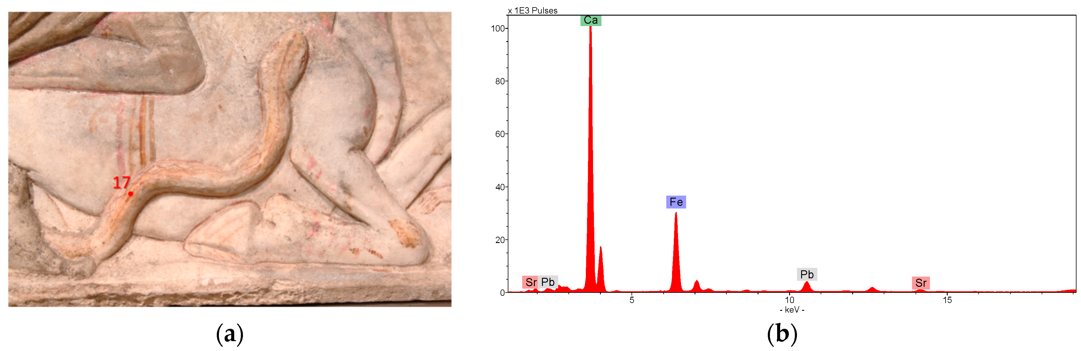
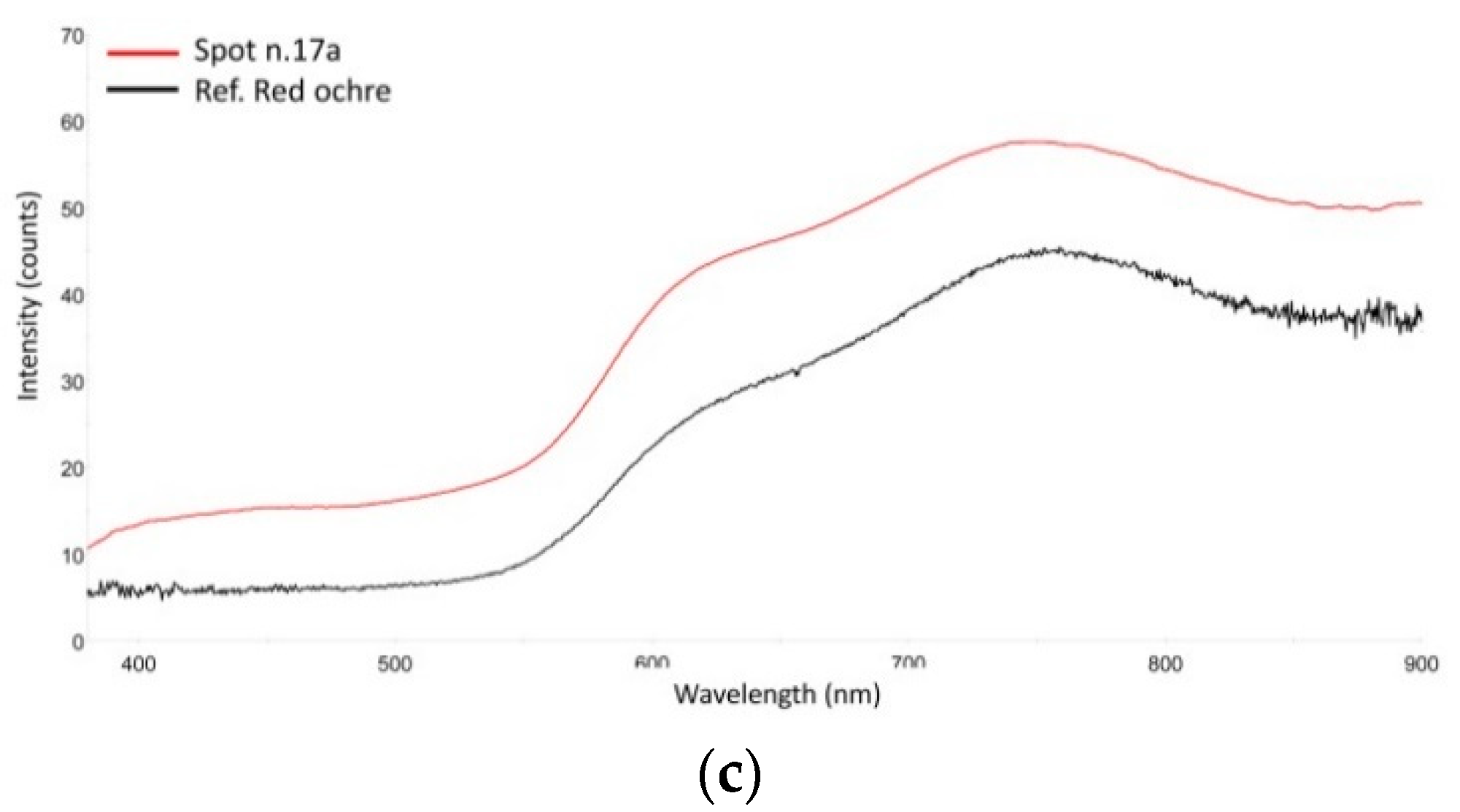
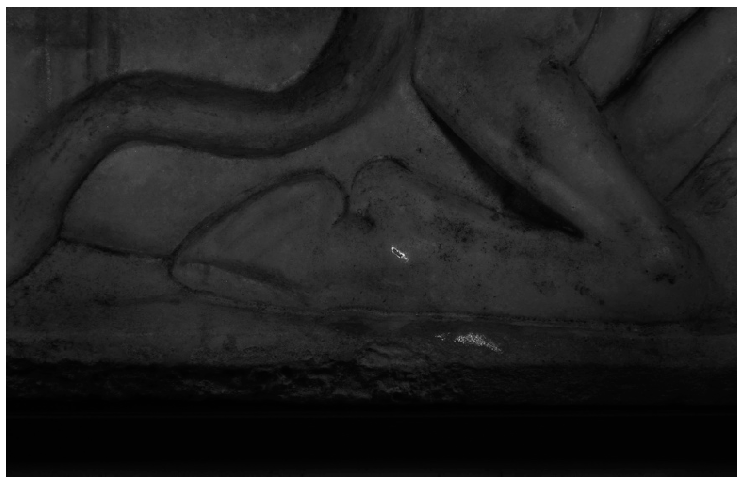
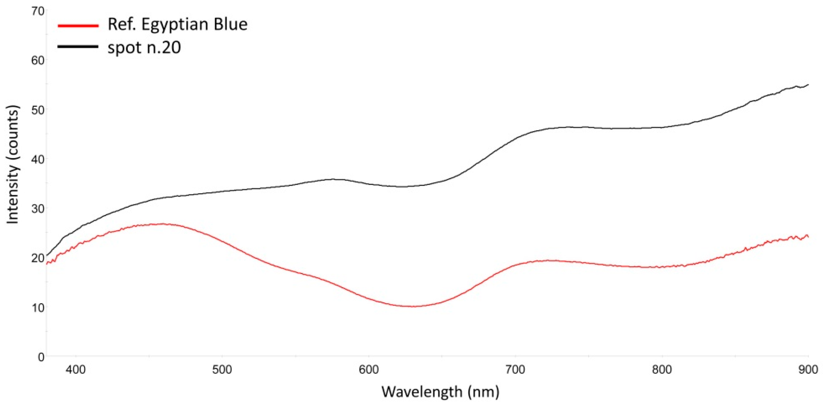
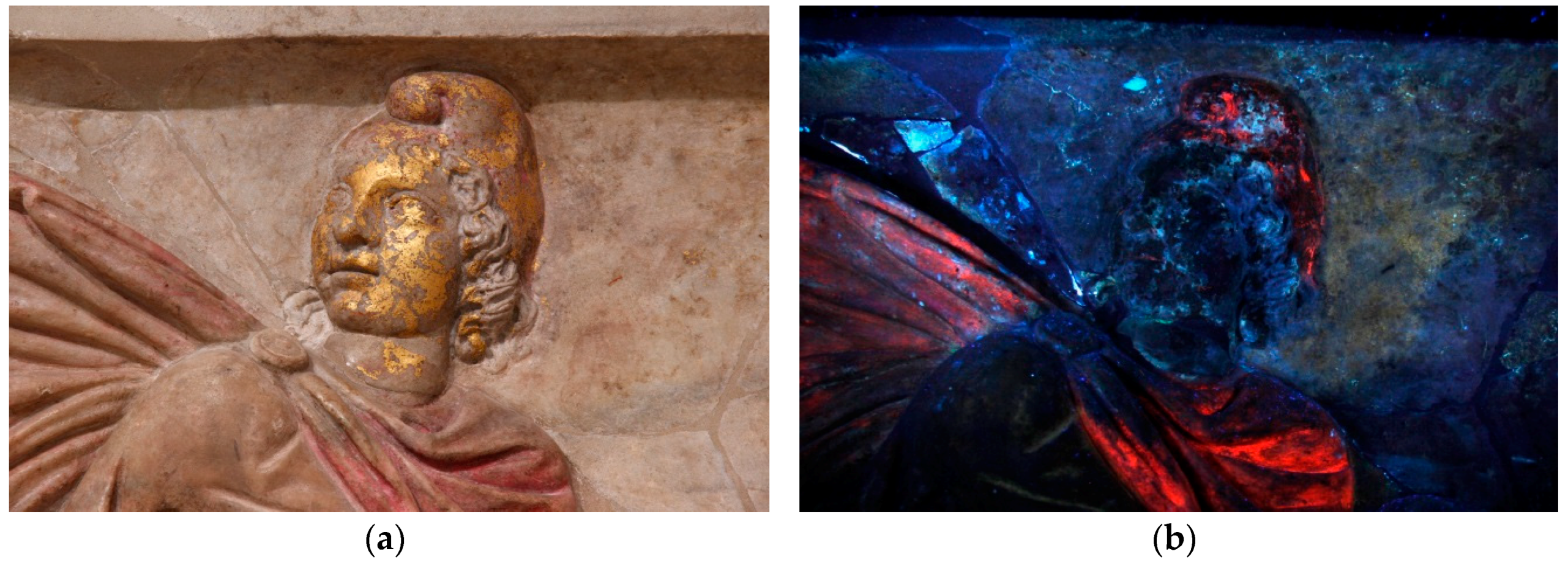
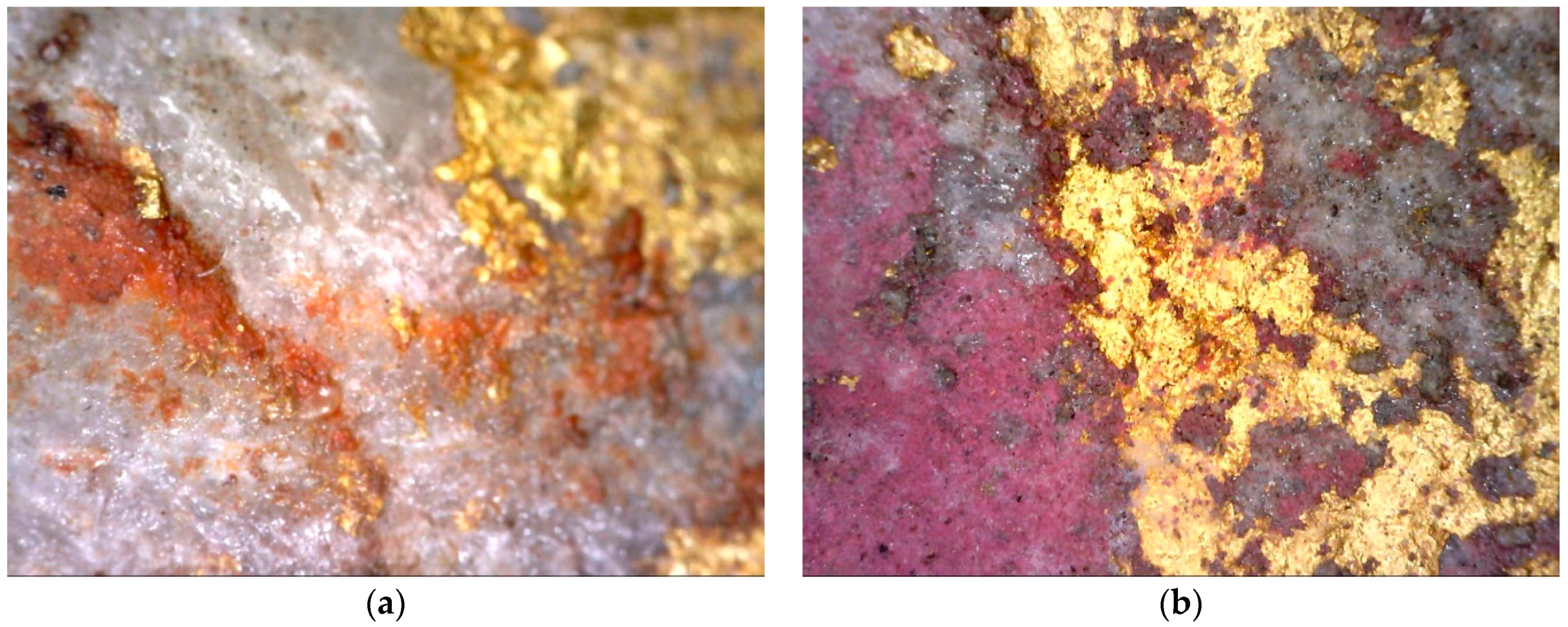
3. Results and Discussion

The technical examination was concentrated on studying the composition of the residual traces of polychromy on the stone surface. The examination showed that the slab was originally almost completely painted and today the residual polychromy is still visible and well preserved.
We carried out 30 spot analyses on the surface by both p-XRF and FORS. Digital portable microscope was also used to document the spot locations and to observe at high magnification the surfaces and traces of polychromy.
As observed comparing the results, a fairly wide range of colors was used to achieve different painterly effects. Red were used to highlight many details, such as eyebrows and eyelashes. Red-painted outlines are visible to the naked eye along the contours of some carved elements in order to make the relief stand out more clearly. Brown color appeared on the rooster close to Cautes as well and in some areas on both rocks. A few simple brushstrokes of red and yellow accentuate the essential lines of the bas-relief and define details, as is the case of the decoration of the bull’s body or the dog face. A definite diamond-shaped brown pattern is preserved on the snake body surface (Figure 3a). The red-brownish color, showing high iron (Fe) content in the XRF spectra suggested the use of ochre for these details (Figure 3b). These data were confirmed by FORS spectra showing the characteristic spectral features of iron oxides [10,11,12] (Figure 3c).
UVL was performed to reveal the presence and spatial distribution of organic materials or pigments [13,14,15]. On the slab, this photographic technique was able to capture the extensive use of a colorant with a distinctive pink-orange fluorescence [16,17].
This colorant has a strong emission in the visible and was identified as a madder lake, a red dye from the plant of the family of Rubiaceae, commonly used in the past [18,19,20,21]. Large areas of the slab, such as the Mithras’ mantle, some details of the bull, the blood and the torch were colored using this bright pink pigment (Figure 4).
The UVL images of the eyes and mouths of Hesperos, and the inner part of the mouth of the dog and bull, revealed also the use of the madder lake to accentuate the flesh parts along the rim of the eyes and between the lips (Figure 5).
The presence of the red lake was confirmed by FORS revealing in the spectrum (Figure 6) the characteristic patterns in the visible region, which includes a strong absorption band, structured into two main sub-bands, from 500–560 nm, a shoulder at 480–500 nm, a peak of reflectance at about 420 nm, and a sharp increase in reflectance at about 600 nm into the NIR due to the carbonyl groups of this anthraquinone based dye [22].
Visible induced luminescence photography of the slab showed the presence of a broad emission for few areas where color traces were not easily discernible. These unique luminescent properties are typical of the ancient blue pigment Egyptian Blue (CaCuSi4O10) [23,24,25]. The photographic technique was able to unmistakably identify and map Egyptian Blue, while it also provided support to better understand the iconography and color distribution in this slab.
The large amount of Egyptian Blue, in terms of traces, were found on the hoof and on the underlying frame (Figure 7). The latter was the only trace of Egyptian Blue found on the frame, likely a not intentional transfer. Few particles were found also in correspondence of the pink lines on the bull’s body.
Erratic particles, in very little amount, were found on other areas of the slab, but the amount and distribution were not enough to infer if these zones were intentionally painted or if Egyptian blue presence was completely random due to mechanical transfer from other areas/objects.
Spots more or less gathered were visible in some areas of the slab, but their amount cannot be related to a well specified hypothesis.
Validation of the presence of this pigment was provided by p-XRF and FORS (Figure 8) with the identification of a copper-based pigment with characteristic absorption maxima at ~560 and 628 nm and a minor absorption at ~790 nm [26].
This bas-relief is also characterized by an extensive presence of gold. In addition to the face and the cap of Mithra, as well as the cuff and some part of the knife, traces of gold were also visible in small areas on the background and within the folds of the dress.
Observing in detail the gilding, the gold leaf on the face and cap was applied on two different underlying layers. This was also observable from the UV induced luminescence image of the head, the red layer under the gold on the face didn’t show any fluorescence being practically similar to gold, while the pinkish layer under the gold on the Phrygian cap showed the characteristic fluorescence of a lake (Figure 9a,b).
In Figure 10a, acquired on the chin of Mithra, the microscopic examination highlighted the presence of a red ground layer below the golden foil, while the detail of the cap at high magnification (Figure 10b), confirmed the use of a red madder as ground layer. Single spot analyses confirmed the presence of an iron-based pigment on the chin and madder on the cap.
The reasons why this difference exists in the use of gilding preparation levels does not have a clear explanation. While the use of red ocher (bolus) is widely reported in the literature as a preparation layer for the adhesion of gold leaf, the use of a madder lake is rather strange and uncommon. It could be a stylistic subtlety of the painter to give different shades to gold. To the authors knowledge no similar cases are reported in the literature. Although a clear explanation is not possible, one hypothesis can be the following: the slab was originally painted without gilding. Only the Mithras’ face could be gilded, as well as the stucco Mithras’ head of the first phase Mithraeum. So, Mithras’ cap was painted with a red lake, as well as his mantle. In a second moment, also other parts of the garments were gilded. It is important to notice that, as well as the Mithraeum itself, also this slab could have various phases in its painting: it could be repainted and changed in its polychromy. The example of artworks with repainting and changing in their final color is quite common in antiquity [27,28]. Although very preliminary, this is one of the possible explanations for the presence of a gold foil on a layer of red lake.
A different ground layer was probably applied underneath the cuff. In this case XRF spectrum recorded high counts for lead.
Black pigment was found in two areas on the scorpion, together with small traces of gold, and on the raven. XRF spectrum on the scorpion’s back showed the presence of calcium, gold, and small traces of iron, the latter referable to the presence of soil traces on the surface. In the spectrum acquired on the raven, only counts for calcium with traces of iron and lead were detected. For both areas the presence of carbon black can be suggested. This hypothesis arises from the fact that no absorption bands or key-elements typical of different black pigments, such as manganese black, were present neither in FORS nor in XRF spectra.
The background of the bas-relief has some traces of red painting on the right upper corner (E. Lissi-Caronna described these traces of painting as possible stars), but no other trace of color was detected. One possible hypothesis for the color of the background is that he was originally unpainted. A comparison can be easily found in some Roman marble sarcophagi, with an intentionally unpainted background [7].

4. Conclusions

The investigation of the polychromy of the slab provided us with valuable insights into chromatic choices of Roman painting techniques. It demonstrates that the range of the palette was fairly wide and the use of it was subject to certain technical choices. An extensive color palette, yellow ochre, red iron oxide, pink madder lake, Egyptian blue, organic black were applied, combined with gilding added in large areas of the slab, covering most of its surface.
The colors’ choices were also related to a specific color code in the Mithraic cult: comparing this slab with other images with Mithras tauroctonos, such as the wall paintings in the Santa Maria Capua Vetere, Marino and Palazzo Barberini (Rome), is recognizable a similar color scheme. Mithras’ garments are often red, the bull is white, Cautes wears clothes filled with brighter colors than Cautopates (and this could be a direct reference of their opposite meaning in Mithraic cult). So, the colors of the slab in the Museo Nazionale Romano were applied following a specific color code. However, this particular meaning of the color could not be completely understood, without a comparison with the wall paintings and other color traces on artworks related with Mithraic cult. For this purpose, an interdisciplinary research is necessary. By means of a combined non-invasive approach using visual observations, multi-band imaging, microscopy, XRF and FORS this study allowed to acquire new data and provide a detailed documentation of surviving color and its relationship with other polychrome representations of Mithras. Moreover the well-established protocol allowed us to obtain as much information as possible without a sampling campaign and as little stress as possible for the artefact, avoiding the handling, thus permitting the development of the research on other artworks, in order to understand deeply the color choices of Mithraic cult.

Author Contributions

Data curation, D.M., S.B., G.B., R.I., S.L. and P.L.; Formal analysis, D.M., S.B., G.B. and R.I.; Investigation, D.M., S.B., G.B., R.I., S.L. and P.L.; Methodology, D.M., S.B., G.B., R.I. and S.L.; Supervision, S.B. and P.L.; Writing—original draft, D.M., R.I. and S.L.

Funding

This research received no external funding.

Conflicts of Interest

The authors declare no conflict of interest.

References

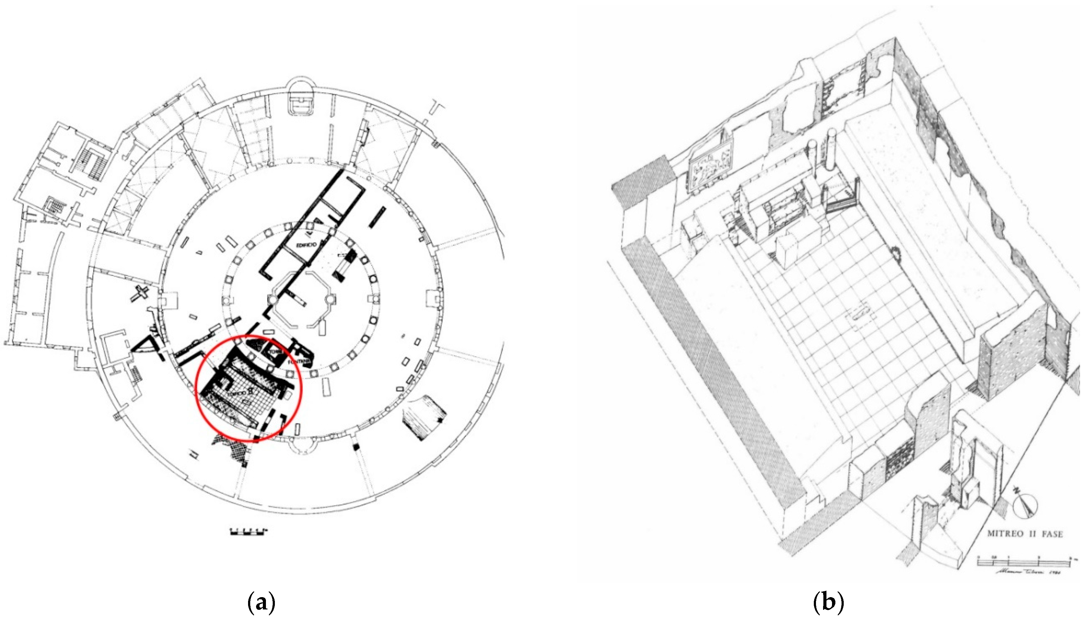
  1. Lissi-Caronna, E. IL Mitreo dei Castra Peregrinorum (S. Stefano Rotondo); Brill Publisher: Leiden, UK, 1986; ISBN 9789004296565. [Google Scholar]
  2. Brandenburg, H. Santo Stefano Rotondo sul Celio, l’ultimo edificio monumentale di Roma fra antichità e medioevo. In Roma Dall’antichità al Medioevo, 2 vol. Contesti tardoantichi e Altomedievali; Paroli, L., Vendittelli, L., Eds.; Mondadori Electa: Milan, Italy, 2004; pp. 480–505. [Google Scholar]
  3. Di Giacomo, G. Una statua di Mitra che nasce dalla roccia. In Catalogo del Museo Nazionale Romano—Terme di Diocleziano; Friggeri, R., Granino, M.G., Gregori, G.L., Eds.; Electa: Milano, Italy, 2012; pp. 642–643. ISBN 88-370-8934-1. [Google Scholar]
  4. Borgognoni, C. Un rilievo mitraico. In Catalogo del Museo Nazionale Romano—Terme di Diocleziano; Friggeri, R., Granino, M.G., Gregori, G.L., Eds.; Electa: Milano, Italy, 2012; pp. 647–648. ISBN 88-370-8934-1. [Google Scholar]
  5. Borgognoni, C.B. Un piccolo rilievo mitraico. In Catalogo del Museo Nazionale Romano—Terme di Diocleziano; Friggeri, R., Granino, M.G., Gregori, G.L., Eds.; Electa: Milano, Italy, 2012; ISBN 88-370-8934-1. [Google Scholar]
  6. Iannaccone, R.; Bracci, S.; Cantisani, E.; Mazzei, B. An integrated multi-methodological approach for characterizing the materials and pigments on a sarcophagus in St. Mark, Marcellian and Damasus catacombs. Appl. Phys. A 2015, 121, 1235–1242. [Google Scholar] [CrossRef]
  7. Liverani, P.; Bracci, S.; Iannaccone, R.; Lenzi, S.; Magrini, D.; Mazzei, B. Colours in the dark: New researches into catacombs. In Polychromy in Ancient Sculpture and Architecture; Bracci, S., Giachi, G., Liverani, P., Pallecchi, P., Paolucci, F., Eds.; Sillabe: Livorno, Italy, 2018; pp. 94–105. ISBN 978-88-8347-997-7. [Google Scholar]
  8. Noferi, C.; Bracci, S.; Iannaccone, R.; Lenzi, S.; Magrini, D. Consideration on Polychromy from a group of nenfro sarcophagi at the Archaeological Museum in Florence, from Gens Statlane Tomb in Tuscania (Viterbo). In Polychromy in Ancient Sculpture and Architecture; Bracci, S., Giachi, G., Liverani, P., Pallecchi, P., Paolucci, F., Eds.; Sillabe: Livorno, Italy, 2018; pp. 27–34. ISBN 978-88-8347-997-7. [Google Scholar]
  9. Karidas, A.G.; Brecoulaki, H.; Bourgeois, B.; Jockey, P.H. In-situX-Ray Fluorescence analysis of raw pigments and traces of polychromy on Hellenistic sculptureat the archaeological museum of Delos. In Proceedings of the 7th International Conference of ASMOSIA, Association for the Study of Marble and Other Stones in Antiquity, Thassos, Greece, 15–20 September 2003; pp. 811–829. [Google Scholar]
  10. Bacci, M. Fibre optics applications to works of art. Sens. Actuators 1995, 29, 190. [Google Scholar] [CrossRef]
  11. Bacci, M. UV-VIS-NIR, FT-IR and FORS Spectroscopies. In Modern Analytical Methods in Art and Archaeology; Ciliberto, E., Spoto, G., Eds.; Wiley: Chichester, UK, 2000; pp. 321–360. [Google Scholar]
  12. Bacci, M.; Fabbri, M.; Picollo, M.; Porcinai, S. Non-invasive fibre optic Fourier transform-infrared reflectance spectroscopy on painted layers. Anal. Chim. Acta 2001, 446, 15–21. [Google Scholar] [CrossRef]
  13. Aldrovandi, A.; Bertani, D.; Cetica, M.; Matteini, M.; Moles, A.; Poggi, P. Multispectral image processing of paintings. Stud. Conserv. 1988, 33, 154–159. [Google Scholar]
  14. Aldrovandi, A.; Buzzegoli, E.; Keller, A.; Kunzelman, D. Investigation of painted surfaces with a reflected UV false colour technique. In Proceedings of the 8th International Conference on Non Destructive Investigations and Micro Analysis for the Diagnostics and Conservation of the Cultural and Environmental Heritage, Lecce, Italy, 15–19 May 2005; pp. 3–18. [Google Scholar]
  15. Fischer, C.; Kakoulli, I. Multispectral and hyperspectral imaging technologies in conservation: Current research and potential applications. Rev. Conserv. 2006, 7, 3–16. [Google Scholar] [CrossRef]
  16. Costello, S.D.; Klausmeyer, P. A re-united pair: The conservation, technical study, and ethical decisions involved in exhibiting two terracotta orante statues from Canosa. Stud. Conserv. 2014, 59, 377–390. [Google Scholar] [CrossRef]
  17. De la Rie, E.R. Fluorescence of paint and varnish layers (part 1). Stud. Conserv. 1982, 27, 1–7. [Google Scholar]
  18. Hofenk de Graaff, J.H.; Roelofs, W.G.T.; van Bommel, M.R. The Colourful Past: Origins, Chemistry and Identification of Natural Dyestuffs, Archetype; Riggisberg Abegg-Stiftung: London, UK, 2004. [Google Scholar]
  19. Daniels, V.; Devièse, T.; Hacke, M.; Higgitt, C. Technological Insights into Madder Pigment Production in Antiquity; Technical Research Bulletin; British Museum: London, UK, 2014; pp. 13–28. [Google Scholar]
  20. Fostiridou, A.; Karapanagiotis, I.; Vivdenko, S.; Lampakis, D.; Mantzouris, D.; Achilara, L. Identification of pigments in hellenistic and Roman funeral figurines. Archaeometry 2016, 58, 453–464. [Google Scholar] [CrossRef]
  21. Kakoulli, I.; Radpour, R.; Lin, Y.; Svoboda, M.; Fischer, C. Application of forensic photography for the detection and mapping of Egyptian blue and madder lake in Hellenistic polychrome terracottas based on their photophysical properties. Dyes Pigments 2017, 136, 104–115. [Google Scholar] [CrossRef]
  22. Bisulca, C.; Picollo, M.; Bacci, M.; Kunzelman, D. UV-Vis-NIR Reflectance Spectroscopy of Red Lakes in Paintings. In Proceedings of the 9th International Conference on Nondestructive Testing of Art, Jerusalem, Israel, 25–30 May 2008; pp. 25–30. [Google Scholar]
  23. Accorsi, G.; Verri, G.; Bolognesi, M.; Armaroli, N.; Clementi, C.; Miliani, C.; Romani, A. The exceptional near infrared luminescence properties of cuprorivaite (Egyptian Blue). Chem. Commun. 2009, 23, 3392–3394. [Google Scholar] [CrossRef] [PubMed]
  24. Verri, G. The spatial resolved characterization of Egyptian Blue, Han Blue and Han Purple by photo-induced luminescence digital imaging. Anal. Bioanal. Chem. 2009, 394, 1011–1021. [Google Scholar] [CrossRef] [PubMed]
  25. Verri, G. The ‘Treu Head’: A case study in Roman sculptural polychromy. Br. Mus. Tech. Res. Bull. 2010, 4, 39–54. [Google Scholar]
  26. Mirti, P.; Appolonia, L.; Casoli, A.; Ferrari, R.P.; Laurenti, E.; Amisano Canesi, A.; Chiari, G. Spectrochemical and structural studies on a roman sample of Egyptian blue. Spectrochim. Acta Part A Mol. Biomol. Spectrosc. 1995, 51, 437–446. [Google Scholar] [CrossRef]
  27. Bourgeois, B. Thérapéia. Taking Care of Colour in Hellenistic Greece. In Transformations. Classical Sculpture in Colour; Østergaard, J.S., Nielsen, A.M., Eds.; Ny Carlsberg Glyptotekpublisher: Copenhagen, Denmark, 2014; pp. 190–207. [Google Scholar]
  28. Liverani, P. L’Augusto di Prima Porta. In I Colori del Bianco. Policromia Nella Scultura Antica; Casa Editrice Vaticano: Rome, Italy, 2004; pp. 235–242. [Google Scholar]
Figure 1. Reconstruction of the Mithraeum under Santo Stefano Rotondo, Rome: (a) the church of Santo Stefano Rotondo and the Castra Peregrinorum. The building with the Mithraeum is encircled in red (photo: elaboration from Lissi—Caronna 1986), (b) reconstruction of the second-phase Mithraeum (from Lissi—Caronna 1986).
Heritage 02 00130 g001
Figure 2. The big slab with Mithras slaying the bull from the Mithraeum under Santo Stefano Rotondo, National Museum of Rome, inv. n. 205837.
Heritage 02 00130 g002
Figure 3. Detail of the red-brown decorations; (a) Spot on the diamond shape pattern on the snake (spot location n.17) (b) X-ray fluorescence spectroscopy (XRF) spectrum; (c) fiber optics reflectance spectroscopy (FORS) spectrum of the spot measurement compared with a reference of red ochre.
Heritage 02 00130 g003aHeritage 02 00130 g003b
Figure 4. Ultraviolet induced luminescence (UVL) images (a) detail of Cautopates; (b) Mithra’s mantle.
Heritage 02 00130 g004
Figure 5. UVL images (a) the knife and the bull’s blood; (b) detail of Hesperos’ face and torch.
Heritage 02 00130 g005
Figure 6. FORS spectrum of an area on the mantle of Mithras (black) compared with the spectrum of reference mock-up of a madder lake (red).
Heritage 02 00130 g006
Figure 7. Visible induced luminescence (VIL) detail of the frame and of the bull’s pawn.
Heritage 02 00130 g007
Figure 8. FORS spectrum of an area where luminescence of EB was found (black) compared with the spectrum of a reference mock-up of EB (red).
Heritage 02 00130 g008
Figure 9. Detail of the face of Mithra: (a) visible image; (b) ultraviolet induced luminescence image.
Heritage 02 00130 g009
Figure 10. (a) Magnified detail of the remnants of gold applied on red ochre ground layer on the chin of Mithra; (b) magnified detail of the remnants of gold applied on the madder lake ground layer on the cap of Mithra.
Heritage 02 00130 g010
Table 1. Schematic table explicating the investigation plan. Analytical techniques and number of acquisitions.
Analytical Techniques AppliedNumber of Acquisitions
Multispectral imaging: Visible reflected (400–700 nm)70
Ultraviolet-induced luminescence (400–700 nm)70
Visible-induced luminescence (800–1100 nm)70
Optical microscopy70
UV-ViS Fiber optics reflectance spectroscopy (FORS) (350–900 nm)30
Portable X-ray fluorescence spectroscopy (p-XRF)30

