Article

Molecular Detection of a Bartonella sp. in Fleas and Mites Collected from Wild Rodents (Cricetidae: Sigmodontinae) in the Northeast of Argentine Patagonia


by
Winter Marina
1,2,†,
Acosta Diana Belén
3,†,
Abate Sergio Damián
1 and
Sanchez Juliana Patricia
2,4,*
1
Centro de Investigaciones y Transferencia de Rio Negro (CONICET-UNRN), Universidad Nacional de Río Negro-Sede Atlántica, Viedma 8500, Argentina
2
Consejo Nacional de Investigaciones Científicas y Técnicas (CONICET), Ciudad Autónoma de Buenos Aires C1425FQB, Argentina
3
Centro de Bioinvestigaciones-CeBio, Centro de Investigaciones y Transferencia del Noroeste de la Provincia de Buenos Aires-CIT NOBA (CONICET-UNNOBAUNSAdA), Pergamino 2700, Argentina
4
Instituto Nacional de Parasitología (INP)—ANLIS-Malbrán, Ciudad Autónoma de Buenos Aires C1282AFF, Argentina
*
Author to whom correspondence should be addressed.
These authors contributed equally to this work.
Zoonotic Dis. 2026, 6(3), 29; https://doi.org/10.3390/zoonoticdis6030029
Submission received: 23 June 2026 / Revised: 18 July 2026 / Accepted: 24 July 2026 / Published: 28 July 2026

Simple Summary

This study investigated fleas and mites from wild mice in Argentine Patagonia, detecting Bartonella DNA. This specific pathogen had not previously been reported in the region. The proximity of these wild hosts to human settlements and workspaces suggests a potential risk of transmission to people and pets. These results contribute to a better understanding of the distribution and epidemiology of ectoparasite-borne zoonotic diseases.

Abstract

Ectoparasite-borne infections have become a global public health problem. This study investigated the diversity of fleas and mites associated with wild rodents in the northeastern region of Argentine Patagonia and analyzed the molecular presence of Rickettsia and Bartonella within these arthropods. Research was conducted in the Monte Desierto biome, focusing on areas where wildlife, livestock, and human activities overlap. Ectoparasites (n = 32) were collected from captured rodents and subjected to PCR amplification and phylogenetic analysis using gltA and rpoB for Bartonella and OmpA, OmpB and gltA for Rickettsia. Bartonella DNA was detected in all studied ectoparasites, with the obtained gltA and rpoB sequences showing 100% and 99% identity, respectively, to reference sequences of Bartonella sp. closely related to Bartonella quintana, whereas no Rickettsia DNA was detected. Specifically, the pathogen was identified in four flea species (Craneopsylla minerva wolffhuegeli, Neotyphloceras crackensis, Polygenis rimatus, and Ectinorus sp.) and the mite Gigantolaelaps wolffsohni, while Rickettsia was not detected in any samples. These findings constitute the first report of Bartonella sp. closely related to B. quintana in this geographical region as well as the first detection of the bacterium in these specific flea and mite species in South America. Our results suggest an active wild cycle of Bartonella sp., emphasizing the importance of the One Health approach in monitoring emerging infectious diseases.

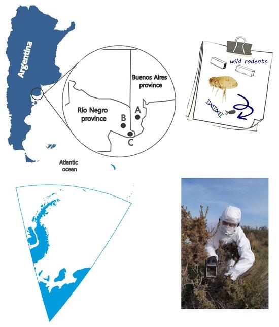
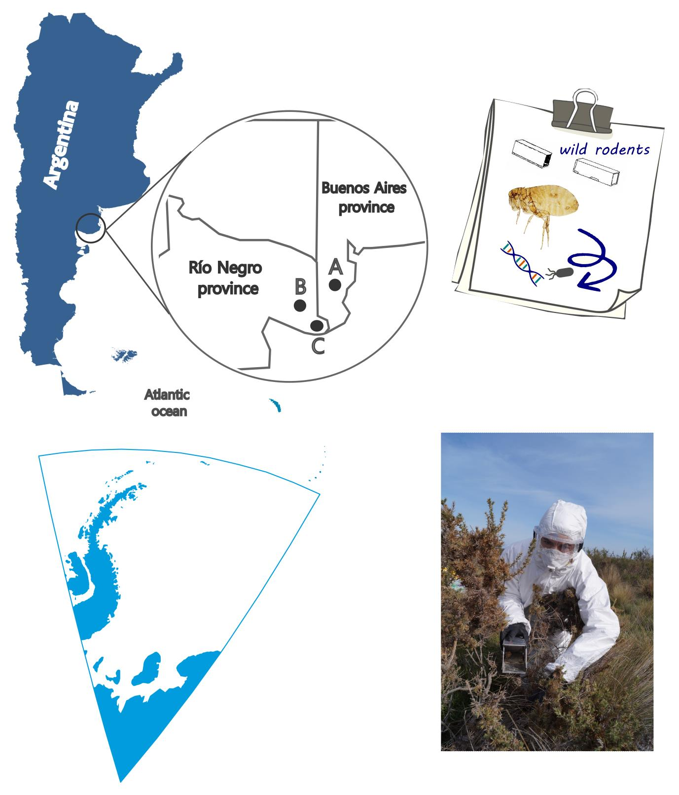
Graphical Abstract

1. Introduction

Sigmodontine rodents (Cricetidae) constitute the most diverse mammalian subfamily and serve as primary hosts for fleas (Siphonaptera) [1]. The high species richness of fleas associated with these rodents suggests a deep coevolutionary history between host and ectoparasite [1,2]. Beyond these evolutionary dynamics, fleas are critical vectors for pathogens; specifically, bartonellosis, rickettsiosis, and yersiniosis represent the primary zoonotic diseases transmitted by these insects [3]. However, while research has predominantly focused on ticks and secondarily on fleas, the role of Mesostigmata mites as rodent ectoparasites and potential pathogen carriers remains comparatively understudied, particularly in South America [4].
Mites and fleas serve as vectors for bacteria of the genus Bartonella. This genus comprises a large number of facultative intracellular bacteria. Currently, there are 131 species and candidates of Bartonella in the List of Prokaryotic names with Standing in Nomenclature (LPSN) (https://lpsn.dsmz.de/search?word=bartonella accessed on 18 June 2026), with at least 17 identified as zoonotic pathogens affecting humans and domestic animals [5,6]. Given its broad impact, bartonellosis is increasingly regarded as a major emerging infectious disease [3]. In Argentina, clinical bartonellosis has been documented in both pediatric and adult patients. Reported cases include cat scratch disease (CSD) caused by Bartonella henselae (Hyphomicrobiales; Bartonellaceae) in Buenos Aires and San Luis provinces, as well as B. quintana-induced infective endocarditis in an adult from Buenos Aires [7,8,9]. Moreover, Bartonella DNA has been detected in Argentine fleas associated with domestic and wild meso mammals and wild rodent fleas [10,11,12,13,14,15,16].
Similarly, the genus Rickettsia comprises a diverse group of obligate intracellular bacteria. Currently, 177 species and candidates are listed in LPSN (https://lpsn.dsmz.de/search?word=Rickettsia accessed on 18 June 2026). The environmental circulation of rickettsiae depends strictly on vector-host interaction, and recent years have seen a significant rise in reported cases globally [17]. However, according to the Subcommittee on Rickettsiosis to the Tick-Borne Disease Working Group (2022), official figures are likely underestimated due to the lack of rapid diagnostic tests at primary healthcare centers [18]. In Argentina, human rickettsiosis has been primarily linked to Rickettsia rickettsii and R. parkeri, both transmitted by ticks [17,19,20]. While the growing body of knowledge in Argentina has focused heavily on ticks and ectoparasites associated with domestic animals, studies regarding wild species remain comparatively limited [14,15,16,21,22,23,24,25,26,27].
Over the past decade, arthropod-borne infections have become a global public health problem. These include diseases transmitted by ectoparasites, with outbreaks and emergencies in countries where they have caused serious cases in humans [28]. Anthropogenic pressure, rather than wildlife presence alone, dictates disease emergence risks at the human–animal interface. Activities like forestry, mining, agriculture, and urbanization alter host-vector-pathogen dynamics and increase spatial overlap. Furthermore, climate-driven rodent migrations into human settlements exacerbate zoonotic transmission. Therefore, research into rodent-ectoparasite vector associations is crucial to understanding these shifting dynamics and establishing baseline data [29,30]. The aim of this study was to evaluate the diversity of ectoparasites associated with wild rodents in the extreme northeast of Argentinian Patagonia, and analyze the molecular presence of Rickettsia and Bartonella in these arthropods.

2. Material and Methods

2.1. Study Area

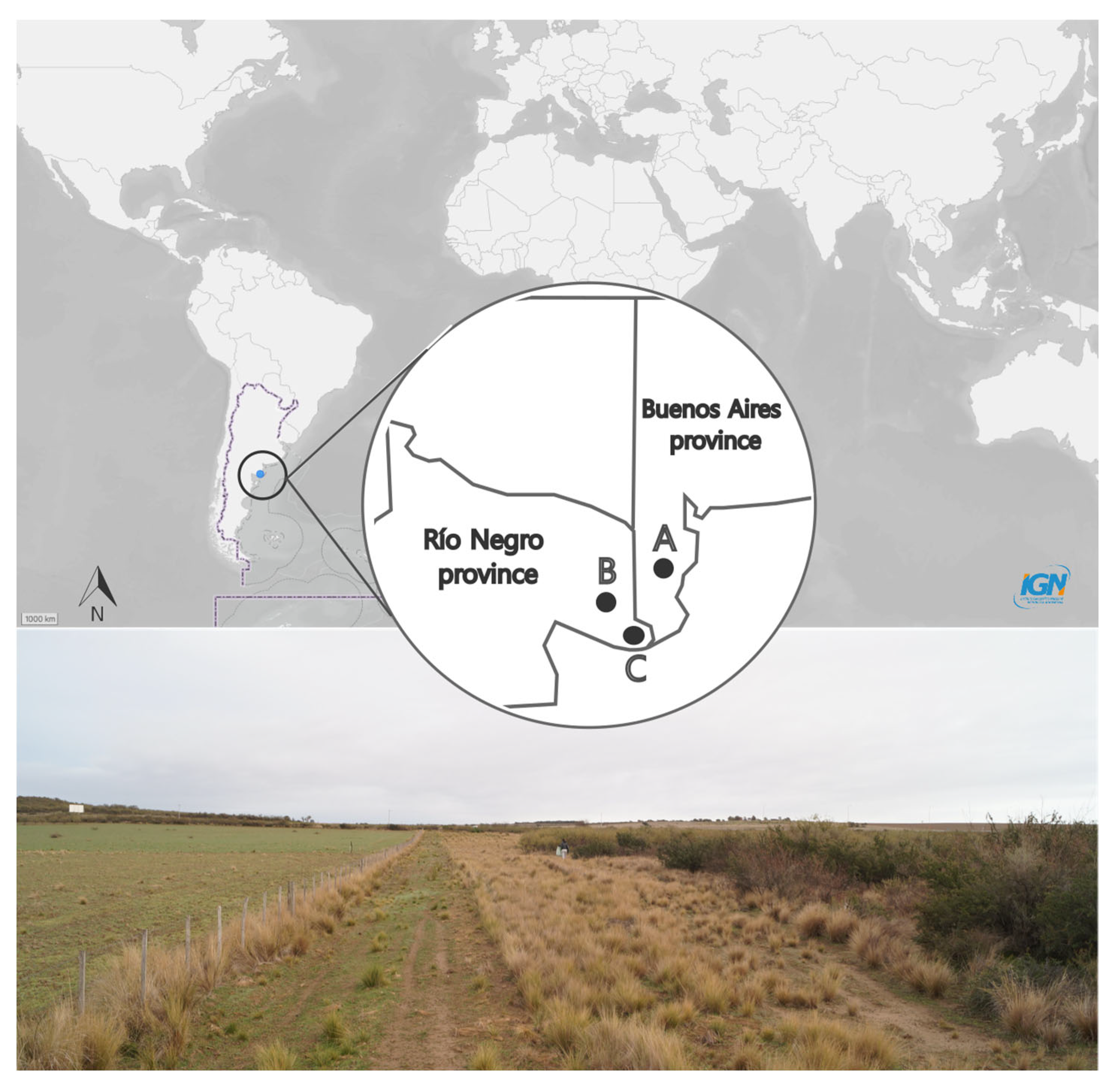
The study area was located in the northeastern-most part of Argentine Patagonia, on the border between the provinces of Buenos Aires and Rio Negro (Figure 1). The natural ecosystem corresponds to the Monte Desierto biome [31]. The climate is semi-arid to arid, with high evaporation exacerbated by wind, and average annual precipitation ranges from less than 100 to 450 mm. The vegetation is fairly uniform in terms of appearance and floristic composition [31,32]. Native xerophytic flora alternates with exotic species and areas of semi-extensive livestock farming (mainly cattle) and agriculture. Rodent traps were placed on private properties with a weak or nonexistent biosecurity protocol. Dogs and cats were observed coexisting with livestock and wildlife. Thus, it is common for dogs to travel weekly between rural and urban areas with their owners.

2.2. Host and Ectoparasites Collection and Identification

Between June and August of 2017 and 2018, eight rodent trapping sessions were conducted across three sampling sites: Site A (4 trapping sessions; −40°27′55″/−62°47′55″), Site B (2 trapping sessions; −40°43′35″/−63°17′56″), and Site C (2 trapping sessions; −40°58′34″/−62°53′08″) (Figure 1). Rodents were captured using Sherman live traps (23 × 8 × 9 cm3) baited with oats. Traps were inspected daily in the early morning for three consecutive days. At each site, 40 traps were deployed nightly in two parallel lines of 20, spaced 5 m apart, following the local topography and vegetation. Species identification followed the criteria of Gómez Villafañe et al. [33]. Biosecurity protocols were strictly observed during animal handling, and an open-air field laboratory was established at each site. The study was approved by the Secretary of Environment and Sustainable Development of Río Negro (085206SAYDS/218/2015) and the Office of Flora and Fauna of Buenos Aires (22500-2952). Additionally, protocols were reviewed and authorized by the Institutional Care and Use Committee (CICUAL) of the Faculty of Veterinary Sciences, University of Buenos Aires (2016/4, dated 16 June 2016).
Ectoparasites were collected by forceps and stored in 96% ethyl alcohol until molecular analyses were carried out in the Centro de Bioinvestigaciones (CeBio—UNNOBA). Details on parasitological examination as well as on flea identification can be found in Sanchez (2013), Sanchez and Lareschi (2019) and for mites in Furman (1972) [34,35,36].
After DNA extraction, fleas and mites were prepared for morphological study and identification. Fleas were cleared and softened in an aqueous solution of potassium hydroxide 10% (KOH), dehydrated in an increasing series of ethanol (80–100%), further diaphanized in eugenol, and mounted on permanent slides with Canadian balsam and studied under light microscopy. Identification keys used included those of Hopkins and Rothschild (1953), Johnson (1957) and Smit (1987) [37,38,39]. Mites were cleared in lactophenol and mounted individually in Hoyer’s medium for their identification in accordance with the original descriptions and drawings of Furman (1972) [36].
DNA 70 ethanol followed by sterile distilled water to remove external contaminants before processing. For fleas, a small incision was made between the third and fourth abdominal tergites using a sterile scalpel to obtain material for DNA extraction, while preserving the remaining specimen for subsequent morphological identification. For mites, a small incision was made through the body to obtain material for DNA extraction, while maintaining the remaining specimen for morphological examination. The material used to handle the ectoparasites was sterilized between each sample. Genomic DNA extraction was performed from individual ectoparasites per host, using the Chelex®-100 (Bio-Rad Laboratories, Hercules, CA, USA) method described by Acosta et al. [16]. The genomic DNA obtained was stored at −20 °C under sterile conditions. Following the DNA extraction, the fleas and mites’ exoskeletons were recovered and stored in 96% ethanol and subsequently mounted for species identification.

2.3. Molecular Detection of Bartonella spp. and Rickettsia spp.

The presence of Bartonella was screened using the citrate synthase (gltA) and RNA polymerase beta-subunit (rpoB) genes, while the presence of Rickettsia was screened using the citrate synthase (gltA), outer membrane protein A (ompA) and outer membrane protein B (ompB) genes (Table 1). Using the gltA gene, the genus of both bacteria can be confirmed, while the other molecular markers show the specific identity. For the amplification, the polymerase chain reaction (PCR) program started with an initial denaturation for 5 min at 95 °C, followed by 40 cycles (95 °C for 30 s, gene-specific annealing °C for 30 s, and 72 °C for 30 s), and a final extension step at 72 °C for 5 min (Table 1). PCR was set to a final volume of 20 μL, containing 1 ng/µL of template DNA for mite samples and up to 20 ng/µL for flea samples, according to the DNA yield obtained from individual ectoparasite extractions, 1.5 mM MgCl2, 0.2 μM of each primer, 0.2 mM of each dNTP, 1X reaction buffer, 0.5U of Pegasus DNA polymerase and ultrapure sterile water to come to final volume. All amplifications were performed in conjunction with a negative (distilled water) and positive (DNA of Bartonella spp. provided by the Servicio Bacteriología Especial, “Dr. Carlos G. Malbrán,” INEI-ANLIS, Buenos Aires, Argentina, and DNA of Rickettsia spp. provided by the Instituto Nacional de Enfermedades Virales Humanas “Dr. Julio I. Maiztegui,” INEV-ANLIS, Pergamino, Argentina) controls. Additionally, all PCR setup procedures were performed under sterile conditions in a laminar flow cabinet, using dedicated materials and aerosol-resistant filter tips to minimize the risk of cross-contamination. DNA fragment amplification was confirmed by electrophoresis on 1% m/v agarose gel, stained with ethidium bromide (10 µg/μL) and visualized under UV light.
In the samples with positive PCR for the genes analyzed, we proceeded to purification using 10U of Exonuclease I (Thermo Fisher Scientific, Waltham, MA, USA) and 1U of FastAp thermosensible alkaline phosphatase (Thermo Fisher Scientific), incubating at 37 °C for 15 min and a subsequent 15 min at 85 °C to stop the reaction. The purified samples were sequenced by Macrogen® (Seoul, Republic of Korea).

2.4. Data Analysis

The obtained sequences for the genes were analyzed and manually edited using the BioEdit program [44]. To assign identity to each sequence with statistical significance, it was subjected to a homology comparison against the GenBank nucleotide database, making use of the nucleotide BLAST algorithm (https://blast.ncbi.nlm.nih.gov/Blast.cgi accessed on 15 May 2026). The sequences have been deposited in the GenBank nucleotide database (https://www.ncbi.nlm.nih.gov/genbank/ accessed on 15 May 2026) under accession numbers PZ407960 to PZ407968.
To perform the phylogenetic analysis, sequences were taken from the GenBank nucleic acid database. The complete set of gene sequences was employed for a multiple alignment performed with the ClustalW algorithm in MEGA v.11 [45]. The resulting alignment was checked and manually corrected. Moreover, phylogenetic trees were built using the Maximum Likelihood (ML) and Neighbor Joining (NJ) methods of statistical inference, both for individual genes and for concatenated sequences. In the case of the concatenation of genes, the Farris test was initially performed using methods PAUP* based on the inference on parsimony to establish whether these genes could be used [46,47]. Through the Mesquite program, the sequences were concatenated [48]. The evolutionary history was inferred using the ML method based on the Tamura 3-parameter (I + G) model with 10,000 replicates of random-addition taxa and tree bisection and reconnection branch swapping. Branch support was assessed using 1000 bootstrap replicates. All positions were weighted equally.

3. Results

With a total sampling effort of 910 trap-nights, we recorded 127 captures (61 in 2017 and 66 in 2018). Ectoparasites were recovered from 16 rodents: 26 fleas—Craneopsylla minerva wolffhuegeli (18), Neotyphloceras crackensi (3), Polygenis rimatus (4), Ectinorus sp. (1)—and 6 Gigantolaelaps wolffsohni mites (6). Specific host–parasite associations are detailed in Table 2.
Total genomic DNA was extracted from fleas and mites collected from all infested rodents. The genus Bartonella was detected for the gltA and rpoB genes in all the ectoparasites studied. Both genes were detected in fleas of the species Neotyphloceras crackensis (3/3), Craneopsylla minerva wolffhuegeli (18/18), Ectinorus sp. (1/1) and Polygenis rimatus (4/4), and in the mite species Gigantolaelaps wolffsohni (6/6). Among the PCR-positive specimens, five individual ectoparasites, representing each positive ectoparasite species, were selected for sequencing and molecular analyses (Supplementary Material Table S1). The resulting sequences were deposited in GenBank under accession numbers PZ407960–PZ407963 (gltA) and PZ407964–PZ407968 (rpoB).
Sequence analysis revealed genetic variation among the detected Bartonella variants. The rpoB sequences corresponded to five distinct haplotypes, each showing nucleotide differences among samples (Supplementary Material Table S1). Similarly, the gltA marker showed four haplotypes and 29 variable nucleotide positions among the analyzed sequences (Supplementary Material Table S1).
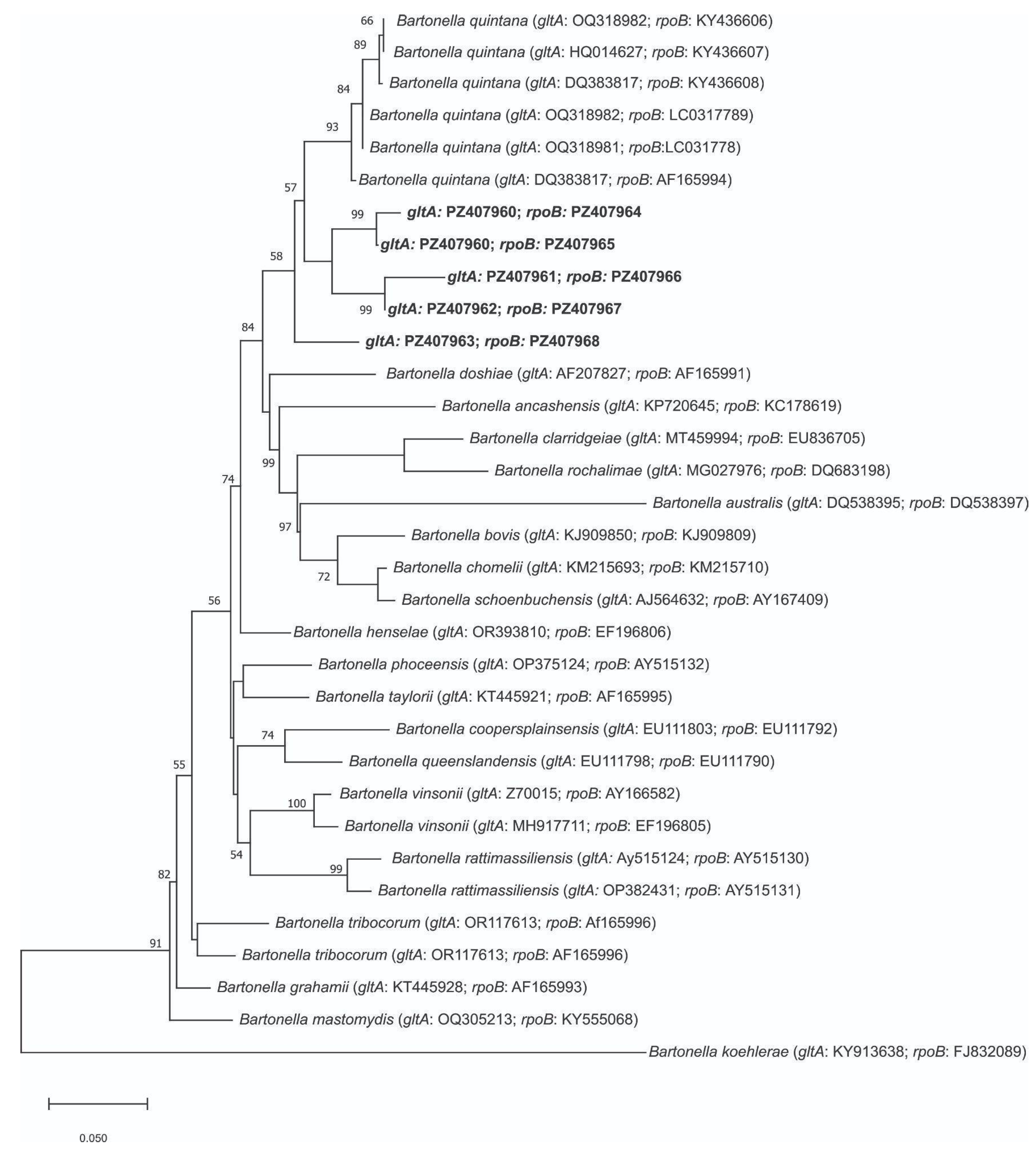
For gltA, nBLAST analysis indicated 100% (query cover 100%; e-value = 0.0) identity with Bartonella spp. For the rpoB gene, the nBLAST analysis indicated 99% (query cover 97%; e-value = 3e−159) identity with Bartonella quintana. When both genes were concatenated, it gave an identity of 98% with Bartonella quintana (query cover 100%; e-value = 2e−157). Phylogenetic analyses through ML and NJ inferences were inferred from the gltA and rpoB, separately analyzed, as well as by concatenating these genes, resulting in a total length of 309 bp (Figures S1 and S2 in Supplementary Materials). The phylogenetic analysis, both the ML and NJ inferences, showed the same topology; thus, only the ML is shown. These results showed that all sequences obtained in this study are grouped with B. quintana (Figure 2; Supplementary Material Figures S1 and S2). Although the amplified gltA and rpoB fragments were shorter than those proposed by La Scola et al. (2003) for species-level identification, these loci have been widely used in molecular surveys of Bartonella because they contain informative polymorphic sites that facilitate preliminary genetic characterization [49,50]. However, given the limited phylogenetic resolution provided by these short fragments, we conservatively refer to the detected bacterium as Bartonella sp. closely related to B. quintana rather than assigning it unequivocally to that species.
The genus Rickettsia was not detected in the ectoparasites studied.

4. Discussion

Some studies have shown that the diversity of ectoparasites remains constant among rodent communities in anthropogenic environments with varying degrees of alteration [51].
Interactions between wild and domestic animals facilitate horizontal transmission and create new niches for pathogen evolution and persistence. Zoonotic agents can spread to humans through any point of contact with wildlife or livestock. In this context, wild rodents often act as silent links; by sharing micro- and macro-environments, they establish epidemiological chains that bridge the gap between species. Most studies of rodent–ectoparasites–pathogen associations have been conducted near natural protected areas [52]. Therefore, the information obtained in this study becomes even more relevant when one considers that the sampling sites are private fields that are the visual essence of the One Health approach. These areas blend wild environments with the presence of domestic production animals: primarily cattle and sheep, and domestic dogs used for fieldwork but living alongside people. The role of dogs, in this case, could be fundamental in linking infections from wild and urban environments. Dogs act as reservoirs for various Bartonella species; notably, high prevalence rates have been reported in clinically asymptomatic individuals [53,54,55,56].
Human body lice, Pediculus humanus humanus, are considered the main vectors of trench fever caused by B. quintana [57]. However, B. quintana DNA and other species have been amplified from bed bugs and cosmopolitan fleas including Ctenocephalides canis, Ctenocephalides felis felis, and Pulex irritans (Pulicidae) [58]. Furthermore, B. quintana infections have been reported to be one of the major causes of endocarditis. A recent review highlights the lack of data on other arthropods-beyond lice-and a marked geographical asymmetry in research; while some regions have abundant studies on the vectors of this zoonosis, in others the information is practically nonexistent [59].
In southern South America, research has documented the circulation of Bartonella among various flea species infesting rodents and other wild mammals. In Brazil, Bartonella sp. DNA was identified in Craneopsylla minerva minerva from Oligoryzomys nigripes (3/6); in Polygenis occidentalis from Oxymycterus nasutus and O. nigripes (2/5); and in P. platensis parasitizing Akodon azarae and Scapteromys tumidus (2/2) [60]. Additionally, B. clarridgeiae and Bartonella sp. were detected in Ctenocephalides felis (2/6) collected from wild felids such as the margay (Leopardus wiedii) and jaguarundi (Herpailurus yagouaroundi), respectively [61]. In Chile, B. tribocorum was found in Plocopsylla sp. and Nosopsyllus sp. from Mus musculus and Abrothrix spp. (11/39) [62]. Regional studies in Argentina show diverse findings: in southern Patagonia, Bartonella sp. was detected in Neotyphloceras crackensis (28/36) from Phyllotis xanthopygus, Eligmodontia morgani and Abrothrix hirta, whereas in northern Argentina, López Berrizbeitia et al. (2024) reported B. quintana in C. m. minerva (10/22) and Polygenis acodontis (6/14) from several wild rodents [13,14]. Most recently, B. rochalimae DNA was amplified from Phthiropsylla agenoris (1/13) on armadillos (Chaetophractus villosus), and from Pulex irritans (2/2) on plains vizcachas (Lagostomus maximus) and gray foxes (Lycalopex gymnocercus) [16]. Research on the presence of Bartonella DNA in fleas in southern South America shows wide variability in the apparent prevalences detected, both when analyzing each flea species individually and when evaluating all samples. In the region, reported values range from 8% to 100%, a range consistent with the findings of this study. Notably, sample sizes per species are often relatively small, underscoring the logistical and technical challenges inherent in wildlife field research.
The genus Gigantolaelaps is a group of large ectoparasitic mites that infest New World sigmodontine rodents, specifically of the tribe Oryzomyini (such as O. longicaudatus) [57]. In this system, males reside exclusively in the host’s nest, whereas females live on the host for feeding and dispersal. Although females acquire Bartonella (and other pathogens) via hematophagy, they return to the protected nest microclimate to oviposit. So, the host nest acts as a permanent physical and biological reservoir, buffered against the external environmental fluctuations that typically constrain flea populations. Gigantolaelaps ensures enzootic persistence to the host species and its offspring within the nest. This sex-biased division of roles functions as an ecological strategy that prevents Bartonella clearance from the rodent population, underscoring the critical and understudied epidemiological role of this mite genus [63,64,65,66].
Although Rickettsia DNA was not amplified in this study, its presence has been documented for the study area in C. felis collected from a white-eared opossum (Didelphis albiventris) [16]. D. albiventris is known to interact with rodents primarily through predation, sharing habitats, and serving as a host for common parasites. R. felis is an etiological agent within the Spotted Fever Group (SFG). Although it is a cosmopolitan zoonosis, in countries like Argentina it remains a neglected disease [67]. It is prevalent in both domestic animals and humans, yet frequently underdiagnosed in its early stages due to clinical symptoms that overlap with more common illnesses such as influenza and dengue [68,69]. Sampling point C of this work is located very close to the Rickettsia record of Acosta et al. (2025) [16]. The geographical coverage is an interface area between natural, rural and urban ecosystems with abundant domestic canids and felids roaming about. The absence of this bacterium in this rodent-focused study reinforces the need and importance of conducting comprehensive studies, evaluating the role of rodents as reservoirs.
Local studies are foundational for mapping the epidemiological networks of pathogenic microorganisms. Although these findings are site-specific and based on a relatively small sample, the data collected between 2017 and 2018 constitute an essential historical baseline for retrospectively assessing the impact of recent environmental changes. Consequently, replicating and expanding this line of research is critical to validating these trends over broader spatial and temporal scales. These results also serve as crucial indicators for neighboring regions or countries with similar bioclimatic conditions. Furthermore, fleas are well-known vectors; our positive molecular results in mites provide valuable information about other ectoparasites that could be involved in the maintenance and transmission cycles of Bartonella. These findings suggest an active wild cycle for this bacterium. Within the One Health framework, information regarding the links between wildlife, ectoparasites, and pathogens should ideally be prioritized when planning new urban developments.

5. Conclusions

This study confirms for the first time the presence of Bartonella sp., closely related to B. quintana, in fleas and mites from wild rodents in northeastern Argentine Patagonia. These results represent the first record for this region, as well as the first detection in South America of Bartonella in the flea species Craneopsylla minerva wolffhuegeli, Ectinorus sp., and Polygenis rimatus, and in mites of the genus Gigantolaelaps. These findings constitute an essential baseline for the surveillance of environmental impacts on the epidemiology of regional vector-borne diseases. The absence of Rickettsia in this study, despite previous reports in the area, highlights the need for continued, comprehensive research. In future research, it will be necessary to expand the sample size of ectoparasites and include the hosts in the studies, in order to define their role as reservoirs of both Bartonella and Rickettsia.

Supplementary Materials

The following supporting information can be downloaded at: https://www.mdpi.com/article/10.3390/zoonoticdis6030029/s1, Supplementary Material Table S1. Molecular characterization of Bartonella-positive ectoparasite specimens analyzed in this study. The table shows the ectoparasite species, GenBank accession numbers for the gltA and rpoB sequences, and the polymorphic nucleotide sites identified among the sequences generated in this study. Nucleotide positions correspond to the aligned sequences for each marker. Dots (.) indicate nucleotide identity with the reference sequence used for the alignment, whereas letters indicate nucleotide substitutions; Supplementary Material Figure S2. Phylogenetic tree inferred using the Maximum Likelihood method based on partial rpoB sequences of Bartonella spp. detected in fleas and mites from Argentine Patagonia. Bootstrap support values > 50% are shown at the nodes. Bartonella genotypes identified in the present study are highlighted in bold; Supplementary Material Figure S3. Phylogenetic tree inferred using the Maximum Likelihood method based on partial gltA sequences of Bartonella spp. detected in fleas and mites from Argentine Patagonia. Bootstrap support values > 50% are shown at the nodes. Bartonella genotypes identified in the present study are highlighted in bold.

Author Contributions

Conceptualization, W.M., A.D.B., A.S.D. and S.J.P.; methodology: W.M., A.D.B., A.S.D. and S.J.P.; investigation, W.M., A.D.B., A.S.D. and S.J.P.; data curation, W.M., A.D.B., A.S.D. and S.J.P.; writing—original draft, W.M., A.D.B. and S.J.P.; writing—review and editing, W.M., A.D.B., A.S.D. and S.J.P.; visualization, W.M. and A.D.B.; supervision, A.S.D. and S.J.P.; project administration, W.M., A.D.B. and S.J.P.; funding acquisition, W.M., A.D.B., A.S.D. and S.J.P. All authors have read and agreed to the published version of the manuscript.

Funding

This research was funded by Universidad Nacional de Río Negro projects number 40-C-665 and 40-C-983.

Institutional Review Board Statement

The rodent capture and removal protocol was approved by the Institutional Committee for the Care and Use of Laboratory Animals (CICUAL) of the Faculty of Veterinary Sciences of the University of Buenos Aires (2016/4, dated 16 June 2016). The study was approved by the Secretary of Environment and Sustainable Development of Río Negro (085206SAYDS/218/2015) and the Office of Flora and Fauna of Buenos Aires (22500-2952).

Informed Consent Statement

Not applicable.

Data Availability Statement

The original contributions presented in this study are included in the article/Supplementary Material. Further inquiries can be directed to the authors.

Acknowledgments

The authors thank the Secretary of Environment of the province of Río Negro, the Office of Flora and Fauna of the province of Buenos Aires, and the owners of fields where the rodent sampling was carried out.

Conflicts of Interest

The authors declare no conflicts of interest.

References

  1. Whiting, M.F.; Whiting, A.S.; Hastriter, M.W.; Dittmar, K. A molecular phylogeny of fleas (Insecta: Siphonaptera): Origins and host associations. Cladistics 2008, 24, 677–707. [Google Scholar] [CrossRef]
  2. Krasnov, B.R. Functional and Evolutionary Ecology of Fleas: A Model for Ecological Parasitology; Cambridge University Press: New York, NY, USA, 2008; p. 610. [Google Scholar]
  3. Cutillas, C.; Trujillo, I.; Zurita, A.; García-Sánchez, A.M. Highlighting zoonotic importance of synanthropic fleas through microbiome analysis. Sci. Rep. 2025, 15, 37651. [Google Scholar] [CrossRef] [PubMed]
  4. Herrera-Mares, A.; Guzmán-Cornejo, C.; Ulloa-García, A.; Córdoba-Aguilar, A.; Silva-de la Fuente, M.C.; Suzán, G. Mites, rodents, and pathogens: A global review for a multi-species interaction in disease ecology. Acta Trop. 2022, 232, 106509. [Google Scholar] [CrossRef] [PubMed]
  5. Breitschwerdt, E.B.; Maggi, R.G.; Chomel, B.B.; Lappin, M.R. Bartonellosis: An emerging infectious disease of zoonotic importance to animals and human beings. J. Vet. Emerg. Crit. Care 2010, 20, 8–30. [Google Scholar] [CrossRef] [PubMed]
  6. García-Alvarez, L.; García-García, C.; Muñoz, P.; Fariñas-Alvarez, M.D.C.; Cuadra, M.G.; Fernández-Hidalgo, N.; García-Vázquez, E.; Moral-Escudero, E.; Alonso-Socas, M.d.M.; On behalf of Grupo de Apoyo al Manejo de la Endocarditis infecciosa en España (GAMES); et al. Bartonella endocarditis in Spain: Case reports of 21 cases. Pathogens 2022, 11, 561. [Google Scholar] [CrossRef] [PubMed]
  7. Deregibus, M.I.; Bagnara, E.I.; Buchovsky, A. Enfermedad por arañazo de gato: Experiencia en un hospital pediátrico de tercer nivel. Arch. Argent. Pediatr. 2023, 121, e202202592. [Google Scholar] [CrossRef]
  8. Lorenzatti, J.; De Salvo, M.N.; Díaz Pérez, P.; Cicuttin, G.L.; Samartino, L.E. Enfermedad por arañazo de gato en la interfaz humano-animal. Reporte de caso en la Ciudad de San Luis, Argentina. Rev. Med. Vet. 2022, 103, 97–99. [Google Scholar]
  9. Garré, L.; Guaraglia, W.; Cuatz, D.; Kaufman, S.; Gil, H.; De Rosa, A.F. Infective endocarditis due to Bartonella quintana. Medicina 2008, 68, 144–146. [Google Scholar] [PubMed]
  10. Cicuttin, G.L.; Brambati, D.F.; De Gennaro, M.F.; Carmona, F.; Isturiz, M.L.; Pujol, L.E.; Belerenian, G.C.; Gil, H. Bartonella spp. in cats from Buenos Aires, Argentina. Vet. Microbiol. 2014, 168, 225–228. [Google Scholar] [CrossRef] [PubMed]
  11. Cicuttin, G.; De Salvo, M.N.; Sanchez, J.; Cañón, C.; Lareschi, M. Molecular detection of Bartonella in fleas (Hexapoda, Siphonaptera) collected from wild rodents (Cricetidae, Sigmodontinae) from Argentina. Med. Vet. Entomol. 2019, 33, 541–545. [Google Scholar] [CrossRef] [PubMed]
  12. De Salvo, M.N.; Hercolini, C.; Arístegui, E.; Bruno, A.; Brambati, D.F.; Cicuttin, G.L. Bartonella spp. associated with rodents in an urban protected area, Buenos Aires (Argentina). Comp. Immunol. Microbiol. Infect. Dis. 2020, 72, 101515. [Google Scholar] [CrossRef] [PubMed]
  13. Urdapilleta, M.; Cicuttin, G.L.; De Salvo, M.N.; Pech-May, A.; Salomon, O.D.; Lareschi, M. Molecular detection and identification of Bartonella in the cat flea Ctenocephalides felis felis collected from companion animals in a border area in northeastern Argentina. Vet. Parasitol. Reg. Stud. Rep. 2020, 19, 100361. [Google Scholar] [CrossRef] [PubMed]
  14. López Berrizbeitia, M.F.; Acosta, D.B.; Sanchez, J.P. Wild rodent fleas carrying Bartonella and Rickettsia in an area endemic for vectorborne diseases from Argentina. Sci. Rep. 2024, 14, 23269. [Google Scholar] [CrossRef] [PubMed]
  15. Acosta, D.B.; Zanocco, F.A.; Ruiz, M.; Sánchez, J.P. Domestic dogs as host of ectoparasites carrying Rickettsia, Bartonella and Mycoplasma in urban, peri-urban and rural areas from center Argentina. Mastozool. Neotrop. 2023, 30, e0991. [Google Scholar] [CrossRef]
  16. Acosta, D.B.; Winter, M.; Abate, S.D.; Sanchez, J.P. Fleas of wild mammals carrying pathogenic bacteria in Argentinian Patagonia: A study based on wildlife roadkill. Med. Vet. Entomol. 2025, 39, 653–663. [Google Scholar] [CrossRef] [PubMed]
  17. Armitano, R.I.; Guillemi, E.; Escalada, V.; Govedic, F.; Lopez, J.L.; Farber, M.; Borras, P.; Prieto, M. Fiebre manchada en Argentina. Descripción de dos casos clínicos. Rev. Argent. Microbiol. 2019, 51, 339–344. [Google Scholar] [CrossRef] [PubMed]
  18. Walker, D.H.; Myers, C.T.E.; Blanton, L.S.; Bloch, K.C.; Fowler, V.G., Jr.; Gaines, D.N.; Paddock, C.D.; Yaglom, H.D. Rickettsiosis subcommittee report to the tick-borne disease working group. Ticks Tick Borne Dis. 2022, 13, 101855. [Google Scholar] [CrossRef] [PubMed]
  19. Rymaszewska, A.; Piotrowski, M. Rickettsia Species: Genetic Variability, Vectors, and Rickettsiosis—A Review. Pathogens 2024, 13, 661. [Google Scholar] [CrossRef] [PubMed]
  20. Romer, Y.; Seijo, A.C.; Crudo, F.; Nicholson, W.L.; Varela-Stokes, A.; Lash, R.R.; Paddock, C.D. Rickettsia parkeri Rickettsiosis, Argentina. Emerg. Infect. Dis. 2011, 17, 1169–1173. [Google Scholar] [CrossRef] [PubMed]
  21. Nava, S.; Perez-Martinez, L.; Venzal, J.; Portillo, A.M.; Santibanez, S.; Oteo, J.A. Rickettsia felis in Ctenocephalides felis from Argentina. Vector-Borne Zoonotic Dis. 2008, 8, 465–466. [Google Scholar] [CrossRef] [PubMed]
  22. Ruiz, M.; Acosta, D.B.; Baricalla, A.; Sánchez, J.P. Molecular detection of Rickettsia in ectoparasites (Siphonaptera and Phthiraptera) of domestic and feral pigs from Argentina. Parasitol. Res. 2021, 120, 3611–3618. [Google Scholar] [CrossRef] [PubMed]
  23. Maas, L.M.I.; Winter, M.; Herrmann, V.; Abate, S.D.; Obiegala, A.; Nava, S.; Sebastian, P.S. Anaplasma platys and Rickettsia massiliae in Rhipicephalus sanguineus sensu stricto ticks collected on dogs in the Patagonian region of Argentina. Parasitology 2024, 151, 848–852. [Google Scholar] [CrossRef] [PubMed]
  24. Sebastian, P.S.; Winter, M.; Abate, S.D.; Tarragona, E.L.; Nava, S. Molecular Detection of Candidatus Rickettsia andeanae and Ehrlichia sp. in Amblyomma pseudoconcolor Aragão, 1908 (Acari: Ixodidae) from the Argentinian Patagonia. Animals 2022, 12, 3307. [Google Scholar] [CrossRef] [PubMed]
  25. Oscherov, E.B.; Milano, A.M.F.; Lobo, B.; Anda, P.; Escudero, R. Detection of Anaplasma platys and other pathogens in ectoparasites from urban hosts in Northeast Argentine. Rev. Ibero-Latinoam. Parasitol. 2011, 70, 42–48. [Google Scholar]
  26. Lamattina, D.; Tarragona, E.L.; Nava, S. Molecular detection of the human pathogen Rickettsia parkeri strain Atlantic rainforest in Amblyomma ovale ticks in Argentina. Ticks Tick-Borne Dis. 2018, 9, 1261–1263. [Google Scholar] [CrossRef] [PubMed]
  27. Ruiz, M.; Alonso, R.J.; Rospide, M.; Acosta, D.B.; Cavia, R.; Sanchez, J.P. Diversity and eco-epidemiology of ectoparasites and Rickettsia spp. associated with the opossums Didelphis albiventris Lund in livestock farms from Argentinian Pampas region. Med. Vet. Entomol. 2025, 39, 431–444. [Google Scholar] [CrossRef] [PubMed]
  28. Bitam, I.; Dittmar, K.; Parola, P.; Whiting, M.F.; Raoult, D. Fleas and flea-borne diseases. Int. J. Infect. Dis. 2010, 14, e667–e676. [Google Scholar] [CrossRef] [PubMed]
  29. Hassell, J.M.; Begon, M.; Ward, M.J.; Fèvre, E.M. Urbanization and Disease Emergence: Dynamics at the Wildlife–Livestock–Human Interface. Trends Ecol. Evol. 2017, 32, 55–67. [Google Scholar] [CrossRef] [PubMed]
  30. Rodriguez-Morales, A.J.; Shehata, A.A.; Parvin, R.; Tasnim, S.; Duarte, P.M.; Basiouni, S. Rodent-Borne Parasites and Human Disease: A Growing Public Health Concern. Animals 2025, 15, 2681. [Google Scholar] [CrossRef] [PubMed]
  31. Abraham, E.; del Valle, H.F.; Roig, F.; Torres, L.; Ares, J.O.; Coronato, F.; Godagnone, R. Overview of the geography of the Monte Desert biome (Argentina). J. Arid. Environ. 2009, 73, 144–153. [Google Scholar] [CrossRef]
  32. Oyarzabal, M.; Clavijo, J.; Oakley, L.; Biganzoli, F.; Tognetti, P.; Barberis, I.; Maturo, H.M.; Aragón, R.; Campanello, P.I.; Prado, D.; et al. Unidades de vegetación de la Argentina. Ecol. Austral 2018, 28, 40–63. [Google Scholar] [CrossRef]
  33. Gómez Villafañe, I.E.; Miño, M.; Cavia, G.; Hodara, K.; Courtalón, P.; Suárez, O.; Busch, M. Roedores: Guía de la Provincia de Buenos Aires; Colin Sharp: Buenos Aires, Argentina, 2005; 96p. [Google Scholar]
  34. Sanchez, J.P. Sifonápteros Parásitos de los Roedores Sigmodontinos de la Patagonia Norte de la Argentina: Estudios Sistemáticos y Ecológicos. Ph.D. Thesis, Facultad de Ciencias Naturales y Museo, Universidad Nacional de La Plata, Buenos Aires, Argentina, 2013. [Google Scholar]
  35. Sanchez, J.P.; Lareschi, M. Diversity, distribution and parasitism rates fleas (Insecta: Siphonaptera) on sigmodontine rodents (Cricetidae) from Argentinian Patagonia. Bull. Entomol. Res. 2019, 109, 72–83. [Google Scholar] [CrossRef] [PubMed]
  36. Furman, D.P. Mites of the Family Laelapidae in Venezuela (Acarina: Laelapidae). Brigh. Young Univ. Sci. Bull. Biol. Ser. 1972, 17, 1–58. [Google Scholar]
  37. Hopkins, G.H.; Rothschild, M. An Illustrated Catalogue of Rothschild Collection of Fleas (Siphonaptera) in the British Museum (Natural History), British Museum, NH; Tungidae and Pulicidae: London, UK, 1953; Volume 1. [Google Scholar]
  38. Johnson, P.T. A Classification of the Siphonaptera of South America with Descriptions of New Species; Memoirs of the Entomological Society of Washington: Washington, DC, USA, 1957. [Google Scholar]
  39. Smit, F.G.A.M. An illustrated catalogue of the Rothschild collection of fleas (Siphonaptera) in the British museum (natural history). VII. In Malacopsylloidea (Malacopsyllidae and Rhopalopsyllidae); Oxford University: Oxford, UK, 1987. [Google Scholar]
  40. Moreno Salas, L.; Espinoza-Carniglia, M.; Lizama-Schmeisser, N.; Torres, L.G.; Silva-De La Fuente, M.C.; Lareschi, M.; González-Acuña, D. Fleas of black rats (Rattus rattus) as reservoir host of Bartonella spp. in Chile. PeerJ 2019, 7, e7371. [Google Scholar] [CrossRef] [PubMed]
  41. Labruna, M.B.; Whitworth, T.; Horta, M.C.; Bouyer, D.H.; McBride, J.W.; Pinter, A.; Popov, V.; Gennari, S.M.; Walker, D.H. Rickettsia species infecting Amblyomma cooperi ticks from an area in the state of São Paulo, Brazil, where Brazilian spotted fever is endemic. J. Clin. Microbiol. 2004, 42, 90–98. [Google Scholar] [CrossRef] [PubMed]
  42. Roux, V.; Fournier, P.E.; Raoult, D. Differentiation of spotted fever group rickettsiae by sequencing and analysis of restriction fragment length polymorphism of PCR-amplified DNA of the gene encoding the protein rOmpA. J. Clin. Microbiol. 1996, 34, 2058–2065. [Google Scholar] [CrossRef] [PubMed]
  43. Roux, V.; Raoult, D. Phylogenetic analysis of members of the genus Rickettsia using the gene encoding the outer-membrane protein rOmpB (ompB). Int. J. Syst. Evol. Microbiol. 2000, 50, 1449–1455. [Google Scholar] [CrossRef] [PubMed]
  44. Hall, T. BioEdit 6.0.7; Department of Microbiology, North Carolina State University: Raleigh, NC, USA, 2004. [Google Scholar]
  45. Tamura, K.; Stecher, G.; Kumar, S. MEGA11: Molecular evolutionary genetics analysis version 11. Mol. Biol. Evol. 2021, 38, 3022–3027. [Google Scholar] [CrossRef] [PubMed]
  46. Swofford, D.L.; Sullivan, J. Phylogeny inference based on parsimony and other methods using PAUP*. In The phylogenetic Handbook: A Practical Approach to DNA and Protein Phylogeny, Cáp; Cambridge University Press: Cambridge, UK, 2003; Volume 7, pp. 160–206. [Google Scholar]
  47. Farris, J.S.; Kallersjo, M.; Kluge, A.G.; Bult, C. Testing significance of incongruence. Cladistics 1994, 10, 315–319. [Google Scholar] [CrossRef]
  48. Maddison, W.P.; Maddison, D.R. Mesquite: A Modular System for Evolutionary Analysis, Version 3.70. 2021. Available online: https://www.mesquiteproject.org (accessed on 15 May 2026).
  49. Birtles, R.J.; Raoult, D. Comparison of partial citrate synthase gene (gltA) sequences for phylogenetic analysis of Bartonella species. Int. J. Syst. Evol. Microbiol. 1996, 46, 891–897. [Google Scholar] [CrossRef]
  50. La Scola, B.; Zeaiter, Z.; Khamis, A.; Raoult, D. Gene-sequence-based criteria for species definition in bacteriology: The Bartonella paradigm. Trends Microbiol. 2003, 11, 318–321. [Google Scholar] [CrossRef] [PubMed]
  51. Gil-Fernández, M.; Vargas-Sandoval, M.; Delfín-Alfonso, C.A.; Mendoza, E.; Godínez-Gómez, O.; Jiménez-Lara, N.K.; MacSwiney, G.M.C.; Carthey, A.; Blanco-García, A.; Le Roux, J.J. Host sweet host: Rodent communities support similar ectoparasite diversity regardless of anthropogenic disturbance. J. Appl. Entomol. 2024, 148, 537–552. [Google Scholar] [CrossRef]
  52. Mendoza, H.; Rubio, A.V.; García-Peña, G.E.; Suzán, G.; Simonetti, J.A. Does land-use change increase the abundance of zoonotic reservoirs? Rodents say yes. Eur. J. Wildl. Res. 2020, 66, 6. [Google Scholar] [CrossRef]
  53. Perez, C.; Maggi, R.G.; Diniz, P.P.; Breitschwerdt, E.B. Molecular and serological diagnosis of Bartonella infection in 61 dogs from the United States. J. Vet. Intern. Med. 2011, 25, 805–810. [Google Scholar] [CrossRef] [PubMed]
  54. Brenner, E.C.; Chomel, B.B.; Singhasivanon, O.U.; Namekata, D.Y.; Kasten, R.W.; Kass, P.H.; Cortes-Vecino, J.A.; Gennari, S.M.; Rajapakse, R.P.; Huong, L.T.; et al. Bartonella infection in urban and rural dogs from the tropics: Brazil, Colombia, Sri Lanka and Vietnam. Epidemiol. Infect. 2013, 141, 54–61. [Google Scholar] [CrossRef] [PubMed]
  55. Müller, A.; Soto, F.; Sepúlveda, M.; Bittencourt, P.; Benevenute, J.L.; Ikeda, P.; Machado, R.Z.; André, M.R. Bartonella vinsonii subsp. berkhoffii and B. henselae in dogs. Epidemiol. Infect. 2018, 146, 1202–1204. [Google Scholar] [CrossRef] [PubMed]
  56. Álvarez-Fernández, A.; Breitschwerdt, E.B.; Solano-Gallego, L. Bartonella infections in cats and dogs including zoonotic aspects. Parasites Vectors 2018, 11, 624. [Google Scholar] [CrossRef] [PubMed]
  57. Barbieri, R.; Drancourt, M.; Raoult, D. The role of louse-transmitted diseases in historical plague pandemics. Lancet Infect. Dis. 2021, 21, e17–e25. [Google Scholar] [CrossRef] [PubMed]
  58. Raoult, D.; Angelakis, E.; Socolovschi, C. Bartonella quintana in Cimex hemipterus, Rwanda. Am. J. Trop. Med. Hyg. 2013, 89, 986–987. [Google Scholar] [CrossRef]
  59. Boodman, C.; Gupta, N.; van Griensven, J.; Van Bortel, W. Bartonella quintana detection among arthropods and their hosts: A systematic review and meta-analysis. Parasites Vectors 2024, 17, 328. [Google Scholar] [CrossRef] [PubMed]
  60. Schott, D.; Umeno, K.; Dall’Agnol, B.; Araújo Souza, U.; Webster, A.; Michel, T.; Peters, F.; Uarth Christoff, A.; Rogério André, M.; Ricardo Ott, R.; et al. Detection of Bartonella sp. and a novel spotted fever group Rickettsia sp. in neotropical fleas of wild rodents (Cricetidae) from southern Brazil. Comp. Immunol. Microbiol. Infect. Dis. 2020, 73, 101568. [Google Scholar] [CrossRef] [PubMed]
  61. Araújo Souza, U.; Webster, A.; Dall’Agnol, B.; Bortolotto Peters, F.; Ochoa Favarini, M.; Diogo Schott, D.; Caló Zitelli, L.; Dias Mazim, F.; Benhur Kasper, C.; Ott, R.; et al. Ticks, mites, fleas, and vector-borne pathogens in free-ranging neotropical wild felids from southern Brazil. Ticks Tick Borne Dis. 2021, 12, 101706. [Google Scholar] [CrossRef] [PubMed]
  62. Müller, A.; Gutiérrez, R.; Seguel, M.; Monti, G.; Otth, C.; Bittencourt, P.; Sepúlveda, P.; Alabí, A.; Nachum-Biala, Y.; Harrus, S. Molecular survey of Bartonella spp. in rodents and fleas from Chile. Acta Trop. 2020, 212, 105672. [Google Scholar] [CrossRef] [PubMed]
  63. Lareschi, M.; Nieri-Bastos, F.; Barros-Battesti, D.M.; Nava, S.; Beldomenico, P.; Autino, A.; Gettinger, D. Gigantolaelaps gilmorei Fonseca, 1939 (Acari: Laelapidae): Taxonomic status, lectotype and paralectotype designation and new distributional records. Syst. Parasitol. 2004, 59, 235–236. [Google Scholar] [CrossRef] [PubMed]
  64. Lareschi, M. Artrópodos ectoparásitos. In Macroparásitos. Diversidad y Biología; Drago, F.B., Ed.; Editorial de la Universidad Nacional de La Plata (EDULP): Buenos Aires, Argentina, 2017; pp. 167–185. [Google Scholar]
  65. Gettinger, D.; Martins-Hatano, F.; Gardner, S.L. Some laelapine mites (Acari: Laelapidae) ectoparasitic on small mammals in the Galapagos Islands, including a new species of Gigantolaelaps from Aegialomys galapagoensis. J. Parasitol. 2011, 97, 574–576. [Google Scholar] [CrossRef] [PubMed][Green Version]
  66. Fuenzalida-Araya, K.; González-Aguayo, F.; Moreno, L.; Landaeta-Aqueveque, C.; Santodomingo, A.; Silva-de la Fuente, C.; González-Acuña, D. New records of Gigantolaelaps wolffsohni (Mesostigmata: Laelapidae) in Chile, an ectoparasite of Oligoryzomys longicaudatus (Rodentia: Cricetidae): Ecological aspects and relation to body size and sex of their host. Acarologia 2022, 62, 965–973. [Google Scholar] [CrossRef]
  67. Venzal, J.M.; Nava, S. El género Rickettsia como agente de zoonosis en el Cono Sur de Sudamérica. Rev. Méd. Urug. 2011, 27, 98–106. [Google Scholar]
  68. López, J.A.K.; Azócar, T. Evidencia clínica y serológica de rickettsiosis canina en Chile. Rev. Chil. Infectol. 2007, 24, 189–193. [Google Scholar] [CrossRef] [PubMed][Green Version]
  69. Piranda, E.M.; Faccini, J.L.; Pinter, A.; Saito, T.B.; Pacheco, R.C.; Hagiwara, M.; Labruna, M.B. Experimental infection of dogs with a Brazilian strain of Rickettsia fleas: Clinical and laboratory findings. Mem. Inst. Oswaldo Cruz 2008, 103, 696–701. [Google Scholar] [CrossRef] [PubMed]
Figure 1. Wild rodent capture points: site A (−40°27′55″/−62°47′55″), site B (−40°43′35″/−63°17′56″) and site C (−40°58′34″/−62°53′08″) in Argentine Patagonia, and a panoramic view of capture site A (below).
Zoonoticdis 06 00029 g001
Figure 2. Phylogenetic analysis obtained with the Maximum Likelihood methodology of Bartonella spp. found in fleas and mites from Argentine Patagonia. A total of 309 bp were aligned nucleotide sites of the bartonellosis genes gltA and rpoB, which were concatenated and subjected to analysis by the Maximum Likelihood method. In the nodes, bootstrap values > 50% are shown. The GenBank accession number for each genotype is indicated in parentheses after each Bartonella species.
Zoonoticdis 06 00029 g002
Table 1. Primers used for PCR amplification of Bartonella and Rickettsia genes.
SpecificityTarget GenePrimer NameNucleotide Sequence (5′–3′)Annealing T (°C)Product Length (bp)Reference
BartonellagltABaGlta_FTCTACGGTACGTCTTGCTGGATCA56.2201[40]
BaGlta_RGCCCATAAGGCGGAAAGGATCATT
rpoBBaRpoB_FCGCGCGATCATGTTGATTTGATGG56.6159
BaRpoB_RATGGTGCTTCAGCACGTACAAGAG
RickettsiagltACS-239GCTCTTCTCATCCTATGGCTATTAT60834[41]
CS-1069CAGGGTCTTCGTGCATTTCTT
ompARr190.70ATGGCGAATATTTCTCCAAAA46632[42]
190-701GTTCCGTTAATGGCAGCATCT
ompB120-M59CCGCAGGGTTGGTAACTGC51820[43]
120-807CCTTTTAGATTACCGCCTAA
Table 2. Rodent–flea (F) and rodent–mite (M) associations found per year. The number indicates how many times that association was recorded in that year.
YearRodentEctoparasiteSite
2017Graomys griseoflavusNeotyphloceras crackensis (1) FA
Craneopsylla minerva wolffhuegeli (1) FA
Calomys musculinusCraneopsylla minerva wolffhuegeli (1) FA
2018Graomys griseoflavusNeotyphloceras crackensis (3) FA
Craneopsylla minerva wolffhuegeli (4) FA, C
Ectinorus sp. (1) FA
Polygenis rimatus (2) FA
Eligmodontia typusCraneopsylla minerva wolffhuegeli (1) FC
Calomys musculinusCraneopsylla minerva wolffhuegeli (1) FA
Oligoryzomys longicaudatusGigantolaelaps wolffsohni (3) MA, C
Craneopsylla minerva wolffhuegeli (1) FA
Disclaimer/Publisher’s Note: The statements, opinions and data contained in all publications are solely those of the individual author(s) and contributor(s) and not of MDPI and/or the editor(s). MDPI and/or the editor(s) disclaim responsibility for any injury to people or property resulting from any ideas, methods, instructions or products referred to in the content.