Bioinspired Honokiol Analogs and Their Evaluation for Activity on the Norepinephrine Transporter
by
 , and
, and
Kristen Stout
1,2, 
Marketa Bernaskova
3,
Gary W. Miller
1,2,
Antje Hufner
3 and
Wolfgang Schuehly
4,5,* 1
Rollins School of Public Health, Emory University, 1518 Clifton Road, NE, Claudia Nance Rollins Bldg, Atlanta, GA 30322, USA
2
Columbia University Mailman School of Public Health, 722 West 168th Street, Room 1411B, New York, NY 10032, USA
3
Institute of Pharmaceutical Sciences, Pharmaceutical Chemistry, Universitätsplatz 1, University of Graz, 8010 Graz, Austria
4
Institute of Pharmaceutical Sciences, Pharmacognosy, Universitätsplatz 4, University of Graz, 8010 Graz, Austria
5
Institute of Biology, Universitätsplatz 2, University of Graz, 8010 Graz, Austria
*
Author to whom correspondence should be addressed.
Molecules 2018, 23(10), 2536; https://doi.org/10.3390/molecules23102536
Submission received: 4 August 2018
/
Revised: 24 September 2018
/
Accepted: 26 September 2018
/
Published: 4 October 2018
(This article belongs to the Special Issue Special Issue in Honor of Professor Nikolaus (Klaus) Fischer on the Occasion of His 80th Birthday)
Abstract
:In traditional Asian medicinal systems, preparations of the root and stem bark of Magnolia species are widely used to treat anxiety and other nervous disturbances. The biphenyl-type neolignans honokiol and magnolol are the main constituents of Magnolia bark extracts. In the central nervous system, Magnolia bark preparations that contain honokiol are thought to primarily interact with γ-aminobutyric acid A (GABAA) receptors. However, stress responses inherently involve the noradrenergic system, which has not been investigated in the pharmacological mechanism of honokiol. We present here interactions of honokiol and other synthesized biphenyl-type neolignans and diphenylmethane analogs with the norepinephrine transporter (NET), which is responsible for the synaptic clearance of norepinephrine and the target of many anxiolytics. Of the synthesized compounds, 16 are new chemical entities, which are fully characterized. The 52 compounds tested show mild, non-potent interactions with NET (IC50 > 100 µM). It is thus likely that the observed anxiolytic effects of, e.g., Magnolia preparations, are not due to direct interaction with the noradrenergic system.
1. Introduction
For centuries, traditional Chinese and Japanese medicinal preparations from Magnolia bark have been used to treat neurological diseases, including anxiety and sleep disorders [1]. Study of these preparations led to the identification of the biphenyl neolignans honokiol (H) and magnolol (M) as the major active constituents [2]. While these compounds act promiscuously in the periphery [3], the central nervous system activity of H and M has only been linked to interaction with GABAA receptors [4].
It is possible that these biphenyl-type neolignans exert pharmacological activity on other CNS targets. In Western medicine, anxiolytic and antidepressant therapeutics (e.g., norepinephrine reuptake inhibitors, norepinephrine-dopamine reuptake inhibitors, or tricyclic antidepressants) generally increase noradrenaline levels by inhibiting the norepinephrine transporter (NET) [5]. NET localizes to the presynaptic membrane, where it acts to clear synaptic norepinephrine, thereby terminating signalling and recycling the neurotransmitter for subsequent release. Given the anxiolytic efficacy of H and M, we hypothesized biphenyl neolignans may similarly inhibit NET, a question that has not been addressed in the context of active ingredients from Magnolia-containing preparations. In addition to anxiolytic treatment, NET inhibitors are effective in the treatment of depression and attention deficit hyperactive disorder (ADHD). It is also considered a target for therapeutics against neurodegenerative diseases, such as Alzheimer’s and Parkinson’s disease [6], and in sleep regulation [7]. Thus, these compounds may offer therapeutic efficacy for a myriad of CNS disturbances.
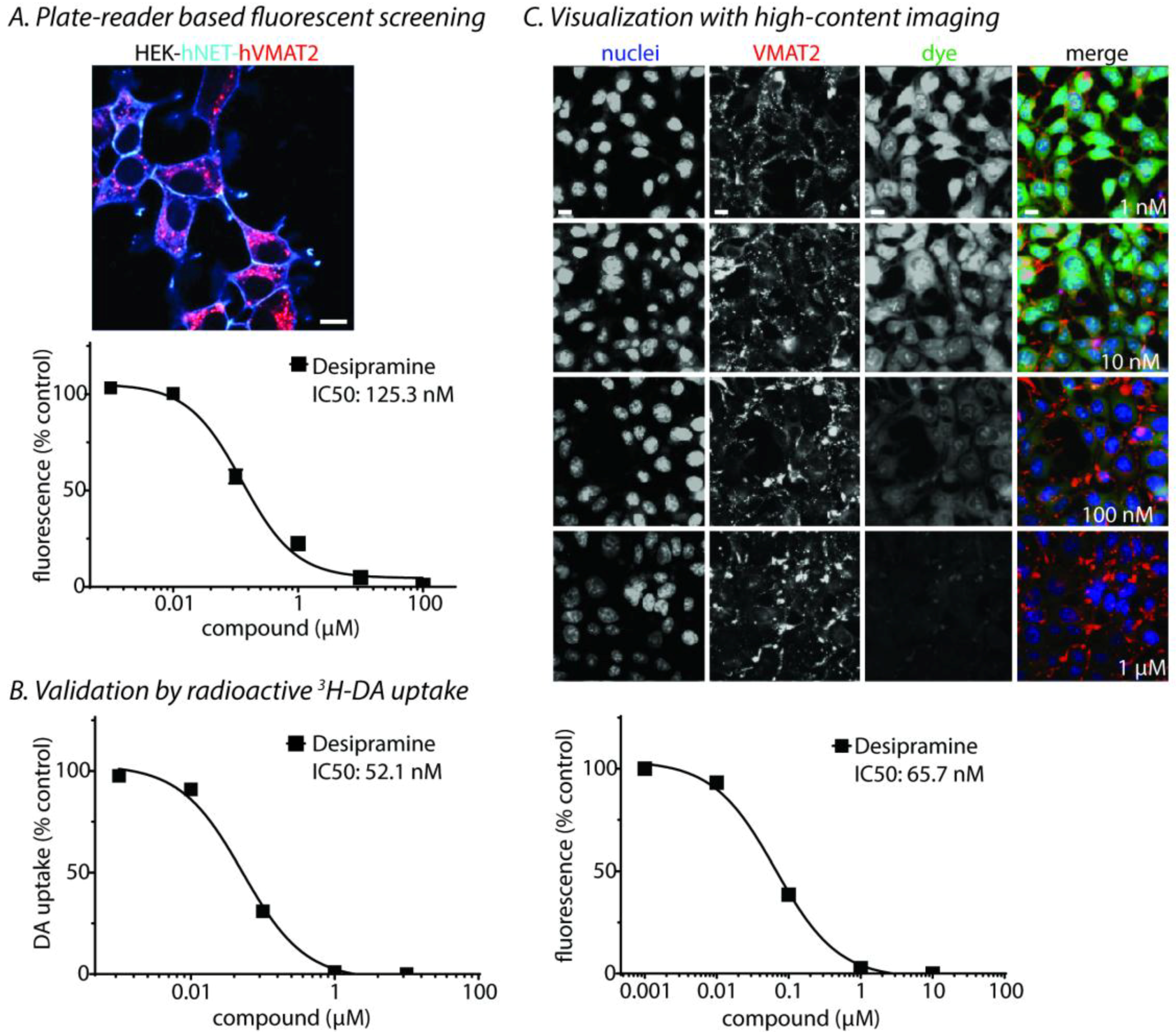
Biphenyls are considered privileged structures due to their over-proportionally frequent biological activity [8]. In our previous investigations, we focused on the diverse biological activities of H, M, and derivatives thereof, such as COX-1/2, 5-LOX and LTB4-formation [9], GABAA receptor activity [10,11], CB2 receptor activity [12], and toxicity on several cancer cell lines [13]. In this paper, we focus on the activity of the compounds at the NET. We tested 32 compounds of our already existing compound library and 16 additional compounds, of which 14 are new chemical substances (see Table 1, Table 2, Table 3, Table 4 and Table 5). Altogether 52 compounds have been evaluated in a norepinephrine (NE) uptake assay using a human embryonic kidney cell line stably expressing human NET (hNET) and human vesicular monoamine transporter 2 (VMAT2); human VMAT2 (hVMAT2) was added in addition to hNET to more faithfully recapitulate norepinephrine neurons in vitro (see characterization in Figure 1).
2. Results and Discussion
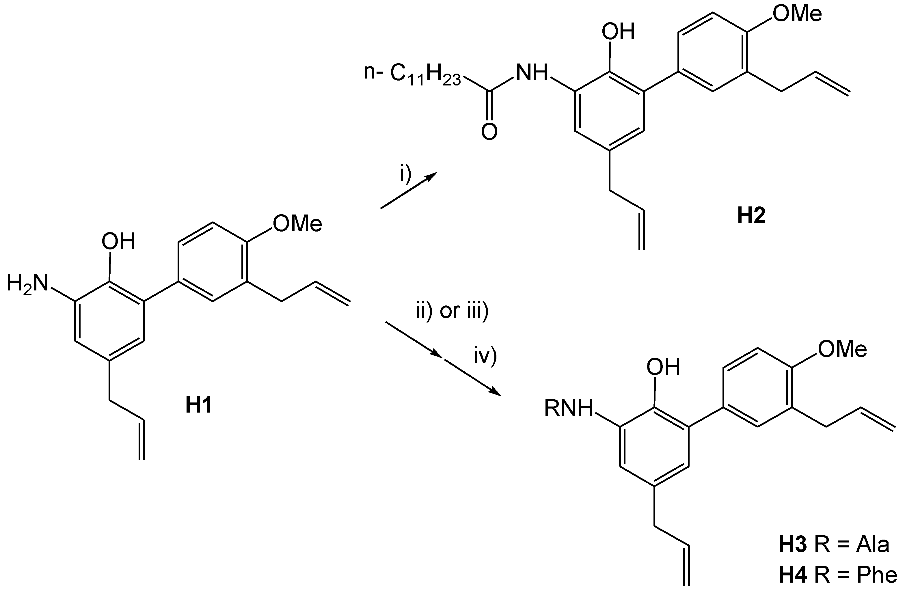
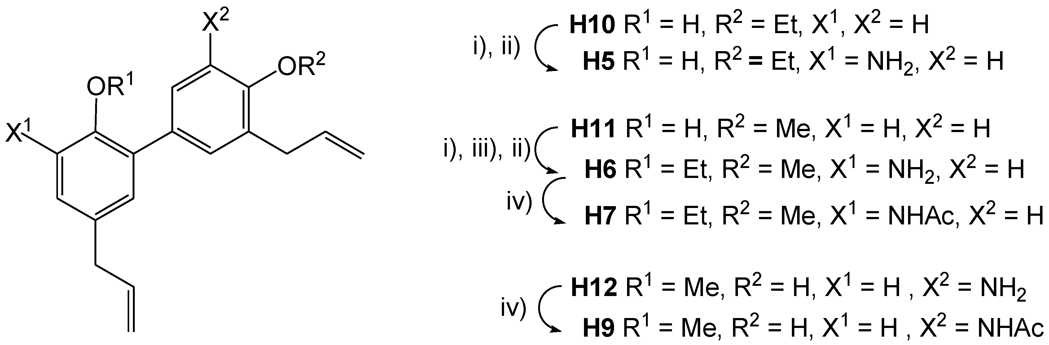
Previous studies indicated a significant increase in pharmacological activity when nitrogen-containing functional groups were introduced into H and other compounds [10]. We enhanced our existing compound library with further nitrogen-bearing honokiol derivatives with significantly changed polarity, either by N-acylation of 3-amino-4′-O-methylhonokiol (H1) with a long chain fatty acid (H2) or an amino acid (H3, H4), by formally replacing the methoxy group of H1 with an ethoxy group (H5), or di-O-alkylation without or with N-acetylation (H6, H7). In contrast to amid H8 of our parent library, in H9 acetamido and O-methyl groups are located at C-5′ and C-2, respectively. In the course of the syntheses, intermediates were fully characterized by their chemical and physical properties, but not tested in the biological assay. Compound data is provided for all synthesized chemicals, including compounds mentioned in the literature for which chemical and physical data were not fully provided.
All synthesized compounds were tested for pharmacological activity at the NET and compared with selective NET inhibitor, desipramine (Figure 1). Compounds with any apparent pharmacological activity were further characterized by radioactive uptake and high content imaging. No compound showed robust NET activity (Table 1).
As outlined in Scheme 1, N-acylations of H1 were performed using the corresponding carboxylic acid chlorides [14,15]. An Fmoc-protection group was used for the preparation of the peptide-type amides H3 and H4 [16,17] (see Scheme 1). N-acylation of H6 resulted in H7 (see Scheme 2).
As outlined in Scheme 2, H5 was prepared analogously to H1 from 4′-O-ethylhonokiol (H10) via nitration with nitric acid followed by reduction with SnCl2 × 2H2O [10,18,19]. To avoid N-alkylation in the synthesis of di-O-alkylated amine H6, the O-alkylation of the hydroxy group of 4′-O-methylhonokiol honokiol (H11) had to be performed after nitration and prior to reduction with SnCl2 × 2 H2O. N-acetylation of H6 with acetic anhydride resulted in H7 [10]. H9 was prepared by a similar N-acetylation of the corresponding amine H12.
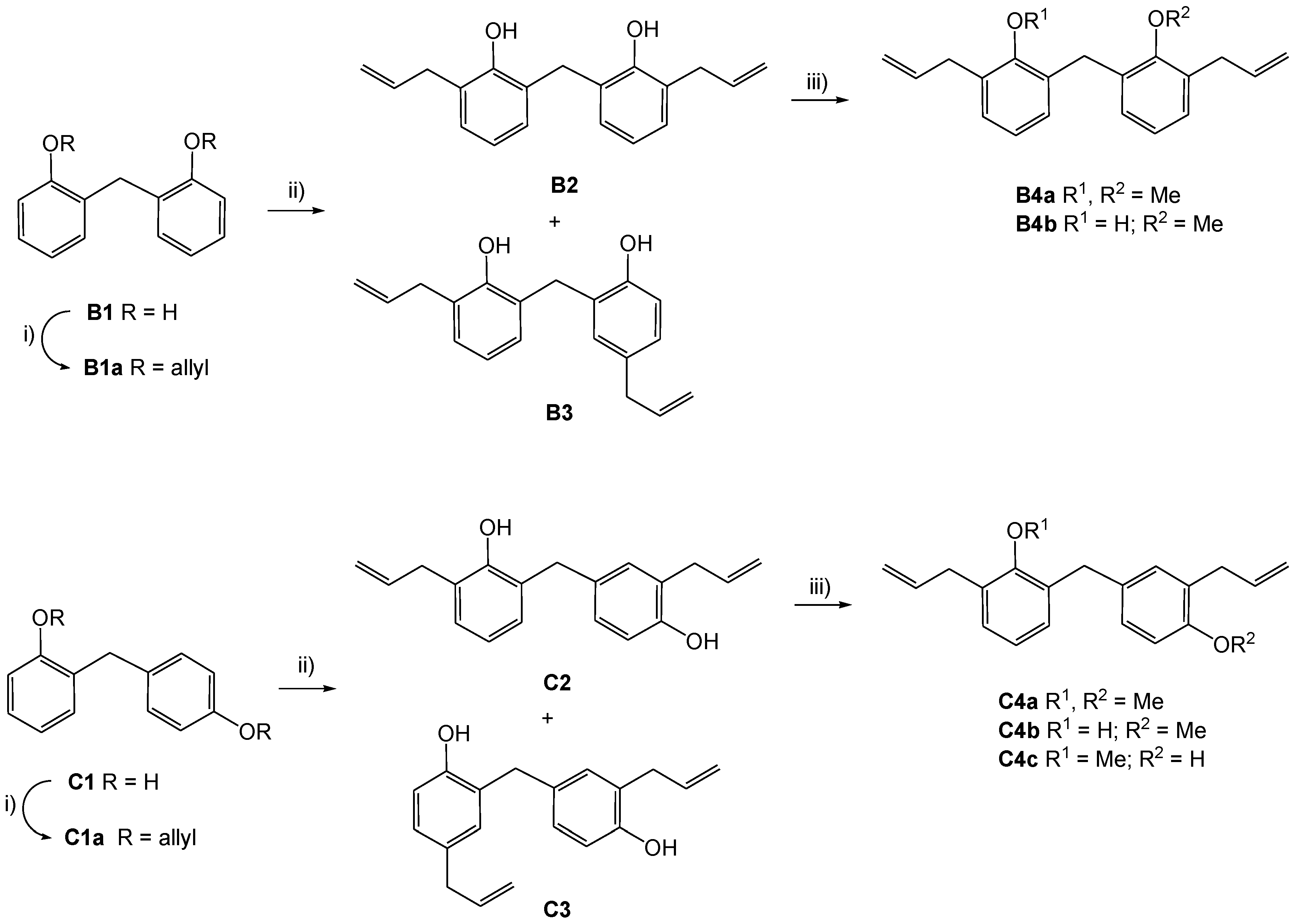
To go one step further, we tested several dioxygenated diphenylmethanes to see if insertion of a methylene group between the two aromatic moieties affects biological activity. As outlined in Scheme 3, the synthesis started from bis(2-hydroxyphenyl)methane (B1) and 2,4′-dihydroxydiphenylmethane (B1), which are commercially available but were also prepared from salicylic alcohol and phenol [20]. Double O-allylation and Claisen rearrangement according to Chattopadhyay et al. [21] yielded mainly the ortho rearrangement products (B2 and C2 resp.) together with little of the para rearrangement products (B3 and C3 resp.). B2 and C2 were submitted to incomplete O-methylation to yield the monoethers B4a, C4b, and C4c together with the respective diether B4a or C4a.
3. Experimental
3.1. General
Microwave reactions were carried out on a CEM Corp. Discover laboratory microwave equipped with an Explorer unit (CEM Corp. Matthews, NC, USA). Infrared spectra were recorded on a Bruker Alpha Platinum ATR spectrometer (Bruker, Kennewick, WA, USA) or as KBr pellets on a Perkin-Elmer 281 B spectrometer (PerkinElmer, Waltham, MA, USA). 1H and 13C-NMR spectra were recorded on a Varian 400 MHz UnityINOVA spectrometer (400 and 100 MHz, respectively, Varian, Palo Alto, CA, USA) and unless otherwise stated in CDCl3 with undeuterated solvent as an internal standard (7.26 ppm and 77.0 ppm, respectively). For convenience, atoms were numbered according to structures in Table 1, Table 4 and Table 5 with double-primed numbers for substituents at ring A and triple primed numbers for substituents at ring B, respectively. ESI-MS were recorded in ESI positive and negative mode on an LC Ultimate 3000 (Thermo, San José, CA, USA) with DAD detection in line with a Thermo Scientific LTQ XL mass spectrometer. Column: Knauer (Berlin, Germany) RP-18 (1.8 μm; 125 × 2.1 mm) with a guard cartridge at a flowrate of 150 μL/min. ESI-MS were recorded on an Agilent Technologies HP 7890A instrument fitted with detector HP 5975C VL MSD (70 eV, ion source 250 °C, quadrupole temperature 150 °C, Santa Clara, CA, USA). An Agilent HP-5MS column (30 m, ID 0.25 mm, film 5%pheny l95%, methylpolysiloxane 0.25 μm) was used. The oven temperature was kept at 45 °C for 2 min and programmed to increase to 300 °C at a rate of 3 °C/min, then kept constant at 300 °C for 20 min, with a total run time of 64.5 min. Helium was used as a carrier gas. The injection volume was 1 μL (≈0.5% solution) and a split ratio of 1:50.
For high resolution mass spectrometry, a Waters GCT Premier instrument was used with electron impact ionization (70 eV) at an ion source temperature of 200 °C.
General Information on Syntheses
Compounds were synthesized as described below. Solvents were of analytical quality, if not stated otherwise. The purity of all synthesized compounds was verified using NMR and analytical HPLC. Analytical thin layer chromatography (TLC) was performed using aluminium foil coated with silica 60 F254 (Merck, Darmstadt, Germany). Preparative thin layer chromatography (PTLC) was performed using glass plates coated with silica 60 F254 (Merck). Detection was done using UV/254 nm and spraying with molybdophosphoric acid and subsequent heating. Compound mixtures were separated using column chromatography (CC) on silica gel 60 (63–200 µm, Merck) using cyclohexane/ethyl acetate mixtures. Further purification was performed using preparative HPLC (Varian Prepstar with a Dynamax Rainin detector; column SepServ, Berlin, Germany, 250 × 21 mm, RP-18, 7 μm, flow rate 15 mL). Honokiol (purity > 98%) was purchased from APIChem Technology Co. (Hangzhou, China). Proton NMR spectra of the newly described compounds are given under “Supplementary Materials”.
3-Dodecanoylamino-4′-O-methylhonokiol (H2)
Dodecanoyl chloride (0.34 mmol, 80 μL) was added with intense stirring at room temperature (RT) to a solution of 100 mg (0.34 mmol) of H1 and pyridine (0.41 mmol, 33 μL) in abs. Et2O (4 mL). The reaction mixture was stirred at room temperature overnight and filtered. The precipitate was extracted with Et2O (4 mL). NaHCO3 (1 M, 10 mL) was added to the combined filtrate and washings. The organic phase was separated, and the aqueous phase was extracted with Et2O (3 × 10 mL). The combined organic phases were washed with brine (10 mL), dried over Na2SO4, concentrated under reduced pressure, and purified using CC (silica, cyclohexane/AcOEt 5:1) resulting in 107 mg (59%) of a light brown solid (H2).
Compound H2: IR (ATR, νmax, cm−1): 3304, 2956, 2919, 2847, 2485, 1639, 1606, 1539, 1499, 1467, 1411, 1243, 906, 725, 577; 1H-NMR (CDCl3) δ 7.57 (bs, 1H, NH), 7.40 (d, J = 1.5 Hz, 1H, H-4), 7.34 (dd, J = 8.4, 2.3 Hz, 1H, H-6′), 7.27 (d, J = 2.1 Hz, 1H, H-2′), 7.17 (s, 1H, OH), 6.94 (d, J = 8.4 Hz, 1H, H-5′), 6.89 (d, J = 1.9 Hz, 1H, H-6), 6.01 (ddt, J = 16.8, 10.2, 6.6 Hz, 1H, H-2‴), 5.96 (ddt, J = 16.8, 10.0, 6.7 Hz, 1H, H-2″), 5.02–5.13 (m, 4H, H-3‴, H-3″), 3.87 (s, 3H, OCH3), 3.43 (d, J = 6.6 Hz, 2H, H-1‴), 3.33 (d, J = 6.7 Hz, 2H, H-1″), 2.42 (t, J = 7.5 Hz, 2H, Acyl-Hα), 1.74 (quint, J = 7.3 Hz, 2H, Acyl-Hβ), 1.21–1.42 (m, 16H, Acyl-Hγ to Hω-1), 0.88 (t, J = 6.8 Hz, 3H, Acyl-Hω), 13C-NMR (CDCl3) δ 172.7 (CO), 156.8 (C-4′), 142.3 (C-2), 137.5 (C-2″), 136.7 (C-2‴), 132.2 (C-5), 130.7 (C-2′), 130.7 (C-1), 129.6 (C-1′), 129.0 (C-3′), 128.1 (C-6′), 126.7 (C-6), 126.2 (C-3), 120.3 (C-4), 115.8 (C-3″), 115.6 (C-3‴), 110.5 (C-5′), 55.5 (OCH3), 39.5 (C-1″), 37.5 (Acyl-Cα), 34.3 (C-1‴), 31.9 (Acyl-Cω-2), {2 × 29.6, 29.4, 2 × 29.3, 29.2 (Acyl-Cγ-to Cω-3)}, 25.7 (Acyl-Cβ), 22.7 (Acyl-Cω-1), 14.1 (Acyl-Cω), ESI+ calcd for C31H43NO3: [M]+ 477.324; found ESI-MS m/z (rel. int.): 478.63 [M + H]+ (100). From the same batch, the compound has been tested in parallel towards CB1/CB2 receptor agonistic activity by Bertini et al. [22]; however, spectroscopic data and synthesis are only given here.
3-(N-l-Alanyl)-4′-O-methylhonokiol (H3)
l-N-(9-Fluorenylmethoxycarbonyl)alanylchloride, Fmoc-l-Ala-Cl: Fmoc-l-Ala-OH · H2O was first dehydrated at 50 °C over P2O5 in vacuo for 12 h. Under dry conditions 400 mg (1.28 mmol) l-N-(9-Fluorenylmethoxycarbonyl)alanin was suspended in 5 mL dichloromethane, thionyl chloride (freshly distilled, 1.23 mL, 17.7 mmol) was added, and the mixture was sonicated at RT. After 7 min, the reaction mixture was a homogeneous solution and it was sonicated for an additional 15 min. Dichloromethane and the excess of thionyl chloride were removed in vacuo to yield 404 mg (96%) of Fmoc-l-Ala-Cl as a grey solid, which was used without further purification for a second step.
l-3-(N-(9-Fluorenylmethoxycarbonyl)alanyl)-4′-O-methylhonokiol (H3a): Zn (dust) was treated with HCl (2 M, aqueous solution), washed subsequently with water, EtOH and diethylether, dried in vacuo, and stored under argon. Under argon, H1 (259 mg, 0.841 mmol), Fmoc-l-Ala-Cl (279 mg, 0.846 mmol), and Zn dust (55 mg, 0.841 mmol) were suspended in abs. THF (50 mL) and stirred at RT overnight. Over a period of two hours, the reaction color turned from brownish to yellowish. Reaction completion was proven using TLC analysis (cyclohexane/AcOEt = 5:3). Zn dust was filtered off and the filtrate was evaporated in vacuo. The residue was dissolved in dichloromethane (15 mL), washed with brine (2 × 5 mL), and dried over Na2SO4. Evaporation of the solvent in vacuo and CC (silica, cyclohexane/AcOEt = 5:1) yielded in H3a (428 mg; 87%).
3-(N-l-Alanyl)-4′-O-methylhonokiol (H3): H3a (401 mg, 0.682 mmol) was dissolved in diethyl ether (3 mL) and piperidine (1 mL, 1 mmol) was added. The mixture was stirred at RT for 1 h. The reaction mixture was evaporated under reduced pressure and dichloromethane (5 mL) was added to the residue. The resulting precipitate was filtered off and the filtrate was concentrated under reduced pressure to dryness. The residue (377 mg) was purified using CC (silica, dichloromethane/MeOH, gradient: 0–10% v/v methanol) to yield 220 mg (88%) of H3 as an orange oil.
Compound H3: IR (ATR, νmax, cm−1): 3272, 3075, 2974, 2929, 2834, 1638, 1606, 1590, 1530, 1500, 1460, 1440, 1244, 1029, 993, 910, 813, 594; 1H-NMR (CDCl3 + D2O) δ 7.35 (dd, J = 8.1, 1.8 Hz, 1H, H-6′), 7.29 (d, J = 2.2 Hz, 1H, H-2′), 7.18 (s, 1H, H-4), 6.92 (s, 1H, H-6), 6.91 (d, J = 8.4 Hz, 1H, H-5′), 6.01 (ddt, J = 16.8, 9.8, 6.6 Hz, 1H, H-2‴), 5.95 (ddt, J = 16.9, 9.9, 6.6 Hz, 1H, H-2″), 5.01–5.13 (m, 4H, H-3″, H-3‴), 3.85 (s, 3H, O–CH3), 3.73 (q, J = 6.2 Hz, 1H, CH3CH(NH2)CO), 3.42 (d, J = 6.6 Hz, 2H, H-1‴), 3.32 (d, J = 6.2 Hz, 2H, H-1″), 2.03 (s, 2H, NH2), 1.45 (d, J = 6.6 Hz, 3H, CH3CH(NH2)CO); 13C-NMR (CDCl3 + D2O) δ 174.6 (CO), 156.7 (C-4′), 143.2 (C-2), 137.5 (C-2″), 136.8 (C-2‴), 131.9 (C-5), 131.0 (C-1), 130.8 (C-2′), 130.1 (C-1′), 128.6 (C-3′), 128.2 (C-6′), 127.5 (C-6), 125.6 (C-3), 120.6 (C-4), 115.7 (C-3″), 115.5 (C-3‴), 110.3 (C-5′), 55.5 (O–CH3), 50.5 (CH3CH(NH2)CO), 39.4 (C-1″), 34.3 (C-1‴), 21.1 (CH3CH(NH2)CO); ESI+ calcd for C22H26N2O3: [M]+ 366.194; found ESI-MS m/z (rel. int.): 367.18 [M + H]+ (100).
3-(N-l-Phenylalanyl)-4′-O-methylhonokiol (H4)
l-N-(9-Fluorenylmethoxycarbonyl)phenylalanylchloride, Fmoc-l-Phe-Cl: Under dry conditions 300 mg (0.774 mmol) l-N-(9-Fluorenylmethoxycarbonyl)phenylalanin was suspended in dry dichloromethane (4 mL), thionyl chloride (freshly distilled, 0.776 mL, 10.7 mmol) was added, and the mixture was sonicated at RT for 45 min. The sonication bath was heated up to 40 °C over that period of time. Dichloromethane and the excess of thionyl chloride were removed in vacuo to yield 294 mg (94%) of Fmoc-l-Phe-Cl as a grey solid, which was used without further purification in the next step.
3-(N-l-Phenylalanyl)-4′-O-methylhonokiol (H4): Zn dust was treated with aqueous HCl (2 M), washed subsequently with water, ethanol, and diethylether, dried in vacuo, and stored under argon. Under argon, H1 (75 mg, 0.25 mmol), Fmoc-l-Phe-Cl (103 mg, 0.254 mmol), and Zn dust (17 mg, 0.25 mmol) were suspended in dry THF (10 mL) and stirred for 4 h. Reaction completion was proven using TLC analysis (cyclohexane/AcOEt = 5:3). Piperidine (0.5 mL, 5 mmol) was added and the reaction mixture was stirred for 20 min at RT, the solvent was evaporated under reduced pressure and the residue was diluted with dichloromethane (5 mL), filtered, and evaporated under reduced pressure. The residue (170 mg) was purified using CC (silica, cyclohexane/AcOEt, gradient: 0–100% v/v AcOEt) to yield 50 mg (45%) of H4 as an orange oil.
Compound H4: IR (ATR, νmax, cm−1): 3270, 3073, 2974, 2834, 1638, 1605, 1530, 1500, 1453, 1439 s, 1244, 1028, 910; 1H-NMR (CDCl3) δ 9.69 (bs, 1H, CONH), 7.39 (dd, J = 8.4, ≈2 Hz, 1H, H-6′), 7.30–7.36 (m, 3H, Ph-Hmeta Ph-Hpara), 7.22–7.30 (m, 3H, H-2′, Ph-Hortho), 7.10 (s, 1H, H-4), 6.95 (s, 1H, H-6), 6.92 (d, J = 8.4 Hz, 1H, H-5′), 6.01 (ddt, J = 16.9, 9.9, 6.6 Hz, 1H, H-2‴), 5.97 (ddt, J = 16.9, 10.2, 6.6 Hz, 1H, H-2″), 5.02–5.14 (m, 4H, H-3″, H-3‴), 3.86 (s, 3H, OCH3), 3.82 (m, 1H, PhCH2CH(NH2)CO), 3.43 (d, J = 6.6 Hz, 2H, H-1‴), 3.34 (m, 1H, PhCH2CH(NH2)CO), 3.33 (d, J = 6.9 Hz, 2H, H-1″), 2.84 (dd, J = 13.6, 9.2 Hz, 1H, PhCH2CH(NH2)CO); 13C-NMR (CDCl3) δ 173.7 (CO), 156.6 (C-4′), 143.5 (C-2), 139.1 (Ph-Cipso), 137.5 (C-2″), 136.8 (C-2‴), 131.8 (C-5), 131.2 (C-1), 130.8 (C-2′), 130.2 (C-1′), 129.3 (Ph-Cortho), 128.8 (Ph-Cmeta), 128.6 (C-3′), 128.2 (C-6′), 127.6 (C-6), 127.0 (Ph-Cpara), 125.7 (C-3), 120.5 (C-4), 115.7 (C-3″), 115.5 (C-3‴), 110.3 (C-5′), 56.3 (PhCH2CH(NH2)CO), 55.5 (OCH3), 40.5 (PhCH2CH(NH2)CO), 39.3 (C-1″), 34.3 (C-1‴); ESI+ calcd for C28H30N2O3: [M]+ 442.226; found ESI-MS m/z (rel. int.): 442.20 [M + H]+ (100).
3-Amino-4′-O-ethylhonokiol (H5)
4′-O-Ethyl-3-nitro-honokiol (H5a): Aqueous nitric acid (65%, 0.474 mL, 6.79 mmol) was added under intense stirring within ≈5 s to a solution of H10 (200 mg, 0.679 mmol; synthesis see Schuehly et al. [12] supplemental; cpd 16b) in AcOEt (5 mL) at RT. The reaction mixture was stirred for 60 s and quenched with NaHCO3 (1 M, 10 mL). The organic phase was separated, and the aqueous phase was extracted with AcOEt (3 × 10 mL). The combined organic layers were washed with brine (10 mL), dried over Na2SO4, concentrated under reduced pressure, and purified using CC (silica, cyclohexane/AcOEt 99:1) to yield 120 mg (50%) of 4′-O-ethyl-3-nitro-honokiol (H5a) as a dark yellow oil.
Compound H5a: IR (ATR, νmax, cm−1): 3177, 3078, 2978, 2903, 1638, 1607, 1537, 1502, 1314, 1244, 1045, 912, 672, 555 cm−1; 1H-NMR (CDCl3) δ 11.03 (s, 1H, OH), 7.90 (d, J = 2.2 Hz, 1H, H-4), 7.45 (d, J = 2.2 Hz, 1H, H-6), 7.38 (dd, J = 8.4, 2.2, 1H, H-6′), 7.33 (d, J = 2.2 Hz, 1H, H-2′), 6.92 (d, J = 8.4 Hz, 1H, H-5′), 6.03 (ddt, J = 16.9, 10.3, 6.6 Hz, 1H, H-2‴), 5.96 (ddt, J = 16.5, 10.3, 6.6 Hz, 1H, H-2″), 5.03–5.17 (m, 4H, H-3″ and H-3‴), 4.10 (q, J = 7.2 Hz, 2H, OCH2CH3), 3.45 (d, J = 6.4 Hz, 2H, H-1‴), 3.40 (d, J = 6.6 Hz, 2H, H-1″), 1.46 (t, J = 6.9 Hz, 3H, OCH2CH3); 13C-NMR (CDCl3) δ 156.6 (C-4′), 151.3 (C-2), 138.9 (C-6), 136.8 (C-2‴), 136.0 (C-2″), 133.8 (C-3), 132.9 (C-1), 131.7 (C-5), 130.7 (C-2′), 128.5 (C-3′), 128.2 (C-6′), 127.4 (C-1′), 122.8 (C-4), 117.1 (C-3‴), 115.6 (C-3″), 111.0 (C-5′), 63.7 (OCH2CH3), 38.9 (C-1″), 34.5 (C-1‴), 14.9 (OCH2CH3); ESI– calcd for C20H21NO4: [M]− 339.147; found ESI-MS m/z (rel. int.): 338.21 ([M − H]− (100).
3-Amino-4′-O-ethylhonokiol (H5): SnCl2×2H2O (1.62 g, 7.16 mmol) was added to a solution of H5a (270 mg, 0.796 mmol) in abs. MeOH (10 mL). The reaction mixture was stirred for 48 h at RT, concentrated under reduced pressure and diluted with AcOEt (15 mL). The spumy precipitate resulting from the addition of aqueous NaHCO3 (1 M, 20 mL) was filtered off with Celite® (Sigma-Aldrich, Buchs, Switzerland) and rinsed with AcOEt (30 mL). The organic layer was separated from the combined filtrate and washings, dried over Na2SO4, and concentrated under reduced pressure. The residue (127 mg) was purified using PTLC (silica, cyclohexane/AcOEt = 5:1) to yield 65 mg (26%) of H5 as a brown oil.
Compound H5: IR (ATR, νmax, cm−1): 3544, 3370, 3075, 2977, 2900, 1637, 1607, 1503, 1486,1474, 1242, 1215, 1122, 1044, 908, 808; 1H-NMR (CDCl3) δ 7.26 (d ≈ 9 Hz, 1H, H-6′), 7.25 (s, 1H, H-2′), 6.93 (d, J = 8.1 Hz, 1H, H-5′), 6.61 (s, 1H, H-4), 6.51 (s, 1H, H-6), 6.00 (ddt, J = 16.9, 9.9, 6.6 Hz, 1H, H-2‴), 5.96 (ddt, J = 17.0, 9.9, 6.9 Hz, 1H, H-2″), 5.02–5.14 (m, 4H, H-3″, H-3‴), 4.19 (bs, 2H, NH2), 4.09 (q, J = 7.0 Hz, 2H, OCH2CH3), 3.43 (d, J = 6.6 Hz, 2H, H-1‴), 3.28 (d, J = 6.9 Hz, 2H, H-1″), 1.46 (t, J = 7.0 Hz, 3H, OCH2CH3); 13C-NMR (CDCl3) δ 156.4 (C-4′), 139.0 (C-2), 137.9 (C-2″), 136.6 (C-2‴), 133.6 (C-3), 132.4 (C-5), 130.3 (C-2′), 129.9 (C-3′), 129.0 (C-1′), 127.9 (C-1), 127.7 (C-6′), 120.5 (C-6), 115.8 (C-3‴), 115.7 (C-4), 115.4 (C-3″), 111.9 (C-5′), 63.7 (OCH2CH3), 39.6 (C-1″), 34.5 (C-1‴), 14.9 (OCH2CH3); ESI+ calcd C20H23NO2: [M]+ 309.173; found ESI-MS m/z (rel. int.): 310.11 [M + H]+ (100).
3-Amino-2-O-ethyl-4′-O-methylhonokiol (H6)
In a microwave process vial, KOH (132 mg, 2.36 mmol) was added to a solution of 4′-O-methyl-3-nitro-honokiol (synthesis see [10]) (192 mg, 0.590 mmol) in abs. MeOH (2 mL) and the mixture was stirred at RT for 10 min. Diethyl sulfate (155 μL, 2.36 mmol) was added and the reaction mixture was irradiated in a microwave oven at 90 °C for 60 min. After cooling to RT, the reaction mixture was neutralized with aqueous HCl (2 M). MeOH was evaporated under reduced pressure (40 °C, 100 mbar). The residue was extracted with dichloromethane (3 × 2 mL), the organic phase washed with water (3 × 5 mL), dried over Na2SO4, and concentrated under reduced pressure, resulting in 198 mg of crude 2-O-ethyl-4′-O-methyl-3-nitro-honokiol, which was used without further purification.
SnCl2×2H2O (1.09 g, 4.84 mmol) was added to a solution of crude 2-O-ethyl-4′-O-methyl-3-nitro-honokiol (190 mg, 0.54 mmol) in abs. EtOH (10 mL). The reaction mixture was stirred at RT for 48 h. The spumy precipitate resulting from the addition of aqueous NaHCO3 (1 M, 20 mL) was filtered using Celite®, the residue was extracted with EtOH (50 mL). Combined organic layers were evaporated under reduced pressure (100 mbar). The residue was extracted with dichloromethane (3 × 10 mL), which was washed with brine (3 × 5 mL), dried over Na2SO4, and concentrated under reduced pressure to yield 107 mg of a crude product, which after purification using PTLC (silica, cyclohexane/AcOEt = 5:1), yielded 26 mg (14%) of H6.
Compound H6: IR (ATR, νmax, cm−1): 3417, 3310, 3076, 2975, 2928, 2836, 1674, 1638, 1607, 1522, 1502, 1435, 1245, 1026, 910, 812, 600, 523; 1H-NMR (CDCl3) δ 7.47 (d, J = 2.2 Hz, 1H, H-2′), 7.46 (dd, J = 8.1, 2.2 Hz, 1H, H-6′), 6.92 (d, J = 8.4 Hz, 1H, H-5′), 6.60 (s, 2H, H-4, H-6), 6.07 (ddt, J = 16.6, 10.2, 6.2 Hz, 1H, H-2‴), 6.00 (ddt, J = 16.9, 9.9, 6.9 Hz, 1H, H-2″), 5.04–5.17 (m, 4H, H-3″, H-3‴), 3.96 (bs, 2H, NH2), 3.89 (s, 3H, OCH3), 3.57 (q, J = 6.9 Hz, 2H, OCH2CH3), 3.46 (d, J = 6.6 Hz, 2H, H-1‴), 3.33 (d, J = 6.9 Hz, 2H, H-1″), 1.16 (t, J = 7.1 Hz, 3H, OCH2CH3); 13C-NMR (CDCl3) δ 156.4 (C-4′), 142.1 (C-2), 140.0 (C-3), 137.6 (C-2″),137.0 (C-2‴), 136.0 (C-5), 134.4 (C-1), 131.0 (C-1′), 130.5 (C-2′), 128.0 (C-3′), 127.6 (C-6′), 120.5 (C-6), 115.5 (C-3″), 115.2 (C-3‴), 114.7 (C-4), 109.9 (C-5′), 67.6 (C–OCH2CH3), 55.4 (OCH3), 39.9 (C-1″), 34.3 (C-1‴), 15.7 (OCH2CH3); ESI+ calcd for C21H25NO2: [M]+ 323.189; found ESI-MS m/z (rel. int.): 324.23 [M + H]+ (100).
3-Acetylamino-2-O-ethyl-4′-O-methylhonokiol (H7)
In a 10 mL round-bottom flask, H6 (34 mg, 0.105 mmol) was mixed with water (0.3 mL), and acetic anhydride (0.21 mmol, 20 μL) was added. The flask was rotated at 80 °C for about 10 min in a water bath. After cooling to RT, the reaction mixture was quenched with aqueous NaHCO3 (1 M, 2 mL) and extracted with dichloromethane (3 × 2 mL). The combined extracts were washed with aqueous NaHCO3 (1 M, 2 mL), water (2 mL), dried over Na2SO4, and concentrated under reduced pressure. The crude product (27 mg) was purified using PTLC (cyclohexane/AcOEt = 5:3), yielding 21 mg (55%) of H7 as a brownish oil.
Compound H7: IR (ATR, νmax, cm−1): 3469, 3368, 3075, 2974, 2928, 2903, 2834, 1638, 1606, 1504, 1438, 1243, 1209, 1030, 909, 810, 596; 1H-NMR (CDCl3) δ 8.17 (s, 1H, H-4), 7.96 (bs, 1H, NH), 7.38 (s, 1H, H-2′), 7.37 (dd, J ≈ 8, 2.2 Hz, 1H, H-6′), 6.90 (d, J = 8.1 Hz, 1H, H-5′), 6.86 (d, J = 1.1 Hz, 1H, H-6), 6.03 (ddt, J = 16.8, 9.9, 6.9 Hz, 1H, H-2‴), 5.98 (ddt, J = 16.9, 9.9, 6.6 Hz, 1H, H-2″), 5.02–5.15 (m, 4H, H-3″, H-3‴), 3.87 (s, 3H, OCH3), 3.52 (q, J = 7.1 Hz, 2H, OCH2CH3), 3.42 (d, J = 6.6 Hz, 2H, H-1‴), 3.38 (d, J = 7.0 Hz, 2H, H-1″), 2.21 (s, 3H, CH3CO), 1.14 (t, J = 7.0 Hz, 3H, OCH2CH3); 13C-NMR (CDCl3) δ 168.09 (CO), 156.7 (C-4′), 143.1 (C-2), 137.3 (C-2″), 136.9 (C-2‴), 136.2 (C-5), 133.5 (C-1), 131.9 (C-3), 130.4 (C-1′), 130.3 (C-2′), 128.4 (C-3′), 127.6 (C-6′), 125.3 (C-6), 118.7 (C-4), 115.8 (C-3″), 115.4 (C-3‴), 110.2 (C-5′), 68.7 (O–CH2–CH3), 55.4 (OCH3), 40.0 (C-1″), 34.3 (C-1‴), 24.9 (CH3–C=O), 15.6 (OCH2CH3); ESI+ calcd for C23H27NO3: [M]+ 365.199; found ESI-MS m/z (rel. int.): 366.29 [M + H]+ (100).
5′-(N-Acetylamino)-2-O-methyl-honokiol (H9)
In a 10 mL round-bottom flask, H12 (for synthesis, see Reference [11]) (37 mg, 0.125 mmol) was suspended with water (1 mL), and acetic anhydride (0.21 mmol, 20 μL) was added. The flask was allowed to rotate for 10 min at 80 °C in a water bath. After cooling to RT, the reaction mixture was quenched with aqueous NaHCO3 (1 M, 1 mL) and extracted with dichloromethane (3 × 1.5 mL). The combined extracts were washed with aqueous NaHCO3 (1 M, 1.5 mL) and water (1.5 mL), dried over Na2SO4, and concentrated under reduced pressure. The crude product (29 mg) was purified using PTLC (silica, cyclohexane/AcOEt 5:3), yielding 16 mg (39%) of H9, a light orange solid.
Compound H9: IR (ATR, νmax, cm−1): 3276, 3075, 3001, 2917, 2848, 1750, 1637, 1595, 1540, 1480, 1239, 1239, 1139, 1025, 910, 873, 809; 1H-NMR (CDCl3) δ 7.62 (s, br 1H, NH), 7.18 (s, 1H, H-2′), 7.10 (d ≈ 8 Hz, 1H, H-4), 7.07 (s, 2H, H-6, H-6′), 6.89 (d, J = 8 Hz, 1H, H-3), 6.06 (ddt, J = 16.9, 10.3, 6.6 Hz, 1H, H-2‴), 5.97 (ddt, J ≈ 17, 9.9, 6.9 Hz, 1H, H-2″), 5.03–5.16 (m, 4H, H-3″, H-3‴), 3.77 (s, 3H, OCH3), 3.50 (d, J = 5.9 Hz, 2H, H-1‴), 3.35 (d, J = 6.6 Hz, 2H, H-1″), 2.22 (s, 3H, CO-CH3); 13C-NMR (CDCl3) δ 170.5 (CO), 154.7 (C-2), 146.0 (C-4′), 137.7 (C-2″), 136.8 (C-2‴), 132.3 (C-5), 130.9 (C-6), 130.4 (C-5′), 130.2 (C-3′), 129.6 (C-1), 128.8 (C-2′), 128.2 (C-4), 125.2 (C-1′), 121.4 (C-6′), 115.7 (C-3‴), 115.6 (C-3″), 111.4 (C-3), 55.8 (OCH3), 39.4 (C-1″), 35.9 (C-1‴), 23.7 (CO–CH3); ESI+ calcd for C21H23NO3: [M]+ 337.168; found ESI-MS m/z (rel. int.): 338.08 [M + H]+ (100).
Bis(2-Allyloxyphenyl)-methane (B1a)
B1 (400 mg, 2.0 mmol) was O-allylated with allylbromide (968 mg, 0.70 mL, 8.0 mmol) and K2CO3 (2.21 g, 16 mmol) in acetone (10 mL). The reaction mixture was stirred for 5 h with reflux, and a further 12 h at RT and filtered. The residue was extracted with acetone (50 mL) and the combined organic solutions were evaporated under reduced pressure yielding B1a (559 mg, 73%).
Compound B1a: IR (ATR, νmax, cm−1): 3025, 2920, 2905, 1595, 1584, 1491, 1450, 1421, 1248, 1116, 1103, 1022, 924, 751 cm−1; 1H-NMR (CDCl3) δ 7.19 (td, J = 7.6, 1.5 Hz, 2H, H-4), 7.14 (dd, J = 7.5, 1.2 Hz, 2H, H-6), 6.90 (t, J ≈ 8 Hz, 2H, H-5), 6.89 (d, 2H, J = 7.5 Hz, H-3), 6.02–6.13 (m, 2H, CH2–CH=CH2), 5.42 (dquint, J = 17.2, 4.3 Hz, 2H, CH2–CH=CH2), 5.27 (dquint, J = 10.5, 1.4 Hz, 2H, CH2–CH=CH2), 4.58 (dq, J = 5.0, 1.5 Hz, 2H, CH2–CH=CH2), 4.10 (s, 2H, Ar–CH2–Ar); 13C-NMR (CDCl3) δ 156.5 (C-2), 133.6 (CH=CH2), 130.5 (C-6), 129.6 (C-1), 126.9 (C-4), 120.5 (C-5), 116.8 (CH=CH2), 111.6 (C-3), 30.8 (O–CH2), 29.8 (Ar–CH2–Ar); EI+ calcd for C19H20O2: [M]+ 280.1463; found EI-MS m/z (rel. int.): 280.1465 [M]+ (100). Compound B1a has been mentioned in several patents; however, chemical data were not given therein.
2-Allyl-6-(3-allyl-2-hydroxybenzyl)-phenol (B2) and 4-Allyl-2-(3-allyl-2-hydroxybenzyl)-phenol (B3)
Claisen rearrangement of B1a (561 mg, 2 mmol) in N,N-diethylaniline (4 mL) following the procedure of Chattopadhyay et al. [21] resulted in B2 (410 mg; 73%) and B3 (89 mg; 15%) as white wax like solids.
Compound B2: IR (KBr, νmax, cm−1): 3407 (br, OH), 3073, 3035, 2975, 2937, 1639, 1590, 1461, 1448, 1372, 1294, 1234, 1210, 1192, 1086, 999, 922, 912, 754 cm−1; 1H-NMR (CDCl3) δ 7.20 (dd, J = 7.7, 1.5 Hz, 2H, H-6), 7.02 (d, J = 7.7 Hz,2H, H-4), 6.88 (t, J = 7.7 Hz, 2H, H-5), 6.43 (s, br, 2H, OH), 6.06 (ddt, J = 16.8, 10.3, 6.2 Hz, 2H, CH2–CH=CH2), 5.18–5.24 (m, 4H, CH2–CH=CH2), 3.97 (s, 2H, Ar–CH2–Ar), 3.45 (d, J = 6.6 Hz, 4H, CH2–CH=CH2); 13C-NMR (CDCl3) δ 151.4 (C-2), 136.6 (CH2–CH=CH2), 129.1 (C-6), 128.7 (C-4), 127.1 (C-1), 125.8 (C-3), 121.1 (C-5), 116.6 (CH2–CH=CH2), 35.4 (CH2–CH=CH2), 30.8 (Ar–CH2–Ar); EI+ calcd for C19H20O2: [M]+ 280.1463; found EI-MS m/z (rel. int.): 280.1469 [M]+ (58). Compound B2 has been mentioned in several patents; however, chemical data were not given therein.
Compound B3: IR (KBr, νmax, cm−1): 3387 (OH), 3077, 2853, 1638, 1611, 1592, 1506, 1465, 1434, 1256, 1232, 1215, 1107, 996, 914, 759 cm−1. 1H-NMR (CDCl3) δ 7.17 (dd, J = 7.8, 1.5 Hz, 1H, H-6), 7.06 (d, J = 2.2 Hz,1H, H-6′), 6.98 (dd, J = 7.3, 1.5 Hz, 1H, H-4), 6.91 (dd, J = 8.4, 2.2 Hz, 1H, H-4′), 6.84 (t, J = 7.5 Hz, 1H, H-5), 6.74 (d, J = 8.0 Hz, 1H, H-3′), 6.31 (s, vb, 2H, OH), 6.02 (ddt, J = 16.8, 10.3, 6.2 Hz, 1H, 2″-H), 5.92 (ddt, J = 16.9, 10.3, 6.6 Hz, 1H, H-2″), 5.19 (dq, J ≈ 16, 1.7 Hz, 1H, H-3″), 5.18 (dq, J ≈ 9, 1.5 Hz, 1H, H-3″), 5.03 (dq, J ≈ 17, 1.8 Hz, 1H, H-3‴), 5.04 (dq, J ≈ 9, 1.3 Hz, 1H, H-3‴), 3.89 (s, 2H, Ar–CH2–Ar), 3.41 (d, J = 6.2 Hz, 2H, H-1″), 3.28 (d, J = 7.0 Hz, 2H, H-1‴); 13C-NMR (CDCl3) δ 151.3 (C-2), 151.2 (C-2′), 137.8 (C-2‴), 136.5 (C-2″), 132.8 (C-5′), 130.8 (C-6′), 129.1 (C-6), 128.8 (C-4), 128.0 (C-4′), 127.1 (C-1), 126.6 (C-1′), 125.6 (C-3), 121.2 (C-5), 116.9 (C-3″), 116.0 (C-3′), 115.4 (C-3‴), 39.4 (C-1‴), 35.6 (C-1″), 30.8 (Ar–CH2–Ar); EI+ calcd for C19H20O2: [M]+ 280.1463; found EI-MS m/z (rel. int.): 280.1464 [M]+ (100).
2,4′-Diallyloxy-diphenylmethane (C1a)
C1 (3.77 g, 18.8 mmol) was allylated with allylbromide (8.97 g, 6.5 mL, 74.1 mmol) and K2CO3 (20.8 g, 0.15 mol) in acetone (100 mL) as described above yielding the diallyloxy-diphenylmethane (4.12 g, 78%).
Compound C1a: IR (ATR, νmax, cm−1): 3077, 3022, 2915, 2860, 1610, 1599, 1584, 1508, 1490, 1451, 1237, 1221, 1020, 996, 921, 749 cm−1; 1H-NMR (CDCl3) δ 7.22 (td, J ≈ 8, 1.3 Hz, 1H, H-4), 7.21 (d, J ≈ 8 Hz, 2H, H-2′ and H-6′), 7.14 (d, J = 7.7 Hz, 1H, H-6), 6.94 (t, J = 7.3 Hz, 1H, H-5), 6.90 (d, 1H, J ≈ 8 Hz, H-3), 6.89 (d, 2H, J = 8.4 Hz, H-3′ and H-5′), 6.03–6.17 (m, 2H, H-2″ and H-2‴), 5.45 (dq, J = 17.2, 1.5 Hz, 1H, H-3‴), 5.44 (dq, J = 17.3, 1.5 Hz, 1H, H-3″), 5.32 (dq, J = 10.5, 1.5 Hz, 1H, H-3‴), 5.31 (dq, J = 10.5, 1.5 Hz, 1H, H-3″), 4.58 (dt, J = 5.1, 1.5, 2H, H-1″), 4.55 (dt, J = 5.1, 1.5, 2H, H-1‴), 4.02 (s, 2H, Ar–CH2–Ar); 13C-NMR (CDCl3) δ 156.7(C-4′), 156.2 (C-2), 2 × 133.5 (2 × CH=CH2), 133.3 (C-1′*), 130.4 (C-1*), 130.2 (C-6), 129.8 (C-2′ and C-6′), 127.2 (C-C-4), 120.6 (C-5), 117.4 (C-3‴), 116.9 (C-3″), 114.5 (C-3′ and C-5′), 111.7 (C-3), 68.8 and 68.7 (2 × O–CH2), 35.1 (Ar–CH2–Ar); EI+ calcd for C19H20O2: [M]+ 280.1463; found EI-MS m/z (rel. int.): 280.1 [M]+ (45), 133.1 [C6H4–O–allyl]+ (100).
2-Allyl-4-(3-allyl-2-hydroxybenzyl)-phenol (C2) and 2-Allyl-4-(5-allyl-2-hydroxybenzyl)-phenol (C3)
Claisen rearrangement of C1a (14.5 g, 51.7 mmol) in N,N-diethylaniline (115 mL) following the procedure of Chattopadhyay et al. [21] resulted in C2 (10.6 g; 73%) and C3 (1.2 g; 8.3%)
Compound C2: IR (ATR, νmax, cm−1): 3507 (br, OH), 3076, 3009, 2977, 2906, 1637, 1609, 1591, 1501, 1461, 1432, 1328, 1255, 1182, 996, 911, 751 cm−1; 1H-NMR (CDCl3) δ 6.95–7.06 (m, 4H, H-4,H-6, H-2′, H-6′), 6.86 (t, J = 7.5 Hz, 1H, H-5), 6.74 (d, J = 7.7 Hz, 1H, H-5′), 5.94–6.09 (m, 2H, H-2″, H-2‴), 5.12–5.20 (m, 4H, H-3″, H-3‴), 4.99 (s, b, 2H, OH), 3.92 (s, 2H, Ar–CH2–Ar), 3.41 (d, J = 6.2 Hz, 2H, H-1″), 3.38 (d, J = 6.2 Hz, 2H, H-1‴); 13C-NMR (CDCl3) δ 152.6 (C-4′), 152.4 (C-2), 136.5 (C-2″), 136.4 (C-2‴), 132.1 (C-1′), 130.7 (C-2′), 129.1 (C-6), 128.6 (C-4), 128.0 (C-6′), 127.6 (C-1), 125.6 (C-3, C-3′), 120.6 (C-5), 116.4 (C-3″, C-3‴), 116.0 (C-5′), 35.7 (Ar–CH2–Ar), 35.3 (C-1″), 35.1′ (C-1‴); EI+ calcd for C19H20O2: [M]+ 280.1463; found EI-MS m/z (rel. int.): 280.1465 [M]+ (100).
Compound C3: IR (ATR, νmax, cm−1): 3277 (br, OH), 3078, 3013, 2977, 2902, 1638, 1608, 1502, 1434, 1341, 1247, 1207, 1094, 992, 793, 634, 617 cm−1; 1H-NMR (CDCl3) δ 6.90–7.04 (m, 4H, H-4, H-6, H-2′, H-6′), 6.68–6.78 (m 2H, H-3, H-5′), 5.88–6.07 (m, 2H, H-2″, H-2‴), 5.10–5.20 (m, 2H, H-3‴), 4.99–5.10 (m, 2H, H-3″), 4.87 (s, 1H, OH), 4.57 (s, 1H, OH), 3.89 (s, 2H, Ar–CH2–Ar), 3.38 (d, J = 6.2 Hz, 2H, 1‴-H), 3.30 (d, J = 6.6 Hz, 2H, H-1′); 13C-NMR (CDCl3) δ 152.6 (C-4′), 152.1 (C-2), 137.9 (C-2″), 136.4 (C-2‴), 132.2 (C-5), 132.0 (C-1′), 131.0 (C-6), 130.2 (C-2′), 127.9 (C-6′), 127.7 (C-4), 127.1 (C-1), 125.6 (C-3′), 116.3 (C-3‴), 116.0, 115.8 (C-3, C-5′), 115.3 (C-3″), 39.3 (C-1″), 35.7 (Ar–CH2–Ar), 35.1 (C-1‴ EI+ calcd for C19H20O2: [M]+ 280.1463; found EI-MS m/z (rel. int.): 280.1 [M]+ (100), 147.1 [CH2–C6H4–O–allyl]+ (100).
General Procedure for the Methylation of Dihydroxy-Diphenylmethanes
The corresponding diol (1 eq) was dissolved in aqueous KOH (10%, 2.5 eq) and stirred for 10 min. Me2SO4 (1.2 eq) was added and the resulting mixture was stirred for 20 min at RT and for 1 h at 95 °C. After cooling to RT, the mixture was acidified with aqueous HCl (2M) and extracted with chloroform (3 ×). The organic layers were dried over Na2SO4 and concentrated under reduced pressure. The residue was chromatographed over silica with cyclohexane/AcOEt = 5:1.
Bis(3-allyl-2-methoxyphenyl)methane (B4a) and 6-allyl-2-(3-allyl-2-methoxybenzyl)-phenol (B4b)
Methylation of 420 mg (1.5 mmol) B2 resulted in 39 mg of starting material, 40 mg B4a (8.6%) and 249 mg B4b (57%).
Compound B4a: IR (ATR, νmax, cm−1): 3001, 2978, 2941, 2827, 1463, 1427, 1291, 1164, 1080, 1009, 911, 766 cm−1; 1H-NMR (CDCl3) δ 7.09 (dd, 2H, J = 7.6 and 1.3 Hz, H-4), 7.00 (t, 2H, J = 7.5 Hz, H-5), 6.93 (dd, 2H, J = 7.4 and 1.3 Hz, H-6), 5.97–6.09 (m, 2H, -CH=CH2), 5.06–5.21 (m, 4H, -CH=CH2), 4.10 (s, 2H, Ar–CH2–Ar), 3.72 (s, 6H, OCH3), 3.46 (d, 4H, J 6.6 Hz, CH2-CH=CH2); 13C-NMR (CDCl3) δ 154.4 (C-2), 137.3 (-CH=CH2), 134.0 (C-1), 132.9 (C-3), 128.9 (C-6), 128.5 (C-4), 124.1 (C-5), 115.8 (-CH=CH2), 34.0 (-CH2-CH=CH2), 29.6 (Ar–CH2–Ar). EI+ calcd for C21H24O2: [M]+ 308.1776; found EI-MS m/z (rel. int.): 308.1776 [M]+ (100).
Compound B4b: IR (ATR, νmax, cm−1): 3320 (br, OH), 3001, 2978, 2975, 2944, 2908, 1641, 1632, 1591, 1462, 1446, 1430, 1220, 1086, 998, 910, 780, 604 cm−1; 1H-NMR (CDCl3) δ 7.35 (s, 1H, OH), 7.18 (dd, J = 7.1, 1.8 Hz, 1H, H-6′), 7.13 (d, J = 7.1 Hz, 1H, H-6), 7.08 (dd, J ≈ 7, ≈ 2 Hz, 1H, H-4′), 7.06 (t, J = 7.5 Hz, 1H, H-5′), 7.00 (d, J = 7.7 Hz, 1H, H-4), 6.81 (t, J = 7.5 Hz, 1H, H-5), 5.92–6.08 (m, 2H, H-2′ and H-2‴), 5.03–5.15 (m, 4H, H-3″ and H-3‴), 3.92 (2 × s, 5H, Ar–CH2–Ar and OCH3), 3.47 (d, J = 6.3 Hz, 2H, H-1‴), 3.40 (d, J = 6.6 Hz, 2H, H-1″); 13C-NMR (CDCl3) δ 154.2 (C-2′), 151.2 (C-2), 137.0 (C-2″), 136.8 (C-2‴), 2 × 133.0, (C-1′ and C-3′), 129.1 (C-4′), 128.9 (C-6′), 128.6 (C-4), 128.4 (C-6), 127.2 (C-3), 126.6 (C-1), 125.3 (C-5′), 120.1 (C-5), 116.2 (C-3‴), 115.4 (C-3″), 62.3 (OCH3), 34.7 (C-1″), 33.8 (C-1‴), 31.4 (Ar–CH2–Ar); EI+ calcd for C21H24O2: [M]+ 294.1620; found EI-MS m/z (rel. int.): 294.1619 [M]+ (100).
3,3′-Diallyl-2,4′-dimethoxy-diphenyl-methan (C4a), 6-allyl-2-(3-allyl-4-methoxybenzyl)-phenol (C4b) and 2-allyl-4-(3-allyl-2-methoxybenzyl)-phenol (C4c)
Methylation of 2.10 g (7.5 mmol) C2 resulted in 143 mg of starting material, 318 mg C4a (14%), 665 mg C4b (30%) and 442 mg C4c (20%).
Compound C4a: IR (ATR, νmax, cm−1): 3001, 2977, 2939, 2833, 1638, 1500, 1463, 1429, 1248, 1125, 1080, 1033, 1010, 910, 768 cm−1; 1H-NMR (CDCl3) δ 7.07 (dd, J = 7.3, 1.5 Hz, 1H, H-4), 6.98–7.02 (m, 3H, H-2′, H-5 and H-6′), 6.96 (dd, J = 7.6, 1.3 Hz, 1H, H-6), 6.78 (d, J = 8.4 Hz, 1H, H-5′), 5.92–6.07 (m, 2H, H-2″ and H-2‴), 5.06–5.13 (m, 2H, H-3″), 4.99–5.06 (m, 2H, H-3‴), 3.96 (s, 2H, Ar–CH2–Ar), 3.80 (s, 3H, C4′-OCH3), 3.68 (s, 3H, C2-OCH3), 3.46 (d, J = 6.3 Hz, 2H, H-1″), 3.36 (d, J = 6.6 Hz, 2H, H-1‴); 13C-NMR (CDCl3) δ 156.3 (C-2), 155.6 (C-2′), 137.3 (C-2″), 137.1 (C-2‴), 134.7 (C-1), 133.0 (C-3), 132.8 (C-1′), 130.2 (C-2′), 129.0 (C-6), 128.5 (C-4), 128.4 (C-3′), 127.5 (C-6′), 120.5 (C-5), 115.8 (C-3″), 115.3 (C-3‴), 110.3 (C-5′), 61.2 (C2-OCH3), 55.4 (C4′-OCH3), 34.9 (Ar–CH2–Ar), 34.3 (C-1‴), 34.0 (C-1″); MS (ESI+) m/z (%): calculated for C21H24O2: [M]+ 308.178; found: EI-MS m/z (rel. int.): 308.4 [M]+ (100).
D4b: IR (ATR, νmax, cm−1): 3526 (br, OH), 3003, 2976, 2905, 2834, 1637, 1608, 1592, 1499, 1461, 1247, 1182, 1032, 996, 910, 752 cm−1; 1H-NMR (CDCl3) δ 6.97–7.02 (m, 4H, H-4, H-6, H-2′ and H-6′), 6.82 (t, J = 7.5 Hz, 1H, H-5), 6.76 (d, J = 8.8 Hz, 1H, H-5′), 5.90–6.05 (m, 2H, H-2″and H-2‴), 5.11–5.17 (m, 2H, H-3″), 4.98–5.05 (m, 2H, H-3‴), 4.94 (s, 1H, OH), 3.91 (s, 2H, Ar–CH2–Ar), 3.78 (s, 3H, OCH3), 3.38 (d, J = 6.2 Hz, 2H, H-1″), 3.34 (d, J = 6.5 Hz, 2H, H-1‴); 13C-NMR (CDCl3) δ 152.9 (C-4′), 152.4 (C-2), 136.9 (C-2‴), 136.5 (C-2″), 131.4 (C-1′), 130.2 (C-2′), 129.1 (C-6′), 128.8 (C-3′), 128. 6 (C-4), 127.6 (C-1), 127.2 (C-6), 125.5 (C-3), 120.5 (C-5), 116.4 (C-3″), 115.3 (C-3‴), 55.4 (OCH3), 35.8 (Ar–CH2–Ar), 35.3 (C-1″), 34.3 (C-1‴); MS (EI) m/z (%): calculated for C20H22O2: [M]+ 294.162; found: EI-MS m/z (rel. int.): 294.4 [M]+ (80), 253.2 (65).
D4c: IR (ATR, νmax, cm−1): 3393 (br, OH), 3005, 2977, 2940, 2911, 2830, 1638, 1610, 1591, 1505, 1464, 1428, 1251, 1198, 1080, 996, 910, 768 cm−1; 1H-NMR (CDCl3) δ 7.09 (dd, J = 7.3, 1.7 Hz, 1H, H-4), 7.01 (t, J = 7.3 Hz, 1H, H-5), 6.93–6.99 (m, 3H, H-2′, H-6 and H-6′), 6.73 (d, J = 7.7 Hz, 1H, H-5′), 5.94–6.08 (m, 2H, H-2″ and H-2‴), 5.06–5.20 (m, 4H, H-3″ and H-3‴), 4.95 (s, 1H, OH), 3.96 (s, 2H, Ar–CH2–Ar), 3.68 (s, 3H, OCH3), 3.46 (d, J = 6.6 Hz, 2H, H-1″), 3.38 (d, J = 6.5 Hz, 2H, H-1‴), 13C-NMR (CDCl3) δ 156.3 (C-2), 152.3 (C-4′), 137.3 (C-2″), 136.5 (C-2‴), 134.6 (C-1), 133.3 (C-1′), 133.0 (C-3), 130.1 (C-2′), 129.0 (C-6), 128.6 (C-4), 128.2 (C-6′), 125.1 (C-3′), 124.1 (C-5), 116.3 (C-3″), 115.8 (C-3‴), 115.7 (C-5′), 61.2 (OCH3), 35.2 (C-1‴), 34.9 (Ar–CH2–Ar), 33.9 (C-1″); MS (EI) m/z (%): calculated for C20H22O2: [M]+ 294.162; found: EI-MS m/z (rel. int.): 294.4 [M]+ (75), 253.2 (40).
3.2. Biological Assays
Cell line: All screening was done in a line of human embryonic kidney cells stably expressing the human norepinephrine transporter and the vesicular monoamine transporter 2 (HEK-hNET- -hVMAT2-mCherry). Human VMAT2 expression, in addition to hNET, more fully recapitulates a neuronal phenotype in cell culture. Cells were imaged and fully characterized for functionality (Figure 1). For imaging, cells were fixed in 4% paraformaldehyde and protein expression of NET determined by immunolabelling (primary antibody: Novus, anti-norepinephrine transporter, 1:1000; secondary antibody: Thermo Fisher, Alexafluor goat anti-rabbit-488, 1:5000) followed by confocal imaging (Nikon A1R, Nikon Instruments, Melville, NY, USA). Screening: Synthesized compounds were tested for interaction with NET via a three-step process. First, all compounds were tested using a commercially available fluorescent assay (neurotransmitter transport uptake kit, Molecular Devices, San Jose, CA, USA) for the assessment of plasmalemmal monoamine transport ability [23,24]. Cells were plated in poly-d-lysine (PDL)-coated 96-well plates (Corning Inc., Corning, NY, USA) to 100% confluence. The following day, cell media (DMEM, 10% fetal bovine serum, 1% penicillin/streptomycin) was removed, and experimental media (Hank’s balanced salt solution, Sigma-Aldrich) was added. Following a 30-min incubation at 37 °C, dye was added to all wells, with or without compounds (3 nM–100 μM final concentration) or selective NET inhibitor desipramine (3 nM–10 μM final concentration, control). Plates were incubated for 1 h, then read on a fluorescent plate reader (Thermo Fisher). Experiments were conducted in triplicate. Second, the most potent modulators of transport activity (C4a, H8, H11, H16, H20 and M6) were further assessed for radiolabeled dopamine uptake in the same cell line, as dopamine is a substrate of NET [25,26]. Cells were plated to confluence in 48-well PDL-coated plates the day prior to the experiment. For experiments, media was removed, cells were rinsed with phosphate buffered saline (PBS), and incubated with or without compounds or desipramine (10 nM to 10 μM, non-specific uptake measured by addition of 100 μM desipramine) for ten minutes at 37 °C. Dopamine (1 μM total concentration with 200 nM 3H-dopamine tracer) was added and uptake proceeded for 10 min at 37 °C. Cells were then rinsed three times with PBS, lysed with 0.1 N NaOH, and measured via liquid scintillation counting (Beckman Coulter, Brea, CA, USA). Finally, each selected compound was visualized with high-content imaging using the Array Scan VTI HCS (Cellomics). Cells were seeded to 80% confluence in PDL-coated, glass-bottom 96-well plates. Media was removed and DMEM without phenol red with or without compounds or desipramine (10 nM to 10 μM) was added. Following a one-hour incubation, cells were imaged. Forty-nine contiguous fields per well were taken and total fluorescence in the green channel was reported. No compound demonstrated potent activity at NET (IC50 > 100 µM), as compared with the selective NET inhibitor, desipramine, which inhibited fluorescent (plate reader: IC50 = 125.3 nM; imaging: IC50 = 65.7 nM, Figures 1.1 and 3) and radioactive uptake (IC50 = 52.1 nM, Figure 1.2). Dose-Response curves (measured as fluorescence) for tested compounds can be found in “Supplementary Materials”.
4. Conclusions
Altogether, we tested a series of 52 biphenyl neolignans and derivatives (including diphenyl-methane analogs) found in biological preparations that are used to treat anxiety and insomnia in Eastern medicine, for pharmacological efficacy at the NET. No tested compound potently inhibited uptake via NET, suggesting acutely that these compounds may work exclusively through GABAA receptor modulation. It is also possible that these compounds still modulate the noradrenergic system; long-term treatment with the compounds could change tissue expression of receptors or affect the tissue level of norepinephrine, potentially by modulating enzymes that produce and metabolize the neurotransmitter. Future experiments are needed to determine if chronic administration of drugs may change noradrenergic signaling. Within the context of these experi-ments, however, we see no evidence for acute modulation of NET activity by biphenyl neolignans or derivatives. It is also of note that serotonergic signaling also plays a major role in anxiety and depression. Though beyond the scope of this work, investigation of honokiol derivatives at serotonin receptors may provide further information as to how these compounds exert their anxiolytic effect.
Supplementary Materials
Proton NMR spectra of the new compounds and dose-response curves of the tested compounds are available in “Supplementary Materials”.
Author Contributions
W.S. concept, funding acquisition, HPLC separation, GC-MS measurements and purity control of compounds, analyzing data, writing the manuscript. K.S. and G.W.M. design and carryout of pharmacological investigation, data analysis, writing and correcting the manuscript. M.B. design and performing of the syntheses. A.H. Design of chemical syntheses, performing synthetic research, performing NMR spectroscopic experiments and data analysis, writing of the manuscript.
Funding
Parts of this work were supported by the Austrian Science Fund (FWF) under a grant “Synthesis of new GABAA receptor modulators” (P21241).
Acknowledgments
We are indebted to Andreas Leitner for recording IR spectra. We thank R. Bauer and F. Bucar (Institute of Pharmaceutical Sciences Graz) for the use of preparative HPLC, ESI-MS and GC-MS facil-ities, respectively.
Conflicts of Interest
The authors declare no conflict of interest.
References
- Chen, Y.L.; Lee, C.Y.; Huang, K.H.; Kuan, Y.H.; Chen, M.P. Prescription patterns of Chinese herbal products for patients with sleep disorder and major depressive disorder in Taiwan. J. Ethnopharm. 2015, 171, 307–316. [Google Scholar] [CrossRef] [PubMed]
- Maruyama, Y.; Kuribara, H.; Morita, M.; Yuzurihara, M.; Weintraub, S.T. Identification of magnolol and honokiol as anxiolytic agents in extracts of saiboku-to, an oriental herbal medicine. J. Nat. Prod. 1998, 61, 135–138. [Google Scholar] [CrossRef] [PubMed]
- Maruyama, Y.; Kuribara, H. Overview of the pharmacological features of honokiol. CNS Drug Rev. 2000, 6, 35–44. [Google Scholar] [CrossRef]
- Ai, J.; Wan, X.; Nielsen, M. Honokiol and Magnolol Selectively Interact with GABAA Receptor Subtypes in vitro. Pharmacology 2001, 63, 34–41. [Google Scholar] [CrossRef] [PubMed]
- Benmansour, S.; Altamirano, A.V.; Jones, D.J.; Sanches, T.A.; Gould, G.G.; Pardon, M.-C.; Morilak, D.A.; Frazer, A. Regulation of the norepinephrine transporter by chronic administration of antidepressants. Biol. Psychiatry 2004, 55, 313–316. [Google Scholar] [CrossRef]
- Chalermpalanupap, T.; Kinkead, B.; Hu, W.T.; Kummer, M.P.; Hammerschmidt, Th.; Heneka, M.T.; Weinshenker, D.; Levey, A.I. Targeting norepinephrine in mild cognitive impairment and Alzheimer’s disease. Alzheimers Res. Ther. 2013, 5, 21. [Google Scholar] [CrossRef] [PubMed]
- Mitchell, H.A.; Weinshenker, D. Good night and good luck: Norepinephrine in sleep pharmacology. Biochem. Pharm. 2010, 79, 801–809. [Google Scholar] [CrossRef] [PubMed] [Green Version]
- Costantino, L.; Barlocco, D. Privileged structures as leads in medicinal chemistry. Curr. Med. Chem. 2006, 13, 65–85. [Google Scholar] [CrossRef] [PubMed]
- Schühly, W.; Hüfner, A.; Pferschy-Wenzig, E.; Prettner, E.; Adams, M.; Bodensieck, A.; Kunert, O.; Oluwemimo, A.; Haslinger, E.; Bauer, R. Design and synthesis of ten biphenyl-neolignan derivatives and their in vitro inhibitory potency against cyclooxygenase-1/2 activity and 5-lipoxygenase-mediated LTB4-formation. Bioorg. Med. Chem. 2009, 17, 4459–4465. [Google Scholar] [CrossRef] [PubMed]
- Taferner, B.; Schuehly, W.; Huefner, A.; Baburin, I.; Wiesner, K.; Ecker, G.F.; Hering, S. Modulation of GABAA-Receptors by Honokiol and Derivatives: Subtype Selectivity and Structure Activity Relationship. J. Med. Chem. 2011, 54, 5349–5361. [Google Scholar] [CrossRef] [PubMed]
- Bernaskova, M.; Schoeffmann, A.; Schuehly, W.; Huefner, A.; Baburin, I.; Hering, S. Nitrogenated honokiol derivatives allosterically modulate GABAA receptors and act as strong partial agonists. Bioorg. Med. Chem. 2015, 23, 6757–6762. [Google Scholar] [CrossRef] [PubMed]
- Schuehly, W.; Viveros Paredes, J.M.; Kleyer, J.; Huefner, A.; Anavi-Goffer, S.; Raduner, S.; Altmann, K.H.; Gertsch, J. Mechanisms of Osteoclastogenesis Inhibition by a Novel Class of Biphenyl-Type Cannabinoid CB2 Receptor Inverse Agonists. Chem. Biol. 2011, 18, 1053–1064. [Google Scholar] [CrossRef] [PubMed]
- Bernaskova, M.; Kretschmer, N.; Schuehly, W.; Huefner, A.; Weis, R.; Bauer, R. Synthesis of Tetrahydrohonokiol Derivates and Their Evaluation for Cytotoxic Activity against CCRF-CEM Leukemia, U251 Glioblastoma and HCT-116 Colon Cancer Cells. Molecules 2014, 19, 1223–1237. [Google Scholar] [CrossRef] [PubMed] [Green Version]
- Wheeler, K.W. Some 2-Substituted 2H-l,4-Benzoxazin-3(4H)-ones. J. Med. Chem. 1962, 5, 1378–1383. [Google Scholar] [CrossRef]
- Groenvik, M.E. Sur l’action de l’éther chloxycarbonique sur l’ammidophenol. Bull. Soc. Chim. Fr. 1876, 25, 177–180. [Google Scholar]
- Kantharaju, B.S.P.; Suresh Babu, V.V. Synthesis of Fmoc-amino acid chlorides assisted by ultrasonication, a rapid approach. Lett. Pept. Sci. 2002, 9, 227–229. [Google Scholar] [CrossRef]
- Gopi, H.N.; Suresh Babu, V.V.S. Synthesis of peptides employing Fmoc-amino acid chlorides and commercial zinc dust. Tetrahedron Lett. 1998, 39, 9769–9772. [Google Scholar] [CrossRef] [Green Version]
- Johnson, T.W.; Corey, E.J. Enantiospecific Synthesis of the Proposed Structure of the Antitubercular Marine Diterpenoid Pseudopteroxazole: Revision of Stereochemistry. J. Am. Chem. Soc. 2001, 123, 4475–4479. [Google Scholar] [CrossRef] [PubMed]
- Widdowson, K.L.; Elliott, J.D.; Veber, D.F.; Nie, H.; Rutledge, M.C.; McCleland, B.W.; Xiang, J.N.; Jurewicz, A.J.; Hertzberg, R.P.; Foley, J.J.; et al. Evaluation of potent and selective small-molecule antagonists for the CXCR2 chemokine receptor. J. Med. Chem. 2004, 47, 1319–1321. [Google Scholar] [CrossRef] [PubMed]
- Dorrestijn, E.; Kranenburg, M.; Ciriano, M.V; Mulder, P. The Reactivity of o-Hydroxybenzyl Alcohol and Derivatives in Solution at Elevated Temperatures. J. Org. Chem. 1999, 64, 3012–3018. [Google Scholar] [CrossRef] [PubMed]
- Chattopadhyay, S.K.; Biswas, T.; Maity, S. Sequential double Claisen rearrangement and two-directional ring-closing metathesis as a route to various benzofused bisoxepin and bisoxocin derivatives. Synlett 2006, 14, 2211–2214. [Google Scholar] [CrossRef]
- Bertini, S.; Chicca, A.; Arena, C.; Chicca, S.; Saccomanni, G.; Gertsch, J.; Manera, C.; Macchia, M. Synthesis and pharmacological evaluation of new biphenylic derivatives as CB2 receptor ligands. Eur. J. Med. Chem. 2016, 116, 252–266. [Google Scholar] [CrossRef] [PubMed]
- Jørgensen, S.; Nielsen, E.Ø.; Peters, D.; Dyhring, T. Validation of a fluorescence-based high throughput assay for the measurement of neurotransmitter transporter uptake activity. J. Neurosci. Methods 2008, 169, 168–176. [Google Scholar] [CrossRef] [PubMed]
- Bernstein, A.I.; Stout, K.A.; Miller, G.W. A fluorescent-based assay for live cell, spatially resolved assessment of vesicular monoamine transporter 2-mediated neurotransmitter transport. J. Neurosci. Methods 2012, 209, 357–366. [Google Scholar] [CrossRef] [PubMed] [Green Version]
- Yamamoto, B.K.; Novotney, S. Regulation of extracellular dopamine by the norepinephrine transporter. J. Neurochem. 1998, 71, 274–280. [Google Scholar] [CrossRef] [PubMed]
- Torres, G.E.; Gainetdinov, R.R.; Caron, M.G. Plasma membrane monoamine transporters: Structure, regulation and function. Nat. Rev. Neurosci. 2003, 4, 13–25. [Google Scholar] [CrossRef] [PubMed]
Sample Availability: Samples of the compounds are available from WS and AH. The availability can only be guaranteed for compounds that remain stable under storage conditions. |
Figure 1.
Plate reader-based fluorescent assay using human NET in a validation assay of the scree-ning strategy using the known NET inhibitor desipramine as a positive control. Compound screening workflow: (A) All compounds were screened for fluorescent uptake in human embryonic cidney HEK-hNET-hVMAT2 cells (blue hNET, red VMAT2) using a dye that is a substrate for NET, as indicated by inhibition by addition of a selective NET inhibitor, desipramine. (B) Next, fluorescent counts were confirmed with a radiolabelled uptake of dopamine, which is also a substrate of NET, as demonstrated by the lack of uptake in the presence of desipramine. (C) Finally, uptake was visualized using high content imaging with the Array Scan VTI HCS (Cellomics, Thermo Fisher Scientific, Waltham, MA, USA) to visualize nuclei (stained with DAPI), VMAT2-mCherry, and dye. All experiments were conducted in triplicate and analyzed with nonlinear regression analysis in GraphPad Prism (GraphPad Software, La Jolla, CA, USA). Scale: 10 nm.

Scheme 1.
Synthesis of honokiol derivatives. Reagents and conditions: i) n-C11H23COCl, pyridine; ii) Fmoc-l-Ala-Cl, Zn, THF; iii) Fmoc-l-Phe-Cl, Zn, THF; iv) piperidine.
Scheme 1.
Synthesis of honokiol derivatives. Reagents and conditions: i) n-C11H23COCl, pyridine; ii) Fmoc-l-Ala-Cl, Zn, THF; iii) Fmoc-l-Phe-Cl, Zn, THF; iv) piperidine.

Scheme 2.
Synthesis of honokiol derivatives. Reagents and conditions: i) HNO3 (65%), AcOEt; ii) SnCl2×2 H2O, EtOH; iii) MW irradiation 1. KOH, 2. Me2SO4 or Et2SO4; iv) Ac2O, H2O.
Scheme 2.
Synthesis of honokiol derivatives. Reagents and conditions: i) HNO3 (65%), AcOEt; ii) SnCl2×2 H2O, EtOH; iii) MW irradiation 1. KOH, 2. Me2SO4 or Et2SO4; iv) Ac2O, H2O.

Scheme 3.
Synthesis of diphenylmethane derivatives. Reagents and conditions: i) K2CO3, CH2=CH–CH2–Br, acetone; ii) Δ, Ph-NMe2; iii) 1. KOH, 2. Me2SO4.
Scheme 3.
Synthesis of diphenylmethane derivatives. Reagents and conditions: i) K2CO3, CH2=CH–CH2–Br, acetone; ii) Δ, Ph-NMe2; iii) 1. KOH, 2. Me2SO4.


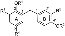
Table 1.
Structures of Compounds Based on Honokiol.
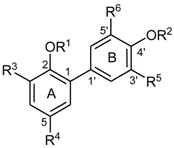
| Cpd. | R1 | R2 | R3 | R4 | R5 | R6 |
|---|---|---|---|---|---|---|
| H | H | H | H | allyl | allyl | H |
| H1 | H | Me | NH2 | allyl | allyl | H |
| H2 | H | Me | NH-COC11H23 | allyl | allyl | H |
| H3 | H | Me | NH-Ala | allyl | allyl | H |
| H4 | H | Me | NH-Phe | allyl | allyl | H |
| H5 | H | Et | NH2 | allyl | allyl | H |
| H6 | Et | Me | NH2 | allyl | allyl | H |
| H7 | Et | Me | NH-Ac | allyl | allyl | H |
| H8 | H | Me | NH-Ac | allyl | allyl | H |
| H9 | Me | H | H | allyl | allyl | NH-Ac |
| H10 | H | Et | H | allyl | allyl | H |
| H11 | H | Me | H | allyl | allyl | H |
| H12 | Me | H | H | allyl | allyl | NH2 |
| H13 | H | H | H | allyl | 1-propenyl | H |
| H14 | H | H | H | n-propyl | n-propyl | H |
| H15 | Me | H | H | allyl | allyl | H |
| H16 | Me | Me | H | allyl | allyl | H |
| H17 | H | Me | H | allyl | 1-propenyl | H |
| H18 | H | Me | H | 2,3-dihydroxypropyl | 2,3-dihydroxypropyl | H |
| H19 | H | Me | H | n-propyl | n-propyl | H |
| H20 | Me | Me | H | n-propyl | n-propyl | H |
| H21 | Et | H | H | allyl | allyl | H |
| H22 | H | Et | H | allyl | allyl | H |
| H23 | Et | H | H | n-propyl | n-propyl | H |
| H24 | Et | Et | H | n-propyl | n-propyl | H |
| H25 | H | Me | H | 2-bromopropyl | allyl | H |
| H26 | Me | H | H | allyl | allyl | NO2 |
| H27 | Me | H | H | n-propyl | n-propyl | NO2 |
| H28 | H | Me | NO2 | allyl | allyl | H |
| H29 | Me | H | H | n-propyl | n-propyl | NH2 |
| H30 | H | Me | NH2 | n-propyl | n-propyl | H |
| H31 | Me | H | H | n-propyl | n-propyl | NH-Ac |
| H32 | H | Me | NH-Ac | n-propyl | n-propyl | H |
Table 2.
Structures of Compounds Based on Biphenyl-2,2′-diol.
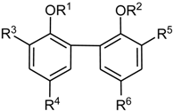
| Cpd. | R1 | R2 | R3 | R4 | R5 | R6 |
|---|---|---|---|---|---|---|
| M | H | H | H | allyl | H | allyl |
| M1 | H | H | H | H | H | H |
| M2 | H | H | H | n-propyl | H | n-propyl |
| M3 | Me | H | H | allyl | H | allyl |
| M4 | Me | Me | H | allyl | H | allyl |
| M5 | H | H | H | Me | H | Me |
| M6 | H | H | H | t-butyl | H | t-butyl |
| M7 | H | H | allyl | H | allyl | H |
Table 3.
Structures of Compounds Based on Biphenyl-4,4′-diol.
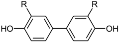
| Cpd. | R |
|---|---|
| A1 | H |
| A2 | allyl |
Table 4.
Structures of Compounds Based on Bis(2-hydroxyphenyl)methane.
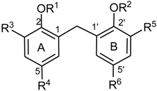
| Cpd. | R1 | R2 | R3 | R4 | R5 | R6 |
|---|---|---|---|---|---|---|
| B1 | H | H | H | H | H | H |
| B1a * | allyl | allyl | H | H | H | H |
| B2 | H | H | allyl | H | allyl | H |
| B3 * | H | H | allyl | H | H | allyl |
| B4b | Me | H | allyl | H | allyl | H |
| B4a | Me | Me | allyl | H | allyl | H |
* Compounds not tested.
Table 5.
Structures of Compounds Based on 2,4′-Dihydroxydiphenylmethane.
| Cpd. | R1 | R2 | R3 | R4 | R5 |
|---|---|---|---|---|---|
| C1 | H | H | H | H | H |
| C1a * | allyl | allyl | H | H | H |
| C2 | H | H | allyl | H | allyl |
| C3 * | H | H | H | allyl | allyl |
| C4c | Me | H | allyl | H | allyl |
| C4b | H | Me | allyl | H | allyl |
| C4a | Me | Me | allyl | H | allyl |
* Compounds not tested.
© 2018 by the authors. Licensee MDPI, Basel, Switzerland. This article is an open access article distributed under the terms and conditions of the Creative Commons Attribution (CC BY) license (http://creativecommons.org/licenses/by/4.0/).
Share and Cite
MDPI and ACS Style
Stout, K.; Bernaskova, M.; Miller, G.W.; Hufner, A.; Schuehly, W. Bioinspired Honokiol Analogs and Their Evaluation for Activity on the Norepinephrine Transporter. Molecules 2018, 23, 2536. https://doi.org/10.3390/molecules23102536
AMA Style
Stout K, Bernaskova M, Miller GW, Hufner A, Schuehly W. Bioinspired Honokiol Analogs and Their Evaluation for Activity on the Norepinephrine Transporter. Molecules. 2018; 23(10):2536. https://doi.org/10.3390/molecules23102536
Chicago/Turabian StyleStout, Kristen, Marketa Bernaskova, Gary W. Miller, Antje Hufner, and Wolfgang Schuehly. 2018. "Bioinspired Honokiol Analogs and Their Evaluation for Activity on the Norepinephrine Transporter" Molecules 23, no. 10: 2536. https://doi.org/10.3390/molecules23102536