Article

A Novel Postbiotic Improves Stool Consistency in Dogs: A Randomized, Double-Blind Placebo-Controlled Study


Kingdom, Brooklyn, NY 11205, USA
*
Authors to whom correspondence should be addressed.
Submission received: 16 February 2026 / Revised: 14 April 2026 / Accepted: 27 April 2026 / Published: 30 April 2026

Abstract

Postbiotics are an emerging ingredient class which have promising potential to support canine gut function by delivering beneficial microbial compounds directly to the gut. We tested a canine immune health postbiotic (CIHP) in a randomized, double-blind, placebo-controlled study of twenty colony-housed dogs (ten per group) selected for having consistently loose stools but with no diagnosed gastrointestinal disease. After a 5-day wash-in and 5-day baseline, dogs received 12 mg/kg body weight per day of CIHP or a placebo for 28 days mixed with their normal dry diet. The primary outcome was stool consistency (Waltham fecal score), measured on Days 0, 14, and 28; secondary outcomes included fecal gut-health biomarkers and fecal microbiome composition from 16S rRNA sequencing, measured on Days 0 and 28. CIHP improved stool quality (p-value = 0.03), while placebo did not (p-value = 0.5), and CIHP showed a trend toward increasing the odds of individual dogs showing improved fecal scores by Day 28 compared to placebo (p-value = 0.07). Microbiome profiling revealed broader community remodeling with CIHP than the placebo (16 taxa significantly shifted with CIHP vs. 1 with the placebo), consistent with stool quality being impacted by both gastrointestinal and gut microbiome functions. Fecal biomarkers that reflect gut health (pH, dry matter, short-chain fatty acids, dysbiosis index, calprotectin) were within reference ranges at baseline and remained stable in both groups, indicating benefits occurred within a normal physiological window. Together, these findings show that CIHP can improve stool consistency while reshaping the gut microbiome in otherwise healthy dogs prone to loose stools. Future studies in home-environment dogs across breeds, ages, and living conditions are needed to generalize these findings to the broader canine population.

1. Introduction

Gut health in dogs depends on the coordinated function of the gastrointestinal tract and its resident microbiota, which together regulate digestion, nutrient absorption, and immune balance [1,2,3,4,5,6]. A healthy gut is characterized by a diverse and balanced microbiome that supports nutrient metabolism, inhibits colonization by potential pathogens, and contributes to mucosal and systemic immune regulation. As such, studies in dogs have demonstrated that gastrointestinal disease states are consistently associated with significant reductions in bacterial diversity indices and depletion of short-chain fatty acid-producing bacteria, concurrent with overgrowth of potentially pathogenic taxa including Clostridium perfringens [7,8,9]. Furthermore, these compositional shifts correlate with altered fecal concentrations of propionate and butyrate, decreased serum tryptophan metabolites involved in immune homeostasis, and elevated intestinal inflammatory markers including calprotectin [7,10,11], and microbial imbalance, or dysbiosis, has been associated with diarrhea and inflammatory changes in dogs [2,7,8,12]. Accordingly, nutritional strategies that support a healthy gut microbiome are a growing focus in companion animal research [2,13,14,15].
Stool quality can often provide a practical, non-invasive indicator of gastrointestinal function and is widely used to assess the impact of dietary interventions in dogs [13,16,17,18]. Variations in stool consistency offer a readout of gut health as they reflect integrated changes in digestion, water reabsorption, and microbial metabolism [19,20,21,22]. The Waltham fecal scoring system provides a standardized, validated framework for quantifying stool consistency and is sensitive to subtle dietary effects in healthy dogs [16,23,24].
Microbially derived ingredients, including probiotics and postbiotics, are increasingly being utilized to support gut health in dogs, and there is a growing body of evidence that probiotics can improve canine stool quality. Lactobacillus probiotics improved stool quality in dogs with diarrhea within five to seven days across two studies [25,26]. Additionally, dogs fed a Bacillus subtilis probiotic for 28 days had firmer stools compared to a control group; however, no baseline stool quality data was collected prior to treatment [27]. Probiotic viability can be compromised during pet food processing and storage, limiting their reliability. An analysis of dog and cat diets that contained probiotics found that many did not include the organisms listed on the label, and that the number of colony forming units was about half of the dose known to impart a benefit [28]. Additionally, probiotic efficacy is highly variable across individuals and species, largely due to differences in the baseline gut microbiome that determine colonization compatibility [29]. Postbiotics, defined as inanimate microbial cells and their metabolites that confer a health benefit, offer a promising alternative to probiotics in terms of stability and consistency [30]. While a postbiotic derived from Lacticaseibacillus paracasei, Lactiplantibacillus plantarum, and Bifidobacterium animalis subsp. lactis strains has been shown to improve stool quality in humans with chronic diarrhea [31], evidence that postbiotics can impact canine stool quality is lacking [32,33,34,35,36,37,38,39,40].
Previously, a canine immune health postbiotic (CIHP) was shown to promote a healthy gut–skin axis in dogs with elevated itching, reducing scratching by 20% as measured by accelerometer, improving human-perceived itch scores, enhancing coat quality, and favorably modulating the gut microbiome [41]. Considering these findings, we hypothesized that CIHP supplementation may also support gut health. To further investigate its potential to support gut health, the present study evaluated CIHP in a population of dogs with consistently loose stools but without diagnosed gastrointestinal disease. The objective was to determine whether CIHP supplementation could improve stool quality and support a healthy gut microbiome.

2. Materials and Methods

2.1. Animals and Housing

The study was conducted at an independent, registered contract research organization, separate from the commercial entity to which the authors are affiliated. The contract research organization complied with all local regulations governing the care and use of laboratory animals and was conducted in accordance with the Ontario Ministry of Agriculture, Food and Rural Affairs (OMAFRA), the Canadian Council on Animal Care (CCAC) Guide to the Care and Use of Experimental Animals. To ensure compliance, the protocol was reviewed and approved by the facility’s Institutional Animal Care and Use Committee (IACUC; ONL-AUP-2025-03). A protocol including the research question, key design features, and analysis plan was prepared before the study, and this protocol was not registered.
All dogs were part of a permanent colony made up of beagles and small mixed breed dogs. All participants were between 1 and 14 years old. Both intact and neutered animals of both sexes were included in the study. Dogs were allowed to socialize in groups in outside dog runs for at least one hour each day and had supervised access to a larger dog park at least twice weekly for robust play and exercise. Dogs received daily one-on-one human socialization from a regular attendant (physical affection, play, and leash-walking), as well as additional regular human contact (bathing, grooming, walking, and play) from enrichment staff.
All dogs were either pair-housed in 10-foot × 10-foot runs that could be divided into 5-foot × 10-foot runs for individual housing or group-housed in 20-foot × 16-foot rooms (6–8 dogs/room). Beds and blankets were provided to all dogs. Fresh, clean, drinking water was provided ad libitum. Dogs remained in the same room for the duration of the study. Animal rooms were cleaned at least once daily, disinfected twice weekly, and descaled when needed. Dogs were monitored for coprophagy, and any instances of coprophagy were recorded.
All dogs were classified as USDA Category C for the full duration of the study: Animal use activities that involve no more than momentary or slight pain or distress for which there is no need for use of pain-relieving drugs.

2.2. Study Design

The study was a double-blind, placebo-controlled, randomized trial. No gut health treatments, including supplements, were administered for the duration of the study. The duration of the study was 38 days (Days −9 to 28), and consisted of a 5-day wash-in period (Days −9 to −5), a 5-day baseline observation period (Days −4 to 0), and a 28-day intervention period (Days 1 to 28).
Twenty dogs were enrolled in this study. The population was defined by general and gut-specific health eligibility criteria. The generic health eligibility criteria were that the dog (1) is not taking any prescribed medications or antibiotics beyond standard preventative flea, tick, and heartworm medication. The gut-specific health eligibility criteria were the following: the dog (2) is not currently diagnosed with any gastrointestinal condition, such as irritable bowel syndrome, (3) does not have food allergies, (4) does not currently have any parasitic infections, (5) has not been prescribed medication for any food allergy or gastrointestinal conditions by a veterinarian in the past year, (6) does not currently take any supplements or products to relieve gastrointestinal issues, and (7) consistently has a Waltham Fecal Score of three to four, inclusive (see Stool Quality Assessment and Stool Collection).
Participants were stratified randomly into 2 groups of 10 subjects, N, based on the average stool score from Day −4 to Day −1 and sex (Table 1 and Table S1). Potential confounders such as the order of interventions and measurements, or animal location, were not controlled. All dogs at the registered research facility meeting all inclusion criteria at the time of recruitment and available were enrolled in the study.
There were two termination criteria for the study: (1) abnormal changes in a dog’s health as assessed by a veterinarian followed by a recommendation by a veterinarian to remove the dog from the study, and (2) refusal to eat more than 4 consecutive meals containing the ingredient (not eating over the course of 2 days).

2.3. Intervention

CIHP is a commercially available ingredient (Superculture® Pet Immune ingredient, Kingdom, Brooklyn, NY, USA) composed of a tapioca maltodextrin carrier and dried Pediococcus acidilactici fermentation product. The fermentation product is heat treated to inactivate live cells, then spray or freeze dried. The placebo was tapioca maltodextrin, the same carrier utilized in the ingredient.
Animals were individually housed for feeding and the amount of food offered was based on the resting energy requirement. Each dog’s food intake was recorded to ensure that the majority of the offering was regularly consumed (Table S2) and food offerings were updated weekly based on the dog’s current body weight. On Days 1–28, each dog received a 6 mg/kg body weight dose of CIHP or the placebo added to both their first and second meal of the day (the regularly provided diet; Purina Dog Chow; guaranteed analysis and ingredient list provided in Table S3) as a powder topper, for a total daily dose of 12 mg/kg body weight. The daily dose was updated on a weekly basis based on the dog’s current body weight. The daily food portion was placed into a bowl and sprayed with enough water to moisten the food and promote adhesion of the powder topper to the food. The powder topper was mixed into the food with a spoon until evenly distributed. The dog was served the food containing the powder topper and given up to 30 min to eat all the food. The study employed a double-blind design, in which the individuals performing intervention administration, data collection, or data analysis were blind to group assignments.

2.4. Stool Quality Assessment and Stool Collection

On Days 0, 14, and 28, dogs were brought into the collection runs first thing in the morning and were observed by a technician until a fecal sample was produced. Fecal samples were scored and collected within 15 min of defecation. The time of scoring was recorded.
The Waltham fecal score was recorded according to the following scale (1–5): 1.0 Bullet shaped, chalk like consistency; 1.5 Hard, dry crumbly stool; 2.0 Well-formed, solid with pronounced grooves; 2.5 Well-formed, slightly moist with visible grooves; 3.0 Moist, beginning to lose form, grooves still visible; 3.5 Very moist, has form but no grooves; 4.0 Most of the form is lost, consistency is viscous; 4.5 Liquid stool with slight consistency; 5.0 Entirely liquid stool.
Total sample weight was recorded, and samples were stored for analysis as follows: samples of the first evacuated feces were stored in propylene cryovials (VWR Low-Temperature Freezer Vials, item # CVI-2.0-B) for microbiome and stool biomarker analyses. The cryovials were flash frozen in liquid nitrogen, and stored at −80 °C until shipment to external partners for analysis. Samples were shipped on dry ice. Further samples were stored in a whirl-pak bag for pH and dry matter analysis, which was conducted at the research facility.

2.5. Stool Microbiome Analysis

Approximately 100 mg (wet weight) of each fecal sample was extracted using the MoBio PowerSoil DNA Isolation Kit (MoBio Laboratories, Carlsbad, CA, USA) per manufacturer instructions. The V4 region of the bacterial 16S rRNA gene was amplified with primers 515F (5′-GTGCCAGCMGCCGCGGTAA-3′) and 806R (5′-GGACTACVSGGGTATCTAAT-3′) using HotStarTaq Plus Master Mix (Qiagen, Hilden, Germany) under the following program: 94 °C for 3 min; 28 cycles of 94 °C for 30 s, 53 °C for 40 s, 72 °C for 60 s; final extension 72 °C for 5 min. Amplicons were sequenced on an Illumina platform at MR DNA (Shallowater, TX, USA). A total of 40 fecal samples were analyzed by 16S rRNA gene sequencing (n = 20 dogs × 2 timepoints: Day 0 and Day 28). All twenty enrolled dogs contributed samples at both timepoints, and all samples were included in the relevant analysis.
Reads with expected number of errors (E) greater than 1 were filtered out. Reads shorter than 120 bp were removed. Counts of the zero-radius operational taxonomic units (ZOTUs) [42] were determined for each library using USEARCH (https://www.drive5.com/usearch/download.html (accessed on 1 August 2025)), and taxonomic assignments for the ZOTUs were made using the RDP classifier [43]. The median read count was 509,038.5 reads with samples ranging from 207,599 to 798,959 reads. For fold change calculations, taxa with less than 500 reads in at least 20% of samples were removed, and read counts were standardized to the median sequencing depth. Read counts were analyzed in R using Phyloseq (Version 1.46.0) [44] and DESeq2 (Version 1.42.1) [45] packages. Alpha diversity describes the variety of individuals that make up a community on a local scale [46]. The log fold change in number of reads was calculated for each taxa for each group of dogs. Taxa are considered significantly changed if the log fold change from Day 0 to Day 28 has a Benjamini–Hochberg–adjusted p-value < 0.05, controlling the false discovery rate (FDR).

2.6. Stool Dry Matter Analysis

The fecal sample was assigned a code which matches a labelled crucible. The labelled crucible was weighed (Sartorius PRACTUM 124-1S, Sartorius AG, Göttingen, Germany) and the weight recorded in the internal software system up to 0.0001 g. A sample of approximately 2.0 ± 0.01 g of feces was weighed and the combined weight of the crucible and feces is recorded. The sample was placed in a drying oven (Fisher Scientific Isotemp Oven Model 630G, Thermo Fisher Scientific, Waltham, MA, USA) at 135 ± 2 °C for 2 h. Upon removal, the sample was placed in a lidded desiccator (Nalgene Polypropylene Desiccator (Waltham, MA, USA) with Stopcock, Catalog No.5310-0250) and allowed to cool to room temperature before reweighing the crucible and sample. The remaining weight is recorded as the dry matter of the sample.

2.7. Stool pH Analysis

Fecal samples were homogenized prior to pH analysis. To prepare the pH meter (HANNA HI 8424 pH meter, Hanna Instruments, Woonsocket, RI, USA) for analysis it was turned on and then the probe was rinsed with distilled water and dried with a Kim wipe. Standard pH solutions (Fisher Chemical Buffer Solutions pH 4.00 and 7.00, Thermo Fisher Scientific, Waltham, MA, USA) were freshly prepared, and the meter was calibrated. Once calibrated, the probe was placed into the middle of the fecal sample, ensuring that no part of the bulb is touching the side of the bag or exposed to air. After waiting for the meter to stabilize, the pH was recorded. The probe was cleaned with distilled water between samples and kept in distilled water between sampling times.

2.8. Stool Calprotectin Analysis

Frozen stool aliquots were thawed on ice and extracted per the manufacturer’s instructions for the BÜHLMANN fCAL® turbo particle-enhanced turbidimetric immunoassay (BÜHLMANN Laboratories AG, Schönenbuch, Switzerland). Briefly, samples were processed with the CALEX® Cap device to a final 1:500 dilution (or by manual weighing to 1:50 followed by a 1:10 dilution in extraction buffer (BÜHLMANN Laboratories AG, B-CAL-EX12) when CALEX® was not used), vortexed thoroughly for 20 min, and centrifuged (5 min, 1500× g). Clear extracts were loaded onto a clinical chemistry analyzer (Beckman Coulter AU480,Beckman Coulter, Brea, CA, USA) configured with fCAL® turbo reagents (R1 reaction buffer; R2 calprotectin-antibody–coated immunoparticles). A six-point calibration curve (BÜHLMANN fCAL® turbo Calibrator Kit, Catalog# B-KCAL-CASET) was run and verified with low/high controls prior to samples. Turbidity (absorbance increase due to immune agglutination) was measured by the analyzer and calprotectin concentrations (μg/g) were automatically interpolated from the calibration curve; results were reported after applying the manufacturer-specified extraction and dilution factors [47].

2.9. Stool IgA Analysis

Frozen stool aliquots were thawed on ice, homogenized in the kit diluent (Bethyl Dog IgA ELISA quantification set, Bethyl Laboratories, Montgomery, TX, USA; 1× Dilution Buffer B), and clarified by centrifugation to remove particulates. Supernatants were serially diluted (matrix-appropriate dilutions chosen to fall within the assay range) and assayed in duplicate on pre-coated anti-dog IgA 96-well plates following the manufacturer’s protocol: 100 μL/well sample or standard (0–1000 ng/mL), 1 h at room temperature; wash ×4; 100 μL Detection Antibody, 1 h; wash ×4; 100 μL HRP Solution A, 30 min; wash ×4; 100 μL TMB, 30 min in the dark; 100 μL Stop Solution. Absorbance at 450 nm was read within 30 min of stopping (Biotek 800TS Absorbance Reader, BioTek Instruments, Winooski, VT, USA). Concentrations were calculated by 4-parameter logistic regression against the standard curve and multiplied by the appropriate dilution and extraction factors to yield IgA levels in the original stool extracts.

2.10. Stool Short-Chain Fatty Acids

Methods followed Minamoto et al. 2019 [48]. Frozen fecal aliquots were thawed, diluted 1:5 (w/v) in 2 N HCl, homogenized at room temperature, and centrifuged (2100× g, 20 min, 4 °C) to obtain clarified supernatants. An internal standard (internal standard mix (200 mM d4-acetic acid, 50 mM d5-propionic acid, 25 mM d7-butyric acid, 781.25 μM d9-valeric acid in water)) was added before extraction on C18 columns (Discovery® DSC-18 SPE Tube, Supelco, Bellefonte, PA, USA). Extracts were derivatized with MTBSTFA (60 min, RT) and analyzed by GC–MS (Agilent 6890N/5975C, Agilent Technologies, Santa Clara, CA, USA, DB-1 ms column) following a temperature program of 40–280 °C. Quantification of acetate, propionate, and butyrate was performed by selective ion monitoring (m/z 117 (acetate), 120 (d4-acetate), 131 (propionate), 136 (d5-propionate), 145 (isobutyrate and butyrate), 152 (d7-butyrate), 159 (valerate and isovalerate), and 168 (d9-valerate)), using area-ratio calibration against internal standards. Results were expressed as μmol/g dry matter, adjusting for fecal water content.

2.11. Dysbiosis Index

Fecal DNA was quantified by SYBR-green qPCR for seven taxa—Faecalibacterium, Turicibacter, Streptococcus, Escherichia coli, Blautia, Fusobacterium, and Clostridium hiranonis—together with a universal 16S “total bacteria” assay [49]. Reactions were run in duplicate and reported as log DNA mass per 10 ng total input DNA. For each sample, target abundances were normalized to universal bacteria, and the resulting seven-dimensional vector was entered into a nearest-centroid model trained on reference healthy and chronic enteropathy cohorts. The Dysbiosis Index (DI) was computed as the Euclidean-distance difference between the sample and the healthy vs diseased class centroids (DI =   z μ H 2 z μ D 2 ), yielding a single scalar per sample (negative values closer to normobiotic, positive values more dysbiotic).

2.12. Statistical Analysis

The use of one-tailed within group tests for stool quality and fecal biomarkers was pre-determined based on the results of a previous clinical trial [41]. A Shapiro–Wilk test was used to assess if the data were normally distributed. A one-tailed exact Wilcoxon signed-rank test (Pratt version) was performed on the stool score to assess how stool quality changed from baseline within a group. A two-tailed ranked Mann–Whitney test on the absolute and relative change in stool score was used to assess any differences between the two groups. If the fecal biomarker data were normally distributed, a t-test was used to assess the change from baseline within a group. If the data were not normally distributed, a one-tailed Wilcoxon signed-rank test was used to assess the change from baseline within a group. Results were considered statistically significant at p-value < 0.05, while results with 0.05 ≤ p-value < 0.10 were reported as trends.
All dogs started the study with loose stools. Any reduction in stool score indicated a relatively firmer stool and was categorized as improved. A positive change or no change was categorized as not improved. A logistic regression was performed on the categorical data to determine the odds of improvement.

3. Results

No adverse events or meal refusals were recorded, and no participants were removed from the study. One incidence of coprophagy was recorded during the wash-in period. All dogs were used in all analysis.
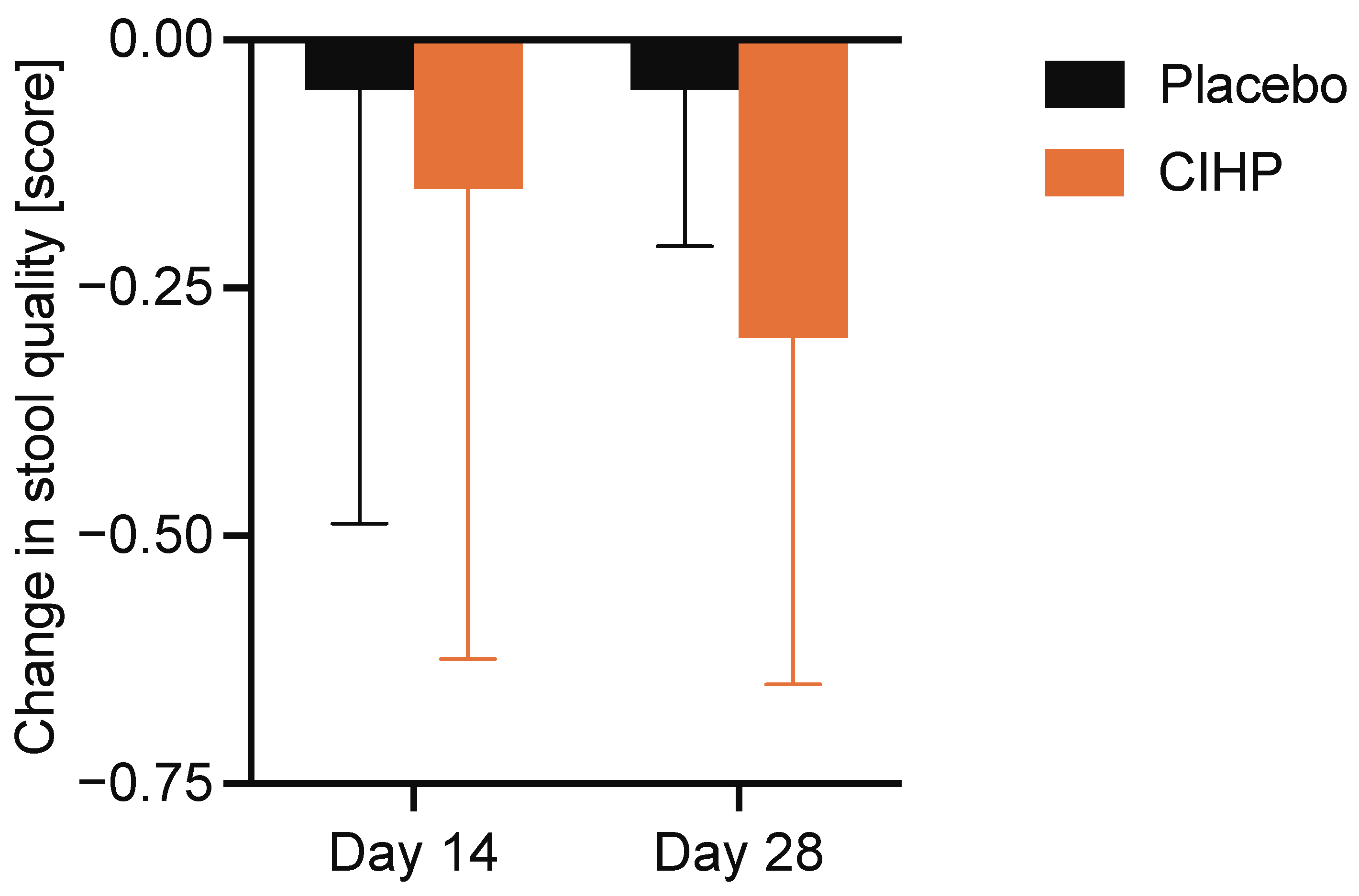
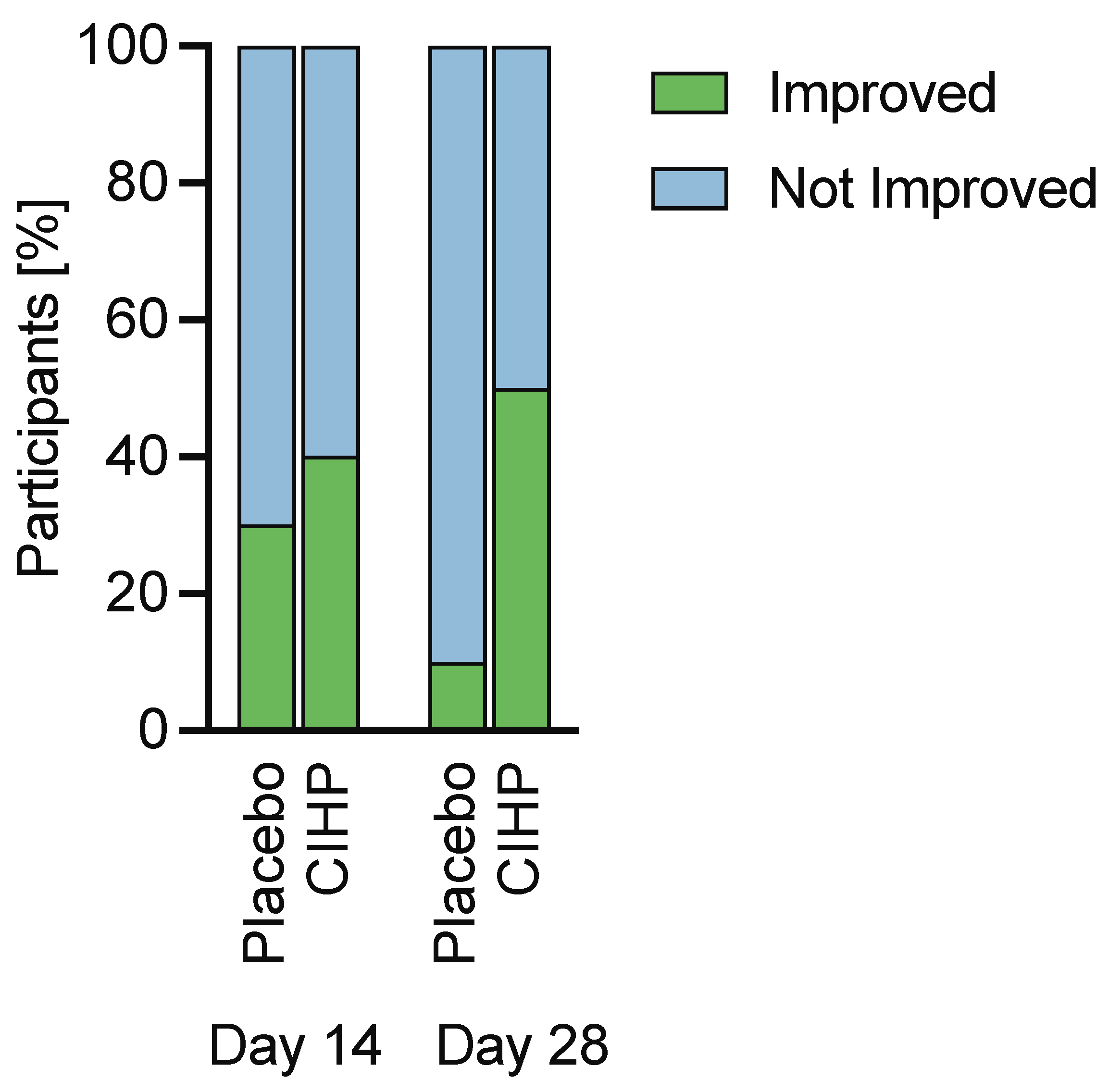
CIHP effectively improved stool quality in dogs with noticeably loose stools but without diagnosed gastrointestinal disease. At baseline (Day 0), fecal scores ranged from 2.5 (“well-formed, slightly moist with visible grooves”) to 3.5 (“very moist, has form but no grooves”) in both groups. On average, the scores were 3.1 ± 0.4 in the CIHP group and 3.0 ± 0.3 in the placebo group; scores in this range correspond to the “moist, beginning to lose form” category of the Waltham scale, confirming that both groups entered the intervention period with consistently loose stools. No change in stool score was observed in either group at Day 14 (CIHP: p-value = 0.3; placebo: p-value = 0.3), suggesting that any intervention effect required more than two weeks to manifest. By Day 28, CIHP significantly reduced the Waltham fecal score (median change = −0.25, p-value = 0.03), reflecting an improvement in stool consistency (Figure 1 and Figure S1). Given the scale’s 0.5-point scoring increments, a median change of −0.25 represents movement halfway toward the next firmness category. No corresponding change was observed in the placebo group at Day 28 (p-value = 0.5). In line with this result, 50% of dogs in the CIHP group showed improved fecal scores at Day 28 compared with only 10% in the placebo group (Figure 2), with CIHP trending toward a significantly increased odds of improvement versus placebo (p-value = 0.07). Additionally, considering both the absolute and relative change in stool score, there was a trend toward a group difference at Day 28 (p-values > 0.08), but not at Day 14 (p-values > 0.81). Convergence of evidence across the within-group test, the between-group responder analysis, and the absolute and relative change analyses provides a coherent and consistent picture of the intervention effect.
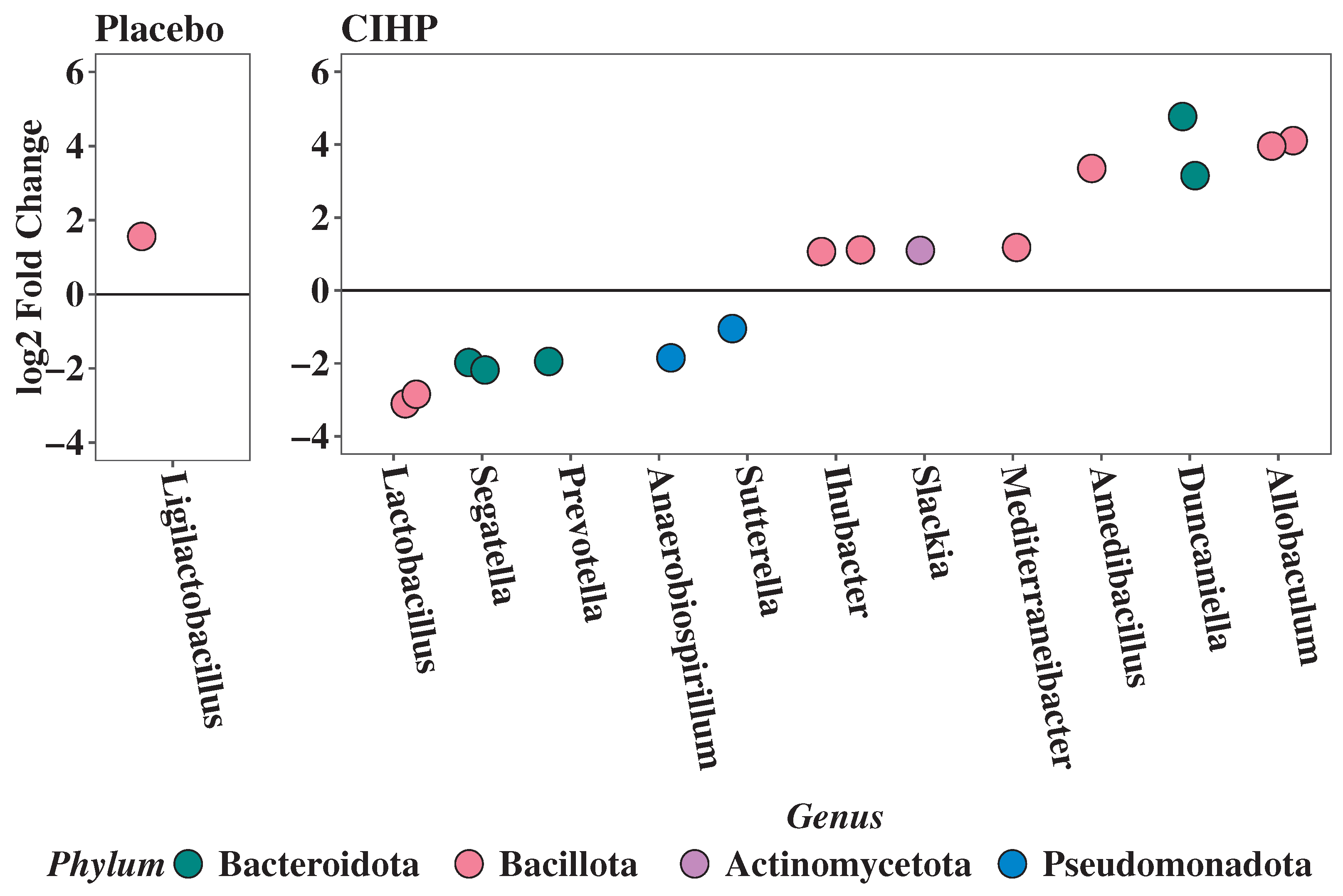
Improvement in stool quality was accompanied by a shift in gut microbiome composition, with CIHP having a larger impact on gut microbiome composition compared to placebo at Day 28. More taxa increased and decreased in abundance over the course of the study for dogs in the CIHP group (Table 2) than in the placebo group (Table 3) (Figure 3). Only one taxon significantly increased in abundance in the placebo group, while sixteen taxa exhibited changed abundance in the CIHP group (alpha = 0.05; 7 taxa decrease and 9 taxa increase). A threshold of 0.8 was set for genus confidence with regards to confirming taxa identity [50,51]. Ligilactobacillus ( log 2 fold change = 1.44; confidence 0.93) increased in the placebo group. Allobaculum (confidence ≥ 0.89; log 2 fold change = 3.99 and 4.10), Mediterraneibacter (confidence 0.91; log 2 fold change = 1.17), and Slackia (confidence 0.99; log 2 fold change = 1.10) increased in the CIHP group (Table 2), and Lactobacillus (confidence 0.87; log 2 fold change = −3.11) and Segatella (confidence ≥ 0.97; log 2 fold change = −1.98 and −2.18) decreased in the CIHP group (Table 2).
Fecal gut health biomarkers were maintained in both groups throughout the study (p-value ≥ 0.24, Table 4). Fecal pH, dry matter content (% DM), short-chain fatty acids (acetate, propionate, and butyrate; SCFA), and dysbiosis index (DI) were within normal range at baseline, and remained within normal range throughout the study (Table 4). Fecal calprotectin was below the level of detection in the majority of dogs (8/10 in CIHP; 9/10 in placebo; Table 4).

4. Discussion

The postbiotic CIHP improved stool quality and produced measurable changes in the fecal microbiome of dogs with consistently loose stools but without diagnosed gastrointestinal disease, indicating a general enhancement of gut health. CIHP induced compositional shifts involving sixteen taxa (nine increasing and seven decreasing) compared to a single taxon in the placebo group, suggesting effects on the gut microbiome composition underlie stool quality changes. Taken together, the results of this study suggest that microbiome remodeling likely contributed to more efficient digestive processing and intestinal water balance, resulting in improved stool formation. These results are aligned with other studies that quantify both stool consistency and the gut microbiome: Stool consistency was found to be tightly linked to gut microbial community composition in human cohorts [19], and a study in mice has shown that antibiotic-induced gut dysbiosis caused delayed gut motility, which led to increased water content in the stool [56]. Additionally, dogs with both acute loose stools and gut inflammation have been found to have distinct microbiomes from healthy dogs [8].
The taxa with significantly shifted abundance in the CIHP group that could be assigned at the genus level with confidence ≥ 0.80 spanned three phyla and included both increases and decreases, consistent with broad community remodeling. Among the three taxa of high identity confidence that increased, the most robust signal was observed in Allobaculum, whose co-increase across two independent ZOTUs strengthens this observation. Allobaculum has been associated with reduced gut inflammatory state in rodent colitis models, with its abundance increasing alongside resolution of intestinal mucosal inflammation and restoration of epithelial barrier function [57,58]. It was also observed that its abundance increased in obese dogs undergoing dietary weight loss intervention [33]; given that obesity is associated with chronic inflammation and gut dysbiosis in dogs [59,60,61], this is consistent with Allobaculum tracking a less inflamed community state. The remaining two increasing ZOTUs are more difficult to interpret in the context of gut health. Mediterraneibacter (formerly Ruminococcus) [62], has a complex, context-dependent role in gut homeostasis [63,64]. Slackia is an anaerobic commensal found in healthy human feces [65] that has been associated with gut inflammation and barrier dysfunction in some human studies [66,67].
Among the two taxa with high identity confidence that decreased, the most robust signal was observed in two ZOTUs assigned to Segatella (formerly Prevotella copri clade; [68]), found in both the human and canine gut [69,70]. This taxon is associated with pro-inflammatory signaling, including arthritic states [69,71,72], and its abundance correlated with pain indices in senior dogs [73]. One ZOTU assigned to Lactobacillus also decreased; as Lactobacillus probiotic strains improve stool quality in dogs with diarrhea [25,26], this decrease most plausibly reflects intra-community niche remodeling given the simultaneous expansion of multiple Bacillota taxa, though strain-level data would be required to confirm this.
In the placebo group, only one taxon increased significantly: a ZOTU assigned to Ligilactobacillus, species of which have been isolated from healthy canine feces and demonstrated antimicrobial properties against enteric pathogens in vitro [74]. Its modest increase most likely reflects spontaneous community fluctuation rather than a directed intervention response.
Importantly, the study population consisted of dogs that were otherwise healthy and free of gastrointestinal disease. The gut-specific health eligibility criteria ensured that the population of dogs in this study do not have known pathology, and that consistently loose stool is a suboptimal-but-healthy phenotype. As expected, biomarkers such as fecal pH, dry matter, calprotectin, and dysbiosis index were within reference ranges at baseline and remained stable throughout the study. However, it should be noted that inter-laboratory differences exist, even when using the same assay technologies, resulting in different reference ranges across laboratories. Indeed, the high proportion of undetectable calprotectin values (80–90%) in this study reflects the relatively high limit of detection in the calprotectin assay (65 μg/g), which is substantially higher than the limit of detection of 3 μg/g in the report by Enderle et al. (2022) that validated the fCAL® turbo assay in dogs [47]. The healthy dog median in Enderle et al. was 9.0 μg/g, and the upper limit of their reference interval was 41 μg/g, suggesting that the majority of values in a healthy population would fall below our limit of detection. The stability of fecal biomarkers across both groups is consistent with the study population being within a healthy physiological range at baseline. While the absence of biomarker changes does not independently demonstrate an intervention effect, the findings here are consistent with the stool quality improvements observed in the CIHP group occurring within normal physiological bounds, and demonstrate that CIHP was well-tolerated; stable biomarker profiles in healthy dogs during dietary interventions have been used as evidence of tolerability in similar recent canine studies [35,37]. Future studies with larger sample sizes and more targeted biomarker panels, including measures of intestinal permeability and immune function, would be better positioned to identify within-normal-range biomarker changes that correlate with microbiome remodeling and to characterize the mechanistic pathways through which CIHP supports gut health.
Results from this and prior studies of CIHP indicate that the ingredient produces measurable improvements in stool quality and microbiome composition in dogs, with effects that appear more robust than those reported for other postbiotic formulations in similar research. Other postbiotic ingredients have failed to show a significant change in stool quality [32,33,34,35,36,37,38,39], and a Saccharomyces cerevisiae postbiotic was shown to worsen stool quality in healthy dogs at Day 28 [40]. Additionally, while CIHP has been shown to increase Shannon diversity in a population of dogs with elevated itching and shifted the abundance of four times more taxa than the placebo in that study [41] and sixteen times more taxa in this study, other postbiotics have less robust data to demonstrate their ability to reorganize the microbiome. Other postbiotics have not modulated alpha diversity metrics, including Shannon diversity, and generally are less able to modulate microbiome composition compared to CIHP [32,33,34,35,36,37,38,39]. A Lacticaseibacillus paracasei postbiotic failed to modulate the composition of the microbiome across two studies, one in healthy dogs and one in dogs subjected to an abrupt diet change [37,38]. The same Saccharomyces cerevisiae postbiotic that worsened stool quality modulated the abundance of only one or two taxa in two different studies [32,40], and during an experiment in which dogs underwent transport stress it was observed that different taxa changed in abundance, depending on whether dogs received the Saccharomyces cerevisiae postbiotic or the control [33]. A second Saccharomyces cerevisiae postbiotic was found to affect the abundance of only one taxa after transport stress, and this effect was not consistent across two doses administered [39]. Other postbiotics or heat-treated strains show some microbiome modulation, but on longer timescales: a Lactobacillus postbiotic modulated the magnitude of changes in taxa abundance for six taxa compared to a control at Day 35 [34] and heat-treated Bifidobacterium animalis modulated the magnitude of changes in taxa abundance for twenty taxa compared to a control at Day 90 [35]. Overall, the results indicate that CIHP provides more comprehensive gut health support relative to alternative postbiotics currently under investigation.
Some limitations of this study should be noted. Individual host factors, including sex, reproductive status, age, and breed, are known to influence gut microbiome composition in dogs [75,76,77,78]. Although sex was used as a stratification variable during randomization, the limited sample size did not allow for stratification by additional covariates or formal assessment of the contribution of these physiological variables to the observed microbiome response. Future studies with larger, more diverse cohorts should consider incorporating these variables as covariates in the microbiome analysis. The study population was restricted to a small population of Beagles and small mixed-breed dogs in a controlled research environment, which has the advantage of limiting environmental effects on the gut microbiome; however, it limits generalizability to the broader pet dog population. Colony-housed dogs may differ systematically from owned pet dogs in baseline gut microbiome composition, stress exposure, dietary history, and inflammatory status. Future studies in home-environment dogs with heterogeneous breeds, ages, and housing conditions are needed to confirm that these findings extend to the broader canine population. Performing a study in home-environment dogs would also allow us to increase the sample size to improve statistical confidence and generalizability of the findings while still focusing on the most relevant dog population, healthy dogs with noticeable loose stools. Moreover, longitudinal trials assessing persistence of effects could further elucidate whether CIHP supports stabilization of gut function over time, and studies in which healthy animals undergo a transport stress or diet-change could show whether CIHP’s ability to support gut health persists in scenarios known to induce loose stools. Additionally, in order to better understand mechanism of action, further studies could include an explicit assessment of gut permeability, such as through the administration of a tracer molecule that is later quantified in the urine or blood [79]. This study did not include direct measures of immune function, such as serum cytokine profiles, immunoglobulin levels, or immune cell counts. Future studies would benefit from incorporating systemic immune function endpoints to directly characterize the effects of CIHP supplementation on the immune system. Additionally, serum cobalamin and folate, which are commonly used to screen for chronic enteropathy in dogs, were not included in the health assessment. While eligibility criteria excluded all dogs with a prior gastrointestinal diagnosis or use of relevant medications, and all participants were under regular veterinary oversight, inclusion of these biomarkers in future studies would provide a more comprehensive exclusion of gastrointestinal pathology.
This 28-day study duration supports short-term tolerability but is insufficient to characterize the long-term safety profile of CIHP [80]. Given the compositional complexity inherent to fermentation products [81,82], conclusions regarding the safety of chronic supplementation require dedicated long-term safety studies. A study evaluating CIHP over a period of several months or more would be required to characterize its safety profile under conditions of prolonged exposure.
Overall, the results demonstrate that CIHP supports a healthy gut microbiome and healthy digestion to improve stool quality. These findings strengthen the evidence that postbiotics represent a promising, non-invasive nutritional approach for supporting gut health in companion animals.

5. Conclusions

In colony-housed dogs exhibiting consistently loose stools but without diagnosed gastrointestinal disease, a 28-day intervention with a novel postbiotic improved stool quality and drove substantially broader shifts in gut microbial composition than a placebo, while fecal health markers remained within normal ranges throughout the study. When considered alongside prior work linking the same ingredient to higher Shannon diversity and skin/coat benefits in dogs with elevated itching [41], the present findings strengthen the idea that targeted postbiotics provide a practical strategy to support canine gut health. These findings were obtained under controlled research facility conditions, and replication in dogs across diverse settings would substantially improve their generalizability.

Supplementary Materials

The following supporting information can be downloaded at: https://www.mdpi.com/article/10.3390/pets3020019/s1, Table S1: Initial study group composition showing microchip identification, breed, sex/neutered status, age, baseline body weight, and mean stool score during the pre-study period, (Days −4 to −1) for each subject; Table S2: Individual mean food consumption (% of food offered) during the intervention period (1–28 May 2025); Table S3: Purina Dog Chow guaranteed analysis and ingredient list, as reported by the manufacturer; Figure S1: The distribution of stool score changes in each group. Bars represent the number of dogs with each score change on Day 14 (left) or Day 28 (right). A decrease in score is indicative of an improvement in stool quality.

Author Contributions

Conceptualization, R.U.S., R.T. and A.S.; methodology, A.S., R.T. and J.H.; formal analysis, A.S. and J.H.; investigation, A.S. and J.H.; data curation, A.S. and J.H.; writing—original draft preparation, J.H. and A.S.; writing—review and editing, J.H., A.S., R.T. and R.U.S.; visualization, A.S. and J.H.; supervision, R.T.; validation, A.S., J.H. and R.T.; project administration, A.S. All authors have read and agreed to the published version of the manuscript.

Funding

This research received no external funding.

Institutional Review Board Statement

The animal study protocol was approved by the Institutional Animal Care and Use Committee (IACUC) of the research facility and was conducted in accordance with the Ontario Ministry of Agriculture, Food and Rural Affairs (OMAFRA) and the Canadian Council on Animal Care (CCAC) Guide to the Care and Use of Experimental Animals.

Informed Consent Statement

Not applicable.

Data Availability Statement

The data presented in this study are available on request from the corresponding authors.

Acknowledgments

We would like to thank all Kingdom employees for their contributions to this project. In particular, we would like to thank Antonio Diaz for his contributions to the project administration, and Emily Daley for her review of the manuscript.

Conflicts of Interest

All authors are employees of Kingdom and hold stocks and/or stock options in the company. Kingdom funded this research and is the supplier of the commercial material assessed in this study. The authors are committed to maintaining scientific integrity and adhering to ethical research practices. The paper reflects the view of the scientists and not the company.

Abbreviations

The following abbreviations are used in this manuscript:
AhRAryl hydrocarbon receptor
CCACCanadian Council on Animal Care
CIHPCanine immune health postbiotic
DIDysbiosis index
DMDry matter
FDRFalse discovery rate
IACUCInstitutional Animal Care and Use Committee
OMAFRAOntario Ministry of Agriculture, Food and Rural Affairs
PXRPregnane X receptor
rRNARibosomal RNA
SCFAShort chain fatty acid
ZOTUZero-radius operational taxonomic unit

References

  1. Suchodolski, J.S. Analysis of the gut microbiome in dogs and cats. Vet. Clin. Pathol. 2022, 50, 6–17. [Google Scholar] [CrossRef]
  2. Pilla, R.; Suchodolski, J.S. The Role of the Canine Gut Microbiome and Metabolome in Health and Gastrointestinal Disease. Front. Vet. Sci. 2019, 6, 498. [Google Scholar] [CrossRef] [PubMed]
  3. Yang, Q.; Wu, Z. Gut Probiotics and Health of Dogs and Cats: Benefits, Applications, and Underlying Mechanisms. Microorganisms 2023, 11, 2452. [Google Scholar] [CrossRef]
  4. Grześkowiak, L.; Endo, A.; Beasley, S.; Salminen, S. Microbiota and probiotics in canine and feline welfare. Anaerobe 2015, 34, 14–23. [Google Scholar] [CrossRef]
  5. Hooda, S.; Minamoto, Y.; Suchodolski, J.S.; Swanson, K.S. Current state of knowledge: The canine gastrointestinal microbiome. Anim. Health Res. Rev. 2012, 13, 78–88. [Google Scholar] [CrossRef] [PubMed]
  6. Mondo, E.; Marliani, G.; Accorsi, P.A.; Cocchi, M.; Di Leone, A. Role of gut microbiota in dog and cat’s health and diseases. Open. Vet. J. 2019, 9, 253–258. [Google Scholar] [CrossRef] [PubMed]
  7. Guard, B.C.; Barr, J.W.; Reddivari, L.; Klemashevich, C.; Jayaraman, A.; Steiner, J.M.; Vanamala, J.; Suchodolski, J.S. Characterization of Microbial Dysbiosis and Metabolomic Changes in Dogs with Acute Diarrhea. PLoS ONE 2015, 10, e0127259. [Google Scholar] [CrossRef]
  8. Suchodolski, J.S.; Markel, M.E.; Garcia-Mazcorro, J.F.; Unterer, S.; Heilmann, R.M.; Dowd, S.E.; Kachroo, P.; Ivanov, I.; Minamoto, Y.; Dillman, E.M.; et al. The Fecal Microbiome in Dogs with Acute Diarrhea and Idiopathic Inflammatory Bowel Disease. PLoS ONE 2012, 7, e51907. [Google Scholar] [CrossRef]
  9. Minamoto, Y.; Cristiane, O.C.; Steelman, S.M.; Büyükleblebici, O.; Steiner, J.M.; Jergens, A.E.; Suchodolski, J.S. Alteration of the fecal microbiota and serum metabolite profiles in dogs with idiopathic inflammatory bowel disease. Gut Microbes 2015, 6, 33–47. [Google Scholar] [CrossRef]
  10. Herstad, K.M.V.; Vinje, H.; Skancke, E.; Næverdal, T.; Corral, F.; Llarena, A.K.; Heilmann, R.M.; Suchodolski, J.S.; Steiner, J.M.; Nyquist, N.F. Effects of Canine-Obtained Lactic-Acid Bacteria on the Fecal Microbiota and Inflammatory Markers in Dogs Receiving Non-Steroidal Anti-Inflammatory Treatment. Animals 2022, 12, 2519. [Google Scholar] [CrossRef]
  11. Omori, M.; Maeda, S.; Igarashi, H.; Ohno, K.; Sakai, K.; Yonezawa, T.; Horigome, A.; Odamaki, T.; Matsuki, N. Fecal microbiome in dogs with inflammatory bowel disease and intestinal lymphoma. J. Vet. Med. Sci. 2017, 79, 1840–1847. [Google Scholar] [CrossRef]
  12. Galler, A.I.; Suchodolski, J.S.; Steiner, J.M.; Sung, C.H.; Hittmair, K.M.; Richter, B.; Burgener, I.A. Microbial dysbiosis and fecal metabolomic perturbations in Yorkshire Terriers with chronic enteropathy. Sci. Rep. 2022, 12, 12977. [Google Scholar] [CrossRef] [PubMed]
  13. Barry, K.A.; Wojcicki, B.J.; Middelbos, I.S.; Vester, B.M.; Swanson, K.S.; Fahey, G.C. Dietary cellulose, fructooligosaccharides, and pectin modify fecal protein catabolites and microbial populations in adult cats. J. Anim. Sci. 2010, 88, 2978–2987. [Google Scholar] [CrossRef]
  14. Schauf, S.; de la Fuente, G.; Newbold, C.J.; Salas-Mani, A.; Torre, C.; Abecia, L.; Castrillo, C. Effect of dietary fat to starch content on fecal microbiota composition and activity in dogs. J. Anim. Sci. 2018, 96, 3684–3698. [Google Scholar] [CrossRef]
  15. Kerr, K.R.; Forster, G.; Dowd, S.E.; Ryan, E.P.; Swanson, K.S. Effects of Dietary Cooked Navy Bean on the Fecal Microbiome of Healthy Companion Dogs. PLoS ONE 2013, 8, e74998. [Google Scholar] [CrossRef] [PubMed]
  16. Cavett, C.L.; Tonero, M.; Marks, S.L.; Winston, J.A.; Gilor, C.; Rudinsky, A.J. Consistency of faecal scoring using two canine faecal scoring systems. J. Small Anim. Pract. 2021, 62, 167–173. [Google Scholar] [CrossRef]
  17. Nery, J.; Biourge, V.; Tournier, C.; Leray, V.; Martin, L.; Dumon, H.; Nguyen, P. Influence of dietary protein content and source on fecal quality, electrolyte concentrations, and osmolarity, and digestibility in dogs differing in body size. J. Anim. Sci. 2010, 88, 159–169. [Google Scholar] [CrossRef] [PubMed]
  18. Jackson, M.I.; Jewell, D.E. Balance of saccharolysis and proteolysis underpins improvements in stool quality induced by adding a fiber bundle containing bound polyphenols to either hydrolyzed meat or grain-rich foods. Gut Microbes 2019, 10, 298–320. [Google Scholar] [CrossRef]
  19. Vandeputte, D.; Falony, G.; Vieira-Silva, S.; Tito, R.Y.; Joossens, M.; Raes, J. Stool consistency is strongly associated with gut microbiota richness and composition, enterotypes and bacterial growth rates. Gut 2016, 65, 57–62. [Google Scholar] [CrossRef]
  20. Jackson, M.I.; Wernimont, S.M.; Carnagey, K.; Jewell, D.E. Nutrient Digestive Bypass: Determinants and Associations with Stool Quality in Cats and Dogs. Animals 2024, 14, 2778. [Google Scholar] [CrossRef]
  21. Deschamps, C.; Humbert, D.; Zentek, J.; Denis, S.; Priymenko, N.; Apper, E.; Blanquet-Diot, S. From Chihuahua to Saint-Bernard: How did digestion and microbiota evolve with dog sizes. Int. J. Biol. Sci. 2022, 18, 5086–5102. [Google Scholar] [CrossRef] [PubMed]
  22. Waclawiková, B.; Codutti, A.; Alim, K.; El Aidy, S. Gut microbiota-motility interregulation: Insights from in vivo, ex vivo and in silico studies. Gut Microbes 2022, 14, 1997296. [Google Scholar] [CrossRef]
  23. Vogel, C.L.; Geary, E.L.; Oba, P.M.; Mioto, J.C.; Rudolph, B.C.; Rens, L.; Swanson, K.S. Effects of corn protein inclusion on apparent total tract macronutrient digestibility, palatability, and fecal characteristics, microbiota, and metabolites of healthy adult dogs. J. Anim. Sci. 2025, 103, skaf122. [Google Scholar] [CrossRef] [PubMed]
  24. Rolfe, V.E.; Adams, C.A.; Butterwick, R.F.; Batt, R.M. Relationship between faecal character and intestinal transit time in normal dogs and diet-sensitive dogs. J. Small Anim. Pract. 2002, 43, 290–294. [Google Scholar] [CrossRef]
  25. Gómez-Gallego, C.; Junnila, J.; Männikkö, S.; Hämeenoja, P.; Valtonen, E.; Salminen, S.; Beasley, S. A canine-specific probiotic product in treating acute or intermittent diarrhea in dogs: A double-blind placebo-controlled efficacy study. Vet. Microbiol. 2016, 197, 122–128. [Google Scholar] [CrossRef]
  26. Delucchi, L.; Fraga, M.; Zunino, P. Effect of the probiotic Lactobacillus murinus LbP2 on clinical parameters of dogs with distemper-associated diarrhea. Can. J. Vet. Res. 2017, 81, 118–121. [Google Scholar]
  27. De Lima, D.C.; Souza, C.M.M.; Nakamura, N.; Mesa, D.; De Oliveira, S.G.; Félix, A.P. Dietary supplementation with Bacillus subtilis C-3102 improves gut health indicators and fecal microbiota of dogs. Anim. Feed Sci. Technol. 2020, 270, 114672. [Google Scholar] [CrossRef]
  28. Weese, J.S.; Arroyo, L. Bacteriological evaluation of dog and cat diets that claim to contain probiotics. Can. Vet. J. 2003, 44, 212–215. [Google Scholar]
  29. Zmora, N.; Zilberman-Schapira, G.; Suez, J.; Mor, U.; Dori-Bachash, M.; Bashiardes, S.; Kotler, E.; Zur, M.; Regev-Lehavi, D.; Brik, R.B.Z.; et al. Personalized Gut Mucosal Colonization Resistance to Empiric Probiotics Is Associated with Unique Host and Microbiome Features. Cell 2018, 174, 1388–1405.e21. [Google Scholar] [CrossRef] [PubMed]
  30. Salminen, S.; Collado, M.C.; Endo, A.; Hill, C.; Lebeer, S.; Quigley, E.M.M.; Sanders, M.E.; Shamir, R.; Swann, J.R.; Szajewska, H.; et al. The International Scientific Association of Probiotics and Prebiotics (ISAPP) consensus statement on the definition and scope of postbiotics. Nat. Rev. Gastroenterol. Hepatol. 2021, 18, 649–667. [Google Scholar] [CrossRef]
  31. Guo, S.; Ma, T.; Kwok, L.Y.; Quan, K.; Li, B.; Wang, H.; Zhang, H.; Menghe, B.; Chen, Y. Effects of postbiotics on chronic diarrhea in young adults: A randomized, double-blind, placebo-controlled crossover trial assessing clinical symptoms, gut microbiota, and metabolite profiles. Gut Microbes 2024, 16, 2395092. [Google Scholar] [CrossRef]
  32. Oba, P.M.; Carroll, M.Q.; Sieja, K.M.; de Souza Nogueira, J.P.; Yang, X.; Epp, T.Y.; Warzecha, C.M.; Varney, J.L.; Fowler, J.W.; Coon, C.N.; et al. Effects of a Saccharomyces cerevisiae fermentation product on fecal characteristics, metabolite concentrations, and microbiota populations of dogs subjected to exercise challenge. J. Anim. Sci. 2022, 101, skac424. [Google Scholar] [CrossRef] [PubMed]
  33. Oba, P.M.; Carroll, M.Q.; Sieja, K.M.; Yang, X.; Epp, T.Y.; Warzecha, C.M.; Varney, J.L.; Fowler, J.W.; Coon, C.N.; Swanson, K.S. Effects of a Saccharomyces cerevisiae fermentation product on fecal characteristics, metabolite concentrations, and microbiota populations of dogs undergoing transport stress. J. Anim. Sci. 2023, 101, skad191. [Google Scholar] [CrossRef]
  34. Koziol, S.A.; Oba, P.M.; Soto-Diaz, K.; Steelman, A.J.; Suchodolski, J.S.; Eckhardt, E.R.M.; Swanson, K.S. Effects of a Lactobacillus fermentation product on the fecal characteristics, fecal microbial populations, immune function, and stress markers of adult dogs. J. Anim. Sci. 2023, 101, skad160. [Google Scholar] [CrossRef] [PubMed]
  35. Kayser, E.; He, F.; Nixon, S.; Howard-Varona, A.; Lamelas, A.; Martinez-Blanch, J.; Chenoll, E.; Davenport, G.M.; de Godoy, M.R.C. Effects of supplementation of live and heat-treated Bifidobacterium animalis subspecies lactis CECT 8145 on glycemic and insulinemic response, fecal microbiota, systemic biomarkers of inflammation, and white blood cell gene expression of adult dogs. J. Anim. Sci. 2024, 102, skae291. [Google Scholar] [CrossRef]
  36. Lin, C.Y.; Carroll, M.Q.; Miller, M.J.; Rabot, R.; Swanson, K.S. Supplementation of Yeast Cell Wall Fraction Tends to Improve Intestinal Health in Adult Dogs Undergoing an Abrupt Diet Transition. Front. Vet. Sci. 2020, 7. [Google Scholar] [CrossRef] [PubMed]
  37. Wren, J.F.; Wilson, S.M.; Kang, Y.; Oba, P.M.; Menton, J.F.; Vinay, E.; Millette, M.; Kelly, M.R.; Swanson, K.S. Effects of live Bacillus pumilus SG154 or a Lacticaseibacillus paracasei 327 postbiotic on apparent total tract nutrient digestibility and the immune function and fecal characteristics, metabolites, and microbiota of healthy adult dogs. J. Anim. Sci. 2025, 103, skaf231. [Google Scholar] [CrossRef]
  38. Wren, J.F.; Wilson, S.M.; Kang, Y.; Oba, P.M.; Menton, J.F.; Vinay, E.; Millette, M.; Kelly, M.R.; Swanson, K.S. Effects of Bacillus pumilus SG154 or Lacticaseibacillus paracasei 327 Postbiotic on the Fecal Characteristics and Microbiota of Healthy Adult Dogs Subjected to an Abrupt Diet Change. Pets 2025, 2, 30. [Google Scholar] [CrossRef]
  39. Timlin, C.L.; Mccracken, F.B.; Dickerson, S.M.; Skaggs, P.M.; Fowler, J.W.; Jalukar, S.; Coon, C.N. Effects of a Saccharomyces cerevisiae-Derived Postbiotic in Adult Labrador Retrievers Undergoing Exercise and Transport Stress. Pets 2024, 1, 350–371. [Google Scholar] [CrossRef]
  40. Lin, C.Y.; Alexander, C.; Steelman, A.J.; Warzecha, C.M.; de Godoy, M.R.C.; Swanson, K.S. Effects of a Saccharomyces cerevisiae fermentation product on fecal characteristics, nutrient digestibility, fecal fermentative end-products, fecal microbial populations, immune function, and diet palatability in adult dogs1. J. Anim. Sci. 2019, 97, 1586–1599. [Google Scholar] [CrossRef]
  41. Sordillo, A.; Heldrich, J.; Turcotte, R.; Sheth, R.U. An Indole-Rich Postbiotic Reduces Itching in Dogs: A Randomized, Double-Blinded Placebo-Controlled Study. Animals 2025, 15, 2019. [Google Scholar] [CrossRef]
  42. Edgar, R.C. UNOISE2: Improved error-correction for Illumina 16S and ITS amplicon sequencing. bioRxiv 2016. [Google Scholar] [CrossRef]
  43. Wang, Q.; Cole, J.R. Updated RDP taxonomy and RDP Classifier for more accurate taxonomic classification. Microbiol. Resour. Announc. 2024, 13, e0106323. [Google Scholar] [CrossRef]
  44. McMurdie, P.J.; Holmes, S. phyloseq: An R Package for Reproducible Interactive Analysis and Graphics of Microbiome Census Data. PLoS ONE 2013, 8, e61217. [Google Scholar] [CrossRef] [PubMed]
  45. Love, M.I.; Huber, W.; Anders, S. Moderated estimation of fold change and dispersion for RNA-seq data with DESeq2. Genome Biol. 2014, 15, 550. [Google Scholar] [CrossRef]
  46. Kers, J.G.; Saccenti, E. The Power of Microbiome Studies: Some Considerations on Which Alpha and Beta Metrics to Use and How to Report Results. Front. Microbiol. 2022, 12, 796025. [Google Scholar] [CrossRef]
  47. Enderle, L.L.; Köller, G.; Heilmann, R.M. Verification of the fCAL turbo immunoturbidimetric assay for measurement of the fecal calprotectin concentration in dogs and cats. J. Vet. Diagn. Investig. 2022, 34, 813–824. [Google Scholar] [CrossRef] [PubMed]
  48. Minamoto, Y.; Minamoto, T.; Isaiah, A.; Sattasathuchana, P.; Buono, A.; Rangachari, V.R.; McNeely, I.H.; Lidbury, J.; Steiner, J.M.; Suchodolski, J.S. Fecal short-chain fatty acid concentrations and dysbiosis in dogs with chronic enteropathy. J. Vet. Intern. Med. 2019, 33, 1608–1618. [Google Scholar] [CrossRef]
  49. AlShawaqfeh, M.; Wajid, B.; Minamoto, Y.; Markel, M.; Lidbury, J.; Steiner, J.; Serpedin, E.; Suchodolski, J. A dysbiosis index to assess microbial changes in fecal samples of dogs with chronic inflammatory enteropathy. FEMS Microbiol. Ecol. 2017, 93, fix136. [Google Scholar] [CrossRef] [PubMed]
  50. Johnson, J.S.; Spakowicz, D.J.; Hong, B.Y.; Petersen, L.M.; Demkowicz, P.; Chen, L.; Leopold, S.R.; Hanson, B.M.; Agresta, H.O.; Gerstein, M.; et al. Evaluation of 16S rRNA gene sequencing for species and strain-level microbiome analysis. Nat. Commun. 2019, 10, 5029. [Google Scholar] [CrossRef]
  51. Murali, A.; Bhargava, A.; Wright, E.S. IDTAXA: A novel approach for accurate taxonomic classification of microbiome sequences. Microbiome 2018, 6, 140. [Google Scholar] [CrossRef]
  52. Abd El-Wahab, A.; Zeiger, A.L.; Chuppava, B.; Visscher, C.; Kamphues, J. Effects of poultry by-products inclusion in dry food on nutrient digestibility and fecal quality in Beagle dogs. PLoS ONE 2022, 17, e0276398. [Google Scholar] [CrossRef]
  53. Tress, U.; Suchodolski, J.S.; Williams, D.A.; Steiner, J.M. Development of a fecal sample collection strategy for extraction and quantification of fecal immunoglobulin A in dogs. Am. J. Vet. Res. 2006, 67, 1756–1759. [Google Scholar] [CrossRef] [PubMed]
  54. Zaine, L.; Ferreira, C.; Gomes, M.O.S.; Monti, M.; Tortola, L.; Vasconcellos, R.S.; Carciofi, A.C. Faecal IgA concentration is influenced by age in dogs. Br. J. Nutr. 2011, 106, S183–S186. [Google Scholar] [CrossRef]
  55. Satyaraj, E.; Reynolds, A.; Engler, R.; Labuda, J.; Sun, P. Supplementation of diets with Spirulina influences immune and gut function in dogs. Front. Nutr. 2021, 8, 667072. [Google Scholar] [CrossRef]
  56. Ge, X.; Ding, C.; Zhao, W.; Xu, L.; Tian, H.; Gong, J.; Zhu, M.; Li, J.; Li, N. Antibiotics-induced depletion of mice microbiota induces changes in host serotonin biosynthesis and intestinal motility. J. Transl. Med. 2017, 15, 13. [Google Scholar] [CrossRef]
  57. Wu, X.; Chen, H.; Gao, X.; Gao, H.; He, Q.; Li, G.; Yao, J.; Liu, Z. Natural Herbal Remedy Wumei Decoction Ameliorates Intestinal Mucosal Inflammation by Inhibiting Th1/Th17 Cell Differentiation and Maintaining Microbial Homeostasis. Inflamm. Bowel Dis. 2022, 28, 1061–1071. [Google Scholar] [CrossRef]
  58. Seo, S.H.; Unno, T.; Park, S.E.; Kim, E.J.; Lee, Y.M.; Na, C.S.; Son, H.S. Korean Traditional Medicine (Jakyakgamcho-tang) Ameliorates Colitis by Regulating Gut Microbiota. Metabolites 2019, 9, 226. [Google Scholar] [CrossRef] [PubMed]
  59. Li, Y.; Rahman, S.U.; Huang, Y.; Zhang, Y.; Ming, P.; Zhu, L.; Chu, X.; Li, J.; Feng, S.; Wang, X.; et al. Green tea polyphenols decrease weight gain, ameliorate alteration of gut microbiota, and mitigate intestinal inflammation in canines with high-fat-diet-induced obesity. J. Nutr. Biochem. 2020, 78, 108324. [Google Scholar] [CrossRef] [PubMed]
  60. Cortese, L.; Terrazzano, G.; Pelagalli, A. Leptin and Immunological Profile in Obesity and Its Associated Diseases in Dogs. Int. J. Mol. Sci. 2019, 20, 2392. [Google Scholar] [CrossRef]
  61. Thomson, P.; Santibáñez, R.; Rodríguez-Salas, C.; Flores-Yañez, C.; Garrido, D. Differences in the composition and predicted functions of the intestinal microbiome of obese and normal weight adult dogs. PeerJ 2022, 10, e12695. [Google Scholar] [CrossRef]
  62. Togo, A.H.; Diop, A.; Bittar, F.; Maraninchi, M.; Valero, R.; Armstrong, N.; Dubourg, G.; Labas, N.; Richez, M.; Delerce, J.; et al. Description of Mediterraneibacter massiliensis, gen. nov., sp. nov., a new genus isolated from the gut microbiota of an obese patient and reclassification of Ruminococcus faecis, Ruminococcus lactaris, Ruminococcus torques, Ruminococcus gnavus and Clostridium glycyrrhizinilyticum as Mediterraneibacter faecis comb. nov., Mediterraneibacter lactaris comb. nov., Mediterraneibacter torques comb. nov., Mediterraneibacter gnavus comb. nov. and Mediterraneibacter glycyrrhizinilyticus comb. nov. Antonie Van Leeuwenhoek 2018, 111, 2107–2128. [Google Scholar] [CrossRef]
  63. Tiffany, E.; Kim, K.S.; Sittipo, P.; Lee, D.W.; Lee, Y.K. Mucin-degrading gut bacteria: Context-dependent roles in intestinal homeostasis and disease. Gut Microbes 2026, 18, 2614054. [Google Scholar] [CrossRef]
  64. Grabinger, T.; Glaus Garzon, J.F.; Hausmann, M.; Geirnaert, A.; Lacroix, C.; Hennet, T. Alleviation of Intestinal Inflammation by Oral Supplementation with 2-Fucosyllactose in Mice. Front. Microbiol. 2019, 10, 1385. [Google Scholar] [CrossRef]
  65. Tamura, M.; Hori, S.; Nakagawa, H.; Yamauchi, S.; Sugahara, T. Effects of an equol-producing bacterium isolated from human faeces on isoflavone and lignan metabolism in mice. J. Sci. Food Agric. 2016, 96, 3126–3132. [Google Scholar] [CrossRef] [PubMed]
  66. Munjoma, P.T.; Chandiwana, P.; Wyss, J.; Mazhandu, A.J.; Jordi, S.B.U.; Gutsire, R.; Katsidzira, L.; Yilmaz, B.; Misselwitz, B.; Duri, K. Immune activation and inflammation in lactating women on combination antiretroviral therapy: Role of gut dysfunction and gut microbiota imbalance. Front. Immunol. 2023, 14, 1280262. [Google Scholar] [CrossRef] [PubMed]
  67. Maqoud, F.; Calabrese, F.M.; Celano, G.; Mallardi, D.; Goscilo, F.; D’Attoma, B.; Ignazzi, A.; Linsalata, M.; Bitetto, G.; Chito, M.D.; et al. Role of Increasing Body Mass Index in Gut Barrier Dysfunction, Systemic Inflammation, and Metabolic Dysregulation in Obesity. Nutrients 2024, 17, 72. [Google Scholar] [CrossRef]
  68. Hitch, T.C.A.; Bisdorf, K.; Afrizal, A.; Riedel, T.; Overmann, J.; Strowig, T.; Clavel, T. A taxonomic note on the genus Prevotella: Description of four novel genera and emended description of the genera Hallella and Xylanibacter. Syst. Appl. Microbiol. 2022, 45, 126354. [Google Scholar] [CrossRef] [PubMed]
  69. Abdelsalam, N.A.; Hegazy, S.M.; Aziz, R.K. The curious case of Prevotella copri. Gut Microbes 2023, 15, 2249152. [Google Scholar] [CrossRef]
  70. Wang, N.; Sieng, S.; Chen, P.; Liang, T.; Xu, J.; Han, Q. Regulation Effect of Toxocara canis and Anthelmintics on Intestinal Microbiota Diversity and Composition in Dog. Microorganisms 2024, 12, 2037. [Google Scholar] [CrossRef]
  71. Sepúlveda-Pontigo, A.; Chávez-Villacreses, K.; Madrid-Muñoz, C.; Conejeros-Lillo, S.; Parra, F.; Melo-González, F.; Regaldiz, A.; González, V.P.I.; Méndez-Pérez, I.; Castillo-Godoy, D.P.; et al. Segatella copri Outer-Membrane Vesicles Are Internalized by Human Macrophages and Promote a Pro-Inflammatory Profile. Int. J. Mol. Sci. 2025, 26, 3630. [Google Scholar] [CrossRef]
  72. Ajith, T.A.; Anita, B. Impact of Gut Microbiota and Probiotics on Rheumatoid Arthritis: A Potential Treatment Challenge. Int. J. Rheum. Dis. 2025, 28, e70266. [Google Scholar] [CrossRef]
  73. Saral, B.; Atilgan, D.; Adiay, D.; Filazi, N.; Ozturk, H.; Kismali, G.; Da Graca Pereira, G.; Ozkul, A.; Salgirli Demirbas, Y. An exploratory study of behavioral, cognitive, physiological, and microbiota profiles in senior dogs. Front. Behav. Neurosci. 2026, 20, 1689807. [Google Scholar] [CrossRef] [PubMed]
  74. Jimenez-Trigos, E.; Toquet, M.; Barba, M.; Gómez-Martín, Á.; Quereda, J.J.; Bataller, E. Search of antimicrobial lactic acid bacteria from Salmonella-negative dogs. BMC Vet. Res. 2022, 18, 12. [Google Scholar] [CrossRef] [PubMed]
  75. Choi, S.Y.; Choi, B.H.; Cha, J.H.; Lim, Y.J.; Sheet, S.; Song, M.J.; Ko, M.J.; Kim, N.Y.; Kim, J.S.; Lee, S.J.; et al. Insight into the Fecal Microbiota Signature Associated with Growth Specificity in Korean Jindo Dogs Using 16S rRNA Sequencing. Animals 2022, 12, 2499. [Google Scholar] [CrossRef]
  76. Morelli, G.; Patuzzi, I.; Losasso, C.; Ricci, A.; Contiero, B.; Andrighetto, I.; Ricci, R. Characterization of intestinal microbiota in normal weight and overweight Border Collie and Labrador Retriever dogs. Sci. Rep. 2022, 12, 9199. [Google Scholar] [CrossRef] [PubMed]
  77. Asaduzzaman, M.; Oláh, P.; Yaseen, N.J.; Taifi, A.; Járay, T.; Gulyás, G.; Boldogkői, Z.; Tombácz, D. Longitudinal long-read microbiome profiling in a canine model reveals how age, diet, and birth mode shape gut community dynamics. mSystems 2026, 11, e01279-25. [Google Scholar] [CrossRef]
  78. Yang, K.; Lin, X.; Jian, S.; Wen, J.; Jian, X.; He, S.; Wen, C.; Liu, T.; Qi, X.; Yin, Y.; et al. Changes in gut microbiota and short-chain fatty acids are involved in the process of canine obesity after neutering. J. Anim. Sci. 2023, 101, skad283. [Google Scholar] [CrossRef]
  79. González-González, M.; Díaz-Zepeda, C.; Eyzaguirre-Velásquez, J.; González-Arancibia, C.; Bravo, J.A.; Julio-Pieper, M. Investigating Gut Permeability in Animal Models of Disease. Front. Physiol. 2019, 9, 1962. [Google Scholar] [CrossRef]
  80. Schauss, A.G.; Glavits, R.; Endres, J.; Jensen, G.S.; Clewell, A. Safety evaluation of a proprietary food-grade, dried fermentate preparation of Saccharomyces cerevisiae. Int. J. Toxicol. 2012, 31, 34–45. [Google Scholar] [CrossRef]
  81. Vinderola, G.; Benkowski, A.; Bernardeau, M.; Chenoll, E.; Collado, M.C.; Cronin, U.; Eckhardt, E.; Green, J.B.; Ipharraguerre, I.R.; Kemperman, R.; et al. Postbiotics: A perspective on their quantification. Front. Nutr. 2025, 12, 1582733. [Google Scholar] [CrossRef] [PubMed]
  82. Caffrey, E.B.; Sonnenburg, J.L.; Devkota, S. Our extended microbiome: The human relevant metabolites and biology of fermented foods. Cell Metab. 2024, 36, 684–701. [Google Scholar] [CrossRef] [PubMed]
Figure 1. CIHP decreased fecal score at Day 28, indicating an improvement in stool quality. Bars indicate the mean and error bars indicate the standard deviation.
Pets 03 00019 g001
Figure 2. Dogs fed CIHP trended toward an increased odds of improvement in fecal score at Day 28. 50% of dogs improved in the CIHP group on Day 28, while only 10% of dogs improved in the Placebo group.
Pets 03 00019 g002
Figure 3. Taxa with significant fold changes. More fluctuation in individual taxa occurs in the CIHP group than in the placebo group during the study. Upward deflection from the black line at zero represents increased abundance and downward deflection represents decreased abundance (FDR corrected p-value less than 0.05 for log fold change from Day 0 to Day 28). The x-axis indicates the genus of each taxa. The color of the points indicates the phylum of each taxa.
Pets 03 00019 g003
Table 1. Baseline characteristics by treatment group. Values are presented as mean ± SD.
GroupNMale:FemaleAge (Years)Body Weight (kg)Stool Score
CIHP106:46 ± 211.8 ± 3.83.1 ± 0.4
Placebo106:48 ± 313.5 ± 3.43.1 ± 0.3
Table 2. Significantly changed taxa for Superculture® Pet Immune group.
ZOTUPhylumGenusGenus Confidencelog2 Fold ChangeAdjusted p-Value
Zotu4BacillotaLactobacillus0.87−3.110.0044
Zotu17BacillotaAllobaculum0.924.100.0019
Zotu27BacteroidotaPrevotella0.42−1.950.012
Zotu16BacillotaAllobaculum0.893.990.015
Zotu24BacteroidotaSegatella0.97−1.980.012
Zotu80BacteroidotaDuncaniella0.193.140.031
Zotu9BacillotaAmedibacillus0.203.350.031
Zotu37BacillotaIhubacter0.411.120.015
Zotu460BacillotaLactobacillus0.33−2.850.028
Zotu58BacteroidotaDuncaniella0.494.770.015
Zotu69BacillotaMediterraneibacter0.911.170.0019
Zotu42PseudomonadotaSutterella0.56−1.040.031
Zotu316BacillotaIhubacter0.701.070.015
Zotu81BacteroidotaSegatella0.98−2.180.015
Zotu73PseudomonadotaAnaerobiospirillum0.72−1.840.012
Zotu85ActinomycetotaSlackia0.991.100.0019
Table 3. Significantly changed taxa for placebo group.
ZOTUPhylumGenusGenus Confidencelog2 Fold ChangeAdjusted p-Value
Zotu2BacillotaLigilactobacillus0.931.440.042
Table 4. Fecal biomarkers were maintained within a healthy range throughout the study. 1 65 μg/g is the limit of detection for this assay. 2 Sum of short-chain fatty acids: butyrate, acetate, and propionate. 3 Range provided by Texas A&M GI Lab.
Fecal BiomarkerGroupDay 0 (Mean ± SD)Day 28 (Mean ± SD)Healthy RangeSignificant Change (Test, p-Value)
pHCIHP6.1 ± 0.26.2 ± 0.26.0–6.9Paired t-test (D28 < D0), p = 0.9
Placebo6.1 ± 0.36.11 ± 0.19 Paired t-test (D28 < D0), p = 0.4
Dry matter (%)CIHP27 ± 428 ± 228–38% [52]Paired t-test (D28 > D0), p = 0.4
Placebo30 ± 328 ± 4 Paired t-test (D28 > D0), p = 0.9
IgA (mg/g)CIHP10 ± 1016 ± 25≥2.3 mg/g [53,54,55]Paired Wilcoxon (D28 > D0), p = 0.58
Placebo10 ± 134 ± 4 Paired Wilcoxon (D28 > D0), p = 0.96
Calprotectin 1 (μg/g)CIHP9/10 dogs undetectable8/10 dogs undetectable<133 μg/g 3
Placebo8/10 dogs undetectable10/10 dogs undetectable
SCFA 2 (μg/g)CIHP507 ± 63473 ± 53127–927 μg/g [48]Paired t-test (D28 > D0), p = 0.88
Placebo456 ± 61476 ± 84 Paired t-test (D28 > D0), p = 0.24
Dysbiosis indexCIHP−2.9 ± 1.2−2.3 ± 1.6<0Paired Wilcoxon (D28 < D0), p = 0.94
Placebo−3.4 ± 0.9−3.6 ± 1.4 Paired Wilcoxon (D28 < D0), p = 0.95
Disclaimer/Publisher’s Note: The statements, opinions and data contained in all publications are solely those of the individual author(s) and contributor(s) and not of MDPI and/or the editor(s). MDPI and/or the editor(s) disclaim responsibility for any injury to people or property resulting from any ideas, methods, instructions or products referred to in the content.