Review

Clostridioides difficile Infection in Tuberculosis Patients: An Underrecognized Complication of Antituberculous Therapy


by
Jovan Javorac
1,2,3,*,
Ana Milenković
1,2,
Dragica Kovačević
1,2,
Emilija Vujičić
1,2 and
Dejan Živanović
1,3,4
1
Faculty of Medicine, University of Novi Sad, 21000 Novi Sad, Serbia
2
Institute for Pulmonary Diseases of Vojvodina, 21204 Sremska Kamenica, Serbia
3
College of Vocational Studies for the Education of Preschool Teachers and Sports Trainers, 24000 Subotica, Serbia
4
Department of Psychology, College of Human Development, 11000 Belgrade, Serbia
*
Author to whom correspondence should be addressed.
J. Oman Med. Assoc. 2026, 3(2), 11; https://doi.org/10.3390/joma3020011
Submission received: 28 March 2026 / Revised: 2 June 2026 / Accepted: 25 June 2026 / Published: 3 July 2026

Abstract

Clostridioides difficile infection (CDI) is the leading cause of antibiotic-associated diarrhea; however, its association with antituberculous therapy (ATT) remains underrecognized. This review examines the risk, mechanisms, and clinical implications of CDI in patients with active tuberculosis (TB). Although ATT is traditionally considered low risk, prolonged exposure, particularly to rifampicin, may lead to cumulative disruption of gut microbiota and loss of colonization resistance. A characteristic feature of CDI in this setting is delayed onset, typically occurring several weeks to months after ATT initiation, which complicates timely diagnosis. Rifampicin plays a dual role, contributing to microbiota alterations and selection of resistant strains, while potentially affecting the pharmacokinetics of co-administered drugs. CDI in TB patients is associated with increased morbidity and mortality, with reported mortality rates up to 9.9%. Fidaxomicin is preferred due to its microbiota-sparing effect and lower recurrence rates, although cost remains a major limitation in high TB-burden settings, since vancomycin is an appropriate alternative. Management requires an individualized approach that balances the risk of CDI recurrence against the necessity of maintaining effective TB therapy.

1. Introduction

Clostridioides difficile infection (CDI) is the leading cause of antibiotic-associated diarrhea and a major contributor to morbidity and mortality in both hospital and community settings [1,2]. Although CDI is classically associated with broad-spectrum antibiotics [3], emerging evidence suggests that antituberculous therapy (ATT) may also contribute to its development despite its relatively narrow antimicrobial spectrum (see Table 1).
Tuberculosis (TB) remains a major global health problem, requiring prolonged multidrug therapy, including isoniazid, rifampicin, pyrazinamide, and ethambutol. This extended antibiotic exposure, combined with frequent comorbidities and healthcare contact, may predispose patients to gut microbiota disruption and CDI. Despite increasing reports and case series [1,2,4,5,6], CDI in patients receiving ATT remains underrecognized and insufficiently characterized, particularly regarding optimal management strategies.
The aim of this review is to summarize current evidence on the epidemiology, pathophysiology, risk factors, clinical features, and therapeutic challenges of CDI in patients undergoing ATT.

2. Materials and Methods

This narrative review was conducted through a comprehensive search of the PubMed, Scopus, and Web of Science databases for articles published between 2013 and 2025. Search terms included combinations of “tuberculosis”, “antituberculosis drugs”, “Clostridioides difficile infection”, “dysbiosis”, and “rifampicin”. Priority was given to systematic reviews, meta-analyses, clinical guidelines, and large cohort studies investigating the epidemiology, mechanisms, diagnosis, and management of CDI under ATT. The selected literature was analyzed and synthesized to summarize current knowledge regarding the definition, pathogenesis, clinical presentation, diagnosis, management, and prevention of CDI in patients with TB.

3. Results

3.1. Epidemiology and Risk Factors of CDI in TB Patients

European estimates suggest that the incidence of CDI in community settings is approximately 1.35 cases per 1000 hospital admissions, while in hospital settings it ranges from 2.58 to 7.04 cases per 10,000 patient-days, depending largely on adherence to infection prevention and control measures [7]. Although a marked increase in CDI incidence was observed during the early 2000s, more recent studies indicate a declining trend, likely reflecting the impact of improved preventive strategies [8]. Mortality rates vary considerably across studies, ranging from 6% to 50%. The most significant risk factors for CDI include antibiotic exposure, recent hospitalization, use of proton pump inhibitors, and advanced age [3].
An additional important aspect is recurrent CDI (rCDI), defined as the reappearance of symptoms within eight weeks after the initial episode, with reported rates ranging from 4% to 40% [7]. Risk factors for recurrence include age over 65 years, healthcare-associated CDI, hospitalization within the previous three months, repeated antibiotic exposure, use of proton pump inhibitors during or after CDI treatment, and a history of prior CDI episodes [9]. The risk of recurrence increases with the accumulation of these factors.
ATT is generally considered a less common cause of CDI; however, its true incidence in TB patients remains insufficiently characterized. Available evidence suggests that CDI occurs less frequently compared to cases associated with broad-spectrum antibiotics, but it is not negligible. A nationwide Korean study [6] demonstrated an increasing incidence of CDI among patients with TB, rising from 12 to 33 cases per 1000 patients over a five-year period. The same study reported higher mortality in patients with both TB and CDI (9.9%) compared to those with TB alone (6.9%). In a study by Kurahara et al. [5], the prevalence of CDI among patients receiving ATT was 1.4% (2.1 cases per 10,000 patient-days), with rifampicin implicated as the likely trigger in more than half of cases, particularly among elderly patients with comorbidities. Similarly, another Korean study reported an incidence of 15 cases per 10,000 patient-days, with a recurrence rate of 26% [2], highlighting the growing clinical relevance of this complication of TB treatment. Despite growing recognition of CDI during ATT, epidemiological evidence remains limited, and no dedicated meta-analysis has yet been performed. Most currently available data originate from a small number of observational studies, highlighting the need for prospective multicenter investigations.
Patients at higher risk of developing CDI during TB treatment include those admitted to intensive care units, individuals with significant comorbidities (Charlson Comorbidity Index ≥ 3), and patients receiving concomitant antibiotics for other infections. Additional risk factors, consistent with the general CDI population, include age > 65 years, proton pump inhibitor use, immunosuppression (e.g., HIV infection or corticosteroid therapy), and prior CDI. Multidrug-resistant TB and extrapulmonary TB may further increase the risk, as their treatment often involves agents with broader effects on the gut microbiota [6,10]. Importantly, the risk appears to be cumulative.

3.2. Pathophysiology and Mechanism of CDI Development in TB Patients

Clostridioides difficile is a Gram-positive, spore-forming, strictly anaerobic bacillus whose spores can persist in the external environment as well as in the human gastrointestinal tract for prolonged periods. Colonization with C. difficile refers to the presence of the bacterium in asymptomatic individuals, with a prevalence of 4–15% among healthy adults, approximately 21% among hospitalized patients, and 15–30% among residents of long-term care facilities [8]. Although these individuals are asymptomatic, colonization represents a reservoir from which, in the presence of additional risk factors, clinically manifest infection may develop.
Under physiological conditions, the complex gut microbiota provides so-called colonization resistance, limiting the proliferation of pathogenic bacteria through competition for nutrients and receptor sites, as well as through the production of antimicrobial substances. Disruption of this balance, i.e., dysbiosis, particularly when associated with a reduction in obligate anaerobes, creates conditions for uncontrolled proliferation of toxin-producing C. difficile strains [11]. The key pathogenic mechanism of CDI is based on the production of exotoxins—namely, toxin A (enterotoxin) and toxin B (cytotoxin)—which lead to inactivation of Rho GTPases in enterocytes, disruption of the cytoskeleton, increased intestinal permeability, and induction of a pronounced inflammatory response [7,12]. Consequently, the clinical presentation ranges from mild diarrhea to pseudomembranous and fulminant colitis [8,9].
The most significant risk factor for the development of CDI is prior or ongoing exposure to antibiotics. Antibiotic therapy leads to both quantitative and qualitative alterations in the gut microbiota, primarily through the reduction in non-pathogenic anaerobic bacteria that normally inhibit the growth of C. difficile. This reduction diminishes competition for nutrients and allows unchecked colonization and proliferation of the pathogen [13]. Additionally, an inadequate host immune response, particularly reduced antibody production against toxins, contributes to more severe disease and an increased risk of recurrence [14]. Although nearly all antibiotics may be associated with CDI, certain classes carry a significantly higher risk, especially those with broad-spectrum activity and pronounced effects on anaerobic flora. The most commonly implicated antibiotics include clindamycin, fluoroquinolones, cephalosporins (particularly third- and fourth-generation), and broad-spectrum penicillins combined with β-lactamase inhibitors. The risk of CDI depends on the antimicrobial spectrum, duration of therapy, and cumulative exposure [3].
In comparison, ATT is traditionally considered to carry a relatively lower risk for CDI, primarily due to its narrower spectrum of activity directed against Mycobacterium tuberculosis and its limited impact on dominant components of the gut microbiota. However, emerging evidence suggests that ATT, particularly regimens containing rifampicin, may induce substantial alterations in gut microbial composition and diversity. Studies evaluating standard first-line regimens have demonstrated reductions in several beneficial bacterial taxa, including Clostridium, Ruminococcus, Faecalibacterium, and Bifidobacterium, accompanied by shifts in the relative abundance of Firmicutes and Bacteroidetes. Such changes may impair the production of short-chain fatty acids and weaken colonization resistance, thereby creating a favorable environment for C. difficile proliferation. Importantly, these microbiota alterations may persist long after treatment initiation and even after completion of therapy, potentially contributing to the delayed onset of CDI frequently observed in patients receiving ATT [15]. Although most microbiome studies have evaluated combination ATT rather than rifampicin in isolation, available evidence suggests that rifampicin is likely the major contributor to ATT-associated dysbiosis and disruption of colonization resistance [4,16,17]. Although C. difficile may exhibit in vitro susceptibility to rifampicin, rapid selection of resistant strains can occur during therapy, further complicating its role in CDI pathogenesis. The emergence of rifampicin-resistant strains represents an additional challenge, as the inhibitory activity of rifampicin against C. difficile may be rapidly lost during therapy due to the selection of resistant isolates [13,18,19]. In contrast to rifampicin, other first-line ATT, such as isoniazid, ethambutol, and pyrazinamide, are thought to have minimal direct impact on the gut microbiota due to their narrow spectrum and specific mechanisms of action and are therefore considered to have a negligible individual contribution to CDI development.
In addition to pharmacological effects, the risk of CDI in patients receiving ATT is influenced by several other factors, including advanced age, comorbidities, and frequent or prolonged hospitalization. Particular attention should be given to elderly patients, individuals with significant comorbidities or immunosuppression, and those requiring prolonged hospitalization or intensive care, as the coexistence of host-related and treatment-related risk factors may further increase susceptibility to CDI [2,5,6,10]. Therefore, CDI risk in this population should be viewed as the result of a complex interaction between therapeutic, host-related, and healthcare system–related factors.

3.3. Diagnostic Challenges of CDI in the TB Setting

Diagnosis of CDI, regardless of the cause, is based on a combination of clinical, laboratory, and, in selected cases, imaging and endoscopic criteria.
The primary clinical prerequisite for suspecting CDI is the presence of new-onset diarrhea, defined as ≥3 unformed stools within 24 h, particularly in patients with recent or ongoing antibiotic exposure or healthcare contact, and in the absence of an alternative explanation (e.g., laxative use or enteral feeding) [20]. Diarrhea is typically watery and foul-smelling, rarely accompanied by blood (an indicator that may suggest more severe disease) and may be associated with fever, malaise, and abdominal pain, usually localized to the lower abdomen [3].
The clinical presentation of CDI in patients with TB does not differ substantially from that observed in the general population and commonly includes diarrhea, abdominal pain, and fever [2,4]. However, several important distinctions should be considered. First, CDI associated with ATT may present with milder but more prolonged symptoms compared to CDI related to conventional antibiotics, although severe forms such as pseudomembranous colitis have also been reported [21,22]. Second, gastrointestinal symptoms, including diarrhea, nausea, vomiting, and abdominal discomfort, are frequently misattributed to adverse effects of ATT, leading to potential delays in CDI diagnosis [10]. That is the reason why persistence or progression of symptoms despite supportive measures, as well as the presence of systemic manifestations such as leukocytosis or fever, should prompt consideration of CDI and appropriate microbiological testing. Third, available evidence suggests that CDI in this population typically occurs later, most often between the first and second month of therapy, in contrast to antibiotic-associated CDI, which usually develops within 7–10 days of treatment initiation [2,4,23]. This delayed onset likely reflects cumulative microbiota disruption rather than acute dysbiosis, particularly in the case of rifampicin, which induces more gradual but sustained alterations compared to high-risk antibiotics such as cephalosporins and fluoroquinolones.
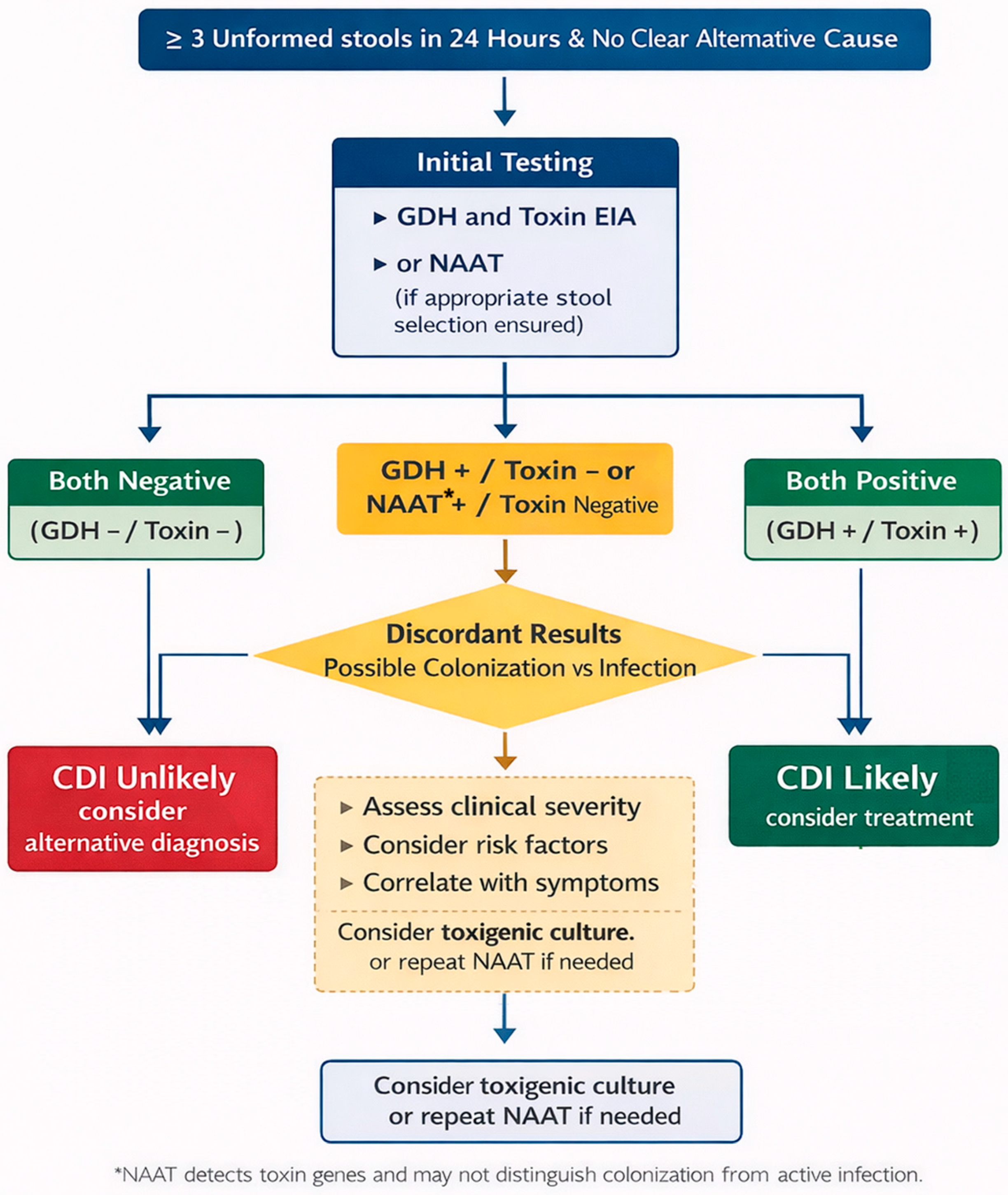
Laboratory confirmation of CDI involves the detection of toxins or toxin genes of C. difficile in stool samples. Testing is recommended exclusively in symptomatic patients and should not be performed on formed stool or routinely repeated during the same diarrheal episode [3]. Reference methods include the cell cytotoxicity neutralization assay (CCNA), which detects free toxins, and toxigenic culture (TC), which identifies toxin-producing strains. Although highly sensitive and specific, these methods are not routinely used due to their complexity and prolonged turnaround time [13]. In clinical practice, multistep diagnostic algorithms are recommended (Figure 1). These typically combine a highly sensitive screening test, such as glutamate dehydrogenase (GDH) detection or nucleic acid amplification tests (NAAT), with a toxin enzyme immunoassay (EIA). GDH and NAAT offer high sensitivity and negative predictive value, whereas toxin EIAs, although less sensitive, are more specific for active toxin production. In cases of discordant results (e.g., GDH-positive/toxin-negative or NAAT-positive/toxin-negative), additional testing and careful clinical correlation are required to distinguish active infection from asymptomatic colonization [20,24]. Importantly, the widespread use of NAAT has raised concerns regarding overdiagnosis, as these methods detect toxin genes rather than active toxin production. Consequently, positive NAAT results may reflect colonization rather than true infection, particularly in populations with a high prevalence of asymptomatic carriage, such as hospitalized or elderly patients. Therefore, laboratory findings must always be interpreted in conjunction with clinical presentation. Certain strains, such as ribotype 017, represent clinically relevant variants characterized by the absence of toxin A production and the presence of toxin B. These strains have been associated with antimicrobial resistance, particularly to fluoroquinolones, and may contribute to persistence and transmission in patients receiving prolonged antibiotic therapy, including ATT [10].
In selected cases, particularly in severe or atypical presentations, imaging and endoscopic evaluation may be required. Computed tomography may demonstrate colonic wall thickening, dilation, or complications, while endoscopy can reveal pseudomembranes, which are characteristic but not universally present in CDI. These methods are not routinely indicated and are reserved for specific clinical scenarios [20].
According to ESCMID guidelines [9], the diagnosis of CDI requires a compatible clinical presentation (≥3 unformed stools within 24 h) combined with microbiological evidence of toxigenic C. difficile. This may include detection of free toxins in stool, a positive NAAT in an appropriate clinical context, or isolation of a toxigenic strain. Endoscopic or histopathological evidence of pseudomembranous colitis is also considered diagnostic, regardless of laboratory findings.

3.4. Management of CDI in the TB Setting

According to current ESCMID [9] and IDSA/SHEA guidelines [25], management of CDI is based on prompt discontinuation of the inciting antibiotic, when feasible, or its substitution with a lower-risk alternative, and initiation of targeted therapy. Fidaxomicin is recommended as the preferred first-line agent due to its comparable efficacy to vancomycin and lower recurrence rates, particularly in patients at high risk of recurrent disease [26]. An additional benefit of fidaxomicin is its microbiota-sparing effect, which is especially relevant in patients who require continuation of concomitant antibiotic therapy, such as those receiving ATT. Oral vancomycin remains an acceptable alternative when fidaxomicin is unavailable (most commonly due to cost constraints). Metronidazole, once widely used, is now reserved for selected mild cases when other options are not available, due to its inferior efficacy. Treatment of severe and fulminant CDI requires a more aggressive approach, including high-dose oral vancomycin combined with intravenous metronidazole, with additional supportive or surgical management in selected cases. Management of recurrent CDI depends on prior therapy and may include fidaxomicin, tapered vancomycin regimens, or fecal microbiota transplantation (FMT) in patients with multiple recurrences. In patients with multiple CDI recurrences, FMT may represent an effective microbiota restoration strategy and is recommended by several contemporary guidelines following failure of standard antimicrobial therapies [8,9]. Management of the initial CDI episode, severe CDI and recurrent CDI is displayed in more detail in Table 2.
Routine use of probiotics is not currently recommended by international guidelines due to insufficient evidence and lack of standardization [9,25]. Nevertheless, they are sometimes used in clinical practice as adjunctive therapy, primarily for microbiota support after completion of CDI treatment [21].
The management of CDI in patients receiving ATT generally follows the same principles as in the general population, with comparable treatment outcomes [4]. At present, no dedicated evidence-based guidelines specifically address CDI management in patients receiving ATT. Consequently, treatment decisions are largely extrapolated from general CDI recommendations listed above. However, several important challenges arise in this context. From a practical perspective, although fidaxomicin has been associated with lower CDI recurrence rates and is increasingly recommended as a first-line treatment for CDI, its substantially higher cost and limited availability may restrict its use in many high TB-burden settings. Consequently, oral vancomycin often remains the most practical and accessible therapeutic option in low- and middle-income countries, where the global burden of TB is greatest. Furthermore, unlike most antibiotics, ATT, particularly rifampicin, often cannot be discontinued due to the severity and public health implications of TB. Consequently, ATT is frequently continued alongside CDI-directed therapy, requiring careful individualized risk–benefit assessment. Available evidence suggests that continuation of ATT does not significantly impair CDI treatment outcomes, although data remain limited [4]. Considering that, routine interruption or modification of ATT is generally not recommended, as available evidence suggests that many patients can successfully continue ATT during CDI treatment [4,5]. Nevertheless, individualized treatment adjustments may occasionally be required in selected cases. Additionally, rifampicin is a potent inducer of hepatic enzymes and drug transporters and may reduce the plasma concentrations of certain co-administered medications, potentially complicating overall pharmacological management. However, clinically significant pharmacokinetic interactions with oral vancomycin and fidaxomicin are expected to be limited because both agents exhibit minimal systemic absorption. Importantly, despite the risk of recurrence, reintroduction of rifampicin after successful CDI treatment may be clinically justified. Studies have shown that rifampicin re-administration is associated with faster sputum conversion and shorter hospital stays, underscoring its critical role in TB management. Although CDI recurrence has been reported in approximately one-third of patients, more than half were able to successfully continue full ATT [5]. These findings support an individualized management approach, balancing the risk of CDI recurrence against the necessity of maintaining effective tuberculosis treatment. Although evidence specifically addressing patients receiving ATT remains limited, FMT may be considered in selected cases of recurrent CDI when conventional treatment approaches have been unsuccessful.

3.5. Prevention of CDI in the TB Setting

Prevention of CDI, regardless of the cause, relies on a multifaceted approach that includes surveillance, infection control measures, and environmental interventions [3]. Continuous monitoring of CDI incidence at both hospital and ward levels, combined with timely feedback, has been shown to reduce infection rates.
Hand hygiene remains a cornerstone of prevention, with preference given to washing with soap and water due to the resistance of C. difficile spores to alcohol-based disinfectants. This should be complemented by consistent use of gloves and appropriate protective equipment. Early identification and prompt isolation of patients with suspected or confirmed CDI, along with strict adherence to contact precautions (gloves, gowns, and patient cohorting), are essential for limiting transmission. Environmental decontamination is equally important, given the prolonged persistence of spores on surfaces. Daily and terminal cleaning with sporicidal agents, such as chlorine-based disinfectants or hydrogen peroxide, is recommended, with particular attention to high-touch surfaces. Adjunctive technologies, including ultraviolet (UV-C) disinfection, may further reduce transmission in high-incidence settings [27].
Finally, preventive strategies should also include antimicrobial stewardship and individualized risk assessment. Although interruption or shortening of ATT is generally not feasible, unnecessary exposure to additional broad-spectrum antibiotics (such as fluoroquinolones) should be avoided whenever possible, as cumulative microbiota disruption may further increase CDI risk. Optimizing overall antibiotic use and considering microbiota-preserving or restorative approaches in high-risk patients may further reduce the burden of CDI [28].

4. Conclusions

ATT in patients with active TB represents a “double-edged sword”. While these agents are essential for controlling the global TB epidemic, their prolonged use, particularly rifampicin, is associated with a clinically relevant risk of CDI. Although ATT has traditionally been considered a lower-risk group compared to broad-spectrum antibiotics, its cumulative impact on gut microbiota and disruption of colonization resistance should not be underestimated.
A key challenge in the TB population is the delayed onset of CDI, with symptoms typically emerging several weeks to months after initiation of therapy, which may lead to underrecognition and diagnostic delay. In addition, the role of rifampicin is complex: beyond contributing to dysbiosis and the selection of resistant strains (such as ribotypes 017 and 046), it may also influence treatment outcomes through enzyme induction, potentially reducing the effectiveness of concomitant therapies.
Given that ATT often cannot be discontinued, fidaxomicin emerges as a preferred therapeutic option due to its microbiota-sparing properties and lower recurrence rates. However, its high cost and limited availability in many healthcare systems, particularly in regions with a high TB burden, represent significant barriers to optimal management. Consequently, oral vancomycin remains the most widely available and practical treatment option in many clinical settings and continues to play a central role in CDI management among patients receiving ATT. Improving access to fidaxomicin may further optimize outcomes in selected high-risk patients.
Despite growing recognition of CDI as a complication of ATT, available evidence remains limited and largely based on retrospective studies. Future prospective research is needed to better define risk factors and optimal management strategies, ultimately supporting the development of evidence-based recommendations for this unique patient population.

Author Contributions

Conceptualization, J.J.; methodology, J.J. and D.Ž.; formal analysis, J.J., A.M., D.K., E.V. and D.Ž.; investigation, J.J., A.M., D.K., E.V. and D.Ž.; writing—original draft preparation, J.J.; writing—review and editing, J.J., A.M., D.K., E.V. and D.Ž.; visualization, J.J.; supervision, D.Ž. All authors have read and agreed to the published version of the manuscript.

Funding

This research received no external funding.

Institutional Review Board Statement

Ethical review and approval were waived for this study due to the type of study (review).

Informed Consent Statement

Not applicable.

Data Availability Statement

The original contributions presented in this study are included in the article. Further inquiries can be directed to the corresponding author.

Conflicts of Interest

The authors declare no conflicts of interest.

Abbreviations

The following abbreviations are used in this manuscript:
ATTAntituberculous therapy
CDIClostridioides difficile infection
CCNACell cytotoxicity neutralization assay
EIAEnzyme immunoassay
ESCMIDEuropean Society of Clinical Microbiology and Infectious Diseases
GDHGlutamate dehydrogenase
GIGastrointestinal
ICUIntensive Care Unit
IDSAInfectious Diseases Society of America
IQRInterquartile range
IVIntravenous
METMetronidazole
NAATNucleic acid amplification tests
rCDIRecurrent Clostridioides difficile infection
SHEASociety for Healthcare Epidemiology of America
TBTuberculosis
TCToxigenic culture
VANVancomycin
WBCWhite blood cells

References

  1. Kullin, B.R.; Reid, S.; Abratt, V. Clostridium difficile in patients attending tuberculosis hospitals in Cape Town, South Africa, 2014–2015. Afr. J. Lab. Med. 2018, 7, 846. [Google Scholar] [CrossRef] [Scilit]
  2. Ha, S.W.; Hwang, S. Clinical Characteristics and Impact of Clostridium difficile Infection during Treatment of Rifampicin-Susceptible Pulmonary Tuberculosis. Tuberc. Respir. Dis. 2025, 88, 718–729. [Google Scholar] [CrossRef] [Scilit] [PubMed]
  3. Di Bella, S.; Sanson, G.; Monticelli, J.; Zerbato, V.; Principe, L.; Giuffrè, M.; Pipitone, G.; Luzzati, R. Clostridioides difficile infection: History, epidemiology, risk factors, prevention, clinical manifestations, treatment, and future options. Clin. Microbiol. Rev. 2024, 37, e0013523. [Google Scholar] [CrossRef] [Scilit] [PubMed]
  4. Lee, Y.M.; Huh, K.C.; Yoon, S.M.; Jang, B.I.; Shin, J.E.; Koo, H.S.; Jung, Y.; Kim, S.H.; Moon, H.S.; Lee, S.W.; et al. Incidence and Clinical Outcomes of Clostridium difficile Infection after Treatment with Tuberculosis Medication. Gut Liver 2016, 10, 250–254. [Google Scholar] [CrossRef] [Scilit] [PubMed]
  5. Kurahara, Y.; Kobayashi, T.; Tachibana, K.; Tsuyuguchi, K. The feasibility of rifampicin Re-administration in patients with tuberculosis and Clostridioides difficile infection. J. Infect. Chemother. 2022, 28, 558–562. [Google Scholar] [CrossRef] [Scilit] [PubMed]
  6. Suh, J.W.; Jeong, Y.J.; Ahn, H.G.; Kim, J.Y.; Sohn, J.W.; Yoon, Y.K. Epidemiologic characteristics and risk factors of Clostridioides difficile infection in patients with active tuberculosis in the Republic of Korea: A nationwide population-based study. J. Hosp. Infect. 2024, 154, 1–8. [Google Scholar] [CrossRef] [Scilit] [PubMed]
  7. Gagliano, M.C.; D’Agati, G.; Medaglia, A.A.; Pipitò, L.; Catania, B.; Conti, C.; Tuttolomondo, A.; Cefalù, A.B.; Cammà, C.; Scichilone, N.; et al. Burden of Clostridioides difficile Infection and Risk Factors for Recurrences in an Italian Tertiary Care University Hospital: A Prospective Observational Study. Antibiotics 2025, 15, 23. [Google Scholar] [CrossRef] [Scilit] [PubMed]
  8. Kelly, C.R.; Fischer, M.; Allegretti, J.R.; LaPlante, K.; Stewart, D.B.; Limketkai, B.N.; Stollman, N.H. ACG Clinical Guidelines: Prevention, Diagnosis, and Treatment of Clostridioides difficile Infections. Am. J. Gastroenterol. 2021, 116, 1124–1147, Erratum in Am. J. Gastroenterol. 2022, 117, 358. https://doi.org/10.14309/ajg.0000000000001529. [Google Scholar] [CrossRef] [Scilit] [PubMed]
  9. van Prehn, J.; Reigadas, E.; Vogelzang, E.H.; Bouza, E.; Hristea, A.; Guery, B.; Krutova, M.; Norén, T.; Allerberger, F.; Coia, J.E.; et al. Guideline Committee of the European Study Group on Clostridioides difficile. European Society of Clinical Microbiology and Infectious Diseases: 2021 update on the treatment guidance document for Clostridioides difficile infection in adults. Clin. Microbiol. Infect. 2021, 27, S1–S21. [Google Scholar] [CrossRef] [Scilit] [PubMed]
  10. Cioboata, R.; Balteanu, M.A.; Osman, A.; Vlasceanu, S.G.; Zlatian, O.M.; Mitroi, D.M.; Catana, O.M.; Socaci, A.; Tieranu, E.N. Coinfections in Tuberculosis in Low- and Middle-Income Countries: Epidemiology, Clinical Implications, Diagnostic Challenges, and Management Strategies-A Narrative Review. J. Clin. Med. 2025, 14, 2154. [Google Scholar] [CrossRef] [Scilit] [PubMed]
  11. Kesavelu, D.; Jog, P. Current understanding of antibiotic-associated dysbiosis and approaches for its management. Ther. Adv. Infect. Dis. 2023, 10, 20499361231154443. [Google Scholar] [CrossRef] [Scilit] [PubMed]
  12. Orrell, K.E.; Melnyk, R.A. Large Clostridial Toxins: Mechanisms and Roles in Disease. Microbiol. Mol. Biol. Rev. 2021, 85, e0006421. [Google Scholar] [CrossRef] [Scilit] [PubMed]
  13. Markantonis, J.E.; Fallon, J.T.; Madan, R.; Alam, M.Z. Clostridioides difficile Infection: Diagnosis and Treatment Challenges. Pathogens 2024, 13, 118. [Google Scholar] [CrossRef] [Scilit] [PubMed]
  14. Nibbering, B.; Gerding, D.N.; Kuijper, E.J.; Zwittink, R.D.; Smits, W.K. Host Immune Responses to Clostridioides difficile: Toxins and Beyond. Front. Microbiol. 2021, 12, 804949. [Google Scholar] [CrossRef] [Scilit] [PubMed]
  15. Chai, Y.; Li, M.; Deng, X.; Ma, C.; Zhou, N.; Chen, Y.; Yao, Y.; Li, K.; Gong, W.; Lei, H. Gut microbiota and tuberculosis infection: Interaction and therapeutic potential. Gut Microbes 2025, 17, 2531201. [Google Scholar] [CrossRef] [Scilit] [PubMed]
  16. Shveid, G.D.; Canales, A.S.J.; Muñoz, B.J.M.; Sánchez, F.M. Clostridium difficile colitis during tuberculosis treatment. An. Med. Asoc. Med. Hosp. ABC 2018, 63, 122–124. [Google Scholar]
  17. Balaban, D.V.; Popescu, A.L.; Pătrășescu, M.; Bucurică, S.; Costache, R.S.; Ionită-Radu, F.; Jinga, M. Clostridium difficile colitis during antituberculous treatment. Images Med. 2021, 18, 89–90. [Google Scholar] [CrossRef] [Scilit]
  18. Mutai, W.C.; Mureithi, M.W.; Anzala, O.; Revathi, G.; Kullin, B.; Burugu, M.; Kyany’a, C.; Odoyo, E.; Otieno, P.; Musila, L. High Prevalence of Multidrug-Resistant Clostridioides difficile Following Extensive Use of Antimicrobials in Hospitalized Patients in Kenya. Front. Cell. Infect. Microbiol. 2021, 10, 604986. [Google Scholar] [CrossRef] [Scilit] [PubMed]
  19. Obuch-Woszczatyński, P.; Dubiel, G.; Harmanus, C.; Kuijper, E.; Duda, A.; Wultańska, D.; van Belkum, A.; Pituch, H. Emergence of Clostridium difficile infection in tuberculosis patients due to a highly rifampicin-resistant PCR ribotype 046 clone in Poland. Eur. J. Clin. Microbiol. Infect. Dis. 2013, 32, 1027–1030. [Google Scholar] [CrossRef] [Scilit] [PubMed]
  20. McDonald, L.C.; Gerding, D.N.; Johnson, S.; Bakken, J.S.; Carroll, K.C.; Coffin, S.E.; Dubberke, E.R.; Garey, K.W.; Gould, C.V.; Kelly, C.; et al. Clinical Practice Guidelines for Clostridium difficile Infection in Adults and Children: 2017 Update by the Infectious Diseases Society of America (IDSA) and Society for Healthcare Epidemiology of America (SHEA). Clin. Infect. Dis. 2018, 66, 987–994. [Google Scholar] [CrossRef] [Scilit] [PubMed]
  21. Kim, M.J.; Jeong, H.W.; Choi, Y.R. A case of pseudomembranous colitis associated with antituberculosis therapy in a patient with tuberculous meningitis. J. Biomed. Res. 2014, 15, 44–48. [Google Scholar] [CrossRef] [Scilit]
  22. Rehsi, S.S.; Roy, N.; Nair, V.V.; Baranwal, A.K.; Sengupta, P.; Sharma, P.; Kumar, A. A rare case of tuberculosis spine on antitubercular drug succumbing to pseudomembranous enterocolitis. J. NTR Univ. Health Sci. 2021, 10, 105–108. [Google Scholar] [CrossRef] [Scilit]
  23. Sun, Y.X.; Zhao, Y.T.; Teng, L.L.; Ge, J.L.; Jiang, H.; Shao, L. Clostridium difficile infection associated with antituberculous agents in a patient with tuberculous pericarditis. Intern. Med. 2013, 52, 1495–1497. [Google Scholar] [CrossRef] [Scilit] [PubMed]
  24. Crobach, M.J.; Planche, T.; Eckert, C.; Barbut, F.; Terveer, E.M.; Dekkers, O.M.; Wilcox, M.H.; Kuijper, E.J. European Society of Clinical Microbiology and Infectious Diseases: Update of the diagnostic guidance document for Clostridium difficile infection. Clin. Microbiol. Infect. 2016, 22, S63–S81. [Google Scholar] [CrossRef] [Scilit] [PubMed]
  25. Johnson, S.; Lavergne, V.; Skinner, A.M.; Gonzales-Luna, A.J.; Garey, K.W.; Kelly, C.P.; Wilcox, M.H. Clinical Practice Guideline by the Infectious Diseases Society of America (IDSA) and Society for Healthcare Epidemiology of America (SHEA): 2021 Focused Update Guidelines on Management of Clostridioides difficile Infection in Adults. Clin. Infect. Dis. 2021, 73, 755–757. [Google Scholar] [CrossRef] [Scilit] [PubMed]
  26. Zhao, Z.; Wu, Y.; Geng, X.; Yuan, C.; Fu, Y.; Yang, G. Efficacy of fidaxomicin versus vancomycin in the treatment of Clostridium difficile infection: A systematic meta-analysis. Medicine 2024, 103, e39213. [Google Scholar] [CrossRef] [Scilit] [PubMed]
  27. Tschudin-Sutter, S.; Kuijper, E.J.; Durovic, A.; Vehreschild, M.J.G.T.; Barbut, F.; Eckert, C.; Fitzpatrick, F.; Hell, M.; Norèn, T.; O’Driscoll, J.; et al. Guidance document for prevention of Clostridium difficile infection in acute healthcare settings. Clin. Microbiol. Infect. 2018, 24, 1051–1054. [Google Scholar] [CrossRef] [Scilit] [PubMed]
  28. Reigadas, E.; van Prehn, J.; Falcone, M.; Fitzpatrick, F.; Vehreschild, M.J.G.T.; Kuijper, E.J.; Bouza, E. European Society of Clinical Microbiology and Infectious Diseases Study Group on Clostridioides difficile (ESGCD) and Study Group for Host and Microbiota interaction (ESGHAMI). How to: Prophylactic interventions for prevention of Clostridioides difficile infection. Clin. Microbiol. Infect. 2021, 27, 1777–1783. [Google Scholar] [CrossRef] [Scilit] [PubMed]
Figure 1. Diagnostic algorithm for CDI (regardless of the cause). Abbreviations: GDH—Glutamate dehydrogenase; EIA—Enzyme immunoassay; NAAT—Nucleic acid amplification tests; CDI—Clostridioides difficile infection.
Joma 03 00011 g001
Table 1. Case series from the literature that explore CDI as a complication of ATT.
Study/
Country/
Time Period
CDI
Incidence
Risk Factors for CDITime to CDI OnsetCDI TreatmentComplicationsATT Continuation
Lee et al. [4]
South Korea
2008–2013
54 CDI/19,080 TB patients;
(2.83/1000)
GI surgery;
Cytostatics;
Immunosuppression
41.8 ± 35.0
days
Oral MET (87%);
IV MET (11.1%);
Oral VAN (1.9%)
Acute kidney injury (3.8%);
Death (1.9%)
Continued incl. rifampicin (42.6%); discontinued all (38.9%); rifampicin only stopped (5.6%)
Ha & Hwang [2]
South Korea
2022–2023
46 CDI/168 TB (27.3%; 15.0/10,000 patient-days)Age ≥ 70;
Malnutrition
57 days (IQR 32–98)Not specifiedMortality (13%);
Recurrent CDI (26%)
Not specified
Kurahara et al. [5]
Japan
1999–2021
156 CDI/11,230 TB;
(1.4%; 2.1/10,000 patient-days)
Elderly men;
Underlying diseases
Delayed onset (weeks after therapy initiation)Vancomycin-based therapyNot specifiedRifampicin often reintroduced after CDI control
Suh et al. [6]
South Korea
2018–2022
2901 CDI/131,950 TB (~2.2%)ICU stay;
Underlying diseases;
Antibiotic
exposure
Not specifiedOral MET (81.94%)Mortality higher in CDI vs. non-CDI (9.9% vs. 6.9%)Not specified
Abbreviations: ATT—antituberculous therapy; CDI—Clostridioides difficile infection; GI—gastrointestinal; ICU—Intensive Care Unit; IV—intravenous; IQR—interquartile range; MET—metronidazole; TB—tuberculosis; VAN—vancomycin.
Table 2. Treatment recommendations for CDI according to ESCMID and IDSA/SHEA guidelines [9,25].
Category of CDIDefinitionRecommended Therapy (ESCMID & IDSA/SHEA)
Initial episode
(Non-severe)
Diarrhea (≥3 unformed stools/24 h) + positive test for C. difficile;
no signs of severe disease (WBC < 15 × 109/L, normal creatinine, hemodynamically stable)
First-line: Fidaxomicin (200 mg orally twice daily for 10 days)
Alternative: Vancomycin (125 mg orally four times daily for 10 days)
If unavailable: Metronidazole (500 mg orally three times daily for 10 days)
Severe CDIESCMID: Temperature > 38.5 °C, WBC > 15 × 109/L, creatinine increase > 50%
IDSA: WBC > 15 × 109/L or creatinine ≥ 1.5 mg/dL
Fidaxomicin (200 mg orally twice daily for 10 days)
or
Vancomycin (125 mg orally four times daily for 10 days)
Fulminant CDI
(Severe-complicated)
Hypotension, shock, ileus, elevated lactate, toxic megacolonVancomycin (500 mg orally or via nasogastric tube four times daily)
+ Metronidazole (500 mg IV every 8 h)
+ Rectal vancomycin (500 mg in 100 mL saline every 6 h) if ileus present;
Consider surgical consultation and tigecycline (100 mg loading dose, then 50 mg every 12 h)
First recurrenceRecurrence of symptoms within 8 weeks after completion of prior
therapy
If vancomycin used initially → Fidaxomicin (standard regimen: 200 mg twice daily for 10 days or extended regimen: 200 mg twice daily for 5 days, then every other day until day 25)
If fidaxomicin used initially → Fidaxomicin or Vancomycin
tapered/pulsed regimen:
125 mg orally 4× daily for 14 days →
125 mg orally 2× daily for 7 days →
125 mg orally 1× daily for 7 days →
125 mg orally every other day for 8 days (4 doses) →
125 mg orally every 3 days for 2 weeks (5 doses)
Second or subsequent recurrencesMultiple recurrences after previous successful treatmentsFidaxomicin (standard or extended regimen)
or Vancomycin tapered/pulsed regimen
Strong recommendation: Fecal microbiota transplantation
Abbreviations: CDI—Clostridioides difficile infection; ESCMID—European Society of Clinical Microbiology and Infectious Diseases; IDSA—Infectious Diseases Society of America; IV—intravenously; SHEA—Society for Healthcare Epidemiology of America; WBC—white blood cells.
Disclaimer/Publisher’s Note: The statements, opinions and data contained in all publications are solely those of the individual author(s) and contributor(s) and not of MDPI and/or the editor(s). MDPI and/or the editor(s) disclaim responsibility for any injury to people or property resulting from any ideas, methods, instructions or products referred to in the content.