Review

Tropical and Arboviral Causes of Febrile Illness in International Travelers: A Focused Review

Department of Pharmacotherapeutics and Clinical Research, Taneja College of Pharmacy, University of South Florida, Tampa, FL 33602, USA
*
Author to whom correspondence should be addressed.
Emerg. Care Med. 2026, 3(2), 16; https://doi.org/10.3390/ecm3020016
Submission received: 27 January 2026 / Revised: 4 April 2026 / Accepted: 8 April 2026 / Published: 17 April 2026

Abstract

Background/Objectives: Febrile illness in returning travelers presents a diagnostic and operational challenge for emergency medicine clinicians as early symptoms of high-consequence tropical infections often overlap with common viral syndromes. This review synthesizes current evidence to guide frontline clinicians in the systematic evaluation, diagnosis, and management of internally acquired febrile illnesses with a focus on pathogen of greatest relevance to United States (US) emergency departments (ED). Methods: We conducted a narrative review of the literature addressing epidemiology, clinical presentation, diagnostic testing, and management strategies for key travel-associated infections. Special consideration was given to rapid diagnostic modalities, pediatric risk factors, and infections most frequently implicated in returning travelers, including chikungunya (CHIK), dengue virus (DENV) disease, Ebola virus (EBV) disease, malaria, Mpox, typhoid fever (TF), yellow fever (YF), and Zika virus (ZIKV) disease. Results: Effective evaluation begins with a detailed travel and exposure history, recognition of epidemiologic and clinical red flags, and targeted use of rapid diagnostic tests. Malaria remains the most common life-threatening cause of post-travel fever and the only pathogen with reliable Food and Drug Administration (FDA)-cleared rapid testing available in the ED. Arboviral infections such as DENV, CHIK, ZIKV, and YFrequire region-specific consideration and phase-appropriate molecular or serologic evaluation. Emerging and high-consequence pathogens, including Mpox and EBV, necessitate strict infection control measures and coordination with public health authorities. Pediatric travelers, particularly those visiting friends and relatives, face disproportionate risk for severe systemic infections and often require broader diagnostic testing. Conclusions: A structured approach integrating travel history, focused examination, rapid diagnostics, and early recognition of high-risk features is essential to improving outcomes for febrile returning travelers. Strengthened vector control, enhanced vaccination uptake, and global surveillance are critical to reducing future disease burden.

1. Introduction

Evaluating a febrile patient with a recent history of international travel presents a unique diagnostic challenge for emergency physicians, requiring a structured and geographically informed approach. A meticulous history and physical examination remain the foundation of assessment, as clinical findings alone are often nonspecific and early disease manifestations can overlap with common community-acquired infections [1].
With international travel projected to reach 2 billion annual trips by 2030, travel-associated infections represent a rapidly growing global health burden [2]. Global surveillance networks have documented hundreds of thousands of travel-related diagnoses over the last decade, with malaria and acute diarrheal illnesses remaining the most frequently reported [2,3]. Beyond morbidity, these infections impose a substantial economic toll. The financial impact includes significant out-of-pocket healthcare costs for acute hospitalizations, which frequently occur upon return to the traveler’s home country, as well as severe productivity losses and lost wages associated with prolonged recovery from vector-borne illnesses like DENV and CHIK [4,5].
When evaluating a febrile returning traveler, clinicians must maintain a broad differential diagnosis. Cosmopolitan and globally prevalent infections, including respiratory viruses such as influenza and COVID-19, zoonotic and bacterial diseases like leptospirosis and rickettsioses, and systemic viral illnesses such as acute human immunodeficiency virus (HIV) syndrome, remain frequent causes of post-travel fever. However, this review deliberately narrows its focus to high-consequence tropical, arboviral, and emerging pathogens. While less frequently encountered in the daily clinical setting, these specific diseases present unique diagnostic and operational challenges. They frequently mimic benign viral syndromes in their early stages but demand specialized rapid diagnostic testing, immediate implementation of stringent infection control measures, and pathogen-specific acute resuscitation strategies that are critical for frontline and emergency practitioners to master.
While extensive literature exists on the epidemiology of travel medicine, the rapid expansion of vector habitats and the resurgence of global travel necessitate an updated, acute care-focused framework. Emergency departments frequently serve as the initial, and sometimes only, point of contact for returning travelers presenting with undifferentiated febrile illnesses. Therefore, the rationale for this review is to bridge the gap between global infectious disease surveillance and frontline acute care operations. The primary goals of this review are to: (1) synthesize current evidence regarding the high-consequence tropical and emerging pathogens most relevant to US emergency departments; (2) provide a detailed pharmacotherapeutic perspective on empiric treatment, critical medication contraindications, and preventive vaccine landscapes to fill existing gaps in diagnostic-heavy literature; (3) provide a practical, algorithm-driven framework to guide early triage, rapid diagnostic testing, and empiric management.

2. Methods

A narrative review of the medical literature was conducted to evaluate the epidemiology, clinical presentation, diagnostic testing, and management of high-consequence pathogens in febrile international travelers presenting to acute care settings. During the preparation of this manuscript/study, the author(s) used ChatGPT-5 (OpenAI, San Francisco, CA, USA) for the purposes of generating text. The authors have reviewed and edited the output and take full responsibility for the content of this publication.

2.1. Search Strategy and Data Sources

We searched the PubMed database for articles published from 1 January 2015, through 13 August 2025. The following Boolean search strategy, combining Medical Subject Headings (MeSH) and text words, was utilized: (“Travel” [Mesh] OR “Travel Medicine”[Mesh] OR “travel related disease” OR traveler* [tiab] OR traveller*[tiab]) AND (“Fever”[Mesh] OR febrile OR fever OR pyrexia) AND (“Emergency Service, Hospital”[Mesh] OR “Emergency Room Visits” [Mesh] OR “Emergency Treatment” [Mesh] OR “emergency health service” OR emergency[tiab] OR “urgent care”[tiab]) AND (“United States”[Mesh] OR “united states”[tiab] OR usa[tiab]).

2.2. Study Selection and Inclusion Criteria

Articles were included if they were published in the English language and addressed the epidemiology, diagnosis, or management of travel-related febrile illnesses. Included study designs comprised systematic reviews, meta-analyses, randomized controlled trials, and clinical practice guidelines published by major health organizations (e.g., Centers of Disease Control [CDC], World Health Organization [WHO]). Studies were specifically selected if they addressed rapid diagnostic modalities, high-consequence pathogens, or vulnerable pediatric populations.

3. Results of the Literature Search

Our targeted literature search yielded a broad array of clinical practice guidelines, retrospective cohorts, and narrative syntheses relevant to emergency and travel medicine. Rather than a quantitative meta-analysis, the extracted data from the final selection of articles were synthesized thematically to construct a practical, symptom-based framework for the acute care clinician.
The analysis of the literature revealed three primary domains critical to the evaluation of the febrile returning traveler, which form the results of this review: (1) diagnostic triage and initial approach: evidence highlighting the necessity of detailed travel histories and the strategic deployment of rapid diagnostic tests (RDTs) to immediately identify life-threatening infections and guide isolation protocols; (2) vulnerable populations: literature emphasizing the disproportionate risk and unique, often deceptive, clinical presentations of internationally acquired severe systemic infections (ISSIs) in pediatric patients and those visiting friends and relatives (VFRs); (3) high-consequence pathogen profiles: synthesized epidemiological, diagnostic, and therapeutic updates for the most critical infections encountered in US emergency departments, including malaria, high-consequence arboviruses (DENV, CHIK, ZIKV, YF), enteric fever, and emerging zoonoses (EBV, Mpox).
These thematic results are detailed in the subsequent sections to bridge the gap between epidemiologic surveillance and frontline clinical execution. While the differential diagnosis for post-travel fever is vast, this review deliberately centers on eight core pathogens: malaria, DENV, ZIKV, CHIK, EVD, YF, TF, and Mpox. These specific diseases were selected as the focal point of this review because they represent the highest epidemiological burden encountered in typical returning travelers, or they present an acute threat requiring immediate, high-level infection control and public health intervention upon presentation. This targeted approach is designed to provide acute care and adult medicine clinicians with a high-yield, operational framework rather than an exhaustive epidemiological catalog.

4. Emergency Department Triage and Diagnostic Framework

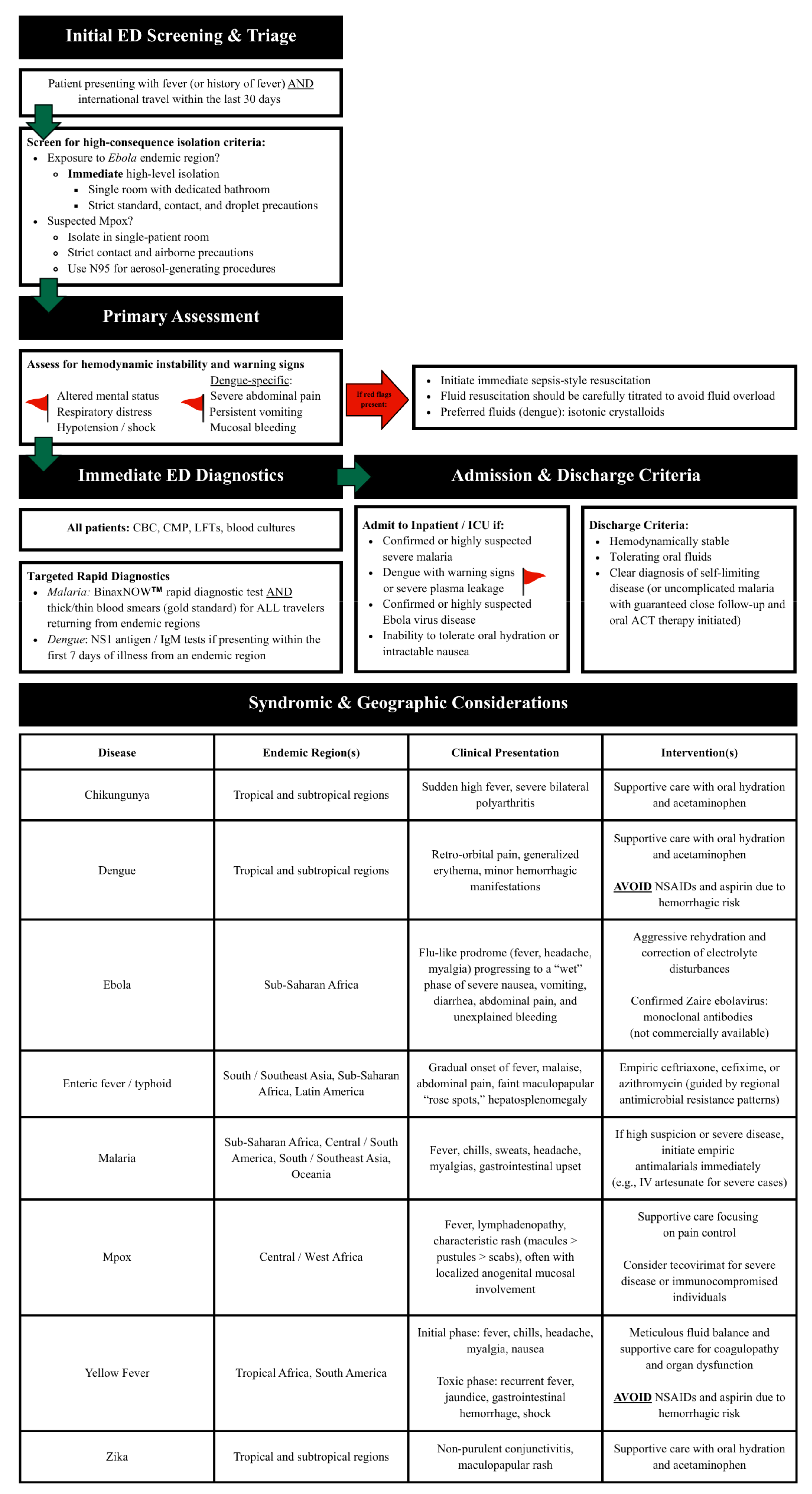
To manage the diagnostic complexity of the febrile returning traveler, our review of the literature indicates that emergency clinicians should utilize a structured, syndromic, and geographically informed algorithm. Rather than relying on standard textbook history-taking, the evidence supports a targeted framework (Figure 1) prioritizing immediate infection control, rapid diagnostics, and early recognition of clinical red flags [1]. It is critical to note that this framework is designed to facilitate immediate infection control and rapid rule-outs for specific high-yield pathogens. It is not a substitute for a comprehensive infectious disease workup if initial targeted diagnostics are negative.

4.1. Triage Priorities and the Targeted Exposure History

Effective evaluation begins with a detailed travel and exposure history, which dictates the immediate algorithmic branching. Essential historical outcomes identified in the literature include verifying the exact travel itinerary, the purpose of travel (e.g., visiting friends and relatives), and, critically, an assessment of pre-travel preparations. Inquiry into exact malaria chemoprophylaxis regimens and adherence is a vital operational step, as failure rates or non-adherence heavily influence the differential diagnosis and empiric therapy selection [1].
Because the risk of acquiring specific pathogens, particularly Mpox, CHIK, and ZIKV, is heavily dependent on active epidemic transmission rather than baseline endemicity, historical screening must be paired with real-time epidemiological data. Clinicians should routinely consult dynamic surveillance resources, such as the CDC Travel Health Notices or the WHO Disease Outbreak News, to accurately determine a traveler’s true epidemiological risk based on current outbreak activity.

4.2. Immediate Laboratory Investigations and Antimicrobial Stewardship

Baseline laboratory evaluation for the toxic-appearing traveler should include a complete blood count (CBC), comprehensive metabolic panel (CMP), liver function testing (LFTs), and blood cultures. RDTs play a critical role in the early evaluation of febrile illness among returning travelers, allowing for timely differentiation of life-threatening tropical infections and optimization of antimicrobial therapy [6].
Malaria remains the only pathogen for which reliable, US FDA-cleared rapid diagnostics (e.g., BinaxNOW, Abbott Diagnostics, Scarborough, ME, USA) can be routinely available in the ED. Literature strongly supports the integration of malaria RDTs into ED triage protocols. This allows rapid identification and isolation of high-risk patients while serving as an antimicrobial stewardship tool through immediate confirmation and species-specific management pathways [6].

4.3. Primary Assessment and Clinical Red Flags

Following isolation protocols, clinicians must assess for signs of hemodynamic instability and target organ damage. Early recognition of red flag findings, including altered mental status, respiratory distress, hypotension, or bleeding manifestations, should prompt immediate resuscitative measures [1]. Fluid resuscitation must be carefully titrated. While isotonic crystalloid solutions are required for managing shock due to plasma leakage in severe dengue, cautious administration is necessary to avoid iatrogenic fluid overload [7,8].

4.4. Syndrome and Geographic Considerations

Categorizing the patient’s presentation by syndrome and travel history refines empiric management:
  • Travel to Malaria-Endemic Regions (Sub-Saharan Africa, Latin America, Asia, Oceania) with Undifferentiated Fever: In travelers presenting with fever, chills, headache, and myalgias, malaria must be presumed until proven otherwise. If clinical suspicion is high or if the patient exhibits severe disease, empiric antimalarial therapy, such as intravenous artesunate, should be initiated promptly without waiting for confirmatory testing [9].
  • Travel to Tropical/Subtropical Regions with Arthropod-Borne Syndrome: In travelers presenting with a combination of severe polyarthritis (CHIK), retro-orbital pain/erythema (DENV), or conjunctivitis (ZIKV), meticulous supportive care is required. A critical pharmacological intervention in the ED is the strict avoidance of nonsteroidal anti-inflammatory drugs (NSAIDs) and aspirin due to the heightened risk of hemorrhage. Acetaminophen (APAP) is the preferred antipyretic until dengue is definitively excluded [7,10,11].
  • Travel to Hyperendemic Regions (South/Southeast Asia, Sub-Saharan Africa) with Enteric Syndrome: In patients presenting with a gradual onset of fever, abdominal pain, hepatosplenomegaly, and “rose spots,” evaluate for enteric (typhoid) fever. Empiric antimicrobial therapy with ceftriaxone or azithromycin should be initiated, guided closely by regional resistance patterns [12].
  • Travel to Endemic African or South American Regions with Viral Hemorrhagic Toxidrome: In patients presenting with severe gastrointestinal symptoms, jaundice, unexplained mucosal bleeding, or shock, suspect EBV or YF and initiate immediate aggressive supportive care, correction of coagulopathy, and coordination with public health authorities [13,14].

4.5. Admission and Disposition Criteria

Hospitalization, often in an intensive care setting, is required for patients demonstrating severe disease manifestations. Admission criteria include confirmed or highly suspected severe malaria (e.g., parasitemia > 5%, severe anemia, acute kidney injury, altered mental status), dengue with warning signs (e.g., severe abdominal pain, persistent vomiting, mucosal bleeding), or any patient exhibiting hemodynamic instability or an inability to tolerate oral hydration [9,15,16]. Patients with EBV require care in a specialized facility capable of high-level infection prevention [13].

4.6. Public Health Notification and Reporting

A critical operational requirement in the acute care setting is the immediate notification of local and state public health authorities when high-consequence, travel-associated infections are suspected, such as those pathogens highlighted in this review. In the US, disease reporting mandates are dictated by state and local laws; however, all of the primary pathogens highlighted in this review are classified as Nationally Notifiable Conditions by the CDC.
Emergency clinicians do not report cases directly to the CDC. Instead, suspected or confirmed cases must be reported to the local or state health department, which then securely transmits the data to the CDC via the National Notifiable Diseases Surveillance System (NNDSS). While routine surveillance pathogens may have standard reporting windows, suspected cases of high-consequence pathogens such as EBV, YF, and Mpox require immediate, 24 h telephone notification to local health officials to trigger urgent epidemiological investigations and infection control protocols. Furthermore, early engagement with public health authorities is often a necessity rather than a mere reporting formality for guidance [17].

5. Special Populations: Pediatrics

Fever is one of the most common presenting symptoms in pediatric EDs in the US. While most children have self-limited viral illnesses, those who have recently returned from international travel warrant additional consideration for ISSIs, which can present with few distinguishing features on initial examination. A 2021 retrospective study by Khan et al. reviewed a 10-year cohort of febrile children presenting to two pediatric EDs in New York following international travel and identified important diagnostic predictors to aid early recognition of ISSIs [6].
Among 353 pediatric patients presenting with fever within 30 days of international travel, 12% were diagnosed with ISSI. The most common etiologies were malaria (57%), DENV (14%), and pathogenic bacteria (30%), primarily due to Salmonella typhi. Geographic risk was notable. Most ISSIs occurred in children who had traveled to Africa (57%) or Asia (27%), often for the purpose of visiting friends or relatives, a demographic known to have higher exposure risk and lower uptake of preventive measures. Despite the severity of these infections, nearly one-fifth of affected children had sought prior medical care in the US but did not undergo diagnostic bloodwork, emphasizing the potential for missed or delayed diagnosis in non-endemic settings [6].
The study highlights that pediatric travelers often appear well on presentation despite severe underlying disease. Clinical findings such as fever, headache, and nonspecific constitutional symptoms may overlap with benign viral illnesses, making a detailed travel history essential to guide diagnostic testing. The authors recommend that all febrile children presenting within one month of international travel, especially those returning from Africa or Asia, undergo complete blood count, blood culture, and liver function testing, with malaria testing performed if travel to an endemic region is reported. Early identification of thrombocytopenia and elevated transaminases can serve as critical laboratory clues suggestive of malaria, DENV, or TF[6].
Children are more susceptible to internationally acquired infections than adults, particularly when traveling to visit family and friends in endemic regions, due to longer exposure duration, closer contact with local populations, consumption of local foods and water, and lack of chemoprophylaxis or age-appropriate vaccinations. Infants and young children also face heightened risks of severe disease and dehydration due to immature immune systems and limited physiological reserves [6].
From a systems perspective, this study reinforces the importance of pediatric-specific ED protocols for the evaluation of post-travel fever, including standardized prompts for travel history documentation, laboratory order sets based on destination risk, and early consultation with infectious disease specialists. Educational initiatives targeting clinicians in pediatric emergency settings are crucial for enhancing recognition and facilitating timely testing for imported infections. Integrating these tools into electronic health records and triage workflows can reduce diagnostic delays and prevent adverse outcomes [6].

6. Select Causes of Febrile Illness in Travelers

6.1. Chikungunya Virus

CHIK is an emerging arboviral illness caused by an alphavirus transmitted to humans primarily by Aedes aegypti and Aedes albopictus mosquitoes (Table 1). Once considered confined to Africa and Southeast Asia, CHIK has expanded globally over the past two decades, with major outbreaks reported in the Indian Ocean islands, India, Europe, and across the Caribbean and the Americas. This expansion underscores the disease’s clinical relevance to returning travelers and the risk of establishment in previously nonendemic regions due to widespread vector distribution, high levels of viremia in infected individuals, and lack of population immunity [10,18]. However, for the typical returning traveler, acquisition in a purely endemic setting is rare. Clinical risk is almost exclusively associated with travel to regions experiencing active, documented epidemics.
The incubation period of CHIK is typically three to seven days, though it can range from one to twelve days. Illness is generally self-limiting and death is rare. Clinical manifestations vary by age, with distinct patterns observed in pediatric and adult populations. In adults, acute illness is characterized by abrupt high fever, often exceeding 39 °C and accompanied by chills and rigors. Severe, bilateral, symmetric polyarthritis affecting the distal joints begins two to five days after the onset of fever. Other common features include maculopapular rash, pruritus, lymphadenopathy, myalgias, fatigue, nausea, diarrhea, and ocular inflammation. Cutaneous manifestations such as diffuse erythema of the face and trunk, hyperpigmentation, intertrigo, and penoscrotal or perianal ulceration have also been described. In most cases, symptoms resolve within 7–10 days; however, a substantial proportion of patients develop chronic or relapsing arthritis and arthralgias that persist for months to years. Prolonged CHIK arthritis can mimic seronegative rheumatoid arthritis, spondyloarthritis, or systemic lupus erythematosus, complicating diagnosis and management [1,18].
In contrast, CHIK disease in children is often more variable in its presentation. Fever, rash, and polyarthritis occur in more than half of pediatric cases, but joint pain tends to be better tolerated and less frequently disabling than in adults. Neurologic and dermatologic complications are relatively more common in the pediatric population. Encephalitis, febrile seizures, and acute encephalopathy among hospitalized children have been reported. Cerebrospinal fluid findings are often unremarkable and neuroimaging reveals nonspecific abnormalities. Infants, particularly those under six months of age, frequently exhibit distinctive cutaneous findings such as diffuse erythema, maculopapular rash, peeling, and vesiculobullous lesions. Other features include peripheral cyanosis without hemodynamic abnormalities, edema of the extremities, and hyperpigmentation that may persist beyond recovery [10,18].
Severe or atypical manifestations, though rare, include encephalitis, myocarditis, hepatitis, Guillain-Barré syndrome, multiorgan failure, and ocular complications. These are more common among neonates, older adults, and individuals with cardiovascular, respiratory, or neurologic comorbidities. Hemorrhagic complications are uncommon; therefore, patients presenting with hemoconcentration, lymphopenia, or thrombocytopenia should be evaluated for a concurrent dengue infection [10,18].
Vertical transmission can occur, particularly during intrapartum maternal viremia, and neonatal disease is frequently severe, with neurologic and cardiac involvement. Although data are limited, transmission of CHIK through breastmilk has not been demonstrated [10,18].
Diagnosis is based on clinical suspicion in travelers from endemic regions combined with laboratory testing. During the viremic phase, reverse transcription–polymerase chain reaction (RT-PCR) provides a sensitive and specific means of detection. Serologic assays detecting IgM (appearing after five to seven days and persisting for up to three months) and IgG (appearing after two weeks and persisting for years) are widely used; however, these should be interpreted with caution as CHIK IgM antibodies may cross-react with similar viruses in the same antigenic complex (e.g., Ross River virus, Mayaro virus). Laboratory abnormalities may include leukopenia with relative lymphocytosis, mild hepatic enzyme elevations, and elevated acute-phase reactants [1].
There is no specific antiviral therapy for CHIK, and management is supportive. Acetaminophen is preferred for fever and analgesia, as aspirin carries a risk of hemorrhage and Reye syndrome. NSAIDs are commonly employed for persistent joint symptoms, while corticosteroids, chloroquine, and disease-modifying antirheumatic drugs such as methotrexate may be considered in cases of chronic inflammatory arthritis, although evidence for their effectiveness is limited. Biologic agents, including tumor necrosis factor inhibitors, are contraindicated in acute infection and may worsen disease. Supportive care measures such as hydration, rest, antipyretics, analgesics, and physical therapy for chronic arthritis are central to management. Hospitalization is required for severe disease, including neurologic involvement, multiorgan dysfunction, neonatal infection, and in the elderly or those with comorbid conditions. The use of convalescent plasma has been reported as a potential strategy for treatment and prevention in patients at high-risk for progression to severe infection. Patients receiving care in endemic areas should be treated in mosquito-free zones or use repellents to prevent the spread of infection. The risk of transmission from an infected patient to a susceptible Aedes mosquito is highest during the viremic phase (e.g., the first two to six days of illness) [1,18].
Currently, one licensed vaccine exists for CHIK, although promising candidates, including virus-like particle vaccines and monoclonal antibodies, are in clinical development (Table 1). Another vaccine was available but suspended due to concerns about causing CHIK-like illness in vaccine recipients. Prevention relies on mosquito bite avoidance and vector control. Recommended personal protective measures include the use of repellents, insecticide-treated bed nets, and protective clothing. Community-level control of mosquito breeding sites, such as covering water storage containers, removing standing water, and managing waste, is essential. For travelers and residents in endemic areas, barriers to prevention include the ubiquity of Aedes vectors, lack of access to the vaccine or antiviral prophylaxis therapy, inadequate education during pre-travel health consultation appointments, inconsistent adherence to mosquito avoidance measures, and frequent misdiagnosis due to overlap with other febrile and rheumatologic illnesses [1,15]. Co-infections with DENV or ZIKV further complicate a definitive diagnosis [1].
CHIK represents an important and growing global health threat, with significant implications for clinicians evaluating febrile travelers. Early recognition and accurate diagnosis are critical to appropriate supportive management, reduction in unnecessary therapies, and mitigation of the risk of local transmission [10].

6.2. Dengue Virus

DENV disease is the most common arboviral infection worldwide. It is caused by one of four antigenically distinct dengue viruses (DENV-1 to DENV-4) transmitted by Aedes species mosquitoes (Table 1). Global incidence has nearly doubled in the past three decades, with an estimated 390 million infections and nearly 100 million symptomatic cases annually. Half of the world’s population now lives in areas at risk of transmission. Climate change, rapid urbanization, poverty, migration, and the expansion of competent vectors continue to drive the spread of dengue. In the US, dengue is most relevant to returning travelers and to residents of Puerto Rico, the US Virgin Islands, and American Samoa [7]. Sporadic autochthonous cases have also been identified in Florida and Texas [7,19].
Most infections are asymptomatic, but symptomatic dengue typically presents with fever, nausea, vomiting, headache, retro-orbital pain, rash, myalgias, arthralgias, and leukopenia [7]. Rash associated with DENV is a characteristic generalized erythema with circular areas of sparing and has been described as “isles of white in a sea of red” [1]. Long-term physical, immunological, and cognitive sequelae have been reported, even up to three years following convalescence. Severe dengue develops in about five percent of cases and is marked by plasma leakage leading to third spacing and resulting in shock, coagulopathy, or organ impairment. Atypical presentations are increasing in frequency and include cardiac manifestations such as conduction abnormalities, myocarditis, and pericarditis [20]. Warning signs that can predict progression to severe disease include severe abdominal pain or tenderness, persistent vomiting, mucosal bleeding, fluid accumulation, postural hypotension, hepatomegaly, lethargy, and rising hematocrit with falling platelet count. High-risk groups for more severe infections include infants, children, pregnant individuals, older adults, and patients with comorbidities such as diabetes, obesity, hypertension, asthma, chronic liver or kidney disease, and coagulopathies [7].
Diagnosis relies on a combination of clinical suspicion and laboratory confirmation. While RDTs detecting non-structural protein-1 (NS1) antigen and dengue-specific IgM or IgG antibodies are available, these tests demonstrate variable accuracy (NS1 antigen RDT sensitivity generally ranges from 60 to 80% with a specificity > 90%). The diagnostic gold standard during the acute phase is reverse-transcription polymerase chain reaction (RT-PCR) or nucleic acid amplification testing (NAAT). Nucleic acid amplification testing (NAAT) or nonstructural protein 1 (NS1) antigen assays are most sensitive in the first 7 days of illness. After this viremic phase, IgM detection can provide evidence of recent infection, although cross-reactivity with other flaviviruses, including ZIKV, may complicate interpretation. In such cases, plaque reduction neutralization testing can sometimes differentiate DENV from other flaviviruses, though this is rarely available in clinical practice [7].
Treatment remains supportive, as no specific antivirals have demonstrated efficacy in reducing viremia or complications. APAP is the preferred antipyretic, while NSAIDs should be avoided due to bleeding risk. Early, aggressive oral hydration is strongly recommended, particularly in children, as it reduces hospitalization rates. Patients with warning signs, severe disease, or high-risk features should be hospitalized for careful monitoring, serial laboratory testing, and prompt initiation of intravenous fluid resuscitation when indicated. Isotonic crystalloid solutions are preferred for managing shock due to plasma leakage, with careful titration to avoid fluid overload. Corticosteroids, immunoglobulins, and prophylactic platelet transfusions are not recommended due to a lack of demonstrated benefit. The typical duration of symptomatic illness is 2–7 days, although recovery from severe disease may require prolonged supportive care. Untreated severe DENV carries a mortality rate exceeding 10%, but with timely recognition and appropriate fluid management, case fatality can be reduced to less than 1% [7,8].
Prevention focuses on both individual protection and community-level vector control. Travelers to endemic areas should use Environmental Protection Agency (EPA)–approved insect repellents, wear protective clothing, utilize screened windows, doors, and bed nets, and eliminate standing water that serves as mosquito breeding sites. Traditional vector control efforts are challenged by insecticide resistance and sustainability issues, prompting the development of novel approaches such as genetically modified mosquitoes and Wolbachia-based interventions, which have demonstrated reductions in DENV transmission and hospitalizations of up to 80% in field trials [7].
Vaccination represents an important new tool in DENV control. Dengvaxia® (CYD-TDV, Sanofi Pasteur, Swiftwater, PA, USA) is the first and only licensed dengue vaccine in the U.S. and is approved for children aged 6–16 years with laboratory-confirmed prior infection living in endemic areas. This three-dose, live attenuated tetravalent vaccine reduces symptomatic and severe disease among seropositive individuals but increases the risk of hospitalization and severe dengue among seronegative recipients due to antibody-dependent enhancement (ADE). For this reason, pre-vaccination serologic testing is required, representing both a barrier to implementation and an unprecedented step in vaccine policy [21]. Another tetravalent live vaccine, Qdenga (TAK-003, Takeda, Singen, Germany), is approved for use in more than 40 countries. In contrast to Dengvaxia®, Qdenga (TAK-003) may be used in individuals 4 years of age and older regardless of prior dengue infection history [22]. Data suggests strong efficacy, particularly against certain serotypes, and without the safety concerns associated with Dengvaxia® [23].
Barriers to dengue prevention include the global expansion of Aedes vectors due to climate change, inadequate housing and water management infrastructure in endemic regions, insecticide resistance, and the logistical complexity of vaccine roll-out requiring prior infection documentation. Misconceptions among previously exposed individuals that past infection provides lifelong immunity further undermine preventive efforts, despite the risk of more severe disease with secondary infections due to antibody-dependent enhancement (ADE). Circulation of four antigenically distinct DENVserotypes allows for sequential infections, and individuals in endemic settings may be infected by different serotypes on up to four occasions during their lifetime, leading to a more severe infection secondary to the ADE phenomenon [7].
DENV infection represents a growing global health challenge with significant implications for clinicians evaluating febrile returning travelers and populations living in endemic areas. Early recognition, close monitoring for warning signs, supportive fluid-based management, and implementation of preventive strategies, including emerging vaccines and vector control innovations, are critical to reducing morbidity and mortality [7].

6.3. Ebola Virus

EVD is a severe zoonotic infection caused by the Ebola virus (EBOV), a member of the Filoviridae family (Table 1). EBOV is the most extensively studied filovirus and remains the principal cause of large-scale outbreaks in humans [13]. The clinical, epidemiologic, and therapeutic understanding of EVD expanded dramatically following the 2013–2016 West African epidemic, which resulted in more than 28,000 cases and 11,000 deaths. The disease remains a critical consideration in febrile returning travelers from sub-Saharan Africa and in healthcare settings with potential exposure to infected patients or body fluids [13]. Although the absolute incidence of EVDamong routine international travelers to the US is rare, it is included in this clinical framework because it represents the paradigm for high-consequence pathogens; the failure to immediately recognize and isolate a suspected case carries catastrophic operational and public health implications for frontline healthcare workers.
Transmission to humans occurs through direct contact with infected animal blood or tissue and subsequently through human-to-human spread via contact with blood, secretions, or contaminated surfaces. Nosocomial transmission is common in the absence of strict infection control measures, and traditional funeral practices involving handling of deceased bodies are a major driver of community spread. Viral persistence in immune-privileged sites, such as semen, has been documented for over 500 days, representing a rare but important route of delayed transmission [13].
The incubation period ranges from 2 to 21 days, typically 6 to 10 days, depending on exposure route and viral load. EVD is not considered seasonal but demonstrates recurrent regional outbreaks, reflecting sporadic zoonotic spillover events from animal reservoirs [13].
EVD begins as a nonspecific illness characterized by fever, headache, malaise, and myalgia. Gastrointestinal symptoms typically develop several days later and may lead to profound dehydration and hypovolemia. Rash, conjunctival injection, and dysphagia are common, while bleeding manifestations occur in fewer than half of patients, usually as petechiae, mucosal bleeding, or oozing from venipuncture sites. As the disease progresses, multiorgan dysfunction may develop, including hepatic injury, renal failure, coagulopathy, and shock. Survivors frequently experience post-Ebola syndrome, characterized by arthralgia, headache, ocular inflammation, and neurocognitive deficits [13].
Diagnosis is confirmed by quantitative real-time polymerase chain reaction (qRT-PCR), the gold standard for detecting viral ribonucleic acid (RNA) in blood or body fluids. Testing should target at least two distinct genome regions to minimize false negatives due to viral mutation. Early in the disease course, results may be negative; thus, repeat testing is recommended if clinical suspicion persists [13]. Rapid immunochromatographic antigen detection tests (e.g., OraQuick, OraSure Technologies, Bethlehem, PA, USA) are now available for field triage, offering a sensitivity of approximately 94% and specificity of 100%, though negative results must always be confirmed with the qRT-PCR gold standard [24,25].
All patients with suspected or confirmed EVD should receive care in a facility capable of implementing high-level infection prevention and control practices for special pathogens. Any patient who meets criteria for a person under investigation (e.g., compatible symptoms within 21 days of exposure in an area with EVD transmission or contact with a known case) should be placed immediately in a single patient room with a dedicated bathroom and restricted access, and managed with appropriate standard, contact, and droplet precautions. Admission is recommended until infection has been excluded with appropriate diagnostic testing or an alternative diagnosis fully accounts for the clinical presentation in consultation with public health authorities [26].
Prompt recognition and supportive care remain the cornerstone of management. Aggressive rehydration with oral or intravenous fluids, correction of electrolyte disturbances, and management of hypoglycemia are essential to reduce mortality. Antipyretics, antiemetics, and analgesics provide symptomatic relief, and broad-spectrum antibiotics and antimalarial agents are often administered empirically in endemic regions due to frequent coinfection. Advanced supportive measures, such as parenteral nutrition, renal replacement therapy, and oxygen support, are recommended in specialized units [13].
The 2018–2020 outbreak in the Democratic Republic of Congo (DRC) marked the first use of effective antiviral and monoclonal antibody therapies. The randomized Pamoja Tulinde Maisha (PALM) trial demonstrated significantly improved survival with MAb114 (EBANGA™, Ridgeback Biotherapeutics, Baltimore, MD, USA) and REGN-EB3 (Inmazeb®, Regeneron Pharmaceuticals, Tarrytown, NY, USA) compared with remdesivir or ZMapp [27]. Both monoclonal antibody regimens received regulatory approval and represent the current standard of care for confirmed EBOV infection [13,28]. Remdesivir, though less effective, retains utility in combination or for expanded-spectrum coverage. Early initiation of therapy markedly improves outcomes, highlighting the importance of rapid diagnosis and referral to specialized Ebola treatment units [13].
Vaccine development has been a major success of the past decade. The rVSV-ZEBOV-GP (ERVEBO, Merck, Rahway, NJ, USA) single-dose, live-attenuated recombinant vaccine demonstrated 97.5% efficacy in vaccination trials and is now licensed by the FDA and European Medicines Agency (EMA) for individuals ≥ 12 months [13,28]. It is the primary vaccine used in emergency response settings and may also reduce disease severity if administered within a short window following exposure [13].
Travelers to endemic regions should be counseled on the risk of contact with wildlife or body fluids, adherence to infection prevention protocols, and avoidance of funeral attendance. Healthcare and aid workers should ensure full vaccination prior to deployment, maintain high-level personal protective equipment (PPE) use, and participate in pre-departure and post-travel monitoring programs. Major barriers to prevention include delayed recognition of index cases, community mistrust, cultural burial practices, resource limitations, and challenges in vaccine distribution and cold-chain maintenance [13,28].

6.4. Malaria

Malaria remains a major global health threat, responsible for approximately 500,000 deaths annually, with the greatest burden in sub-Saharan Africa and a disproportionate impact on children younger than five years of age. The disease is caused by protozoan parasites of the Plasmodium genus, of which P. falciparum accounts for the most severe disease and mortality. Transmission occurs primarily through the bite of the female Anopheles mosquitoes; however, rare cases arise from transfusion, congenital spread, and local outbreaks (Table 1). While endemic transmission was eliminated in the US during the mid-20th century, approximately 2000 cases occur annually, almost extensively from travelers from endemic regions [9].
The incubation period of malaria is typically six to seven days but may be delayed for months. Seasonality plays a role, with the highest transmission during and immediately after the rainy season, typically May through December in endemic areas. Clinical presentation varies widely, ranging from asymptomatic parasitemia to severe disease. Common symptoms include fever, chills, headache, myalgias, malaise, and gastrointestinal upset, particularly in children. No specific fever pattern is diagnostic, and malaria is often misdiagnosed as a non-specific viral illness. Suspicion for P. falciparum should be treated as a medical emergency, as clinical deterioration or death can occur within 24 to 36 h in malaria-naïve patients [9].
Accurate, species-specific diagnosis is essential for effective management. BinaxNOW™ (Abbott Diagnostics, Scarborough, ME, USA) is an immunochromatographic test offering a sensitivity exceeding 95% for P. falciparum (though lower for other species) and a specificity of >99%. However, regardless of RDT results, microscopic evaluation of Giemsa-stained thick and thin blood smears remains the diagnostic gold standard to confirm infection and quantify parasitemia [9,29,30].
Treatment is guided by clinical status, infecting species, prophylaxis history, and geographic region. The WHO recommends artemisinin-based combination therapies (ACTs), such as artemether-lumefantrine, as first-line treatment for uncomplicated malaria across all species. In the U.S., intravenous artesunate is the treatment of choice for severe malaria and should be initiated promptly. If clinical suspicion for malaria is high, empiric treatment is warranted, particularly in the setting of severe disease or when diagnostic testing is delayed. Furthermore, patients who developed malaria despite using chemoprophylaxis should be treated with antimalarial agents different from those used for prophylaxis. If ACTs are unavailable, alternative regimens include chloroquine or hydroxychloroquine for chloroquine-sensitive regions, or atovaquone-proguanil, quinine plus doxycycline, tetracycline or clindamycin, or mefloquine where resistance is documented. Infections due to P. vivax and P. ovale require additional eradication of dormant hypnozoite stages with primaquine or tafenoquine, provided glucose-6-phosphate dehydrogenase (G6PD) deficiency has been excluded [9].
For severe malaria, intravenous artesunate is administered at 2.4 mg/kg at 0, 12, and 24 h, followed by once-daily dosing until parasitemia falls below 1%, not exceeding seven days. Blood smears should be obtained every 12 h to monitor parasite density. If parasitemia is less than 1% at least four hours after the third dose, patients may be transitioned to a full course of oral therapy, ideally with an ACT [9].
Criteria for admission include severe manifestations (e.g., altered mental status, seizures, severe anemia, acute kidney injury, acute respiratory distress syndrome, shock, disseminated intravascular coagulation, acidosis, or parasitemia > 5%), pregnancy, young age, immunocompromised status, or absence of prior malarial immunity. Supportive care, including intravenous fluids, antipyretics, and antiemetics, is essential, and hospitalization should be pursued whenever red flag criteria are met [9].
Pregnant patients represent a uniquely vulnerable group, with higher risks of severe malaria, maternal complications, and adverse fetal outcomes. ACTs may be used in the second and third trimesters, while artemether-lumefantrine is considered acceptable in all trimesters when no alternatives are available. Chloroquine, hydroxychloroquine, and quinine with clindamycin or mefloquine are safe options throughout pregnancy. Primaquine and tafenoquine should not be used in pregnancy [9].
Clinicians in the U.S. can access expert consultation through the CDC malaria hotline. This service operates 24 hours a day, 365 days a year and can be reached at 770-488-7788. The hotline provides real-time guidance on diagnostic interpretation, therapeutic selection, and management of severe malaria, including coordination of intravenous (IV) artesunate which is only available in the U.S. through the CDC [31].
Preventive strategies focus on both chemoprophylaxis and vector control. Travelers should undergo individualized pre-travel risk assessment, including evaluation of itinerary, destination, and timing relative to the rainy season. Chemoprophylaxis options include atovaquone-proguanil, doxycycline, mefloquine, chloroquine or hydroxychloroquine (if regional sensitivity exists), primaquine, or tafenoquine, with drug choice guided by local resistance patterns and patient factors. These medications must be started prior to travel, continued throughout exposure, and maintained for a defined period post-travel. In addition, travelers should be counseled to employ insect avoidance strategies such as insecticide-treated bed nets, permethrin-treated clothing, and repellents containing 20–30% DEET or 20% picaridin. Barriers to prevention include antimalarial drug resistance, medication side effects limiting adherence, the cost of prophylaxis, limited vaccine availability, the circulation of counterfeit or substandard medications in some regions, and a frequent misconception among individuals originally from endemic areas. Many such patients believe they retain lifelong immunity after moving away; however, partial immunity wanes over time, leaving them susceptible and still in need of prophylaxis when returning to endemic regions [9].
A significant recent development is the WHO’s recommendation of RTS,S/AS01 (Mosquirix, GlaxoSmithKline, Rixensart, Belgium), the first malaria vaccine approved for widespread use, for the prevention of P. falciparum in African children. Although this represents a major advance, vaccine access remains limited, and chemoprophylaxis with personal protective measures continues to be critical for travelers. Overall, timely recognition, accurate diagnosis, empiric initiation of therapy when clinically indicated, and robust preventive counseling remain essential to reducing the morbidity and mortality of malaria, particularly among returning travelers [9].

6.5. Monkeypox Virus

Mpox is a zoonotic viral disease caused by monkeypox virus (MPXV), an orthopoxvirus closely related to variola virus, the causative agent of smallpox. Its natural animal reservoir has not been definitively identified, though African rodents and nonhuman primates are suspected sources [32,33,34]. Historically confined to rural regions of Central and West Africa, Mpox has undergone a rapid epidemiological transformation since 2022, resulting in widespread human-to-human transmission and large global outbreaks [32,33]. Genomic and epidemiologic data from recent outbreaks show multiple co-circulating clades, including Clade I (Congo Basin) and Clade II (West African) [32,33,34,35]. The virus spreads primarily through direct skin-to-skin or mucosal contact, most often during sexual activity, though transmission may also occur via contaminated fomites or respiratory droplets during prolonged close contact [32,33].
The 2022–2025 outbreaks marked a turning point in the global understanding of this reemerged pathogen. Once a neglected zoonosis affecting children in rural Africa, Mpox now sustains transmission in urban centers and across diverse sexual networks. While early cases predominated among men who have sex with men, more recent transmission has involved heterosexual contacts and other high-risk populations, disproportionately affecting marginalized groups [32,33]. From 2022 through 2025, more than 99,000 cases were reported globally, including over 33,000 in the US. In Africa, 45,652 cases and 1492 deaths were documented across 12 countries between 2022 and late 2024, with the DRC accounting for most cases and fatalities [32]. These surges prompted emergency declarations by both the Africa Centers for Disease Control and the WHO in 2024 to accelerate vaccination, surveillance, and outbreak response [33]. While historically endemic to rural regions of Central and West Africa, the disease is currently characterized by distinct epidemic patterns. Outside of specific, highly interconnected sexual networks, Mpox should not be considered a high-probability diagnosis in the typical returning traveler unless they are returning from a region experiencing a documented, active outbreak (such as the ongoing Clade I epidemic in the DRC and neighboring countries).
The incubation period typically ranges from 7 to 21 days. Clinical presentation has evolved from the traditional febrile, disseminated pustular rash to more localized mucocutaneous disease, often involving the anogenital region. Many patients now present with mild or absent prodromal symptoms [32,33]. Common manifestations include rash, fever, lymphadenopathy, sore throat, headache, myalgia, back pain, and fatigue. Lesions progress from macules and papules to vesicles and pustules before crusting over. They may be painful, pruritic, or ulcerative, and bacterial superinfection can occur [33,34]. Severe disease, including extensive mucosal or ocular involvement, occurs more frequently in immunocompromised individuals, particularly those with advanced HIV infection [32,33]. While NAAT remains the diagnostic gold standard, the Cepheid Xpert(R) (Cepheid, Sunnyvale, CA, USA) Mpox real-time PCR test offers rapid point-of-care evaluation under FDA Emergency Use Authorization. Returning results from lesion swabs within 40 min, the assay detects Mpox (clade II) and non-variola Orthopoxvirus deoxyribonucleic acid (DNA) with a sensitivity of 88.6% to 97.7% and a specificity of 77.4% to 97.4% [36,37].
Clinicians should institute prompt isolation for patients with suspected or confirmed Mpox to limit nosocomial transmission. In healthcare settings, staff should place affected patients in a single-patient room with a dedicated bathroom; however, special air handling is not required. Recommended personal protective equipment consists of gloves, gown, medical mask, and eye protection, with use of an N95 respiratory during aerosol-generating procedures. Strict adherence to hand hygiene, appropriate handling of contaminated linens and waste, and environmental cleaning of high-touch surfaces are essential components of infection prevention [38].
Most Mpox infections are self-limited and resolve with supportive care. Management focuses on pain control, skin hygiene, and prevention of secondary bacterial infection. Antiviral therapy is reserved for patients with severe or progressive disease or at high risk for complications. Available agents include tecovirimat, brincidofovir, cidofovir, and vaccinia immune globulin (VIGIV, Cangene Corporation, Winnipeg, Manitoba, Canada) [34]. Tecovirimat has demonstrated favorable safety and tolerability, though randomized trials have not demonstrated a significant reduction in time to lesion resolution for Clade I or II disease [39,40,41]. Clinical use should therefore prioritize immunocompromised patients, those with extensive mucosal involvement, or those with ocular, neurologic, or hemorrhagic manifestations [34,39].
Vaccination remains central to Mpox prevention. The nonreplicating Modified Vaccinia Ankara–Bavarian Nordic (MVA-BN, Bavarian Nordic, Hellerup, Denmark) vaccine (marketed as JYNNEOSor IMVANEX) is recommended for both pre-exposure and post-exposure prophylaxis [42]. The replication-competent ACAM2000 (Emergent BioSolutions, Gaithersburg, MD, USA)vaccine is also approved but is rarely used due to adverse event risks and limited efficacy data in Clade II outbreaks. Real-world studies estimate MVA-BN vaccine effectiveness at approximately 36–75% after one dose and 66–89% after the full two-dose series, with breakthrough infections generally milder [34,35,42]. In the U.S., the CDC recommends two doses of MVA-BN, separated by 28 days, for adults at increased risk. High-risk populations include men who have sex with men, individuals with multiple recent sexual partners, persons with a recent sexually transmitted infection, or those engaging in sex in settings associated with Mpox transmission [43].
Public health responses continue to emphasize risk-based vaccination, contact tracing, health education, and community engagement. Global efforts led by WHO and the Africa CDC aim to improve vaccine access, reduce hesitancy, and expand diagnostic capacity in resource-limited settings where disease burden remains highest. Despite progress, barriers such as vaccine scarcity, cost, and inequitable distribution persist [31,34]. Sustained surveillance, equitable vaccine allocation, and targeted prevention strategies remain essential to curbing Mpox transmission and mitigating future outbreaks.

6.6. Typhoid Fever

TF, a systemic infection caused by Salmonella enterica serovar Typhi, and the related paratyphoid fevers caused by S. enterica serovars Paratyphi A, B, and C, remain major global public health challenges. These pathogens spread via the fecal–oral route, primarily through ingestion of contaminated food or water in areas with inadequate sanitation and hygiene [12,44]. Despite progress in water quality, sanitation, and vaccination in many regions, enteric fever continues to cause significant morbidity, mortality, and economic burden, particularly among children in low- and middle-income countries. The disease is highly endemic in South and Southeast Asia and sub-Saharan Africa, with recent estimates indicating 9–14 million cases and more than 100,000 deaths annually [12,45,46,47]. The highest incidence occurs in children under 15 years, with hyperendemic areas in Bangladesh, Nepal, and Pakistan exceeding 900 cases per 100,000 person-years [12,46]. Limited diagnostic capacity, underreporting, and widespread empirical antibiotic use contribute to substantial uncertainty in global estimates [12,48]. Chronic carriers, often individuals with gallbladder colonization, serve as persistent reservoirs of infection, facilitating ongoing community transmission [12,44]. Climate change, rapid urbanization, and antimicrobial resistance (AMR) are expected to further influence the global epidemiology of TF [12,49].
Following ingestion, S. typhi, a motile Gram-negative bacillus, penetrates the intestinal epithelium via M cells overlying Peyer’s patches, invades macrophages, and disseminates through the lymphatic system to the liver, spleen, and bone marrow [12]. The organism’s virulence factors, including the Vi capsular antigen and type III secretion systems, promote intracellular survival and systemic spread [12,44]. The incubation period ranges from 7 to 21 days, after which bacteremia produces a gradual onset of fever, malaise, abdominal pain, and hepatosplenomegaly. Some patients develop rose spots (faint, blanching maculopapular lesions on the trunk), although these occur in fewer than 10% of cases and may be difficult to visualize on darker skin tones [12]. Without treatment, disease progression can result in gastrointestinal bleeding, intestinal perforation, encephalopathy, or sepsis [12,49].
Diagnosis often relies on blood culture, which remains the gold standard despite its limited sensitivity of 40–80% when compared to a more invasive bone marrow culture. The low amount of S. typhi in blood and the frequency of previous antimicrobial treatment are key factors for the low sensitivity of blood culture [12]. Multiple cultures improve diagnostic yield. Several commercial rapid diagnostic tests exist, though their sensitivity and specificity remain variable and suboptimal [50]. The sensitivity of PCR for Salmonella enterica serovar Typhi ranges from 40 to 100% and the specificity ranges from 76.9 to 100%, contingent on the PCR methodology, target gene, and specimen type [51,52]. Laboratory findings such as leukopenia, thrombocytopenia, and elevated liver transaminases may support the diagnosis [53].
Antimicrobial therapy remains the cornerstone of treatment, but rising drug resistance presents a major clinical concern. Resistance to fluoroquinolones, azithromycin, and third-generation cephalosporins has been increasingly reported, particularly in South Asia [12,49,50,54,55]. The WHO currently recommends azithromycin, ciprofloxacin, or ceftriaxone as first-line options, while meropenem is reserved for extensively drug-resistant (XDR) strains, such as those circulating in Pakistan [12,55]. Supportive care focused on hydration, electrolyte balance, and management of complications is critical, as is identification and eradication of chronic carriage. Ongoing surveillance of local resistance patterns and robust antimicrobial stewardship programs are essential to slow the spread of resistant strains [49].
Vaccination provides the most effective long-term prevention strategy. The WHO recommends routine administration of typhoid conjugate vaccines (TCVs) for individuals living in or traveling to endemic regions [12,48,49,54]. TCVs such as Vi-TT and Vi-CRM197 demonstrate high efficacy (approximately 80–85%) and durable protection across age groups [12,49,54]. Broader adoption of these vaccines could significantly reduce disease burden and interrupt transmission. Despite strong evidence of efficacy and cost-effectiveness, global uptake remains limited due to supply constraints, funding challenges, and gaps in national immunization programs [12,45,49].
Sustained control of TF will depend on a multifaceted approach combining vaccination, improved water and sanitation infrastructure, enhanced surveillance, and responsible antimicrobial use. Without renewed investment in these areas, the global community risks continued resurgence of a disease that remains both preventable and curable.

6.7. Yellow Fever

YF is an acute viral hemorrhagic disease caused by the YF virus (YFV), a flavivirus transmitted primarily by Aedes aegypti mosquitoes. The virus infects both humans and non-human primates and remains endemic across tropical regions of Africa and the Americas. Historically, YF caused devastating epidemics in port cities such as Havana and New Orleans throughout the 17th to 19th centuries, shaping the early history of tropical medicine. The vectorial role of mosquitoes was first proposed by Carlos Finlay and later confirmed by Walter Reed’s team in the early 1900s, leading to widespread mosquito eradication programs that eliminated urban YF from much of the Americas by the mid-20th century [14].
The virus was first isolated in Ghana in 1927, paving the way for the development of the live-attenuated 17D vaccine by Max Theiler in the 1930s, a discovery that remains the cornerstone of prevention. Despite the availability of an effective vaccine, eradication has proven impossible because YFV persists in non-human primate reservoirs and forest-dwelling mosquito populations, maintaining ongoing zoonotic transmission. Discontinuation of routine vaccination and the reestablishment of A. aegypti in urban centers have enabled recurrent outbreaks, particularly in areas with low vaccine coverage [14].
Transmission occurs through three overlapping cycles: sylvatic (jungle), intermediate (savanna), and urban. In the sylvatic cycle, forest mosquitoes transmit the virus among wild primates, with incidental human infection occurring in those who work or travel in forested areas. The intermediate cycle involves transmission between semi-domestic mosquitoes and both humans and primates in rural settings. The urban cycle, which poses the greatest epidemic potential, arises when infected travelers introduce the virus into densely populated areas where A. aegypti is prevalent, leading to rapid human-to-mosquito-to-human spread. Increasing urbanization, declining vaccination rates, and global vector resurgence have heightened the risk of re-emergent urban epidemics in recent decades [14,56].
The incubation period is typically 3–6 days. Clinical manifestations range from mild, self-limited febrile illness to severe, life-threatening disease characterized by hepatic and renal failure, coagulopathy, and jaundice, the feature that gives the disease its name. Early symptoms include fever, chills, headache, myalgia, and nausea. After a brief remission, approximately 15% of patients progress to a toxic phase marked by recurrent fever, jaundice, “black vomit” from gastrointestinal hemorrhage, and multiorgan failure. Case-fatality rates in severe disease range from 20% to 50%, underscoring the need for early recognition and supportive management [14,56].
Diagnosis relies on clinical suspicion in travelers or residents from endemic regions and confirmation through detection of YF-specific IgM antibodies, viral RNA by RT-PCR, or virus isolation [14,56]. Because many cases occur in remote settings with limited diagnostic capacity, underreporting remains significant; the WHO estimates that true case counts may be hundreds of times higher than reported [14]. The gold standard for diagnosing YF is the laborious plaque reduction neutralization test, which is the most specific serological test for differentiating flavivirus infections [57,58]. While serologic assays can detect virus-specific IgM and IgG antibodies, cross-reactivity may occur between antibodies against other flaviviruses. However, the optimal diagnostic approach usually depends on the onset of signs and symptoms [5]. NAAT or virus isolation are preferred during the first several days of illness, but the virus levels wane over time and are unreliable for exclusion of YF [14,56,58]. The sensitivity and specificity of an NAAT test were ≥88% and ≥95%, depending on the assay and laboratory [59,60].
There is no specific antiviral therapy for YF, and management remains supportive, focusing on meticulous fluid balance, treatment of coagulopathy, renal and hepatic support, and prevention of secondary infections [14]. Vaccination and vector control remain the primary strategies for prevention, with proof of vaccination required for entry into many countries [14,56,61]. A single dose of the live-attenuated 17D vaccine confers lifelong immunity in more than 95% of recipients [14]. Rare vaccine-associated viscerotropic and neurotropic adverse events have been documented, but their incidence remains extremely low compared with the benefits of immunization [14,56]. The WHO’s Eliminate Yellow Fever Epidemics (EYE) strategy, launched in 2017, continues to promote mass vaccination, strengthened surveillance, and vector control in high-risk areas [61].
Despite the success of the vaccine, challenges persist. Global vaccine supply remains limited, routine immunization coverage has declined in several endemic regions, and rapid urbanization continues to expand the habitat of A. aegypti. These factors, combined with the persistence of sylvatic reservoirs, make YF a resurgent threat to global public health [14]. Sustained vigilance, expanded vaccination campaigns, and integrated vector control management are essential to prevent future outbreaks and protect both endemic populations and international travelers. Vector control efforts are challenged by the ability of mosquitoes to develop various resistance mechanisms against insecticides, requiring the development of different approaches such as genetically modified mosquitoes to suppress and replace the current population [62,63].

6.8. Zika Virus

ZVD is caused by ZIKV, an arthropod-borne flavivirus closely related to DENV, YF, and West Nile viruses. First identified in Uganda in 1947, ZIKV was initially confined to sporadic infections in Africa and Asia until widespread outbreaks occurred across the Pacific from 2007 to 2015 and subsequently throughout the Americas in 2015–2016. The Brazilian epidemic marked a pivotal moment in global public health, establishing the link between maternal Zika infection and congenital malformations, most notably microcephaly, and leading the WHO to declare a Public Health Emergency of International Concern in 2016. Although transmission has sharply declined since that time, isolated outbreaks continue to occur in South and Southeast Asia, India, and other tropical regions. In 2023, only seven ZIKV cases were reported among U.S. international travelers, reflecting the current low global incidence [11]. Similar to CHIK, the acquisition of ZIKV in a purely endemic setting is currently very rare for the average traveler. Clinical suspicion should remain low unless the patient has recently returned from a region with an active, ongoing epidemic.
ZIKV is transmitted primarily through the bite of infected Aedes aegypti mosquitoes, which also serve as vectors for dengue and chikungunya. Additional routes of transmission include vertical transmission from mother to fetus, sexual contact, blood transfusion, and laboratory exposure, distinguishing ZIKV from most other arboviruses. The American pandemic strain originated from the Asian lineage. It spread rapidly through immunologically naïve populations, resulting in infection rates exceeding 60% in some affected communities during the outbreak’s peak. Although immunity following infection is long-lasting, the high proportion of asymptomatic cases poses challenges for surveillance and outbreak detection [11].
The incubation period ranges from 3 to 14 days, with most symptomatic infections presenting as a mild, self-limited illness characterized by maculopapular rash, low-grade fever, arthralgia, and non-purulent conjunctivitis. Severe complications, although uncommon, are clinically significant. Neurologic sequelae include Guillain–Barré syndrome, while congenital Zika syndrome (CZS) represents the most devastating manifestation. CZS encompasses a spectrum of fetal abnormalities, including microcephaly, intracranial calcifications, cortical thinning, ocular anomalies, and arthrogryposis. The risk of congenital infection is greatest when maternal infection occurs in the first trimester, and up to 5–14% of infants born to infected mothers may develop CZS. Long-term neurodevelopmental monitoring is recommended for all infants with confirmed or suspected prenatal exposure [11].
Diagnosis is based on RT-PCR testing of serum, urine, or cerebrospinal fluid during the acute viremic phase, generally within the first week of illness [11]. Studies have shown NAAT sensitivity of 96.1% and specificity of 100% if testing is conducted during the appropriate window (serum 1–7 days, urine and blood specimens ≥ 2–3 weeks) [64]. Serologic testing for Zika-specific IgM may assist in later diagnosis but is complicated by cross-reactivity with other flaviviruses, particularly dengue. The laborious plaque reduction neutralization test is considered the gold standard confirmatory assay and can help distinguish Zika infection when diagnostic uncertainty persists, with a sensitivity and specificity of ≥90% [11,65].
Management is entirely supportive, focusing on rest, hydration, and analgesia. Acetaminophen is preferred for pain and fever control, while NSAIDs should be avoided until DENV coinfection has been excluded due to bleeding risk. No specific antiviral therapy or licensed vaccine currently exists. Multiple vaccine candidates, including DNA, inactivated, and mRNA platforms, are in various stages of clinical development; however, declining global incidence has hindered efficacy trials [11].
Prevention centers on the avoidance of mosquito bites and sexual transmission during and after travel to endemic regions. Travelers should be advised to use EPA-approved repellents, wear long sleeves and pants, and sleep under insecticide-treated nets. Sexual precautions are critical. Men with possible exposure should abstain or use condoms for at least three months, and women for at least two months, following infection or travel to endemic areas. Pregnant individuals or those planning pregnancy should defer travel to regions with ongoing transmission whenever possible [11].
Persistent gaps in surveillance, diagnostic capacity, and reproductive health infrastructure, particularly in resource-limited settings, continue to impede rapid response to reemerging arboviral threats. Although the ZIKV pandemic has subsided, the virus remains a potential cause of explosive outbreaks in Aedes-infested regions with susceptible populations. Sustained investment in vector control, vaccine development, and global preparedness is essential to mitigate the future impact of ZIKV and related emerging infections in returning travelers.
Table 1. Summary of Travel-Related Febrile Infectious Diseases.
PathogenEndemicitySymptomsFirst-Line TreatmentAlternative TreatmentMonitoringVaccine
Chikungunya virus
[66,67,68,69,70,71]
Worldwide, tropical and subtropical areas in Africa, the Americas, Asia, Europe, and islands in the Indian and Pacific OceansIncubation: 3–7 days
(range 1–12 days)
Rapid onset of high fever (>39 °C); bilateral, symmetric, joint pains
(may persist for months or years); conjunctivitis, headache, myalgia, nausea, vomiting, mild LFT elevations, maculopapular, pruritic rash
Supportive care: rest, hydration, antipyretics and analgesics (APAP), tepid sponge baths.
Avoid: NSAIDs (e.g., ibuprofen) until dengue virus infection
ruled out, low-impact exercises
No specific antiviral agents approved.
For persistent inflammatory arthritis: short course corticosteroids or disease-modifying agents (e.g., methotrexate, hydroxychloroquine, sulfadiazine) under specialist care
Analgesics: hepatotoxicity with excess APAP
NSAIDs: bleeding risk if dengue is present.
Chronic/post-viral arthralgia may persist for months or years
VIMKUNYA inactivated virus-like particle authorized ≥ 12 years
IXCHIQ live attenuated previously authorized ≥ 18 years—now suspended as of 8/22/25 due to safety concerns
Dengue virus
[15,16,21,22,72,73]
Worldwide, tropical and subtropical areas in > 100 countries worldwideIncubation: 5–7 days
(range 3–10 days)
Three phases: febrile (2–7 days); critical (begins at defervescence and last 1–2 days); convalescence or death:
Febrile: may be biphasic; severe headache, retro-orbital pain; bone, joint, or muscle pain; generalized erythema or macular or maculopapular rash; and minor hemorrhagic manifestations (e.g., ecchymosis, epistaxis, bleeding gums, hematuria, petechiae, purpura, or positive tourniquet test)
Critical: may improve or if vascular leakage may progress to severe disease (e.g., ascites or pleural effusions, hemoconcentration, hemodynamically unstable, shock, and/or hypoproteinemia)
Convalescence or death: clinically improve, reabsorption of fluid or effusions, hemodynamic stabilization, confluent rash with spared areas of skin, “islands of white in a sea of red”, and may be pruritic and desquamate
Supportive care: rest, hydration, antipyretics and analgesics (APAP), tepid sponge baths.
Avoid: NSAIDs
Severe disease: IV crystalloids for plasma leakage/severe dengue, RBC or whole blood transfusions (not platelets).
No specific antivirals approved: manage clinical manifestations and complications Fluid overload if excessive IV fluids; occult bleeding if NSAIDs/aspirin used; severe dengue monitor for shock, hemorrhage, organ dysfunction.
Monitor CBC and CMP, hemodynamics
Avoid NSAIDs/aspirin, corticosteroids due to increased risk of bleeding
Dengvaxia (CYD-TDV) live attenuated, tetravalent
FDA approved 9–16 years laboratory confirmed prior dengue infection. WHO recommends for seropositive 9–45 years
Qdenga (TAK-003) live, attenuated tetravalent ages 4 and older. Unavailable in United States; WHO recommends 6–16 years with high disease burden
Ebola virus
[13,26,74,75,76,77,78]
Sporadic outbreaks in West, East, and Central Africa (Democratic Republic of Congo, Gabon, Ivory Coast, Republic of Congo, Sudan, Uganda)Incubation: 8–10 days
(range: 2–21 days)
Three phases
flu-like (“dry”): fever, headache, myalgias, arthralgias, chills
GI (“wet”): nausea, vomiting, diarrhea, abdominal pain, unexplained bleeding, loss of appetite
Convalescence or disease progression (coagulopathies, lymphopenia, thrombocytopenia, shock) to death
Supportive care plus
Inmazeb (REGN-EB3) -OR- EBANGA (mAb114)) for Zaire ebolavirus (unknown efficacy against other spp.)
Supportive care if monoclonal antibodies unavailable Monoclonal antibodies: infusion reactions; supportive care complications (renal, hepatic dysfunction)ERVEBO, (rVSVΔG-ZEBOV-GP) live recombinant vesicular stomatitis virus for ≥1 years for outbreak control and at-risk populations against Zaire virus (not commercially available—part of US stockpile)
MPOX
[42,79,80,81,82,83,84]
North America, South America, Central East, and West Africa, Australia, Asia Incubation: 3–17 days
Two phases (14–28 days)
Prodrome: fever, chills, headache, myalgias, backpain, fatigue, lymphadenopathy
Lesions (1–3 days after symptom onset): macular, popular, vesicular, pustular then scab over and desquamation
Supportive care
Consider antiviral for severe disease or at high risk of complications tecovirimat (no benefit for low-risk groups)
Trifluridine ophthalmic solution for ocular manifestations
Consider in severely ill or immunocompromised
Cidofovir or brincidofovir
Lesion resolution, secondary bacterial infection, side effects associated with tecovirimat (nausea, vomiting, abdominal pain, headache)JYNNEOS (MVA-BN) recommended for ≥18 years pre-exposure in some high-risk groups and for post-exposure prophylaxis
Plasmodium spp. (most common: P. falciparum, less common: P. vivax, P. malariae, P. ovale, P. knowlesi)
[85,86,87,88,89,90]
Tropical and subtropical regions
Sub-Saharan Africa, Central and South America, Southeast Asia, Oceania
Incubation 9–30 days (P. vivax and P. falciparum 10–15 days to weeks to months, P. malariae 28 days)
Continuous cycling or delayed multiplication in causes periodic relapse over 1–2 years in P. ovale and 3–5 years in P. vivax
Milder with P. vivax, P. malariae, and P. ovale
Fever, chills, sweats, flu-like illness (headache, weakness, nausea, vomiting, diarrhea, fatigue, confusion)
Rapidly progress and may be fatal with P. falciparum and P. knowlesi
Severe illness: hypoglycemia, severe anemia, metabolic disturbances, jaundice, hemoglobinuria, jaundice, encephalopathy, coma, seizure, renal failure, respiratory distress, hyperparasitemia
Treatment selection based on disease classification (uncomplicated vs severe), parasitic species, origin of travel, and exposure to chemoprophylaxis
Uncomplicated or unknown species: treat chloroquine-resistant P. falciparum until susceptibility results are available with one of the following agents:
  • Artemether-lumefantrin
  • Atovaquone-proguanil
  • Quinine
  • Mefloquine (last line due to side effects)
Severe illness (vital organ involvement): artemisinin-containing combination therapy with IV artesunate therapy followed by one of the following artemether-lumefantrine, atovaquone-proguanil, quinine plus doxycycline or clindamycin or mefloquine
Quinine + doxycycline/clindamycin (older regimens), atovaquone-proguanil for some species/susceptibility patterns.Supportive care and other symptom management
Drug specific side effects
Artemether/lemefantrin: headache, dizziness, pyrexia, asthenia, GI,
myalgia, arthralgia
sleep disorder, QTc prolongation
Artemisinin: QTc prolongation
Atovaquone-prograunil: GI, headache
Doxycycline: GI, photosensitivity
Mefloquine: neuropsychiatric side effects, seizures
Hydroxychloroquine: GI, headache, blurry vision, skin rash, pruritic (contraindicated > 10 days if pre-existing retinopathy and age < 6 years)
Mosquirix (RTS,S/AS01) 3-dose primary series for 6 weeks to 17 months of age followed by booster dose 18 months later
Matrix-M/R21 3-dose primary series for 5–36 months followed by booster dose 12 months later
Salmonella Enterica Serotypes Typhi and Paratyphi A, B, and C
[12,50]
Africa
South Asia
Latin America
Incubation: 7–21 days
Gradual onset of fever, malaise, abdominal pain, hepatosplenomegaly, rose spots
Progression to GI bleeding, intestinal perforation, encephalopathy, or sepsis if untreated
First-line: ceftriaxone (severe/systemic) or cefixime/azithromycin for uncomplicated oral therapy—choice guided by regional antimicrobial resistance.Fluoroquinolones if susceptible (increasing resistance in many regions); TMP-SMX where susceptible.Ceftriaxone: biliary sludging, allergy (penicillin cross-reactivity rare); azithromycin: GI upset, QT prolongation risk.Oral Ty21a, Vi capsular polysaccharide (Vi-PS), and Vi-conjugate typhoid vaccines (TCV) are available; TCV preferred for children in many programs
Yellow fever virus
[14,56,91]
Tropical South America, Sub-Saharan AfricaIncubation: 3–6 days
Phase 1 (infection): symptomatic or minimal symptoms—backache, chills, fever, headache, myalgia, nausea, vomiting, loss of appetite, photosensitivity, prostration
Phase 2 (remission): resolution ≤ 48 h
Phase 3 (12%) (intoxication): around 3–6 days from onset return of fever, jaundice, hemorrhagic symptoms from nose, mouth, ears, eyes, passing via feces or vomit (“coffee grounds”), jaundice, shock, delirium, coma, multiorgan failure (liver, kidneys, heart)
No specific antivirals. Supportive care: rest, hydration, antipyretics and analgesics (APAP), tepid sponge baths.
Avoid: NSAIDs due to concerns for hemorrhagic illness and
No approved antiviral. No difference in outcomes with use of investigated antiviralsSupportive care complications (renal, hepatic dysfunction)Live attenuated YF-VAX live, attenuated vaccine in ≥9 months of age
Precaution ages 6–8 months and ≥ years
Booster not recommended unless booster dose last dose of vaccine ≥ 10 years ago and traveler going to higher-risk settings based on activities, duration of stay, location, and season
Verify country entry rules prior to travel
Zika virus
[11,92]
Worldwide, periodic outbreaks in tropical and subtropical regions Sub-Saharan Africa, Pacific Islands, Oceania, Caribbean Islands, Southeast Asia, North and South AmericaIncubation: 3–14 days
50–80% asymptomatic or mild clinical illness (20–50%, up to 7 days): acute onset of fever, arthralgia, non-purulent conjunctivitis, maculopapular rash
Other: edema, headache, lymphadenopathy, myalgias, retro-orbital pain, vomiting
Rare: Guillain-Barré syndrome, encephalopathy, meningoencephalitis, myelitis, uveitis, severe thrombocytopenia
Teratogenic (20–30%): microcephaly, brain and ocular anomalies, fetal loss
No antivirals approved
Supportive care: rest, fluids, antipyretics (APAP or NSAIDS).
Avoid: NSAIDs until dengue virus excluded
Guillain-Barré syndrome-plasma exchange or IV immune globulin Fever, arthralgia, conjunctivitis; major concern for fetal risk (congenital Zika syndrome) if infection during pregnancy with greatest risk during the first trimesterNot available

7. Conclusions

For the emergency physician, a structured approach that combines a chronological symptom timeline, a comprehensive travel and exposure history, and targeted physical examination findings provides the most effective framework for identifying potentially severe infections in the febrile returning traveler. This method ensures timely diagnostic testing, appropriate infection control, and early consultation with infectious diseases specialists—measures essential to improving outcomes and preventing secondary transmission [1]. While federal travel advisories and pre-travel prophylaxis remain critical upstream public health interventions, persistent systemic gaps in global surveillance and vaccine accessibility highlight the ongoing need for robust, ED-level clinical preparedness to manage these emerging threats.

Author Contributions

Conceptualization, S.H.; writing—original draft preparation, S.H., B.I., L.C., P.H. and K.M.S.; writing—review and editing, S.H. All authors have read and agreed to the published version of the manuscript.

Funding

This research received no external funding.

Institutional Review Board Statement

Not applicable.

Informed Consent Statement

Not applicable.

Data Availability Statement

No new data were created or analyzed in this study. Data sharing is not applicable to this article.

Acknowledgments

During the preparation of this manuscript/study, the authors used ChatGPT-5 for the purposes of generating text. The authors have reviewed and edited the output and take full responsibility for the content of this publication.

Conflicts of Interest

The authors declare no conflicts of interest.

Abbreviations

The following abbreviations are used in this manuscript:
ACTArtemisinin-based combination therapy
ALTAlanine aminotransferase
AMRAntimicrobial resistance
ASTAspartate aminotransferase
CDCCenters for Disease Control and Prevention
CZSCongenital Zika syndrome
DENVDengue virus
DRCDemocratic Republic of Congo
EVDEbola virus disease
GBSGuillain-Barre syndrome
G6PDGlucose-6-phosphate dehydrogenase
HRP2Histidine-rich protein 2
IgGImmunoglobulin G
IgMImmunoglobulin M
IVIntravenous
MVA-BNModified vaccinia Ankara-Bavarian Nordic vaccine
MpoxMonkeypox virus
NAATNucleic acid amplification test
NS1Dengue nonstructural protein 1 antigen
PCRPolymerase chain reaction
PPEPersonal protective equipment
qRT-PCRQuantitative reverse-transcription polymerase chain reaction
RDTRapid diagnostic test
RNARibonucleic acid
RT-PCRReverse-transcription polymerase chain reaction
TCVTyphoid vaccine conjugate
USUnited States
VFRVisiting friends and relatives
VIGIVVaccinia immune globulin intravenous
Vi-TTTyphoid conjugate vaccine using tetanus toxoid
WHOWorld Health Organization
XDRExtensively drug resistant
YFYellow fever
YFVYellow fever virus

References

  1. Blumrick, C. Severe Infections in Returning Travelers. Med. Clin. N. Am. 2025, 109, 695–704. [Google Scholar] [CrossRef] [PubMed]
  2. Haidar, G.; Singh, N. Fever of Unknown Origin. N. Engl. J. Med. 2022, 386, 463–477. [Google Scholar] [CrossRef] [PubMed]
  3. Brown, A.B.; Miller, C.; Hamer, D.H.; Kozarsky, P.; Libman, M.; Huits, R.; Rizwan, A.; Emetulu, H.; Waggoner, J.; Chen, L.H.; et al. Travel-Related Diagnoses Among U.S. Nonmigrant Travelers or Migrants Presenting to U.S. GeoSentinel Sites—GeoSentinel Network, 2012–2021. MMWR Surveill. Summ. 2023, 72, 1–22. [Google Scholar] [CrossRef] [PubMed]
  4. Tozan, Y.; Headley, T.Y.; Sewe, M.O.; Schwartz, E.; Shemesh, T.; Cramer, J.P.; Eberhardt, K.A.; Ramharter, M.; Harrison, N.; Leder, K.; et al. A Prospective Study on the Impact and Out-of-Pocket Costs of Dengue Illness in International Travelers. Am. J. Trop. Med. Hyg. 2019, 100, 1525–1533. [Google Scholar] [CrossRef]
  5. Tozan, Y.; Headley, T.Y.; Javelle, E.; Gautret, P.; Grobusch, M.; de Pijper, C.; Asgeirsson, H.; Chen, L.H.; Bourque, D.L.; Menéndez, M.D.; et al. Impact, Healthcare Utilization and Costs of Travel-Associated Mosquito-Borne Diseases in International Travellers: A Prospective Study. J. Travel Med. 2023, 30, taad060. [Google Scholar] [CrossRef]
  6. Khan, M.A.; Rosenberg, M.G.; Fein, D.M.; Quezada, X.H.; Reingold, R.E.; Tadros, F.K.; Wolnerman, Y.Y.; Yao, J.Y.; Schneider, C.; Meltzer, J.A. Internationally Acquired Severe Systemic Infections in Febrile Pediatric Travelers Presenting to the Emergency Department. Pediatr. Emerg. Care 2021, 37, e1315–e1320. [Google Scholar] [CrossRef]
  7. Ginsburg, S.; May-Smith, E.; Smith, E.; Freeman, C. The Eyes Cannot See What the Mind Does Not Know: Autochthonous Dengue in South Florida. J. Am. Coll. Emerg. Physicians Open 2025, 6, 100003. [Google Scholar] [CrossRef]
  8. Fischer, R.S.B.; Vilchez, S.; Ronca, S.E.; Kairis, R.; Lino, A.; Maliga, A.; Gunter, S.M.; Murray, K.O. Persistence of Dengue Serotype 2 Viral RNA in Blood Cells of a Returned Traveler with Dengue Fever. Travel Med. Infect. Dis. 2024, 59, 102699. [Google Scholar] [CrossRef]
  9. Gerstenlauer, C. Recognition and Management of Malaria. Nurs. Clin. N. Am. 2019, 54, 245–260. [Google Scholar] [CrossRef]
  10. Millman, A.J.; Esposito, D.H.; Biggs, H.M.; Decenteceo, M.; Klevos, A.; Hunsperger, E.; Munoz-Jordan, J.; Kosoy, O.I.; McPherson, H.; Sullivan, C.; et al. Chikungunya and Dengue Virus Infections Among United States Community Service Volunteers Returning from the Dominican Republic, 2014. Am. J. Trop. Med. Hyg. 2016, 94, 1336–1341. [Google Scholar] [CrossRef]
  11. CDC. Zika. Yellow Book. Available online: https://www.cdc.gov/yellow-book/hcp/travel-associated-infections-diseases/zika.html (accessed on 2 February 2026).
  12. Buzilă, E.R.; Dorneanu, O.S.; Trofin, F.; Sima, C.M.; Iancu, L.S. Assessing Salmonella Typhi Pathogenicity and Prevention: The Crucial Role of Vaccination in Combating Typhoid Fever. Int. J. Mol. Sci. 2025, 26, 3981. [Google Scholar] [CrossRef] [PubMed]
  13. Coltart, C.E.M.; Lindsey, B.; Ghinai, I.; Johnson, A.M.; Heymann, D.L. The Ebola Outbreak, 2013–2016: Old Lessons for New Epidemics. Philos. Trans. R. Soc. Lond. B Biol. Sci. 2017, 372, 20160297. [Google Scholar] [CrossRef] [PubMed]
  14. CDC. Yellow Fever. Yellow Book. Available online: https://www.cdc.gov/yellow-book/hcp/travel-associated-infections-diseases/yellow-fever.html (accessed on 31 January 2026).
  15. Wong, J.M.; Adams, L.E.; Durbin, A.P.; Muñoz-Jordán, J.L.; Poehling, K.A.; Sánchez-González, L.M.; Volkman, H.R.; Paz-Bailey, G. Dengue: A Growing Problem With New Interventions. Pediatrics 2022, 149, e2021055522. [Google Scholar] [CrossRef] [PubMed]
  16. Wong, J.M.; Rivera, A.; Volkman, H.R.; Torres-Velasquez, B.; Rodriguez, D.M.; Paz-Bailey, G.; Adams, L.E. Travel-Associated Dengue Cases—United States, 2010–2021. MMWR Morb. Mortal. Wkly. Rep. 2023, 72, 821–826. [Google Scholar] [CrossRef]
  17. Kimberlin, D.W.; Banerjee, R.; Barnett, E.; Lynfield, R.; Sawyer, M.H. Red Book: 2024–2027 Report of the Committee on Infectious Diseases, 33rd ed.; American Academy of Pediatrics (Red Book): Itasca, IL, USA, 2024. [Google Scholar]
  18. Vijayan, V.; Sukumaran, S. Chikungunya Virus Disease: An Emerging Challenge for the Rheumatologist. J. Clin. Rheumatol. 2016, 22, 203–211. [Google Scholar] [CrossRef]
  19. Reddy, M.; Curtin, J. Fever and Rash in a Traveler. Clin. Infect. Dis. 2025, 80, 215–218. [Google Scholar] [CrossRef]
  20. Eng, S.; Frishman, W.H.; Aronow, W.S. Cardiovascular Complications of Dengue Virus. Cardiol. Rev. 2025, 10–1097. [Google Scholar] [CrossRef]
  21. Center for Biologics Evaluation and Research. DENGVAXIA; FDA: Silver Spring, MD, USA, 2023. [Google Scholar]
  22. Qdenga|European Medicines Agency (EMA). Available online: https://www.ema.europa.eu/en/medicines/human/EPAR/qdenga (accessed on 2 February 2026).
  23. Van Truong, L.; Thuy, L.T.; Hien, L.T.; Tran, T.Q.M.; Gad, A.; Tran, L.; Aziz, J.M.A.; Ahmed, O.; Mahabir, S.; Tiwari, R.; et al. From Controversy to Confidence: Strengthening Dengue Vaccines Safety Reporting. Vaccine 2025, 62, 127489. [Google Scholar] [CrossRef]
  24. CDC. Ebola Disease Basics. Ebola. Available online: https://www.cdc.gov/ebola/about/index.html (accessed on 31 January 2026).
  25. Rapid Ebola Test. Available online: https://orasure.com/products/OraQuick-Ebola.html (accessed on 2 February 2026).
  26. CDC. Infection Prevention and Control Recommendations for Patients in U.S. Hospitals Who Are Suspected or Confirmed to have Selected Viral Hemorrhagic Fevers (VHF). Viral Hemorrhagic Fevers (VHFs). Available online: https://www.cdc.gov/viral-hemorrhagic-fevers/hcp/infection-control/index.html (accessed on 31 January 2026).
  27. Mulangu, S.; Dodd, L.E.; Davey, R.T.; Tshiani Mbaya, O.; Proschan, M.; Mukadi, D.; Lusakibanza Manzo, M.; Nzolo, D.; Tshomba Oloma, A.; Ibanda, A.; et al. A Randomized, Controlled Trial of Ebola Virus Disease Therapeutics. N. Engl. J. Med. 2019, 381, 2293–2303. [Google Scholar] [CrossRef]
  28. Center for Biologics Evaluation and Research. ERVEBO; FDA: Silver Spring, MD, USA, 2023. [Google Scholar]
  29. Stauffer, W.M.; Cartwright, C.P.; Olson, D.A.; Juni, B.A.; Taylor, C.M.; Bowers, S.H.; Hanson, K.L.; Rosenblatt, J.E.; Boulware, D.R. Diagnostic Performance of Rapid Diagnostic Tests versus Blood Smears for Malaria in US Clinical Practice. Clin. Infect. Dis. 2009, 49, 908–913. [Google Scholar] [CrossRef]
  30. Dimaio, M.A.; Pereira, I.T.; George, T.I.; Banaei, N. Performance of BinaxNOW for Diagnosis of Malaria in a U.S. Hospital. J. Clin. Microbiol. 2012, 50, 2877–2880. [Google Scholar] [CrossRef]
  31. Perry, S.G.; Grewal, H.; Castillo, C.; Love, G.; Younus, A. Fatal Cerebral Malaria in a Returning Traveler: Challenges in Recognition, Treatment, and Resource Accessibility in the United States. J. Brown Hosp. Med. 2025, 4, 140959. [Google Scholar] [CrossRef] [PubMed]
  32. Mpox. Available online: https://www.who.int/news-room/fact-sheets/detail/mpox (accessed on 31 January 2026).
  33. Titanji, B.K.; Hazra, A.; Zucker, J. Mpox Clinical Presentation, Diagnostic Approaches, and Treatment Strategies: A Review. JAMA 2024, 332, 1652–1662. [Google Scholar] [CrossRef] [PubMed]
  34. Barton, T.; Oladokun, R. Monkeypox: How Globalization, Host Immunity, and Viral Evolution Create a New Pathogen. Pediatr. Ann. 2022, 51, e431–e435. [Google Scholar] [CrossRef] [PubMed]
  35. PALM007 Writing Group; Ali, R.; Alonga, J.; Biampata, J.-L.; Kombozi Basika, M.; Maljkovic Berry, I.; Bisento, N.; Blum, E.; Bonnett, T.; Cone, K.; et al. Tecovirimat for Clade I MPXV Infection in the Democratic Republic of Congo. N. Engl. J. Med. 2025, 392, 1484–1496. [Google Scholar] [CrossRef]
  36. Xpert Mpox Prescribing Information. Available online: https://cepheid.widen.net/s/smrkldnb8s/cepheid-xpert-mpox-ifu-us-ivd-302-9630-english (accessed on 6 March 2026).
  37. Romero-Ramirez, A.; Somasundaran, A.; Kontogianni, K.; Parkes, J.; Hussain, Y.; Gould, S.; Williams, C.T.; Wooding, D.; Body, R.; Hardwick, H.E.; et al. Evaluation of the Diagnostic Accuracy of Xpert® Mpox and STANDARDTM M10 MPX/OPX for the Detection of Monkeypox Virus. J. Infect. 2025, 90, 106413. [Google Scholar] [CrossRef]
  38. CDC. Monkeypox in the United States and Around the World: Current Situation. Monkeypox. Available online: https://www.cdc.gov/monkeypox/situation-summary/index.html (accessed on 31 January 2026).
  39. Wilkin, T.; Fischer, W.; Zheng, L.; McCarthy, C. 201—Tecovirimat Is Safe but Not Efficacious in People with Clade II Mpox. CROI Conference. Available online: https://www.croiconference.org/abstract/3822-2025/ (accessed on 31 January 2026).
  40. NIH. Tecovirimat Is Safe but Ineffective as Treatment for Clade II Mpox|National Institutes of Health (NIH). Available online: https://www.nih.gov/news-events/news-releases/tecovirimat-safe-ineffective-treatment-clade-ii-mpox (accessed on 31 January 2026).
  41. JYNNEOS (Smallpox and Monkeypox Vaccine, Live, Non-Replicating). JYNNEOS (Smallpox and Monkeypox Vaccine, Live, Non-replicating). Available online: https://jynneos.com/ (accessed on 2 February 2026).
  42. CDC. Vaccine for Monkeypox Prevention in the United States. Monkeypox. Available online: https://www.cdc.gov/monkeypox/hcp/vaccine-considerations/index.html (accessed on 31 January 2026).
  43. Kuehn, R.; Rahden, P.; Hussain, H.S.; Karkey, A.; Qamar, F.N.; Rupali, P.; Parry, C.M. Enteric (Typhoid and Paratyphoid) Fever. Lancet 2025, 406, 1283–1294. [Google Scholar] [CrossRef]
  44. Piovani, D.; Figlioli, G.; Nikolopoulos, G.K.; Bonovas, S. The Global Burden of Enteric Fever, 2017–2021: A Systematic Analysis from the Global Burden of Disease Study 2021. eClinicalMedicine 2024, 77, 102883. [Google Scholar] [CrossRef]
  45. John, J.; Bavdekar, A.; Rongsen-Chandola, T.; Dutta, S.; Gupta, M.; Kanungo, S.; Sinha, B.; Srinivasan, M.; Shrivastava, A.; Bansal, A.; et al. Burden of Typhoid and Paratyphoid Fever in India. N. Engl. J. Med. 2023, 388, 1491–1500. [Google Scholar] [CrossRef]
  46. GBD 2017 Typhoid and Paratyphoid Collaborators. The Global Burden of Typhoid and Paratyphoid Fevers: A Systematic Analysis for the Global Burden of Disease Study 2017. Lancet Infect. Dis. 2019, 19, 369–381. [Google Scholar] [CrossRef]
  47. Hancuh, M.; Walldorf, J.; Minta, A.A.; Tevi-Benissan, C.; Christian, K.A.; Nedelec, Y.; Heitzinger, K.; Mikoleit, M.; Tiffany, A.; Bentsi-Enchill, A.D.; et al. Typhoid Fever Surveillance, Incidence Estimates, and Progress Toward Typhoid Conjugate Vaccine Introduction—Worldwide, 2018–2022. MMWR Morb. Mortal. Wkly. Rep. 2023, 72, 171–176. [Google Scholar] [CrossRef] [PubMed]
  48. Carey, M.E.; McCann, N.S.; Gibani, M.M. Typhoid Fever Control in the 21st Century: Where Are We Now? Curr. Opin. Infect. Dis. 2022, 35, 424–430. [Google Scholar] [CrossRef] [PubMed]
  49. CDC. Typhoid and Paratyphoid Fever. Yellow Book. Available online: https://www.cdc.gov/yellow-book/hcp/travel-associated-infections-diseases/typhoid-and-paratyphoid-fever.html (accessed on 31 January 2026).
  50. Meiring, J.E.; Khanam, F.; Basnyat, B.; Charles, R.C.; Crump, J.A.; Debellut, F.; Holt, K.E.; Kariuki, S.; Mugisha, E.; Neuzil, K.M.; et al. Typhoid Fever. Nat. Rev. Dis. Primers 2023, 9, 71. [Google Scholar] [CrossRef] [PubMed]
  51. Tennant, S.M.; Toema, D.; Qamar, F.; Iqbal, N.; Boyd, M.A.; Marshall, J.M.; Blackwelder, W.C.; Wu, Y.; Quadri, F.; Khan, A.; et al. Detection of Typhoidal and Paratyphoidal Salmonella in Blood by Real-Time Polymerase Chain Reaction. Clin. Infect. Dis. 2015, 61, S241–S250. [Google Scholar] [CrossRef]
  52. Khan, S.; Harish, B.N.; Menezes, G.A.; Acharya, N.S.; Parija, S.C. Early Diagnosis of Typhoid Fever by Nested PCR for Flagellin Gene of Salmonella Enterica Serotype Typhi. Indian J. Med. Res. 2012, 136, 850–854. [Google Scholar]
  53. Birkhold, M.; Mwisongo, A.; Pollard, A.J.; Neuzil, K.M. Typhoid Conjugate Vaccines: Advancing the Research and Public Health Agendas. J. Infect. Dis. 2021, 224, S781–S787. [Google Scholar] [CrossRef]
  54. Masuet-Aumatell, C.; Atouguia, J. Typhoid Fever Infection—Antibiotic Resistance and Vaccination Strategies: A Narrative Review. Travel Med. Infect. Dis. 2021, 40, 101946. [Google Scholar] [CrossRef]
  55. Birger, R.; Antillón, M.; Bilcke, J.; Dolecek, C.; Dougan, G.; Pollard, A.J.; Neuzil, K.M.; Frost, I.; Laxminarayan, R.; Pitzer, V.E. Estimating the Effect of Vaccination on Antimicrobial-Resistant Typhoid Fever in 73 Countries Supported by Gavi: A Mathematical Modelling Study. Lancet Infect. Dis. 2022, 22, 679–691. [Google Scholar] [CrossRef]
  56. Solomon, I.H.; Milner, D.A. Histopathology of Vaccine-Preventable Diseases. Histopathology 2017, 70, 109–122. [Google Scholar] [CrossRef]
  57. Dia, M.; Bob, N.S.; Talla, C.; Dupressoir, A.; Escadafal, C.; Thiam, M.S.; Diallo, A.; Ndiaye, O.; Heraud, J.M.; Faye, O.; et al. Performance Assessment and Validation of a Plaque Reduction Neutralization Test (PRNT) in Support to Yellow Fever Diagnostic and Vaccine Clinical Trials. J. Med. Virol. 2023, 95, e28700. [Google Scholar] [CrossRef]
  58. Staples, J.E.; Gershman, M.; Fischer, M.; Centers for Disease Control and Prevention (CDC). Yellow Fever Vaccine: Recommendations of the Advisory Committee on Immunization Practices (ACIP). MMWR Recomm. Rep. 2010, 59, 1–27. [Google Scholar]
  59. Basile, A.J.; Niedrig, M.; Lambert, A.J.; Meurant, R.; Brault, A.C.; Domingo, C.; Goodman, C.H.; Johnson, B.W.; Mossel, E.C.; Mulders, M.N.; et al. Laboratory Evaluation of RealStar Yellow Fever Virus RT-PCR Kit 1.0 for Potential Use in the Global Yellow Fever Laboratory Network. PLoS Negl. Trop. Dis. 2022, 16, e0010770. [Google Scholar] [CrossRef] [PubMed]
  60. Stittleburg, V.; Rojas, A.; Cardozo, F.; Muñoz, F.M.; Asturias, E.J.; Olson, D.; Paniaga-Avila, A.; Abeynayake, J.; Anderson, E.J.; Waggoner, J.J. Dengue Virus and Yellow Fever Virus Detection Using Reverse Transcription-Insulated Isothermal PCR and Comparison with Real-Time RT-PCR. Am. J. Trop. Med. Hyg. 2020, 103, 157–159. [Google Scholar] [CrossRef] [PubMed]
  61. WHO. Eliminate Yellow Fever Epidemics (EYE): A Global Strategy, 2017–2026. Wkly Epidemiol Rec. 2017, 92, 193–204. [Google Scholar]
  62. Owusu-Akyaw, M.; Owusu-Asenso, C.M.; Abdulai, A.; Mohammed, A.R.; Sraku, I.K.; Boadu, E.N.; Aduhene, E.; Attah, S.K.; Afrane, Y.A. Risk of Arboviral Transmission and Insecticide Resistance Status of Aedes Mosquitoes during a Yellow Fever Outbreak in Ghana. BMC Infect. Dis. 2024, 24, 731. [Google Scholar] [CrossRef]
  63. Patil, P.B.; Dasgupta, S.K.; Gorman, K.; Pickl-Herk, A.; Puinean, M.; McKemey, A.; Char, B.; Zehr, U.B.; Barwale, S.R. Elimination of a Closed Population of the Yellow Fever Mosquito, Aedes Aegypti, through Releases of Self-Limiting Male Mosquitoes. PLoS Negl. Trop. Dis. 2022, 16, e0010315. [Google Scholar] [CrossRef]
  64. Balmaseda, A.; Zambrana, J.V.; Collado, D.; García, N.; Saborío, S.; Elizondo, D.; Mercado, J.C.; Gonzalez, K.; Cerpas, C.; Nuñez, A.; et al. Comparison of Four Serological Methods and Two Reverse Transcription-PCR Assays for Diagnosis and Surveillance of Zika Virus Infection. J. Clin. Microbiol. 2018, 56, e01785-17. [Google Scholar] [CrossRef]
  65. Medialdea-Carrera, R.; Levy, F.; Castanha, P.; Carvalho de Sequeira, P.; Brasil, P.; Lewis-Ximenez, L.L.; Turtle, L.; Solomon, T.; Bispo de Filippis, A.M.; Brown, D.W.; et al. A Systematic Evaluation of IgM and IgG Antibody Assay Accuracy in Diagnosing Acute Zika Virus Infection in Brazil: Lessons Relevant to Emerging Infections. J. Clin. Microbiol. 2021, 59, e0289320. [Google Scholar] [CrossRef]
  66. VIKMUNYA (Chikungunya Vaccine, Recombinant). Available online: https://vimkunya.com/ (accessed on 2 February 2026).
  67. Amaral, J.K.; Lucena, G.; Schoen, R.T. Chikungunya Arthritis Treatment with Methotrexate and Dexamethasone: A Randomized, Double-Blind, Placebo-Controlled Trial. Curr. Rheumatol. Rev. 2024, 20, 337–346. [Google Scholar] [CrossRef]
  68. Kennedy Amaral Pereira, J.; Schoen, R.T. Management of Chikungunya Arthritis. Clin. Rheumatol. 2017, 36, 2179–2186. [Google Scholar] [CrossRef]
  69. CDC. Chikungunya. Yellow Book. Available online: https://www.cdc.gov/yellow-book/hcp/travel-associated-infections-diseases/chikungunya.html (accessed on 2 February 2026).
  70. Center for Biologics Evaluation and Research. FDA Update on the Safety of Ixchiq (Chikungunya Vaccine, Live); FDA: Silver Spring, MD, USA, 2025. [Google Scholar]
  71. CDC. Treatment and Prevention of Chikungunya Virus Disease. Chikungunya Virus. Available online: https://www.cdc.gov/chikungunya/hcp/treatment-prevention/index.html (accessed on 31 January 2026).
  72. WHO Prequalifies New Dengue Vaccine. Available online: https://www.who.int/news/item/15-05-2024-who-prequalifies-new-dengue-vaccine (accessed on 31 January 2026).
  73. Dengue Vaccine: WHO Position Paper, September 2018—Recommendations. Vaccine 2019, 37, 4848–4849. [CrossRef] [PubMed]
  74. CDC. Outbreak History. Ebola. Available online: https://www.cdc.gov/ebola/outbreaks/index.html (accessed on 31 January 2026).
  75. Beeching, N.J.; Fenech, M.; Houlihan, C.F. Ebola Virus Disease. BMJ 2014, 349, g7348. [Google Scholar] [CrossRef] [PubMed]
  76. El Ayoubi, L.W.; Mahmoud, O.; Zakhour, J.; Kanj, S.S. Recent Advances in the Treatment of Ebola Disease: A Brief Overview. PLoS Pathog. 2024, 20, e1012038. [Google Scholar] [CrossRef]
  77. CDC. Signs and Symptoms of Ebola Disease. Ebola. Available online: https://www.cdc.gov/ebola/signs-symptoms/index.html (accessed on 31 January 2026).
  78. Feldmann, H.; Sprecher, A.; Geisbert, T.W. Ebola. N. Engl. J. Med. 2020, 382, 1832–1842. [Google Scholar] [CrossRef]
  79. CDC. Caring for Patients with Monkeypox. Monkeypox. Available online: https://www.cdc.gov/monkeypox/hcp/clinical-care/index.html (accessed on 31 January 2026).
  80. CDC. Clinical Signs and Symptoms of Monkeypox. Monkeypox. Available online: https://www.cdc.gov/monkeypox/hcp/clinical-signs/index.html (accessed on 31 January 2026).
  81. WHO. Clinical Management of Mpox (Monkeypox). Available online: https://www.who.int/teams/health-care-readiness/clinical-management-of-monkeypox (accessed on 31 January 2026).
  82. NIH. NIH Study Finds Tecovirimat Was Safe but Did Not Improve Mpox Resolution or Pain|National Institutes of Health (NIH). Available online: https://www.nih.gov/news-events/news-releases/nih-study-finds-tecovirimat-was-safe-did-not-improve-mpox-resolution-or-pain (accessed on 31 January 2026).
  83. Ndembi, N.; Folayan, M.O.; Komakech, A.; Mercy, K.; Tessema, S.; Mbala-Kingebeni, P.; Ngandu, C.; Ngongo, N.; Kaseya, J.; Abdool Karim, S.S. Evolving Epidemiology of Mpox in Africa in 2024. N. Engl. J. Med. 2025, 392, 666–676. [Google Scholar] [CrossRef]
  84. Riedel, S.; Hobden, J.A.; Miller, S.; Morse, S.A.; Mietzner, T.A.; Detrick, B.; Mitchell, T.G.; Sakanari, J.A.; Hotez, P.; Mejia, R.; et al. (Eds.) Jawetz, Melnick, & Adelberg’s Medical Microbiology, 28th ed.; McGraw-Hill’s AccessMedicine: New York, NY, USA; McGraw-Hill Education LLC: New York, NY, USA, 2019. [Google Scholar]
  85. Shahbodaghi, S.D.; Rathjen, N.A. Malaria: Prevention, Diagnosis, and Treatment. Am. Fam. Physician 2022, 106, 270–278. [Google Scholar]
  86. WHO. Fact Sheet About Malaria. Available online: https://www.who.int/news-room/fact-sheets/detail/malaria (accessed on 2 February 2026).
  87. Mosquirix Mosquirix (Plasmodium Falciparum and Hepatitis B Vaccine, Recombinant, AS01E Adjuvanted). Available online: https://www.ema.europa.eu/en/documents/outside-eu-product-information/mosquirix-product-information_en.pdf/ (accessed on 2 February 2026).
  88. CDC. Treatment of Uncomplicated Malaria. Malaria. Available online: https://www.cdc.gov/malaria/hcp/clinical-guidance/treatment-uncomplicated.html (accessed on 31 January 2026).
  89. WHO. R21/Matrix-M Vaccine. Available online: https://www.who.int/groups/global-advisory-committee-on-vaccine-safety/topics/malaria-vaccines/r21-matrix-m-vaccine (accessed on 31 January 2026).
  90. CDC. Treatment of Severe Malaria. Malaria. Available online: https://www.cdc.gov/malaria/hcp/clinical-guidance/treatment-of-severe-malaria.html (accessed on 31 January 2026).
  91. Gardner, C.L.; Ryman, K.D. Yellow Fever: A Reemerging Threat. Clin. Lab. Med. 2010, 30, 237–260. [Google Scholar] [CrossRef]
  92. Musso, D.; Ko, A.I.; Baud, D. Zika Virus Infection—After the Pandemic. N. Engl. J. Med. 2019, 381, 1444–1457. [Google Scholar] [CrossRef]
Figure 1. ED Evaluation and Management Algorithm for the Febrile Returning Traveler. Disclaimer: This algorithm serves as a rapid triage and immediate-management tool for the highest-burden and highest-consequence viral and parasitic pathogens encountered in the ED. It does not represent an exhaustive differential diagnosis. Patients testing negative or presenting atypically require expanded evaluation for other travel-associated infections, including rickettsial diseases, leptospirosis, and alternative hemorrhagic fevers.
Ecm 03 00016 g001
Disclaimer/Publisher’s Note: The statements, opinions and data contained in all publications are solely those of the individual author(s) and contributor(s) and not of MDPI and/or the editor(s). MDPI and/or the editor(s) disclaim responsibility for any injury to people or property resulting from any ideas, methods, instructions or products referred to in the content.