Case Report

A Case of Delayed Cholecystitis Caused by Blunt Traumatic Gallbladder Hemorrhage

1
Department of Emergency, Critical Care Medicine, Showa Medical University Fujigaoka Hospital, 1-30 Fujigaoka, Aoba-ku, Yokohama City 227-8501, Kanagawa, Japan
2
Department of Emergency, Critical Care and Disaster Medicine, Showa Medical University School of Medicine, 1-5-8 Hatanodai, Shinagawa-ku 142-8666, Tokyo, Japan
3
Department of Pathology and Laboratory Medicine, Showa Medical University Fujigaoka Hospital, 1-30 Fujigaoka, Aoba-ku, Yokohama City 227-8501, Kanagawa, Japan
*
Author to whom correspondence should be addressed.
Emerg. Care Med. 2026, 3(2), 15; https://doi.org/10.3390/ecm3020015
Submission received: 24 January 2026 / Revised: 29 March 2026 / Accepted: 7 April 2026 / Published: 15 April 2026

Abstract

Background: Isolated gallbladder injuries are rare, especially when initial imaging is normal. Advanced imaging is required to detect delayed complications. Moreover, it is necessary to make an appropriate diagnosis while selecting the most suitable treatment option. Case Presentation: A 49-year-old man fell while cycling and developed worsening abdominal pain. Initial contrast-enhanced computed tomography (CT) scans showed no abnormalities. However, the patient later developed cholangitis and cholecystitis caused by biliary obstruction from a delayed gallbladder hematoma. Magnetic resonance imaging (MRI) and magnetic resonance cholangiopancreatography (MRCP) were used to diagnose this condition. The patient was initially managed conservatively with antibiotics, which led to temporary symptomatic improvement. Notably, the patient developed a delayed recurrence of suspected acute cholangitis (Grade I) on Day 12 due to hematoma migration. After recurrence, endoscopic nasobiliary drainage was performed as a step-up approach, in accordance with the Tokyo Guidelines 2018 management bundle, to achieve biliary decompression, followed by elective laparoscopic cholecystectomy. Pathological examination revealed chronic cholecystitis with hematoma. Conclusions: Isolated gallbladder injuries should be considered in patients with blunt abdominal trauma. Delayed hematoma formation can lead to biliary obstruction, even without initial CT findings. In such cases, early implementation of MRI and MRCP, along with close clinical monitoring for delayed recurrence, is essential. A strategic “step-up approach” incorporating endoscopic drainage is a safe and effective management option prior to definitive surgery.

1. Introduction

Gallbladder trauma is rare because the organ is anatomically protected by the liver, kidneys, ribs, vertebrae, and intestines. Isolated gallbladder injuries are even less common and are usually accompanied by damage to adjacent organs [1]. Furthermore, only a few cases of gallbladder hemorrhage without perforation have been reported. When gallbladder injury occurs, the passage of biliary sludge or an intra-gallbladder hematoma into the biliary tract may result in biliary obstruction, subsequently leading to delayed-onset cholecystitis and/or cholangitis. As the clinical and pathological features of these conditions often overlap, differentiation between them can be challenging. We report a case of isolated gallbladder injury caused by blunt abdominal trauma, in which a delayed-onset gallbladder hematoma resulted in biliary obstruction and subsequent progression to cholangitis and cholecystitis, along with a discussion of the associated diagnostic and therapeutic considerations.

2. Case Presentation

A 49-year-old man presented to the emergency department several hours after falling on his right side while cycling. His medical history indicated autonomic dysfunction, with no prior history of gallstones, and he had been prescribed psychotropic medications, but not anticoagulants or antiplatelet medication. He reported no loss of consciousness but was unsure of the speed or exact impact location. Immediately after the accident, he experienced pain in the right side of the abdomen and lower back. The pain gradually worsened over several hours, and he was brought to the hospital by ambulance. On admission, he presented with E(4)V(5)M(6) on the Glasgow Coma Scale, pulse rate of 60 beats/min, blood pressure of 122/85 mmHg, and body temperature of 36.8 °C. Primary and secondary surveys revealed moderate right upper quadrant tenderness but were otherwise unremarkable, with no conjunctival pallor or obvious external signs of injury to the abdomen or elsewhere on the body. Laboratory findings were as follows: white blood cell (WBC) count, 10,870/µL; hemoglobin (Hgb), 15.0 g/dL; hematocrit (Hct), 46.4%; platelet count (PLT), 28.6 × 10⁴/μL; serum urea nitrogen (BUN), 15.4 mg/dL; serum creatinine, 0.80 mg/dL; total bilirubin (T-Bil), 0.6 mg/dL; aspartate aminotransaminase (AST), 26 IU/L; alanine aminotransferase (ALT), 25 IU/L; gamma-glutamyl transferase (γ-GTP), 16 IU/L; lactic dehydrogenase (LDH), 297 IU/L; C-reactive protein (CRP), 0.24 mg/dL; activated partial thromboplastin time, 28.0 s; prothrombin time international normalized ratio, 0.95; fibrinogen, 404 mg/dL; D-dimer, 2.8 μg/mL.
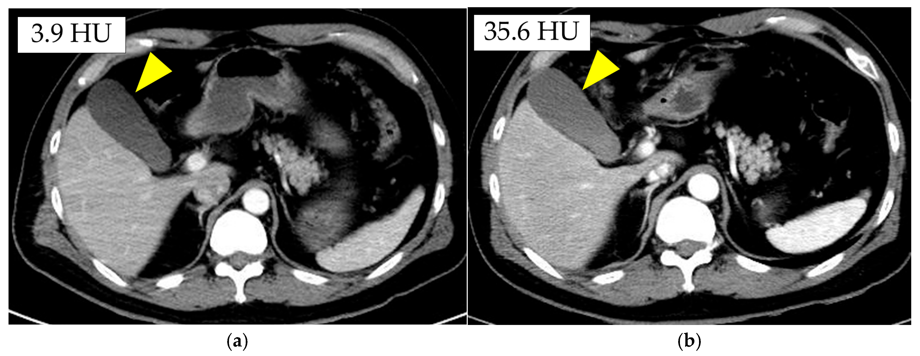
Abdominal contrast-enhanced computed tomography (CT) showed no obvious traumatic changes (Figure 1a). Despite a normal examination, the patient’s abdominal pain persisted, even after administering analgesics. Therefore, the patient was hospitalized for observation due to the risk of delayed injuries associated with the trauma. The following day, repeat abdominal contrast-enhanced CT demonstrated areas of increased attenuation within the gallbladder with mild enlargement; however, no significant interval changes were observed (Figure 1b).
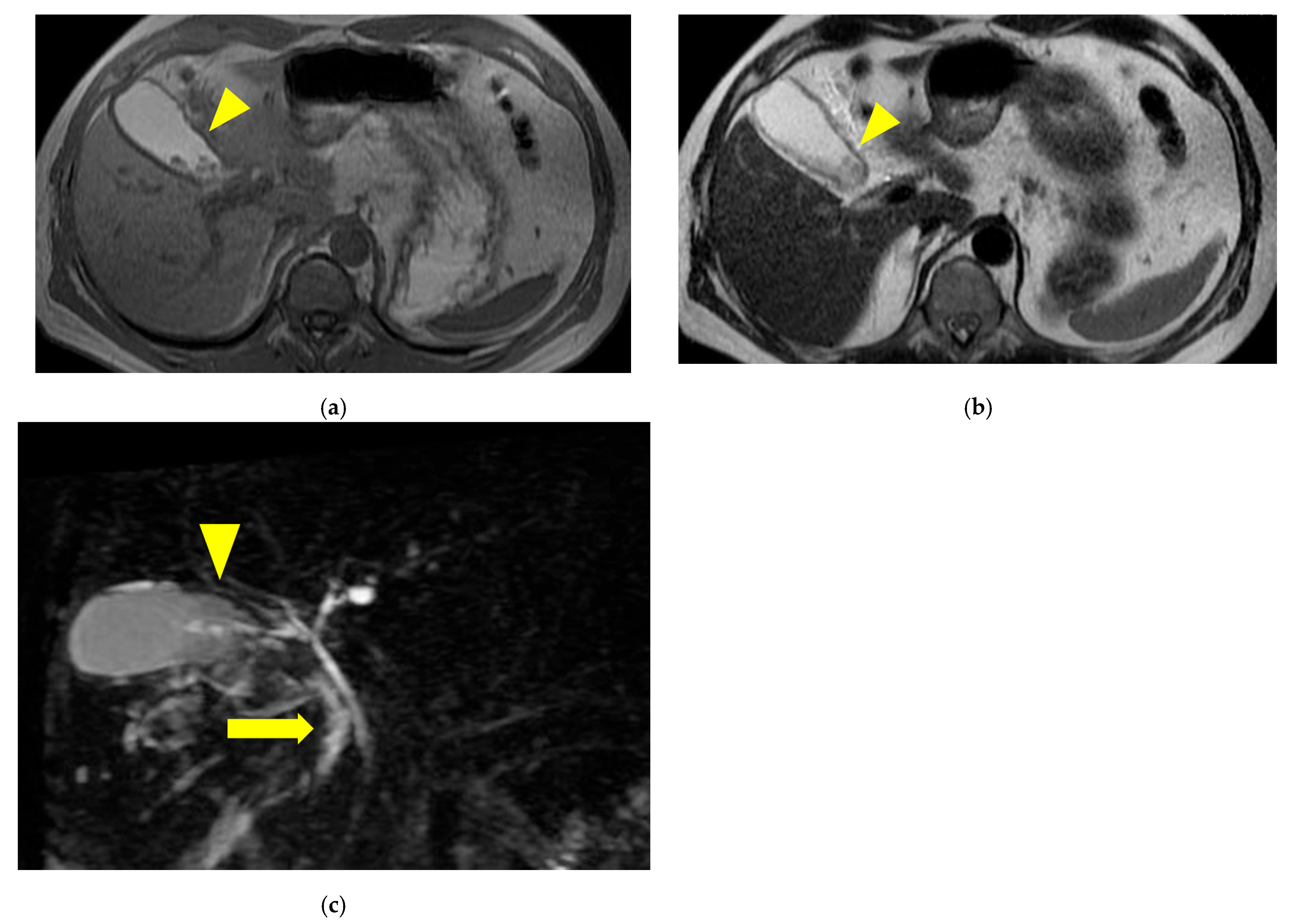
Blood tests revealed leukocytosis, elevated inflammatory markers, and increased hepatobiliary enzyme levels. Although these findings were not sufficient to establish a definitive diagnosis at that time, the patient met several components of the Tokyo Guidelines 2018 (TG18) diagnostic criteria, prompting consideration of acute cholangitis and cholecystitis in the differential diagnosis [2]. As traumatic gallbladder hemorrhage with obstructive jaundice was suspected, magnetic resonance cholangiopancreatography (MRCP) was performed to evaluate for cholangitis, while ultrasonographic examination was performed concurrently. Ultrasonography revealed circumferential gallbladder wall thickening, biliary sludge, and a small amount of ascites. MRCP revealed hyperintense areas within the collum vesicae felleae of the gallbladder and common bile duct on T1-weighted images, suggestive of bleeding. Additionally, hypointense areas in the posterior gallbladder on both T1- and T2-weighted images appeared hyperintense on diffusion-weighted imaging, indicating the presence of a clot; however, no obvious dilation of the bile duct was observed (Figure 2).
Based on these findings, delayed-onset cholecystitis or cholangitis due to traumatic gallbladder hemorrhage was strongly suspected, and it was determined that emergency surgery would not be necessary if the bleeding did not progress. Although MRCP confirmed intra-gallbladder hematoma, we determined that obstructive jaundice, if caused by hematoma rather than ongoing bleeding, would not require invasive surgery. Therefore, instead of proceeding with emergency surgery for this potentially highly invasive form of cholecystitis, a less invasive approach was prioritized. Initial management consisted of conservative treatment with antibiotics (sulbactam/cefoperazone), with a plan to perform biliary drainage via endoscopic retrograde cholangiopancreatography (ERCP) for decompression if clinical deterioration occurred. Under this approach, the patient’s clinical course remained favorable with conservative therapy alone.
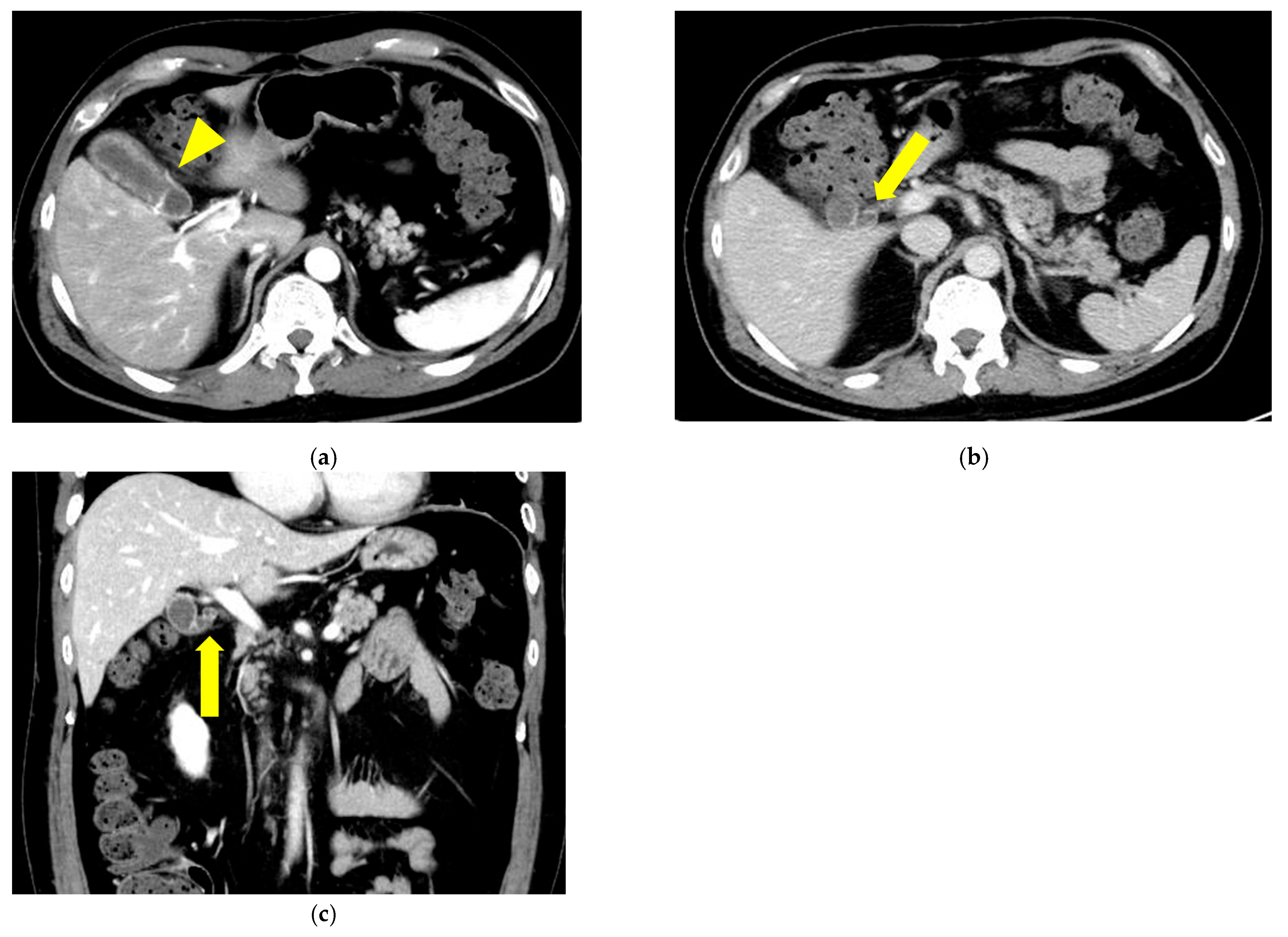
On Day 12, he complained of abdominal pain. Laboratory findings were as follows: WBC count, 6250/µL; Hgb, 12.9 g/dL; Hct, 40.3%; PLT, 30.6 × 10⁴/μL; T-Bil, 0.9 mg/dL; D-Bil, 0.5 mg/dL; AST, 452 IU/L; ALT, 366 IU/L; γ-GTP, 234 IU/L; LDH, 488 IU/L; CRP, 2.29 mg/dL. Abdominal contrast-enhanced CT demonstrated gallbladder wall thickening (Figure 3a). No dilation of the intrahepatic bile ducts or the common bile duct was observed (Figure 3b,c). Although the T-Bil level remained within the normal range and definitive biliary dilation was absent, the clinical presentation, including elevated inflammatory markers and liver enzymes, led us to suspect acute cholangitis (Grade I, mild), based on the TG18 diagnostic criteria, potentially caused by the passage of hematoma. In addition, the newly developed gallbladder wall thickening—absent on initial imaging—supported a concomitant diagnosis of acute cholecystitis [2,3]. Therefore, endoscopic nasobiliary drainage (ENBD) was performed as an initial intervention, after which elective surgery was planned.
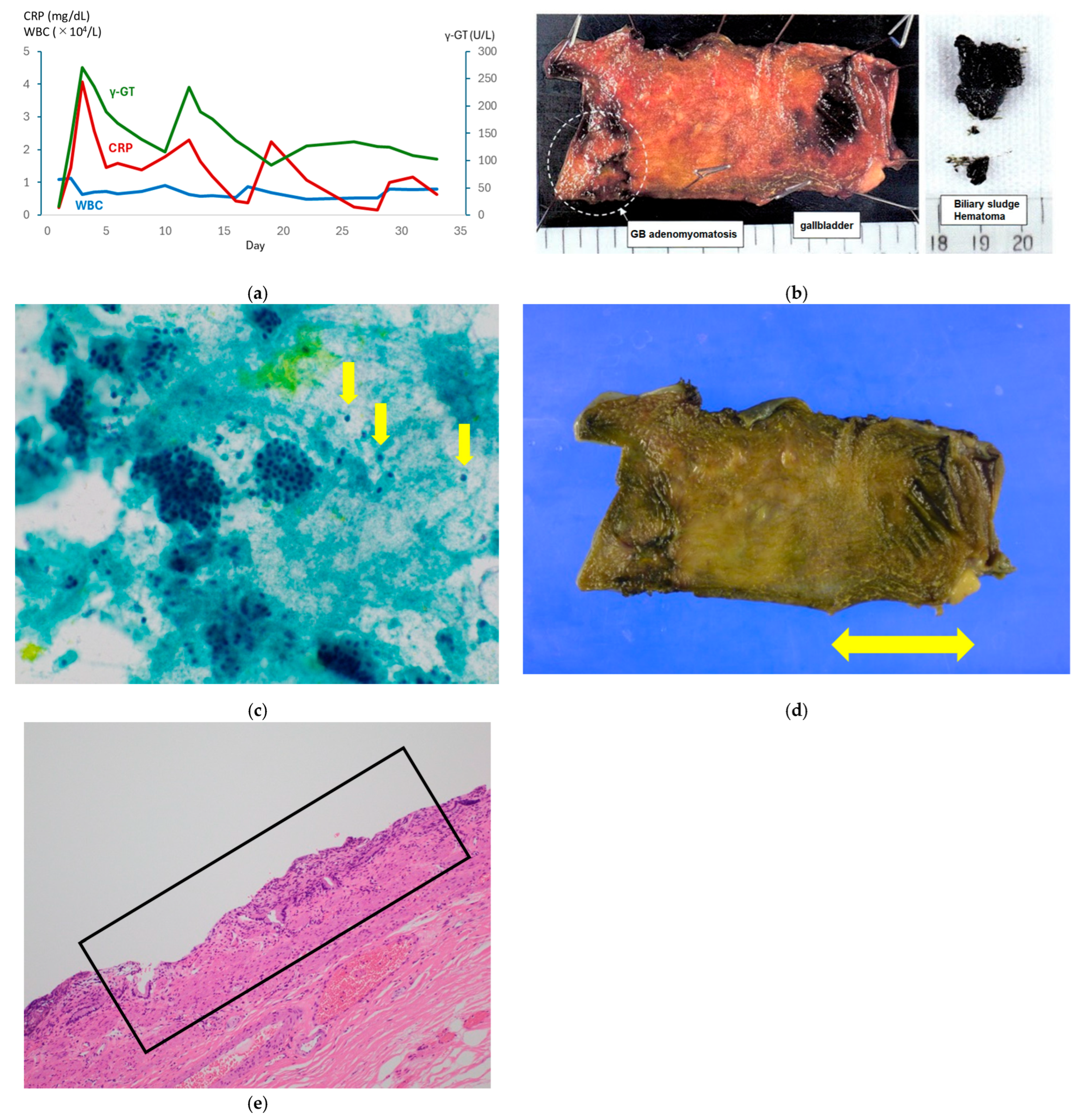
On Day 16, laboratory findings were as follows: WBC count, 5410/µL; T-Bil, 1.0 mg/dL; D-Bil, 0.5 mg/dL; AST, 31 IU/L; ALT, 103 IU/L; γ-GTP, 137 IU/L; LDH, 191 IU/L; CRP, 0.43 mg/dL (Figure 4a). Although the data exhibited a steady downward trend, an ENBD tube was placed to ensure definitive decompression and prevent any further inflammatory exacerbation. Given the risk of recurrent cholecystitis, elective surgery was strategically scheduled for three days later to optimize the patient’s perioperative condition. However, the surgery was postponed because a drug-induced eruption accompanied by mucosal edema developed on Day 20, which was suspected to represent an anaphylactic reaction. Laparoscopic cholecystectomy was performed on Day 28 of hospitalization. Extensive adhesions were found in the transverse colon, greater omentum, and duodenum around the gallbladder, likely due to cholecystitis. The gallbladder wall was partially scarred, resulting in thinning and fragility; however, the gallbladder was successfully removed. The excised specimen had an attached hematoma, which likely caused bile duct obstruction (Figure 4b). Pathological examination revealed lymphocytic infiltration in some areas, indicating that acute cholecystitis was superimposed on chronic cholecystitis. Although the gallbladder was successfully removed, the procedure was not considered routine because of the elevated risk of postoperative complications. Therefore, the ENBD tube was intentionally maintained to monitor for bile leakage and ensure continued biliary decompression. No postoperative complications occurred, including after subsequent ENBD removal. After careful observation confirming resolution of biliary inflammation and the associated systemic eruption, the patient was discharged on Day 35. At the 2-week follow-up, the surgical site was well healed, and no postoperative concerns were noted. The overall clinical course and laboratory trends during hospitalization are summarized in Figure 4a.
On bile cytology, lymphocytic infiltration, a characteristic feature of chronic cholecystitis, was more prominent than neutrophilic infiltration, which is typically observed in acute inflammation (Figure 4c). Macroscopic examination of the gallbladder revealed no evidence of hemorrhage or tumorous lesions (Figure 4d). Histopathological analysis using hematoxylin and eosin staining demonstrated no findings indicative of acute cholecystitis, such as wall destruction, necrosis, or neutrophilic infiltration. As shown in Figure 4e, the gallbladder wall was composed of collagenous fibers; however, neither fibroblast proliferation nor fibrosis was observed. The epithelial cells remained intact, and lymphocytic infiltration was evident, leading to a diagnosis of mild chronic cholecystitis (Figure 4e).

3. Discussion

Gallbladder injuries occur in approximately 2% of abdominal trauma cases, with isolated gallbladder injuries accounting for approximately 3% of these cases [1]. Soderstrom et al. classified gallbladder injuries into three categories: contusion (intramural hematoma), avulsion (partial or complete detachment from the liver), and perforation (wall defect). This classification is useful for guiding clinical assessment and management, as more severe injuries often require surgical intervention [4]. The present case was considered to correspond to the contusion category within this classification.
Regarding the diagnosis, several studies have demonstrated that CT revealed gallbladder enlargement, wall thickening, pericholecystic fluid accumulation, or contrast medium leakage, leading to the diagnosis of gallbladder hemorrhage. However, diagnosing isolated gallbladder injury caused by blunt abdominal trauma may be difficult in the early stages using ultrasound or CT, depending on the extent of bleeding and the hematoma size [1]. There have been case reports, in which a definitive diagnosis could not be established even after performing MRCP in addition to ultrasonography and CT [1].
In the present case, initial examinations after blunt abdominal trauma showed no traumatic changes as described in the aforementioned classification; however, delayed hemorrhage led to the formation of an intracholecystic hematoma, subsequently resulting in acute cholangitis and cholecystitis. Gallbladder hemorrhage was not detected on laboratory testing or CT imaging but was identified by MRCP after acute cholangitis and cholecystitis were suspected based on the TG18 criteria. This case highlights the value of magnetic resonance imaging (MRI) and MRCP for the early detection of gallbladder injury; however, when a definitive diagnosis cannot be established, careful follow-up and close clinical monitoring remain essential.
With respect to treatment, previous reports indicate that emergency surgery was performed in some cases where contrast-enhanced CT revealed contrast extravasation after blunt trauma [5]. In other cases with no obvious traumatic findings, there were instances where conservative treatment led to recovery [5,6], while other reports indicated that biliary obstruction occurred 2 weeks post-injury, necessitating pancreatoduodenectomy [7]. Additionally, gallbladder hemorrhage and cholecystitis have occurred in patients on direct oral anticoagulants despite no trauma [8].
In the present case, conservative antibiotic therapy was initially selected in accordance with our institutional protocol based on the TG18 criteria for Grade I/II severity and was effective for both cholangitis and cholecystitis. However, the patient experienced recurrence on Day 12. In the setting of clinical deterioration, pseudoaneurysm of the cystic or hepatic artery should also be considered as a potential cause of delayed hemobilia. A recent review reported that cystic artery pseudoaneurysms most commonly occur secondary to cholecystitis (61.2%) or cholecystectomy (26.8%), whereas traumatic etiology is exceedingly rare [9]. After MRCP, we re-examined the arterial-phase CT on Day 12 and found no evidence of pseudoaneurysm or extravasation. Given the absence of active hemorrhage, immediate highly invasive surgery was deemed unnecessary. Instead, we adopted a “step-up approach,” as prioritized in the TG18 management bundle, proceeding with endoscopic intervention (ERCP/ENBD) to ensure biliary decompression [3]. This strategy aimed to reduce surgical risk by stabilizing the patient’s systemic condition and local inflammation before definitive surgery. Delayed surgery related to a drug-induced eruption may have contributed to the chronic cholecystitis findings on pathology. Therefore, although no consensus exists regarding the timing of surgery after blunt abdominal trauma, hospitalization in a setting where elective surgery can be arranged is recommended when TG18 criteria raise suspicion of gallbladder injury or delayed complications, such as hematoma-induced biliary obstruction [10].
Furthermore, the ENBD tube was maintained postoperatively as a precaution. Severe local inflammation and dense adhesions encountered intraoperatively raised concern for potential bile leakage from the gallbladder bed. Maintaining the tube allowed for continuous monitoring of biliary integrity and ensured decompression during the early recovery phase. It was removed only after confirming the absence of bile leakage and stabilization of the patient’s clinical status.
The clinical significance of this case lies in highlighting the potential for delayed complications following blunt gallbladder trauma. Even when initial CT findings are negative for major injury, late migration of an intracholecystic hematoma can result in severe obstructive jaundice and cholangitis. Our experience suggests that a step-up approach—early endoscopic decompression followed by elective surgery—is an effective strategy for managing such unstable presentations. This approach facilitates resolution of acute inflammation and stabilization of systemic conditions, particularly when unexpected complications, such as drug-induced eruption, occur. Moreover, the discrepancy between the acute clinical presentation and chronic histopathological findings suggests that preoperative management may alter tissue characteristics. Clinicians should remain vigilant for delayed biliary obstruction in patients with trauma and prioritize strategic decompression to ensure surgical safety.


4. Conclusions

Gallbladder injuries isolated from blunt trauma are rare and challenging, often presenting with deceptive clinical onset. This case underscores the utility of MRI/MRCP for early detection and the importance of close monitoring in an elective surgical setting for potentially delayed complications, even after initially negative imaging. Specifically, our experience demonstrates that a step-up approach incorporating early endoscopic decompression followed by elective surgery is not a temporary measure but a safe and effective strategy for managing hematoma-induced biliary obstruction. By providing a necessary cooling down period in an inflamed, post-traumatic field, this approach effectively minimizes the risk of bile duct injury. This clinical framework offers practical guidance for emergency physicians and surgeons to optimize patient safety, particularly when unexpected systemic complications require postponement of definitive surgery.

Author Contributions

Conceptualization, A.M. and C.M.; methodology, M.H., J.S., A.M. and C.M.; software, A.M. and C.M.; validation, K.D., A.M. and C.M.; formal analysis, A.M. and C.M.; investigation, A.M., H.T., Y.N. and C.M.; resources, A.M., H.T., Y.N. and C.M.; data curation, Y.U.; writing—original draft preparation, A.M. and C.M.; writing—review and editing, K.D., A.M. and C.M.; visualization, Y.U. and C.M.; supervision, K.D., M.H., J.S. and A.M.; project administration, K.D., A.M. and C.M.; funding acquisition, K.D. All authors have read and agreed to the published version of the manuscript.

Funding

This research received no external funding.

Institutional Review Board Statement

Ethical review and approval were waived for this study because it is a case report of a single patient, and all clinical data were anonymized to protect patient privacy in accordance with the ethical guidelines of our institution.

Informed Consent Statement

Informed consent was obtained from the patient at the time of hospital admission for the use of clinical data and the publication of this case report and any accompanying images.

Data Availability Statement

The original contributions presented in this study are included in the article. Further inquiries can be directed to the corresponding author.

Conflicts of Interest

The authors declare no conflicts of interest.

Abbreviations

The following abbreviations are used in this manuscript:
WBCWhite blood cell
HgbHemoglobin
HctHematocrit
PLTPlatelets
T-BilTotal Bilirubin
ASTAspartate aminotransferase
ALTAlanine aminotransferase
γ-GTPGamma-glutamyl transferase
LDHLactic dehydrogenase
CRPC-reactive protein
MRIMagnetic resonance imaging
MRCPMagnetic resonance cholangiopancreatography
CTComputed tomography
ENBDEndoscopic nasobiliary drainage

References

  1. Birn, J.; Jung, M.; Dearing, M. Isolated gallbladder injury in a case of blunt abdominal trauma. Radiol. Case Rep. 2012, 6, 25–30. [Google Scholar] [CrossRef] [PubMed]
  2. Kiriyama, S.; Kozaka, K.; Takada, T.; Strasberg, S.M.; Pitt, H.A.; Gabata, T.; Hata, J.; Liau, K.H.; Miura, F.; Horiguchi, A.; et al. Tokyo Guidelines 2018: Diagnostic criteria and severity grading of acute cholangitis (with videos). J. Hepatobiliary Pancreat. Sci. 2018, 25, 17–30. [Google Scholar] [CrossRef] [PubMed]
  3. Mayumi, T.; Okamoto, K.; Takada, T.; Strasberg, S.M.; Solomkin, J.S.; Schlossberg, D.; Pitt, H.A.; Yoshida, M.; Gomi, H.; Miura, F.; et al. Tokyo Guidelines 2018: Management bundles for acute cholangitis and cholecystitis. J. Hepatobiliary Pancreat. Sci. 2018, 25, 96–100. [Google Scholar] [CrossRef] [PubMed]
  4. Soderstrom, C.A.; Maekawa, K.; Dupriest, R.W., Jr.; Cowley, R.A. Gallbladder injuries resulting from blunt abdominal trauma: An experience and review. Ann. Surg. 1981, 193, 60–66. [Google Scholar] [CrossRef] [PubMed]
  5. Jang, H.; Park, C.H.; Park, Y.; Jeong, E.; Lee, N.; Kim, J.; Jo, Y. Spontaneous resolution of gallbladder hematoma in blunt traumatic injury: A case report. World J. Clin. Cases 2021, 9, 8518–8523. [Google Scholar] [CrossRef] [PubMed]
  6. Wiebe, I.; Baig, Z.; Sothilingam, N. Hemorrhagic cholecystitis from isolated gallbladder injury following blunt abdominal trauma: An unusual case report. Int. J. Surg. Case Rep. 2022, 90, 106680. [Google Scholar] [CrossRef] [PubMed]
  7. Takahashi, D.; Koizumi, M.; Matsumoto, S.; Kiguchi, E. A case of blunt traumatic intra-gallbladder bleeding without rupture. J. Jpn. Surg. Assoc. 2015, 76, 2788–2793. [Google Scholar] [CrossRef]
  8. Azam, M.U.; Ibrahim, M.A.; Perry, I.; Ellison, S.B.; Barrett, A.; Vega, K.J. It’s the Bloody Gallbladder! Spontaneous gallbladder hemorrhage following factor Xa inhibition. J. Natl. Med. Assoc. 2021, 113, 252−254. [Google Scholar] [CrossRef] [PubMed]
  9. Taghavi, S.M.J.; Jaya Kumar, M.; Damodaran Prabha, R.; Puhalla, H.; Sommerville, C. Cystic artery pseudoaneurysm: Current review of aetiology, presentation, and management. Surg. Res. Pract. 2021, 2021, 4492206. [Google Scholar] [CrossRef] [PubMed]
  10. Jaggard, M.K.J.; Johal, N.S.; Choudhry, M. Blunt abdominal trauma resulting in gallbladder injury: A review with emphasis on pediatrics. J. Trauma 2011, 70, 1005–1010. [Google Scholar] [CrossRef] [PubMed]
Figure 1. (a) Abdominal contrast-enhanced computed tomography (CT) (Day 1): There were no obvious traumatic changes. (b) Abdominal contrast-enhanced CT (Day 2): There were areas of increased attenuation within the gallbladder and mild gallbladder enlargement; however, there was no significant difference compared with the findings from the previous day. In addition, the CT values of the areas indicated by the yellow triangles are shown in each image.
Ecm 03 00015 g001
Figure 2. (a) Magnetic resonance imaging (MRI) (T1-weighted images): There were hypointense areas in the collum vesicae felleae of the gallbladder. (the yellow triangle) (b) MRI (T2-weighted images): Low signal areas were observed in the collum vesicae felleae of the gallbladder, consistent with findings on T1-weighted images. (the yellow triangle) (c) Magnetic resonance cholangiopancreatography (Day 2): There were hyperintense areas suspected to be hematoma within the collum vesicae felleae of the gallbladder (the yellow triangle) and common bile duct (the yellow arrow); however, no dilation of the bile duct was observed.
Ecm 03 00015 g002
Figure 3. Abdominal contrast-enhanced computed tomography on Day 12. (a,b) Axial view: The gallbladder exhibits wall thickening with internal hematoma (the yellow triangle), and no dilation of the adjacent bile duct was observed compared to the initial imaging on Day 2 (the yellow arrow). (c) Coronal view: No dilation of the common bile duct or intrahepatic bile ducts was observed (the yellow arrow).
Ecm 03 00015 g003
Figure 4. (a) Chronological changes in key clinical findings and laboratory parameters from admission (Day 1) to the day of surgery. Inflammatory markers and hepatobiliary enzymes: Significant elevation in white blood cell (WBC) count and C-reactive protein (CRP) was observed on Day 2, coinciding with the onset of suspected cholangitis. A secondary spike in aspartate aminotransferase (AST) and alanine aminotransferase (ALT) levels occurred on Day 12, indicating potential recurrence of biliary obstruction due to hematoma passage. Clinical symptoms: Abdominal pain intensity (visual analog scale) peaked on Days 2 and 12, paralleling the deterioration in laboratory parameters. (b) The excised gallbladder contained bile sludge and hematoma. (c) Results of bile cytology (Papanicolaou staining). The bile pigment was stained yellow, while the cell nuclei were stained purple. Neutrophils were not detected, but some lymphocytes, which have cell nuclei without cytoplasm, were observed (the yellow arrows). (d) Gallbladder tissue specimen. The right side corresponds to the neck, and the left to the fundus. No tumorous lesions were observed. (e) Hematoxylin and eosin staining (the yellow arrowed area in (d)). The gallbladder wall consists of collagenous fibers (inside the black frame), with no prominent inflammatory findings, such as neutrophil infiltration. This suggests chronic cholecystitis.
Ecm 03 00015 g004
Disclaimer/Publisher’s Note: The statements, opinions and data contained in all publications are solely those of the individual author(s) and contributor(s) and not of MDPI and/or the editor(s). MDPI and/or the editor(s) disclaim responsibility for any injury to people or property resulting from any ideas, methods, instructions or products referred to in the content.