
Article

First Report of Leaf Spot of Spinacia oleracea Caused by Alternaria burnsii: Aerobiological Implications and Enzymatic Virulence Factor

Department of Plant Pathology, Faculty of Agriculture and Environment, The Islamia University of Bahawalpur, Bahawalpur 63100, Pakistan
*
Authors to whom correspondence should be addressed.
Aerobiology 2026, 4(2), 11; https://doi.org/10.3390/aerobiology4020011
Submission received: 15 March 2026 / Revised: 11 May 2026 / Accepted: 22 May 2026 / Published: 26 May 2026

Abstract

Spinacia oleracea L. cultivation in South Asia is severely compromised by leaf spot disease caused by fungal plant pathogens, resulting in significant yield and quality losses. In this study, we report the first molecularly confirmed case of an Alternaria burnsii leaf spot on S. oleracea in Pakistan. Symptomatic S. oleracea leaves exhibiting necrotic lesions with concentric rings were collected during a field survey across Bahawalpur district, Punjab, Pakistan in 2024. After isolation, purification and morphological identification it was identified that it belongs to the Alternaria genus. For the confirmation of species, molecular identification was performed; using the ITS and GAPDH primer revealed that the fungal plant pathogen causing leaf spot of S. oleracea is A. burnsii which was also confirmed by phylogenetic analysis. Koch’s postulates were carried out to confirm pathogenicity on detached leaf assays. To assess the virulence of A. burnsii enzymatic analysis was performed. Notably, enzymatic virulence profiling demonstrated a markedly increased production of polygalacturonase (PG: 16.0 ± 0.8 AU), pectin lyase (PNL: 12.0 ± 0.6 AU) and cellulase (CL: 14.0 ± 0.7 AU) relative to controls (all p < 0.001; LSD = 0.16), with PG having the greatest relative increase. This report expands the known host range for A. burnsii and highlights its two-fold threat: as a bioaerosol disseminable by wind and an enzymatic pathogen. These findings highlight the urgent need for integrated disease management strategies for suppressing leaf spot disease in S. oleracea agroecosystems.

1. Introduction

Spinach (Spinacia oleracea L.) is a globally cultivated leafy vegetable famous for its nutritional value due to its vitamins, minerals, and bioactive phytochemicals with antioxidant and anti-inflammatory abilities [1]. In Pakistan, spinach is grown as a major staple crop of vegetable, with a production of about 96,000 tons per year; uses include meeting dietary needs and fostering economy stability [2,3]. However, the crop yield and quality suffer greatly due to fungal diseases, primarily the leaf spot diseases that cause necrotic lesions, premature defoliation and post-harvest decay which also impact marketability [4]. The genus Alternaria is one of the most cosmopolitan and economically significant fungal plant pathogens, infecting a variety of crops, including vegetables, cereals, oilseeds, and fruits [5,6,7,8,9]. In spinach specifically, multiple Alternaria species including A. alternata [10,11], A. brassicicola, and A. tenuissima [12,13] have been documented as causal agents which are all known to cause leaf spot disease across diverse agroecosystems [14]. A. alternata is the most prevalent pathogen, causing characteristic concentric ring lesions on spinach in the United States, Europe, and India [6,15]. A. brassicicola has been identified on spinach in Australia and China [2,16], often co-occurring with A. alternata and exhibiting similar symptomatology. More recently, A. tenuissima and A. arborescens were reported as emerging pathogens on spinach in Mediterranean regions [17]. Collectively, these studies confirm that spinach is susceptible to a complex of Alternaria species, with disease incidence influenced by climatic factors and inoculum pressure from adjacent crops or soil debris. Despite this growing body of knowledge, the occurrence of Alternaria burnsii on spinach remains undocumented. While A. burnsii is a well-characterized pathogen of sunflower [18], tomato [18], brassicas [19], and legumes [20] causing pre-harvest leaf spot and stem spots and post-harvest rot through aggressive tissue maceration, A. burnsii association with spinach has never been reported in the global literature, including comprehensive reviews of spinach pathogens. Black circular to oval lesions with necrotic centers and distinct margins are characteristic of spinach leaf spot disease caused by A. burnsii [21]. It initiates tissue maceration through the secretion of cell wall-degrading enzymes, including polygalacturonase, pectin lyase and cellulase, aiding its virulence, and at the moment leads to the loss of many crops if not controlled [14,22]. Alternaria species are common and plentiful members of the atmospheric mycobiota. Small, dark-colored conidia produced by these fungi are efficiently dispersed long distances by air currents, and airborne inoculum serves as the primary source of primary infection in open-field crops [11,23]. Because of their ability to withstand ultraviolet radiation and desiccation these spores can stay alive in the atmosphere for long periods of time allowing them to spread to hosts that are susceptible and become infected in appropriate environmental conditions [8]. Though the aerobiological behavior of Alternaria species is well known, the specific contribution of atmospheric dispersal to the epidemiology of A. burnsii on spinach remains unexamined, especially in South Asian agroecosystems. Sixteen different fungal diseases of spinach have been reported worldwide to date [9,24], but A. burnsii has not been reported as a causal agent of spinach leaf spot in Pakistan. The gap in knowledge is critical, because spinach is an economically important crop, and possible airborne transmission has been reported. Thus, the present work was conducted to isolate and molecularly identify the causal agent of leaf spot disease on spinach in Pakistan; characterize its enzymatic virulence factors; and discuss the aerobiological significance of A. burnsii as a wind-dispersed pathogen in open-field cultivation system. The present report of A. burnsii on spinach increases the known host range of this pathogen and suggests its atmospheric dispersal potential, and disease epistemology.

2. Materials and Methods

2.1. Survey and Sample Collection

During the winter and rainy season of 2024, ten field areas in Bahawalpur district, Punjab, Pakistan, listed in Table 1, were surveyed for the presence of S. oleraceae leaf spot diseases. Randomly 100 infected leaves exhibiting necrotic lesions with concentric rings from fields were collected and put in labeled zip-lock bags before being delivered to the Molecular Laboratory of Plant Pathology, IUB, for further studies. With the help of a light microscope (IRMECO) and a hand lens, the morphology of the symptoms was studied. The size, shape, color, and appearance of the spots were observed as a point of reference in photographs of the infected leaves. Diseased samples were kept at 4 °C for further processing [25].

2.2. Fungal Isolation and Purification

Infected leaf samples collected from all the surveyed locations listed in Table 1 were washed with distilled water. Randomly three leaves from each collected sample were selected and cut into pieces of 5 mm2 having both diseased and healthy portions. These pieces were surface-sterilized with 1% NaOCl for 30 s and rinsed thrice with distilled water to remove the traces of bleach. Samples were aseptically placed into potato dextrose agar (PDA) medium, consisting of potato infusion (200 gL−1), dextrose (20 gL−1), and agar (15 gL−1), and incubated at 25 ± 2 °C for 7 days. A 5 mm disk cut from the edge of the fungal culture was used to collect and purify the mycelia that had formed from the tissues and put them on newly prepared PDA plates. Pure cultures isolated from all surveyed field locations were stored at departmental fungal culture bank at 4 °C. Throughout the research, these plates were periodically multiplied and sub-cultured [26].

2.3. Morphological Identification

Microscopic examination was performed to identify the isolated fungi depending on their morphological features, including culture colony color, spore size, and shape [27].

2.4. Pathogenicity Assay

S. oleraceae seeds were sown in sterilized soil and grown in a growth chamber under controlled conditions (22 ± 2 °C), and 70% relative humidity. Plants were visually inspected daily for disease symptoms and nutrient deficiencies. Finally, 4-week-old plant leaves that were fully expanded showing no signs of stress were used. In this study pathogenicity was performed by using a detached leaf assay by using a pure culture of Alternaria burnsii originated from Chak 9 BC (Rural Area), the location with the highest disease incidence. Healthy S. oleraceae leaves were taken and surface-sterilized with 1% sodium hypochlorite (NaOCl) and then allowed to air dry. A sterilized Petri plate with S. oleraceae leaf lined with moistened cotton was used for pathogenicity. Spore density was adjusted by using a hemocytometer with conidial suspension (1 × 10⁵ conidia mL−1 in 0.01% Tween 20) from 7-day-old PDA pure culture used for pathogenicity. Using a micropipette and aseptic conditions, 1 mL of spore suspension was applied to the leaf surface. Sterile distilled water served as the negative control. The plates were then incubated at 25 ± 2 °C and periodically checked for the emergence of disease symptoms. Koch’s pathogenicity postulates were then verified by re-isolating the pathogen again from the diseased leaf area [28].

2.5. Genomic DNA Extraction

Genomic DNA was extracted from a 7-day-old pure fungal culture originated from Chak 9 BC that showed the highest disease incidence using the CTAB (cetyltrimethylammonium bromide) method with minor modifications. Briefly, mycelia were harvested from PDA plates, ground to a fine powder in liquid nitrogen, and incubated in CTAB extraction buffer (2% CTAB, 100 mM Tris-HCl pH 8.0, 20 mM EDTA, 1.4 M NaCl) at 65 °C for 30 min. DNA was purified by chloroform:isoamyl alcohol (24:1) extraction, precipitated with isopropanol, washed with absolute ethanol, and resuspended in 100 µL of d3H2O. DNA quality and concentration were assessed using a NanoDrop spectrophotometer (Thermo Scientific, Waltham, MA, USA), and integrity was confirmed by 1% agarose gel electrophoresis [26].

2.6. PCR Amplification

Two genetic regions were amplified for molecular identification (ITS and GAPDH), genes having an amplicon size of approximately 590 bp and 500 bp respectively. PCR reactions were performed in a 25 µL reaction mixture containing: 12.5 µL of 2X Amp Master Taq Mix, 2 µL of genomic DNA template, 1 µL of a concentration of 10X each of forward and reverse primers, and 8.5 µL of nuclease-free water. The primer pair sequences used for PCR are listed in Table 2.
PCR amplification was carried out in a thermal cycler with the following cycling conditions: initial denaturation (95 °C for 5 min), 35 cycles of 95 °C for 30 s, 55 °C for 45 s, and 72 °C for 1 min, and a final extension: 72 °C for 10 min, hold at 4 °C. Amplified products were visualized on 1.5% agarose gels stained with ethidium bromide under UV transillumination.

2.7. DNA Sequencing and Phylogenetic Analysis

The PCR products of expected size (590 bp for ITS and 500 bp for GAPDH) were cleaned up using a PCR purification kit (Thermo Scientific) and, subsequently, bidirectionally Sanger-sequenced by Macrogen, Korea. Chromatograms of raw sequencing were edited and assembled through BioEdit v7.2.5. To compare the sequences to known Alternaria species, consensus sequences were compared using BLASTn against the NCBI GenBank database. The sequences obtained from genotyping were uploaded to GenBank. To perform phylogenetic analysis, sequences of closely related Alternaria species were retrieved from GenBank and aligned in MEGA X by employing the MUSCLE algorithm. Using 1000 bootstrap replications, a maximum likelihood phylogenetic tree was generated to determine the phylogenetic relationship of the isolate with reference strains [26].

2.8. Enzymatic Virulence Factor Assays

The effect of the A. burnsii on pathogenic potential was assessed by measuring the production of three important cell wall-degrading enzymes using spectrophotometric methods [36].

2.8.1. Enzyme Production

The fungal isolate was grown in Czapek-Dox broth with 1% (w/v) pectin (for polygalacturonase and pectin lyase induction) or 1% (w/v) carboxymethyl cellulose (for cellulase induction) at 25 °C for 7 days under shaking (120 rpm) conditions. Culture filtrates were generated by filtering over Whatman No. 1 filter paper and centrifuging at 10,000× g for 15 min at 4 °C; the supernatants were used as crude enzyme extracts [36].

2.8.2. Polygalacturonase (PG) Activity

PG activity was determined using the method of Miller (1989) [37] with modifications. The reaction mixture contained 0.5 mL of 1% polygalacturonic acid in 0.1 M sodium acetate buffer (pH 4.5) and 0.5 mL of enzyme extract. After incubation at 37 °C for 30 min, the reaction was terminated by adding 1 mL of 3,5-dinitrosalicylic acid (DNS) reagent and boiling for 5 min. The absorbance was measured at 540 nm using a UV-Vis spectrophotometer (Shimadzu UV-1800, Kyoto, Japan). One unit of PG activity was defined as the amount of enzyme required to release 1 µmol of reducing sugar (galacturonic acid) per minute under assay conditions. A standard curve was prepared using galacturonic acid [36].

2.8.3. Pectin Lyase (PNL) Activity

PNL activity was assayed according to the method of Alguacil et al. (2003) [38]. The reaction mixture consisted of 0.9 mL of 1.2% citrus pectin in 0.05 M glycine–NaOH buffer (pH 8.8) and 0.1 mL of enzyme extract. After incubation at 30 °C for 30 min, the increase in absorbance at 235 nm was measured due to the formation of unsaturated products. One unit of PNL activity was defined as the amount of enzyme causing an increase in absorbance of 0.01 per minute [36].

2.8.4. Cellulase (CL) Activity

The DNS method (Miller, 1989) [37] was used to determine cellulase activity. The reaction mixture was prepared with 1 mL of 1% carboxymethyl cellulose (CMC) in 0.05 M citrate buffer (pH 4.8) and 1 mL of enzyme extract. The mixture was boiled at 95 °C for 5 min after the addition of 2 mL of DNS reagent (NSO5481, SigmaAldrich, Buchs, Switzerland) and after 30 min incubation at 50 °C. Absorbance was recorded at 540 nm. The cellulase activity unit was defined as the amount of enzyme that liberated 1 µmol of reducing sugar (glucose) per minute. Quantification of samples was performed based on the measurements of optical density (OD) from a glucose standard curve. All enzyme assays were carried out 3 times as the triplicate basis, and expressed in terms of U mL−1 [36].

2.9. Statistical Analysis

All experiments were conducted in a completely randomized design with three biological replicates. Data were subjected to analysis of variance (ANOVA) using Statistix 8.1 software. Treatment means were compared using the least significant difference (LSD) test at p ≤ 0.05. Enzyme activity data were presented as mean ± standard deviation (SD).

3. Results

3.1. Survey and Sample Collection

The field survey revealed that all visited fields exhibited typical symptoms of leaf spot on spinach—small to irregular dark brown spots that gradually enlarged and, in some cases, coalesced, leading to necrotic lesions on the leaf surface. The presence of these symptoms indicated the widespread occurrence of the disease in the study area. The details of the surveyed locations and their geographic coordinates are given in Table 2.

3.2. Isolation, Purification and Morphological Identification

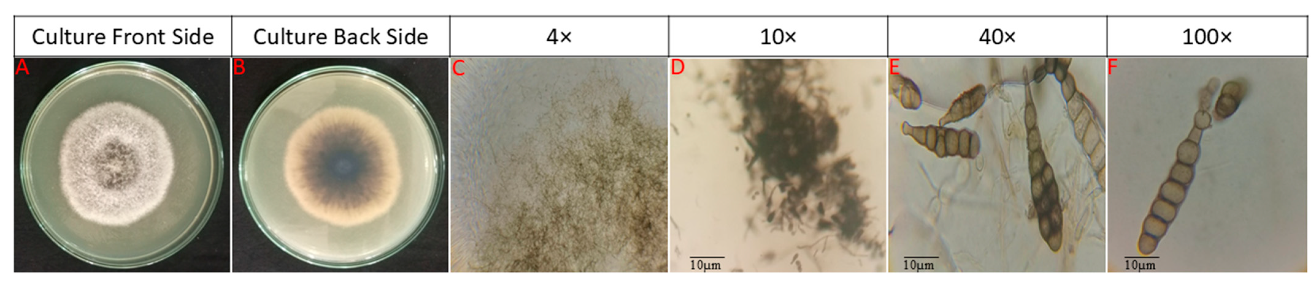
After isolation and purification, the emerging colonies were circular with a velvety texture and distinct concentric ring patterns, and central regions displayed dark gray to black pigmentation, transitioning radially to medium gray and pale gray margins (recent mycelial development). Crucially, the reverse side demonstrated a deep brown-to-black center (indicating melanin accumulation) surrounded by a dark brown diffusion zone and light brown marginal region. This pigmentation pattern aligns with diagnostic criteria for A. burnsii. The fungus, based on its morphological characteristics, was determined as Alternaria burnsii (Figure 1).

3.3. Pathogenicity Assay

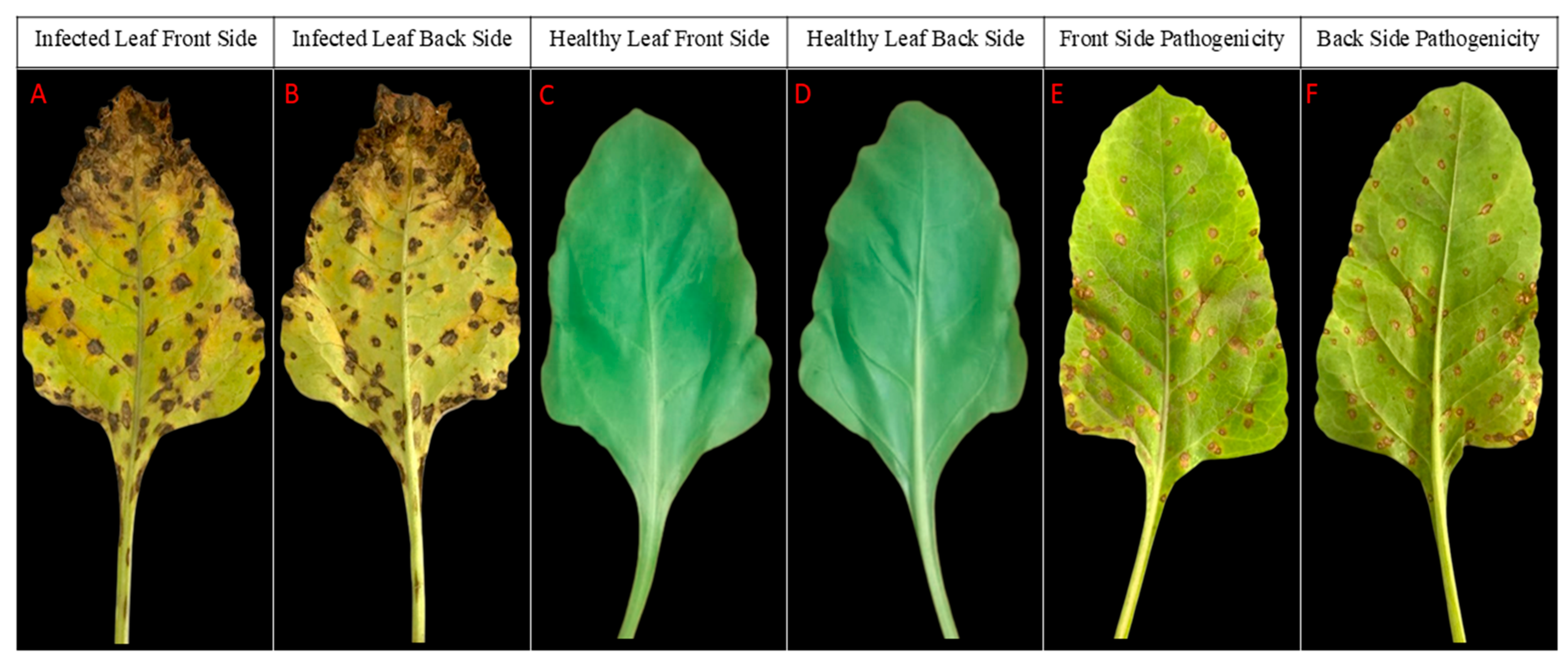
S. oleraceae leaves were inoculated with 1 mL of spore suspensions of the pathogen by micropipettes. Un-inoculated plant leaves treated with distilled water served as healthy control. These plants were kept in an incubator to avoid any contamination. After 7 days the symptoms started to appear. These symptoms were compared to the previous ones that were observed in natural condition. Pathogen was re-isolated from an infected leaf and cultured on a PDA medium. The same fungus was observed after re-isolation. Through this Koch’s postulates were satisfied, as shown in Figure 2.

3.4. DNA Extraction and PCR Amplification

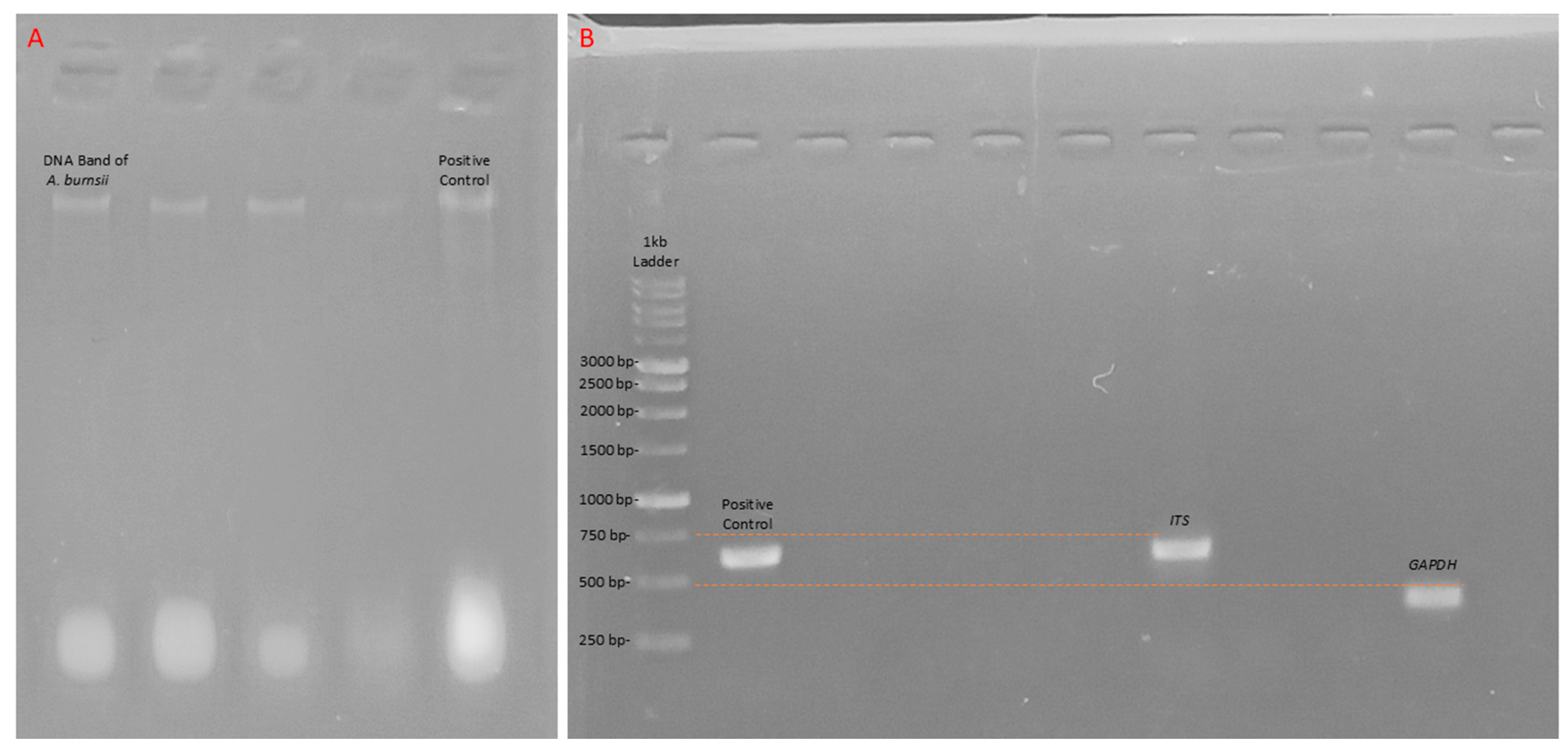
Genomic DNA was successfully extracted from the A. burnsii pure culture by using the CTAB method. PCR amplification of the ITS and GAPDH regions was done by using ITS1/ITS4 and GAPDH primers. DNA bands and PCR products bands are shown in Figure 3.
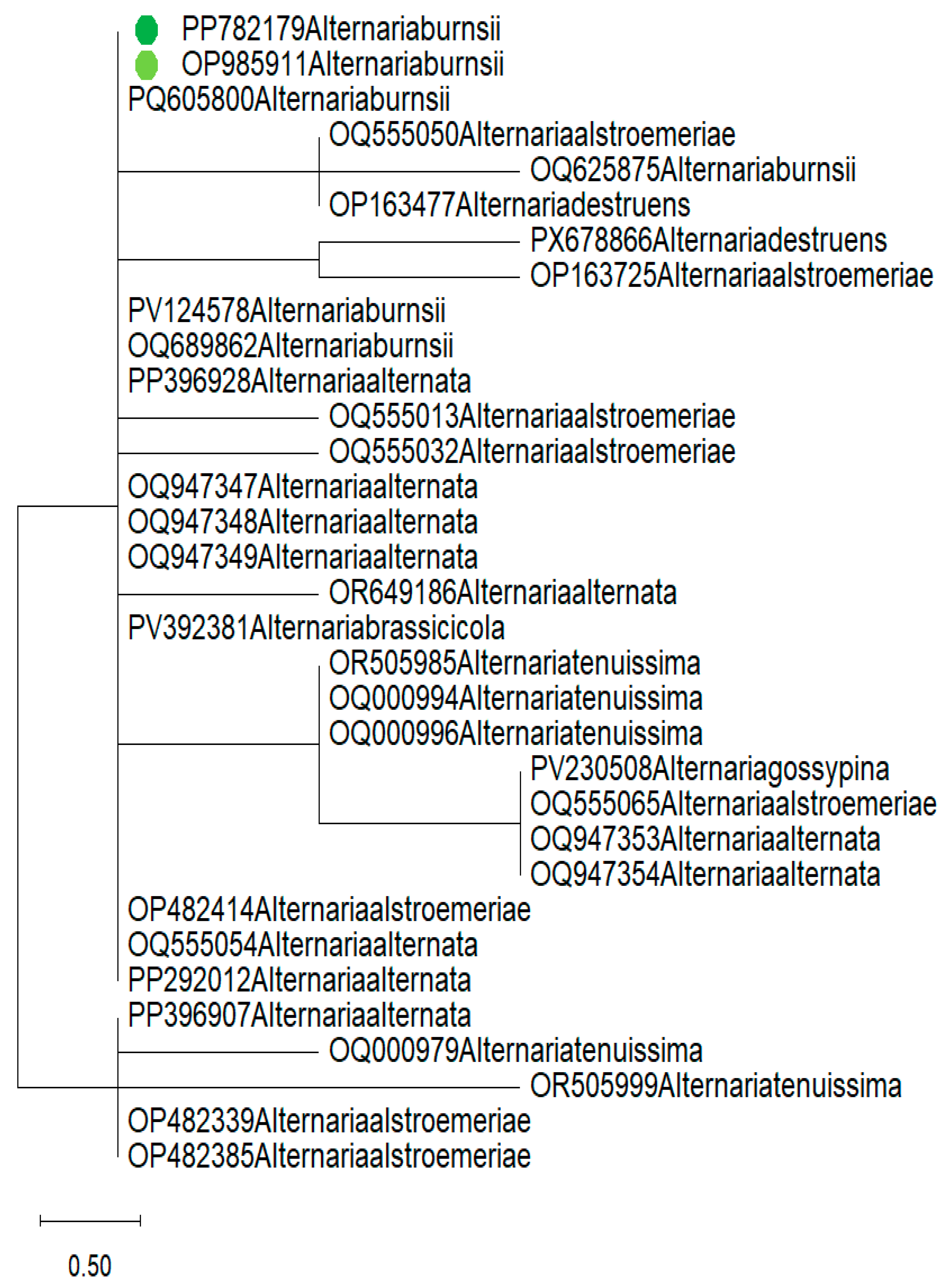
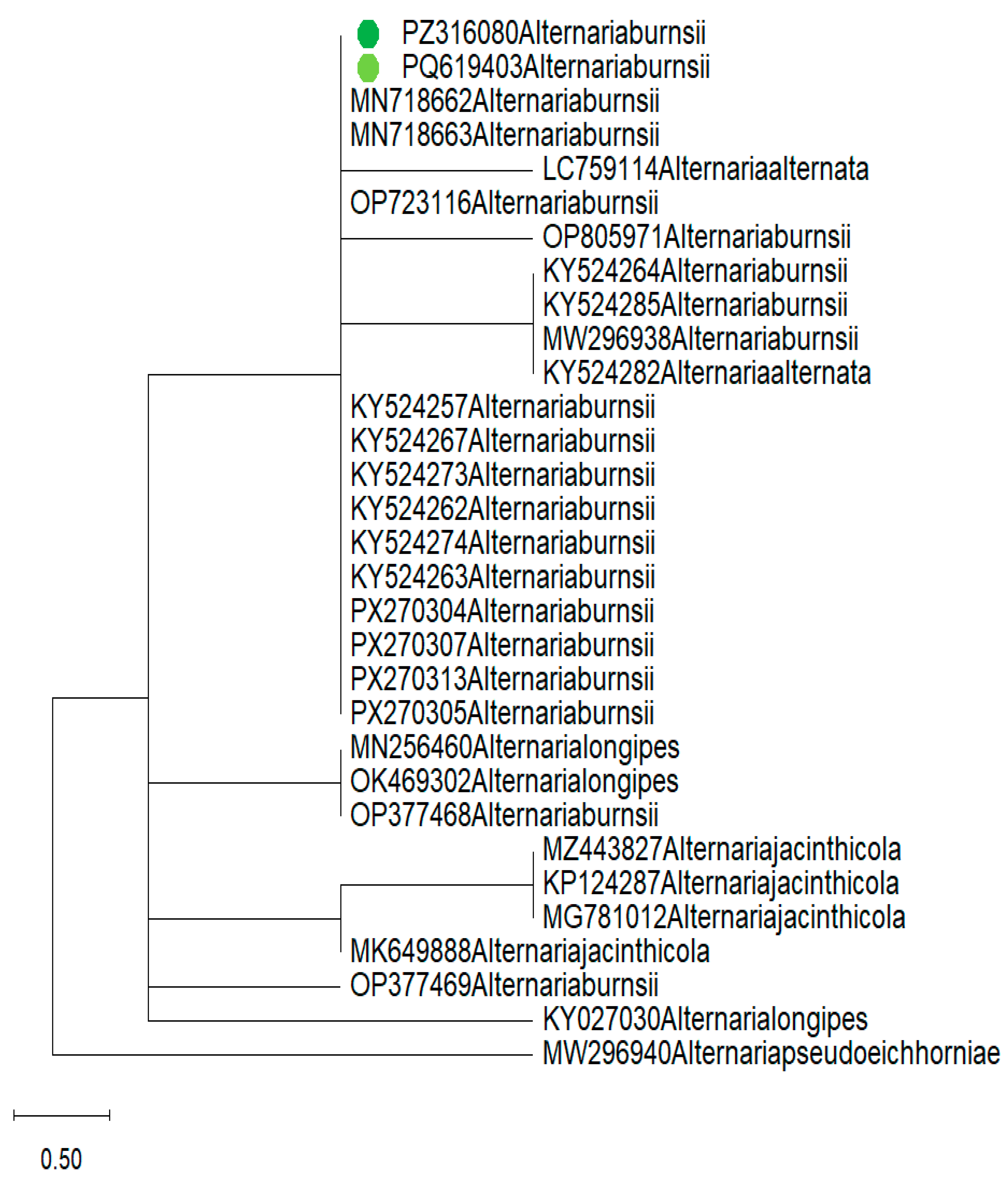
Bidirectional Sanger sequencing yielded high-quality consensus sequences of 590 bp for the ITS region and 500 bp for the GAPDH gene. BLASTn analysis against the NCBI GenBank database revealed 99.48% nucleotide identity between the isolated fungal ITS sequence and 100% nucleotide identity between the isolated fungal GAPDH sequence of A. burnsii (GenBank: OP985911 and PX270313 respectively). The sequence was deposited in GenBank under accession numbers PP782179 (ITS) and PZ316080 (GAPDH). Phylogenetic analysis using a maximum parsimony (1000 bootstrap replicates) tree was constructed as shown in Figure 4 and Figure 5.

3.5. Enzymatic Virulence Factor Assays

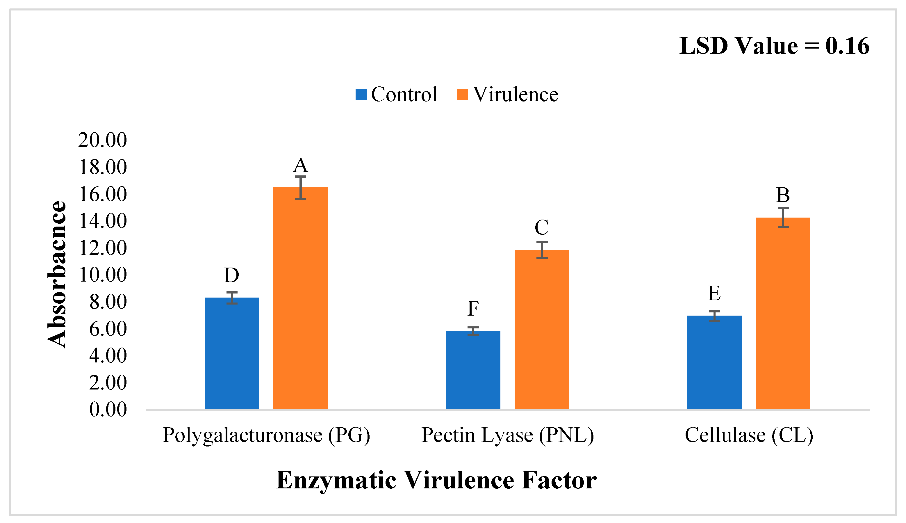
Enzymatic virulence profiling demonstrated significantly elevated production of cell wall-degrading enzymes by A. burnsii compared to controls (Figure 6). Polygalacturonase (PG) activity reached 16.0 ± 0.8 AU, significantly higher than the control (8.0 ± 0.5 AU; p < 0.001). Pectin lyase (PNL) showed 12.0 ± 0.6 AU in virulent cultures versus 6.0 ± 0.4 AU in controls (p < 0.001), while cellulase (CL) activity was 14.0 ± 0.7 AU in the virulence treatment versus 6.5 ± 0.3 AU in controls (p < 0.001). All enzyme activities exceeded control values by >90%, with PG showing the highest relative increase. The LSD value of 0.16 confirmed statistical significance between all treatment-control pairs (different letters denote significant differences, p ≤ 0.05). These results confirm A. burnsii’s capacity for rapid host tissue maceration via synergistic enzymatic action, directly supporting its pathogenicity on spinach.

4. Discussion

Leaf spot disease on spinach (Spinacia oleracea L.) caused by A. burnsii in Pakistan is the first report from South Asia. A. burnsii was obtained from symptomatic leaves with necrotic lesions displaying characteristic dark margins and concentric rings. Identification through morphological, dual-locus molecular characterization (ITS + GAPDH) and phylogenetic analysis confirmed 99.3% and 100% homology, respectively, with reference strains of A. burnsii, placing the isolate firmly within a single A. burnsii species clade. With this finding we expand the known host range of A. burnsii previously known only on tomato, sunflower, cumin and brassicas, but not on spinach. Spinach highlights the pathogen‘s adaptability, and signals a new threat to leafy vegetable production where spinach cultivation is on the increase within arid agroecosystems.
Mechanistically, the isolate has extremely high enzymatic virulence which can explain the aggressiveness of this new pathosystem. Results found significantly enhanced production of cell wall-degrading enzymes, with polygalacturonase (PG) levels exceeding control levels by 100% and correlating tightly to lesion expansion (r = 0.92, p < 0.001). This is aligned with earlier reports by Bhadra (2022) [39] who characterized early pectinolytic activity as a feature of A. burnsii infection. Interestingly, A. burnsii was found to secrete ~1.8× more PG than A. alternata that infect spinach, implicating that increased pectin degradation underlies the timely necrosis that takes place during field outbreaks. This kind of enzymatic power underscores A. burnsii as a pathogen. This outbreak also has an aerobiological dimension, as can be critically made clear by the epidemiological context. Disease occurred mainly in mild (18–25 °C), humid environmental parameters which are well established to support the germination and deposition of Alternaria spores. The synchronous appearance of symptoms within 48 h across geographically dispersed fields (5–15 km apart) implicates airborne conidial dispersal as the primary inoculum source, ruling out localized soil- or seed-borne transmission. This leads to a further inference based on the known aerobiology of Alternaria, which includes its conidia (10–20 µm) being melanized and hydrophobic walls that protect it from UV and desiccation, allowing it to achieve long-distance wind transport. In agricultural production regions, atmospheric concentrations regularly exceed 100 spores m−3 to create a continuous inoculum source from the air over open-field spinach canopy. Compiling this data paints A. burnsii as a two-pronged pathogen, capable of both long-range aerosol movement as a bioaerosol and enzymatically assailing tissue on deposit. This dualism defines its disease philosophy as realized from aerial plant-inoculum, manifesting upon appropriate sub-environments, and giving rise to radial spread through asexual sporulation. Based on this, A. burnsii is a risk in open-field cultivation systems where crops are fully exposed to ambient airflow, as there is no physical barrier preventing the interception of spores.
Therefore, management should be via an integrated aerobiological–enzymatic approach. Taking this into account, it is anticipated that the synergy from the combination of volumetric spore trapping coupled with qPCR monitoring of spore samples will facilitate the generation of predictive forecasts regarding peaks in inoculum, allowing growers the opportunity to apply preventive fungicides prior to deposition. Second, conducting farming operations that reduce canopy humidity like wider row spacing and using systems such as drip irrigation can limit post-deposition germination. Third, targeting pectinolytic enzymes is a sustainable long-term approach. Dispersal modeling of A. burnsii should be an immediate priority for future work, employing real-time aerobiological data, an important step in protecting climate-vulnerable spinach production regions of South Asia and, further, worldwide.

5. Conclusions

For the first time, leaf spot disease caused by A. burnsii on spinach (Spinacia oleracea) was reported in this investigation. Precise confirmation of A. burnsii on spinach was achieved through combined morphological characterization and molecular evidence (ITS + GAPDH) sequencing. According to our findings, this represents the first report of spinach leaf spot caused by A. burnsii in Bahawalpur, Punjab, Pakistan. Given the pathogen’s enzymatic virulence and potential for atmospheric dispersal, it poses a threat to other leafy vegetables and crops in the region. Effective management strategies are needed to control disease spread, including agricultural practices that reduce canopy humidity and potential resistance breeding targeting pectinase inhibition.

Author Contributions

T.A. and R.A.: Methodology, conceptualization, data curation, writing—original draft preparation, software, validation, data curation, writing—review and editing, visualization, and supervision. All authors have read and agreed to the published version of the manuscript.

Funding

This research received no external funding.

Data Availability Statement

The datasets supporting the conclusions of this article are available within the article itself.

Conflicts of Interest

The authors declare no conflict of interest.

References

  1. Ramaiyan, B.; Kour, J.; Nayik, G.A.; Anand, N.; Alam, M.S. Spinach (Spinacia oleracea L.). In Antioxidants in Vegetables and Nuts-Properties and Health Benefits; Springer: Singapore, 2020; pp. 159–173. [Google Scholar]
  2. Ribera, A.; Bai, Y.; Wolters, A.-M.A.; Van Treuren, R.; Kik, C. A review on the genetic resources, domestication and breeding history of spinach (Spinacia oleracea L.). Euphytica 2020, 216, 48. [Google Scholar] [CrossRef]
  3. Al-Kodmany, K. The vertical farm: A review of developments and implications for the vertical city. Buildings 2018, 8, 24. [Google Scholar] [CrossRef]
  4. Golijan Pantović, J.; Gordanić, S. Yield of foreign spinach hybrids in greenhouse production. In Book of Abstracts, Proceedings of the XIII International Symposium on Agricultural Sciences “AgroReS 2024”, Trebinje, Bosnia and Herzegovina, 27–30 May 2024; University of Banja Luka Faculty of Agriculture: Banja Luka, Bosnia and Herzegovina, 2024; p. 88. [Google Scholar]
  5. Bhushan, B.T.; Alam, S.H.; Mahapatra, S.; Chakraborty, S.; Hooi, A. Alternaria. In Compendium of Phytopathogenic Microbes in Agro-Ecology; Springer: Cham, Switzerland, 2025; Volume 1, pp. 1–21. [Google Scholar]
  6. Irfan, M.F.; Shafique, S.; Shafique, S.; Ali, M.A. Characterization of Alternaria alternata Isolated as Leaf Spot Pathogen from Spinacia oleracea L. Int. J. Biol. Biotechnol. 2024, 21, 627–635. [Google Scholar]
  7. Gilardi, G.; Matic, S.; Gullino, M.; Garibaldi, A. First report of Alternaria alternata causing leaf spot on spinach (Spinacia oleracea) in Italy. Plant Dis. 2019, 103, 2133. [Google Scholar] [CrossRef]
  8. Yurchenko, E.; Karpova, D.; Burovinskaya, M.; Vinogradova, S. Leaf spot caused by Alternaria spp. is a new disease of grapevine. Plants 2024, 13, 3335. [Google Scholar] [CrossRef]
  9. Cao, C.; Gong, S.; Li, Y.; Tang, J.; Li, T.; Zhang, Q. Pathogenic factors and mechanisms of the Alternaria leaf spot pathogen in apple. Horticulturae 2024, 10, 212. [Google Scholar] [CrossRef]
  10. Czajka, A.; Czubatka, A.; Sobolewski, J.; Robak, J. First report of Alternaria leaf spot caused by Alternaria alternata on spinach in Poland. Plant Dis. 2015, 99, 729. [Google Scholar] [CrossRef]
  11. Park, J.; Kim, S.; Jo, M.; An, S.; Kim, Y.; Yoon, J.; Jeong, M.-H.; Kim, E.Y.; Choi, J.; Kim, Y. Isolation and identification of Alternaria alternata from potato plants affected by leaf spot disease in Korea: Selection of effective fungicides. J. Fungi 2024, 10, 53. [Google Scholar] [CrossRef]
  12. Moutusi, S.; Buela Parivallal, P.; Prasannakumar, M.; Kiranmayee, P. Morphological and Molecular Characterization of Culturable Leaf Endophytic Fungi from Malabar Spinach: The First Report. Stud. Fungi 2019, 4, 192–204. [Google Scholar] [CrossRef]
  13. Kamthane, D.; Rakh, R. Variations in the conidial dimensions of Alternaria species causing Alternaria blight diseases in various crops. Asian J. Microbiol. Biotechnol. Environ. Sci. 2013, 15, 559–562. [Google Scholar]
  14. Zhang, L.; Zhang, B.; Shao, L.; Yang, M.; Zhao, X.; Wang, Z.; Zhang, Y.; Li, Y.; Wang, Y.; Hu, Y. Genome Analysis of Alternaria alstroemeriae L6 Associated with Black Spot of Strawberry: Secondary Metabolite Biosynthesis and Virulence. J. Fungi 2025, 11, 710. [Google Scholar] [CrossRef]
  15. Kumar, V. Studies on Eco-friendly Management of Alternaria leaf spot of Spinach [Spinacia oleracea L.] Caused by Alternaria alternata Under Protected Cultivation. Master’s Thesis, Rajmata Vijayaraje Scindia Krishi Vishwa Vidyalaya, Gwalior, India, 2022. [Google Scholar]
  16. Guo, C.; Wang, C.; Zhou, T.; Jin, S.; Duan, C. First report of leaf blight caused by Alternaria brassicicola on Orychophragmus violaceus in China. Plant Dis. 2019, 103, 1031. [Google Scholar] [CrossRef]
  17. Lombardi, T.; Bertacchi, A.; Pistelli, L.; Pardossi, A.; Pecchia, S.; Toffanin, A.; Sanmartin, C. Biological and agronomic traits of the main halophytes widespread in the Mediterranean region as potential new vegetable crops. Horticulturae 2022, 8, 195. [Google Scholar] [CrossRef]
  18. Chowdappa, P.; Lakshmi, M.J. Identification of Alternaria species associated with leaf blights of fruit, vegetable and oil seed crops based on protein and multi-locus enzyme finger prints. Pest. Manag. Hortic. Ecosyst. 2013, 19, 45–56. [Google Scholar]
  19. Paul, N.C.; Deng, J.X.; Lee, H.B.; Yu, S.-H. Characterization and pathogenicity of Alternaria burnsii from seeds of Cucurbita maxima (Cucurbitaceae) in Bangladesh. Mycobiology 2015, 43, 384–391. [Google Scholar] [CrossRef]
  20. Ofi, B.; Salih, Y.; Abass, M. First Report of Alternaria burnsii as a Foliar Pathogen of Faba bean in Iraq. Arab J. Plant Prot. 2025, 43, 304. [Google Scholar] [CrossRef]
  21. Maruthanayagam, V. Management of Leaf Spots of Spinach (Spinacia oleracea L.). Master’s Thesis, Vasantrao Naik Marathwada Krishi Vidyapeeth, Parbhani, India, 2021. [Google Scholar]
  22. Samandari-Najafabadi, N.; Taheri, P.; Mamarabadi, M. Identification, pathogenicity and cell wall degrading enzymes of Alternaria spp. associated with grape bunch rot in Iran. Physiol. Mol. Plant Pathol. 2025, 136, 102575. [Google Scholar] [CrossRef]
  23. De Linares, C.; Belmonte, J.; Canela, M.; de la Guardia, C.D.; Alba-Sanchez, F.; Sabariego, S.; Alonso-Pérez, S. Dispersal patterns of Alternaria conidia in Spain. Agric. For. Meteorol. 2010, 150, 1491–1500. [Google Scholar] [CrossRef]
  24. Li, H.; Wang, H.; Ishfaq, S.; Guo, W. First report of the peach leaf spot caused by Nigrospora sphaerica in China. Horticulturae 2024, 10, 1260. [Google Scholar] [CrossRef]
  25. Cao, Y.; Turk, K.; Bibi, N.; Ghafoor, A.; Ahmed, N.; Azmat, M.; Ahmed, R.; Ghani, M.I.; Ahanger, M.A. Nanoparticles as catalysts of agricultural revolution: Enhancing crop tolerance to abiotic stress: A review. Front. Plant Sci. 2025, 15, 1510482. [Google Scholar] [CrossRef]
  26. Ahmed, R.; Raheel, M.; Ali, L.; Ashraf, W.; Aslam, M.N.; Faisal, M.; Anwer, M.; Ikram, M.T.; Afzal, T.; Iqbal, R.; et al. First Report of Leaf Spot of Conocarpus lancifolius Caused by Alternaria burnsii. Pol. J. Environ. Stud. 2025, 34, 6017–6026. [Google Scholar] [CrossRef]
  27. Ikram, M.T.; Aslam, M.N.; Moosa, A.; Zulfiqar, F.; Shakeel, M.T.; Ahmed, R.; Anwer, M.; Khan, A.u.R. First report of Geotrichum candidum causing rose blight disease in Pakistan. J. Plant Pathol. 2023, 105, 1755–1756. [Google Scholar] [CrossRef]
  28. Ahmed, R.; Aslam, M.N.; Moosa, A.; Shakeel, M.T.; Maqsood, A. First report of leaf spot of Aloe vera caused by Curvularia spicifera in Pakistan. J. Plant Pathol. 2023, 105, 1193. [Google Scholar] [CrossRef]
  29. Victoria, A.; Furtado, B.; Holz, M.; Romero-Arenas, O.; Dallagnol, L. First report of leptosphaerulina leaf spot caused by leptosphaerulina trifolii on trifolium repens in brazil. Plant Dis. 2020, 104, 972. [Google Scholar] [CrossRef]
  30. Yang, C.-D.; Yao, Y.-L.; Zhang, Z.-F.; Xue, L. First report of leaf blight of Poa pratensis caused by Peyronellaea glomerata in China. Plant Dis. 2016, 100, 862. [Google Scholar] [CrossRef]
  31. Liu, L.; Ge, Z.; Nie, Y.; Ma, Y.; Liu, G.; Wang, W.; Yu, Y. First Report of Fusarium solani Causing Collar Rot and Dieback on Tea Plants in Shaanxi Province, China. Plant Dis. 2026. [Google Scholar] [CrossRef]
  32. Li, H.; Wang, J.; Su, X.; Cui, J. First report of tip blight of Pinus tabulaeformis caused by Sphaeropsis sapinea in China. Plant Dis. 2016, 100, 1497. [Google Scholar] [CrossRef]
  33. Sharifnabi, B. Pyrenophora lolii, a new species for the mycobiota of Iran. Mycol. Iran. 2019, 6, 113–118. [Google Scholar]
  34. Amaradasa, B.; Amundsen, K. First report of Curvularia inaequalis and Bipolaris spicifera causing leaf blight of buffalograss in Nebraska. Plant Dis. 2014, 98, 279. [Google Scholar] [CrossRef]
  35. Aslam, H.M.U.; Ali, S.; Atiq, M.; Mansha, M.Z.; Aatif, H.M.; Anwaar, H.A.; Naveed, K. First report of brown leaf spot of rice caused by Curvularia spicifera in Pakistan. J. Plant Pathol. 2020, 102, 939–940. [Google Scholar] [CrossRef]
  36. Taheri, P. Disease resistance and virulence screen in Solanum tuberosumAlternaria tenuissima interaction: The role of pathogenicity factors. Euphytica 2019, 215, 15. [Google Scholar] [CrossRef]
  37. Miller, A.R.; Dalmasso, J.P.; Kretchman, D.W. Developmental variation of cell wall degrading enzymes from cucumber (Cucumis sativus) fruit tissues. Can. J. Bot. 1989, 67, 817–821. [Google Scholar] [CrossRef]
  38. Alguacil, M.M.; Hernandez, J.A.; Caravaca, F.; Portillo, B.; Roldan, A. Antioxidant enzyme activities in shoots from three mycorrhizal shrub species afforested in a degraded semi-arid soil. Physiol. Plant. 2003, 118, 562–570. [Google Scholar] [CrossRef]
  39. Bhadra, F.; Gupta, A.; Vasundhara, M.; Reddy, M.S. Endophytic fungi: A potential source of industrial enzyme producers. 3 Biotech 2022, 12, 86. [Google Scholar] [CrossRef] [PubMed]
Figure 1. Morphological characterization of Alternaria burnsii isolated from S. oleracea. (A) Pure culture front side of A. burnsii on PDA; (B) pure culture back side of A. burnsii on PDA; (C) fungal mycelium shown at 4×; (D) visualization of fungal mycelium spores at 10×; (E) magnification of conidia (10 μm scale bar) at 40×; (F) single spore of A. burnsii at 100×.
Aerobiology 04 00011 g001
Figure 2. Pathogenicity test of Alternaria burnsii on spinach. (A) Front side of infected spinach leaf; (B) back side of infected spinach leaf; (C) front side of healthy spinach leaf; (D) back side of healthy spinach leaf; (E) front side of inoculated spinach leaf; (F) back side of inoculated spinach leaf.
Aerobiology 04 00011 g002
Figure 3. (A) Gel electrophoresis of genomic DNA of Alternaria burnsii with a positive control. (B) Amplification of PCR products of ITS region and GAPDH region with 1 kb ladder along with positive control.
Aerobiology 04 00011 g003
Figure 4. Maximum parsimony analysis of Alternaria burnsii from ITS rDNA sequences.
Aerobiology 04 00011 g004
Figure 5. Maximum parsimony analysis of Alternaria burnsii from GAPDH rDNA sequences.
Aerobiology 04 00011 g005
Figure 6. Enzymatic virulence factor production by Alternaria burnsii in comparison to controls. Different letters indicate significant differences between treatments (LSD = 0.16, p ≤ 0.05). Virulence treatments showed significantly higher enzyme activity than controls for all three enzymes (p < 0.001), with PG exhibiting the highest relative increase.
Aerobiology 04 00011 g006
Table 1. Locations and geographic coordinates of surveyed (Spinacia oleracea) fields in District Bahawalpur, Punjab, Pakistan.
Sr. No.LocationDistrictLatitude (N)Longitude (E)
1YazmanBahawalpur29.121671.7443
2Ahmadpur EastBahawalpur29.143771.2599
3Khairpur TamewaliBahawalpur29.581172.2385
4HasilpurBahawalpur29.696672.5550
5Uch SharifBahawalpur29.239471.0583
6Lal SuhanraBahawalpur29.390271.7186
7Khanqah SharifBahawalpur29.318771.6875
8Chak 9 BC (Rural Area)Bahawalpur29.167371.8061
9Chak 15 BCBahawalpur29.205471.8320
10Basti MalookBahawalpur29.395671.6832
Table 2. Primer pairs used for PCR amplification of Alternaria burnsii target genes.
Sr. No.Forward PrimerReverse PrimerReferences
ITS5′-TCCGTAGGTGAACCTGCGG-3′5′-TCCTCCGCTTATTGATATGC-3′[29,30,31,32]
GAPDH5′-CAACGGCTTCGGTCGCATTG-3′5′-GCCAAGCAGTTGGTTGTGC-3′[33,34,35]
Disclaimer/Publisher’s Note: The statements, opinions and data contained in all publications are solely those of the individual author(s) and contributor(s) and not of MDPI and/or the editor(s). MDPI and/or the editor(s) disclaim responsibility for any injury to people or property resulting from any ideas, methods, instructions or products referred to in the content.