1. Introduction
Descending necrotizing mediastinitis represents a severe and life-threatening complication of head and neck infections, posing a major challenge for emergency departments due to its high mortality and rapid progression [
1]. Consequently, an aggressive surgical approach is often favored when this diagnosis is suspected.
Chylothorax is defined as the accumulation of chyle in the pleural space, most often presenting as an exudate [
2,
3]. It results from rupture or obstruction of the intrathoracic lymphatic pathway, which shows numerous anatomical variations and has an embryological origin from the confluence of multiple lymphatic sacs [
4]. The causes of chylothorax may be divided into traumatic and non-traumatic; among non-traumatic causes, oncologic diseases are the most frequent (45%) [
5,
6,
7], including both lymphomatous and solid tumors.
In the assessment of spontaneous mediastinal fluid collection, not related to previous thoracic surgery, necrotizing mediastinitis is the most clinically significant event, and rapid surgical intervention is the cornerstone of treatment and the main prognostic factor [
1]. The purpose of this case presentation is to highlight the possible difficulties in assessing these conditions in an emergency department. We therefore wish to emphasize the importance of being able to promptly recognize misleading diagnosis and to know when to pause and reassess.
2. Case Presentation
A 68-year-old man presented to the emergency department with a rapidly enlarging swelling in the left cervical region that had developed over the previous 24 h. At presentation, the patient reported new onset sialorrhea and dysphagia without history of trauma or cough or vomiting. His medical history included only an anxiety–depressive disorder under symptomatic treatment and ulcerative colitis treated with a monoclonal antibody (Infliximab) and no oncological history.
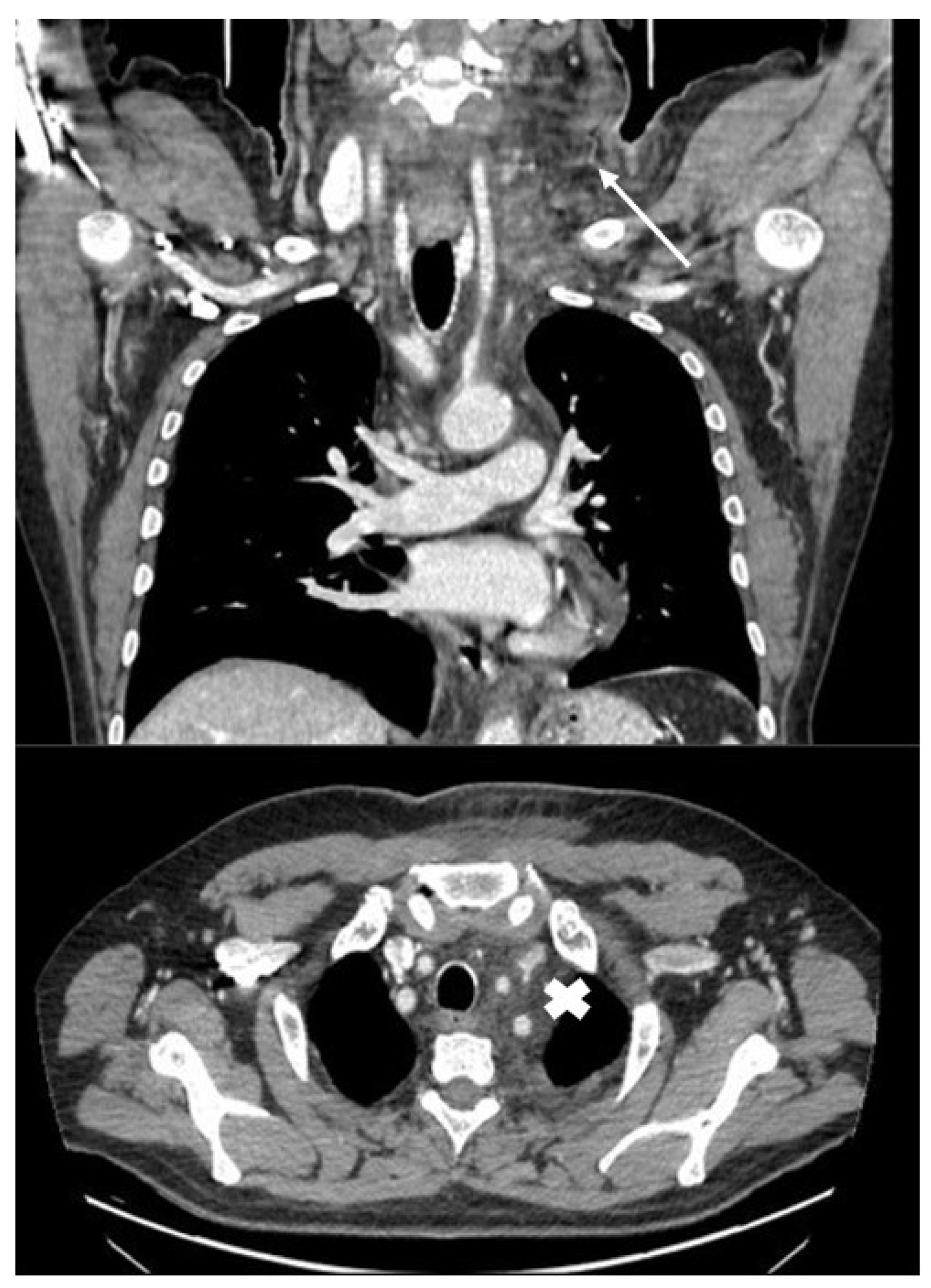
Upon admission, a neck ultrasound revealed a fluid collection with supraliquid characteristics corresponding to the palpable swelling. A subsequent CT scan of the neck and chest confirmed the presence of a fluid collection with soft tissue edema and partial extension into the anterior mediastinum
A loculated collection lateral to the aortic arch was observed, as shown in Figure 1, with no gas collection or pleural thickening; minimal tracheal compression was described.Despite the absence of significant inflammatory markers (CPR 5.6 mg/dL WB 11.08 × 10⁹/L and procalcitonin < 0.1) or fever, a strong clinical suspicion of a tonsillar abscess with early progression to descending mediastinitis prompted the initiation of broad-spectrum antibiotic therapy with meropenem and caspofungin. The patient was evaluated by both thoracic and otorhinolaryngologic surgeons, who recommended surgical drainage and evacuation of the collection through a combined cervical-mediastinal approach.
3. Intra-Operative Findings
A cervicotomy incision was performed to explore the left lateral cervical fascia. Intraoperative findings revealed soft tissue imbibition and a collection of opalescent, milky fluid consistent with chyle under the superficial cervical fascia and in the retropharyngeal region. A total of nearly 350 mL of liquid was collected, and it tested positive for high triglyceride levels (480 mg/dL) Supraclavicular dissection was performed to evaluate the subclavian vessels; no vascular anomalies were identified, and the precise site of lymphatic leakage could not be clearly localized. A left pleural drain was placed and revealed fluid with an identical appearance, consistent with chylothorax, prompting the surgical team to reconsider and modify the therapeutic approach. A cervical drain was positioned, and no mediastinal incision or drainage was performed.
Both cervical and pleural fluids were submitted for culture (both negative) and biochemical analysis, which showed markedly elevated triglyceride levels (525 mg/dL), confirming the diagnosis of chylothorax associated with a chylocervical collection (cervical chyloma).
Total parenteral nutrition (TPN) and a nil per os (NPO) regimen were initiated, followed by a conservative management strategy that led to rapid clinical improvement. Therapy with somatostatin analogues was also started, with subcutaneous octreotide 0.2 mg three times a day. A total-body CT scan was performed to exclude occult intra-abdominal masses, with negative findings, allowing for antibiotic therapy to be discontinued. When thoracic daily output was <250 mL, we gradually reintroduce a low-fat diet without modification of the pleural fluid in quality or quantity. Cervical and thoracic drains were consequently removed on postoperative day 8 and 12.
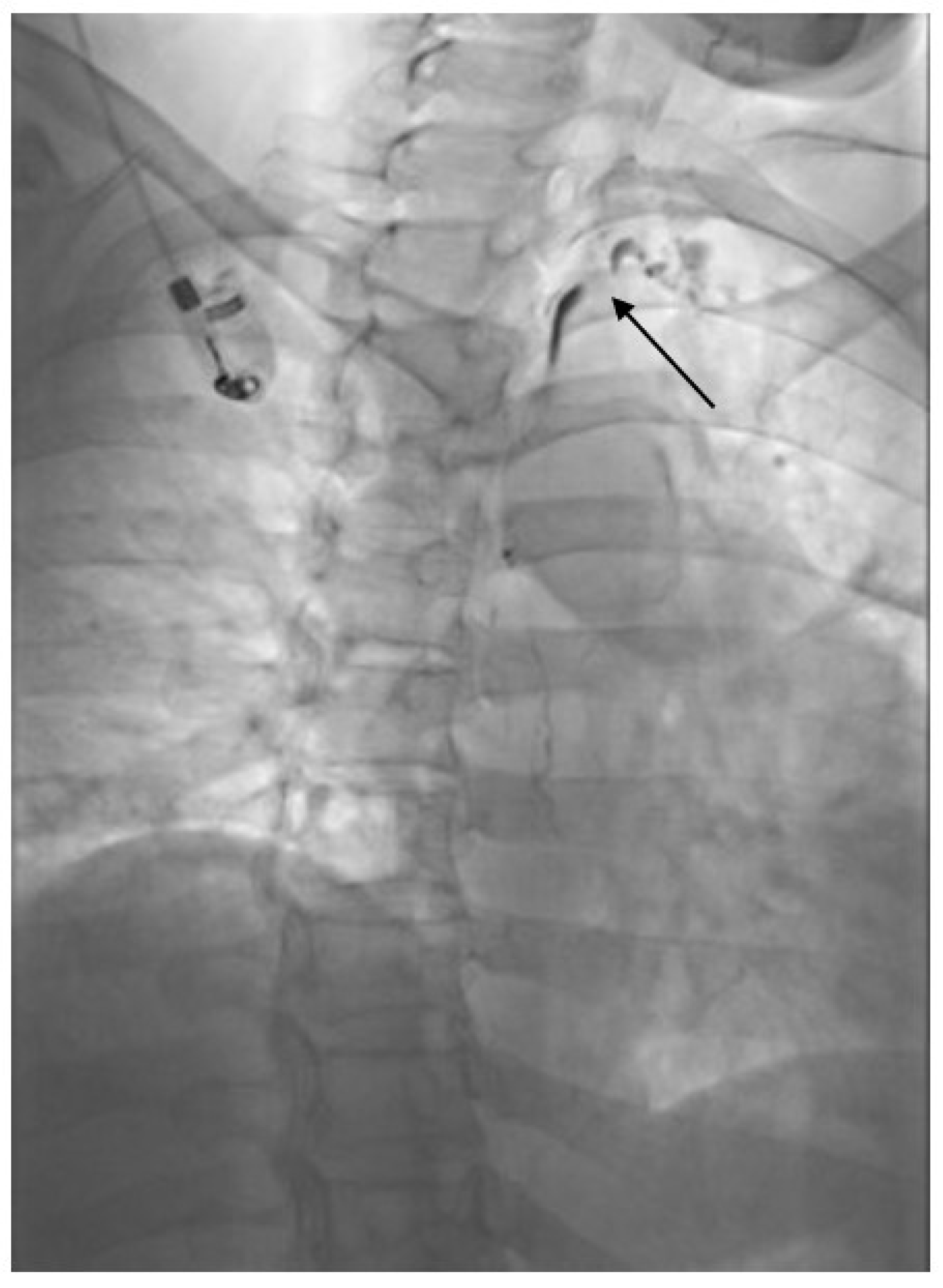
To complete the diagnostic work-up, thoraco-abdominal lymphangiography was performed via bilateral inguinal lymph node puncture with Lipiodol injection (9 mL per side)
before removal of the chest tube. This showed normal opacification of the thoracic and abdominal lymphatic axis up to the left supraclavicular lymph nodes, with no evidence of lymphatic leakage (
Figure 2).
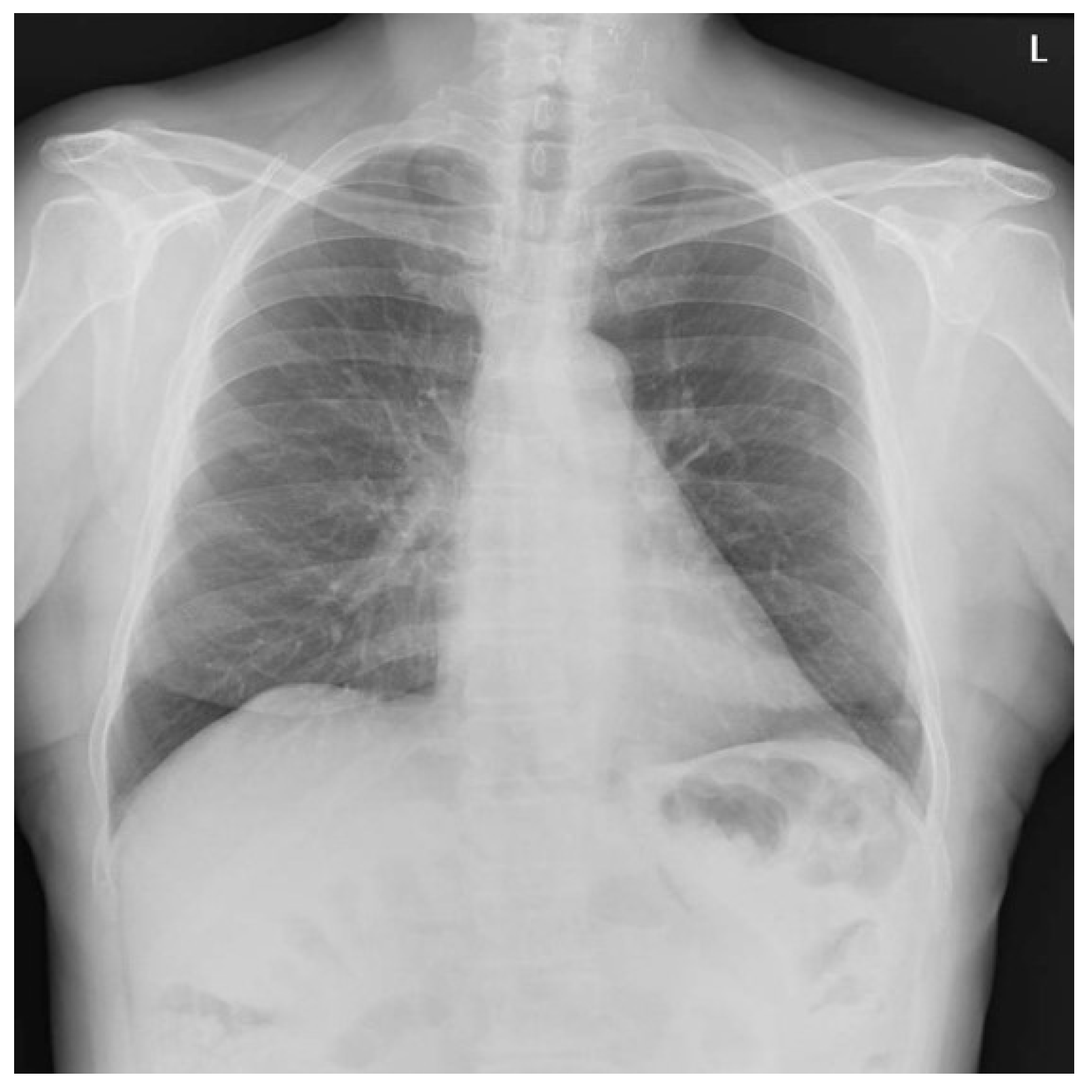
The patient was discharged home in good condition. A chest X-ray performed 20 days later demonstrated complete resolution of the collections, with no recurrence of cervical swelling or new fluid accumulation (
Figure 3).
Three months later, the patient was in excellent general condition, with no signs of regional recurrence in the neck or new intrathoracic collections. He had also resumed a normal diet and completely resumed his daily activities. 4. Discussion
Descending necrotizing mediastinitis is a rapidly progressive and potentially fatal complication of head and neck infections, prompting aggressive surgical management when suspected [
1]. Acute cervical swelling accompanied by dysphagia and sialorrhea, together with imaging evidence of a cervico-mediastinal fluid collection, strongly supports this diagnosis in the emergency setting. However, this case demonstrates how a sterile chylous collection can closely mimic deep neck infection, creating a substantial risk of diagnostic misclassification and unnecessary surgical intervention.
In this patient, dysphagia and sialorrhea were most likely caused by mass effect from a rapidly expanding cervical fluid collection rather than infection. Accumulation of chyle within the deep cervical spaces can compress the pharynx and esophagus, impair swallowing, and interfere with normal saliva clearance, resulting in symptoms classically attributed to deep neck space infection. The rapid onset of symptoms further reinforced the initial assumption of an infectious process, despite the absence of confirmatory systemic signs.
Retrospectively, the lack of fever and leukocytosis represented an important missed red flag. While early deep neck infections may occasionally present without overt systemic inflammation, extensive cervico-mediastinal fluid collections in the absence of fever or inflammatory marker elevation should prompt consideration of noninfectious etiologies, including chylous leakage, hematoma, or seroma [
8,
9,
10]. This case highlights the importance of integrating clinical and laboratory findings with radiological data rather than relying on imaging alone when planning urgent surgical exploration [
11].
The cervical anatomy of the thoracic duct is central to understanding this presentation. After ascending through the thorax, the thoracic duct typically arches laterally at the T1 level in posterior mediastinum, terminating at the lymphovenous junction, most commonly at the confluence of the left internal jugular and subclavian veins. This cervical segment demonstrates marked anatomical variability and structural fragility, predisposing it to spontaneous or minimally traumatic disruption. Injury at this level can result in both cervical chyle accumulation and secondary mediastinal extension [
12,
13].
The distribution of chyle in this case suggests that the leak most likely originated along the cervical arch of the thoracic duct rather than exclusively at the lymphovenous junction. A defect at the arch would allow chyle to dissect superiorly into cervical fascial planes and inferiorly into the mediastinum, accounting for the combined cervical swelling and chylothorax observed. In contrast, isolated disruption at the lymphovenous junction would be expected to preferentially decompress into the venous system, making extensive cervical and mediastinal accumulation less likely.
Spontaneous chylothorax is exceedingly rare, and idiopathic cases account for only a small proportion of non-traumatic presentations [
2,
5]. Presentation with acute cervical swelling in the absence of malignancy or prior surgery has not, to our knowledge, been previously reported. The rapid progression observed effectively excludes congenital lymphatic malformations, while the absence of lymphadenopathy or solid tumors argues against malignant obstruction. Micro-rupture of the thoracic duct related to minor mechanical stress or underlying anatomical variants remains the most plausible mechanism.
5. Conclusions
This case underscores how presumed mediastinal infection can drive extensive, time-critical surgical exploration, potentially exposing patients with alternative pathologies to avoidable risk. Continuous reassessment of intraoperative findings is essential, particularly when they conflict with the preoperative diagnosis. Although rare, chylous leakage should be included in the differential diagnosis of acute cervical-mediastinal fluid collections, especially when systemic signs of infection are absent. Extensive head and neck examination, even with laryngoscopy, is always recommended before deciding to proceed with surgical intervention [
14].
Author Contributions
Conceptualization, G.G. and L.V.; methodology, L.V.; investigation, G.G.; writing—original draft preparation, G.G.; writing—review and editing, I.L.; supervision, A.D. All authors have read and agreed to the published version of the manuscript.
Funding
This research received no external funding.
Institutional Review Board Statement
Ethical review and approval were waived for this study as they are not required in this type of study according to local legislation (DGR XII/1292 2023).
Informed Consent Statement
Written informed consent has been obtained from the patient(s) to publish this paper.
Data Availability Statement
No new data were created or analyzed in this study. Data sharing is not applicable to this article.
Conflicts of Interest
The authors declare no conflicts of interest.
References
- Prado-Calleros, H.M.; Jiménez-Fuentes, E.; Jiménez-Escobar, I. Descending necrotizing mediastinitis: Systematic review on its treatment in the last 6 years, 75 years after its description. Head Neck 2016, 38, E2275–E2283. [Google Scholar] [CrossRef] [Scilit] [PubMed]
- Maldonado, F.; Hawkins, F.J.; Daniels, C.E.; Doerr, C.H.; Decker, P.A.; Ryu, J.H. Pleural fluid characteristics of chylothorax. Mayo Clin. Proc. 2009, 84, 129–133. [Google Scholar] [CrossRef] [Scilit] [PubMed]
- Agrawal, V.; Doelken, P.; Sahn, S.A. Pleural fluid analysis in chylous pleural effusion. Chest 2008, 133, 1436–1441. [Google Scholar] [CrossRef] [Scilit] [PubMed]
- Doerr, C.H.; Allen, M.S.; Nichols, F.C., 3rd; Ryu, J.H. Etiology of chylothorax in 203 patients. Mayo Clin. Proc. 2005, 80, 867–870. [Google Scholar] [CrossRef] [Scilit] [PubMed]
- Valentine, V.G.; Raffin, T.A. The management of chylothorax. Chest 1992, 102, 586–591. [Google Scholar] [CrossRef] [Scilit] [PubMed]
- Williams, K.R.; Burford, T.H. The Management of Chylothorax. Ann. Surg. 1964, 160, 131–140. [Google Scholar] [CrossRef] [Scilit] [PubMed]
- Robinson, C.L. The management of chylothorax. Ann. Thorac. Surg. 1985, 39, 90–95. [Google Scholar] [CrossRef] [Scilit] [PubMed]
- García-Aparicio, J.; Herrero-Herrero, J.-I.; Corral-Gudino, L.; Jorge-Sánchez, R.J. Bilateral idiopathic chylothorax associated with light physical exercise. Respir. Med. CME 2009, 2, 68–69. [Google Scholar] [CrossRef] [Scilit]
- Candas, F.; Yildizhan, A.; Gorur, R.; Isitmangil, T. Is bilateral chylothorax possible after simple cough? Yes. Asian Cardiovasc. Thorac. Ann. 2015, 23, 471–473. [Google Scholar] [CrossRef] [Scilit] [PubMed]
- Reilly, K.M.; Tsou, E. Bilateral chylothorax. A case report following episodes of stretching. JAMA 1975, 233, 536–537. [Google Scholar] [CrossRef] [Scilit] [PubMed]
- McGrath, E.E.; Blades, Z.; Anderson, P.B. Chylothorax: Aetiology, diagnosis and therapeutic options. Respir. Med. 2010, 104, 1–8. [Google Scholar] [CrossRef] [Scilit] [PubMed]
- Matsumoto, T.; Yamagami, T.; Kato, T.; Hirota, T.; Yoshimatsu, R.; Masunami, T.; Nishimura, T. The effectiveness of lymphangiography as a treatment method for various chyle leakages. Br. J. Radiol. 2009, 82, 286–290. [Google Scholar] [CrossRef] [Scilit] [PubMed]
- Cope, C.; Salem, R.; Kaiser, L.R. Management of chylothorax by percutaneous catheterization and embolization of the thoracic duct: Prospective trial. J. Vasc. Interv. Radiol. 1999, 10, 1248–1254. [Google Scholar] [CrossRef] [Scilit] [PubMed]
- Boffa, D.J.; Sands, M.J.; Rice, T.W.; Murthy, S.C.; Mason, D.P.; Geisinger, M.A.; Blackstone, E.H. A critical evaluation of a percutaneous diagnostic and treatment strategy for chylothorax after thoracic surgery. Eur. J. Cardiothorac. Surg. 2008, 33, 435–439. [Google Scholar] [CrossRef] [Scilit] [PubMed]
| Disclaimer/Publisher’s Note: The statements, opinions and data contained in all publications are solely those of the individual author(s) and contributor(s) and not of MDPI and/or the editor(s). MDPI and/or the editor(s) disclaim responsibility for any injury to people or property resulting from any ideas, methods, instructions or products referred to in the content. |