Review

Fulminant Necrotizing Soft Tissue Infection Following Abdominal Liposuction: Comprehensive Literature Review and Case Report

by
Claudia Viviana Jaimes Gonzalez
1,*,
Joan Sebastian Barrera Guaca
1,
Maria Angela Gomez Martinez
2,
Felipe Caballero Paz
2 and
Luis Fernando Alvarez Molina
3
1
Department of Surgery, Clínica de Marly, Bogotá 110231, Colombia
2
Department of Plastic Surgery, Clínica de Marly, Bogotá 110231, Colombia
3
Department of Anesthesiology, Clínica de Marly, Bogotá 110231, Colombia
*
Author to whom correspondence should be addressed.
Complications 2025, 2(3), 23; https://doi.org/10.3390/complications2030023
Submission received: 9 July 2025 / Revised: 27 August 2025 / Accepted: 9 September 2025 / Published: 11 September 2025

Abstract

Necrotizing soft tissue infection (NSTI) is a rapidly progressive, life-threatening soft tissue infection that involves the skin, subcutaneous tissue, and fascia, with a natural evolution to septic shock and death if not treated. NSTI is typically associated with trauma, and rarely reported as a complication of aesthetic procedures such as liposuction. We report the case of a previously healthy 34-year-old woman who developed fulminant necrotizing soft tissue infection shortly after undergoing abdominal liposuction in a suspected non-accredited facility. She arrived at our institution with severe thoracoabdominal pain, ecchymosis, and refractory shock within hours postoperatively. Imaging studies performed in the emergency department revealed gas in the soft tissues, confirming the diagnosis. Emergent surgical debridement exposed extensive necrosis affecting a large body surface area. The patient required multiple surgeries, intensive care support, and broad-spectrum antibiotics to achieve infection control. Reconstructive treatment was initiated once stabilization was achieved, leading to full wound closure and a favorable clinical outcome. In addition, we provide a comprehensive review of the current literature regarding necrotizing soft tissue infection, emphasizing this condition as a postoperative complication, including its epidemiology, microbial etiology, pathophysiology, diagnostic challenges, treatment strategies, and reported cases. This review aims to contextualize this rare but severe postoperative complication and to guide clinicians in its early recognition and management, while also raising awareness about the potential consequences of aesthetic procedures performed in unregulated settings.

1. Introduction

Liposuction is one of the most widely performed cosmetic procedures worldwide [1]. When carried out under appropriate conditions by trained professionals, it is generally considered safe, with complication rates as low as 2.62% in recent meta-analyses. The most prevalent complications reported, in order of frequency, are contour deformity (2.35%), hyperpigmentation (1.49%), seroma (0.65%), and hematoma (0.27%) [2]. However, similar to any surgical procedure, liposuction carries inherent risks that, although infrequent, can be serious and even fatal. Among the serious postoperative complications described for liposuction are skin necrosis, infection, venous thromboembolism, and local anesthesia toxicity—events that, while potentially life-threatening, fortunately remain rare, with prevalences consistently reported below 0.05% in the literature [2,3]. These risks are further compounded by the fact that cosmetic procedures are sometimes performed by healthcare professionals without surgical specialties and even by non-medical personnel in outpatient settings.
Nevertheless, rare but life-threatening complications have been reported, including necrotizing soft tissue infection (NSTI). Necrotizing soft tissue infection following liposuction is a rarely described complication, with only a few cases documented in the literature due to its very low incidence. In a review of postoperative infections in cosmetic surgery, Gravante et al. reported seven isolated case reports of NSTI after liposuction and noted that, in a series of 1047 cases, no such complication was observed [4]. This infection, which involves the fascia and soft tissues, is in most cases polymicrobial, in 70 to 80% of cases. Monomicrobial cases are usually caused by Streptococcus and Staphylococcus, and to a lesser extent by Clostridium [5,6]. It is a rapidly progressive infection and therefore potentially fatal; therefore, early diagnosis and prompt management are key to improving the prognosis in terms of both morbidity and mortality [6].
In this context, NSTI represents a medical emergency that requires immediate surgical management, in order to control the infectious process by reducing bacterial load and debriding necrotic tissue secondary to the infection. Taking the above into account, we present the case of a young, previously healthy patient who developed fulminant necrotizing soft tissue infection following abdominal liposuction in a suspected non-accredited setting; we also provide a comprehensive review of the literature, aiming to contextualize this rare but severe complication of aesthetic surgery and to assist clinicians in its early recognition and appropriate management.

2. Case Report

A 34-year-old previously healthy female patient was admitted to the emergency department of our tertiary care hospital in critical condition, with severe deterioration of her general condition and persistent hemodynamic instability. She reported having undergone abdominal liposuction performed at a cosmetic clinic only a few hours before the onset of symptoms. She subsequently developed intense thoracoabdominal pain associated with a syncopal episode. She was initially evaluated at a walk-in clinic and later at a public hospital, where severe anemia was documented; however, according to the patient, no additional treatment was provided. Due to clinical deterioration and by personal decision, she requested voluntary discharge and presented to our institution.
On admission, the patient exhibited marked pallor, extensive abdominal ecchymosis, and intense pain. Initial differential diagnoses included vascular injury and pulmonary thromboembolism. Fluid resuscitation and blood transfusion were initiated; nevertheless, clinical deterioration persisted, with development of refractory shock despite treatment.
Given the initial suspicion of pulmonary thromboembolism and the simultaneous evaluation of the cutaneous lesions (Figure 1), together with paraclinical findings—complete blood count without leukocytosis but with 82% polymorphonuclear predominance, anemia with hemoglobin of 7.4 g/dL, elevated C-reactive protein (44.44 mg/dL), creatinine 1.64 mg/dL, glucose 218 mg/dL, and sodium 134 mmol/L—an LRINEC (Laboratory Risk Indicator for Necrotizing Fasciitis) score of 11 was calculated, corresponding to the high-risk category for NSTI.
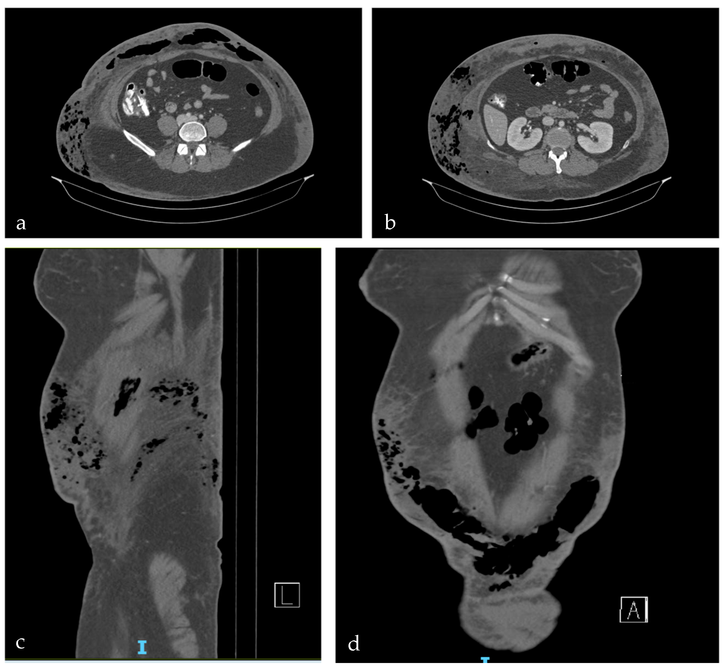
An abdominal CT angiography was requested to rule out pulmonary thromboembolism and evaluate the possibility of NSTI. The study revealed the presence of gas in soft tissues, edema and fluid collections (Figure 2). By integrating the rapid clinical deterioration, the cutaneous manifestations, and the imaging and laboratory findings, a diagnosis of rapidly progressive NSTI was established.
The patient was taken for urgent surgical debridement, which revealed extensive necrosis, gangrene, and foul-smelling fluid in deep tissues, involving full thickness of the skin, subcutaneous cellular tissue, and fascia. No tracts or perforating injuries of the abdominal wall were observed that could suggest penetrating trauma by cannulas. Approximately 25–29% of total body surface area was debrided at that time, and multiple samples were obtained for Gram stain, cultures, and histopathology. Chlorhexidine-impregnated gauze dressings were applied, and she was transferred to the intensive care unit (ICU) due to high postoperative mortality risk.
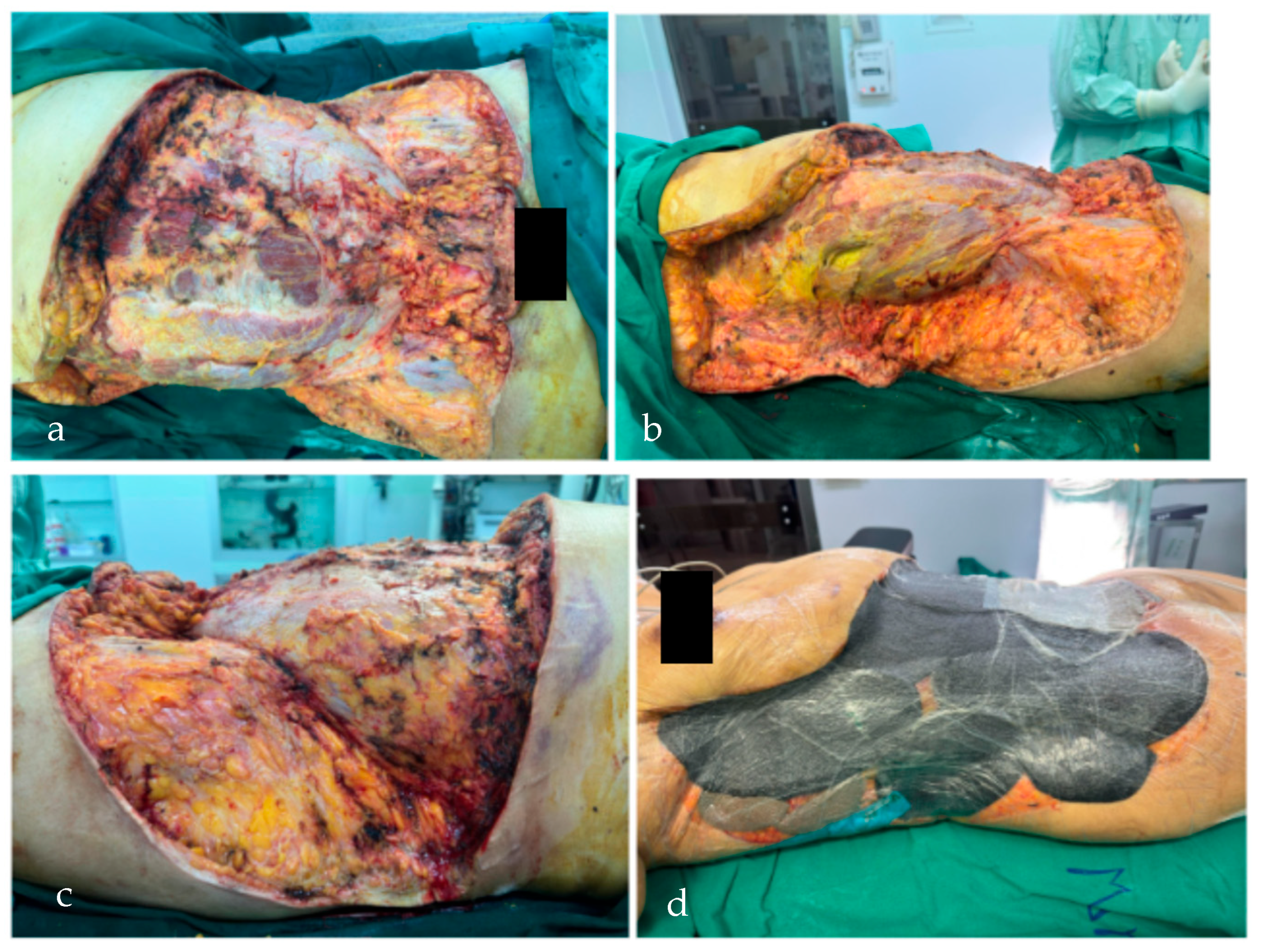
In the ICU, she remained in critical condition with severe hemodynamic instability, requiring multiple vasopressors, invasive mechanical ventilation, and presenting multiorgan dysfunction (hepatic, renal, and coagulopathy). At 24 h postoperative, severe sepsis persisted, attributed to NSTI, despite antibiotic escalation to meropenem and linezolid as recommended by the infectious diseases team. A second surgical procedure was therefore performed for infection control, consisting of additional debridement (20–30% non-excisional, <10% excisional) and placement of negative pressure wound therapy (Figure 3).
Histopathological analysis of the samples obtained during the first and second procedures confirmed NSTI, specifically classified as necrotizing fasciitis (NF). Initial microbiological evaluation showed Gram-negative bacilli (+) and Gram-positive bacilli (++). Subsequently, cultures reported scant isolation of Niallia circulans in liquid, muscle, and fascia samples from the abdominal wall. It was not possible to establish a complete antimicrobial resistance profile due to technical limitations in the laboratory for this microorganism. Considering the clinical severity and microbiological findings, along with the risk of a polymicrobial process, and in light of reports in the literature describing Niallia circulans as a multidrug-resistant organism, the infectious diseases team recommended continuation of broad-spectrum antibiotic coverage, which was instituted.
Although the precise source of the infection could not be definitively established, the multidisciplinary management team supported the suspicion that contamination during the liposuction procedure, suspected to have been performed in a non-accredited or inadequate facility, was the most probable trigger of the necrotizing soft tissue infection. This suspicion was reinforced by the fact that Niallia circulans is primarily found in soil, but has also been identified in sewage and food, which suggested a probable environmental origin of contamination in this case.
With serial surgical management and comprehensive supportive care, partial clinical improvement was achieved, with decreased leukocytosis and improved metabolic response, suggesting containment of the infectious process. Subsequent debridements and washouts revealed viable margins, without residual gas or muscle necrosis.
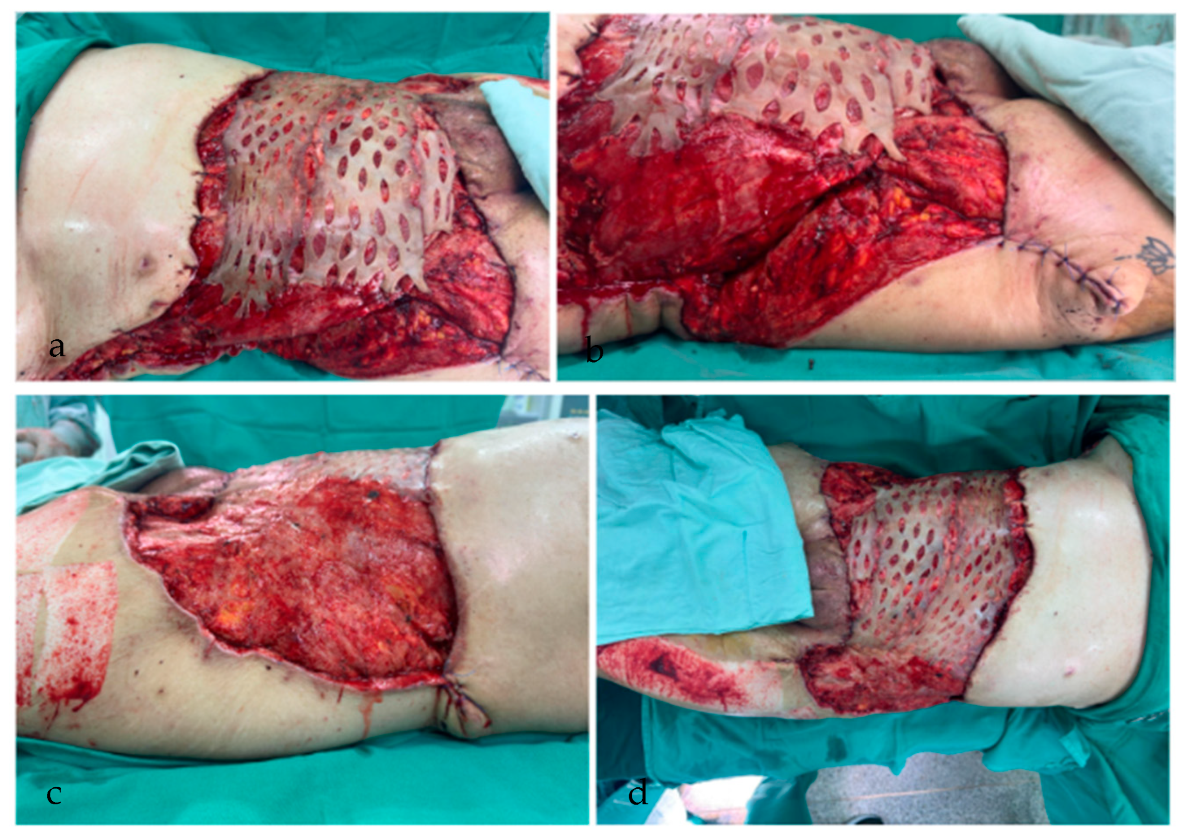
In total, four surgical procedures were required to achieve infection control, including serial debridements, surgical lavage, and exchanges of the negative pressure system. The fifth procedure was performed by a multidisciplinary team of general and plastic surgeons. At that time, an open wound involving 26% of the body surface was observed, with muscle exposure and healthy granulation tissue. In view of the coverage defect and persistence of febrile spikes, interpreted as a metabolic response to trauma and a partially controlled infectious process, a staged closure strategy was initiated. During the first reconstructive stage, with the objective of minimizing intraoperative bleeding, midline coverage was performed with split-thickness skin grafts and advancement flaps from the thigh, along with reconstruction of the thoracic and right breast regions (Figure 4), followed by reapplication of negative pressure therapy.
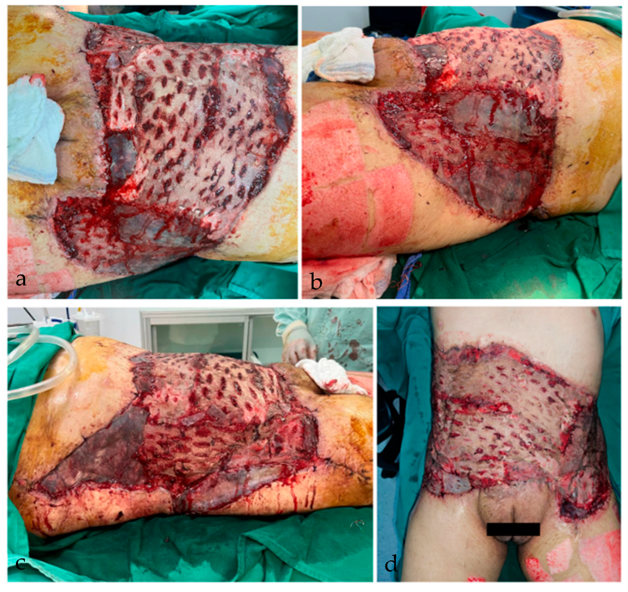
Subsequently, the patient evolved favorably during hospitalization in the general ward, remaining afebrile, hemodynamically stable, and without evidence of active infection or new complications. Laboratory tests showed normalization of inflammatory markers, preserved renal function, and negative blood and wound cultures. She completed a 14-day course of antibiotic therapy with meropenem and linezolid, with good clinical response. She was also evaluated by nutrition, speech therapy, psychiatry, and rehabilitation teams for comprehensive management. The reconstructive protocol continued with the plastic surgery team, including wound washouts, split-thickness skin grafts on the flanks during the second reconstructive stage, and two additional procedures for grafting of residual raw areas in the hypochondria and iliac fossae, ultimately achieving complete wound closure (Figure 5).
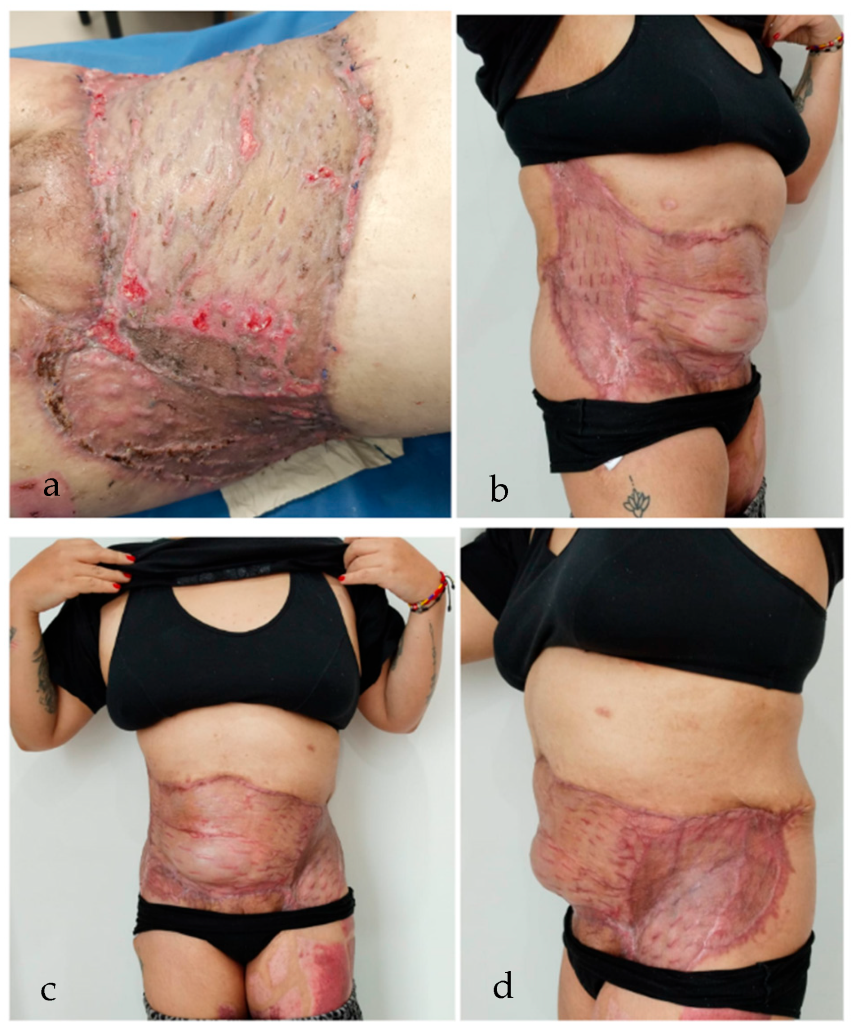
Once surgical coverage was complete and the patient remained clinically stable, she was discharged with outpatient wound clinic follow-up, achieving appropriate graft integration and coverage of the extensive defect resulting from NSTI (Figure 6).
In accordance with public health local regulations, once the case was admitted to our institution, it was reported to “Secretaria Distrital de Salud de Bogotá” the regulatory body for public health in the region, as required in cases of complications derived from cosmetic surgical procedures, to ensure monitoring of the institution and the professionals involved in the initial intervention.

3. Postoperative Necrotizing Soft Tissue Infection

3.1. Epidemiology and Risk Factors

Although NSTI incidence remains low, recent epidemiological data show a growing trend in reported cases. In a large longitudinal cohort study conducted in England over 16 years, Bodansky et al. reported a significant increase in age-standardized incidence from 9 per million in 2002 to 21 per million in 2017, particularly affecting older males and individuals from socioeconomically deprived backgrounds [7]. Despite this rise in incidence, the in-hospital mortality rate remained at approximately 16%, showing the persistent challenges in early recognition and effective intervention. Additionally, nearly 27% of cases had diabetes mellitus, although diabetes alone was not independently associated with higher mortality, suggesting the contribution of multiple interacting comorbidities [7].
In the context of postoperative settings, NSTI represents a particularly devastating complication. Miller et al. identified a distinct subgroup of patients who developed NSTI following surgical or invasive procedures, drawing attention to the absence of typical early signs, and with variable onset ranging from days to months in the postoperatory phase [8]. Most of these cases required multiple surgical debridements and demonstrated high mortality, especially those with underlying immunosuppression or morbid obesity. Lehnhardt et al. specifically highlighted the occurrence of fatal postoperative necrotizing soft tissue infection (PNSTI) after cosmetic liposuction in outpatient clinics lacking adequate infection control standards, reporting 14 cases of NF among 72 severe complications, with nine deaths attributed to delayed diagnosis and inadequate postoperative monitoring [9]. Furthermore, Gaston and Kuremsky emphasized the role of modifiable preoperative factors such as malnutrition, uncontrolled diabetes, MRSA colonization, and smoking, which are all related to compromised host immune response and wound healing capacity [10]. These findings support the multifactorial nature of PNSTI risk and the critical need for stringent perioperative assessment, infection control, and early surgical consultation in high-risk patients.

3.2. Microbial Etiology and Pathophysiology

Microbiologically, PNSTI can be classified using the same taxonomy applied to general necrotizing soft tissue infections, most commonly as type I (polymicrobial) or type II (monomicrobial) infections [6,11]. Type I, the most frequent form, involves synergistic combinations of facultative and obligate anaerobic organisms, including Escherichia coli, Bacteroides spp., Enterococcus spp., Streptococcus spp., and Peptostreptococcus spp. [6]. These bacteria often present as endogenous flora—skin, gastrointestinal, or genitourinary—that are introduced during surgery, especially when a strict aseptic technique is compromised or when the operating area has heavy colonization, such as the perineum or the lower abdomen. The synergistic effect of these organisms leads to rapid tissue destruction through enzymatic degradation, toxin release, and impairment of neutrophil function, particularly in immunocompromised or diabetic individuals [12].
In contrast, type II PNSTI, is typically monomicrobial, most commonly associated with Streptococcus pyogenes (Group A Streptococcus, GAS), and less frequently with Staphylococcus aureus (including MRSA); these organisms are capable of producing severe infections in healthy individuals. Postoperative GAS infections tend to occur through either direct contamination of surgical sites or via hematogenous dissemination from distant foci, such as pharyngeal colonization [6,11]. Experimental studies have shown that S. pyogenes can bind to vimentin expressed on injured skeletal muscle cells, allowing bacterial trafficking to surgical wounds during transient bacteremia, even in the absence of a visible entry [6]. Once present in the deep tissue, these bacteria produce exotoxins—such as streptolysin O and pyrogenic exotoxins—that trigger massive cytokine release, endothelial damage, platelet-leukocyte aggregation, and vascular thrombosis. The result is a cascade of ischemia, necrosis, and systemic inflammatory response syndrome (SIRS), often culminating in septic shock.
Regardless of the microbial origin, the pathophysiological hallmark of PNSTI is the rapid spread of infection along fascial planes, which is facilitated by poor vascularity, low local immune surveillance, and the absence of anatomical barriers. In the early disease course, the overlying skin may appear normal or only mildly erythematous, while deep tissue necrosis and microvascular thrombosis already progress beneath the surface [11]. This anatomical dissociation explains why clinical signs lag behind pathologic damage, and why pain out of proportion to findings is often the earliest—and most important—clinical clue. Surgical trauma itself, especially in procedures involving large tissue undermining (e.g., liposuction, abdominoplasty), creates potential spaces for bacterial proliferation, while devitalized tissues serve as a fertile substrate for anaerobic growth.
Immunosuppression, diabetes, malnutrition, and recent NSAID or corticosteroid consumption are frequently identified as co-factors that impair host defenses. In particular, the use of NSAIDs has been associated with accelerated disease progression in GAS infections, both by masking early symptoms and by facilitating bacterial adhesion and toxin-induced immunosuppression [6]. Moreover, postoperative patients are at risk of diagnostic delay due to the attribution of early symptoms—such as pain, edema, or mild fever—to normal postoperative inflammation, rather than infection. This delay in recognition is particularly dangerous, as the time to initial surgical debridement remains the most critical prognostic factor.
Finally, while Clostridium perfringens and other clostridial species are more commonly associated with traumatic gas gangrene, they have also been implicated in PNSTI, especially in the setting of bowel surgery, gynecologic instrumentation, or injection-related injuries. These organisms thrive in anaerobic, devitalized tissue and produce alpha toxins that lead to myonecrosis and systemic toxicity. In such cases, gas formation, palpable crepitus, and rapidly advancing shock are typical, although early findings may still be subtle [11].

3.3. Diagnostic Challenges

Postoperative necrotizing soft tissue infection arises from a complex synergy between microbial virulence, surgical disruption of tissue integrity, and host immunological vulnerability. Given its frequently cryptic presentation and catastrophic potential, a high index of suspicion is essential, especially in patients with disproportionate pain, systemic signs of deterioration, or known risk factors.
Diagnosing PNSTI remains one of the greatest challenges in surgical and emergency medicine due to its variable presentation, rapid progression, and overlap with benign postoperative findings. In contrast to typical surgical site infections, which often follow a predictable and localized course, PNSTI frequently presents with non-specific symptoms such as disproportionate pain, edema, or mild erythema, particularly during the first 48–72 h post-procedure. This subtle presentation leads to high rates of initial misdiagnosis, with studies reporting incorrect or delayed diagnosis in up to 96% of NSTI cases, even in tertiary centers [8]. Pain out of proportion to physical findings is a critical early clinical clue, but it may be masked in patients receiving NSAIDs, opioids, or corticosteroids—commonly used in the postoperative setting [11,12].
The diagnostic process is further complicated by the overlap between early PNSTI and expected postoperative inflammation, such as erythema, swelling, and low-grade fever. Classic findings such as crepitus, skin necrosis, bullae, or gas in tissues are absent in more than 50% of early presentations [12,13]. In abdominal or aesthetic surgery, the risk of delayed recognition is magnified by the perception of liposuction and abdominoplasty as low-risk procedures, often performed in outpatient settings without robust postoperative monitoring. Imaging may support the diagnosis, particularly CT scans showing fascial thickening, subcutaneous emphysema, or fluid tracking along fascial planes, but imaging should not delay surgical exploration if clinical suspicion is high [13,14].
Scoring systems like the LRINEC score have been proposed to aid diagnosis. However, sensitivity ranges from 49% to 75%, limiting their standalone utility, especially in early stages [13]. A recently developed tool, the NECROSIS score, incorporates laboratory and clinical data, improving specificity, but this score has not yet been widely validated in postoperative cohorts [8]. Ultimately, clinical judgment and early surgical consultation remain the cornerstones of diagnosis. Operative exploration is both diagnostic and therapeutic; in fact, the presence of gray, friable fascia and absence of purulence are hallmark intraoperative findings that confirm NSTI [11,15]. Given the potential for rapid deterioration, surgical exploration should not be delayed by inconclusive labs or imaging when suspicion is moderate to high.

3.4. Treatment Strategies

Rapid surgical exploration and broad-spectrum empiric antimicrobial therapy remain the pillars of management, regardless of the eventual microbial classification. The cornerstone of treatment for PNSTI is early and aggressive surgical debridement, which remains the most significant determinant of survival. Time to first debridement is inversely correlated with mortality, with delays beyond 24 h significantly increasing the risk of multiorgan failure and death [13]. Surgical exploration should be performed urgently when PNSTI is suspected, even in the absence of definitive imaging or laboratory confirmation. Intraoperatively, findings such as gray necrotic fascia, lack of bleeding, muscle non-contractility, and “dishwater” fluid confirm the diagnosis [11]. Repeat debridements are often necessary every 24–48 h until viable, bleeding tissue is achieved [13].
Empiric broad-spectrum antibiotic therapy should be initiated promptly and later tailored based on culture results. Standard regimens include a carbapenem or beta-lactam/beta-lactamase inhibitor combination (e.g., piperacillin–tazobactam), plus clindamycin (for inhibition of bacterial toxin production), and vancomycin or linezolid to cover MRSA [12,13]. In polymicrobial infections (type I), this regimen provides effective coverage against Gram-positive, Gram-negative, and anaerobic organisms. In monomicrobial Streptococcus pyogenes cases (type II), the addition of clindamycin is particularly critical due to its ability to inhibit toxin production independent of bacterial growth phase (6). In immunocompromised or nosocomial contexts, antifungal agents may be considered if mucormycosis or candidiasis is suspected [12].
Adjunctive therapies, while not definitive, may offer benefits in select cases. Hyperbaric oxygen therapy (HBOT) has been shown to enhance oxygen delivery to hypoxic tissues, inhibit anaerobic bacterial growth, and stimulate angiogenesis, although randomized controlled trial data remain limited [16]. Negative pressure wound therapy (NPWT) is frequently employed in the reconstructive phase, as it promotes granulation tissue formation and facilitates wound bed preparation for closure or grafting [16]. A well-documented case by Chiang et al. illustrated the successful use of HBOT and NPWT in managing PNSTI following abdominal liposuction in a patient with complex burn-related scarring, achieving full recovery without major graft loss [16].
Post-debridement wound management requires an individualized approach, often involving multidisciplinary collaboration between surgery, critical care, infectious disease, and plastic/reconstructive teams. Early planning for reconstructive procedures is essential, particularly in extensive abdominal or limb defects. Gaston and Kuremsky emphasize that nutritional optimization, glycemic control, and mitigation of immunosuppressive medications are crucial during both the acute and recovery phases, as these factors significantly influence wound healing and infection control [10].
Ultimately, the successful management of PNSTI hinges on a triad of early surgical intervention, broad-spectrum antimicrobial therapy, and meticulous critical care support, with adjunctive therapies considered in specific clinical scenarios. Timely recognition and coordinated multidisciplinary care remain the most reliable means to reduce morbidity and mortality in this devastating condition.

3.5. Reported Cases of NSTI Following Liposuction

Although NSTI is a rare complication of aesthetic surgery, liposuction-related cases have been increasingly documented, highlighting the potential lethality of this otherwise routine and widely performed procedure. In a systematic review of NF in aesthetic surgery, Marchesi et al. identified 34 reported cases, of which a substantial proportion followed liposuction or abdominoplasty [17]. Most patients were young and previously healthy women undergoing elective procedures, often in outpatient clinics. The average time from surgery to symptom onset ranged from 2 to 7 days. Clinical deterioration was frequently rapid, with many cases requiring multiple surgical debridements and intensive care support. The authors noted that a delay in diagnosis was a common feature, frequently due to misinterpretation of early signs as routine postoperative inflammation. It is important to highlight that, nearly 50% of the patients developed septic shock, and the overall mortality rate among reviewed cases exceeded 20%, underscoring the severity of this complication [17].
Among individual case reports, Chiang et al. described a striking case of NF following abdominal liposuction in a patient with preexisting burn scars—a factor believed to contribute to compromised vascular supply and impaired immune response. The patient was successfully treated with a multimodal approach including repeated surgical debridements, HBOT, and NPWT, leading to full recovery and minimal reconstructive loss [16]. In contrast, Barillo et al. reported multiple fatal and near-fatal cases of NF and gas gangrene following liposuction, most of which occurred in patients treated in inadequately regulated facilities with limited postoperative surveillance [18]. These cases often involved clostridial infections, with extremely rapid deterioration and mortality within 24–48 h of presentation. Anwar et al. also documented a case of NF developing after liposculpture, emphasizing that even minimally invasive procedures can result in severe complications when early signs are overlooked or dismissed [19].
Taken together, these reports highlight several key themes: the importance of high clinical vigilance in the early postoperative period, the critical role of early surgical intervention, and the need for regulation and standardization of outpatient aesthetic surgical practices. While rare, the severity of NSTI following liposuction mandates that clinicians maintain a high index of suspicion in patients with disproportionate pain, systemic signs of sepsis, or delayed wound healing following body contouring procedures.

4. Discussion

Necrotizing soft tissue infection is a rapidly progressive infection involving superficial and deep fascia, with high mortality and morbidity if not diagnosed and treated promptly [11]. Although it can occur as a complication of various surgical and traumatic procedures, its presentation following aesthetic liposuction is unusual but potentially devastating [4,20], as demonstrated in the case presented. Although the incidence of severe complications after aesthetic procedures is low, deep infections are among the most feared outcomes due to their fulminant course, high lethality, and, in survivors, significant functional and aesthetic impact [17].
Our case illustrates an aggressive form of NSTI in a young, previously healthy patient, with abrupt symptom onset hours after an outpatient procedure. The clinical course included severe hemodynamic compromise, multiorgan failure, the need for multiple surgeries, intensive ICU support, and multidisciplinary care.
According to the literature, early diagnosis of NSTI can be hindered by non-specific initial signs [17,21]. Signs and symptoms presented in our case, like disproportionate pain, progressive ecchymosis, early sepsis, and imaging findings such as gas in deep tissues should alert the clinician [11,15]. However, these findings can sometimes be misinterpreted as expected postoperative changes. The LRINEC index is a helpful auxiliary tool for approaching diagnosis, although it may be limited in certain clinical contexts and cannot replace clinical judgment or diagnostic suspicion based on the patient’s course. Accurate diagnosis requires integration of the patient’s clinical history, physical examination, and complementary diagnostic tools, including imaging and laboratory studies [15,21].
Our patient’s treatment was based on four key pillars, three mentioned in our review, and the last one based on integration and our multidisciplinary management: aggressive and repeated surgical debridement to control the infection source, intensive life support, broad-spectrum antibiotic therapy to modulate and combat the infection, and finally, early coverage of the defect. Timely management allowed us to contain the infection’s progression and preserve the patient’s life, though with significant sequelae from extensive tissue damage and the infection’s complexity. The aggressive debridement was crucial for infection control, but the early soft tissues coverage emerged as a priority to achieve hemodynamic stability; prevent opportunistic infections; and modulate the metabolic response. This strategy was based on the understanding that the trauma response generates an imbalance between inflammation and tissue repair processes, leading to hypermetabolism, nutritional depletion, and production of free radicals that perpetuate cellular damage [22]. This led the team to prioritize initiating the reconstructive process as soon as possible.
Once the acute phase was resolved, the reconstructive challenge emerged. Given the size and depth of the cutaneous defect, an approach similar to that used in major burn patients was adopted. This included preparing an adequate surgical bed through negative pressure therapy and serial debridements, followed by flap rotation and advancement to reduce the area to be covered, and application of meshed split-thickness skin grafts to optimize coverage [23,24]. This individualized approach enabled partial functional and structural recovery, considering the case complexity and the limited literature on reconstructive management of NSTI secondary to aesthetic procedures [16].
From a preventive perspective, this case highlights the importance of ensuring aesthetic procedures are performed exclusively in accredited facilities that guarantee strict aseptic conditions, adequate postoperative monitoring, and clear patient education regarding warning signs and recommendations [9]. Equally important is the responsibility of public health authorities to oversee and regulate both centers and professionals involved in the practice of cosmetic surgery. On the other hand, the case underscores the need to maintain a high index of clinical suspicion in the face of unusual progression despite appropriate management, even in young and previously healthy patients. The comprehensive approach combining intensive support, sequential surgical management, and individualized reconstructive strategy enabled survival and acceptable functional recovery in a highly complex clinical scenario. This case provides precedent for managing severe complications associated with aesthetic procedures, particularly given the lack of standardized guidelines for reconstructive management in such uncommon settings.

5. Conclusions

Necrotizing soft tissue infection in the postoperative period of aesthetic surgeries, although rare, represents a real threat with a potentially fatal outcome that depends directly on the speed of diagnosis and the promptness of medical intervention. From a patient safety perspective, this case reinforces the need to establish and adhere to strict standards for aesthetic surgical practice, as well as to ensure adequate postoperative follow-up and to educate patients about the risks of poor surgical practices and the importance of choosing appropriate settings for their surgical procedures.
The successful management of this case was achieved through a multidisciplinary approach, which included emergency surgical intervention, advanced life support, and a reconstructive plan aimed at restoring function and providing skin coverage in a patient with significant tissue involvement. The magnitude of the defect, multisystem compromise, and the scarce literature on reconstruction in these contexts represented a true challenge for both surgical and clinical management—a challenge that was successfully addressed through stepwise planning, extrapolation of techniques used in major burn management, and the coordination and teamwork of the entire treatment team.

Author Contributions

Conceptualization, C.V.J.G. and J.S.B.G.; methodology, J.S.B.G.; validation, M.A.G.M. and F.C.P.; formal analysis, C.V.J.G. and J.S.B.G.; investigation, C.V.J.G., J.S.B.G., and L.F.A.M.; writing—original draft preparation, J.S.B.G.; writing—review and editing, C.V.J.G., J.S.B.G., and M.A.G.M.; visualization, J.S.B.G.; supervision, C.V.J.G. All authors have read and agreed to the published version of the manuscript.

Funding

This research received no external funding.

Institutional Review Board Statement

This study was conducted in accordance with the Declaration of Helsinki, and the protocol was approved by the Comité de Ética en Investigación Clínica de la Clínica de Marly S.A. (approval code CEISHCM 003-2025) on 25 July 2025, as documented in meeting minutes No. 07 from 21 July 2025. This manuscript presents a comprehensive literature review grounded in a retrospective, non-interventional single-patient case report that describes standard clinical care without any experimental procedures or prospective data collection. According to Colombian national regulations (Resolution 8430 of 1993, Article 11), such studies are classified as risk-free and are generally exempt from mandatory prior IRB approval. However, in alignment with our institutional policy and MDPI’s ethical requirements, the complete manuscript was submitted to the Ethics Committee for formal review once the final content and scope were defined. The committee reviewed and approved the study post hoc, confirming that it adheres to national and international ethical standards and that written informed consent from the patient was obtained for publication.

Informed Consent Statement

Informed consent was obtained from all subjects involved in the study. Written informed consent has been obtained from the patient(s) to publish this paper.

Data Availability Statement

The data presented in this study are available on request from the corresponding author due to identity protection measurements.

Conflicts of Interest

The authors declare no conflicts of interest.

Abbreviations

The following abbreviations are used in this manuscript:
NSTINecrotizing soft tissue infection
NFNecrotizing Fasciitis
ICUIntensive Care Unit
CTComputed Tomography
NPWTNegative Pressure Wound Therapy
PNSTIPostoperative Necrotizing soft tissue infection
SIRSSystemic Inflammatory Response Syndrome
NSAIDsNon-Steroidal Anti-Inflammatory Drugs
GASGroup A Streptococcus
MRSAMethicillin-Resistant Staphylococcus aureus
LRINECLaboratory Risk Indicator for Necrotizing Fasciitis
HBOTHyperbaric Oxygen Therapy

References

  1. Surgical Demand in Aesthetic Surgery. Ann. Plast. Surg. 2023, 91, 206–210. [CrossRef]
  2. Comerci, A.J.; Arellano, J.A.; Alessandri-Bonetti, M.; Mocharnuk, J.W.; Marangi, G.F.; Persichetti, P.; Rubin, J.P.; Egro, F.M. Risks and Complications Rate in Liposuction: A Systematic Review and Meta-Analysis. Aesthet Surg. J. 2024, 44, NP454–NP463. [Google Scholar] [CrossRef]
  3. Montrief, T.; Bornstein, K.; Ramzy, M.; Koyfman, A.; Long, B.J. Plastic Surgery Complications: A Review for Emergency Clinicians. West. J. Emerg. Med. 2020, 21, 179–189. [Google Scholar] [CrossRef]
  4. Gravante, G.; Caruso, R.; Araco, A.; Cervelli, V. Infections after plastic procedures: Incidences, etiologies, risk factors, and antibiotic prophylaxis. Aesthetic Plast. Surg. 2008, 32, 243–251. [Google Scholar] [CrossRef] [PubMed]
  5. Tedesco, S.; Di Grezia, M.; Tropeano, G.; Altieri, G.; Brisinda, G. Necrotizing soft tissue infections: A surgical narrative review. Updates Surg. 2025, 77, 1239–1251. [Google Scholar] [CrossRef] [PubMed]
  6. Hua, C.; Urbina, T.; Bosc, R.; Parks, T.; Sriskandan, S.; de Prost, N.; Chosidow, O. Necrotising soft-tissue infections. Lancet Infect. Dis. 2023, 23, e81–e94. [Google Scholar] [CrossRef]
  7. Bodansky, D.M.S.; Begaj, I.; Evison, F.; Webber, M.; Woodman, C.B.; Tucker, O.N. A 16-year Longitudinal Cohort Study of Incidence and Bacteriology of Necrotising Fasciitis in England. World J. Surg. 2020, 44, 2580–2591. [Google Scholar] [CrossRef]
  8. Miller, A.T.; Saadai, P.; Greenstein, A.; Divino, C.M. Postprocedural Necrotizing Fasciitis: A 10-year Retrospective Review. Am. Surg. 2008, 74, 405–409. [Google Scholar] [CrossRef]
  9. Lehnhardt, M.; Homann, H.H.; Daigeler, A.; Hauser, J.; Palka, P.; Steinau, H.U. Major and Lethal Complications of Liposuction: A Review of 72 Cases in Germany Between 1998 and 2002. Plast Reconstr. Surg. 2008, 121, 396–403. [Google Scholar] [CrossRef]
  10. Gaston, R.G.; Kuremsky, M.A. Postoperative Infections: Prevention and Management. Hand Clin. 2010, 26, 265–280. [Google Scholar] [CrossRef]
  11. Stevens, D.L.; Bryant, A.E. Necrotizing Soft-Tissue Infections. N. Engl. J. Med. 2017, 377, 2253–2265. [Google Scholar] [CrossRef] [PubMed]
  12. Allaw, F.; Wehbe, S.; Kanj, S.S. Necrotizing Fasciitis: An Update on Epidemiology, Diagnostic Methods, and Treatment. Curr. Opin. Infect. Dis. 2024, 37, 105–111. [Google Scholar] [CrossRef] [PubMed]
  13. McDermott, J.; Kao, L.S.; Keeley, J.A.; Grigorian, A.; Neville, A.; de Virgilio, C. Necrotizing Soft Tissue Infections: A Review. JAMA Surg. 2024, 159, 1308–1315. [Google Scholar] [CrossRef]
  14. You, J.S.; Chung, Y.E.; Baek, S.E.; Chung, S.P.; Kim, M.J. Imaging Findings of Liposuction with an Emphasis on Postsurgical Complications. Korean J. Radiol. 2015, 16, 1197–1206. [Google Scholar] [CrossRef]
  15. Stevens, D.L.; Bisno, A.L.; Chambers, H.F.; Dellinger, E.P.; Goldstein, E.J.C.; Gorbach, S.L.; Hirschmann, J.; Kaplan, S.L.; Montoya, J.G.; Wade, J.C.; et al. Practice Guidelines for the Diagnosis and Management of Skin and Soft Tissue Infections: 2014 Update by the Infectious Diseases Society of America. Clin. Infect. Dis. 2014, 59, 147–159. [Google Scholar] [CrossRef]
  16. Chiang, I.H.; Chang, S.C.; Wang, C.H. Management of Necrotising Fasciitis Secondary to Abdominal Liposuction Using a Combination of Surgery, Hyperbaric Oxygen and Negative Pressure Wound Therapy in a Patient with Burn Scars. Int. Wound J. 2017, 14, 989–992. [Google Scholar] [CrossRef]
  17. Marchesi, A.; Marcelli, S.; Parodi, P.C.; Perrotta, R.E.; Riccio, M.; Vaienti, L. Necrotizing Fasciitis in Aesthetic Surgery: A Review of the Literature. Aesthetic Plast. Surg. 2017, 41, 352–358. [Google Scholar] [CrossRef]
  18. Barillo, D.J.; Cancio, L.C.; Kim, S.H.; Shirani, K.Z.; Goodwin, C.W. Fatal and Near-Fatal Complications of Liposuction. South. Med. J. 1998, 91, 487–492. [Google Scholar] [CrossRef]
  19. Anwar, U.M.; Ahmad, M.; Sharpe, D.T. Necrotizing Fasciitis After Liposculpture. Aesthetic Plast. Surg. 2004, 28, 426–427. [Google Scholar] [CrossRef]
  20. Lu, J.; Jiang, X.; Huang, H.; Tang, L.; Zou, X.; Mao, H.; Liu, H. Infectious shock after liposuction. BMC Infect. Dis. 2022, 22, 6178. [Google Scholar] [CrossRef]
  21. Sacks, B.; Dela Cruz, G.; Capstick, R.; Seifman, M. Clinical concern for necrotising fasciitis: A review of referrals to plastic surgery units at two tertiary centres. ANZ J. Surg. 2021, 91, 1724–1732. [Google Scholar] [CrossRef]
  22. Eming, S.A.; Murray, P.J.; Pearce, E.J. Metabolic orchestration of the wound healing response. Cell Metab. 2021, 33, 1726–1743. [Google Scholar] [CrossRef]
  23. Greenhalgh, D.G. Management of Burns. N. Engl. J. Med. 2019, 380, 2349–2359. [Google Scholar] [CrossRef]
  24. Williams, F.N.; Herndon, D.N.; Jeschke, M.G. The hypermetabolic response to burn injury and interventions to modify this response. Clin. Plast Surg. 2009, 36, 583–596. [Google Scholar] [CrossRef]
Figure 1. Clinical findings on admission: (a) Abdominal ecchymosis and edema, with extension to the right dorsal region. (b) Area of involvement by edema and subcutaneous emphysema extending down to the thigh. (c) Involvement by ecchymosis observed while at rest in the supine position. (d) Anterior view shows involvement of the abdominal and pubogenital area.
Complications 02 00023 g001
Figure 2. Admission abdominal CT angiography: (a) Axial slice at the level of the 12th rib showing the presence of air within the subcutaneous and muscular planes of the right lateral and anterior abdominal wall, associated with marked soft tissue edema. (b) Axial slice at the renal level demonstrating continuation of subcutaneous emphysema in the lateral wall, along with significant edema and fluid in the anterior wall. (c) Right-to-left sagittal slice showing air within the muscular and subcutaneous tissues. (d) Coronal slice at the level of the anterior abdominal wall highlighting air at multiple levels of the anterior abdominal wall.
Complications 02 00023 g002
Figure 3. Thoracoabdominal area after second surgical debridement, showing a 29% total body surface area defect: (a) Anterior view with full involvement and muscle exposure. (b) Right lateral view showing thoracoabdominal and gluteal involvement, with exposed musculature and adipose tissue. (c) Left lateral view with visible individualized musculature. (d) Right lateral view with negative pressure wound therapy (NPWT).
Complications 02 00023 g003
Figure 4. First reconstructive intervention (fifth surgical time): (a) General view of the meshed graft area on the anterior thoracoabdominal wall. (b) Right lateral view showing advancement of a Y-shaped flap, with an uncovered area at this stage. (c) Left lateral view showing scant granulation tissue, currently not suitable for grafting. (d) Anterior view showing an overall appreciation of meshed grafts and exposure of the donor site on the left thigh.
Complications 02 00023 g004
Figure 5. Final surgical stage: (a) Anterior view showing evidence of previously placed grafts with adequate integration and patch of new split-thickness skin grafts. (b) Left lateral view showing an area previously undergoing granulation, now covered with grafts. (c) Right lateral view with integration of previously placed meshed grafts and coverage of the last defects. (d) General view, 7 days after the last procedure, showing graft integration in the most recent defect patches.
Complications 02 00023 g005
Figure 6. Final outcome and outpatient follow-up 2 months after the last surgery: (a) First follow-up at the wound care clinic showing minimal raw areas and approximately 98% graft integration. (b) Lateral view during follow-up with complete epithelialization of the coverage defect. (c) Anterior view showing evidence of epithelialization, with complete wound coverage achieved with evidence of secondary graft contracture. (d) Left lateral view with the coverage defect fully covered.
Complications 02 00023 g006
Disclaimer/Publisher’s Note: The statements, opinions and data contained in all publications are solely those of the individual author(s) and contributor(s) and not of MDPI and/or the editor(s). MDPI and/or the editor(s) disclaim responsibility for any injury to people or property resulting from any ideas, methods, instructions or products referred to in the content.