Systematic Review

Complications of Vertebroplasty in Adults: Incidence, Etiology, and Therapeutic Strategies—A Comprehensive, Systematic Literature Review

by
Juan Pablo Zuluaga-Garcia
1,2,*,
Maria Alejandra Sierra
3,4,
Francisco Alfredo Call-Orellana
1,
David Herrera
5,
Romulo A. Andrade-Almeida
1,
Pawan Kishore Ravindran
6 and
Esteban Ramirez-Ferrer
1
1
Department of Neurosurgery, The University of Texas MD Anderson Cancer Center, Houston, TX 77030, USA
2
Faculty of Medicine, Universidad CES, Medellin 050021, Colombia
3
School of Medicine, Universidad del Rosario, Bogota 110221, Colombia
4
Department of Neurosurgery, Center of Research and Training in Neurosurgery, Bogota 110221, Colombia
5
School of Medicine, Universidad Pontificia Bolivariana, Medellin 050031, Colombia
6
Department of Neurosurgery, Maastricht University Medical Center, P.O. Box 616 6200 Maastricht, The Netherlands
*
Author to whom correspondence should be addressed.
Complications 2025, 2(3), 22; https://doi.org/10.3390/complications2030022 (registering DOI)
Submission received: 2 June 2025 / Revised: 3 July 2025 / Accepted: 29 August 2025 / Published: 2 September 2025

Abstract

Percutaneous vertebroplasty (PVP) has emerged as a key intervention for painful vertebral compression fractures in osteoporotic and metastatic disease, but its safety profile warrants comprehensive evaluation. We conducted a PRISMA-compliant systematic review of studies published between 2009 and 2024, identifying 15 clinical studies (n ≈ 8500 patients) that reported PVP-related complications in adults with osteoporotic or neoplastic fractures. Data extraction focused on complication incidence, presentation, imaging findings, risk factors, and management strategies. Cement leakage was the most frequently detected event (20–70% of levels, higher in neo-plastic lesions), yet symptomatic neural or vascular sequelae occurred in <1%. Pulmonary cement embolism appeared on imaging in 2–26% of cases but was clinically evident in ≤0.5%, with conservative management or brief anticoagulation sufficing for most patients. New vertebral fractures developed in 8–20% of osteoporotic and up to 30% of metastatic cohorts, paralleling underlying bone fragility rather than PVP itself. Postprocedural infection (0.2–0.5%) and neurologic injury (<0.5%) were uncommon but required prompt surgical and antibiotic interventions. Overall, PVP’s benefits—rapid pain relief and mechanical stabilization—outweigh its risks when performed with meticulous technique, vigilant imaging, and multidisciplinary follow-up, confirming its favorable safety profile in both osteoporosis and spinal malignancy.

1. Introduction

Vertebral fractures represent the most common osteoporotic fragility fracture worldwide, and their absolute number continues to rise with population aging; by contrast, metastatic (pathologic) vertebral fractures—while less frequent—impose disproportionate morbidity. Global estimates indicate 7.5–8.6 million new and >5 million prevalent osteoporotic vertebral fractures in 2021, with up to one in four women and one in six men over 50 harboring at least one deformity—two-thirds of which are silent yet triple future-fracture risk and increase mortality [1,2]. Metastatic fractures, usually secondary to breast, lung, prostate cancer or myeloma, occur a decade earlier, show a narrower sex gap, and feature cortical destruction and epidural extension that heighten pain, neurological compromise, and the need for aggressive stabilization [3]. These contrasting epidemiologic and pathophysiological profiles underscore the urgency of safe, effective vertebral augmentation strategies tailored to osteoporotic and oncologic disease.
First applied in 1987 for vertebral hemangiomas, percutaneous vertebroplasty (PVP) injects polymethylmethacrylate (PMMA) cement into a fractured vertebral body was used to stabilize the bone and relieve pain [4]. The procedure gained widespread adoption for painful osteoporotic compression fractures that are refractory to conservative therapy, as well as for lytic metastatic lesions causing vertebral collapse [5]. By restoring vertebral stability, PVP can provide rapid pain relief and improved function in these patients. However, along with its growing use, various complications have been increasingly recognized.
Early reports and FDA data have highlighted two main safety concerns: adverse re-actions to PMMA bone cement and complications from cement leakage [6]. In general, PVP is considered a safe procedure when performed by experienced operators, with clinical complication rates often reported well under 5% [7]. For example, a large multicenter series of more than 1000 levels treated found only a 1.8% rate of clinically significant complications [8]. Nonetheless, rare but serious complications –including neurologic injury, pulmonary embolism, infection, and new vertebral fractures– have been documented [6]. Understanding the incidence, presentation, and management of such complications is critical. Several narrative reviews and case series have described individual complications of PVP [5,9,10], but to date no comprehensive systematic reviews focused on all major complications have been published in the last decade. Moreover, new data have emerged regarding risk factors (e.g., cement volume, leakage patterns), differences between osteoporotic vs. oncologic cases, and optimal management strategies (e.g., surgical techniques for cement removal in epidural leaks or infection) [9,11].
We therefore undertook a systematic review of the literature from 2009 to 2024 to compile and analyze the spectrum of complications associated with PVP in adults for the treatment of osteoporotic or neoplastic vertebral fractures. By synthesizing data across a broad range of studies, this review aims to provide an up-to-date reference on PVP complications.

2. Materials and Methods

2.1. Search Strategy

We conducted this systematic comprehensive literature searching Scopus, MEDLINE (via PubMed), the Cochrane Library, and Web of Science for studies published between January 2009 and December 2024. Our search combined keywords and MeSH terms for PVP (“vertebroplasty,” “vertebral augmentation,” “cement injection”) with complication-related terms (“complication,” “adverse event,” “cement leakage,” “embolism,” “fracture,” “infection,” “spondylodiscitis,” “neurologic injury”). No language restrictions were applied, and non-English articles were translated. We imported all results into a reference manager—Covidence—removed duplicates, and updated the search in January 2025 to capture late-2024 publications. The protocol was uploaded to Prospero (ID 1067362).
Titles and abstracts were screened for relevance by two reviewers (J.P.Z.G., M.A.S.), followed by full-text review. We included clinical studies of adults (≥18 years) undergoing PVP (with or without kyphoplasty when data were separable) for osteoporotic or oncologic vertebral compression fractures that reported at least one clinical or imaging-detected complication. Excluded were studies of pediatric or traumatic fractures, balloon kyphoplasty without discrete PVP data, small case series (<5 patients), conference abstracts without full texts, and narrative reviews (though their references were hand-searched for additional eligible reports).

2.2. Data Extraction and Quality Assessment

Extracted variables included study design, sample size, indication, imaging modality, follow up, and detailed complication data (incidence, presentation, management, outcome). Due to heterogeneous designs and predominance of case series, we performed a descriptive, narrative synthesis rather than a meta-analysis, reporting each study’s complication rates as presented. The study selection process is illustrated in the PRISMA flow diagram.

3. Results

3.1. Study Characteristics

The literature search retrieved 1124 records; after full-text screening, 15 studies met all eligibility criteria, representing 8512 unique patients and ≈15,370 treated vertebrae (Figure 1). Five additional sham-controlled randomized trials were identified but excluded from quantitative synthesis because they did not provide complication data in a format comparable with cohort reports; they are discussed qualitatively in the Discussion section. Consequently, the analytic set comprises 13 observational cohorts (prospective = 4; retrospective = 9) and two systematic reviews that focused on adverse events after vertebroplasty. As shown in Table 1, most primary data originate from single-center case series (Level IV evidence), underscoring the practical difficulty of running large, randomized trials powered for rare complications.

3.2. Patient Population

Across osteoporotic series, mean age clustered in the mid-70s (range 68–79 years) with a female predominance of 72–85%. Oncologic cohorts were slightly younger (mid-60 s) and more gender-balanced, reflecting tumor epidemiology (breast, lung, and prostate cancer and myeloma). Overall, osteoporotic vertebral compression fractures (OVCFs) accounted for ≈78% of all treated levels, whereas metastatic and myelomatous lesions comprised the remainder. As detailed in the outcome sections below, oncologic series consistently reported slightly higher absolute complication rates than purely osteoporotic cohorts.

3.3. Pathophysiology of Osteoporotic vs. Neoplastic Fractures

Osteoporotic fractures arise from a generalized loss of trabecular connectivity and modest cortical thinning, collapsing under physiologic loads while the posterior wall usually remains intact. Neoplastic fractures, by contrast, result from lytic tumor infiltration that erodes cortex and endplates, generates high intra-vertebral pressures, and opens veno-venous channels; consequently, they require larger cement volumes and exhibit higher frequencies of extravasation, pulmonary embolism, and symptomatic leakage than osteoporotic fractures in which cement is largely contained within surviving trabeculae [5,12].

3.4. Outcomes

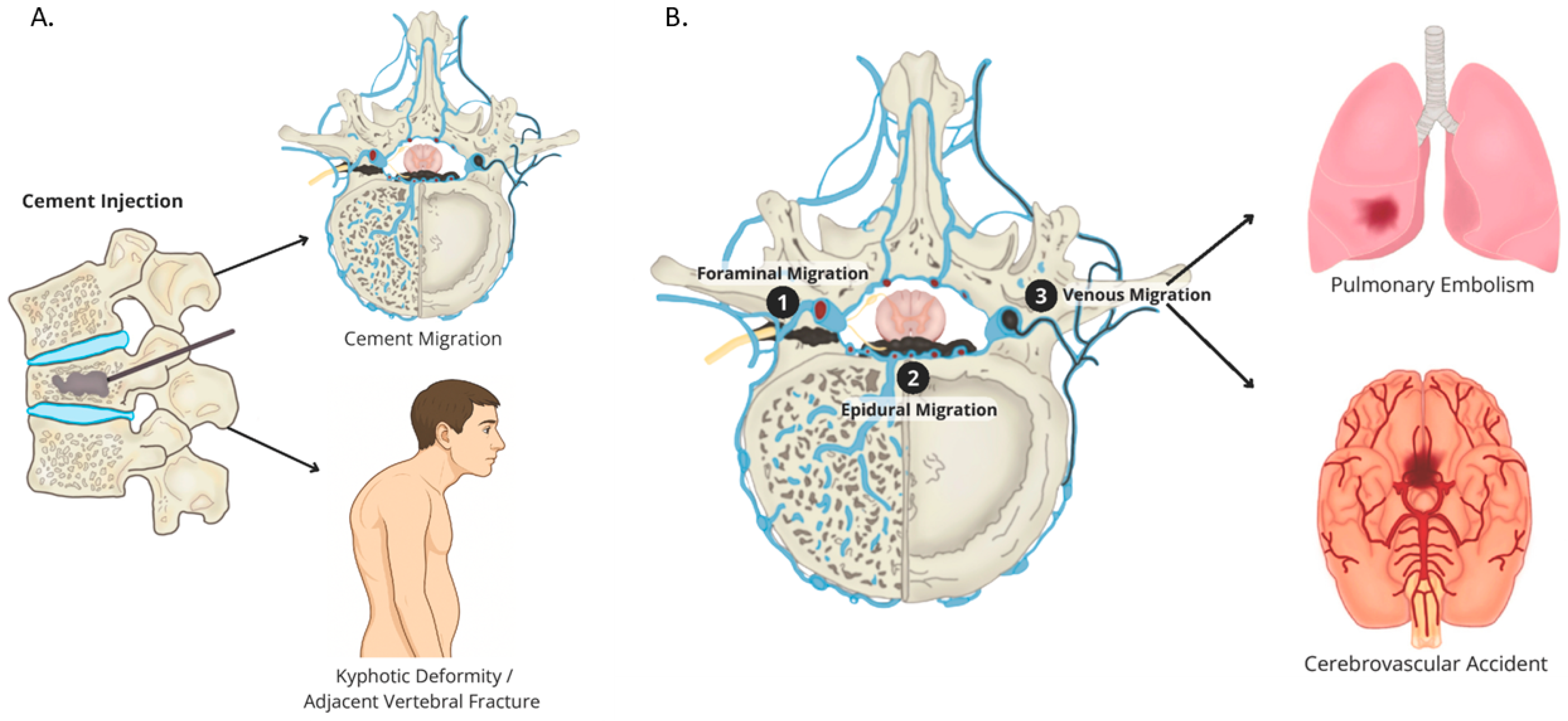
All included studies reported at least one vertebroplasty-related complication. Methodological vigilance varied; interventional-radiology series that obtained routine post-procedure CT detected markedly more asymptomatic leaks than cohorts relying on fluoroscopy or plain radiographs [10]. Nevertheless, consistent patterns emerged across five major complication domains (Figure 2 and Table 1): cement leakage, pulmonary cement embolism, adjacent-level or repeat fractures, infection, and neurologic injury. A synthesis of these findings—stratified by osteoporotic versus oncologic indication—is provided in the subsequent subsections.

3.4.1. Cement Leakage

Cement extravasation is the most frequently reported finding after PVP, with radio-graphic rates of 20–70% per treated level—higher when detected by CT (>50%) versus fluoroscopy alone (10–30%) [11,13,18]. Paravertebral and intradiscal leaks predominate (32–34% and up to 33%, respectively), while venous (5–20%) and epidural (5–15%) leaks are less common (Figure 2A) [19,20].
Using Yeom’s classification [21], most leaks are Type C (cortical breach) through end-plate or cortical defects, followed by Type B (basivertebral) and Type S (segmental vein) leaks; Hsieh et al. [13]. found Type C-intradiscal (22%) and paraspinal (21%) most frequent, with Type B at 12% and Type S at 4%. Although leaks are ubiquitous, the vast majority are asymptomatic. Small paravertebral or intradiscal leaks rarely cause harm, whereas epidural or foraminal leaks can compress neural elements. Clinically significant neurologic injury occurs in <1% (e.g., 0.3% cord compression, 0.13% radiculopathy) and often resolves with prompt decompression surgery [22,23].
On fluoroscopy, leaks appear as radiopaque cement beyond the vertebral margins; CT best defines their location and extent, while MRI reveals any resultant cord or root compression or edema [15,16,17]. Risk factors include cortical disruption (e.g., burst fractures, metastases), low-viscosity cement, large volumes (>7 mL), and central needle placement. Other specific patient characteristics such as older age, female gender, and lower bone mineral density can act as additional risk factors, contributing to vertebral fragility [16,24]; real-time fluoroscopic-monitoring and the use of high viscosity cement placed off-center can reduce leak risk. Most asymptomatic leaks require no treatment, but symptomatic epidural or foraminal leaks warrant urgent laminectomy or foraminotomy to remove cement and decompress neural structures, usually yielding neurologic recovery. Vigilant technique and immediate cessation at the first sign of leakage are essential to prevent serious sequelae.

3.4.2. Pulmonary Cement Embolism (PCE)

Cement may migrate via vertebral veins into the pulmonary arteries, lodging in seg-mental or subsegmental branches. Reported PCE rates vary with the detection method; symptomatic events occur in 0.1–0.5% of patients, while routine chest CT identifies cement emboli in 2–26% of cases—up to 26% in Venmans et al.’s series—most of which are clinically silent [10,16,17]. Larger database analyses note a 1.3% rate of “pulmonary embolism,” though non-cement thrombi are included [15]. Symptomatic PCE typically presents within 24–72 h post-procedure with acute pleuritic chest pain, dyspnea, tachycardia, or hypoxemia; massive emboli can cause hypotension or collapse, but fatalities are exceedingly rare in modern practice [24,25].
Diagnosis relies on chest CT angiography—cement appears as high-density filling defects—since chest X-ray often misses small emboli or overlapping opacities [10,24]. Risk factors mirror those for venous cement leakage: low-viscosity cement, high injection pressure or volume (>7 mL); basivertebral or segmental vein extravasation, and tumor-related cortical defects [13,26]. Asymptomatic, peripheral PCEs are managed conservatively with observation and often short-term anticoagulation to prevent thrombus formation; symptomatic or cardiopulmonary emboli may require surgical or endovascular retrieval [17,26]. Preventive measures include using high-viscosity cement, slow injection under continuous fluoroscopy, limiting volume, and halting immediately upon venous runoff. Overall, clinically significant PCE is uncommon (≤1%), and with prompt recognition and appropriate care, outcomes are favorable.

3.4.3. Adjacent-Level Fractures

Adjacent-level collapse is a shared—but mechanistically distinct—complication after augmentation. In typical osteoporotic cohorts 8–17% of patients sustain a new fracture within 12 months, whereas oncologic series report rates closer to 20–30% because tumor-driven cortical destruction, larger cement volumes, and systemic therapies amplify stress transfer to neighboring vertebrae [27,28,29]. Pooled data show that low bone-mineral density, prior VCFs, intradiscal or venous cement leakage, and chronic glucocorticoid use rank as the strongest osteoporotic predictors, while epidural tumor extension and anterior column lysis dominate in cancer [28,29,30,31,32]. Patients usually present with recurrent pain within three months of the index procedure; STIR-MRI revealing marrow edema in an untreated level confirms the diagnosis [30].
Management follows a stepped algorithm. First-line care combines short-term bracing, analgesia and rapid optimization of anti-osteoporosis medication (anabolic agents in “cascade” phenotypes) [33]. Surgical escalation—repeat PVP or balloon kyphoplasty—is reserved for refractory pain, progressive collapse, or neurologic risk, particularly when the fracture occurs > 6 months after the index level or is caudal to it [34]. Repeat vertebroplasty delivers comparable pain relief to the primary procedure but carries a higher technical-complication rate, with cement leakage and further fractures reported in up to 30–34% of re-augmented cases; meticulous control of cement viscosity and volume, avoidance of intradiscal flow, and staged multilevel treatment mitigate that risk [34,35].
Preventive measures overlap with those detailed in Table 2; they minimize intradiscal leakage (use cavity creation or vertebral body stents in severe osteoporosis), limit cement to ≤ 6 mL, consider prophylactic augmentation of the cephalad level in rapid “fracture-cascade” patients, and ensure all candidates leave the procedure suite with a structured bone-health plan [14]. Adhering to these principles should curb adjacent-level fracture incidence and optimize outcomes for both osteoporotic and metastatic populations.

3.4.4. Infection (Spondylodiscitis)

Although rare (0–1% overall), post-vertebroplasty infection carries substantial morbidity [11,14]. In oncologic cohorts’ immunosuppression and multiple spinal interventions may elevate risk slightly, in comparison to osteoporotic patients. Case series have documented a 1.6% rate of fungal spondylodiscitis in myeloma patients and TB reactivation in endemic areas [14,29].
Infection arises from direct needle inoculation or late hematogenous seeding of cemented bone, presenting in approximately 2–8 weeks post-procedure (though TB can manifest > 1 year later) [11]. Clinically, patients report severe back pain, and present with fever and neurologic deficits in ~39% of cases. MRI with contrast—despite cement artifact—reveals marrow edema, disk enhancement, and abscess formation, while CT shows endplate erosion and paravertebral collections [38,39]. Cultures most commonly grow Staphylococcus aureus (~19%), coagulase-negative staphylococcus, Mycobacterium tuberculosis in endemic regions, and rare fungal pathogens in highly immunocompromised hosts [38].
Management mirrors that of hardware-related osteomyelitis: broad-spectrum intra-venous antibiotics and prompt surgical debridement with corpectomy and reconstruction, often via combined anterior–posterior approaches, followed by long-term targeted antimicrobials [40]. Despite aggressive treatment, mortality can reach ~17% and survivors frequently experience permanent deficits. Prevention hinges on strict sterile technique, peri-procedural antibiotics, and pre-procedure screening for occult periprocedural signs of infections.

3.4.5. Neurological Injury

Neurologic complications—typically radiculopathy or myelopathy—occur in 0.1–1.0% of PVP procedures, with major events (cord compression or cauda equina syndrome) reported in <0.5% of cases [7,13]. The principal mechanism is cement leakage into the spinal canal or neural foramen; though rare, epidural hematoma or direct needle trauma can also injure neural elements. High thoracic or cervical leaks favor cord compression (paraplegia, quadriplegia), while lumbar leaks are likely to cause root symptoms (e.g., L5 radiculopathy).
Patients may develop acute leg pain or weakness during injection (even under conscious sedation). More often, symptoms begin within hours after the procedure, presenting as neuropathic pain, sensory changes, or motor deficits. Urgent CT identifies cement in canal or foramen, and MRI—despite artifact—confirms cord or root compression and rules out hematoma [18]. Significant deficits mandate emergent laminectomy or foraminotomy for cement removal, with most operated patients achieving full or near-full recovery when intervened promptly [13]. Mild, transient deficits may resolve with steroids and observation.
Prevention hinges on meticulous techniques, such as the following: transpedicular needle placement under biplanar fluoroscopy or CT guidance, incremental injection of high-viscosity cement, and immediate cessation upon any leakage or venous intravasation [41].Although open spine surgery carries a higher baseline risk (1–5%) [42], the PVP approach yields a low incidence of neurologic injury—yet clinicians must remain prepared for rapid diagnosis and intervention to preserve neurologic function [13].

4. Discussion

This systematic review of PVP complications in adults with osteoporotic or neo-plastic vertebral fractures supports the idea that when performed by experienced hands and on carefully selected patients, the procedure is both safe and effective. Across 15 studies, major adverse events—such as symptomatic cement embolism, significant neurologic injury, or deep infection—occurred in well under 2% of cases [7,15]. At the same time, we have mapped the full spectrum of potential events, from the frequent but generally benign cement leaks to the very rare catastrophic outcomes, providing clinicians with a clear understanding of the risks to inform their practice.

4.1. Clinical Decision-Making Framework

A growing body of evidence shows that vertebroplasty outcomes depend less on the cement itself than on matching the right technique to the right patient profile. We therefore stratify candidates into seven practical scenarios (Table 3). Low-risk primary osteoporotic fractures (intact posterior cortex, limited comorbidity, T-score > −2.5) achieve predictable pain relief with unilateral high-viscosity PVP and carry < 2% major-event risk [19,43]. Intermediate-risk patients—severe osteoporosis, ≥2 prior fractures, or chronic steroid use—have a 13–20% annual adjacent-level fracture rate; balloon kyphoplasty or low-pressure cavity creation, sometimes combined with prophylactic augmentation of the cephalad level, reduces intradiscal leakage and should be coupled with anabolic osteo-porosis therapy [29,44,45,46].
High-risk oncologic lesions require the greatest nuance; CT-guided cement injection limits Type B/S venous leaks by ~50% [47,48], but extensive cortical destruction, epidural tumor, or coagulopathy mandates staged ablation + kyphoplasty, neuromonitoring, and, when multiple levels are treated, temporary IVC or azygos filters [44]. Procedures are deferred if systemic sepsis, uncontrolled anticoagulation, or cardiorespiratory compromise outweighs expected benefit [29,38].

4.2. Complication Prevention and Technical Optimization

Registry data reveal a 40% reduction in major complications after the first 25 mentored cases, underscoring the learning curve. Core training should include cadaveric simulation, competency checklists for pedicle trajectory, cement viscosity control, and leak recognition [49,50]. Real-time biplanar fluoroscopy with lateral pressure feedback enables instant cessation when cement approaches the dorsal wall; 3-D navigation is advisable for cervicothoracic or highly destructive tumors [46,51]. Emerging technologies—steerable cannulas, closed-loop pressure injectors—show early promise for further reducing epidural migration but await prospective validation.

4.3. Management of Complications

Most cement leaks remain radiographic curiosities, but new radiculopathy or myelopathy warrants emergent CT/MRI and, if canal-filling cement is confirmed, decompression within 12 h to maximize recovery [13,18,52]. Up to 26% of patients show asymptomatic pulmonary cement emboli on screening CT; these may be observed or treated with short-course anticoagulation [16,17]. Symptomatic central emboli require catheter retrieval or surgical embolectomy [24]. Infection, although rare (0.2–0.5%), carries ~17% mortality [11,29]; early MRI, inflammatory markers, and targeted biopsy guide combined anterior–posterior debridement, cement removal, and six-week pathogen-specific therapy [14,38,40]. Recurrent or adjacent fractures should trigger re-assessment of bone health and, in rapid “fracture-cascade” phenotypes, consideration of prophylactic augmentation [38,45].

Informed Consent and Risk Communication

Patients must understand that radiographic cement leakage occurs in up to 70% of levels, yet clinically significant events—paralysis, symptomatic embolism, deep infection—remain < 1%. Metastatic disease confers a higher overall complication rate (5–10%) than osteoporosis (1–3%) [15,44]. Presenting the personalized risk–benefit summaries in Table 2 during counseling helps align expectations and facilitates shared decision-making about alternatives such as bracing, kyphoplasty, or (for tumors) radiation and surgical fixation.

4.4. Future Research Priorities

Four gaps warrant attention: (1) prospective multicenter registries with uniform complication definitions to calibrate risk calculators; (2) randomized trials comparing steerable versus straight cannulas and antibiotic-loaded PMMA for infection prevention; (3) head-to-head studies of vertebroplasty versus kyphoplasty in metastatic disease; and (4) cost-effectiveness analyses incorporating quality-adjusted life-years gained from earlier mobilization. Addressing these questions will refine patient selection and drive the next generation of safer, more efficient vertebral augmentation techniques.

4.5. Limitations of Evidence

Our review is limited by the predominance of observational studies with variable complication definitions and potential under- or over-reporting. Heterogeneity in patient populations and procedural techniques precluded meta-analysis, so our findings rely on reported ranges rather than pooled estimates, which may affect precision.
Innovations such as steerable delivery systems, robotic needle guidance, and novel cement formulations (e.g., resorbable or antibiotic-loaded cements) hold promise for further reducing complication rates. Establishing a multicenter registry with standardized definitions would enable more accurate tracking of outcomes and the identification of modifiable risk factors.

5. Conclusions

PVP provides substantial symptomatic relief for osteoporotic and metastatic vertebral fractures with a favorable safety profile. While cement leakage, pulmonary embolism, infection, and neural compression occur more often in oncologic cases than in osteoporosis, clinically significant events remain uncommon (<5–10%). A meticulous technique—using high-viscosity cement, real-time fluoroscopic or CT guidance, and strict aseptic protocols—minimizes risks. Equally, early recognition of complications allows for rapid interventions mitigating long-term consequences. By pairing careful patient selection with vigilant monitoring and rapid multidisciplinary intervention, clinicians can maximize PVP’s palliative benefits while keeping complications to a minimum in this high-risk population.

Author Contributions

Conceptualization, J.P.Z.-G. and D.H.; methodology, validation, formal analysis, investigation: D.H. and F.A.C.-O.; resources, M.A.S.; data curation, M.A.S. and P.K.R.; writing—original draft preparation, J.P.Z.-G.; writing—review and editing, R.A.A.-A. and E.R.-F.; visualization and illustration, M.A.S. All authors have read and agreed to the published version of the manuscript.

Funding

This research received no external funding.

Institutional Review Board Statement

Not applicable.

Informed Consent Statement

Not applicable.

Data Availability Statement

The original contributions presented in this study are included in the article. Further inquiries can be directed to the corresponding author.

Conflicts of Interest

The authors declare no conflicts of interest.

Abbreviations

PVPPercutaneous Vertebroplasty
RCTRandomized Controlled Trial
OVCFOsteoporotic Vertebral Compression Fracture
PMMAPolymethylmethacrylate
FDAU.S. Food and Drug Administration
SCOPUSA bibliographic database of peer-reviewed literature
MEDLINEMedical Literature Analysis and Retrieval System Online
PRISMAPreferred Reporting Items for Systematic Reviews and Meta-Analyses
CTComputed Tomography
MRIMagnetic Resonance Imaging
PCEPulmonary Cement Embolism
DOACDirect Oral Anticoagulant
UTIUrinary Tract Infection
TBTuberculosis
MSSAMethicillin-Sensitive Staphylococcus Aureus

References

  1. Dong, Y.; Peng, R.; Kang, H.; Song, K.; Guo, Q.; Zhao, H.; Zhu, M.; Zhang, Y.; Guan, H.; Li, F. Global Incidence, Prevalence, and Disability of Vertebral Fractures: A Systematic Analysis of the Global Burden of Disease Study 2019. Spine J. 2022, 22, 857–868. [Google Scholar] [CrossRef]
  2. Lan, Y.; Chen, S.; Lan, G.; Li, C.; Wei, J. Global, Regional, and National Burden of Fracture of Vertebral Column, 1990–2021: Analysis of Data from the Global Burden of Disease Study 2021. Front. Public Health 2025, 13, 1573888. [Google Scholar] [CrossRef]
  3. Curtis, J.R.; Taylor, A.J.; Matthews, R.S.; Ray, M.N.; Becker, D.J.; Gary, L.C.; Kilgore, M.L.; Morrisey, M.A.; Saag, K.G.; Warriner, A.; et al. “Pathologic” Fractures: Should These Be Included in Epidemiologic Studies of Osteoporotic Fractures? Osteoporos. Int. 2009, 20, 1969–1972. [Google Scholar] [CrossRef]
  4. Burton, A.W.; Rhines, L.D.; Mendel, E. Vertebroplasty and Kyphoplasty: A Comprehensive Review. Neurosurg. Focus. 2005, 18, e1. [Google Scholar] [CrossRef] [PubMed]
  5. Cavka, M.; Delimar, D.; Rezan, R.; Zigman, T.; Duric, K.S.; Cimic, M.; Dumic-Cule, I.; Prutki, M. Complications of Percutaneous Vertebroplasty: A Pictorial Review. Medicina 2023, 59, 1536. [Google Scholar] [CrossRef] [PubMed]
  6. Bae, H.; Hatten, H.P.J.; Linovitz, R.; Tahernia, A.D.; Schaufele, M.K.; McCollom, V.; Gilula, L.; Maurer, P.; Benyamin, R.; Mathis, J.M.; et al. A Prospective Randomized FDA-IDE Trial Comparing Cortoss With PMMA for Vertebroplasty: A Comparative Effectiveness Research Study With 24-Month Follow-Up. Spine 2012, 37, 544. [Google Scholar] [CrossRef] [PubMed]
  7. Cosar, M.; Sasani, M.; Oktenoglu, T.; Kaner, T.; Ercelen, O.; Kose, K.C.; Ozer, A.F. The Major Complications of Transpedicular Vertebroplasty. J. Neurosurg. Spine 2009, 11, 607–613. [Google Scholar] [CrossRef]
  8. Layton, K.F.; Thielen, K.R.; Koch, C.A.; Luetmer, P.H.; Lane, J.I.; Wald, J.T.; Kallmes, D.F. Vertebroplasty, First 1000 Levels of a Single Center: Evaluation of the Outcomes and Complications. AJNR Am. J. Neuroradiol. 2007, 28, 683–689. [Google Scholar]
  9. Lee, M.J.; Dumonski, M.; Cahill, P.; Stanley, T.; Park, D.; Singh, K. Percutaneous Treatment of Vertebral Compression Fractures: A Meta-Analysis of Complications. Spine 2009, 34, 1228–1232. [Google Scholar] [CrossRef]
  10. Rahimi, B.; Boroofeh, B.; Dinparastisaleh, R.; Nazifi, H. Cement Pulmonary Embolism after Percutaneous Vertebroplasty in a Patient with Cushing’s Syndrome: A Case Report. Respir. Med. Case Rep. 2018, 25, 78–85. [Google Scholar] [CrossRef]
  11. Liao, J.-C.; Lai, P.-L.; Chen, L.-H.; Niu, C.-C. Surgical Outcomes of Infectious Spondylitis after Vertebroplasty, and Comparisons between Pyogenic and Tuberculosis. BMC Infect. Dis. 2018, 18, 555. [Google Scholar] [CrossRef]
  12. Anselmetti, G.C.; Marcia, S.; Saba, L.; Muto, M.; Bonaldi, G.; Carpeggiani, P.; Marini, S.; Manca, A.; Masala, S. Percutaneous Vertebroplasty: Multi-Centric Results from EVEREST Experience in Large Cohort of Patients. Eur. J. Radiol. 2012, 81, 4083–4086. [Google Scholar] [CrossRef]
  13. Hsieh, M.-K.; Kao, F.-C.; Chiu, P.-Y.; Chen, L.-H.; Yu, C.-W.; Niu, C.-C.; Lai, P.-L.; Tsai, T.-T. Risk Factors of Neurological Deficit and Pulmonary Cement Embolism after Percutaneous Vertebroplasty. J. Orthop. Surg. Res. 2019, 14, 406. [Google Scholar] [CrossRef]
  14. Abdelrahman, H.; Siam, A.E.; Shawky, A.; Ezzati, A.; Boehm, H. Infection after Vertebroplasty or Kyphoplasty. A Series of Nine Cases and Review of Literature. Spine J. 2013, 13, 1809–1817. [Google Scholar] [CrossRef]
  15. Kim, H.-J.; Zuckerman, S.L.; Cerpa, M.; Yeom, J.S.; Lehman, R.A.; Lenke, L.G. Incidence and Risk Factors for Complications and Mortality After Vertebroplasty or Kyphoplasty in the Osteoporotic Vertebral Compression Fracture-Analysis of 1,932 Cases From the American College of Surgeons National Surgical Quality Improvement. Global Spine J. 2022, 12, 1125–1134. [Google Scholar] [CrossRef]
  16. Venmans, A.; Klazen, C.A.H.; Lohle, P.N.M.; van Rooij, W.J.; Verhaar, H.J.J.; de Vries, J.; Mali, W.P.T.M. Percutaneous Vertebroplasty and Pulmonary Cement Embolism: Results from VERTOS II. AJNR Am. J. Neuroradiol. 2010, 31, 1451–1453. [Google Scholar] [CrossRef] [PubMed]
  17. Luetmer, M.T.; Bartholmai, B.J.; Rad, A.E.; Kallmes, D.F. Asymptomatic and Unrecognized Cement Pulmonary Embolism Commonly Occurs with Vertebroplasty. AJNR Am. J. Neuroradiol. 2011, 32, 654–657. [Google Scholar] [CrossRef] [PubMed]
  18. Jing, Z.; Li, L.; Song, J. Delayed Neurological Deficits Caused by Cement Extravasation Following Vertebroplasty: A Case Report. J. Int. Med. Res. 2021, 49, 03000605211019664. [Google Scholar] [CrossRef] [PubMed]
  19. Tang, B.; Cui, L.; Chen, X.; Liu, Y. Risk Factors for Cement Leakage in Percutaneous Vertebroplasty for Osteoporotic Vertebral Compression Fractures: An Analysis of 1456 Vertebrae Augmented by Low-Viscosity Bone Cement. Spine (Phila Pa 1976) 2021, 46, 216–222. [Google Scholar] [CrossRef]
  20. Kasó, G.; Horváth, Z.; Szenohradszky, K.; Sándor, J.; Dóczi, T. Comparison of CT Characteristics of Extravertebral Cement Leakages after Vertebroplasty Performed by Different Navigation and Injection Techniques. Acta Neurochir. 2008, 150, 677–683; discussion 683. [Google Scholar] [CrossRef]
  21. Baek, I.-H.; Park, H.-Y.; Kim, K.-W.; Jang, T.-Y.; Lee, J.-S. Paraplegia Due to Intradural Cement Leakage after Vertebroplasty: A Case Report and Literature Review. BMC Musculoskelet. Disord. 2021, 22, 741. [Google Scholar] [CrossRef] [PubMed]
  22. Hiwatashi, A.; Yoshiura, T.; Yamashita, K.; Kamano, H.; Honda, H. Ultrashort TE MRI: Usefulness after Percutaneous Vertebroplasty. AJR Am. J. Roentgenol. 2010, 195, W365–W368. [Google Scholar] [CrossRef] [PubMed]
  23. Gao, C.; Zong, M.; Wang, W.-T.; Xu, L.; Cao, D.; Zou, Y.-F. Analysis of Risk Factors Causing Short-Term Cement Leakages and Long-Term Complications after Percutaneous Kyphoplasty for Osteoporotic Vertebral Compression Fractures. Acta Radiol. 2018, 59, 577–585. [Google Scholar] [CrossRef]
  24. Barakat, A.S.; Owais, T.; Alhashash, M.; Shousha, M.; El Saghir, H.; Lauer, B.; Boehm, H. Presentation and Management of Symptomatic Central Bone Cement Embolization. Eur. Spine J. 2018, 27, 2584–2592. [Google Scholar] [CrossRef]
  25. Stricker, K.; Orler, R.; Yen, K.; Takala, J.; Luginbühl, M. Severe Hypercapnia Due to Pulmonary Embolism of Polymethylmethacrylate during Vertebroplasty. Anesth. Analg. 2004, 98, 1184–1186. [Google Scholar] [CrossRef]
  26. Sun, H.-B.; Jing, X.-S.; Shan, J.-L.; Bao, L.; Wang, D.-C.; Tang, H. Risk Factors for Pulmonary Cement Embolism Associated with Percutaneous Vertebral Augmentation: A Systematic Review and Meta-Analysis. Int. J. Surg. 2022, 101, 106632. [Google Scholar] [CrossRef]
  27. Dargelos-Descoubez, M.; Martin, F.; Frampas, E.; Perret, C.; David, A.; Volpi, S. Progression toward Vertebral Collapse of Vertebral Metastases Treated with Percutaneous Vertebroplasty: Rate and Risk Factors. J. Vasc. Interv. Radiol. 2024, 35, 59–68. [Google Scholar] [CrossRef]
  28. Mazzantini, M.; Figliomeni, A.; Bottai, V.; Manca, M.L.; Puglioli, M.; Di Munno, O.; Mosca, M. High Rate of Vertebral Refracture after Vertebroplasty in Patients Taking Glucocorticoids: A Prospective Two-Year Study. Clin. Exp. Rheumatol. 2020, 38, 649–653. [Google Scholar]
  29. Park, S.; Sik Choi, S.; Kim, H.; Yoon Byun, S.; Lee, C.H. Risk Factors for New Vertebral Compression Fracture after Vertebroplasty and Efficacy of Osteoporosis Treatment: A STROBE-Compliant Retrospective Study. Medicine 2023, 102, e35042. [Google Scholar] [CrossRef]
  30. Zhai, G.; Li, A.; Liu, B.; Lv, D.; Zhang, J.; Sheng, W.; Yang, G.; Gao, Y. A Meta-Analysis of the Secondary Fractures for Osteoporotic Vertebral Compression Fractures after Percutaneous Vertebroplasty. Medicine 2021, 100, e25396. [Google Scholar] [CrossRef]
  31. Nieuwenhuijse, M.J.; Putter, H.; van Erkel, A.R.; Dijkstra, P.D.S. New Vertebral Fractures after Percutaneous Vertebroplasty for Painful Osteoporotic Vertebral Compression Fractures: A Clustered Analysis and the Relevance of Intradiskal Cement Leakage. Radiology 2013, 266, 862–870. [Google Scholar] [CrossRef]
  32. Marselou, E.; Kelekis, A.; Dimitriadis, Z.; Koumantakis, G.A. Risk Factors for Refracture or New Vertebral Compression Fractures after Percutaneous Vertebroplasty: A Systematic Review and Meta-Analysis. Osteoporos. Int. 2025, 36, 1297–1311. [Google Scholar] [CrossRef]
  33. Huang, C.; Wang, Y.; Li, X.; Ren, L.; Zhao, J.; Hu, Y.; Zhang, L.; Fan, G.; Xu, J.; Gu, X.; et al. Clinical Features of Patients Infected with 2019 Novel Coronavirus in Wuhan, China. Lancet 2020, 395, 497–506. [Google Scholar] [CrossRef]
  34. Wu, B.-S.; Hsu, M.-C.; Yao, Y.-C.; Lin, H.-H.; Chou, P.-H.; Wang, S.-T.; Chang, M.-C.; Hsiung, W.; Wang, C.-Y.; Chen, K.-J. Efficiency and Safety of Repeated Vertebroplasty for Adjacent Segment Fractures. J. Clin. Med. 2024, 14, 166. [Google Scholar] [CrossRef]
  35. Lamy, O.; Uebelhart, B.; Aubry-Rozier, B. Risks and Benefits of Percutaneous Vertebroplasty or Kyphoplasty in the Management of Osteoporotic Vertebral Fractures. Osteoporos. Int. 2014, 25, 807–819. [Google Scholar] [CrossRef]
  36. Khanna, A.J.; Reinhardt, M.K.; Togawa, D.; Lieberman, I.H. Functional Outcomes of Kyphoplasty for the Treatment of Osteoporotic and Osteolytic Vertebral Compression Fractures. Osteoporos. Int. 2006, 17, 817–826. [Google Scholar] [CrossRef] [PubMed]
  37. Essibayi, M.A.; Mortezaei, A.; Azzam, A.Y.; Bangash, A.H.; Eraghi, M.M.; Fluss, R.; Brook, A.; Altschul, D.J.; Yassari, R.; Chandra, R.V.; et al. Risk of Adjacent Level Fracture after Percutaneous Vertebroplasty and Kyphoplasty vs Natural History for the Management of Osteoporotic Vertebral Compression Fractures: A Network Meta-Analysis of Randomized Controlled Trials. Eur. Radiol. 2024, 34, 7185–7196. [Google Scholar] [CrossRef] [PubMed]
  38. Zuluaga-García, J.P.; Leon-Aldana, S.; Herrera, D. Spondylodiscitis: A Comprehensive Review of Diagnostic Challenges, Microbial Etiology, and Management Strategies. SN Compr. Clin. Med. 2025, 7, 92. [Google Scholar] [CrossRef]
  39. Zhou, F.; Yu, T.; Du, R.; Fan, G.; Liu, Y.; Liu, Z.; Xiang, J.; Wang, Y.; Song, B.; Gu, X.; et al. Clinical Course and Risk Factors for Mortality of Adult Inpatients with COVID-19 in Wuhan, China: A Retrospective Cohort Study. Lancet 2020, 395, 1054–1062. [Google Scholar] [CrossRef]
  40. Yuan, H.; Li, Y.; Wang, J.; Wang, X.; Lü, G.; Kuang, L.; Li, J. Spinal Infection after Vertebral Augmentation: A Covert Complication with Serious Havoc. Arch. Orthop. Trauma. Surg. 2024, 144, 1461–1471. [Google Scholar] [CrossRef]
  41. Patel, A.A.; Vaccaro, A.R.; Martyak, G.G.; Harrop, J.S.; Albert, T.J.; Ludwig, S.C.; Youssef, J.A.; Gelb, D.E.; Mathews, H.H.; Chapman, J.R.; et al. Neurologic Deficit Following Percutaneous Vertebral Stabilization. Spine (Phila Pa 1976) 2007, 32, 1728–1734. [Google Scholar] [CrossRef] [PubMed]
  42. Lin, C.-C.; Shen, W.-C.; Lo, Y.-C.; Liu, Y.-J.; Yu, T.-C.; Chen, I.-H.; Chung, H.-W. Recurrent Pain After Percutaneous Vertebroplasty. Am. J. Roentgenol. 2010, 194, 1323–1329. [Google Scholar] [CrossRef] [PubMed]
  43. Barr, J.D.; Jensen, M.E.; Hirsch, J.A.; McGraw, J.K.; Barr, R.M.; Brook, A.L.; Meyers, P.M.; Munk, P.L.; Murphy, K.J.; O’Toole, J.E.; et al. Position Statement on Percutaneous Vertebral Augmentation: A Consensus Statement Developed by the Society of Interventional Radiology (SIR), American Association of Neurological Surgeons (AANS) and the Congress of Neurological Surgeons (CNS), American College of Radiology (ACR), American Society of Neuroradiology (ASNR), American Society of Spine Radiology (ASSR), Canadian Interventional Radiology Association (CIRA), and the Society of NeuroInterventional Surgery (SNIS). J. Vasc. Interv. Radiol. 2014, 25, 171–181. [Google Scholar] [CrossRef] [PubMed]
  44. Corcos, G.; Dbjay, J.; Mastier, C.; Leon, S.; Auperin, A.; De Baere, T.; Deschamps, F. Cement Leakage in Percutaneous Vertebroplasty for Spinal Metastases: A Retrospective Evaluation of Incidence and Risk Factors. Spine (Phila Pa 1976) 2014, 39, E332–E338. [Google Scholar] [CrossRef]
  45. Ebeling, P.R.; Akesson, K.; Bauer, D.C.; Buchbinder, R.; Eastell, R.; Fink, H.A.; Giangregorio, L.; Guanabens, N.; Kado, D.; Kallmes, D.; et al. The Efficacy and Safety of Vertebral Augmentation: A Second ASBMR Task Force Report. J. Bone Miner. Res. 2019, 34, 3–21. [Google Scholar] [CrossRef]
  46. Zhan, Y.; Jiang, J.; Liao, H.; Tan, H.; Yang, K. Risk Factors for Cement Leakage After Vertebroplasty or Kyphoplasty: A Meta-Analysis of Published Evidence. World Neurosurg. 2017, 101, 633–642. [Google Scholar] [CrossRef]
  47. Zhang, Z.; Yang, J.; Jiang, H.; Lai, Z.; Wu, F.; Pan, Y.; Liu, Z. An Updated Comparison of High- and Low-Viscosity Cement Vertebroplasty in the Treatment of Osteoporotic Thoracolumbar Vertebral Compression Fractures: A Retrospective Cohort Study. Int. J. Surg. 2017, 43, 126–130. [Google Scholar] [CrossRef]
  48. Reyad, R.M.; Ghobrial, H.Z.; Hakim, S.M.; Hashem, R.H.; Elsaman, A.; Shaaban, M.H. Thick Cement Usage in Percutaneous Vertebroplasty for Malignant Vertebral Fractures at High Risk for Cement Leakage. Diagn. Interv. Imaging 2017, 98, 721–728. [Google Scholar] [CrossRef]
  49. Wucherer, P.; Stefan, P.; Abhari, K.; Fallavollita, P.; Weigl, M.; Lazarovici, M.; Winkler, A.; Weidert, S.; Peters, T.; de Ribaupierre, S.; et al. Vertebroplasty Performance on Simulator for 19 Surgeons Using Hierarchical Task Analysis. IEEE Trans. Med. Imaging 2015, 34, 1730–1737. [Google Scholar] [CrossRef]
  50. Hollensteiner, M.; Botzenmayer, M.; Fürst, D.; Winkler, M.; Augat, P.; Sandriesser, S.; Schrödl, F.; Esterer, B.; Gabauer, S.; Püschel, K.; et al. Characterization of Polyurethane-Based Synthetic Vertebrae for Spinal Cement Augmentation Training. J. Mater. Sci. Mater. Med. 2018, 29, 153. [Google Scholar] [CrossRef]
  51. Chandra, R.V.; Meyers, P.M.; Hirsch, J.A.; Abruzzo, T.; Eskey, C.J.; Hussain, M.S.; Lee, S.-K.; Narayanan, S.; Bulsara, K.R.; Gandhi, C.D.; et al. Vertebral Augmentation: Report of the Standards and Guidelines Committee of the Society of NeuroInterventional Surgery. J. Neurointerv Surg. 2014, 6, 7–15. [Google Scholar] [CrossRef]
  52. Tanigawa, N.; Kariya, S.; Komemushi, A.; Nakatani, M.; Yagi, R.; Kohzai, M.; Sawada, S. Percutaneous Vertebroplasty for Osteoporotic Compression Fractures: Long-Term Evaluation of the Technical and Clinical Outcomes. AJR Am. J. Roentgenol. 2011, 196, 1415–1418. [Google Scholar] [CrossRef]
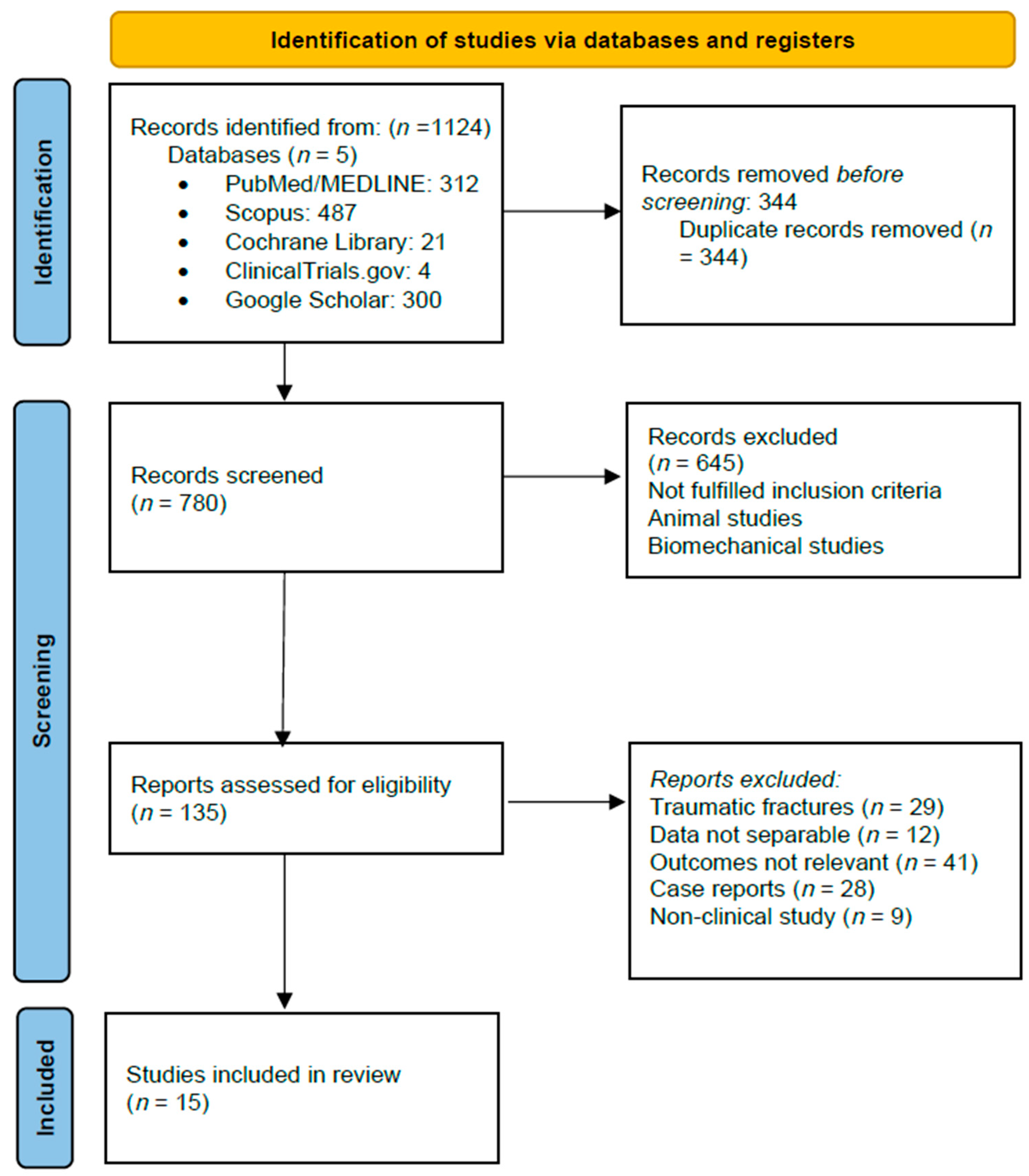
Figure 1. PRISMA flow diagram of study selection.
Complications 02 00022 g001
Figure 2. Cement migration pathways and downstream consequences. (A) Procedure and main groups of complications. (B) Complications related to cement leakages and cement migration.
Complications 02 00022 g002
Table 1. Complication profile in osteoporotic vertebroplasty cohorts.
Author and YearCountryDesign/Evidence Level *Patients (Levels)% OVCFCement Leak%Neuro%PCE%Infection%Adjacent Fx%30-Day Mortality%
Anselmetti 2012 [12]ItalyProspective III4.547 (13.437)7320.50NRNR13NR
Hsieh 2019 [13]TaiwanRetro IV3.175 (3.812)10045 †0.130.280NR0
Liao 2018 [11]TaiwanRetro IV5.749100NR0.13 ‡NR0.32NR0.1 §
Abdelrahman 2013 [14]GermanyRetro IV1.307100NRNRNR0.46NRNR
Kim 2022 [15] USADatabase IV1.932100NR01.30.2NR2.1
Venmans 2010 [16]Neth.Prospective III97 (121)10060 †1260NR0
Luetmer 2011 [17]USARetro IV244100NR09.4 (0.4 symp)0NR0
* Evidence grading: RCT = II; prospective cohort = III; retrospective or registry = IV. † Original data reported per level; converted to approximate per-patient rate by assuming a single leak counts once per patient. ‡ Neurologic deficits occurred only in patients with post-infection epidural compression (7/5 749 = 0.13%). § Three deaths among 1 749 infection-free patients (0.05%) plus 3/18 infection cases (16.7%) ≈ 0.1%. NSQIP database codes all pulmonary emboli; true cement PCE likely lower.
Table 2. Key adverse-event metrics in mixed/oncologic vertebroplasty cohorts.
Author and YearCountryDesign/Evidence Level *Patients (levels)IndicationCement Leak%Neuro%PCE%Infection%Adjacent Fx%
Cavka 2023 [5]CroatiaRetro IV189 (218)75% OVCF/25% pathol.350.50.50.5NR
Anselmetti 2012 [12]ItalyProspective III128100% myeloma35 †00.81.6NR
Khanna 2016 [36]USARetro IV350100% metastaticNR0.80.30.315
Essibayi 2024 [37]MultiSR/meta (RCTs)2050100% OVCF<1 (symp)0<0.1012–20
* Evidence grading: RCT = II, prospective cohort = III, retrospective or registry = IV. † Original data reported per level; converted to approximate per-patient rate by assuming a single leak counts once per patient.
Table 3. Risk-stratified decision matrix for vertebral augmentation.
Clinical ScenarioRisk FactorsPredominant Complication PatternPreferred Augmentation StrategyProcedural SafeguardsPost-Procedure Surveillance
A. Low-risk primary OVCFIntact posterior cortex, BMD > −2.5, no anticoagulation, ASA I–IIRadiographic paravertebral/intradiscal leaks (mostly asymptomatic)Standard PVP, unilateral transpedicular, 4–5 mL high-viscosity PMMAContinuous biplanar fluoro, stop ≤ 5 mm from posterior wall, single level per sessionOut-patient; tele-visit at 2 weeks; no routine CT
B. Severe osteoporosis or steroid use (“cascade” phenotype)≥2 prior VCF, T-score < −3.0, chronic glucocorticoidsHigher adjacent-level fracture rate (13–20%/year)Balloon kyphoplasty or low-pressure cavity creation; consider prophylactic augmentation of cephalad levelVertebral body stent or double balloon to avoid disk leak; cement ≤ 6 mL; initiate anabolic therapy post-opStanding radiograph and serum Ca/Vit-D at 3 months
C. Lytic metastasis without posterior wall breachSingle vertebra, mild cortical erosion, ECOG 0–1Venous (Type B/S) leaks; asymptomatic PCE (≤10%)High-viscosity “doughy” cement PVP, CT-guided cannula placementTest injection with contrast; inject < 10 psi; vascular plug in azygos if large channelChest X-ray in recovery; oncology f/u in 1 month
D. Lytic metastasis with posterior wall destruction/epidural tumorSpinal canal encroachment, >50% cortical loss, ECOG > 1Symptomatic cord/root compression; central PCE; infection (up to 1%)Stage 1: radiofrequency ablation + cavity creation; Stage 2: kyphoplasty with viscous cement < 4 mLCT navigation; neuromonitoring; prophylactic IVC filter for multi-level proceduresIn-hospital neuro checks q2 h × 24 h; MRI if any deficit
E. Coagulopathy/DOAC therapyINR > 1.4, platelets < 100 K, DOAC < 12 h from last doseEpidural hematoma; large venous cement run-offDelay procedure until parameters corrected; if urgent, perform kyphoplasty with viscoelastic cementReversal agents; limit balloon pressure; meticulous hemostasis at trocar siteCBC and neuro exam at 6 h; low-threshold MRI
F. Latent or active infection risk (diabetes, recent UTI, endemic TB)CRP > 10 mg/L, WBC > 10 K, PPD +/IGRA +Pyogenic or tuberculous spondylodiscitis (0.3–0.5%, mortality 17%)Postpone until sepsis ruled out; if imperative, antibiotic-loaded cement (gentamicin 1 g/40 g PMMA)Full surgical prep; new sterile needle for each pedicle; single-shot cefazolin + vancomycinCRP/ESR at 2 and 6 weeks; MRI if pain recurs
G. Multilevel (>3) osteoporotic fracturesFrailty, restrictive lung disease, prone intoleranceCumulative cement volume → higher PCE (up to 26%)Two-stage PVP (max 3 levels/session); total cement ≤ 15 mL/48 hLow-flow injectors; oxygen saturation monitoring; consider prone ventilation breakChest CT only if O2 sat < 94%; repeat DXA 6 mo
BMD = bone mineral density; OVCF = osteoporotic vertebral compression fracture; PMMA = polymethyl-methacrylate; PVP = percutaneous vertebroplasty; BKP = balloon kyphoplasty; PCE = pulmonary cement embolism; RFA = radio-frequency ablation; ECOG = Eastern Cooperative Oncology Group performance status; DOAC = direct oral anticoagulant; CRP = C-reactive protein; ESR = erythrocyte sedimentation rate; DXA = dual-energy X-ray absorptiometry.
Disclaimer/Publisher’s Note: The statements, opinions and data contained in all publications are solely those of the individual author(s) and contributor(s) and not of MDPI and/or the editor(s). MDPI and/or the editor(s) disclaim responsibility for any injury to people or property resulting from any ideas, methods, instructions or products referred to in the content.