Review

Nucleoside Diphosphate Kinases and Arginine Kinase in Trypanosoma cruzi: Versatile Enzymes at the Crossroads of Metabolism, Stress Adaptation, and Drug Development


by
Chantal Reigada
1,2,
Melisa Sayé
1,2,
Fabio Augusto Digirolamo
1,2 and
Mariana Reneé Miranda
1,2,*
1
Instituto de Investigaciones Médicas A. Lanari, Facultad de Medicina, Universidad de Buenos Aires, Avenida Combatientes de Malvinas 3150, Buenos Aires 1427, Argentina
2
Consejo Nacional de Investigaciones Científicas y Técnicas, Laboratorio de Parasitología Molecular, Instituto de Investigaciones Médicas (IDIM), Universidad de Buenos Aires, Avenida Combatientes de Malvinas 3150, Buenos Aires 1427, Argentina
*
Author to whom correspondence should be addressed.
Kinases Phosphatases 2026, 4(1), 2; https://doi.org/10.3390/kinasesphosphatases4010002
Submission received: 27 October 2025 / Revised: 9 December 2025 / Accepted: 24 December 2025 / Published: 9 January 2026

Abstract

Trypanosoma cruzi is the protozoan parasite responsible for Chagas disease, a neglected tropical disease caused by trypanosomatids. Its success as pathogen relies on remarkable metabolic adaptability, stress tolerance, and complex interactions with mammalian hosts. Among the proteins contributing to these processes, nucleoside diphosphate kinases (NDPKs) and arginine kinase (AK) have emerged as central enzymes for parasite metabolism. NDPKs, beyond their canonical role in nucleotide homeostasis, are implicated in DNA repair and oxidative stress responses and are also secreted enzymes. AK, on the other hand, serves as a unique energy-buffering system absent in mammals, supporting parasite growth and adaptation to oxidative and metabolic stresses, including modulation of host immunity. Both enzymes display distinct subcellular localizations all along the parasite and through the life cycle, linking them to multiple roles important for parasite biology and survival. Recent studies have highlighted the impact of interfering these enzymes with several compounds on the viability of the organisms, suggesting new avenues to explore them as drug targets. This review provides a general overview of NDPKs and AK in T. cruzi, aiming to underline their relevance to a broader context of trypanosomatids. Their study not only broadens our understanding of parasite biology but also opens perspectives for applied research, including therapeutic alternatives for Chagas and related diseases.

1. Introduction

Trypanosoma cruzi is the protozoan parasite responsible for Chagas disease, one of the neglected tropical diseases (NTDs) caused by trypanosomatids, remaining a global health problem and affecting millions of people worldwide [1]. Despite more than a century since its discovery, therapeutic options are very limited. The available drugs—benznidazole and nifurtimox—are constrained by severe toxicity, variable efficacy, long treatment courses, and, in some cases, the emergence of resistance. These limitations highlight the urgent need for innovative approaches in understanding parasite biology and identifying new therapeutic targets [2,3].
A distinctive feature of trypanosomatids is their ability to survive under fluctuating and often hostile environments, both in their insect vectors and mammalian hosts. To achieve this, they rely on highly specialized metabolic and stress-response pathways that differ substantially from those of their mammalian hosts. Among the numerous enzymes implicated in these processes, nucleoside diphosphate kinases (NDPKs) and arginine kinase (AK) have emerged as central players. These enzymes illustrate how parasite metabolism intertwines with survival, virulence, and host–parasite interactions. Importantly, both are either absent or different in mammals, making them not only attractive for fundamental biological investigation but also for clinical research [4,5,6,7,8].
NDPKs are evolutionarily conserved enzymes responsible for maintaining intracellular nucleotide pools [9]. While this housekeeping role is elemental, NDPKs have been implicated in a wide array of non-canonical functions [5,10,11,12,13,14,15]. Similarly, AK plays a pivotal role in energy metabolism. Absent in mammals but widespread in invertebrates, AK catalyzes the reversible transfer of phosphate between arginine and ATP, serving as a high-energy phosphate reservoir [16,17,18].
Together, NDPKs and AK exemplify how versatile/pleiotropic enzymes enable parasites to thrive in complex environments towards a successful infection. Their study provides valuable insights into parasite cell biology, stress adaptation, and pathogenicity. Furthermore, exploring their biochemical and unique features offers opportunities for therapeutic development.
This review will provide a general overview of NDPKs and AK in T. cruzi, outlining the principal aspects of both enzymes. The first section will focus on NDPKs and the second section will address AK. Finally, we will integrate these perspectives in a discussion.

2. T. cruzi NDPKs

2.1. General Information

NDPKs (EC 2.7.4.6) are highly conserved enzymes present in all domains of life. Their basic and known function is to maintain balanced intracellular pools of nucleotides by catalyzing the transfer of γ-phosphates from nucleoside triphosphates (NTPs) to nucleoside diphosphates (NDPs) [9,15], covering the full range of ribo- and desoxyribo-substrates following the reaction:
N1TP + N2DP ↔ N1DP + N2TP
This reaction is crucial for sustaining DNA and RNA synthesis and a wide array of metabolic processes. NDPKs are no longer considered as simple housekeeping enzymes, but as players in multiple cellular processes that remain poorly understood. Accumulating studies suggest that NDPKs are multifunctional enzymes involved in processes ranging from signal transduction [13,15], virulence, and host–pathogen interactions [10,14,19,20,21] to metastasis [22], gene regulation and genome integrity [23].
In most eukaryotes, multiple NDPK isoforms exist, localized to different subcellular compartments, underscoring the enzyme’s fundamental role in cellular physiology. NDPKs can be divided into two main groups, the canonical and the divergent. T. cruzi possesses four isoforms, TcNDPK1 to TcNDPK4 (Table 1); while TcNDPK1 is the unique canonical isoform, the other three are divergent. To date, three of these isoforms have been studied, revealing their presence in multiple subcellular compartments—including the cytosol, nucleus, glycosomes, flagellum, and cytoskeleton [5,24,25,26]—suggesting that they are not merely housekeeping enzymes but instead participate in diverse cellular pathways and processes beyond nucleotide homeostasis. Importantly, early studies in trypanosomatids exhibited a metabolism dependency on NDPKs, setting them apart from their mammalian counterparts. Trypanosomatids lack most of the genes for de novo purine biosynthesis and therefore depend on host-derived purines, highlighting a key vulnerability and emphasising the role of NDPKs in subsequent availability of purine nucleotides [27,28].

2.2. Role in DNA Damage Response and Stress Adaptation

An increasing body of evidence implicates canonical NDPKs in DNA damage responses across organisms, from humans to yeasts and trypanosomes. In T. cruzi, TcNDPK1 was able to complement yeast and bacterial strains deficient in NDPK activity, resulting in enhanced tolerance to UV irradiation and reduced spontaneous mutation rates [5]. Moreover, TcNDPK1 localizes to the nucleus and contributes to DNA repair pathways, protecting parasites against genotoxic agents such as hydrogen peroxide and the DNA-damaging drugs hydroxyurea and phleomycin [5]. One proposed mechanism involves the upregulation of DNA repair enzymes, such as TcPARP (Poly (ADP-Ribose) Polymerase), a DNA damage sensor that mediates a more efficient repair response in TcNDPK1-overexpressing parasites. This enhanced response may explain why parasites overexpressing TcNDPK1 are more resistant to diverse genotoxic agents that induce distinct types of DNA lesions and activate different repair pathways [5]. These observations are consistent with previous findings on the TcNDPK1 human orthologue NME2, which was identified as a transcription regulator of c-Myc expression [23].
Hydrogen peroxide and several genotoxic compounds, including the antiparasitic drug benznidazole, used in Chagas’ disease treatment, induce oxidative stress that leads to reactive oxygen species (ROS)-mediated genome instability. TcNDPK1 is overexpressed in benznidazole-resistant isolates and laboratory strains [32], consistent with the higher tolerance to the drug observed in TcNDPK1-overexpressing parasites. Beyond its role in DNA damage repair, NDPKs also appear to contribute to survival under oxidative stress. This function is particularly relevant for trypanosomatids, which must endure ROS generated both by their own metabolism and by the host immune system. Supporting these roles, TcNDPK1 seems to act as a central hub in the parasite’s stress-response network, although the underlying mechanisms remain poorly understood.

2.3. Advances in Structural Characterization

Crystallographic and biochemical studies have expanded our understanding of T. cruzi NDPKs. Structural analyses of canonical TcNDPK1 revealed conserved hexameric folding, together with a parasite-specific oligomerization into higher-order assemblies. Remarkably, TcNDPK1 forms a novel multi-hexameric, left-handed helical oligomer composed of units of four hexamers stacked on top of each other [33], which correlates with the TcNDPK1 granules observed in epimastigotes in vivo [7]. The proposed assembly pathway involves dimerization, formation of hexamers as trimers of dimers, and further oligomerization into helical filaments. Disassembly occurs in the presence of substrate, which implies the release of phosphorylated active enzyme from the quinary structure [33]. This transient TcNDPK1 helical oligomer suggests a regulatory mechanism underlying its multiple functions, while filamentation might potentially contribute to stabilization and protection from degradation [33].
On the other hand, there are no available structures of the divergent TcNDPK2 and TcNDPK3. They possess a structural N-terminal domain DM10 that mediates their subcellular localization. Preliminary bioinformatic analysis of the TcNDPK2 DM10 domain indicates it is an independent structure separate from the rest of the protein, suggesting it may mediate interaction with cellular components (unpublished results). DM10-containing proteins are tightly associated with cytoskeletal structures, as exemplified by NM7 in humans and FAP67 in Chlamydomonas reinhardtii [34,35], both TcNDPK2 homologues. Consistently, the N-terminal domain of TcNDPK2 targets the protein to the cytoskeleton and flagellum of T. cruzi epimastigotes, although it can also be detected in the cytosol [24,26]. In contrast and unexpectedly, TcNDPK3 is not expressed in epimastigotes but localizes to the glycosomes in the mammalian-stage trypomastigote [25]. Further experimental work will be required to determine whether this localization involves association with the outer surface of the organelle through specific structural interactions.

2.4. NDPKs as Potential Drug Targets

Given their essential and multifaceted functions, NDPKs have been explored as potential therapeutic targets. While their high conservation across species raises concerns about selectivity, parasite NDPKs present differences that may be advantageous for drug design. Particular attention has been directed toward pathogen-secreted NDPKs, as one of their relevant functions is the modulation of host signalling pathways to promote infection. Their co-evolution with host targets to maintain proper functionality suggests that NDPKs may be less prone to acquiring drug resistance–conferring mutations [36]. In trypanosomatids, NDPKs have attracted particular interest as molecular targets, since nucleotide metabolism is a potential Achilles’ heel of these parasites due to their inability to perform de novo purine synthesis. Recent studies proved that T. cruzi survival can be affected using small-molecule inhibitors and drug repurposing approaches targeting the canonical TcNDPK1. Starting from earlier reported canonical NDPKs inhibitors [10,36,37,38,39,40] and molecular docking assays using the crystal structure of the enzyme, the study identified telmisartan and nebivolol, two drugs used to treat high blood pressure, as candidate inhibitors of TcNDPK1 possessing high trypanocidal activity [4]; however, target engagement and in vivo validation remain necessary. Since the selectivity of the drugs towards the parasites was high and the effect was comparable to benznidazole, the current Chagas’s treatment, nebivolol and telmisartan are proposed as two medicines that could be repurposed for this disease.
Targeting the processes that pathogen’s NDPKs fulfil could selectively compromise parasite viability while minimizing toxicity in the host. Moreover, the availability of high-resolution structures provides a foundation for rational drug design, although further efforts are needed to validate NDPKs as bona fide drug targets in vivo.

2.5. NDPKs in T. brucei and Leishmania

All T. cruzi NDPK isoforms have homologues in T. brucei and Leishmania spp., except for TcNDPK4, which is absent in Leishmania [24] (Table 2). Despite their conservation, several of these homologues remain uncharacterized. In T. brucei, the canonical TbNDPK1—a homologue of TcNDPK1—is expressed in both bloodstream and procyclic forms, localizes predominantly to the nucleus [41], and has been reported to be secreted by bloodstream trypomastigotes [42]. In Leishmania, studies across multiple species (e.g., L. donovani, L. amazonensis, L. major, L. braziliensis) have shown that the canonical homologues localize to the nucleus and to microsomal fractions enriched in vesicles destined for secretion [43,44]. These secreted NDPKs have been implicated in preventing ATP-mediated cytolysis of macrophages [10,44]. Given these roles, NDPKs have been prioritized as promising drug targets [45] and are currently the focus of multiple anti-Leishmania drug discovery efforts [46,47,48,49]. Recently, the homologue of TcNDPK2 in L. donovani (LdNDK2) was characterized, revealing its localization to distinct regions of the endomembrane system and the essentiality for parasite survival within host cells [50].

2.6. Summary

In summary, trypanosomatid NDPKs exemplify multifunctional enzymes that go beyond nucleotide metabolism. Based on studies mainly conducted in T. cruzi and Leishmania—since only limited research has been performed in T. brucei—these enzymes appear to integrate fundamental housekeeping functions with roles in DNA repair, oxidative stress response, virulence, and host–parasite interactions. Advances in structural biology and biochemistry have expanded our understanding of these enzymes, while emerging pharmacological studies highlight their potential as drug targets. NDPKs thus stand at the crossroads of parasite biology and therapeutic innovation, making them a focal point for both fundamental biologic interest and applied research.

3. T. cruzi AK

3.1. General Information

AK (EC 2.7.3.3) is a phosphagen kinase widely distributed in invertebrates but absent in mammals, which rely instead on creatine kinase. AK catalyses the reversible transfer of a high-energy phosphate group between ATP and arginine, generating phosphoarginine, which serves as a temporal energy reservoir [57].
MgATP + arginine ↔ P-arginine + MgADP + H+
This reaction buffers cellular energy levels, ensuring a steady supply of ATP during periods of high energy demand or metabolic stress.
T. cruzi possesses a unique AK isoform (Table 1), TcAK, that plays important roles in cellular physiology. It is predominantly localized in the cytosol, where it displays a heterogeneous, punctate distribution pattern [30], where it supports energy homeostasis. Life cycle progression, from the insect to human blood and cells impose drastic environmental changes—including variations in nutrient availability, oxidative stress, and temperature shifts—that demand rapid metabolic adaptation. TcAK provides a flexible mechanism for energy storage, contributing to parasite survival under stress conditions.

3.2. Role in Stress Adaptation

Multiple studies have confirmed the critical contribution of AK to parasite biology. In T. cruzi, the enzyme is indispensable for parasite fitness, especially under energy-limiting conditions. Overexpression of TcAK improves the capability of transfectant parasites to grow and survive under nutritional, pH and oxidative stress conditions [6,58]. TcAK expression is regulated by the extracellular conditions, increasing its protein levels when parasites are exposed to ROS or starvation [6,59]. These conditions are encountered, for instance, during the T. cruzi life cycle in the mammalian host, where ROS constitute a key component of the cellular immune response. Conversely, during the insect stage, T. cruzi is frequently subjected to nutritional and pH stress, depending on the feeding status of the vector. These characteristics place TcAK in a central role as mediator of stress responses.

3.3. TcAK Regulation

TcAK expression is regulated by both extracellular and intracellular cues. During epimastigote growth, its expression and activity progressively increase, while arginine uptake declines. This scenario, which implies high TcAK levels and low intracellular arginine concentration, favors AK reaction towards the production of ATP from phosphoarginine reserves, helping parasites withstand nutrient limitation in the stationary phase [59,60]. The opposite pattern is observed in epimastigotes overexpressing the arginine transporter TcAAAP411 (also known as TcAAP3), which exhibits increased arginine transport, elevated intracellular arginine levels [61] and low TcAK expression throughout the culture growth. Upon reaching the stationary phase, ATP levels drop and parasites die. This regulation of TcAK has been interpreted as a compensatory mechanism, in which intracellular arginine excess promotes ATP consumption due to displacement of the TcAK reaction equilibrium. Important complementary information is that, aside from its role in protein synthesis, arginine is only consumed by AKs in trypanosomes [61].

3.4. TcAK as Potential Drug Target

Because TcAK is absent in mammals and plays a role in parasite adaptation to adverse conditions, it has long been regarded as an attractive drug target. Interest in AK inhibitors also extends to pesticide development for controlling cockroach and other arthropod proliferation [62]. In this context, several compounds, including arginine analogues, synthetic molecules, and natural products, have been evaluated for their inhibitory activity against AKs. Rutin, a naturally occurring polyphenol, was identified as an AK inhibitor in insects. Based on the resolved TcAK crystal structure, computer-assisted approaches leaded to the discovery of additional polyphenols with similar activity against T. cruzi enzyme [63,64,65]. Delphinidin, an anthocyanidin, has been shown to act as a non-competitive inhibitor of TcAK, reducing parasite growth and viability in vitro [63]. Similarly, capsaicin, the spicy alkaloid from chili peppers, and resveratrol, a phenolic phytoalexin present in grapes and other plants, exhibited trypanocidal activity associated with TcAK inhibition and disruption of parasite metabolism [64,65]; however, further biochemical and in vivo experiments remain necessary. These findings illustrate the potential of repurposing bioactive natural products as starting points for drug discovery. Although still in its early stages, this line of research provides proof-of-principle that pharmacological targeting of TcAK can compromise parasite survival.

3.5. TcAK as an Allergen

Beyond its role in metabolism, AKs have been recognized as a clinically relevant allergen. This property was first described in invertebrates such as crustaceans and insects (e.g., shrimps, crabs, house dust mites, and moths), where AK elicits IgE-mediated allergic responses [66,67,68,69,70]. Although less extensively studied in trypanosomatids, the allergenic potential of TcAK raises important questions regarding its interactions with the host immune system, as TcAK is a secreted protein [31] and several predicted B-cell epitopes overlap with allergenic peptides previously identified in homologous proteins from other organisms. Specific anti-TcAK IgG and IgE antibodies have been detected in infected individuals, and notably, anti-TcAK IgG4—typically associated with IgE-mediated allergenic processes—was elevated in patients with chronic Chagas disease. These findings suggest that TcAK may potentially contribute to an inefficient B-cell response by promoting a Th2-biased immune profile [8]. This immunological dimension of TcAK biology underscores its pleiotropic nature, linking parasite metabolism, pathophysiology, and host–parasite interactions.

3.6. AKs in T. brucei and Leishmania

According to genomic analyses and experimental evidence, TcAK homologues have been identified only in T. brucei [71] and are absent in Leishmania (Table 2). The prevailing hypothesis proposes that AK was acquired through a horizontal gene transfer event from an ancestral arthropod host. Under this model, the evolutionary retention or loss of AK would be driven by the metabolic incompatibility between arginase and AK within the same cell, as their coexistence exerts strong selective pressure against maintaining both enzymes simultaneously [17]. Consistent with this idea, Leishmania species have retained a functional arginase [72]. In T. brucei, three AK isoforms—TbAK1/AK3, TbAK2/AK2, and TbAK3/AK1—have been described, each exhibiting distinct subcellular localization and stage-specific expression. TbAK1/AK3 is restricted to the flagellum, TbAK2/AK2 localizes to the glycosome, and TbAK3/AK1 is found in the cytosol [53,54]. Functional studies demonstrated that the elimination of total cellular AK activity markedly impairs the growth of procyclic forms [53], whereas the flagellar isoform appears to contribute to parasite motility and successful infection of tsetse flies [54]. TbAKs have emerged as a promising target for chemotherapy, and recent research has explored small-molecule inhibitors and metal-based nanoparticles as potential trypanocidal agents [73,74,75].

3.7. Summary

TcAK represents a parasite-specific enzyme at the intersection of energy metabolism, stress adaptation, and immunological relevance. Its absence in mammals makes it an appealing therapeutic target, while evidence of its allergenic properties expands its significance beyond metabolism. Advances in biochemical characterization and inhibitor studies highlight the feasibility of targeting TcAK for drug discovery, particularly through natural compounds such as polyphenols and alkaloids. At the same time, its role as an allergen opens new avenues for research on host immune responses to parasite enzymes.

4. Discussion and Perspectives

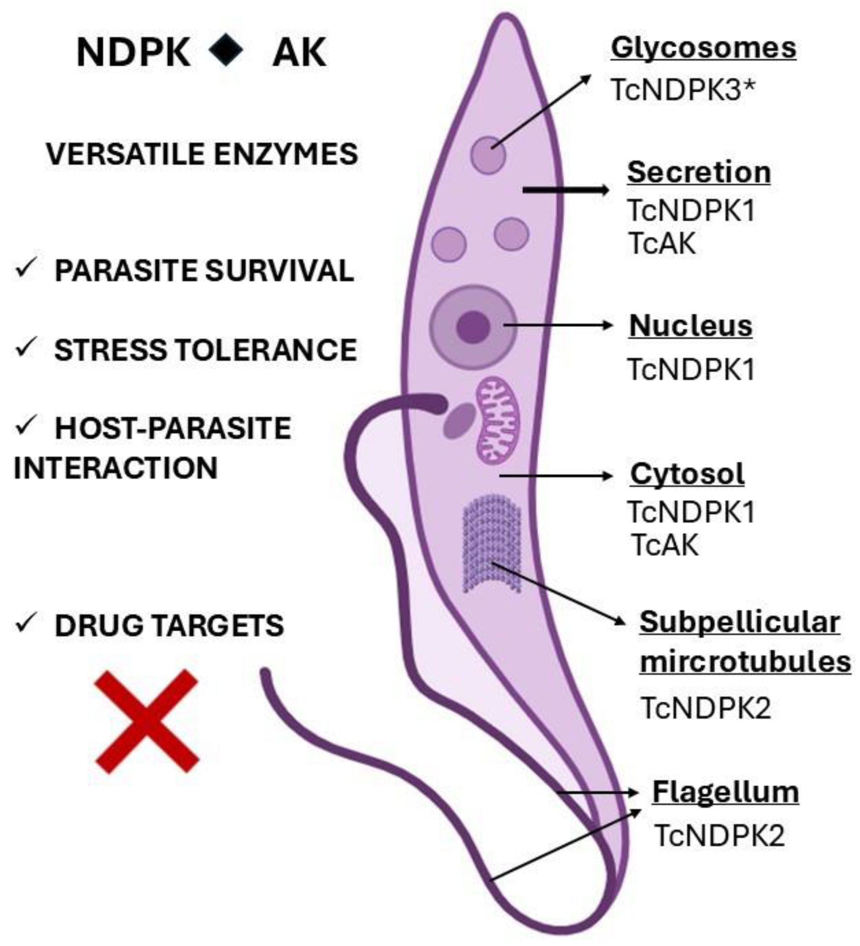
The study of metabolic enzymes in trypanosomatids continues to reveal unexpected complexity and biological versatility. Among these, NDPKs and AKs stand out as proteins that combine homeostatic functions with parasite-specific roles. In this review, using T. cruzi as a model, it was exemplified how these enzymes have evolved to support survival under the challenging conditions faced throughout their life cycles. Figure 1 summarizes what was exposed here, the subcellular distribution, secretion of TcNDPKs and TcAK, and their roles (Figure 1).
From a comparative perspective, both NDPKs and AK illustrate different strategies for parasite adaptation. NDPKs are ubiquitous, conserved and multifunctional; even trypanosomatids have diversified their functions beyond nucleotide homeostasis, incorporating roles in DNA damage repair, oxidative stress management, virulence, and secretion. AK, in contrast, is absent in mammals and fulfils a unique role as an energy buffer in parasites, facilitating rapid metabolic adaptation during environmental transitions. Both enzymes converge on a common theme: enabling parasite persistence in hostile conditions and supporting pathogenesis.
The therapeutic potential of these enzymes lies precisely in their divergence from mammalian biology. AK enzymes, despite being present in T. cruzi and T. brucei but lacking in Leishmania, offer a more straightforward opportunity as a drug target against trypanosomatids because of their absence in humans, reducing concerns of host toxicity (although off-target and ADME/Tox profiles must be assessed for all trypanocidal agents). Natural compounds such as delphinidin and capsaicin have already demonstrated proof-of-principle for pharmacological inhibition of TcAK, highlighting the potential of repurposing bioactive molecules for Chagas ’disease. For NDPKs, the challenge is greater due to their high conservation; however, structural differences and the importance of their non-canonical roles (e.g., DNA repair, secretion) provide a rationale for selective targeting. Nebivolol and telmisartan are two clinically used drugs that reinforced the interest in drug repurposing targeting TcNDPK1. Nonetheless, in both cases, additional genetic approaches and validation in vivo are still needed to establish their relevance.
Beyond chemotherapy, these enzymes hold pathogenic and immunological significance. Canonical NDPKs are secreted and in Leishmania can modulate host signalling, a property that could potentially serve as a biomarker of infection or virulence. AK, on the other hand, is a well-recognized allergen in invertebrates and has been shown to elicit IgE responses in T. cruzi infections. The allergenic potential of AK in trypanosomatids raises intriguing possibilities about its contribution to host immune modulation. Understanding these dimensions may enrich our view of parasite–host interactions and expand the clinical implications of these enzymes.
Looking forward, further investigations must be addressed to fill some knowledge gaps such as, for example, interactomes that will be critical to fully understand their pleiotropic functions.
In conclusion, NDPKs and AK open new opportunities for therapeutic discovery and a deeper understanding of parasite biology. While challenges remain, the progress achieved thus far underscores their potential to inspire innovative strategies against neglected tropical diseases.

Author Contributions

M.R.M.: conceptualization, writing. C.R.: writing, proofreading. M.S.: writing, proofreading. F.A.D.: proofreading. All authors have read and agreed to the published version of the manuscript.

Funding

This work was supported by Consejo Nacional de Investigaciones Científicas y Técnicas (CONICET, PIBAA 28720210100409, PIP 0792) and Agencia Nacional de Promoción Científica y Tecnológica (ANPCyT, FONCYT PICT: 2018-1801, 2018-0585, 2018-01871, 2019-02622).

Institutional Review Board Statement

Not applicable.

Informed Consent Statement

Not applicable.

Data Availability Statement

No new data were created or analyzed in this study. Data sharing is not applicable to this article.

Acknowledgments

We thank Claudio Alejandro Pereira, founder of the Laboratorio de Parasitología Molecular at IDIM (UBA-CONICET), for his guidance and pioneering research on AK and NDPKs in T. cruzi.

Conflicts of Interest

The authors declare no conflicts of interest.

References

  1. Rassi, A.; Rassi, A.; Marin-Neto, J.A. Chagas Disease. Lancet 2010, 375, 1388–1402. [Google Scholar] [CrossRef] [PubMed]
  2. Zuma, A.A.; de Souza, W. Chagas Disease Chemotherapy: What Do We Know So Far? Curr. Pharm. Des. 2021, 27, 3963–3995. [Google Scholar] [CrossRef] [PubMed]
  3. Morillo, C.A.; Marin-Neto, J.A.; Avezum, A.; Sosa-Estani, S.; Rassi, A., Jr.; Rosas, F.; Villena, E.; Quiroz, R.; Bonilla, R.; Britto, C.; et al. Randomized Trial of Benznidazole for Chronic Chagas’ Cardiomyopathy. N. Engl. J. Med. 2015, 373, e124–e125. [Google Scholar] [CrossRef]
  4. Galceran, F.; Digirolamo, F.A.; Rengifo, M.; Reigada, C.; Saye, M.; Maciel, B.J.; Estecho, I.G.; Errasti, A.E.; Pereira, C.A.; Miranda, M.R. Identifying Inhibitors of Trypanosoma cruzi Nucleoside Diphosphate Kinase 1 as Potential Repurposed Drugs for Chagas’ Disease. Biochem. Pharmacol. 2023, 216, 115766. [Google Scholar] [CrossRef] [PubMed]
  5. Reigada, C.; Sayé, M.; Di Girolamo, F.; Valera-Vera, E.A.; Pereira, C.A.; Miranda, M.R. Role of Trypanosoma cruzi Nucleoside Diphosphate Kinase 1 in Dna Damage Responses. Mem. Inst. Oswaldo Cruz 2020, 115, e200019. [Google Scholar] [CrossRef]
  6. Miranda, M.R.; Canepa, G.E.; Bouvier, L.A.; Pereira, C.A. Trypanosoma cruzi: Oxidative Stress Induces Arginine Kinase Expression. Exp. Parasitol. 2006, 114, 341–344. [Google Scholar] [CrossRef]
  7. Pereira, C.A.; Reigada, C.; Sayé, M.; Digirolamo, F.A.; Miranda, M.R. Cytosolic Trypanosoma cruzi Nucleoside Diphosphate Kinase Generates Large Granules That Depend on Its Quaternary Structure. Exp. Parasitol. 2014, 142, 43–50. [Google Scholar] [CrossRef]
  8. Valera-Vera, E.A.; Concepción, J.L.; Cáceres, A.J.; Acevedo, G.R.; Fernández, M.; Hernández, Y.; Digirolamo, F.A.; Duschak, V.G.; Soprano, L.L.; Pereira, C.A.; et al. IgE Antibodies against Trypanosoma cruzi Arginine Kinase in Patients with Chronic Chagas Disease. Mol. Immunol. 2021, 138, 68–75. [Google Scholar] [CrossRef]
  9. Agarwal, R.P.; Robison, B.; Parks, R.E. Nucleoside Diphosphokinase from Human Erythrocytes. Methods Enzymol. 1978, 51, 376–386. [Google Scholar] [CrossRef]
  10. Kolli, B.K.; Kostal, J.; Zaborina, O.; Chakrabarty, A.M.; Chang, K.P. Leishmania-Released Nucleoside Diphosphate Kinase Prevents ATP-Mediated Cytolysis of Macrophages. Mol. Biochem. Parasitol. 2008, 158, 163–175. [Google Scholar] [CrossRef]
  11. Yang, M.; Jarrett, S.G.; Craven, R.; Kaetzel, D.M. YNK1, the Yeast Homolog of Human Metastasis Suppressor NM23, Is Required for Repair of UV Radiation- and Etoposide-Induced DNA Damage. Mutat. Res. Fundam. Mol. Mech. Mutagen. 2009, 660, 74–78. [Google Scholar] [CrossRef]
  12. Zheng, S.; Kusnadi, A.; Choi, J.E.; Vuong, B.Q.; Rhodes, D.; Chaudhuri, J. NME Proteins Regulate Class Switch Recombination. FEBS Lett. 2019, 593, 80–87. [Google Scholar] [CrossRef]
  13. Filić, V.; Marinović, M.; Šoštar, M.; Weber, I. Modulation of Small GTPase Activity by NME Proteins. Lab. Investig. 2018, 98, 589–601. [Google Scholar] [CrossRef]
  14. Yu, H.; Rao, X.; Zhang, K. Nucleoside Diphosphate Kinase (Ndk): A Pleiotropic Effector Manipulating Bacterial Virulence and Adaptive Responses. Microbiol. Res. 2017, 205, 125–134. [Google Scholar] [CrossRef] [PubMed]
  15. Attwood, P.V.; Muimo, R. The Actions of NME1/NDPK-A and NME2/NDPK-B as Protein Kinases. Lab. Investig. 2018, 98, 283–290. [Google Scholar] [CrossRef]
  16. Smith, E.; Morrison, J.F. Kinetic Studies on the Arginine Kinase Reaction. J. Biol. Chem. 1969, 244, 4224–4234. [Google Scholar] [CrossRef] [PubMed]
  17. Hird, F.J.R. The Importance of Arginine in Evolution. Comp. Biochem. Physiol. B Comp. Biochem. 1986, 85, 285–288. [Google Scholar] [CrossRef] [PubMed]
  18. Hird, F.J.; Davuluri, S.P.; McLean, R.M. Evolutionary Relationships between Arginine and Creatine in Muscle. Adv. Exp. Med. Biol. 1982, 153, 401–406. [Google Scholar] [CrossRef] [PubMed]
  19. Yu, H.; Xiong, J.; Zhang, R.; Hu, X.; Qiu, J.; Zhang, D.; Xu, X.; Xin, R.; He, X.; Xie, W.; et al. Ndk, a Novel Host-Responsive Regulator, Negatively Regulates Bacterial Virulence through Quorum Sensing in Pseudomonas Aeruginosa. Sci. Rep. 2016, 6, 28684. [Google Scholar] [CrossRef]
  20. Sun, J.; Singh, V.; Lau, A.; Stokes, R.W.; Obregón-Henao, A.; Orme, I.M.; Wong, D.; Av-Gay, Y.; Hmama, Z. Mycobacterium tuberculosis Nucleoside Diphosphate Kinase Inactivates Small GTPases Leading to Evasion of Innate Immunity. PLoS Pathog. 2013, 9, e1003499. [Google Scholar] [CrossRef]
  21. Wu, M.; Yu, G.; Yan, T.; Ke, D.; Wang, Q.; Liu, R.; Wang, J.Z.; Zhang, B.; Chen, D.; Wang, X. Phosphorylation of SET Mediates Apoptosis via P53 Hyperactivation and NM23-H1 Nuclear Import. Neurobiol. Aging 2018, 69, 38–47. [Google Scholar] [CrossRef]
  22. Steeg, P.S.; Bevilacqua, G.; Kopper, L.; Thorgeirsson, U.P.; Talmadge, J.E.; Liotta, L.A.; Sobel, M.E. Evidence for a Novel Gene Associated with Low Tumor Metastatic Potential. J. Natl. Cancer Inst. 1988, 80, 200–204. [Google Scholar] [CrossRef]
  23. Sengupta, A.; Roy, S.S.; Chowdhury, S. Non-Duplex G-Quadruplex DNA Structure: A Developing Story from Predicted Sequences to DNA Structure-Dependent Epigenetics and Beyond. Acc. Chem. Res. 2021, 54, 46–56. [Google Scholar] [CrossRef]
  24. Miranda, M.R.; Canepa, G.E.; Bouvier, L.A.; Pereira, C.A. Trypanosoma cruzi: Multiple Nucleoside Diphosphate Kinase Isoforms in a Single Cell. Exp. Parasitol. 2008, 120, 103–107. [Google Scholar] [CrossRef]
  25. de los Milagros Cámara, M.; Bouvier, L.; Reigada, C.; Digirolamo, F.A.; Sayé, M.; Pereira, C.A. A Novel Stage-Specific Glycosomal Nucleoside Diphosphate Kinase from Trypanosoma cruzi. Folia Parasitol. 2017, 64, 006. [Google Scholar] [CrossRef] [PubMed]
  26. Miranda, M.R.; De Los Milagros Camara, M.; Bouvier, L.A.; Pereira, C.A. TcNDPK2, a Trypanosoma cruzi Microtubule-Associated Nucleoside Diphosphate Kinase. Mol. Biochem. Parasitol. 2011, 177, 152–155. [Google Scholar] [CrossRef]
  27. Gazanion, E.; Vergnes, B. Protozoan Parasite Auxotrophies and Metabolic Dependencies. Metab. Interact. Infect. 2018, 109, 351–375. [Google Scholar] [CrossRef]
  28. Hammond, D.J.; Gutteridge, W.E. Purine and Pyrimidine Metabolism in the Trypanosomatidae. Mol. Biochem. Parasitol. 1984, 13, 243–261. [Google Scholar] [CrossRef]
  29. Bayer-Santos, E.; Aguilar-Bonavides, C.; Rodrigues, S.P.; Cordero, E.M.; Marques, A.F.; Varela-Ramirez, A.; Choi, H.; Yoshida, N.; Da Silveira, J.F.; Almeida, I.C. Proteomic Analysis of Trypanosoma cruzi Secretome: Characterization of Two Populations of Extracellular Vesicles and Soluble Proteins. J. Proteome Res. 2013, 12, 883–897. [Google Scholar] [CrossRef] [PubMed]
  30. Miranda, M.R.; Bouvier, L.A.; Canepa, G.E.; Pereira, C.A. Subcellular Localization of Trypanosoma cruzi Arginine Kinase. Parasitology 2009, 136, 1201–1207. [Google Scholar] [CrossRef] [PubMed]
  31. Brossas, J.Y.; Gulin, J.E.N.; Bisio, M.M.C.; Chapelle, M.; Marinach-Patrice, C.; Bordessoules, M.; Ruiz, G.P.; Vion, J.; Paris, L.; Altcheh, J.; et al. Secretome Analysis of Trypanosoma cruzi by Proteomics Studies. PLoS ONE 2017, 12, e0185504. [Google Scholar] [CrossRef]
  32. Andrade, H.M.; Murta, S.M.F.; Chapeaurouge, A.; Perales, J.; Nirdé, P.; Romanha, A.J. Proteomic Analysis of Trypanosoma cruzi Resistance to Benznidazole. J. Proteome Res. 2008, 7, 2357–2367. [Google Scholar] [CrossRef]
  33. Gomez Barroso, J.A.; Miranda, M.R.; Pereira, C.A.; Garratt, R.C.; Aguilar, C.F. X-Ray Diffraction and in Vivo Studies Reveal the Quinary Structure of Trypanosoma cruzi Nucleoside Diphosphate Kinase 1: A Novel Helical Oligomer Structure. Acta Crystallogr. D Struct. Biol. 2022, 78, 30–42. [Google Scholar] [CrossRef]
  34. Munier, A.; Serres, C.; Kann, M.L.; Boissan, M.; Lesaffre, C.; Capeau, J.; Fouquet, J.P.; Lacombe, M.L. Nm23/NDP Kinases in Human Male Germ Cells: Role in Spermiogenesis and Sperm Motility? Exp. Cell Res. 2003, 289, 295–306. [Google Scholar] [CrossRef] [PubMed]
  35. Ma, M.; Stoyanova, M.; Rademacher, G.; Dutcher, S.K.; Brown, A.; Zhang, R. Structure of the Decorated Ciliary Doublet Microtubule. Cell 2019, 179, 909–922.e12. [Google Scholar] [CrossRef] [PubMed]
  36. Silvestre, A.; Shintre, S.S.; Rachidi, N. Released Parasite-Derived Kinases as Novel Targets for Antiparasitic Therapies. Front. Cell. Infect. Microbiol. 2022, 12, 825458. [Google Scholar] [CrossRef]
  37. Chou, T.C. Derivation and Properties of Michaelis-Menten Type and Hill Type Equations for Reference Ligands. J. Theor. Biol. 1976, 59, 253–276. [Google Scholar] [CrossRef]
  38. Martin, M.W.; O’Sullivan, A.J.; Gomperts, B.D. Inhibition by Cromoglycate and Some Flavonoids of Nucleoside Diphosphate Kinase and of Exocytosis from Permeabilized Mast Cells. Br. J. Pharmacol. 1995, 115, 1080–1086. [Google Scholar] [CrossRef] [PubMed]
  39. Lin, X.; Momany, C.; Momany, M. SwoHp, a Nucleoside Diphosphate Kinase, Is Essential in Aspergillus Nidulans. Eukaryot. Cell 2003, 2, 1169–1177. [Google Scholar] [CrossRef]
  40. Hemmerich, S.; Yarden, Y.; Pecht, I. A Cromoglycate Binding Protein from Rat Mast Cells of a Leukemia Line Is a Nucleoside Diphosphate Kinase. Biochemistry 1992, 31, 4574–5479. [Google Scholar] [CrossRef]
  41. Hunger-Glaser, I.; Hemphill, A.; Shalaby, T.; Hänni, M.; Seebeck, T. Nucleoside Diphosphate Kinase of Trypanos. brucei. Gene 2000, 257, 251–257. [Google Scholar] [CrossRef]
  42. Geiger, A.; Hirtz, C.; Bécue, T.; Bellard, E.; Centeno, D.; Gargani, D.; Rossignol, M.; Cuny, G.; Peltier, J.B. Exocytosis and Protein Secretion in Trypanosoma. BMC Microbiol. 2010, 10, 20. [Google Scholar] [CrossRef]
  43. de Oliveira, A.H.C.; Ruiz, J.C.; Cruz, A.K.; Greene, L.J.; Rosa, J.C.; Ward, R.J. Subproteomic Analysis of Soluble Proteins of the Microsomal Fraction from Two Leishmania Species. Comp. Biochem. Physiol. Part D Genom. Proteom. 2006, 1, 300–308. [Google Scholar] [CrossRef]
  44. Kushawaha, P.K.; Pati Tripathi, C.D.; Dube, A. Leishmania donovani Secretory Protein Nucleoside Diphosphate Kinase b Localizes in Its Nucleus and Prevents ATP Mediated Cytolysis of Macrophages. Microb. Pathog. 2022, 166, 105457. [Google Scholar] [CrossRef] [PubMed]
  45. Paul, M.L.S.; Kaur, A.; Geete, A.; Sobhia, M.E. Essential Gene Identification and Drug Target Prioritization in Leishmania Species. Mol. Biosyst. 2014, 10, 1184–1195. [Google Scholar] [CrossRef]
  46. Bernardo, V.G.; de Araújo, R.S.A.; de Mélo, N.B.; Coutinho, R.E.P.; de Sousa, J.M.S.; da Silva Sousa, M.G.G.; de Sousa, N.F.; dos Santos Silva, W.F.; da Silva Santos-Júnior, P.F.; da Silva, T.G.; et al. Drug Design and Synthesis of New N-Substituted-Thienopyridine Based on 2-Aminothiophene Derivatives as Antileishmanial Agents. Bioorganic Med. Chem. 2026, 132, 118475. [Google Scholar] [CrossRef] [PubMed]
  47. de Menezes, R.P.B.; de Assis, E.B.; de Sousa, N.F.; de Souza, J.M.S.; da França Rodrigues, K.A.; Scotti, L.; Tavares, J.F.; da Silva, M.S.; Scotti, M.T. Exploring Lamiaceae Diterpenoids as Potential Multitarget Therapeutics for Leishmaniasis and Chagas Disease. Mol. Divers, 2025; online ahead of print. [Google Scholar] [CrossRef]
  48. Vieira, P.S.; Souza, T.d.A.C.B.; Honorato, R.V.; Zanphorlin, L.M.; Severiano, K.U.; Rocco, S.A.; de Oliveira, A.H.C.; Cordeiro, A.T.; Oliveira, P.S.L.; de Giuseppe, P.O.; et al. Pyrrole-Indolinone SU11652 Targets the Nucleoside Diphosphate Kinase from Leishmania Parasites. Biochem. Biophys. Res. Commun. 2017, 488, 461–465. [Google Scholar] [CrossRef]
  49. Mishra, A.K.; Singh, N.; Agnihotri, P.; Mishra, S.; Singh, S.P.; Kolli, B.K.; Chang, K.P.; Sahasrabuddhe, A.A.; Siddiqi, M.I.; Pratap, J.V. Discovery of Novel Inhibitors for Leishmania Nucleoside Diphosphatase Kinase (NDK) Based on Its Structural and Functional Characterization. J. Comput. Aided Mol. Des. 2017, 31, 547–562. [Google Scholar] [CrossRef] [PubMed]
  50. Gupta, A.; Beg, M.A.; Badhwar, S.; Srivastava, S.; Srivastava, R.; Puri, N.; Saxena, A.; Abdin, M.Z.; Selvapandiyan, A. Nucleoside Diphosphate Kinase (LdNDK2): A Metacyclogenesis-Regulating Kinase Essential for Leishmania Parasite Survival within Eukaryotic Host Cells. Microb. Pathog. 2025, 210, 108192. [Google Scholar] [CrossRef]
  51. Moreira, D.S.; Murta, S.M.F. Involvement of Nucleoside Diphosphate Kinase b and Elongation Factor 2 in Leishmania braziliensis Antimony Resistance Phenotype. Parasit. Vectors 2016, 9, 641. [Google Scholar] [CrossRef]
  52. Tonoli, C.C.C.; Vieira, P.S.; Ward, R.J.; Arni, R.K.; De Oliveira, A.H.C.; Murakami, M.T. Production, Purification, Crystallization and Preliminary X-Ray Diffraction Studies of the Nucleoside Diphosphate Kinase b from Leishmania major. Acta Crystallogr. Sect. F Struct. Biol. Cryst. Commun. 2009, 65, 1116–1119. [Google Scholar] [CrossRef] [PubMed]
  53. Voncken, F.; Gao, F.; Wadforth, C.; Harley, M.; Colasante, C. The Phosphoarginine Energy-Buffering System of Trypanosoma brucei Involves Multiple Arginine Kinase Isoforms with Different Subcellular Locations. PLoS ONE 2013, 8, e65908. [Google Scholar] [CrossRef]
  54. Ooi, C.P.; Rotureau, B.; Gribaldo, S.; Georgikou, C.; Julkowska, D.; Blisnick, T.; Perrot, S.; Subota, I.; Bastin, P. The Flagellar Arginine Kinase in Trypanosoma brucei Is Important for Infection in Tsetse Flies. PLoS ONE 2015, 10, e0133676. [Google Scholar] [CrossRef]
  55. Pereira, C. Arginine Kinase: A Potential Pharmacological Target in Trypanosomiasis. Infect. Disord. Drug Targets 2014, 14, 30–36. [Google Scholar] [CrossRef]
  56. Salmazo, P.; De Giuseppe, V.P.O.; Murakami, M.T.; De Oliveira, A.H.C. Crystal Structure and Biophysical Characterization of the Nucleoside Diphosphate Kinase from Leishmania braziliensis. BMC Struct. Biol. 2015, 15, 2. [Google Scholar] [CrossRef][Green Version]
  57. Ellington, W.R. Evolution and Physiological Roles of Phosphagen Systems. Annu. Rev. Physiol. 2001, 63, 289–325. [Google Scholar] [CrossRef] [PubMed]
  58. Pereira, C.A.; Alonso, G.D.; Ivaldi, S.; Silber, A.M.; Alves, M.J.M.; Torres, H.N.; Flawiá, M.M. Arginine Kinase Overexpression Improves Trypanosoma cruzi Survival Capability. FEBS Lett. 2003, 554, 201–205. [Google Scholar] [CrossRef]
  59. Alonso, G.D.; Pereira, C.A.; Remedi, M.S.; Paveto, M.C.; Cochella, L.; Ivaldi, M.S.; Gerez de Burgos, N.M.; Torres, H.N.; Flawiá, M.M. Arginine Kinase of the Flagellate Protozoa Trypanosoma cruzi: Regulation of Its Expression and Catalytic Activity. FEBS Lett. 2001, 498, 22–25. [Google Scholar] [CrossRef]
  60. Pereira, C.A.; Alonso, G.D.; Ivaldi, S.; Silber, A.; Alves, M.J.M.; Bouvier, L.A.; Flawiá, M.M.; Torres, H.N. Arginine Metabolism in Trypanosoma cruzi Is Coupled to Parasite Stage and Replication. FEBS Lett. 2002, 526, 111–114. [Google Scholar] [CrossRef] [PubMed]
  61. Miranda, M.R.; Sayé, M.; Bouvier, L.A.; de los Milagros Cámara, M.; Montserrat, J.; Pereira, C.A. Cationic Amino Acid Uptake Constitutes a Metabolic Regulation Mechanism and Occurs in the Flagellar Pocket of Trypanosoma cruzi. PLoS ONE 2012, 7, e32760. [Google Scholar] [CrossRef]
  62. Gore, J.C.; Schal, C. Cockroach Allergen Biology and Mitigation in the Indoor Environment. Annu. Rev. Entomol. 2007, 52, 439–463. [Google Scholar] [CrossRef]
  63. Valera-Vera, E.; Reigada, C.; Sayé, M.; Digirolamo, F.A.; Galceran, F.; Miranda, M.R.; Pereira, C.A. Trypanocidal Activity of the Anthocyanidin Delphinidin, a Non-Competitive Inhibitor of Arginine Kinase. Nat. Prod. Res. 2022, 36, 3153–3157. [Google Scholar] [CrossRef] [PubMed]
  64. Valera Vera, E.A.; Sayé, M.; Reigada, C.; Damasceno, F.S.; Silber, A.M.; Miranda, M.R.; Pereira, C.A. Resveratrol Inhibits Trypanosoma cruzi Arginine Kinase and Exerts a Trypanocidal Activity. Int. J. Biol. Macromol. 2016, 87, 498–503. [Google Scholar] [CrossRef]
  65. Valera-Vera, E.A.; Reigada, C.; Sayé, M.; Digirolamo, F.A.; Galceran, F.; Miranda, M.R.; Pereira, C.A. Effect of Capsaicin on the Protozoan Parasite Trypanosoma cruzi. FEMS Microbiol. Lett. 2020, 367, fnaa194. [Google Scholar] [CrossRef]
  66. Binder, M.; Mahler, V.; Hayek, B.; Sperr, W.R.; Schöller, M.; Prozell, S.; Wiedermann, G.; Valent, P.; Valenta, R.; Duchêne, M. Molecular and Immunological Characterization of Arginine Kinase from the Indianmeal Moth, Plodia Interpunctella, a Novel Cross-Reactive Invertebrate Pan-Allergen. J. Immunol. 2001, 167, 5470–5477. [Google Scholar] [CrossRef] [PubMed]
  67. Liu, Z.; Xia, L.; Wu, Y.; Xia, Q.; Chen, J.; Roux, K.H. Identification and Characterization of an Arginine Kinase as a Major Allergen from Silkworm (Bombyx Mori) Larvae. Int. Arch. Allergy Immunol. 2009, 150, 8–14. [Google Scholar] [CrossRef]
  68. Popova-Butler, A.; Dean, D.H. Proteomic Analysis of the Mosquito Aedes Aegypti Midgut Brush Border Membrane Vesicles. J. Insect Physiol. 2009, 55, 264–272. [Google Scholar] [CrossRef] [PubMed]
  69. Hales, B.J.; Laing, I.A.; Pearce, L.J.; Hazell, L.A.; Mills, K.L.; Chua, K.Y.; Thornton, R.B.; Richmond, P.; Musk, A.W.; James, A.L.; et al. Distinctive Immunoglobulin E Anti-House Dust Allergen-Binding Specificities in a Tropical Australian Aboriginal Community. Clin. Exp. Allergy 2007, 37, 1357–1363. [Google Scholar] [CrossRef]
  70. Sookrung, N.; Chaicumpa, W.; Tungtrongchitr, A.; Vichyanond, P.; Bunnag, C.; Ramasoota, P.; Tongtawe, P.; Sakolvaree, Y.; Tapchaisri, P. Periplaneta Americana Arginine Kinase as a Major Cockroach Allergen among Thai Patients with Major Cockroach Allergies. Environ. Health Perspect. 2006, 114, 875–880. [Google Scholar] [CrossRef]
  71. Pereira, C.A.; Alonso, G.D.; Torres, H.N.; Flawiá, M.M. Arginine Kinase: A Common Feature for Management of Energy Reserves in African and American Flagellated Trypanosomatids. J. Eukaryot. Microbiol. 2002, 49, 82–85. [Google Scholar] [CrossRef]
  72. Camargo, E.P.; Coelho, J.A.; Moraes, G.; Figueiredo, E.N. Trypanosoma spp., Leishmania spp. and Leptomonas spp.: Enzymes of Ornithine-Arginine Metabolism. Exp. Parasitol. 1978, 46, 141–144. [Google Scholar] [CrossRef] [PubMed]
  73. Ferrins, L.; Diaz, R.; Cordon-Obras, C.; Rojas-Barros, D.; Quotadamo, A.; Oehme, D.P.; Ceballos-Pérez, G.; Swaminathan, U.; Pérez-Moreno, G.; Bosch-Navarrete, C.; et al. Pharmacophore Identification and Structure-Activity Relationship Analysis of a Series of Substituted Azaindoles as Inhibitors of Trypanosoma brucei. J. Med. Chem. 2024, 67, 13985–14006. [Google Scholar] [CrossRef]
  74. Adeyemi, O.S.; Arowolo, A.T.; Hetta, H.F.; Al-Rejaie, S.; Rotimi, D.; El-Saber Batiha, G. Apoferritin and Apoferritin-Capped Metal Nanoparticles Inhibit Arginine Kinase of Trypanosoma brucei. Molecules 2020, 25, 3432. [Google Scholar] [CrossRef] [PubMed]
  75. Adeyemi, O.S.; Whiteley, C.G. Interaction of Nanoparticles with Arginine Kinase from Trypanosoma brucei: Kinetic and Mechanistic Evaluation. Int. J. Biol. Macromol. 2013, 62, 450–456. [Google Scholar] [CrossRef] [PubMed]
Figure 1. Schematic representation of a T. cruzi epimastigote showing the subcellular localization of nucleoside diphosphate kinases and arginine kinase (TcNDPKs and TcAK). These enzymes are distributed throughout the parasite body and can also be secreted, contributing to various cellular processes beyond their canonical metabolic roles, such as stress tolerance and host–parasite interactions. Both kinases represent valuable drug targets for Chagas disease. * TcNDPK3 is expressed exclusively in trypomastigotes.
Kinasesphosphatases 04 00002 g001
Table 1. Summary of T. cruzi NDPKs and AK.
Kinase/
Isoform
Length (aa)TritrypDB IDTypeKinase
Activity
Subcellular Localization *SecretionRef.
TcNDPK1153TcCLB.508707.200CanonicalYesCytosol
Nucleus
Lipidic vesicles[4,5,7,24,29]
TcNDPK2334TcCLB.508461.400DivergentYesCytosol
Cytoskeleton
Flagellum
Nd[24,26]
TcNDPK3349TcCLB.510879.210DivergentYesGlycosomeNd[25]
TcNDPK4632TcCLB.508989.100DivergentPutativeNdNd[24]
TcAK357TcCLB.507241.30-YesCytosol
Heterogeneously
distributed
Yes[30,31]
TritrypDB: https://tritrypdb.org/tritrypdb (accessed on 10 October 2025). Nd: not determined. * Determined in epimastigotes, except for TcNDPK3, which is expressed in trypomastigotes.
Table 2. TcNDPKs and TcAK homologues in the trypanosomatids T. brucei and Leishmania.
Kinase/
Isoform
in T. cruzi
Homologues in T. brucei
TritrypDB ID
Length (aa)
Homologues in L. major a
(TritrypDB ID)
Length (aa)
Ref.
TcNDPK1TbNDPK1
Tb927.11.16130
153
LmNDPK1/LmNDKb
LmjF.32.2950
151
[24,41,42,43,44,51,52]
TcNDPK2TbNDPK2
Tb927.9.11260
334
LmNDPK2
LmjF.35.3870
337
[24,26]
TcNDPK3TbNDPK3
Tb927.4.1720
349
LmNDPK3
LmjF.34.2980
343
[24]
TcNDPK4TbNDPK4
Tb927.8.4510
634
-[24]
TcAKTbAK1/AK3
Tb927.9.6170
404
-[53,54,55]
TbAK2/AK2
Tb927.9.6230
370
TbAK3/AK1
Tb927.9.6290
356
TritrypID (https://tritrypdb.org/tritrypdb/app, accessed on 17 November 2025) and length are shown. a L. major was used as an example, but NDPKs were described in L. braziliensis [51,56], L. donovani [44,50] and L. amazonensis [43].
Disclaimer/Publisher’s Note: The statements, opinions and data contained in all publications are solely those of the individual author(s) and contributor(s) and not of MDPI and/or the editor(s). MDPI and/or the editor(s) disclaim responsibility for any injury to people or property resulting from any ideas, methods, instructions or products referred to in the content.