1. Introduction
Ticks are obligate hematophagous ectoparasites of considerable ecological, medical, and veterinary relevance because they serve as vectors and reservoirs of a wide variety of microorganisms affecting both humans and animals [
1,
2]. Among them, soft ticks (Argasidae) differ from hard ticks by their nidicolous lifestyle, rapid feeding behavior, repeated blood meals, multiple nymphal instars, and remarkable resistance to prolonged starvation. These biological characteristics enable long-term persistence within sheltered microhabitats, repeated interactions with multiple hosts, and continuous exposure to host-associated microorganisms throughout their life cycle [
3,
4]. Consequently, argasid ticks represent particularly valuable models for studying host–microbe interactions and the ecological processes shaping arthropod-associated microbial communities [
5,
6,
7].
Argas persicus, commonly known as the fowl tick, is widely distributed throughout temperate and subtropical regions and is primarily associated with domestic poultry, pigeons, and other birds [
4,
5]. As a nidicolous species, it typically inhabits cracks and crevices within animal shelters, where it can survive for prolonged periods between blood meals. Although birds constitute its principal hosts,
A. persicus has also been reported from livestock facilities and occasionally from mammals, indicating ecological flexibility in the use of sheltered peridomestic habitats rather than strict host specificity [
8]. Owing to its close association with animal shelters and repeated blood-feeding behavior, this tick has long been recognized as a vector or reservoir of microorganisms of veterinary importance, including
Borrelia anserina, several
Rickettsia species, and West Nile virus [
9,
10]. Despite its veterinary relevance, surprisingly little is known about the bacterial communities naturally associated with this species.
Recent advances in high-throughput sequencing have demonstrated that the microbiota of ticks extends far beyond pathogenic bacteria and includes diverse endosymbiotic microorganisms that contribute to essential physiological processes such as nutrient provisioning, vitamin biosynthesis, metabolism, reproduction, osmoregulation, and host survival [
6,
11]. These symbiotic bacteria are increasingly recognized as key components of tick biology because they can influence host fitness, microbial community assembly, and potentially vector competence.
Previous studies on
A. persicus have shown that members of the phylum Proteobacteria dominate the bacterial community, whereas genera such as
Rickettsia,
Stenotrophomonas,
Spiroplasma, and
Coxiella may occur as major bacterial associates depending on the geographical origin and developmental stage of the ticks [
7]. However, investigations conducted in other argasid species indicate that bacterial communities are highly variable across taxa and geographic regions, ranging from relatively diverse assemblages to communities dominated by one or a few maternally inherited endosymbionts, including
Coxiella,
Francisella,
Rickettsia, and
Occidentia [
6,
12]. This growing body of evidence suggests that microbial community structure in argasid ticks is highly context-dependent and likely shaped by complex interactions among host species, endosymbionts, environmental conditions, and ecological history.
Mexico harbors a diverse fauna of soft ticks, with species belonging to the genera
Argas,
Ornithodoros, and
Otobius distributed across different ecological regions. An updated taxonomic synthesis of Mexican Argasidae, including identification keys and distributional records, was provided by Guzmán-Cornejo et al. [
13]. Within this context,
A. persicus has been documented in northern Mexico, although information regarding its associated bacterial microbiota remains virtually absent. This knowledge gap limits our understanding of the ecological diversity of argasid microbiomes and the contribution of dominant endosymbionts to tick-associated microbial communities across different environments. Moreover, characterizing the microbiota of poorly studied tick species not only expands taxonomic knowledge but also provides an important foundation for understanding the ecological and functional interactions that shape tick biology and vector competence. Therefore, the present study aimed to characterize the bacterial community associated with
A. persicus collected from a semi-arid region of northern Mexico using 16S rRNA gene amplicon sequencing. Community composition and alpha diversity were evaluated, and taxonomic assignments of the dominant ASVs were independently validated using the NCBI and EMBL-EBI reference databases to improve species-level confidence. To our knowledge, this study represents the first characterization of the bacterial microbiota of
A. persicus in Mexico and provides new insights into the ecological organization of bacterial communities associated with argasid ticks inhabiting semi-arid environments.
2. Materials and Methods
Sampling was conducted during a single field campaign in May 2025 at a single small rustic goat farm located in Ceballos, Durango, Mexico (26°33′10.99″ N, 104°9′19.91″ W) because a naturally established population of
A. persicus was detected inhabiting cracks and crevices in wooden structures within a goat shed. No additional farms or sampling sites were included in this study. The study area is located within a semi-arid region characterized by a very arid climate [
14], with an average annual temperature of 25.5 °C, and an average annual precipitation of 265 mm [
15]. The predominant vegetation is microphile and rosette scrub, as well as halophyte, and gypsophila plants [
16].
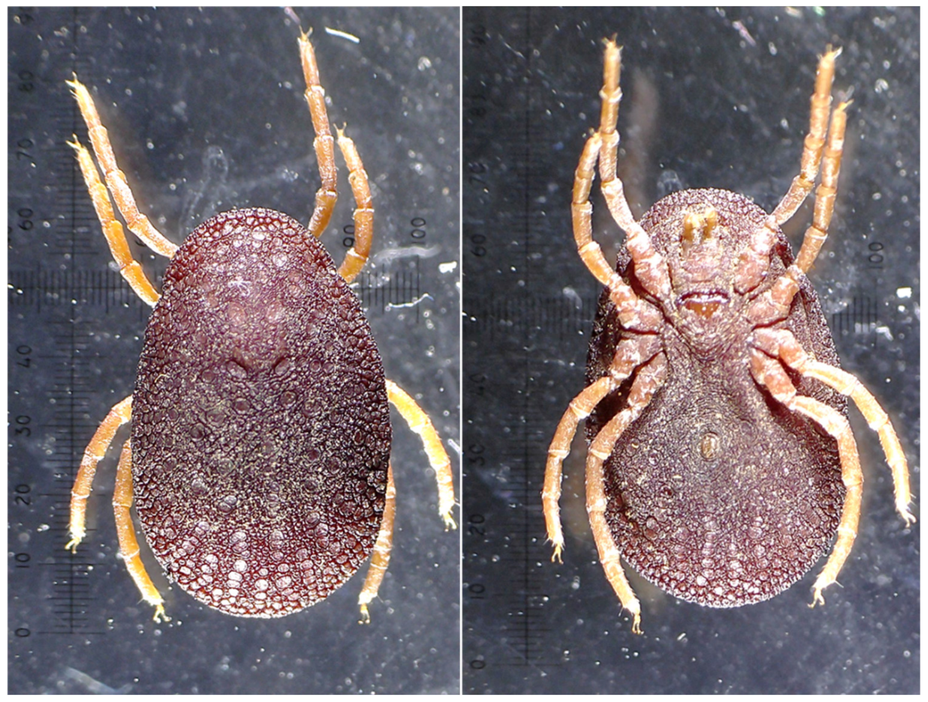
Although a few nymphal specimens were also encountered during field collection, their low numbers precluded meaningful microbiome characterization or the construction of comparable pooled samples. Consequently, only adult ticks were included in the present study. Specimens were manually collected using sterile fine-tipped forceps. Immediately after collection, ticks were individually placed into sterile 2-mL tubes, transported alive to the laboratory, and dissected under sterile conditions on the same day. Ticks were morphologically identified as
Argas persicus using standard taxonomic keys [
17],
Figure 1.
Prior to dissection, ticks were surface sterilized by sequential washing with sterile water and 1% sodium hypochlorite to remove external contaminants, following previously described protocols for tick microbiome studies [
18]. Dissections were performed under sterile conditions using a stereomicroscope and sterile scalpels to isolate the internal tissues. A total of 10 pools were prepared, each consisting of the internal soft tissues recovered from 10 adult
A. persicus ticks following removal of the exoskeleton. Because of the limited number of specimens available, each pool included both male and female adults to maximize DNA yield and provide an overall characterization of the bacterial community associated with the local tick population. Each pool was transferred into BashingBead Zymo Research cell lysis tubes (Zymo Research, Irvine, CA, USA) containing 750 μL of lysis/stabilization solution. The tubes were processed in a TerraLyzer (Zymo Research, Irvine, CA, USA) cellular disruptor for 30 s to ensure efficient homogenization and lysis of tick tissues and associated microorganisms.
Total DNA was extracted from each pool using the Zymobiomics DNA extraction kit (Zymo Research, Irvine, CA, USA), following the manufacturer’s instructions. This process was carried out in a UV laminar flow hood (Thermo Scientific, Marietta, OH, USA) following all sterility protocols. Samples were processed at Novogene Corporation, Inc. (Davis, CA, USA), and the V3–V4 region of the 16S rRNA gene was amplified via the primers 341F (CCTAYGGGRBGCASCAG) and 806R (GGACTACNNGGTATCTAAT). PCR protocol is described in Ontiveros-Chacón et al. [
19]. Sequencing libraries were generated using a PCR-free TruSeq
® DNA sample preparation kit (Illumina, San Diego, CA, USA) following the manufacturer’s instructions, with index codes added. Library quality was assessed using a Qubit
® 2.0 fluorometer (Thermo Scientific, Waltham, MA, USA) and an Agilent Bioanalyzer 2100 (Agilent, Santa Clara, CA, USA) system. Finally, the library was sequenced on an Illumina NovaSeq platform, generating paired-end reads of 250 bp. No extraction blank or PCR negative controls were sequenced in parallel with the samples. This limitation was considered when interpreting rare taxa.
The bioinformatic analysis was performed using Quantitative Insights into Microbial Ecology (QIIME2) on the Linux Ubuntu platform [
20]. The DADA2 algorithm (divisive amplicon denoising algorithm) was employed to remove low-quality sequences, filter chimeric sequences, and generate amplicon sequence variants (ASVs) [
21,
22]. Good’s coverage [
23] was calculated for each pooled sample to assess sequencing depth completeness; values between 0.95 and 1.00 were indicative of adequate coverage. Alpha diversity was estimated for each pooled sample using Observed ASVs, the Shannon diversity index, Pielou’s evenness, and Faith’s phylogenetic diversity (Faith’s PD), calculated with the QIIME2 diversity plugin.
Taxonomic assignment was performed using a Naïve Bayes classifier trained on the Greengenes2 reference database [
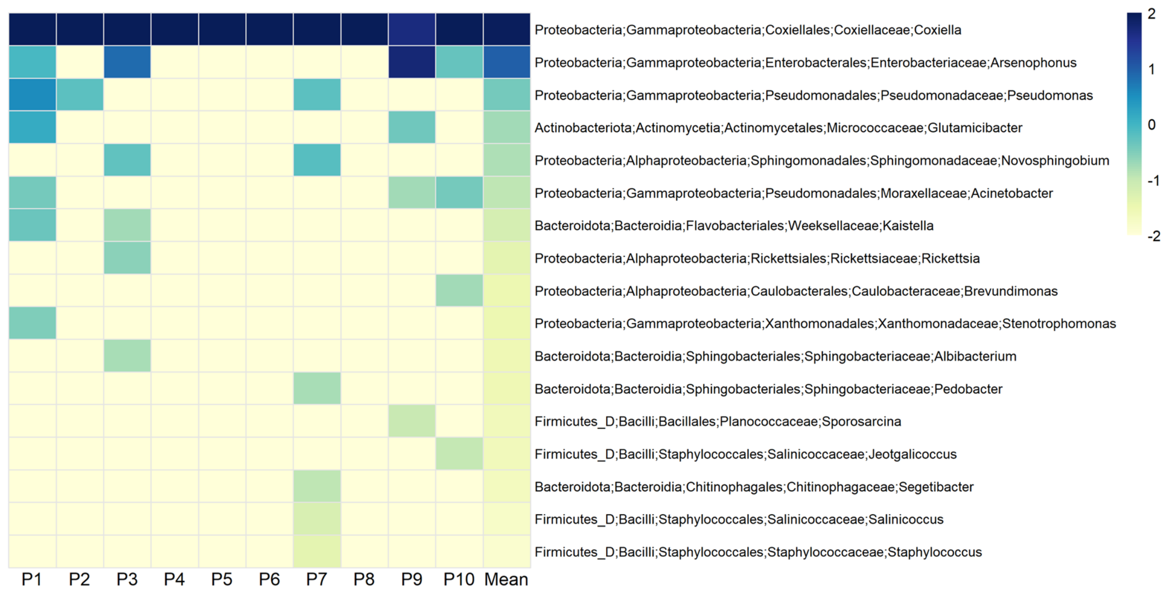
24]. To provide a comprehensive overview of the bacterial community structure, a single heatmap integrating taxonomic assignments from phylum to genus level was generated. Relative abundance values (%) were log10-transformed [log10(x + 0.01)] prior to visualization to enhance contrast among low-abundance taxa. The heatmap was constructed in RStudio (version 2024.12.1). To further evaluate the taxonomic identity of the dominant ASVs, representative sequences were independently compared against the NCBI nucleotide (BLASTn) and EMBL-EBI ENA Ribosomal Sequence databases using nucleotide sequence similarity searches. The highest-scoring matches, sequence identity, query coverage, and alignment statistics obtained from both databases were used to assess the robustness of species-level assignments. Both databases are continuously curated public repositories; therefore, sequence similarity searches were performed using the versions available in July 2026.
3. Results
The average number of reads obtained for the 10 pools was 46,541.1, and the average number of nonchimeric sequences was 21,776.3 (45.7%;
Table S1). Good’s coverage values were 1.00 for each pool, indicating that sequencing depth was sufficient to capture the majority of bacterial diversity. Alpha diversity metrics consistently indicated a low-diversity bacterial community across all pooled samples (
Table S2). Observed ASVs ranged from 4 to 16 (mean = 8.1), while Shannon diversity index values varied between 0.012 and 1.130 (mean = 0.345). Pielou’s evenness ranged from 0.006 to 0.377 (mean = 0.107), and Faith’s PD values varied between 0.680 and 1.891 (mean = 1.251).
The bacterial community associated with
A. persicus exhibited a highly uneven taxonomic structure across all hierarchical levels. At the phylum level, Proteobacteria overwhelmingly dominated the microbiota, accounting for 99.69% of the total relative abundance across samples. In contrast, Actinobacteriota (0.18%), Bacteroidota (0.10%), and Firmicutes (0.03%) were detected only at very low abundances. At the class level, the bacterial community was similarly dominated by Gammaproteobacteria (99.53%), whereas Actinomycetia (0.18%), Alphaproteobacteria (0.17%), Bacteroidia (0.10%), and Bacilli (0.03%) were present in negligible proportions. At the order level, Coxiellales represented 92.63% of the total relative abundance, followed by Enterobacterales (6.30%), while Pseudomonadales (0.57%), Actinomycetales (0.18%), and Sphingomonadales (0.12%) each contributed less than 1%. The same dominance pattern was observed at lower taxonomic ranks. Coxiellaceae accounted for 92.63% of all bacterial sequences at the family level, followed by Enterobacteriaceae (6.30%). At the genus level,
Coxiella remained the predominant taxon (92.63%), whereas
Arsenophonus represented 6.30% of the community. All remaining genera, including
Pseudomonas,
Acinetobacter,
Novosphingobium,
Glutamicibacter,
Kaistella,
Rickettsia, and others, individually accounted for less than 1% of the total relative abundance (
Figure 2). Overall, these findings indicate a highly simplified bacterial community dominated by a single bacterial lineage, consistent with a specialized microbiome characterized by very low richness, diversity, and evenness.
The Greengenes2 classifier assigned the two dominant
Coxiella-associated ASVs to
Coxiella burnetii with high classification confidence (0.96–0.97;
Table S3). However, independent BLASTn searches against the NCBI nucleotide and EMBL-EBI ENA Ribosomal Sequence databases recovered highly similar matches to both
C. burnetii genomes and
Coxiella-like endosymbionts associated with soft ticks (
Table 1,
Tables S4 and S5). Sequence identities ranged from approximately 98–99%, indicating that the short V3–V4 16S rRNA amplicons did not provide sufficient phylogenetic resolution to confidently discriminate pathogenic
C. burnetii from closely related tick-associated endosymbionts. Consequently, these sequences were conservatively interpreted as
Coxiella-like endosymbionts throughout the remainder of the study.
4. Discussion
The present study provides the first characterization of the bacterial microbiota associated with an adult population of
Argas persicus from northern Mexico. Across all analyzed pools, the bacterial community exhibited a remarkably simplified structure, overwhelmingly dominated by Proteobacteria and, more specifically, by
Coxiella-like endosymbionts. Although alpha diversity metrics showed some variation among pools, overall diversity remained consistently low. Observed richness ranged from 4 to 16 ASVs, whereas Shannon diversity, Pielou’s evenness, and Faith’s phylogenetic diversity were uniformly low across samples. The higher Shannon and evenness values observed in a few pools reflected the presence of small proportions of secondary bacterial taxa accompanying the dominant
Coxiella-like endosymbionts, whereas pools with the lowest values were almost exclusively dominated by this single bacterial lineage. Overall, these metrics indicate limited within-population variability and are consistent with the markedly simplified bacterial community observed throughout the dataset. These findings contrast with that reported by Duan et al. [
7], who characterized the bacterial microbiota of
A. persicus across multiple developmental stages in China and found
Rickettsia to be the dominant bacterial genus throughout development, whereas
Coxiella occurred at considerably lower relative abundances. However, unlike Duan et al. [
7], the present study was restricted to adult ticks (males and females pooled) collected from a single naturally occurring population in northern Mexico. Consequently, comparisons between studies should be interpreted cautiously, as differences in bacterial community composition may reflect developmental stage in addition to geographic, ecological, and host-associated factors [
7,
11,
12].
The predominance of
Coxiella-like endosymbionts observed here is consistent with previous reports indicating that argasid ticks may harbor bacterial communities dominated by a limited number of maternally inherited symbionts [
6]. In several tick species,
Coxiella-like endosymbionts have been detected in metabolically active tissues, including the ovaries, Malpighian tubules, salivary glands, and, in
A. persicus, the midgut, where they are thought to participate in nutrient provisioning through the biosynthesis of B vitamins and other essential cofactors lacking in vertebrate blood meals [
7,
11]. These metabolic contributions have been associated with physiological processes such as development, reproduction, osmoregulation, and survival. Nevertheless, the functional role of the lineage detected in the present study cannot be inferred directly from 16S rRNA amplicon sequencing alone. Therefore, although the overwhelming predominance of
Coxiella-like endosymbionts is consistent with a stable host association, their biological significance in this
A. persicus population remains hypothetical and requires experimental validation.
An important outcome of the present study was the independent evaluation of the dominant ASVs using three complementary taxonomic resources. While the Greengenes2 classifier assigned the dominant sequences to
Coxiella burnetii, independent BLASTn searches against the NCBI and EMBL-EBI reference databases consistently recovered similarly high sequence identities to both
C. burnetii and tick-associated
Coxiella-like endosymbionts. These comparisons demonstrate the limited discriminatory power of the short V3–V4 region of the 16S rRNA gene for resolving closely related members of the genus
Coxiella. Rather than maintaining a potentially overconfident species-level assignment, we therefore adopted a conservative interpretation throughout this study, referring to the dominant lineage as
Coxiella-like endosymbionts. Similar observations have been reported in other tick microbiome studies, highlighting the need for taxon-specific molecular markers or genome-resolved approaches to accurately distinguish pathogenic
C. burnetii from closely related symbiotic lineages [
8,
11].
From an epidemiological perspective, the collection site also deserves consideration. Although ticks were collected from a rustic goat farm, where domestic ruminants are recognized reservoirs of
C. burnetii [
25,
26], the present data do not provide sufficient evidence to attribute the detected sequences to the pathogenic species. The high sequence similarity observed against both
C. burnetii and tick-associated
Coxiella-like endosymbionts reflects the intrinsic taxonomic limitation of short 16S rRNA amplicons rather than evidence of pathogen circulation in the study area. Consequently, the bacterial lineage detected here is conservatively interpreted as a
Coxiella-like endosymbiont. Future studies combining pathogen-specific molecular assays, such as quantitative PCR targeting the multicopy insertion element IS1111, together with genome-resolved sequencing approaches, will be necessary to determine whether
C. burnetii is present in local
A. persicus populations.
The ecological setting in which the ticks were collected also provides useful context for interpreting these findings.
Argas persicus is considered a predominantly avian tick that typically inhabits sheltered microhabitats, including cracks and crevices within poultry houses and other protected structures, rather than remaining permanently attached to its hosts [
4,
5,
17]. In the present study, ticks were recovered from wooden structures within a small rustic goat farm, showing that nidicolous populations may persist in livestock-associated shelters even in the apparent absence of poultry. Similar observations have been reported from henhouses and bird nests, where repeated host use of protected refuges facilitates the maintenance and circulation of both ticks and their associated microorganisms [
4,
7,
12]. These observations suggest that the persistence of
A. persicus populations may depend more strongly on the availability of suitable sheltered microhabitats than on host identity alone.
In addition to the dominant
Coxiella-like endosymbionts, only a small number of bacterial genera, including
Arsenophonus,
Pseudomonas,
Acinetobacter, and
Sphingomonas, were detected at low relative abundances. Similar secondary bacterial associates have been reported in
A. persicus and other argasid ticks [
6,
7,
27,
28], although their occurrence is generally sporadic and considerably lower than that of the dominant endosymbionts. Their limited abundance in the present dataset suggests that they are unlikely to represent core members of the microbiota. Nevertheless, because DNA was extracted from a low-biomass biological system, and although ticks were surface-sterilized following a previously published protocol [
18] and only internal soft tissues were processed after aseptic removal of the exoskeleton, the contribution of residual environmental bacteria cannot be completely excluded. Future studies incorporating additional pretreatment steps, such as ethanol or surfactant washes, together with extraction controls, would further strengthen confidence in the characterization of low-abundance bacterial taxa [
29]. Furthermore, although
A. persicus has been recognized as a vector and reservoir of several pathogens of veterinary importance [
9,
10], the present study did not detect convincing evidence of pathogenic bacteria within the analyzed microbiota. Given that this study represents a first characterization based on a single population from one geographic location, these findings should not be interpreted as evidence for either the absence or presence of pathogens in
A. persicus populations from Mexico. Rather, they provide a baseline for future investigations incorporating broader geographic sampling and targeted pathogen-specific molecular assays.
Beyond taxonomic inventories, increasing evidence suggests that understanding the ecological and physiological functions performed by dominant microbial symbionts may provide deeper insight into tick biology than community composition alone. Although the present study focused on bacterial diversity and taxonomic characterization, the overwhelming predominance of Coxiella-like endosymbionts highlights the importance of future investigations integrating genome-resolved sequencing, functional genomics, transcriptomics, or metabolic analyses to determine the ecological role of these bacteria within A. persicus. Such approaches will help clarify whether the dominant bacterial lineage contributes to nutritional symbiosis, host physiology, or other biological processes that shape tick fitness and adaptation.
From an epidemiological perspective, although the present study was not designed to evaluate pathogen transmission among vertebrate hosts, future investigations integrating blood meal identification with comparative microbiome analyses of on-host and off-host A. persicus populations could provide valuable insights into host-associated microbial acquisition and the ecological processes underlying pathogen exchange between avian and non-avian hosts. Such integrative approaches would substantially improve our understanding of the ecological and epidemiological role of this tick species within peridomestic environments.
The present study has several limitations that should be considered when interpreting its findings. The bacterial community described here originated from a single naturally established A. persicus population collected at one geographic location in northern Mexico and therefore should not be considered representative of the species throughout its distribution. In addition, only adult ticks were analyzed, with males and females pooled because of the limited number of available specimens. Consequently, the present results should be interpreted as a baseline characterization of one local population rather than as a comprehensive description of the microbiota associated with A. persicus. Future studies including broader geographic sampling, multiple developmental stages, diverse host environments, species-specific molecular confirmation, and functional analyses will be essential to determine whether the bacterial community reported here represents a local ecological pattern or a widespread feature of A. persicus populations.
5. Conclusions
This study provides the first bacterial microbiome characterization of Argas persicus from Mexico using high-throughput 16S rRNA sequencing. The bacterial community consistently exhibited remarkably low diversity and was consistently dominated by Coxiella-like endosymbionts across all analyzed pools, supporting the view that simplified bacterial communities may characterize some naturally occurring argasid tick populations.
Independent comparisons using Greengenes2, NCBI BLAST, and EMBL-EBI reference databases demonstrated that the short V3–V4 region of the 16S rRNA gene does not reliably distinguish Coxiella burnetii from closely related tick-associated Coxiella-like endosymbionts. Consequently, the dominant bacterial lineage identified in this study is conservatively interpreted as a Coxiella-like endosymbiont rather than confirmed C. burnetii.
Because this study was based on a single adult population collected from one geographic location, the results should be interpreted within that ecological context and should not be generalized to the species as a whole. Nevertheless, these findings establish an important baseline for future microbiome studies of A. persicus in the Americas. Future studies integrating broader geographic sampling, multiple developmental stages, genome-resolved sequencing, pathogen-specific molecular assays, and functional analyses will be essential to clarify the taxonomic identity, ecological significance, and biological role of the dominant Coxiella-like endosymbionts in A. persicus.




 ,
,