Review

CD8+ T Cell Dysfunction in Tumor-Draining Lymph Nodes: A Hallmark of Tumor Immune Escape That May Arise Early During the Course of Cancer Progression

by
Kristian M. Hargadon
Hargadon Laboratory, Department of Biology, Hampden-Sydney College, Hampden-Sydney, VA 23943, USA
Lymphatics 2026, 4(1), 2; https://doi.org/10.3390/lymphatics4010002
Submission received: 16 October 2025 / Revised: 2 January 2026 / Accepted: 5 January 2026 / Published: 8 January 2026

Abstract

Tumor-draining lymph nodes function paradoxically not only as key sites for the priming and coordination of anti-tumor CD8+ T cell responses but also as regional hubs through which invading tumor cells can seed distant metastases. The quality of tumor-specific CD8+ T cells elicited at this site is therefore a critical determinant of the outcome of anti-tumor immunity and cancer progression. Recent studies have demonstrated the significance of CD8+ T cell dysfunction within tumor-draining lymph nodes, highlighting it as an important means of tumor immune escape that may arise early in the course of cancer progression. This review aims to bring attention to emerging data on this topic, with particular focus given to the implications that lymph-node-resident CD8+ T cell dysfunction has both for cancer immunotherapy and for pre-metastatic niche formation during early stages of cancer progression.

1. Introduction

As critical effectors of both natural and therapy-associated anti-tumor immune responses, CD8+ T cells have been the subject of intense study in the field of cancer immunology for over two decades. Recognizing the significance of these cells to the overall outcome of anti-tumor immune responses, in 2013, Chen and Mellman described the Cancer-Immunity cycle, a step-wise sequence of events that are essential to achieving CD8+ T cell-mediated control of tumors [1]. Spanning everything from the initial release and capture of tumor antigen (Ag) by Ag-presenting cells (APC) to the elimination of cancer cells by cytotoxic T lymphocytes (CTL) primed against said tumor Ag, the Cancer-Immunity cycle encompasses a broad spectrum of events that are controlled at each step by a range of competing immunoregulatory factors. Ultimately dictating whether any given step of the cycle is propagated forward or halted from further amplification, it is the balance of these regulatory factors that, in turn, determines whether successful anti-tumor immune reactivity or, alternatively, tumor immune evasion is achieved.
One of the key rate-limiting steps of the Cancer-Immunity cycle that has garnered significant attention over the last several years is the priming and activation of naïve CD8+ T lymphocytes within tumor-draining lymph nodes (TDLNs). As principal sites of anti-tumor T cell priming, TDLNs are also notably one of the first sites of tumor cell metastasis during cancer progression, functioning as conduits for tumor cell access to lymph node blood vessels for subsequent spread to distant organs [2,3]. As such, TDLNs can be viewed not only as key hubs for orchestrating anti-tumor immune responses that limit cancer progression but, paradoxically, also as a gateway for the dissemination of tumor cells that occurs during disease progression. Based on the significance of these processes to overall disease outcome, it is critical that we improve our understanding of the various factors that influence tumor cell invasion of regional lymph nodes (LNs) and the quality of anti-tumor immune responses elicited at these sites. In this regard, we and others have recently documented the induction of CD8+ T cell dysfunction within TDLNs as an early event that may arise during the initial stages of T lymphocyte priming, even in the absence of LN invasion by cancer cells. This review discusses these findings and related work in the field, and it highlights the implications of various forms of TDLN-associated immune dysfunction, both for immunotherapeutic strategies that aim to overcome LN-resident CD8+ T cell dysfunction and for the potential role that early immune dysfunction within TDLNs might play in establishing a pre-metastatic niche that supports eventual LN invasion and cancer progression.

2. CD8+ T Cell Dysfunction Within Tumor-Draining Lymph Nodes

LN invasion and tumor burden within involved LNs are well-established as poor prognostic factors for patient survival [4,5,6,7,8,9,10,11], and numerous reports have documented LN involvement as a correlate of tumor-specific CD8+ T cell dysfunction [12,13,14,15,16]. As described in many of these early studies, the recovery of tumor Ag-specific T cells from lymphoid compartments, tumors, and peripheral blood of melanoma patients provided some of the earliest evidence that CD8+ T cells could be engaged in anti-tumor immunity. At the same time, though the elevated frequency of these cells in many cancer patients reflected the potential of T lymphocytes to mount a proliferative response following tumor Ag recognition, the routine absence of effector function in tumor-specific T cells pointed early on to challenges in eliciting and maintaining robust anti-tumor immunity. Unclear whether the T cell dysfunction observed in these clinical settings reflected suboptimal stimulation of naïve T cells or active inhibition of once appropriately stimulated cells that had fully differentiated but later become suppressed, in 2006 our group became one of the first to describe incomplete differentiation as a novel form of tumor-specific CD8+ T cell dysfunction [17]. Specifically, we found that incomplete differentiation arises shortly after naïve T cell priming within TDLNs and is distinct from other forms of anti-tumor T lymphocyte dysfunction such as anergy (a hyporesponsive state in which T cells also exhibit diminished proliferative capacity) and exhaustion (an impaired state in which previously differentiated effector T cells exhibit reduced functionality over time). Using murine melanomas engineered to express either the OVA257 epitope of cytoplasmic chicken ovalbumin as a model neoAg or the Tyr369 epitope derived from the melanocyte differentiation protein tyrosinase, we found that Ag-specific T cells primed within tumor-involved LNs underwent robust proliferation but failed to acquire IFNγ-secreting or cytolytic effector functions, regardless of whether tumor Ag was cross-presented by host APC or directly presented by LN-infiltrating melanoma cells.
Because suppressive factors within the tumor microenvironment (TME) are known to compromise the quality of T lymphocyte responses [18], we recently extended our earlier work on incomplete differentiation to investigate whether its induction was dependent on lymph node invasion by the tumor. In contrast to other forms of T cell dysfunction that arise specifically within the context of tumor-involved LNs [19,20], we found that incomplete differentiation may arise very early in the course of tumor progression, prior to tumor cell invasion of regional LNs [21]. Employing a melanoma lung colonization model, we examined the differentiation of OT-I cells in lung-draining paratracheal LNs following intravenous challenge with cytoplasmic ovalbumin-expressing variants of the LN-invasive B16-F1 melanoma and the LN-non-invasive D5.1G4 melanoma. Although naive OT-I cells differentiated into functional effectors within the paratracheal LNs following their transfer into mice that had been challenged one day earlier with either melanoma cell line, they failed to acquire effector function when transferred into mice bearing more established tumors in the lung. Interestingly, this dysfunctional phenotype arose whether tumor Ag was derived from the rapidly progressing B16-F1 melanoma or the more stable D5.1G4 melanoma, and it was observed whether OT-I cells encountered tumor Ag within the context of tumor-free LNs (as existed in mice bearing 7-day old B16-F1 and 7-/14-day old D5.1G4 tumors) or tumor-involved LNs (as existed in mice bearing 14-day-old B16-F1 tumors). Importantly, we note that the phenotype we describe did not arise from chronic antigenic stimulation in our model but instead arose during the priming phase of the anti-tumor T cell response. Indeed, though we monitored OT-I priming and differentiation at various stages of tumor progression, we did so by transferring these cells into mice bearing pre-established tumors, and we evaluated their effector function upon recovery from paratracheal LNs five days post-transfer, regardless of the stage of tumor progression at the time of transfer.
A failure of tumor-specific CD8+ T cells to fully differentiate into cytolytic effectors within TDLNs has also been reported by Prokhnevaska et al., who recently demonstrated that anti-tumor T cell activation occurs in two distinct stages: an initial priming phase that takes place in TDLNs and a subsequent effector differentiation phase that occurs upon T cell migration to the tumor [22]. Using various transplantable murine tumor models, they found that LN-resident, tumor-specific CD8+ T cells acquire a stem-like TCF1+ phenotype and undergo several rounds of division upon transfer into tumor-bearing mice, but these cells do not acquire cytolytic effector function until transitioning to terminally differentiated effectors in the tumor, where they receive additional costimulation from tumor-associated APC. Similar observations were also made by Connolly et al. in the KP-NINJA inducible model of lung cancer after evaluating T cell responses to programmable neoAgs. In this model, it was demonstrated that TDLNs maintain a long-term reservoir of stem-like CD8+ T cells that are clonally related to more terminally differentiated T cells within the tumor [23]. Although there are many similarities in the incompletely differentiated T cells described in our various models, it is worth noting that, unlike in our system, the stem-like CD8+ T cells recovered from TDLNs in these other studies did produce effector cytokines upon ex vivo restimulation, highlighting their potential for responsiveness to subsequent Ag recognition. Although the reason for the discrepant responses to restimulation in our models remains unclear, it is possible that differences in the model Ags employed in our studies—and thus the strength of TCR signaling that occurs during priming in our various experimental systems—may account for this difference. Additionally, tumor model-specific variation in the collective signals transmitted through costimulatory/co-inhibitory receptors and cytokine receptors that also contribute to T lymphocyte programming and overall functionality might also contribute to these differences. Regardless, just as LN-resident stem-like T cells retain the potential for effector differentiation and are not irreversibly suppressed in the Prokhnevaska and Connolly studies, we found in our model that tumor-specific CD8+ T cells within the TDLN compartment are indeed capable of acquiring effector functionality, as these cells do differentiate into IFNγ-secreting effectors when IL-12 is administered during the course of their TDLN priming [24].
Though we cannot formally rule out the possibility that the incompletely differentiated phenotype we have described represents an earlier precursor stage in a developmental trajectory that would ultimately yield the stem-like pre-terminally differentiated phenotype reported by Prokhnevaska et al. and Connolly et al., two pieces of evidence suggest that the tumor-reactive but IFNγ- and cytolytic-deficient CD8+ T cells we have observed arise independent of this progressive differentiation program. First, if the CD8+ T cells in our model represented an earlier stage of such a linear differentiation trajectory, we would expect that they should be rescuable by the same interventions that drive effector activity in their would-be stem-like descendants that have not yet progressed to a terminally differentiated state. However, as discussed in more detail below, while checkpoint blockade therapy promotes effector function in stem-like pre-terminally differentiated tumor-specific CD8+ T cells, it does not induce effector activity in the highly proliferative but incompletely differentiated T cells observed in our model [24]. Second, while Horton et al. have recently described a phenotype similar to ours in CD8+ T cells responding to two different models of lung cancer, they found that incomplete differentiation arose specifically in lung-draining LNs of mice bearing orthotopic lung tumors and not in inguinal LN draining tumors implanted in the flank, where T cell priming drove a distinct stem-like precursor-exhausted phenotype more akin to that described in the Prokhnevaska et al. and Connolly et al. studies [25]. Consistent with emerging data on the role of LN heterogeneity as a critical determinant of T cell immunity [26], this same group has more recently demonstrated that dysfunctional CD8+ T cell priming within lung-draining LNs arises from more potent regulatory T cell (Treg)-mediated suppression of type 1 conventional DC within this compartment as compared to flank-draining inguinal LNs [27]. Collectively, these data highlight the role of LN-specific microenvironmental cues as a determinant of CD8+ T cell differentiation outcome, and they suggest that there are indeed distinct trajectories along which tumor-specific T cells may differentiate to yield unique forms of anti-tumor immune dysfunction.
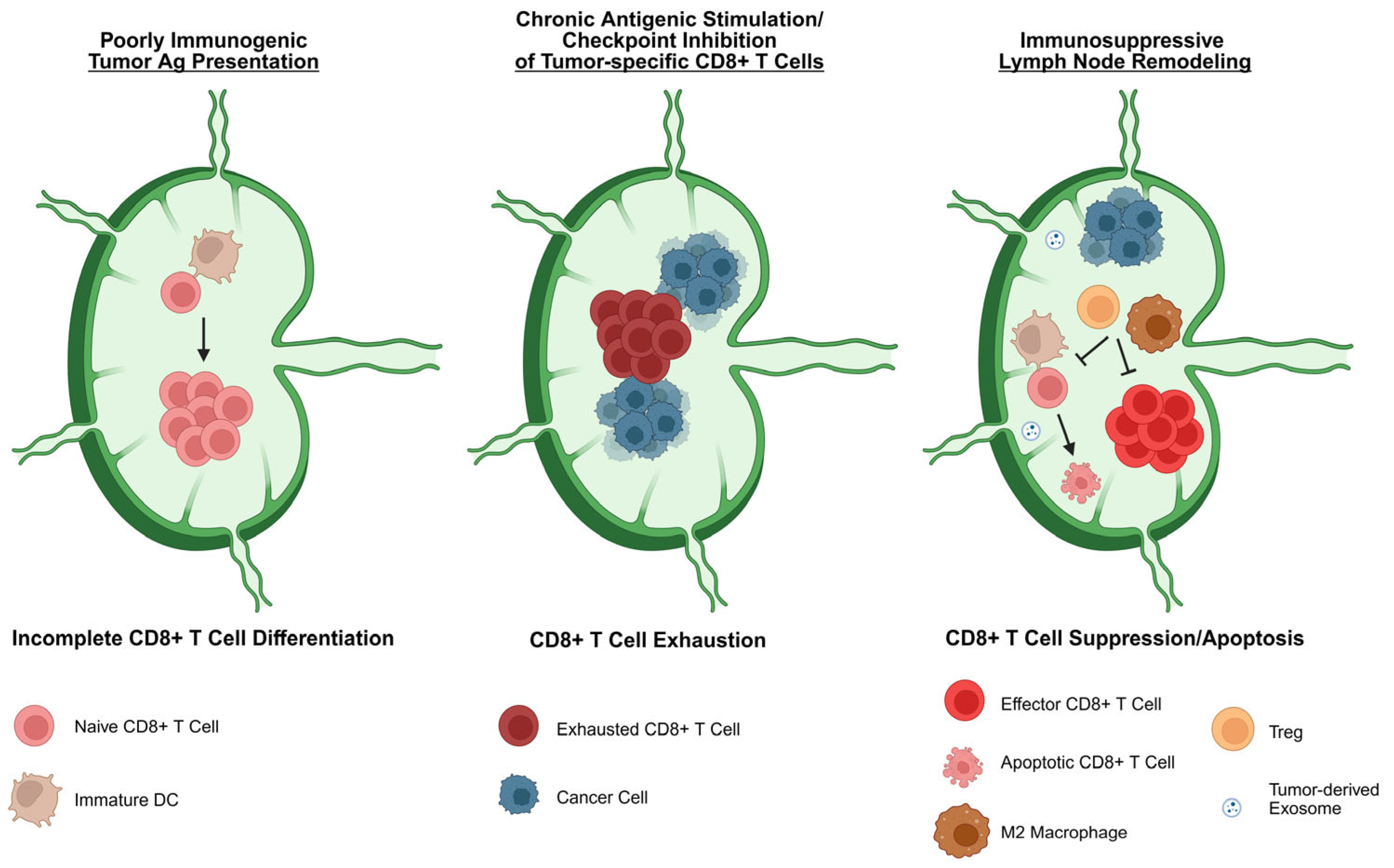
Further distinguishing incomplete differentiation from exhaustion as a form of antitumor immune dysfunction is our formal demonstration that incomplete T cell differentiation may arise within the context of both tumor-free and tumor-involved LNs, a finding that is in contrast with other studies reporting LN-resident T cell exhaustion as a form of anti-tumor immune dysfunction that arises specifically within tumor-involved LNs [20,28]. Such exhaustion is triggered by chronic antigenic stimulation, engagement of co-inhibitory receptors, and immunosuppressive remodeling of LN tissue upon infiltration by the tumor. In contrast to incomplete differentiation, which is associated with suboptimal stimulation during naïve T lymphocyte priming, T cell exhaustion is a more challenging form of dysfunction to overcome due to the stability of the epigenetic reprogramming that occurs during acquisition of the terminally exhausted phenotype [29]. As a result, although anti-tumor T cell dysfunction may arise within TDLNs prior to metastatic infiltration by the tumor, LN invasion presents an additional barrier to the maintenance of responses that have already been initiated in these compartments, as discussed in more detail below.

3. Implications of Lymph-Node-Resident CD8+ T Cell Dysfunction for Cancer Immunotherapy

As a key reservoir of CD8+ T cells capable of responding to tumors, TDLNs have emerged as sites of significant relevance to cancer immunotherapy. In this regard, targeted delivery of CTLA-4 and PD-1 checkpoint inhibitors to TDLNs improves the anti-tumor response to these therapeutics [30], and the efficacy of PD-1/PD-L1 checkpoint blockade has specifically been shown to depend on CD8+ T lymphocytes activated within TDLNs [31,32,33]. In particular, TDLN-resident tumor-specific memory (TTSM) CD8+ T cells and a differentiated subset of this population characterized by Tcf1 expression and termed progenitor exhausted T cells (Tpex) have both been shown to mediate immunotherapy-driven tumor control [33,34,35]. This latter Tpex subset has also been shown to provide the proliferative burst following checkpoint blockade therapy and is clonally related to more differentiated populations of intermediate exhausted cells (Tex-int) and terminally exhausted cells (Tex-term) that accumulate over time in the tumor. As such, this population has garnered significant interest for its potential to serve as a sustainable source of tumor-infiltrating effectors that are capable of maintaining the anti-tumor immune response, especially as those cells previously engaged in the response become terminally exhausted and lose effector function within the TME [20,23,36]. Consistent with recent reports that LN involvement by cancer cells correlates negatively with patient response to immunotherapy [37,38], these LN-resident Tpex, although found to be abundant in tumor-free LNs, were nearly absent in metastatic LNs of head and neck squamous cell carcinoma (HNSCC) patients. Replaced primarily by dysfunctional Tex-term cells, the few CD8+ T lymphocytes with Tpex and Tex-int phenotypes that remained within remodeled tumor-involved LNs of these patients were spatially localized to immunosuppressive niches enriched in tolerogenic DC and Treg populations, further highlighting the potential of LN invasion by tumor cells to disrupt TDLN-associated immune system dynamics [20].
Though the aforementioned studies document important immunologic consequences of LN invasion by the tumor, our recent work demonstrates that CD8+ T cell dysfunction at this site does not necessarily depend on metastatic involvement of the TDLNs. Indeed, while we have observed incomplete differentiation of CD8+ T cells shortly after their priming within tumor-involved LNs, we also consistently observe this incompletely differentiated phenotype when T cells respond within tumor-free LNs as well. We note also that the phenotype we have described is not limited to T cells responding against melanoma, as a similarly dysfunctional phenotype characterized by CD8+ T cell proliferation that is decoupled from acquisition of effector function has also been described in the aforementioned Horton et al. study, both in a murine model of lung cancer and in patients with non-small-cell lung cancer (NSCLC) [25]. Consistent with work in our melanoma model [24], Horton et al. also found that incomplete T cell differentiation arose shortly after tumor Ag recognition and was distinct from T cell exhaustion, as it could not be prevented by checkpoint inhibition during T lymphocyte priming. Interestingly, however, we both found that administration of exogenous IL-12 (or a combination of IL-12 and IL-2) during the priming phase of the anti-tumor response led to functional CD8+ T cell effector differentiation, suggesting that incomplete differentiation is a consequence of suboptimal stimulation rather than active T cell suppression.
Having found that incomplete differentiation arises regardless of whether tumor Ag is cross-presented by endogenous host APC or directly presented by LN-invading tumor cells, and based on the observation that CD8+ T cells in our system were capable of differentiating into functional effectors in both tumor-free and tumor-involved LNs when IL-12 was administered or when Ag was presented by CD40L-activated bone marrow-derived DC [17], we hypothesize that incomplete anti-tumor T cell differentiation arises as a result of the poor immunogenicity of both tumor cells and tumor-associated APC. With regard to the latter, we have previously shown that soluble melanoma-derived factors alter the maturation and activation of DC [39,40], and although Horton et al. did not observe any ex vivo defects in the T cell stimulatory capacity of cross-presenting DC that had acquired tumor Ag in their model, a more recent follow-up study from the Spranger group found that the T cell stimulatory capacity of these DC was indeed restrained when Tregs harvested from LN draining tumor-bearing lung tissue were also included in ex vivo cultures. Importantly, this priming defect was DC-dependent and not the result of direct Treg-mediated suppression of CD8+ T cells, as Tregs from this compartment did not inhibit CD8+ T cell effector differentiation when these cells were stimulated with plate-bound anti-CD3/anti-CD28 in place of cross-presenting TDLN-derived DC. Also of note, the inability of Treg-restrained DC from lung-draining LNs to prime effector T cell differentiation was not due to an inability of these cells to acquire or present tumor Ag but instead could be attributed to insufficient accessory signaling from these cells, a defect that could be overcome by providing artificial anti-CD28 costimulation and supplemental IL-12 [27]. Together, these data provide compelling evidence that the poor immunogenicity of TDLN-resident DC is responsible for the induction of incomplete CD8+ T cell differentiation.
Though we posit that the incompletely differentiated CD8+ T cells described by the Spranger group and our own are distinct from the stem-like Tpex reported by others, it is worth noting that this latter population is also known to arise from aberrant stimulation during priming. Indeed, in their recent study of patients with LN-invasive HNSCC, Rahim et al. found that while most CD8+ T cells had been driven toward a terminally exhausted state of differentiation within metastatic LN, the remaining Tpex in this compartment were cells that had actually failed to differentiate and that were localized near DC with a tolerogenic phenotype [20]. Though such Tpex might still have the potential to become effectors should they receive subsequent costimulation after infiltrating the primary tumor [22], it is just as likely that the immunosuppressive nature of the TME will often preclude the additional costimulation needed for complete effector differentiation. In light of this possibility, interventions that improve the anti-tumor immunogenicity of DC, such as therapeutic vaccination with exogenously stimulated DC or treatment with agents that expand and activate endogenous tumor-associated DC, hold great potential for enhancing the quality of tumor-specific CD8+ T cell responses, particularly as they can support not only the effector differentiation of these cells but also their responsiveness to checkpoint inhibitors whose ability to maintain T cell functionality relies on interference with inhibitory receptors expressed primarily on activated T lymphocytes. In this regard, DC that have been expanded intratumorally with the growth factor Flt3L have been successfully activated in preclinical settings using various strategies, including locally delivered anti-CD40 [36], poly I:C [41], and oncolytic virus therapy [42], with each leading to augmented effector CD8+ T cell activation in TDLN. Such localized approaches to boost anti-tumor immunity are especially attractive as they: (1) overcome toxicity-related issues that otherwise arise from systemic delivery of immunostimulatory cytokines like IL-12 that are needed to support effector CD8+ T cell differentiation but which also have deleterious consequences when administered more broadly; and (2) have potential to bypass the need for CD40L-mediated licensing of DC by CD4+ T cells, an interaction that is critical for optimal priming of anti-tumor CD8+ T cells [43] but which can itself also be compromised within the hostile confines of the TME. The challenge going forward will be to identify biomarkers predictive of the various forms of T cell dysfunction described herein so that these therapeutic interventions can be appropriately leveraged not only in patients with advanced disease but also in early-stage, pre-metastatic patients whose TDLN-resident T cells could still be prone to dysfunctional states that might permit disease progression or recurrence after initial treatment. Though such interventions would not be feasible or even recommended in all cases, they might prove beneficial for patients whose primary tumors display histologic features or molecular signatures/biomarkers associated with aggressive disease.
Finally, it should be noted that although defective CD8+ T cell priming has been reported in multiple cancer types, with both tumor- and context-dependent influences impacting the specific nature of the ensuing T cell differentiation program, such defective priming has not to date been linked with T cell exclusion from tumor tissue. Indeed, Horton et al. found in their model that lung tumors were well-infiltrated by incompletely differentiated CD8+ T cells, and to a significantly greater extent than flank tumors, even though the latter were associated with the activation of a more potent, ICB-responsive T cell differentiation program in cells primed within the skin-draining inguinal LNs [25]. In contrast to the dysfunctional T cell responses that arise from suboptimal priming in TDLNs, T cell exclusion from “immune cold” tumors is most often a consequence of: (1) a lack of tumor immunogenicity altogether or (2) a failure of primed T cells to be recruited into tumor tissue. Because both of these limitations have also been attributed to tumor-associated influences on DC [44,45], interventions such as those discussed above that expand and activate tumor-infiltrating DC also have the potential to transform immunologically “cold” tumors into immunologically “hot” ones that are more responsive to T cell-based immunotherapy.

4. Lymph-Node-Resident CD8+ T Cell Dysfunction as an Early Factor in the Establishment of a Pre-Metastatic Niche That Supports Tumor Progression

It has become widely appreciated that one of the key factors contributing to cancer progression is the formation, often early in the course of tumor development, of a pre-metastatic niche in sites distant from the primary tumor. First recognized by Stephen Paget in 1889 when he proposed his “seed and soil” hypothesis after observing nonrandom patterns of metastasis in patients with breast cancer [46], the idea that developing tumors prepare regional and distant tissues to become favorable sites for subsequent metastatic invasion has received considerable attention in the current era of cellular and molecular oncology [47]. Based on the potential of TDLNs to foster more widespread metastasis [2,3], understanding the factors that support pre-metastatic niche formation at these sites has become a particularly relevant area of investigation in recent years.
One mechanism of pre-metastatic niche formation that has been found to condition TDLNs for cancer cell invasion involves the release of exosomes or other small extracellular vesicles from the primary tumor. As carriers of a diverse range of tumor-derived cargo, such vesicles are capable of remodeling TDLNs in a number of ways that ultimately favor LN invasion. Independent of tumor immunity, these vesicles have been shown to activate cell signaling pathways that support tumor cell migration/recruitment, extracellular matrix deposition, and vascular growth within LN tissue [48]. Additionally, others have reported immunologic consequences of extracellular vesicle-mediated LN conditioning that favor tumor cell invasion and growth within these immune-rich environments. In the Lewis lung carcinoma model, Morrissey et al. found that tumor-derived exosomes reprogram macrophages from oxidative toward glycolytic metabolism, a shift that was driven by exosome-induced GLUT1 expression and increased glucose uptake. This metabolic shift in turn promoted the release of lactic acid that polarized macrophages toward an immunosuppressive phenotype characterized by high expression of PD-L1. Though work in this murine model focused on the role of these macrophages in establishing a pre-metastatic niche within lung tissue rather than LNs, analyses of tumor-free TDLNs of NSCLC patients also revealed elevated M2-like macrophages when compared to LNs obtained from healthy donors, and the frequency of this LN-resident immunosuppressive macrophage population across cancer patients indeed correlated with expression of the exosome-release gene YKT6 in the primary lung tumor, suggesting that this mechanism may promote LN metastasis in patients as well [49]. More direct evidence for this type of TDLN pre-conditioning has come from studies in melanoma, where small tumor-derived extracellular vesicles have likewise been shown to induce immune suppression in TDLNs prior to LN metastasis. In addition to exhibiting nerve growth factor receptor (NGFR)-dependent effects on LN stromal cells that augment lymphangiogenesis and tumor cell adhesion [50], these vesicles have also been found to shuttle tumor Ag to cross-presenting LN endothelial cells that induce apoptosis in Ag-specific CD8+ T cells [51].
Though we have not yet evaluated the role of exosomes in the induction of incomplete CD8+ T cell differentiation in our model, we argue that the suboptimal T cell stimulation that leads to this dysfunctional phenotype may also contribute to pre-metastatic niche formation within TDLNs, setting the stage for invading tumor cells to escape attack by functional effectors. As discussed above, our ability to induce effector T cell differentiation within TDLNs through the administration of exogenous IL-12 or immunization with activated bone marrow-derived DC demonstrates that LN-resident CD8+ T cells are indeed capable of responding to tumor Ag in this compartment, and our findings thus point to the poor immunogenicity of endogenous tumor-associated APC as the root cause of incomplete differentiation. Importantly, we note also that the dysfunctional phenotype we describe is restricted to T lymphocytes responding within TDLNs, as T cells in non-draining LNs of tumor-bearing animals fully differentiate into functional effectors when primed against an irrelevant Ag not expressed by the tumor at the same time that said Ag still fails to elicit effector differentiation by CD8+ T cells within TDLNs [17]. That this non-tumor-associated Ag also induces complete effector differentiation in lung-draining LNs of tumor-free mice indicates that LN heterogeneity alone cannot account for the differential LN-associated responses in our model. Collectively, our data suggest that tumor-associated factors specifically within the TDLNs may preclude immunogenic Ag presentation at this site, and it therefore remains possible in our model that exosome-associated cargo contributes to an overall tolerogenic microenvironment that is overcome only by provision of exogenous immunostimulatory agents. In the absence of such agents, however, the lack of effector function that arises in Ag-specific CD8+ T cells in the early stages of tumor progression might indeed render TDLNs susceptible to successful infiltration by cancer cells that eventually acquire the migratory, invasive, and metabolic properties needed to seed these compartments. Future experiments will be necessary to determine whether interventions that support complete CD8+ T cell differentiation in pre-metastatic LNs specifically preclude LN metastasis or limit tumor burden within involved LNs, either of which outcomes would add support to the hypothesis that induction of T cell dysfunction within TDLNs prior to LN invasion contributes to pre-metastatic niche formation in this compartment.
Finally, in addition to the previously described mechanisms by which LN-extrinsic factors may pre-condition TDLNs for metastatic invasion, it has been shown that LN-intrinsic factors independent of the primary tumor also support metastasis to this site by creating an immunologically favorable environment for infiltrating tumor cells. Specifically, Kahn et al. recently found the following: (1) LN-resident Tregs facilitate tumor growth and survival within TDLNs by locally suppressing tumor-specific CD8+ T cells via IL-2 restriction; and (2) local depletion of Tregs from TDLNs in turn dramatically reduces LN metastasis [52]. Of note, the Treg-mediated immune suppression described in this study did not involve pre-conditioning of the TDLNs by the primary tumor, as Ag-experienced CD8+ T cells within the non-draining contralateral LNs likewise failed to acquire cytolytic effector function. These data thus highlight the LN-intrinsic nature of this immune dysfunction as an inherent barrier to anti-tumor immunity, one that renders LN tissue an immune-privileged site for tumor cells with invasive potential. Together with the studies discussed above, these findings reveal also that there are likely a combination of LN-intrinsic and -extrinsic factors that limit the generation of functional anti-tumor CD8+ T cell responses in TDLNs and that ultimately foster formation of a pre-metastatic niche permissive for tumor engraftment and growth at these sites.

5. Conclusions

The induction and maintenance of anti-tumor CD8+ T cell responses within TDLNs is a critical aspect of the Cancer-Immunity cycle that is necessary for both endogenous and therapy-mediated tumor control. Importantly, T cell dysfunction that arises from sub-optimal priming, chronic antigenic stimulation, or local immunosuppression at this site not only has consequences for the immunologic control of primary tumors and the responsiveness of these cells to immunotherapy, but it also contributes to formation of a pre-metastatic niche in what is often the first site of metastasis from a primary tumor, enabling growth of cancerous cells in a compartment that frequently serves as a conduit for more distant dissemination (Figure 1) [21,29,43,45,46].
While anti-tumor CD8+ T cell dysfunction has long been appreciated as a byproduct of the immunologically hostile TME and is well-recognized as a phenomenon associated with tumor immune escape, emerging evidence has revealed that such dysfunction is not necessarily restricted to the confines of a tumor or even to late stages of tumor progression. Our work and that of others has indeed highlighted the significance of CD8+ T cell dysfunction within TDLNs as a means of immune escape that may arise very early in the course of tumor development, prior to any metastatic spread from the primary tumor. Based on these findings, and as highlighted in some of the preclinical studies discussed herein, LN-targeted immunomodulation is thus emerging as a promising therapeutic approach to render this critical compartment conducive to the activation and maintenance of functional anti-tumor CD8+ T cell effectors. Going forward, insights that inform the most appropriate type and timing of these and related interventions will be key to maximizing the anti-tumor potential of CD8+ T cell responses elicited within TDLNs.

Funding

This research received no external funding. No funding was required for the preparation of this review article.

Data Availability Statement

No new data were created or analyzed in this study. Data sharing is not applicable to this article.

Conflicts of Interest

The author declares no conflicts of interest.

Abbreviations

The following abbreviations are used in this manuscript:
Agantigen
APCantigen-presenting cell
CTLcytotoxic T lymphocyte
HNSCChead and neck squamous cell carcinoma
LNlymph node
NGFRnerve growth factor receptor
NSCLCnon-small-cell lung cancer
TDLNtumor-draining lymph node
TMEtumor microenvironment
Tex-intintermediate exhausted T cell
Tex-termterminally exhausted T cell
Tpexprogenitor exhausted T cell
Tregsregulatory T cells
TTSMtumor-specific memory T cell

References

  1. Chen, D.S.; Mellman, I. Oncology meets immunology: The cancer-immunity cycle. Immunity 2013, 39, 1–10. [Google Scholar] [CrossRef] [PubMed]
  2. Pereira, E.R.; Kedrin, D.; Seano, G.; Gautier, O.; Meijer, E.F.J.; Jones, D.; Chin, S.-M.; Kitahara, S.; Bouta, E.M.; Chang, J.; et al. Lymph node metastases can invade local blood vessels, exit the node, and colonize distant organs in mice. Science 2018, 359, 1403–1407. [Google Scholar] [CrossRef] [PubMed]
  3. Brown, M.; Assen, F.P.; Leithner, A.; Abe, J.; Schachner, H.; Asfour, G.; Bago-Horvath, Z.; Stein, J.V.; Uhrin, P.; Sixt, M.; et al. Lymph node blood vessels provide exit routes for metastatic tumor cell dissemination in mice. Science 2018, 359, 1408–1411. [Google Scholar] [CrossRef]
  4. Lughezzani, G.; Capitanio, U.; Jeldres, C.; Isbarn, H.; Shariat, S.F.; Arjane, P.; Widmer, H.; Perrotte, P.; Montorsi, F.; Karakiewicz, P.I. Prognostic significance of lymph node invasion in patients with metastatic renal cell carcinoma: A population-based perspective. Cancer 2009, 115, 5680–5687. [Google Scholar] [CrossRef] [PubMed]
  5. Chen, J.; Xu, Y.; Zhou, Y.; Wang, Y.; Zhu, H.; Shi, Y. Prognostic role of sentinel lymph node biopsy for patients with cutaneous melanoma: A retrospective study of surveillance, epidemiology, and end-result population-based data. Oncotarget 2016, 7, 45671–45677. [Google Scholar] [CrossRef]
  6. Doepker, M.P.; Thompson, Z.J.; Harb, J.N.; Messina, J.L.; Puleo, C.A.; Egan, K.M.; Sarnaik, A.A.; Gonzalez, R.J.; Sondak, V.K.; Zager, J.S. Dermal melanoma: A report on prognosis, outcomes, and the utility of sentinel lymph node biopsy. J. Surg. Oncol. 2016, 113, 98–102. [Google Scholar] [CrossRef]
  7. Scheipner, L.; Barletta, F.; Cano Garcia, C.; Incesu, R.B.; Morra, S.; Baudo, A.; Assad, A.; Tian, Z.; Saad, F.; Shariat, S.F.; et al. Prognostic significance of pathologic lymph node invasion in metastatic renal cell carcinoma in the immunotherapy era. Ann. Surg. Oncol. 2023, 30, 8780–8785. [Google Scholar] [CrossRef]
  8. Jaganmurugan, R.; Arora, A.; Chandankhede, U.; Prakash, G.; Bakshi, G.; Joshi, A.; Menon, S.; Murthy, V.; Pal, M. Prognostic Significance of lymph node density in pathological node positive urothelial carcinoma of the bladder -upfront surgery and post neoadjuvant chemotherapy cohorts. Clin. Genitourin. Cancer 2024, 22, 385–393. [Google Scholar] [CrossRef]
  9. Pilko, G.; Zgajnar, J.; Music, M.; Hocevar, M. Lower tumour burden and better overall survival in melanoma patients with regional lymph node metastases and negative preoperative ultrasound. Radiol. Oncol. 2012, 46, 60–68. [Google Scholar] [CrossRef]
  10. Ulmer, A.; Dietz, K.; Hodak, I.; Polzer, B.; Scheitler, S.; Yildiz, M.; Czyz, Z.; Lehnert, P.; Fehm, T.; Hafner, C.; et al. Quantitative measurement of melanoma spread in sentinel lymph nodes and survival. PLoS Med. 2014, 11, e1001604. [Google Scholar] [CrossRef]
  11. Kim, C.; Economou, S.; Amatruda, T.T.; Martin, J.C.; Dudek, A.Z. Prognostic significance of microscopic tumor burden in sentinel lymph node in patients with cutaneous melanoma. Anticancer Res. 2015, 35, 301–309. [Google Scholar] [PubMed]
  12. Anichini, A.; Scarito, A.; Molla, A.; Parmiani, G.; Mortarini, R. Differentiation of CD8+ T cells from tumor-invaded and tumor-free lymph nodes of melanoma patients: Role of common gamma-chain cytokines. J. Immunol. 2003, 171, 2134–2141. [Google Scholar] [CrossRef] [PubMed]
  13. Zippelius, A.; Batard, P.; Rubio-godoy, V.; Bioley, G.; Lie, D.; Lejeune, F.; Rimoldi, D.; Guillaume, P.; Meidenbauer, N.; Mackensen, A.; et al. Effector function of human tumor-specific CD8 T cells in melanoma lesions: A state of local functional tolerance. Cancer Res. 2004, 64, 2865–2873. [Google Scholar] [CrossRef] [PubMed]
  14. Mortarini, R.; Piris, A.; Maurichi, A.; Molla, A.; Bersani, I.; Bono, A.; Bartoli, C.; Santinami, M.; Lombardo, C.; Ravagnani, F.; et al. Lack of terminally differentiated tumor-specific CD8+ T cells at tumor site in spite of antitumor immunity to self-Ags in human metastatic melanoma. Cancer Res. 2003, 63, 2535–2545. [Google Scholar]
  15. Negin, B.; Panka, D.; Wang, W.; Siddiqui, M.; Tawa, N.; Mullen, J.; Tahan, S.; Mandato, L.; Polivy, A.; Mier, J.; et al. Effect of melanoma on immune function in the regional lymph node basin. Clin. Cancer Res. 2008, 14, 654–659. [Google Scholar] [CrossRef][Green Version]
  16. Baitsch, L.; Baumgaertner, P.; Devêvre, E.; Raghav, S.K.; Legat, A.; Barba, L.; Wieckowski, S.; Bouzourene, H.; Deplancke, B.; Romero, P.; et al. Exhaustion of tumor-specific CD8+ T cells in metastases from melanoma patients. J. Clin. Investig. 2011, 121, 2350–2360. [Google Scholar] [CrossRef]
  17. Hargadon, K.M.; Brinkman, C.C.; Sheasley-O’neill, S.L.; Nichols, L.A.; Bullock, T.N.J.; Engelhard, V.H. Incomplete differentiation of Ag-specific CD8 T cells in tumor-draining lymph nodes. J. Immunol. 2006, 177, 6081–6090. [Google Scholar] [CrossRef]
  18. Hargadon, K.M. Tumor microenvironmental influences on dendritic cell and T cell function: A focus on clinically relevant immunologic and metabolic checkpoints. Clin. Transl. Med. 2020, 10, 374–411. [Google Scholar] [CrossRef]
  19. Reticker-Flynn, N.E.; Zhang, W.; Belk, J.A.; Basto, P.A.; Escalante, N.K.; Pilarowski, G.O.W.; Bejnood, A.; Martins, M.M.; Kenkel, J.A.; Linde, I.L.; et al. Lymph node colonization induces tumor-immune tolerance to promote distant metastasis. Cell 2022, 185, 1924–1942.e23. [Google Scholar] [CrossRef]
  20. Rahim, M.K.; Okholm, T.L.H.; Jones, K.B.; McCarthy, E.E.; Liu, C.C.; Yee, J.L.; Tamaki, S.J.; Marquez, D.M.; Tenvooren, I.; Wai, K.; et al. Dynamic CD8+ T cell responses to cancer immunotherapy in human regional lymph nodes are disrupted in metastatic lymph nodes. Cell 2023, 186, 1127–1143.e18. [Google Scholar] [CrossRef]
  21. Hargadon, K.M.; Goodloe, T.B.; Woodall, S.L. Lymph node invasion by melanoma cells is not required for the induction of incomplete differentiation by tumor-specific CD8+ T cells. Cancer Rep. 2025, 8, e70145. [Google Scholar] [CrossRef] [PubMed]
  22. Prokhnevska, N.; Cardenas, M.A.; Valanparambil, R.M.; Sobierajska, E.; Barwick, B.G.; Jansen, C.; Reyes Moon, A.; Gregorova, P.; delBalzo, L.; Greenwald, R.; et al. CD8+ T cell activation in cancer comprises an initial activation phase in lymph nodes followed by effector differentiation within the tumor. Immunity 2023, 56, 107–124.e5. [Google Scholar] [CrossRef] [PubMed]
  23. Connolly, K.A.; Kuchroo, M.; Venkat, A.; Khatun, A.; Wang, J.; William, I.; Hornick, N.I.; Fitzgerald, B.L.; Damo, M.; Kasmani, M.Y.; et al. A reservoir of stem-like CD8+ T cells in the tumor-draining lymph node preserves the ongoing antitumor immune response. Sci. Immunol. 2021, 6, eabg7836. [Google Scholar] [CrossRef] [PubMed]
  24. Ferguson, A.R.; Nichols, L.A.; Zarling, A.L.; Thompson, E.D.; Brinkman, C.; Hargadon, K.M.; Bullock, T.N.; Engelhard, V.H. Strategies and challenges in eliciting immunity to melanoma. Immunol. Rev. 2008, 222, 28–42. [Google Scholar] [CrossRef]
  25. Horton, B.L.; Morgan, D.M.; Momin, N.; Zagorulya, M.; Torres-Meji, E.; Bhandarkar, V.; Wittrup, K.D.; Love, J.C.; Spranger, S. Lack of CD8+ T cell effector differentiation during priming mediates checkpoint blockade resistance in non-small cell lung cancer. Sci. Immunol. 2021, 6, eabi8800. [Google Scholar] [CrossRef]
  26. Cruz de Casas, P.; Knöpper, K.; Dey Sarkar, R.; Kastenmüller, W. Same yet different—How lymph node heterogeneity affects immune responses. Nat. Rev. Immunol. 2023, 24, 358–374. [Google Scholar] [CrossRef]
  27. Zagorulya, M.; Yim, L.; Morgan, D.M.; Edwards, A.; Torres-Mejia, E.; Momin, N.; McCreery, C.V.; Zamora, I.L.; Horton, B.L.; Fox, J.G.; et al. Tissue-specific abundance of interferon-gamma drives regulatory T cells to restrain DC1-mediated priming of cytotoxic T cells against lung cancer. Immunity 2023, 56, 386–405.e10. [Google Scholar] [CrossRef]
  28. Canale, F.P.; Ramello, M.C.; Núñez, N.; Furlan, C.L.A.; Bossio, S.N.; Serrán, M.G.; Boari, J.T.; del Castillo, A.; Ledesma, M.; Sedlik, C.; et al. CD39 expression defines cell exhaustion in tumor-infiltrating CD8+ T cells. Cancer Res. 2018, 78, 115–128. [Google Scholar] [CrossRef]
  29. Pauken, K.E.; Sammons, M.A.; Odorizzi, P.M.; Manne, S.; Godec, J.; Khan, O.; Drake, A.M.; Chen, Z.; Sen, D.R.; Kurachi, M.; et al. Epigenetic stability of exhausted T cells limits durability of reinvigoration by PD-1 blockade. Science 2016, 354, 1160–1165. [Google Scholar] [CrossRef]
  30. Francis, D.M.; Manspeaker, M.P.; Schudel, A.; Sestito, L.F.; O’Melia, M.J.; Kissick, H.T.; Pollack, B.P.; Waller, E.K.; Thomas, S.N. Blockade of immune checkpoints in lymph nodes through locoregional delivery augments cancer immunotherapy. Sci. Transl. Med. 2020, 12, eaay3575. [Google Scholar] [CrossRef]
  31. Fransen, M.F.; Schoonderwoerd, M.; Knopf, P.; Camps, M.G.; Hawinkels, L.J.; Kneilling, M.; van Hall, T.; Ossendorp, F. Tumor-draining lymph nodes are pivotal in PD-1/PD-L1 checkpoint therapy. JCI Insight 2018, 3, e124507. [Google Scholar] [CrossRef] [PubMed]
  32. Dammeijer, F.; van Gulijk, M.; Mulder, E.E.; Lukkes, M.; Klaase, L.; van den Bosch, T.; van Nimwegen, M.; Lau, S.P.; Latupeirissa, K.; Schetters, S.; et al. The PD-1/PD-L1-checkpoint restrains T cell immunity in tumor-draining lymph nodes. Cancer Cell 2020, 38, 685–700.e8. [Google Scholar] [CrossRef]
  33. Huang, Q.; Wu, X.; Wang, Z.; Chen, X.; Wang, L.; Lu, Y.; Xiong, D.; Liu, Q.; Tian, Y.; Lin, H.; et al. The primordial differentiation of tumor-specific memory CD8+ T cells as bona fide responders to PD-1/PD-L1 blockade in draining lymph nodes. Cell 2022, 185, 4049–4066.e25. [Google Scholar] [CrossRef] [PubMed]
  34. Im, S.J.; Hashimoto, M.; Gerner, M.Y.; Lee, J.; Kissick, H.T.; Burger, M.C.; Shan, Q.; Hale, J.S.; Lee, J.; Nasti, T.H.; et al. Defining CD8+ T cells that provide the proliferative burst after PD-1 therapy. Nature 2016, 537, 417–421. [Google Scholar] [CrossRef] [PubMed]
  35. Miller, B.C.; Sen, D.R.; Al Abosy, R.; Bi, K.; Virkud, Y.V.; LaFleur, M.W.; Yates, K.B.; Lako, A.; Felt, K.; Naik, G.S.; et al. Subsets of exhausted CD8+ T cells differentially mediate tumor control and respond to checkpoint blockade. Nat. Immunol. 2019, 20, 1556, Erratum in: Nat Immunol. 2019, 11, 1556. https://doi.org/10.1038/s41590-019-0528-5.. [Google Scholar] [CrossRef]
  36. Schenkel, J.M.; Herbst, R.H.; Canner, D.; Li, A.; Hillman, M.; Shanahan, S.L.; Gibbons, G.; Smith, O.C.; Kim, J.Y.; Westcott, P.; et al. Conventional type I dendritic cells maintain a reservoir of proliferative tumor-Ag specific TCF-1+ CD8+ T cells in tumor-draining lymph nodes. Immunity 2021, 54, 2338–2353.e6. [Google Scholar] [CrossRef]
  37. Yang, H.; Sun, B.; Ma, W.; Fan, L.; Xu, K.; Jia, Y.; Xu, J.; Wang, Z.; Yao, F. Multi-scale characterization of tumor-draining lymph nodes in resectable lung cancer treated with neoadjuvant immune checkpoint inhibitors. eBioMedicine 2022, 84, 104265. [Google Scholar] [CrossRef]
  38. Ma, R.; Yang, H.; Ge, Y.; Ma, T.; Wang, J.; Li, S.; Feng, T.; Feng, S.; Zhang, C.; Sun, T.; et al. Prognostic implications of lymph node status in non–small-cell lung cancer patients before and after neoadjuvant chemoimmunotherapy: A multicenter retrospective study. Clin. Lung Cancer 2025, 26, 370–383. [Google Scholar] [CrossRef]
  39. Hargadon, K.M.; Ararso, Y.T.; Forrest, O.A.; Harte, C.M. Melanoma-associated suppression of the dendritic cell lines DC2.4 and JAWSII. Am. J. Immunol. 2012, 8, 179–190. [Google Scholar] [CrossRef][Green Version]
  40. Hargadon, K.M.; Bishop, J.D.; Brandt, J.P.; Hand, Z.C.; Ararso, Y.T.; Forrest, O.A. Melanoma-derived factors alter the maturation and activation of differentiated tissue-resident dendritic cells. Immunol. Cell Biol. 2016, 94, 24–38. [Google Scholar] [CrossRef]
  41. Salmon, H.; Idoyaga, J.; Rahman, A.; Leboeuf, M.; Remark, R.; Jordan, S.; Casanova-Acebes, M.; Khudoynazarova, M.; Agudo, J.; Tung, N.; et al. Expansion and activation of CD103+ dendritic cell progenitors at the tumor site enhances tumor responses to therapeutic PD-L1 and BRAF inhibition. Immunity 2016, 44, 924–938. [Google Scholar] [CrossRef] [PubMed]
  42. Svensson-Arvelund, J.; Cuadrado-Castano, S.; Pantsulaia, G.; Kim, K.; Aleynick, M.; Hammerich, L.; Upadhyay, R.; Yellin, M.; Marsh, H.; Oreper, D.; et al. Expanding Cross-presenting dendritic cells enhances oncolytic virotherapy and is critical for long-term anti-tumor immunity. Nat. Commun. 2022, 13, 7149. [Google Scholar] [CrossRef] [PubMed]
  43. Ferris, S.T.; Durai, V.; Wu, R.; Theisen, D.J.; Ward, J.P.; Bern, M.D.; Davidson, J.T.; Bagadia, P.; Liu, T.; Briseño, C.G.; et al. CDC1 prime and are licensed by CD4+ T cells to induce anti-tumour immunity. Nature 2020, 584, 624–629. [Google Scholar] [CrossRef] [PubMed]
  44. Spranger, S.; Bao, R.; Gajewski, T.F. Melanoma-Intrinsic β-Catenin Signalling Prevents Anti-Tumour Immunity. Nature 2015, 523, 231–235. [Google Scholar] [CrossRef]
  45. Spranger, S.; Dai, D.; Horton, B.; Gajewski, T.F. Tumor-residing Batf3 dendritic cells are required for effector T cell trafficking and adoptive T cell therapy. Cancer Cell 2017, 31, 711–723.e4. [Google Scholar] [CrossRef]
  46. Paget, S. The distribution of secondary growths in cancer of the breast. Lancet 1889, 133, 571–573. [Google Scholar] [CrossRef]
  47. Wang, Y.; Jia, J.; Wang, F.; Fang, Y.; Yang, Y.; Zhou, Q.; Yuan, W.; Gu, X.; Hu, J.; Yang, S. Pre-metastatic niche: Formation, characteristics and therapeutic implication. Signal Transduct. Target. Ther. 2024, 9, 236. [Google Scholar] [CrossRef]
  48. Hood, J.L.; San, R.S.; Wickline, S.A. Exosomes released by melanoma cells prepare sentinel lymph nodes for tumor metastasis. Cancer Res. 2011, 71, 3792–3801. [Google Scholar] [CrossRef]
  49. Morrissey, S.M.; Zhang, F.; Ding, C.; Montoya-Durango, D.E.; Hu, X.; Yang, C.; Wang, Z.; Yuan, F.; Fox, M.; Zhang, H.G.; et al. Tumor-derived exosomes drive immunosuppressive macrophages in a pre-metastatic niche through glycolytic dominant metabolic reprogramming. Cell Metab. 2021, 33, 2040–2058.e10. [Google Scholar] [CrossRef]
  50. García-Silva, S.; Benito-Martín, A.; Nogués, L.; Hernández-Barranco, A.; Mazariegos, M.S.; Santos, V.; Hergueta-Redondo, M.; Ximénez-Embún, P.; Kataru, R.P.; Lopez, A.A.; et al. Melanoma-derived small extracellular vesicles induce lymphangiogenesis and metastasis through an NGFR-dependent mechanism. Nat. Cancer 2021, 2, 1387–1405. [Google Scholar] [CrossRef]
  51. Leary, N.; Walser, S.; He, Y.; Cousin, N.; Pereira, P.; Gallo, A.; Collado-Diaz, V.; Halin, C.; Garcia-Silva, S.; Peinado, H.; et al. Melanoma-derived extracellular vesicles mediate lymphatic remodelling and impair tumour immunity in draining lymph nodes. J. Extracell. Vesicles 2022, 11, e12197. [Google Scholar] [CrossRef]
  52. Kahn, B.M.; Ng, R.W.S.; Kim, I.-K.; Eskandarian, C.; Chen, C.O.; Huang, B.; Lucas, A.; Li, L.; Maillard, I.; Stanger, B.Z. Intrinsic properties of the lymph node render it immunologically susceptible to metastasis. Cancer Discov. 2025, 15, 1949–1968. [Google Scholar] [CrossRef]
  53. Hargadon, K. 2026. Available online: https://BioRender.com/e63k0jp (accessed on 7 December 2025).
Figure 1. CD8+ T cell dysfunction within tumor-draining lymph nodes. Various forms of CD8+ T cell dysfunction have been described in the context of tumor-free and tumor-involved lymph nodes. Incomplete T cell differentiation, characterized by robust proliferation but a failure to acquire IFNγ-secreting or cytolytic effector activity, may arise during early stages of tumor progression and has been associated with suboptimal stimulation by both cross-presenting APC and directly presenting tumor cells in both tumor-free and tumor-involved lymph nodes. T cell exhaustion, typically within the context of tumor-involved lymph nodes, arises later during the course of tumor progression as a result of chronic antigenic stimulation and engagement of co-inhibitory receptors. Non-exhausted forms of anti-tumor T lymphocyte dysfunction may also be induced as a result of immunosuppressive remodeling of lymph nodes by tumor cells, tumor-derived factors, and tumor-associated immunoregulatory cells. Created in BioRender [53].
Lymphatics 04 00002 g001
Disclaimer/Publisher’s Note: The statements, opinions and data contained in all publications are solely those of the individual author(s) and contributor(s) and not of MDPI and/or the editor(s). MDPI and/or the editor(s) disclaim responsibility for any injury to people or property resulting from any ideas, methods, instructions or products referred to in the content.