Review

Neck Dissection in the Era of Immunotherapy: A Narrative Review

by
Andrea Lorenzi
1,2,†,
Carmine Prizio
3,†,
Remo Accorona
3,
Vijay Kumar Srinivasalu
4 and
Narayana Subramaniam
5,*
1
Division of Otolaryngology, Department of Surgical Sciences, University of Turin, 10124 Turin, Italy
2
Department of Mechanical and Aerospace Engineering, Politecnico di Torino, 10129 Turin, Italy
3
Department of Otolaryngology–Head and Neck Surgery, ASST Grande Ospedale Metropolitano Niguarda, 20162 Milan, Italy
4
Department of Medical Oncology, Sparsh Hospitals, Bengaluru 560064, India
5
Department of Head and Neck Surgery & Oncology, Aster International Institute of Oncology, Bengaluru 560092, India
*
Author to whom correspondence should be addressed.
These authors contributed equally to this work.
Lymphatics 2026, 4(1), 13; https://doi.org/10.3390/lymphatics4010013
Submission received: 30 November 2025 / Revised: 2 February 2026 / Accepted: 15 February 2026 / Published: 22 February 2026

Abstract

Cervical lymph node metastases are major prognostic determinants in head and neck squamous cell carcinoma (HNSCC), and neck dissection (ND) has long been central to regional control. As ND has evolved from radical to selective procedures, immune checkpoint inhibitors (ICIs) have emerged as a fourth treatment pillar, reframing tumor-draining lymph nodes (TDLNs) as active immune organs rather than passive conduits of metastatic spread. This narrative review synthesizes surgical, immunologic, and translational evidence on how ND and cervical irradiation interact with immunotherapy. It also examines the historical development of ND, the immunologic structure and function of cervical TDLNs, and the use of neoadjuvant, perioperative, and recurrent/metastatic immunotherapy in HNSCC. Preclinical and early clinical observations suggest that ablating or heavily irradiating non-involved nodal basins may attenuate ICI efficacy by disrupting antigen presentation, progenitor exhausted CD8+ T (Tpex) cell pools, and effector recirculation, supporting the conceptual model of an “immune desert neck.” The review critically appraises timing (pre- versus post-immunotherapy ND), response-adapted or de-escalated surgery, and imaging, tissue-based, and circulating biomarkers to guide individualized management. Current evidence does not support abandoning elective or therapeutic ND, but does highlight the need for biomarker-driven, lymphatic-sparing trials to redefine when ND is essential, modifiable, or potentially avoidable in immunotherapy-treated HNSCC.

1. Introduction

Regional lymph node metastases—present at diagnosis in up to 40% of patients—remain among the strongest prognostic factors in head and neck squamous cell carcinoma (HNSCC) [1]. In oral cavity cancer, cervical nodal metastasis is associated with a 5-year overall survival (OS) below 50%, underscoring the central role of the neck lymphatic basin in disease progression and treatment planning [2,3,4].
Traditionally, neck management has relied on surgery, radiotherapy, and chemotherapy. Since Crile’s original radical neck dissection (RND) and subsequent refinements, surgical clearance of cervical nodes has been integral to curative treatment [5,6,7]. Elective neck dissection (END) became standard for clinically node-negative (cN0) disease because 20–30% of patients harbor occult metastases [8,9]. Randomized trials and meta-analyses, particularly in early oral cavity cancer, showed improved disease-free survival (DFS) and reduced nodal recurrence with END compared with observation [10]. However, END also subjects many ultimately node-negative patients to avoidable surgery, functional morbidity, and removal of immunologically active lymph nodes [11].
Over the past decade, immunotherapy has emerged as a fourth therapeutic pillar in HNSCC. Immune checkpoint inhibitors (ICIs) targeting PD-1/PD-L1 and CTLA-4 have improved outcomes in recurrent and metastatic disease and are now being evaluated in curative-intent settings [12]. At the same time, translational work has reframed cervical lymph nodes as active immune organs: tumor-draining, non-metastatic nodes retain potent antigen-presenting capacity and harbor progenitor exhausted CD8+ T (Tpex) cells that appear critical for effective ICI responses. Experimental and clinical observations suggest that removing these nodes—surgically or with elective irradiation—can attenuate immunotherapy efficacy, whereas preserving uninvolved basins may sustain antitumor immunity [13,14].
As ICIs move into neoadjuvant and perioperative use, the traditional rationale for routine END in anatomically or biologically favorable cN0 disease may need to be revisited. A modern approach to neck dissection (ND) must account not only for oncologic control, but also for the impact of lymphatic disruption on systemic immune responses.

2. Evolution of Neck Dissection

2.1. The Radical Era

Modern cervical lymphadenectomy traces back to George Crile, who in 1906 introduced RND based on Halsted’s principle of en bloc removal of the primary tumor and its draining lymphatic basin (Table 1). RND mandated sacrifice of the sternocleidomastoid muscle (SCM), internal jugular vein (IJV), and spinal accessory nerve (SAN), along with surrounding soft tissue, to remove all cervical lymphatics in continuity [15,16,17].
Hayes Martin later standardized and popularized RND as the reference procedure for cervical metastases [18]. Although RND achieved reliable regional control, it carried substantial morbidity—including classic “shoulder syndrome”—and was poorly suited to bilateral dissections or clinically node-negative patients.

2.2. Modified Radical Neck Dissection

By the mid-20th century, it became clear that non-lymphatic structures did not always need to be sacrificed to achieve oncologic control. In the late 1970s, Richard H. Jesse, Alando J. Ballantyne, and colleagues began preserving one or more key structures (SAN, IJV, and SCM) while maintaining an en bloc dissection of lymph node levels I–V. This approach defined modified radical neck dissection (MRND), designed to reduce functional morbidity while keeping the oncologic footprint of RND [19].
MRND emerged as an alternative for clinically N0 necks and low-volume N+ disease, signaling a shift from maximally ablative to more function-preserving surgery [20].

2.3. Functional and Selective Neck Dissection

In the 1960s, Osvaldo Suárez introduced functional ND as a conceptual break from RND. He emphasized preservation of all non-lymphatic structures by following natural fascial planes, removing the neck lymphatic system only when tumor remained confined to lymphatic compartments [15]. The technique was disseminated in Europe by César Gavilán and Ettore Bocca and later adapted in the United States [20].
In parallel, centers such as Memorial Sloan Kettering and MD Anderson formalized selective neck dissection (SND) based on predictable patterns of cervical metastasis. SND intentionally preserves lymph node levels at low risk for occult spread while resecting high-risk levels according to tumor subsite (e.g., I–III for oral cavity, II–IV for oropharynx). The 1991 American Academy of Otolaryngology–Head and Neck Surgery (AAO-HNS) classification, later refined by Medina, codified SND variants—supraomohyoid, lateral, posterolateral, anterior—based on the levels removed [17,21].
SND is now standard for cN0 and early-stage disease in many head and neck subsites, balancing oncologic safety with lower morbidity.

2.4. Extended and Salvage Neck Dissection

Extended radical neck dissections (ERNDs) were defined for disease involving structures beyond those removed in standard RND—such as the carotid artery, parapharyngeal space, or paratracheal/mediastinal lymphatics—and are reserved for bulky or anatomically invasive disease [22].
Salvage ND remains critical after failed chemoradiation or persistent nodal disease. It can achieve regional control but is associated with higher complication rates and worse function, particularly in previously dissected or heavily irradiated necks [23].

2.5. Contemporary Neck Surgery

Current neck management integrates these historical approaches with modern imaging and technology [24]. Advances in CT, MRI, ultrasound, PET/CT, radiomics, and image-based risk models have refined indications for each dissection type and improved cN0 staging [25,26,27]. Minimally invasive techniques—including endoscopic and robotic ND—aim to decrease scarring and morbidity while respecting oncologic principles [28,29,30].
For decades, more extensive clearance was assumed to provide better regional control. Increasing evidence on the immunologic role of cervical lymph nodes—and the consequences of removing or irradiating them in immunotherapy-treated patients—now challenges this assumption and forces a re-examination of both extent and timing.

3. The Immunological Landscape of the Neck

3.1. Cervical Nodes as Immune Organs

Cervical lymph nodes lie along the lymphatic drainage of the upper aerodigestive tract and act as secondary lymphoid organs. Within specialized microenvironments—including fibroblastic reticular cell–conduit networks and high endothelial venules—antigen-bearing dendritic cells, naïve T cells, and B cells are brought together, allowing efficient priming, expansion, and differentiation of effector and memory lymphocytes [31,32].
In HNSCC, first-echelon (“sentinel”) cervical nodes are often the earliest metastatic sites and key locations for antigen presentation. Immunologic activation or suppression in these nodes can strongly influence downstream systemic antitumor responses [33,34].

3.2. Lymphatic Drainage and the Tumor Microenvironment

Afferent lymphatics from oral, pharyngeal, thyroid, and cutaneous territories transport interstitial fluid, soluble tumor antigens, and migratory dendritic cells to draining cervical nodes, creating a continuous immunologic link between the tumor and regional lymphoid tissue [35]. Conventional dendritic cells acquire tumor antigens in peripheral tissues and deliver them to tumor-draining nodes for cross-presentation to naïve CD8+ T cells, a process essential for initiating and sustaining cytotoxic responses [36].
Tumor colonization remodels tumor-draining nodes, often expanding regulatory T cells and other suppressive populations. Nonetheless, intact nodal architecture remains crucial for effective checkpoint blockade and vaccine-based strategies, which depend on ongoing nodal priming to maintain antitumor immunity [34].

4. The Rise of Immunotherapy in HNSCC

4.1. Checkpoint Inhibitors

Immune checkpoint blockade has transformed management of recurrent and metastatic HNSCC. The phase III KEYNOTE-048 trial established pembrolizumab, alone or with platinum and 5-fluorouracil, as a first-line standard by improving OS versus cetuximab-based EXTREME, particularly in patients with PD-L1 combined positive scores (CPS) ≥ 1 and ≥20 [37]. In platinum-refractory disease, CheckMate 141 demonstrated that nivolumab significantly improved OS and maintained long-term benefit versus investigator’s choice single-agent therapy, supporting its role in recurrent/metastatic HNSCC progressing after platinum-containing therapy [38,39].

4.2. Neoadjuvant Immunotherapy

To exploit the intact tumor–lymph node axis, neoadjuvant PD-1 blockade has been evaluated in resectable HNSCC. In HPV-independent locally advanced disease, single-agent pembrolizumab before surgery was safe and produced pathologic tumor regression in roughly 40–45% of patients, with early signs of reduced relapse in high-risk groups [40]. Phase II trials of neoadjuvant nivolumab, alone or with ipilimumab, have reported major pathologic responses (≤10% viable tumor) and occasional complete responses in primary and nodal specimens, indicating robust immune activation before resection [41].

4.3. Perioperative and Adjuvant Approaches

Perioperative strategies now combine neoadjuvant and adjuvant immunotherapy around surgery. In phase III KEYNOTE-689, perioperative pembrolizumab added to standard surgery and risk-adapted adjuvant therapy improved DFS and achieved higher rates of major pathologic response and pathologic complete response (pCR) at resection, without reducing the likelihood of surgical completion [42]. Other peri- or adjuvant PD-L1–directed approaches (e.g., atezolizumab, durvalumab) have produced mixed or negative DFS results, underscoring that the optimal agent, timing, and integration with (chemo)radiotherapy remain unsettled [43].

5. The Paradox: How Neck Dissection and Radiation May Impair Immunotherapy

5.1. Immunotherapy Relies on Lymphatic Integrity

Immune checkpoint blockade relies on an intact lymphatic network linking the primary tumor to regional tumor-draining lymph nodes (TDLNs). Within these nodes, tumor antigens delivered via afferent lymphatics are processed by dendritic cells, which prime and expand tumor-specific CD8+ T cells, including stem-like progenitor subsets that preferentially respond to PD-1/PD-L1 blockade and give rise to effector cells that return to the tumor [44,45]. Preclinical and translational work across tumor types, including HNSCC, shows that disrupting this nodal axis—through surgery, broad-field irradiation, or blocking lymphocyte egress—can markedly reduce ICI efficacy [46,47]. In HNSCC, cervical TDLNs are enriched with tumor-specific T and B cells and are dynamically remodeled by immunotherapy [48,49].

5.2. Potential Immunologic Consequences of Surgery

Conventional ND, rooted in Halstedian en bloc principles, removes metastatic and non-metastatic nodes along with the intervening lymphatics. In early oral cavity squamous cell carcinoma (OSCC), many nodes removed in END for cT1–T2N0 disease are pathologically negative yet immunologically competent. Bu et al. proposed that such nodes can act as “pivotal responders” to ICIs [10].
In murine models, surgical ablation of TDLNs can abolish responses to PD-1/PD-L1–directed immunotherapy and combined radioimmunotherapy, emphasizing that these nodes are required for durable systemic control [46]. Human studies indicate that non-metastatic cervical nodes may harbor HPV16 E6/E7-specific CD8+ T cells, consistent with ongoing antitumor priming even in basins without overt metastasis [13]. In OSCC, B-cell and regulatory T-cell dysfunction within TDLNs correlates with relapse risk, reinforcing their role as immune “control centers” [50].
Removing this network eliminates major antigen-presenting hubs, interrupts dendritic-cell migration and T-cell recirculation, and is followed by fibrosis and scarring that may further impair immune-cell trafficking into the treated neck [10,13].

5.3. Cervical Radiation and Immunosuppression

Cervical radiation adds a second layer of immune disruption. Standard fields often include bilateral elective nodal regions, exposing non-involved nodes and collecting lymphatics to high doses. In HNSCC and other models, elective nodal irradiation reduces tumor-specific T-cell pools in both TDLNs and the tumor microenvironment and significantly blunts the synergy between stereotactic radiotherapy and PD-1/PD-L1 blockade [51]. Broader radioimmunotherapy studies similarly suggest that large-volume nodal irradiation or early irradiation of TDLNs can dampen local and abscopal ICI responses, whereas lymphatic-preserving sequencing—delivering ICIs while nodal basins remain intact and deferring nodal radiation—may better preserve antitumor immunity [48,49,52].
Clinically, high-dose bilateral neck irradiation is associated with persistent lymphopenia, disruption of nodal architecture, and radiation-induced fibrosis, contributing to a chronic “T-cell exclusion zone” in the treated neck [48,53].

5.4. Clinical Evidence Suggesting Reduced Immunotherapy Efficacy

These mechanistic concerns are mirrored by clinical observations. While PD-1 inhibitors have become standard in recurrent/metastatic HNSCC, several large trials adding ICIs concurrently to definitive chemoradiotherapy in treatment-naïve, locally advanced disease have failed to improve survival [54,55,56,57]. Commentaries on these trials have suggested that extensive elective nodal irradiation and treatment-induced lymphopenia may attenuate ICI activity by ablating or functionally silencing cervical TDLNs [48,49].
Retrospective and modeling studies in OSCC increasingly question routine END in all cN0 patients in the immunotherapy era, proposing that de-escalating ND—when oncologically safe—could preserve nodal immune function in ICI candidates [10,49]. Neoadjuvant trials, in which PD-1 blockade is given before surgery and adjuvant radiotherapy, show substantial pathologic responses in primary tumors and regional nodes while the tumor–TDLN axis is intact, further supporting the importance of lymphatic preservation at the time of immunotherapy exposure [48,49,58].

5.5. The “Immune Desert Neck” Concept

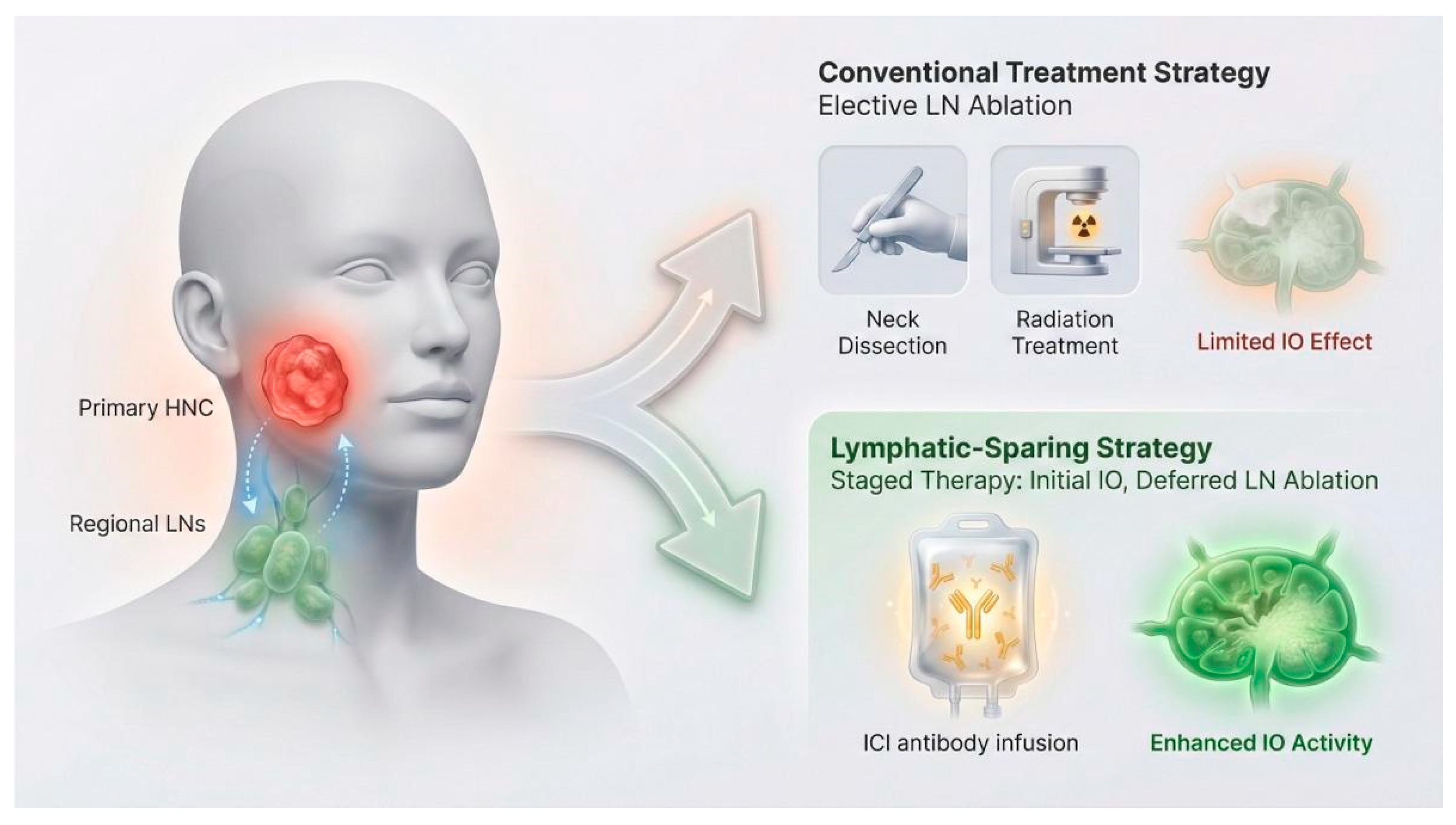
Taken together, these observations support a conceptual model of the “immune desert neck”: a cervical basin rendered biologically inert by lymphatic-targeting therapies (e.g., ND and nodal irradiation). In such a landscape, the structural substrates required for antigen presentation, T-cell priming, and effector recirculation are either surgically removed or chronically damaged, leaving systemic immunotherapy to act in the absence of its most relevant regional immune organ (Figure 1).
Preclinical data showing loss of ICI efficacy after TDLN ablation, coupled with negative concurrent chemoradiotherapy–ICI trials and early neoadjuvant experiences, collectively suggest that future multimodal strategies should deliberately sequence and tailor ND and radiation in ways that maximize oncologic control while preserving, rather than eradicating, cervical immune infrastructure [10,13,48,49,58].

6. Optimal Timing of Neck Dissection in the Immunotherapy Era

6.1. Pre-Immunotherapy vs. Post-Immunotherapy Neck Dissection

Because cervical TDLNs are both the first echelon of metastatic spread and hubs for antigen presentation and Tpex cells, their removal raises a timing dilemma in patients receiving ICIs.
Conceptually, delivering immunotherapy while the lymphatic network is intact has advantages [59,60,61]. Neoadjuvant PD-1/PD-L1 blockade with an undisturbed primary tumor–TDLN axis may maximize antigen exposure, expand tumor-specific T cells in regional nodes, and improve systemic immune surveillance. Immunotherapy-induced nodal regression may also downstage the neck, allowing less extensive surgery in responders [13,47].
Conversely, long-standing oncologic principles caution against delaying surgery in patients with bulky, symptomatic, or unstable nodal disease, where progression can lead to loss of resectability or acute complications. Even in neoadjuvant immunotherapy trials, surgery and ND have remained mandatory and generally feasible after a short preoperative course [62,63].
In practice, sequencing is likely to be risk-adapted. In resectable, locally advanced disease without immediately threatening nodal features, a brief neoadjuvant immunotherapy course followed by timely resection of primary and neck may balance immune priming and safety. In rapidly progressive or high-risk neck disease, upfront or early ND remains the safer default, with immunotherapy integrated peri- or postoperatively rather than used to justify delaying definitive surgery.

6.2. Should Neck Dissection Be Less Aggressive in Immunotherapy-Responders?

Neoadjuvant and perioperative immunotherapy trials have reported major and complete pathologic responses in primary tumors and cervical nodes. This naturally raises the question of whether ND can be de-intensified in strong responders, instead of defaulting to the baseline surgical plan [41,64,65,66].
In early OSCC, END often yields pN0 necks, meaning that many immunologically competent nodes are removed to treat a minority with occult disease [10,11,67,68]. Immunotherapy amplifies this tension: in responders, the very nodes that may have contributed most to systemic immune activation are often targeted for removal.
However, current evidence is not sufficient for routine de-escalation. Nodal responses can be heterogeneous, and small foci of viable carcinoma can still carry significant risk. Robust correlations between nodal pCR and long-term regional control in HNSCC remain limited, and most neoadjuvant trials have required standard ND regardless of response [63,65].
At present, less aggressive ND in immunotherapy responders should be tested within clinical trials. These studies will need clear response-based criteria—combining clinical exam, imaging, and pathology—for safely converting from more extensive to more selective dissections, or for omitting END in selected early-stage cN0 patients.

6.3. Radiologic and Pathologic Response Assessment

Checkpoint blockade often produces nodal “immune flares,” with edema, reactive changes, and tertiary lymphoid structures that can mimic residual disease. PET/CT may remain FDG-avid in nodes without viable tumor, and size criteria may fail to detect scattered residual carcinoma within fibrotic or inflamed nodes. Similarly, pathology must distinguish true residual tumor from regression bed and immune infiltrates. These challenges are discussed in more detail in Section 7.4, where imaging and biomarker-based strategies are integrated.

6.4. “Watch-And-Wait” Strategies

“Watch-and-wait” (W&W) strategies—omitting or delaying major surgery in patients who achieve a robust response after neoadjuvant therapy—are established in rectal cancer and under investigation in esophageal, lung, and other malignancies treated with chemo(radio)therapy or chemo-immunotherapy [69,70]. In carefully selected complete responders, strict surveillance with prompt salvage can preserve organ function without clear survival compromise [71].
Applying W&W to the neck in HNSCC is conceptually attractive. Avoiding unnecessary ablation of cervical TDLNs may preserve immune infrastructure and reduce the risk of an “immune desert neck.” One can envision immunotherapy-based regimens in which patients with compelling nodal responses are monitored rather than undergoing immediate END, reserving early salvage ND for radiologic or clinical recurrence.
However, true W&W for the neck after immunotherapy remains experimental. Current data are limited to small series, retrospective analyses, and extrapolation from other tumor types. Uncontrolled regional failure in HNSCC carries high morbidity and mortality, and the window for effective salvage ND may be narrow, particularly in previously irradiated or heavily pretreated necks.
If W&W is to be adopted, it will require strict eligibility criteria (e.g., early-stage, low initial nodal burden, strong immunotherapy response), multimodal confirmation of response (clinical, imaging, and possibly biopsy), intensive surveillance, and predefined triggers for salvage surgery. Until such protocols are validated and shown to maintain regional control and survival, W&W in the neck should be confined to prospective trials, with conventional ND remaining the benchmark.

7. Management of the Neck in the Immunotherapy Era

7.1. Neck Dissection as an Immunologic and Surgical Intervention

In the immunotherapy era, ND is not simply a mechanical procedure for debulking and staging; it is also an intervention that directly alters TDLN structure and function. Randomized trials and meta-analyses in early OSCC established that END improves DFS and, in many series, OS for cT1N0 and cT2N0 disease, supporting its adoption as standard of care. D’Cruz et al. showed a clear OS advantage for upfront END versus therapeutic ND, while the SEND trial and pooled data confirmed reduced recurrence and improved regional control at the cost of increased morbidity [6,11].
When low-risk stage I (T1N0) tumors are analyzed separately, the benefit of routine END is less clear. Pooled analyses suggest no obvious OS advantage and only a modest reduction in nodal recurrence in carefully selected patients, prompting concern about indiscriminate END in low-risk OSCC, especially when ICIs are planned [72].
Concurrently, modern immunology has reframed cervical TDLNs as dynamic immune organs where antigen-presenting cells, Tpex cells, B cells, and regulatory populations interact under PD-1/PD-L1 blockade. Experimental and translational data indicate that removing uninvolved lymphatic basins or heavily irradiating them can blunt systemic ICI responses by eliminating key sites of antigen presentation and T-cell priming [10,65]. ND should therefore be understood as a modifier of host immunity whose indication, timing, and extent must be reconsidered rather than assumed to be immunologically neutral.

7.2. Neck Dissection After Neoadjuvant Immunotherapy

The integration of neoadjuvant PD-(L)1 blockade into resectable HNSCC has reshaped how ND is used and performed. In modern phase II trials of pembrolizumab and nivolumab (alone or in combination), a substantial share of patients achieve major or complete pathologic response at the primary site, while residual viable disease may persist in cervical nodes, highlighting frequent discordance between primary and nodal responses [40,73].
After neoadjuvant immunotherapy, response-adapted ND is indicated for radiographically residual or progressive nodal disease (on CT, MRI, PET/CT, or diffusion-weighted imaging), biopsy-proven viable metastasis, or high clinical suspicion of persistent nodal involvement despite apparent primary regression. Outside trials, indications remain conservative, given the morbidity and consequences of uncontrolled regional failure.
Technically, post-immunotherapy ND can be more challenging than in treatment-naïve necks. Across tumor types, preoperative PD-(L)1 blockade is associated with dense fibrosis, desmoplastic reaction, and altered tissue planes in both tumor bed and regional lymphatics, which can obscure landmarks and tether vessels and nerves [74]. Early head and neck series suggest overall complication rates comparable to historical controls, but surgeons frequently report tougher tissue and less predictable planes, especially around the carotid sheath, lower cranial nerves, and thoracic duct [66]. Concentrating post-immunotherapy ND in high-volume centers with ready vascular and reconstructive support appears prudent.
Pathologic assessment also changes. Instead of simply measuring residual viable tumor, immune-related pathologic response frameworks emphasize the “regression bed”—proliferative fibrosis, neovascularization, foamy macrophages, cholesterol clefts, and dense lymphoid infiltrates often organized into tertiary lymphoid structures (TLS) [75]. In immunotherapy-treated HNSCC necks, these regression beds can be rich in CD8+ T cells and B-cell follicles, features associated with favorable immunologic profiles [76]. Standardized protocols for grossing, sampling, and grading treatment effect in cervical nodes are needed to avoid overcalling residual carcinoma when much of the mass represents immune-mediated regression.
Oncologic outcomes in this setting come mainly from single-arm studies, but a few themes are consistent. Neoadjuvant pembrolizumab and nivolumab trials report high rates of pathologic response at the primary site and meaningful nodal regression, with low isolated regional failure rates when ND follows standard oncologic principles [40]. Correlative studies document frequent primary–nodal response discordance, sometimes with deeper effects in nodes, suggesting that in the future, some patients with radiographically and pathologically “sterilized” nodal basins might be candidates for de-escalated or omitted completion ND. For now, completion or salvage ND remains the standard when objective disease persists, and omitting ND solely based on imaging or limited pathology should be restricted to controlled trials.

7.3. Response-Adapted and De-Escalation Strategies

Recognizing ND as an immunologic intervention has spurred interest in response-adapted and de-escalation approaches. Early phase neoadjuvant immunotherapy trials show that a meaningful subset of patients achieve major or complete pathologic response with acceptable toxicity, demonstrating that systemic immune control can be established before surgery [10,65].
This is particularly relevant in early OSCC, where END often removes pathologically negative but immunologically competent nodes (Section 7.1). In an immunotherapy context, this trade-off becomes more problematic: responders may derive part of their systemic control from the same nodal basins targeted for extensive dissection [11,67,68]. Bu et al. proposed that in cT1–T2N0 OSCC, neoadjuvant immunotherapy with intact TDLNs might eradicate occult micrometastases and allow de-escalation or omission of END in selected responders, while patients with residual nodal disease would still undergo comprehensive ND [10]. Yang et al. similarly suggest that in stage I OSCC, where the absolute risk of occult nodal spread is relatively low, immune-based strategies may ultimately offer a better balance of control and morbidity than systematic prophylactic lymphadenectomy [72].
Less invasive surgical strategies align naturally with this paradigm. Sentinel lymph node biopsy (SLNB) has shown oncologic non-inferiority to END in early OSCC, with lower morbidity and better functional outcomes, and provides a template for nodal sampling rather than wholesale clearance [77]. In immunotherapy-integrated algorithms, SLNB, limited basin dissection, or image-guided minimally invasive nodal sampling could document eradication of occult disease or detect small residual deposits needing clearance, maximizing regional control while preserving immune-competent nodes.
More radical de-escalation, such as W&W in immunotherapy responders, remains investigational and should follow the principles outlined in Section 6.4. Population-based analyses from large registries suggest that among patients undergoing surgery plus adjuvant immunotherapy, ND itself is not independently associated with worse OS after adjusting for stage and treatment. This implies that nodal surgery and immunotherapy can coexist without obvious detriment in high-burden disease, but also that aggressive ND is not uniformly required in all immunotherapy-treated patients [78]. Implementing response-adapted strategies will require tight multidisciplinary coordination, with surgeons and medical oncologists jointly deciding when ND is indispensable and when it can be limited or deferred, and radiation oncologists using lymphatic-sparing planning where feasible.

7.4. Biomarkers and Imaging to Guide Neck Management

Personalizing neck management in immunotherapy-treated patients ultimately requires accurate characterization of nodal biology before and after treatment. As noted in Section 6.3, immunotherapy-induced immune flares limit the reliability of size and metabolic criteria as surrogates for nodal pCR.
Advanced imaging and quantitative methods offer potential solutions. Early changes in PET/CT standardized uptake value, metabolic tumor volume, and total lesion glycolysis, as well as diffusion-weighted MRI parameters and radiomic signatures, correlate with pathologic response to neoadjuvant chemo-immunotherapy in HNSCC and other cancers and could be incorporated into models predicting nodal pCR non-invasively [79,80,81,82].
At the tissue level, PD-L1 CPS, tumor mutational burden, and immune gene-expression signatures have been associated with higher major and complete pathologic response rates to neoadjuvant PD-1 blockade in HNSCC and related squamous malignancies; PD-L1 CPS ≥ 1 has been linked to improved DFS in a phase II neoadjuvant pembrolizumab study [40,42,62]. TLS density, spatial patterns of tumor-infiltrating lymphocytes, and quantitative immune-histopathologic scores are emerging prognostic markers that may help distinguish immunologically “cured” nodes from those harboring residual disease despite regression [83,84]. Standardized pathologic response criteria for immunotherapy-treated neck specimens are still evolving and will be essential for consistent reporting.
Circulating tumor DNA (ctDNA) assays show promise for detecting minimal residual disease (MRD) and monitoring response to checkpoint blockade in HNSCC; dynamic ctDNA clearance or deep reductions during neoadjuvant therapy may eventually function as systemic surrogates of nodal pCR [85,86,87]. Combining ctDNA kinetics with imaging and tissue-based biomarkers could generate composite algorithms stratifying patients into groups for whom ND is mandatory, optional, or potentially avoidable [85,87].
From a clinical implementation perspective, the clearest pathway for avoiding routine END in cN0 disease relies on a standardized decision tool rather than on any single biomarker. SLNB with ultrastaging, interpreted alongside high-quality anatomic imaging (ultrasound ± fine-needle aspiration cytology [FNAC] and CT/MRI), provides an actionable de-escalation rule in appropriately selected early oral cavity cancers and can preserve regional control in experienced centers [77,88]. In perioperative immunotherapy pathways, a plausible near-term extension is to combine quantitative imaging features with ctDNA MRD dynamics (e.g., ctDNA negativity or clearance together with concordant radiological response) to support omission strategies under intensified surveillance and predefined salvage triggers; however, decision thresholds and sampling timepoints remain under prospective validation [89,90]. Multi-parameter models that integrate ctDNA kinetics, radiomics/deep learning estimates of nodal risk, and tissue immune profiling (including TLS-related metrics and spatial immune signatures) are conceptually aligned with response-adapted neck management after neoadjuvant immunotherapy, but should currently be confined to clinical trials [91].
Ultimately, the evolution of neck management in the immunotherapy era will depend on validating such biomarker-driven, response-adapted strategies in prospective trials. Until robust tools are widely available, ND should continue to follow established oncologic principles in patients at meaningful risk of nodal failure, while de-escalation and omission strategies are tested in carefully controlled research settings [92].

8. Conclusions

Neck dissection has evolved from a uniformly ablative operation into a risk-adapted set of procedures designed to balance oncologic safety with functional preservation. In the era of immune checkpoint inhibition, this surgical evolution intersects with a parallel conceptual shift: cervical TDLNs are increasingly recognized not as passive conduits of metastatic spread, but as active immune organs that support antigen presentation, T-cell priming, and the maintenance of stem-like effector precursors that may be critical for durable responses to PD-1/PD-L1 blockade. Accordingly, ND and elective nodal irradiation should be considered not only as locoregional control measures, but also as interventions capable of reshaping systemic antitumor immunity—potentially contributing, in selected contexts, to an “immune desert neck” if non-involved basins are routinely ablated or heavily irradiated.
Based on currently available evidence, the routine omission of elective or therapeutic ND is not supported in patients at meaningful risk of regional failure, and established oncologic principles remain the benchmark against which any de-escalation strategy must be tested. However, converging mechanistic insights and early perioperative immunotherapy experience define a clear research roadmap aimed at preserving immune-relevant lymphatic infrastructure while maintaining oncologic safety. Future work should prioritize: (a) biomarker-driven, individualized treatment strategies for ND patients, integrating tissue-based immune profiling, circulating markers such as ctDNA kinetics, and response-adapted imaging to determine when ND is essential, when it can be limited to targeted or sentinel-based approaches, and when omission might be studied under rigorous surveillance protocols; (b) the development of immunoprotective radiotherapy techniques, including nodal-volume sparing, refined elective field design, and sequencing approaches that minimize unnecessary injury to non-involved nodal basins during periods when immune priming is most consequential; and (c) AI-based fusion models combining radiomics and histopathomics to predict lymph node immune status, enabling scalable, response-adapted neck management by noninvasively estimating both residual disease risk and nodal immune competence.
Ultimately, the goal is not to discard ND, but to embed it within a multidisciplinary, immunologically informed paradigm in which the timing, extent, and adjunctive radiation strategy are tailored to tumor biology and host immunity—maximizing cure while preserving the regional immune architecture that may be required for sustained benefit from immunotherapy.

Author Contributions

Conceptualization, N.S.; methodology, A.L., C.P. and N.S.; validation, N.S., R.A. and V.K.S.; investigation, A.L., C.P. and N.S.; resources, N.S.; writing—original draft preparation, A.L., C.P. and N.S.; writing—review and editing, A.L., C.P., N.S., R.A. and V.K.S.; supervision, N.S., R.A. and V.K.S. All authors have read and agreed to the published version of the manuscript.

Funding

This research received no external funding.

Data Availability Statement

No new data were created or analyzed in this study. Data sharing is not applicable to this article.

Conflicts of Interest

The authors declare no conflicts of interest.

Abbreviations

The following abbreviations are used in this manuscript:
AAO-HNSAmerican Academy of Otolaryngology–Head and Neck Surgery
AIArtificial intelligence
CPSCombined positive score
CTComputed tomography
ctDNACirculating tumor DNA
CTLA-4Cytotoxic T-lymphocyte–associated protein 4
DFSDisease-free survival
ENDElective neck dissection
ERNDExtended radical neck dissection
FDGFluorodeoxyglucose
FNACFine-needle aspiration cytology
HNSCCHead and neck squamous cell carcinoma
HPVHuman papillomavirus
ICIImmune checkpoint inhibitor
IJVInternal jugular vein
MRDMinimal residual disease
MRIMagnetic resonance imaging
MRNDModified radical neck dissection
NDNeck dissection
OSOverall survival
OSCCOral cavity squamous cell carcinoma
pCRPathologic complete response
PD-1Programmed death-1
PD-L1Programmed death-ligand 1
PET/CTPositron emission tomography/computed tomography
RNDRadical neck dissection
SANSpinal accessory nerve
SCMSternocleidomastoid muscle
SLNBSentinel lymph node biopsy
SNDSelective neck dissection
TDLNTumor-draining lymph node
TpexProgenitor exhausted CD8+ T
TLSTertiary lymphoid structure
W&WWatch-and-wait

References

  1. Rettig, E.M.; D’Souza, G. Epidemiology of Head and Neck Cancer. Surg. Oncol. Clin. N. Am. 2015, 24, 379–396. [Google Scholar] [CrossRef] [Scilit] [PubMed]
  2. Woolgar, J.A.; Scott, J.; Vaughan, E.D.; Brown, J.S.; West, C.R.; Rogers, S. Survival, Metastasis and Recurrence of Oral Cancer in Relation to Pathological Features. Ann. R. Coll. Surg. Engl. 1995, 77, 325–331. [Google Scholar]
  3. Eckert, A.W.; Lautner, M.H.W.; Dempf, R.; Schubert, J.; Bilkenroth, U. Prognostic factors for oral squamous cell carcinoma. Chirurg 2009, 80, 138–143. [Google Scholar] [CrossRef] [Scilit]
  4. Haidari, S.; Obermeier, K.T.; Kraus, M.; Otto, S.; Probst, F.A.; Liokatis, P. Nodal Disease and Survival in Oral Cancer: Is Occult Metastasis a Burden Factor Compared to Preoperatively Nodal Positive Neck? Cancers 2022, 14, 4241. [Google Scholar] [CrossRef] [Scilit]
  5. Myers, E.N.; Gastman, B.R. Neck Dissection: An Operation in Evolution: Hayes Martin Lecture. Arch. Otolaryngol. Head Neck Surg. 2003, 129, 14–25. [Google Scholar] [CrossRef] [Scilit]
  6. Devaraja, K. Historical Milestones in the Evolution of the Procedure of Neck Dissection. World J. Otorhinolaryngol.-Head Neck Surg. 2024, 10, 333–344. [Google Scholar] [CrossRef] [Scilit]
  7. Givens, A.K.; Goyal, N.; Goldenberg, D. Evolution of the Neck Dissection. Oper. Tech. Otolaryngol.-Head Neck Surg. 2024, 35, 290–296. [Google Scholar] [CrossRef] [Scilit]
  8. Farrokhian, N.; Holcomb, A.J.; Dimon, E.; Karadaghy, O.; Ward, C.; Whiteford, E.; Tolan, C.; Hanly, E.K.; Buchakjian, M.R.; Harding, B.; et al. Development and Validation of Machine Learning Models for Predicting Occult Nodal Metastasis in Early-Stage Oral Cavity Squamous Cell Carcinoma. JAMA Netw. Open 2022, 5, e227226. [Google Scholar] [CrossRef] [Scilit] [PubMed]
  9. Payne, K.; Nenclares, P.; Schilling, C. The Impact of Elective Cervical Lymph Node Treatment on the Tumour Immune Response in Head and Neck Squamous Cell Carcinoma: Time for a Change in Treatment Strategy? BJC Rep. 2024, 2, 68. [Google Scholar] [CrossRef] [Scilit]
  10. Bu, L.-L.; Xu, Z.-Y.; Kuo, Z.-Y.; Wei, L.-Y.; Liu, B.; Jia, J. Neck Dissection in cN0 Oral Squamous Cell Carcinoma: A Chance for Immunotherapy. Int. J. Oral Maxillofac. Surg. 2025, 54, 794–805. [Google Scholar] [CrossRef] [Scilit]
  11. D’Cruz, A.K.; Vaish, R.; Kapre, N.; Dandekar, M.; Gupta, S.; Hawaldar, R.; Agarwal, J.P.; Pantvaidya, G.; Chaukar, D.; Deshmukh, A.; et al. Elective versus Therapeutic Neck Dissection in Node-Negative Oral Cancer. N. Engl. J. Med. 2015, 373, 521–529. [Google Scholar] [CrossRef] [Scilit] [PubMed]
  12. Ferris, R.L. Immunology and Immunotherapy of Head and Neck Cancer. J. Clin. Oncol. 2015, 33, 3293–3304. [Google Scholar] [CrossRef] [Scilit] [PubMed]
  13. Nakamura, H.; Ogawa, T.; Yamanaka, S.; Inukai, D.; Maruo, T.; Takahara, T.; Satou, A.; Tsuzuki, T.; Suzuki, S.; Ueda, R.; et al. Immune Status of Cervical Lymph Nodes in Head and Neck Cancer-A Surgical Oncology Perspective. J. Pers. Med. 2023, 13, 1174. [Google Scholar] [CrossRef] [Scilit]
  14. Saddawi-Konefka, R.; Schokrpur, S.; Gutkind, J.S. Let It Be: Preserving Tumor-Draining Lymph Nodes in the Era of Immuno-Oncology. Cancer Cell 2024, 42, 930–933. [Google Scholar] [CrossRef] [Scilit]
  15. Subramanian, S.; Chiesa, F.; Lyubaev, V.; Aidarbekova, A. The Evolution of Surgery in the Management of Neck Metastases. Acta Otorhinolaryngol. Ital. 2006, 26, 309–316. [Google Scholar]
  16. Rinaldo, A.; Ferlito, A.; Silver, C.E. Early History of Neck Dissection. Eur. Arch. Oto-Rhino-Laryngol. 2008, 265, 1535–1538. [Google Scholar] [CrossRef] [Scilit]
  17. Robbins, K.T.; Medina, J.E.; Wolfe, G.T.; Levine, P.A.; Sessions, R.B.; Pruet, C.W. Standardizing Neck Dissection Terminology. Official Report of the Academy’s Committee for Head and Neck Surgery and Oncology. Arch. Otolaryngol. Head Neck Surg. 1991, 117, 601–605. [Google Scholar] [CrossRef] [Scilit]
  18. Martin, H.; Del Valle, B.; Ehrlich, H.; Cahan, W.G. Neck Dissection. Cancer 1951, 4, 441–499. [Google Scholar] [CrossRef] [Scilit]
  19. Jesse, R.H.; Ballantyne, A.J.; Larson, D. Radical or Modified Neck Dissection: A Therapeutic Dilemma. Am. J. Surg. 1978, 136, 516–519. [Google Scholar] [CrossRef] [Scilit]
  20. Ferlito, A.; Rinaldo, A.; Silver, C.E.; Shah, J.P.; Suárez, C.; Medina, J.E.; Kowalski, L.P.; Johnson, J.T.; Strome, M.; Rodrigo, J.P.; et al. Neck Dissection: Then and Now. Auris. Nasus. Larynx 2006, 33, 365–374. [Google Scholar] [CrossRef] [Scilit] [PubMed]
  21. Ferlito, A. Classification and Terminology of Neck Dissection. Arch. Otolaryngol. Head Neck Surg. 2002, 128, 747–748. [Google Scholar] [CrossRef] [Scilit]
  22. Shaha, A.R. Extended Neck Dissection. J. Surg. Oncol. 1990, 45, 229–233. [Google Scholar] [CrossRef] [Scilit] [PubMed]
  23. Okano, W.; Hayashi, R.; Matsuura, K.; Shinozaki, T.; Tomioka, T. Extent of Salvage Neck Dissection Following Chemoradiation for Locally Advanced Head and Neck Cancer. Head Neck 2021, 43, 413–418. [Google Scholar] [CrossRef] [Scilit]
  24. Ferlito, A.; Robbins, K.T.; Shah, J.P.; Medina, J.E.; Silver, C.E.; Al-Tamimi, S.; Fagan, J.J.; Paleri, V.; Takes, R.P.; Bradford, C.R.; et al. Proposal for a Rational Classification of Neck Dissections. Head Neck 2011, 33, 445–450. [Google Scholar] [CrossRef] [Scilit] [PubMed]
  25. Liao, L.-J.; Lo, W.-C.; Hsu, W.-L.; Wang, C.-T.; Lai, M.-S. Detection of Cervical Lymph Node Metastasis in Head and Neck Cancer Patients with Clinically N0 Neck-a Meta-Analysis Comparing Different Imaging Modalities. BMC Cancer 2012, 12, 236. [Google Scholar] [CrossRef] [Scilit]
  26. Kim, S.-J.; Pak, K.; Kim, K. Diagnostic Accuracy of F-18 FDG PET or PET/CT for Detection of Lymph Node Metastasis in Clinically Node Negative Head and Neck Cancer Patients; A Systematic Review and Meta-Analysis. Am. J. Otolaryngol. 2019, 40, 297–305. [Google Scholar] [CrossRef] [Scilit] [PubMed]
  27. Alsibani, A.; Alqahtani, A.; Almohammadi, R.; Islam, T.; Alessa, M.; Aldhahri, S.F.; Al-Qahtani, K.H. Comparing the Efficacy of CT, MRI, PET-CT, and US in the Detection of Cervical Lymph Node Metastases in Head and Neck Squamous Cell Carcinoma with Clinically Negative Neck Lymph Node: A Systematic Review and Meta-Analysis. J. Clin. Med. 2024, 13, 7622. [Google Scholar] [CrossRef] [Scilit]
  28. Lira, R.B.; Kowalski, L.P. Robotic Neck Dissection: State of Affairs. Curr. Opin. Otolaryngol. Head Neck Surg. 2020, 28, 96–99. [Google Scholar] [CrossRef] [Scilit]
  29. Holcomb, A.J.; Richmon, J.D. Robotic and Endoscopic Approaches to Head and Neck Surgery. Hematol. Oncol. Clin. N. Am. 2021, 35, 875–894. [Google Scholar] [CrossRef] [Scilit]
  30. Anand, A.; Vidhyadharan, S.; Subramaniam, N.; Balsubramanian, D.; Battoo, A.J.; Iyer, S.; Thankappan, K. Selective Neck Dissection in Oral Cavity Cancer Is Not Without Morbidity. Indian J. Surg. Oncol. 2021, 12, 5–11. [Google Scholar] [CrossRef] [Scilit]
  31. Malhotra, D.; Fletcher, A.L.; Turley, S.J. Stromal and Hematopoietic Cells in Secondary Lymphoid Organs: Partners in Immunity. Immunol. Rev. 2013, 251, 160–176. [Google Scholar] [CrossRef] [Scilit]
  32. Gasteiger, G.; Ataide, M.; Kastenmüller, W. Lymph Node-an Organ for T-Cell Activation and Pathogen Defense. Immunol. Rev. 2016, 271, 200–220. [Google Scholar] [CrossRef] [Scilit] [PubMed]
  33. Kim, R.; Emi, M.; Tanabe, K.; Arihiro, K. Immunobiology of the Sentinel Lymph Node and Its Potential Role for Antitumour Immunity. Lancet Oncol. 2006, 7, 1006–1016. [Google Scholar] [CrossRef] [Scilit] [PubMed]
  34. Basto, P.A.; Reticker-Flynn, N.E. Interrogating the Roles of Lymph Node Metastasis in Systemic Immune Surveillance. Clin. Exp. Metastasis 2024, 41, 351–359. [Google Scholar] [CrossRef] [Scilit]
  35. Hampton, H.R.; Chtanova, T. Lymphatic Migration of Immune Cells. Front. Immunol. 2019, 10, 1168. [Google Scholar] [CrossRef] [Scilit] [PubMed]
  36. Davis, M.J.; Zawieja, S.D.; King, P.D. Transport and Immune Functions of the Lymphatic System. Annu. Rev. Physiol. 2025, 87, 151–172. [Google Scholar] [CrossRef] [Scilit]
  37. Burtness, B.; Harrington, K.J.; Greil, R.; Soulières, D.; Tahara, M.; de Castro, G.; Psyrri, A.; Basté, N.; Neupane, P.; Bratland, Å.; et al. Pembrolizumab Alone or with Chemotherapy versus Cetuximab with Chemotherapy for Recurrent or Metastatic Squamous Cell Carcinoma of the Head and Neck (KEYNOTE-048): A Randomised, Open-Label, Phase 3 Study. Lancet Lond. Engl. 2019, 394, 1915–1928, Erratum in Lancet Lond. Engl. 2021, 397, 2252. [Google Scholar] [CrossRef] [Scilit]
  38. Ferris, R.L.; Blumenschein, G.; Fayette, J.; Guigay, J.; Colevas, A.D.; Licitra, L.; Harrington, K.; Kasper, S.; Vokes, E.E.; Even, C.; et al. Nivolumab for Recurrent Squamous-Cell Carcinoma of the Head and Neck. N. Engl. J. Med. 2016, 375, 1856–1867. [Google Scholar] [CrossRef] [Scilit]
  39. Ueda, Y.; Okano, S.; Enokida, T.; Fujisawa, T.; Ito, K.; Sato, M.; Tanaka, H.; Wada, A.; Tahara, M. Nivolumab for Recurrent or Metastatic Head and Neck Cancer Patients with Non-Squamous Cell Carcinoma and/or a Primary Subsite Excluded from CheckMate141, a Retrospective Study. Oral Oncol. 2022, 130, 105932. [Google Scholar] [CrossRef] [Scilit]
  40. Uppaluri, R.; Campbell, K.M.; Egloff, A.M.; Zolkind, P.; Skidmore, Z.L.; Nussenbaum, B.; Paniello, R.C.; Rich, J.T.; Jackson, R.; Pipkorn, P.; et al. Neoadjuvant and Adjuvant Pembrolizumab in Resectable Locally Advanced, Human Papillomavirus-Unrelated Head and Neck Cancer: A Multicenter, Phase II Trial. Clin. Cancer Res. 2020, 26, 5140–5152, Correction in Clin Cancer Res. 2021, 27, 357. [Google Scholar] [CrossRef] [Scilit]
  41. Vos, J.L.; Elbers, J.B.W.; Krijgsman, O.; Traets, J.J.H.; Qiao, X.; van der Leun, A.M.; Lubeck, Y.; Seignette, I.M.; Smit, L.A.; Willems, S.M.; et al. Neoadjuvant Immunotherapy with Nivolumab and Ipilimumab Induces Major Pathological Responses in Patients with Head and Neck Squamous Cell Carcinoma. Nat. Commun. 2021, 12, 7348. [Google Scholar] [CrossRef] [Scilit]
  42. Uppaluri, R.; Haddad, R.I.; Tao, Y.; Le Tourneau, C.; Lee, N.Y.; Westra, W.; Chernock, R.; Tahara, M.; Harrington, K.J.; Klochikhin, A.L.; et al. Neoadjuvant and Adjuvant Pembrolizumab in Locally Advanced Head and Neck Cancer. N. Engl. J. Med. 2025, 393, 37–50. [Google Scholar] [CrossRef] [Scilit]
  43. Gohel, S.; Akhade, A. Immunotherapy Makes Inroads in Head and Neck Cancer Treatment. Indian J. Med. Paediatr. Oncol. 2025, 46, 549–552. [Google Scholar] [CrossRef] [Scilit]
  44. Liu, Z.; Yu, Z.; Chen, D.; Verma, V.; Yuan, C.; Wang, M.; Wang, F.; Fan, Q.; Wang, X.; Li, Y.; et al. Pivotal Roles of Tumor-Draining Lymph Nodes in the Abscopal Effects from Combined Immunotherapy and Radiotherapy. Cancer Commun. Lond. Engl. 2022, 42, 971–986. [Google Scholar] [CrossRef] [Scilit]
  45. Reticker-Flynn, N.E.; Engleman, E.G. Lymph Nodes: At the Intersection of Cancer Treatment and Progression. Trends Cell Biol. 2023, 33, 1021–1034. [Google Scholar] [CrossRef] [Scilit]
  46. Buchwald, Z.S.; Nasti, T.H.; Lee, J.; Eberhardt, C.S.; Wieland, A.; Im, S.J.; Lawson, D.; Curran, W.; Ahmed, R.; Khan, M.K. Tumor-Draining Lymph Node Is Important for a Robust Abscopal Effect Stimulated by Radiotherapy. J. Immunother. Cancer 2020, 8, e000867, Erratum in J. Immunother. Cancer 2021, 9, e000867corr1. [Google Scholar] [CrossRef] [Scilit] [PubMed]
  47. Saddawi-Konefka, R.; O’Farrell, A.; Faraji, F.; Clubb, L.; Allevato, M.M.; Jensen, S.M.; Yung, B.S.; Wang, Z.; Wu, V.H.; Anang, N.-A.; et al. Lymphatic-Preserving Treatment Sequencing with Immune Checkpoint Inhibition Unleashes cDC1-Dependent Antitumor Immunity in HNSCC. Nat. Commun. 2022, 13, 4298. [Google Scholar] [CrossRef] [Scilit] [PubMed]
  48. Karam, S.D.; Anderson, C.M.; Ma, D.; Chua, M.L.K.; Kimple, R.J. Immunotherapy in Head and Neck Cancer-Ready for Prime Time or More Research Needed? Int. J. Radiat. Oncol. Biol. Phys. 2021, 109, 647–650. [Google Scholar] [CrossRef] [Scilit] [PubMed]
  49. Sim, E.S.; Nguyen, H.C.B.; Hanna, G.J.; Uppaluri, R. Current Progress and Future Directions of Immunotherapy in Head and Neck Squamous Cell Carcinoma: A Narrative Review. JAMA Otolaryngol.-Head Neck Surg. 2025, 151, 521–528. [Google Scholar] [CrossRef] [Scilit]
  50. Liljeström, V.; Farrajota Neves da Silva, P.; Bark, R.; Elliot, A.; Marklund, L.; Margolin, G.; Kumlien Georén, S.; Cardell, L.-O.; Piersiala, K. B Cell Dysfunction in Tumor-Draining Lymph Nodes Predicts Relapse in Oral Squamous Cell Carcinoma. Cancer Immunol. Immunother. CII 2025, 74, 373. [Google Scholar] [CrossRef] [Scilit]
  51. Marciscano, A.E.; Ghasemzadeh, A.; Nirschl, T.R.; Theodros, D.; Kochel, C.M.; Francica, B.J.; Muroyama, Y.; Anders, R.A.; Sharabi, A.B.; Velarde, E.; et al. Elective Nodal Irradiation Attenuates the Combinatorial Efficacy of Stereotactic Radiation Therapy and Immunotherapy. Clin. Cancer Res. 2018, 24, 5058–5071. [Google Scholar] [CrossRef] [Scilit]
  52. Darragh, L.B.; Gadwa, J.; Pham, T.T.; Van Court, B.; Neupert, B.; Olimpo, N.A.; Nguyen, K.; Nguyen, D.; Knitz, M.W.; Hoen, M.; et al. Elective Nodal Irradiation Mitigates Local and Systemic Immunity Generated by Combination Radiation and Immunotherapy in Head and Neck Tumors. Nat. Commun. 2022, 13, 7015, Erratum in Nat. Commun. 2024, 15, 8781. [Google Scholar] [CrossRef] [Scilit]
  53. Cheng, Y.; Wang, J.; Sun, J.; Zhong, Y.; Wu, Q. Radiotherapy Improves the Clinical Outcomes of Recurrent or Metastatic Head and Neck Cancers Treated with Immunotherapy. Discov. Oncol. 2025, 16, 988. [Google Scholar] [CrossRef] [Scilit] [PubMed]
  54. Tao, Y.; Aupérin, A.; Sun, X.; Sire, C.; Martin, L.; Coutte, A.; Lafond, C.; Miroir, J.; Liem, X.; Rolland, F.; et al. Avelumab-Cetuximab-Radiotherapy versus Standards of Care in Locally Advanced Squamous-Cell Carcinoma of the Head and Neck: The Safety Phase of a Randomised Phase III Trial GORTEC 2017-01 (REACH). Eur. J. Cancer 2020, 141, 21–29. [Google Scholar] [CrossRef] [Scilit]
  55. Lee, N.Y.; Ferris, R.L.; Psyrri, A.; Haddad, R.I.; Tahara, M.; Bourhis, J.; Harrington, K.; Chang, P.M.-H.; Lin, J.-C.; Razaq, M.A.; et al. Avelumab plus Standard-of-Care Chemoradiotherapy versus Chemoradiotherapy Alone in Patients with Locally Advanced Squamous Cell Carcinoma of the Head and Neck: A Randomised, Double-Blind, Placebo-Controlled, Multicentre, Phase 3 Trial. Lancet Oncol. 2021, 22, 450–462. [Google Scholar] [CrossRef] [Scilit] [PubMed]
  56. Machiels, J.-P.; Tao, Y.; Licitra, L.; Burtness, B.; Tahara, M.; Rischin, D.; Alves, G.; Lima, I.P.F.; Hughes, B.G.M.; Pointreau, Y.; et al. Pembrolizumab plus Concurrent Chemoradiotherapy versus Placebo plus Concurrent Chemoradiotherapy in Patients with Locally Advanced Squamous Cell Carcinoma of the Head and Neck (KEYNOTE-412): A Randomised, Double-Blind, Phase 3 Trial. Lancet Oncol. 2024, 25, 572–587. [Google Scholar] [CrossRef] [Scilit] [PubMed]
  57. Haddad, R.; Fayette, J.; Teixeira, M.; Prabhash, K.; Mesia, R.; Kawecki, A.; Dechaphunkul, A.; Dinis, J.; Guo, Y.; Masuda, M.; et al. Atezolizumab in High-Risk Locally Advanced Squamous Cell Carcinoma of the Head and Neck: A Randomized Clinical Trial. JAMA 2025, 333, 1599–1607. [Google Scholar] [CrossRef] [Scilit]
  58. Olmos, M.; Glajzer, J.; Büntemeyer, T.-O.; Frohwitter, G.; Ries, J.; Eckstein, M.; Hecht, M.; Lutz, R.; Kesting, M.R.; Weber, M. Neoadjuvant Immunotherapy of Oral Squamous Cell Carcinoma: Case Report and Assessment of Histological Response. Front. Oncol. 2021, 11, 720951. [Google Scholar] [CrossRef] [Scilit]
  59. Fransen, M.F.; Schoonderwoerd, M.; Knopf, P.; Camps, M.G.; Hawinkels, L.J.; Kneilling, M.; van Hall, T.; Ossendorp, F. Tumor-Draining Lymph Nodes Are Pivotal in PD-1/PD-L1 Checkpoint Therapy. JCI Insight 2018, 3, e124507. [Google Scholar] [CrossRef] [Scilit]
  60. Wei, J.; Li, D.; Long, H.; Han, M. Immune Microenvironment of Tumor-Draining Lymph Nodes: Insights for Immunotherapy. Front. Immunol. 2025, 16, 1562797. [Google Scholar] [CrossRef] [Scilit]
  61. Zhang, J.; Ma, J.; Niu, Y.; Liu, J.; Wang, Z.; Guo, J.; Meng, Y.; Sun, R.; Zhang, Z.; Liu, H.; et al. Tumor-Draining Lymph Nodes Respond to Immune Checkpoint Inhibition and Orchestrate Tumor Immune Remodeling. Cancer Immunol. Immunother. CII 2025, 74, 326. [Google Scholar] [CrossRef] [Scilit]
  62. Wise-Draper, T.M.; Gulati, S.; Palackdharry, S.; Hinrichs, B.H.; Worden, F.P.; Old, M.O.; Dunlap, N.E.; Kaczmar, J.M.; Patil, Y.; Riaz, M.K.; et al. Phase II Clinical Trial of Neoadjuvant and Adjuvant Pembrolizumab in Resectable Local-Regionally Advanced Head and Neck Squamous Cell Carcinoma. Clin. Cancer Res. 2022, 28, 1345–1352. [Google Scholar] [CrossRef] [Scilit]
  63. Widjaja, W.; Ng, I.; Shannon, N.; Iyer, N.G. Neoadjuvant Immunotherapy in Resectable HNSCC: An Updated Systematic Review and Meta-Analysis. Ann. Surg. Oncol. 2025, 32, 2713–2724. [Google Scholar] [CrossRef] [Scilit]
  64. Leidner, R.; Crittenden, M.; Young, K.; Xiao, H.; Wu, Y.; Couey, M.A.; Patel, A.A.; Cheng, A.C.; Watters, A.L.; Bifulco, C.; et al. Neoadjuvant Immunoradiotherapy Results in High Rate of Complete Pathological Response and Clinical to Pathological Downstaging in Locally Advanced Head and Neck Squamous Cell Carcinoma. J. Immunother. Cancer 2021, 9, e002485. [Google Scholar] [CrossRef] [Scilit]
  65. Masarwy, R.; Kampel, L.; Horowitz, G.; Gutfeld, O.; Muhanna, N. Neoadjuvant PD-1/PD-L1 Inhibitors for Resectable Head and Neck Cancer: A Systematic Review and Meta-Analysis. JAMA Otolaryngol.-Head Neck Surg. 2021, 147, 871–878. [Google Scholar] [CrossRef] [Scilit]
  66. Pedroso, C.M.; de Pauli Paglioni, M.; Normando, A.G.C.; Chaves, A.L.F.; Kowalski, L.P.; de Castro Júnior, G.; Matos, L.L.; Willian Junior, W.N.; de Oliveira, T.B.; de Marchi, P.; et al. Preoperative Neoadjuvant Chemotherapy or Immunotherapy in Head and Neck Cancer: A Systematic Review and Meta-Analysis of Surgical Risk and Pathologic Response. Crit. Rev. Oncol. Hematol. 2025, 212, 104742. [Google Scholar] [CrossRef] [Scilit]
  67. Massey, C.; Dharmarajan, A.; Bannuru, R.R.; Rebeiz, E. Management of N0 Neck in Early Oral Squamous Cell Carcinoma: A Systematic Review and Meta-Analysis. Laryngoscope 2019, 129, E284–E298. [Google Scholar] [CrossRef] [Scilit] [PubMed]
  68. Cao, Y.; Wang, T.; Yu, C.; Guo, X.; Li, C.; Li, L. Elective Neck Dissection Versus Wait-and-Watch Policy for Oral Cavity Squamous Cell Carcinoma in Early Stage: A Systematic Review and Meta-Analysis Based on Survival Data. J. Oral Maxillofac. Surg. 2019, 77, 2154–2167. [Google Scholar] [CrossRef] [Scilit] [PubMed]
  69. Tan, L.; Yang, G.; Zeng, C.; Zhang, X. Case Report: Watch-and-Wait Strategy in Resectable Esophageal Cancer Following Neoadjuvant Chemoimmunotherapy: A Case Series. Front. Immunol. 2024, 15, 1502206. [Google Scholar] [CrossRef] [Scilit]
  70. Wang, X.; Li, C.; Cao, Y.; Kang, X.; Leng, X.; Zheng, Y.; Zheng, B.; Guo, W.; Jing, H.; Han, Y.; et al. Neoadjuvant Camrelizumab Combined with Chemoradiotherapy and Watch-and-Wait Strategy versus Neoadjuvant Chemoradiotherapy Followed by Surgery in Locally Advanced Oesophageal Squamous Cell Carcinoma: Study Protocol of a Randomised Controlled Trial (PALACE-3). BMJ Open 2025, 15, e093538. [Google Scholar] [CrossRef] [Scilit] [PubMed]
  71. Zhang, Q.; Huang, W.; Chen, X.; Yan, Z.; Huang, D.; Xu, H.; Zheng, Y.; Zhu, L.; Pan, C.; Pan, J.; et al. Watch-and-Wait Strategy in Patients with Objective Response after Immuno-Chemotherapy for Locally Recurrent Nasopharyngeal Carcinoma: A Pilot Study. BMC Cancer 2025, 25, 1255. [Google Scholar] [CrossRef] [Scilit]
  72. Yang, C.; Wang, Z.; Shi, L.; Liu, W. Evaluation of Neck Control Strategies for Oral Squamous Cell Carcinoma of Stage I: Neck Dissection or Potential Immunotherapy. J. Dent. Sci. 2024, 19, 640–644. [Google Scholar] [CrossRef] [Scilit]
  73. Merlino, D.J.; Johnson, J.M.; Tuluc, M.; Gargano, S.; Stapp, R.; Harshyne, L.; Leiby, B.E.; Flanders, A.; Zinner, R.; Axelrod, R.; et al. Discordant Responses Between Primary Head and Neck Tumors and Nodal Metastases Treated with Neoadjuvant Nivolumab: Correlation of Radiographic and Pathologic Treatment Effect. Front. Oncol. 2020, 10, 566315. [Google Scholar] [CrossRef] [Scilit] [PubMed]
  74. Zotta, A.; Marciano, M.L.; Sabbatino, F.; Ottaiano, A.; Cascella, M.; Pontone, M.; Montano, M.; Calogero, E.; Longo, F.; Fasano, M.; et al. Neoadjuvant Immunotherapy in Head and Neck Cancers: A Paradigm Shift in Treatment Approach. Biomedicines 2024, 12, 2337. [Google Scholar] [CrossRef] [Scilit]
  75. Cottrell, T.R.; Thompson, E.D.; Forde, P.M.; Stein, J.E.; Duffield, A.S.; Anagnostou, V.; Rekhtman, N.; Anders, R.A.; Cuda, J.D.; Illei, P.B.; et al. Pathologic Features of Response to Neoadjuvant Anti-PD-1 in Resected Non-Small-Cell Lung Carcinoma: A Proposal for Quantitative Immune-Related Pathologic Response Criteria (irPRC). Ann. Oncol. 2018, 29, 1853–1860. [Google Scholar] [CrossRef] [Scilit] [PubMed]
  76. Jiang, B.; Wu, Z.; Zhang, Y.; Yang, X. Associations between Tertiary Lymphoid Structure Density and Immune Checkpoint Inhibitor Efficacy in Solid Tumors: Systematic Review and Meta-Analysis. Front. Immunol. 2024, 15, 1414884. [Google Scholar] [CrossRef] [Scilit]
  77. Hasegawa, Y.; Tsukahara, K.; Yoshimoto, S.; Miura, K.; Yokoyama, J.; Hirano, S.; Uemura, H.; Sugasawa, M.; Yoshizaki, T.; Homma, A.; et al. Neck Dissections Based on Sentinel Lymph Node Navigation Versus Elective Neck Dissections in Early Oral Cancers: A Randomized, Multicenter, and Noninferiority Trial. J. Clin. Oncol. 2021, 39, 2025–2036. [Google Scholar] [CrossRef] [Scilit]
  78. Patel, A.M.; Haleem, A.; Cowan, P.T.; Roden, D.F. Neck Dissection and Survival Among Head and Neck Cancer Patients Undergoing Adjuvant Immunotherapy. Laryngoscope Investig. Otolaryngol. 2025, 10, e70120. [Google Scholar] [CrossRef] [Scilit]
  79. Vos, J.L.; Zuur, C.L.; Smit, L.A.; de Boer, J.P.; Al-Mamgani, A.; van den Brekel, M.W.M.; Haanen, J.B.A.G.; Vogel, W.V. [18F]FDG-PET Accurately Identifies Pathological Response Early upon Neoadjuvant Immune Checkpoint Blockade in Head and Neck Squamous Cell Carcinoma. Eur. J. Nucl. Med. Mol. Imaging 2022, 49, 2010–2022. [Google Scholar] [CrossRef] [Scilit] [PubMed]
  80. Wondergem, N.E.; Miedema, I.H.C.; van de Ven, R.; Zwezerijnen, G.J.C.; de Graaf, P.; Karagozoglu, K.H.; Hendrickx, J.-J.; Eerenstein, S.E.J.; Bun, R.J.; Mulder, D.C.; et al. Circulating T Cell Status and Molecular Imaging May Predict Clinical Benefit of Neoadjuvant PD-1 Blockade in Oral Cancer. J. Immunother. Cancer 2024, 12, e009278. [Google Scholar] [CrossRef] [Scilit]
  81. Lyu, W.; Gong, J.; Zhu, L.; Xu, T.; Huang, S.; Shen, C.; Wang, C.; He, X.; Ying, H.; Hu, C.; et al. MR Radiomics Unveils Neoadjuvant Chemo-Responsiveness with Insights into Selective Treatment de-Intensification in HPV-Positive Oropharyngeal Carcinoma. Oral Oncol. 2024, 159, 107049. [Google Scholar] [CrossRef] [Scilit]
  82. Breukers, S.E.; Crommelin, R.D.; Smit, L.A.; de Boer, J.P.; Navran, A.; van Houdt, W.J.; de Bree, R.; Devriese, L.A.; Haanen, J.B.A.G.; Wondergem, M.; et al. [18F]FDG-PET/CT’s Change in Total Lesion Glycolysis Can Accurately Identify Early Response upon Neoadjuvant Immunotherapy Prior to Curative-Intent Surgery in Cutaneous Squamous Cell Carcinoma; MATISSE Trial. Eur. J. Nucl. Med. Mol. Imaging 2025, 53, 2217–2233. [Google Scholar] [CrossRef] [Scilit]
  83. Liu, Z.; Meng, X.; Tang, X.; Zou, W.; He, Y. Intratumoral Tertiary Lymphoid Structures Promote Patient Survival and Immunotherapy Response in Head Neck Squamous Cell Carcinoma. Cancer Immunol. Immunother. CII 2023, 72, 1505–1521. [Google Scholar] [CrossRef] [Scilit]
  84. Cui, M.; Zhou, M.; Zhou, L.; Zhou, G.; Liu, Y. Tertiary Lymphoid Structures Achieve “cold” to “Hot” Transition by Remodeling the Cold Tumor Microenvironment. Biochim. Biophys. Acta Rev. Cancer 2025, 1880, 189312. [Google Scholar] [CrossRef] [Scilit]
  85. Honoré, N.; van der Elst, A.; Dietz, A.; van Marcke, C.; Helaers, R.; Mendola, A.; Dahou, H.; Marbaix, E.; Poncin, R.; Seront, E.; et al. Tumour-Agnostic Plasma Assay for Circulating Tumour DNA Predicts Outcome in Recurrent and/or Metastatic Squamous Cell Carcinoma of the Head and Neck Treated with a PD-1 Inhibitor. Eur. J. Cancer 2023, 195, 113372. [Google Scholar] [CrossRef] [Scilit]
  86. Noji, R.; Tohyama, K.; Nakamura, S.; Naito, T.; Oikawa, Y.; Kuroshima, T.; Tomioka, H.; Michi, Y.; Ikeda, S.; Asakage, T.; et al. Dynamic Changes in Circulating Tumor DNA During Immunotherapy for Head and Neck Cancer: SHIZUKU-HN Study. Int. J. Mol. Sci. 2024, 26, 235. [Google Scholar] [CrossRef] [Scilit] [PubMed]
  87. Gezer, U.; Meral, R.; Özgür, E.; Yörüker, E.E.; Bronkhorst, A.; Holdenrieder, S. A Narrative Review of the Role of Non-Viral Circulating Tumor DNA Profiling in Predicting the Treatment Response and Recurrence in Head and Neck Squamous Cell Carcinoma. Cancers 2025, 17, 2279. [Google Scholar] [CrossRef] [Scilit]
  88. Al-Moraissi, E.A.; Marwan, H.; Elayah, S.A.; Traxler-Weidenauer, D.; Paraskevopoulos, K.; Amir Rais, M.; Zimmermann, M. Sentinel Lymph Node Biopsy versus Elective Neck Dissection in Management of the Clinically Negative (cN0) Neck in Patients with Oral Squamous Cell Carcinoma: A Systematic Review and Meta-Analysis. J. Cranio-Maxillofac. Surg. 2024, 52, 141–150. [Google Scholar] [CrossRef] [Scilit] [PubMed]
  89. Ruiz-Torres, D.A.; Roberts, T.J.; Du, P.; Mendel, J.; Neagele, S.; Bonora, G.; Zhang, F.; Efthymiou, V.; Merkin, R.D.; Lin, D.T.; et al. Prognostic Value of Tumor-Informed Circulating Tumor DNA in HPV-Independent Head and Neck Squamous Cell Carcinoma. JAMA Otolaryngol.-Head Neck Surg. 2026, e254837. [Google Scholar] [CrossRef] [Scilit] [PubMed]
  90. Ventelou, L.; Ginzac, A.; Ferreira, M.-C.; Canetti, L.; Philippe, S.; Pinard, C.; Durando, X.; Bernadach, M. Circulating Tumour DNA in Head and Neck Squamous-Cell Carcinomas: A Literature Review. Cancer Treat. Rev. 2026, 142, 103070. [Google Scholar] [CrossRef] [Scilit]
  91. Lan, T.; Kuang, S.; Liang, P.; Ning, C.; Li, Q.; Wang, L.; Wang, Y.; Lin, Z.; Hu, H.; Yang, L.; et al. MRI-Based Deep Learning and Radiomics for Prediction of Occult Cervical Lymph Node Metastasis and Prognosis in Early-Stage Oral and Oropharyngeal Squamous Cell Carcinoma: A Diagnostic Study. Int. J. Surg. 2024, 110, 4648–4659. [Google Scholar] [CrossRef] [Scilit] [PubMed]
  92. Contrera, K.J.; Kansara, S.; Goyal, N.; Mady, L.J.; Puram, S.V.; Young, G.D.; Zandberg, D.P.; Uppaluri, R.; Ferrarotto, R.; Holsinger, F.C.; et al. Neoadjuvant Therapy for Mucosal Head and Neck Squamous Cell Carcinoma: A Review From the American Head and Neck Society. JAMA Otolaryngol.-Head Neck Surg. 2025, 151, 615–625. [Google Scholar] [CrossRef] [Scilit] [PubMed]
Figure 1. Conceptual framework for managing TDLNs in the immunotherapy era [14]. Effective immune checkpoint blockade depends on an intact tumor–lymph node axis, in which TDLNs function as active immune organs that coordinate antigen presentation, expansion of Tpex cells, and effector recirculation. Traditional strategies—elective ND and broad-field elective nodal irradiation—achieve regional control but may simultaneously ablate or chronically damage non-involved nodal basins, creating an “immune desert neck” that attenuates immunotherapy efficacy. HNC, head and neck cancer; ICI, immune checkpoint inhibitor; IO, immunotherapy; LN, lymph node.
Lymphatics 04 00013 g001
Table 1. Historical evolution and standardization of ND terminology and extent.
TimeframeMilestoneDefining Operative ConceptStandardized Description/Nomenclature OutputRelevance to Current Practice
1906Early systematic en bloc cervical lymphadenectomy (Crile)En bloc clearance of the cervical lymphatic-bearing tissue, emphasizing minimal manipulation and acknowledging the close lymphatic–venous relationship (with venous resection when required).Establishes the “en bloc package” principle that later informs radical templates.Conceptual foundation for comprehensive nodal clearance as an oncologic operation.
1951 (mid-20th century)RND becomes the reference standard (Martin)Comprehensive lymphadenectomy with routine resection of SAN, IJV, and SCM advocated for metastatic neck disease.Canonical definition of “radical” ND as the historical benchmark.Benchmark procedure; morbidity associated with non-lymphatic sacrifice drives subsequent de-escalation.
1956–1960sFormalization of elective treatment of the clinically N0 neckEmergence of elective ND terminology for prophylactic management of occult metastases.Distinction between therapeutic and elective ND enters common usage.Frames the persistent risk–benefit debate in cN0 disease (overtreatment vs. prevention).
1963–1980sFunctional/conservation ND (Suárez; Bocca)Preservation of key non-lymphatic structures (SAN/IJV/SCM) while removing the lymphatic–fibrofatty compartments along fascial planes; subsequent large series support oncologic acceptability in selected settings.Consolidates the principle that lymphadenectomy and functional preservation can be compatible.Conceptual and technical precursor of modified radical and selective approaches.
Late 1970s–1990sMRND validatedComprehensive ND with preservation of ≥1 of SAN/IJV/SCM; comparative experiences reinforce reduced morbidity with maintained control in appropriate patients.MRND defined as levels I–V with preservation of one or more non-lymphatic structures.Establishes “comprehensive yet structure-preserving” surgery as mainstream for many N+ scenarios.
1980s–presentSND (pattern-based, subsite-driven)ND limited to nodal levels at highest risk for a given primary site; intentional omission of low-risk levels.SND variants increasingly reported by levels removed (e.g., SND I–III; SND II–IV).Current default in many cN0 and limited N+ settings to reduce morbidity while maintaining regional control.
1991AAO-HNS classification codifies ND categoriesFour-category taxonomy: RND, MRND, SND, END.Standard reporting framework: procedure name + preserved structures (MRND) or levels removed (SND).Enables inter-study comparability and consistent multidisciplinary communication.
2002–2011Modern emphasis on anatomic granularity and descriptive nomenclatureAddition of sublevels (A/B) to levels I/II/V; removal of “named” SND subtypes; recognition of level VII; recommendation to designate ND by levels + non-lymphatic structures removed.Contemporary best practice: ND (levels ± structures), e.g., ND (I–V, SCM, IJV) or ND (I–III).Minimizes ambiguity around “selective” vs. “comprehensive” and harmonizes operative, imaging, and radiotherapy planning language.
Disclaimer/Publisher’s Note: The statements, opinions and data contained in all publications are solely those of the individual author(s) and contributor(s) and not of MDPI and/or the editor(s). MDPI and/or the editor(s) disclaim responsibility for any injury to people or property resulting from any ideas, methods, instructions or products referred to in the content.