Review

From Mechanisms to Treatment: A Comprehensive View of Lymphatic Metastasis in Cancer

1
New Jersey Medical School, Rutgers Biomedical and Health Sciences, Newark, NJ 07103, USA
2
Division of Plastic and Reconstructive Surgery, Department of Surgery, Stanford University School of Medicine, Palo Alto, CA 94304, USA
*
Author to whom correspondence should be addressed.
Lymphatics 2025, 3(2), 12; https://doi.org/10.3390/lymphatics3020012
Submission received: 2 February 2025 / Revised: 30 April 2025 / Accepted: 15 May 2025 / Published: 19 May 2025

Abstract

:
The lymphatic system, a complex and dynamic network comprising lymphatic vessels, lymph nodes (LNs), and associated lymphoid tissues, plays a pivotal role in regulating interstitial fluid balance and providing immune surveillance across the body. In cancer, however, the lymphatic system often transforms into a pathway for malignant cell dissemination, leading to lymphatic metastasis—a significant step in tumor progression associated with worse patient prognoses. Mechanistically, tumor cells exploit lymphangiogenic pathways to facilitate their entry and spread within the lymphatic network. Key mechanisms in this process include the upregulation of vascular endothelial growth factors C and D (VEGF-C/D), which promote lymphatic endothelial proliferation, vessel dilation, and increased permeability. This review seeks to provide an in-depth examination of the biological mechanisms underpinning lymphatic metastasis, explore its impact on cancer progression, and highlight current and emerging strategies aimed at managing metastatic disease.

1. Introduction

The lymphatic system, a complex and dynamic network comprising lymphatic vessels, lymph nodes (LNs), and associated lymphoid tissues, plays a pivotal role in regulating interstitial fluid balance and providing immune surveillance across the body [1]. This intricate system serves as a conduit for transporting lymph—a fluid rich in immune cells, cytokines, and signaling molecules—essential for the activation and modulation of immune responses [2]. The primary function of this system centers around maintaining tissue fluid homeostasis, immune cell trafficking, and the filtration of pathogens and antigens through lymph nodes, which in turn initiates and amplifies immune responses [2].
In cancer, however, the lymphatic system often transforms into a pathway for malignant cell dissemination, leading to lymphatic metastasis—a significant step in tumor progression associated with worse patient prognoses [3,4]. Lymphatic metastasis refers to the spread of malignant cells from a primary tumor through lymphatic channels, resulting in colonization of regional lymph nodes and often facilitating further spread to distant organs via lymphatic and bloodborne routes [5] This process marks a critical stage of disease advancement and is implicated in worse outcomes in cancers like breast cancer, melanoma, and gastrointestinal malignancies, where lymph node involvement heavily influences clinical staging, treatment planning, and prognosis [2].
Mechanistically, tumor cells exploit lymphangiogenic pathways to facilitate their entry and spread within the lymphatic network. Key mechanisms in this process include the upregulation of vascular endothelial growth factors C and D (VEGF-C/D), which promote lymphatic endothelial proliferation, vessel dilation, and increased permeability [6,7]. Such lymphangiogenic signaling creates an environment conducive to tumor cell invasion, migration, and colonization of lymph nodes [3,8]. The activation of these pathways not only facilitates local lymphatic spread but also primes tumor cells for further systemic dissemination, underscoring the significance of lymphatic involvement in cancer metastasis [9].
The clinical implications of lymphatic metastasis are profound, with lymph node positivity serving as a critical prognostic marker in cancer staging and treatment planning [10]. The presence of metastatic lymph nodes often correlates with increased risks of distant metastasis and diminished survival, making accurate assessment of lymphatic spread vital for therapeutic decision making [2]. Consequently, advances in diagnostic imaging, sentinel lymph node biopsy, and lymphatic-targeted therapies have emerged as key strategies to identify, stage, and inhibit metastatic progression [4].
This review seeks to provide an in-depth examination of the biological mechanisms underpinning lymphatic metastasis, explore its impact on cancer progression, and highlight current and emerging strategies aimed at managing metastatic disease. By delving into the molecular pathways, diagnostic innovations, and therapeutic interventions targeting lymphatic dissemination, we aim to contribute to the growing body of research and inspire further exploration of this critical aspect of oncology.

2. Mechanisms of Lymphatic Metastasis

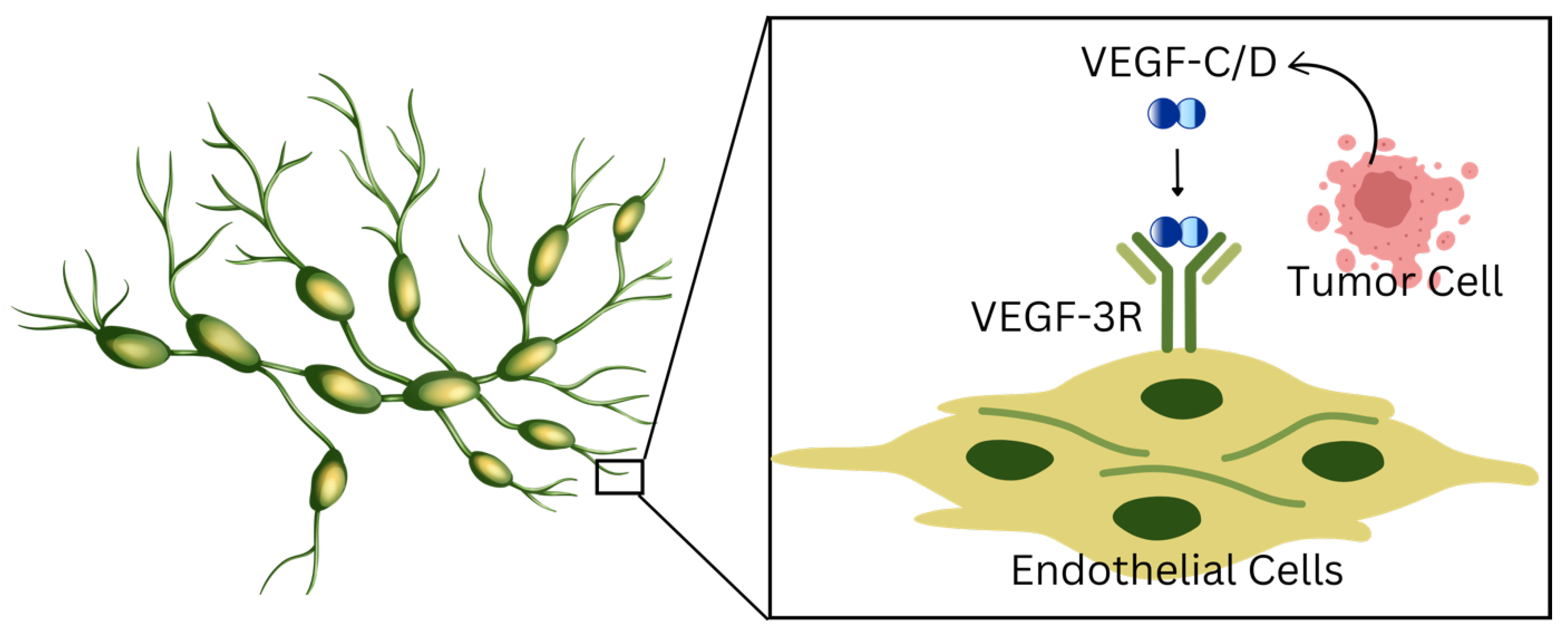
2.1. Role of Lymphangiogenesis

Lymphangiogenesis, the formation of new lymphatic vessels from pre-existing ones, is a key process implicated in both physiological functions and pathological conditions, including cancer metastasis. The lymphatic system maintains fluid homeostasis and immune responses but also provides a route for tumor cell dissemination. This process is significant, as it enables cancer cells to invade and spread to lymph nodes and beyond, contributing to disease progression and poor prognosis across multiple cancer types [11].
The link between lymphangiogenesis and lymphatic metastasis is primarily driven by the overexpression of VEGF-C and VEGF-D. These growth factors bind to their receptor VEGFR-3, expressed on lymphatic endothelial cells (LECs), to promote lymphatic vessel sprouting, expansion, and remodeling, creating a favorable environment for tumor cell entry and dissemination (Figure 1) [12]. This results in the formation of peritumoral lymphatic vessels, which are often dilated and more prone to facilitating tumor cell intravasation and transport to lymph nodes [5]. Tumors overexpressing VEGF-C have been shown to exhibit increased lymphatic vessel density and a higher rate of lymph node metastasis through VEGFR-3 signaling, which regulates lymphatic vessel growth, migration, and permeability, creating conditions conducive to tumor spread pathways [13]. VEGF-D plays a complementary role by enhancing lymphangiogenesis through distinct regulatory mechanisms that facilitate metastatic spread [14].
VEGF-C and VEGF-D not only interact with VEGFR-3; they can also form complexes with neuropilin-2, enhancing lymphangiogenic signaling in LECs. Neuropilin-2 co-internalization along with VEGFR-3 suggests an integrated regulatory role where lymphatic vessel signaling pathways are modulated by multiple interactions, promoting precise and efficient lymphatic growth and development [15]. Downstream signaling pathways, such as PI3K/Akt and MAPK, further regulate LEC proliferation, migration, and survival, which extends to pathologies like cancer metastasis (Figure 2) [16]. VEGF-C-induced activation of lymphatic endothelial cells is essential for processes that facilitate tumor dissemination through the lymphatic system [13].
Overall, VEGF-C and VEGF-D signaling pathways are central to the interplay between lymphangiogenesis and lymphatic metastasis. Tumor-secreted VEGF-C and VEGF-D promote lymphatic vessel sprouting and dilation, creating conditions conducive for tumor cell intravasation and subsequent spread to lymph nodes. This lymphatic expansion enhances the metastatic potential of tumors by providing a more accessible route for dissemination, highlighting their potential as therapeutic targets for inhibiting cancer spread (Table 1).

2.2. Tumor Cell Migration and Intravasation into Lymphatics

Continuing from the critical role of lymphangiogenesis in promoting lymphatic metastasis, tumor cell migration and intravasation into lymphatics mark the next steps in metastatic spread. Tumor cells leverage complex interactions within the lymphatic microenvironment to enter lymphatic vessels, a process that is facilitated by chemokines, growth factors, adhesion molecules, and matrix-modifying enzymes (Table 1).
Flow dynamics within lymphatic vessels play a crucial role in modulating tumor cell behavior. Research has demonstrated that both luminal and transmural fluid flow can increase the rate of tumor cell invasion and intravasation by altering the adhesive properties of the lymphatic endothelium. These biomechanical cues act as critical regulators of tumor–lymphatic interactions, enhancing the ability of tumor cells to penetrate the lymphatic barrier [17].
Chemokines, such as CCL21, secreted by lymphatic endothelial cells (LECs), provide directional cues that attract tumor cells expressing the corresponding receptor, CCR7. This chemokine–receptor interaction creates a chemotactic gradient, guiding tumor cells toward lymphatic vessels and facilitating their intravasation. This mechanism is particularly significant in cancers like melanoma, where CCR7-driven migration enhances metastatic spread to lymph nodes [18].
Matrix metalloproteinases (MMPs), particularly MMP14 and MMP16, further enhance tumor cell migration by degrading extracellular matrix (ECM) components (Figure 1). This proteolytic activity not only clears physical barriers to invasion but also modulates cell adhesion dynamics, thereby promoting tumor cell entry into lymphatic vessels. MMP activity underscores the importance of ECM remodeling in facilitating tumor cell dissemination [19,20].
Adhesion molecules expressed by lymphatic endothelial cells also play a pivotal role in the intravasation process. Molecules like VCAM-1, upregulated in tumor-associated lymphatic vessels, weaken the junctional integrity of LECs, thereby increasing lymphatic permeability. This facilitates tumor cell entry into lymphatic vessels, providing a more accessible route for their dissemination [21,22,23].
Additionally, tumor–lymphatic interactions are modulated by chemokine signaling pathways, such as the CXCL1-integrin β1 axis. CXCL1, secreted by tumor-associated LECs, activates integrin signaling pathways in cancer cells, enhancing their migration, invasion, and adhesion to lymphatic vessels. This interaction highlights the complex signaling crosstalk that facilitates lymphatic invasion and promotes metastatic progression (Wang et al., 2017) [24].
The synergistic effects of VEGF-C signaling and CCR7-mediated chemoinvasion further illustrate the multi-faceted strategies employed by tumor cells to invade the lymphatic system. VEGF-C not only promotes lymphangiogenesis but also upregulates lymphatic secretion of CCL21, driving CCR7-dependent tumor cell migration and invasion. This dual role underscores the importance of targeting both chemokine and growth factor pathways in the management of lymphatic metastasis [25].

2.3. Tumor Cell Survival and Colonization in Lymph Nodes

Tumor cell survival and colonization in lymph nodes are critical steps in cancer metastasis driven by complex immune evasion strategies and microenvironmental adaptations. To persist within lymph nodes, tumor cells must escape immune surveillance. One major mechanism involves the recruitment and modulation of regulatory T cells (Tregs) within the tumor microenvironment. Tregs suppress the activity of cytotoxic T cells and other immune effectors, providing essential survival signals to the tumor and creating a protective niche. This suppression not only blunts the host immune response but actively supports tumor persistence and proliferation [26]. Furthermore, tumors frequently secrete immunosuppressive cytokines, such as TGF-β, which inhibit the function of natural killer (NK) cells and cytotoxic T lymphocytes. This TGF-β-mediated immune suppression can be reversed therapeutically to restore NK cell activity, highlighting potential strategies for disrupting immune evasion (Table 1) [27,28].
Tumor cells also exploit immune checkpoint pathways to evade detection. By overexpressing immune checkpoint proteins, such as programmed death-ligand 1 (PD-L1), tumor cells can inhibit T cell activation by binding to programmed cell death protein-1 (PD-1) receptors on immune cells. This interaction effectively blunts immune responses, allowing the tumor to thrive [29,30].
In addition to immune evasion, tumor cells adapt to the supportive microenvironment of the lymph nodes to ensure their survival. The tumor microenvironment (TME) within lymph nodes is a complex network of immune cells, fibroblasts, and structural elements that work together to support tumor growth [31]. Tumor-associated macrophages (TAMs), often polarized towards an immunosuppressive M2 phenotype, promote tumor survival by secreting anti-inflammatory cytokines and remodeling the extracellular matrix (ECM) to create a favorable environment for cancer cells [31,32]. These interactions enable tumor cells to evade immune attack, proliferate, and establish themselves within lymph nodes (Table 1).
Hypoxia within the lymph node microenvironment further drives tumor adaptation by inducing genetic and epigenetic changes that enhance resistance to treatment and promote survival. Cancer-associated fibroblasts (CAFs) and other stromal cells play a critical role in reorganizing the ECM and secreting chemokines that recruit additional immunosuppressive cells, such as Tregs, which bolster immune evasion mechanisms. This interplay between structural and immune components creates a robust niche that fosters tumor colonization and persistence [33,34].
Beyond immune evasion, metabolic adaptations in the lymphatic environment further enhance tumor cell survival and prepare them for subsequent systemic dissemination [35]. Recent studies have shown that the lymphatic system provides a relatively protective biochemical milieu compared to the bloodstream, with higher concentrations of glutathione and oleic acid and lower levels of free iron [35]. These conditions help tumor cells develop resistance to ferroptosis, a form of iron-dependent programmed cell death driven by oxidative stress [35]. Because the bloodstream represents a more oxidative environment, the metabolic conditioning within the lymphatic system enhances tumor cell survival and metastatic competence during later hematogenous spread [35]. Exposure to the lymphatic microenvironment therefore not only facilitates regional dissemination but also equips tumor cells for successful systemic metastasis [35,36].

2.4. Tumor Types and the Role of Lymphatic Metastasis

Solid and hematologic tumors represent two major categories of cancer, each exhibiting distinct patterns of lymphatic involvement [37]. In solid tumors, such as breast cancer, melanoma, colorectal cancer, and non-small cell lung cancer (NSCLC), lymphatic metastasis plays a critical role in disease progression and prognosis [4]. Tumor cells in these cancers often exploit lymphangiogenic pathways to enter lymphatic vessels, colonize regional lymph nodes, and disseminate to distant organs [38]. Lymph node involvement serves as a key prognostic factor and guides treatment decisions, including the use of lymphadenectomy, sentinel lymph node biopsy, and systemic therapies [39].
In contrast, hematologic malignancies, such as lymphoma and leukemia, inherently involve lymphoid tissues and systemic circulation. While the lymphatic structures are central to disease development in these cancers, the concept of lymphatic metastasis differs from solid tumors, as spread through lymphatic channels is a natural component of disease dissemination rather than a secondary process [40]. Nonetheless, disruption of normal lymphatic architecture and function remains a hallmark of advanced disease in hematologic cancers [41].
A summary of representative solid and hematologic tumors and the role of lymphatic metastasis in their progression is provided in Table 2.

3. Clinical Implications of Lymphatic Metastasis

3.1. Prognostic Value of Lymphatic Involvement in Cancer

Lymphatic involvement, as mentioned earlier, has a significant role in cancer metastasis to different locations throughout the body. The presence of cancer in the lymph nodes is a telltale sign and one of the most common sites of metastasis. Moreover, the presence of cancer cells in the lymphatic vessels, or lymphatic invasion, can also be used for prognostic purposes.
The number of lymph nodes positive for cancer greatly influences mean survival. The TNM classification system helps classify a malignancy and consists of the following factors: size and spread, spread to nearby lymph nodes, and metastasis to other parts of the body. This system separates cancer cases into stages to evaluate the severity of the disease and survival probability [42]. This has been implemented in breast cancer, where N1 represents one to three axillary lymph nodes, N2 represents metastasis in four to nine lymph nodes, and N3 represents metastasis in ten or more lymph nodes [43]. Likewise, for colorectal cancer, N1 indicates the involvement of one to three nodes, N2 indicates four to six nodes, and N3 indicates seven or more regional nodes [42]. This practice is also common in prostate cancer and crucial after prostatectomy to allow for histopathology and staging [44]. This tumor staging system, while slightly modified for different types of cancer, still highlights lymph node metastasis as a crucial component with a significant prognostic role in cancer.
Similar to identifying the number of lymph nodes, the implementation of a lymph node ratio has been utilized to control for the number of lymph nodes removed. Diagnosis and treatment can involve the removal of regional lymph nodes to check for and reduce cancer spread [45]. The lymph node ratio represents the ratio of metastatic lymph nodes to examined lymph nodes. This has been found to be more effective than simply counting the number of lymph nodes in breast cancer, colorectal cancer, gastric cancer, pancreatic cancer, and many others [46,47]. For example, in stage III colorectal cancer, a high lymph node ratio had a pooled hazard ratio of 2.36 for overall survival and 3.71 for disease-free survival [48]. Furthermore, the presence of lymph node metastasis was a significant (p < 0.008) prognostic factor in oral and laryngeal squamous cell carcinomas [49].
Lymphatic vessel invasion (LVI) has also been used to help determine the survival of patients with specific cancers. In a study of 1408 patients with breast cancer, the presence of LVI was shown to have an odds ratio of 1.92 for causing death compared to those without LVI. The incidence of LVI was also highly correlated with the number of lymph node metastases and tumor size, two validated prognostic factors [50]. Even when isolated and used in both univariate and multivariate analyses, LVI was significantly shown to be an independent predictor of overall and disease-free survival [51]. Analyses have also shown LVI to have a higher hazard ratio than the presence of one to two positive lymph nodes. Assessment of LVI is now endorsed as a prognostic factor for breast cancer by the World Health Organization, the European Commission, the American Joint Committee on Cancer, and more sources [52]. For prostate cancer, LVI was similarly found to be linked to prostate-specific antigen recurrence. LVI is identified 10.2% of the time on average and is correlated with outcomes, signifying the need for routine screenings of LVI in prostatectomy specimens [52]. LVI status was unrelated to clinical outcomes in patients with metastasis (N1) but implicated in poor outcomes in patients with no metastasis (N0) [44]. LVI has also been shown to be a significant indicator of poor prognosis in colorectal cancer patients, with an overall survival hazard ratio of 2.25 (p < 0.0001) and a disease-free survival hazard ratio of 2.34 (p < 0.0001) [30]. A higher prevalence of LVI in patients with oral cancer was found in patients with advanced-stage cancer with metastases and has a significant negative impact on patient survival; it may be an indication for preventative neck dissection with adjuvant therapy [53,54]. Non-small cell lung cancer is also correlated with lymphatic vessel invasion, with LVI in 32.1% of patients who were 1.73 times more likely to relapse and showed a pooled hazard ratio of 1.59 compared to patients without LVI [55]. Finally, melanoma patients who presented with LVI had greater lymph node involvement (p = 0), a higher Clark level (p = 0), greater ulcerations (p = 0), and more metastasis (p = 0.008). Lymph node metastasis and lymphatic invasion are both promising prognostic markers for cancer metastasis and signify poorer outcomes in various cancer types. Targeted therapy, such as the use of anti-lymphangiogenic for photodynamic ablation, has shown promising results in preventing lymphatic invasion.

3.2. Diagnostic Approaches

Lymphoscintigraphy is an imaging tool used to assess lymphatic function and map out sentinel lymph nodes (SLN), which are the first lymph nodes where cancer can metastasize from the primary tumor. Lymphoscintigraphy uses a radioactive tracer and gamma cameras to visualize the gamma rays released by the tracer [56]. The sentinel lymph node would have a higher count of radioactive tracers in this scenario, enabling its identification using the gamma cameras [57]. The most common tracer used in lymphoscintigraphy is a technetium-99m-labeled lymphatic-specific tracer. Tracers of smaller size have been shown to have rapid uptake, sparking the use of new, smaller tracers for greater dissemination. The tracer is often injected into the interdigital space of the hands or feet, where it can then help localize SLNs using gamma cameras. Classical lymphoscintigraphy provides planar images of low resolution, which cannot provide accurate anatomical details. However, it still has significant uses for both qualitative and quantitative analyses. Qualitative analysis can analyze the symmetry of tracer intake into lymph nodes and identify dermal backflow. Quantitative analysis can calculate the tracer’s disappearance rate, tracer uptake kinetics, or the quantitative asymmetry index [58,59]. Lymphoscintigraphy can also be used in conjunction with SPECT/CT imaging to provide morphological information [60]. Lymphoscintigraphy is widely utilized, prompting new innovative techniques. The current advances in this field include the use of Indocyanine Green, photoacoustic detection, or superparamagnetic iron oxide [61,62,63].
Sentinel lymph node (SLN) biopsy, guided by lymphoscintigraphy, allows for the targeted removal of sentinel lymph nodes, reducing the need for extensive lymphadenectomy [45]. It is commonly used to stage breast cancer and melanoma [64]. The procedure begins with imaging of the SLN using radiotracers, such as a Tc99m-labeled sulfur colloid or Tc99m tilmanocept. The SLN is then localized with imaging and marked for surgery. Blue dye (Methylene blue, Isosulfan blue, Evans blue, or Patent blue) is then injected at the SLN, allowing for visualization of blue lymph nodes [45,65]. The nodes of interest are then excised. The biopsy procedure usually requires a small incision at the lymph node basin, followed by an excision. The extracted SLN can undergo sectioning, staining, and reverse transcriptase polymerase chain reaction (RT-PCR). Immunohistochemistry staining allows for the identification of tumors using specific proteins, such as MART-1 in melanoma. RT-PCR allows for the identification of clinical markers based on genes that are overexpressed [64]. While the lymph node often presents with tumor cells, SLN biopsy often has a false negative rate where the patient is node positive but the SLN biopsy report is negative. The false negative rate for melanoma is 10.8% [66]. The false negative rates for breast cancer were 7.0% overall, with a lower false negative rate when the radioactive tracer was combined with dye [67]. For prostate cancer, false negative rates had a median of 4.6% [68]. SLN biopsy allows for the diagnosis and staging of multiple cancers and is a very useful method to characterize and treat cancer. Furthermore, recent advancements in diagnostic techniques have significantly improved the precision of detecting lymphatic metastasis, with nanotechnology playing a pivotal role. Nanoparticles enhance imaging modalities, such as MRI, CT, and PET, by selectively targeting metastatic lymph nodes through specific biomarkers, thereby increasing sensitivity and reducing the need for invasive procedures like extensive lymphadenectomy [69].

3.3. Biomarkers for Lymphatic Spread

A variety of molecular and genetic markers characterize lymphatic spread and help identify cancer metastasis. Lymphangiogenesis markers include MMPs, VEGF, fibroblast growth factor (FGF), and platelet-derived growth factors (PDGF) (Table 3). MMPs, specifically MMP9, are collagenases implicated in breast cancer metastasis due to their role in extracellular matrix degradation [70,71]. VEGF plays a key role in the regulation of lymphangiogenesis. FGF and PDGF also promote the growth and stability of lymphatic vessels. Many signaling pathways and interleukins have also been found to be associated with this pathway. The angiopoietin 2 (Ang2)/Tie/PI3K signaling pathway is essential for VEGFR-3 expression on cell surfaces [72,73,74,75]. IL-6 activates the JAK-STAT3-VEGF-C pathway, mediating VEGF-C production and, thus, lymphangiogenesis [76,77,78,79]. IL-7 has also been shown to promote lymphatic vessel development by upregulating VEGF-D [80,81,82].
Lymphatic endothelial markers have also been used to detect lymphatic spread, including the lymphatic vessel endothelial hyaluronan receptor 1 (LYVE-1) gene, podoplanin, and Prospero homeobox 1 (PROX1). LYVE-1 binds with hyaluronic acid on the luminal surface of lymphatic vessels. More importantly, it is not found in blood vessels, allowing for the distinction of lymphatic vessels. Podoplanin is a transmembrane glycoprotein expressed by lymphatic endothelial cells but not vascular endothelial cells. It is also commonly utilized as a specific marker and associated with tumor spreading, inflammation, and metastasis [83]. PROX1 is a key regulator of lymphangiogenesis and also a transmembrane protein expressed on lymphatic endothelial cells. PROX1 is suggested to cause the transformation of vascular endothelium to lymphatic vessels [21]. In addition to these biomarkers, gene expression profile tests can also be performed to analyze chemotaxis, metastasis, adhesion factors, proliferation, and more. This helps identify patients who are at high risk for having a positive SLN [84]. Biomarkers for cancer can help characterize the cancer and develop a prognosis and personalized treatment response for the patient.

4. Therapeutic Strategies Targeting Lymphatic Metastasis

4.1. Surgical Approaches

Surgical strategies are fundamental in the management of lymphatic metastasis, with lymphadenectomy and sentinel lymph node dissection (SLND) being pivotal techniques employed for diagnosis, staging, and treatment. Lymphadenectomy involves the removal of lymph nodes to assess and prevent further metastatic spread of cancer. Traditionally, complete lymph node dissection has been utilized for various cancers, such as melanoma and breast cancer, to mitigate the risk of cancer progression by excising affected and potentially affected nodes. However, extensive lymphadenectomy can lead to significant complications, including lymphedema, pain, and impaired mobility, thus warranting a more selective approach [85].
SLND has emerged as a less invasive and more targeted surgical option. This procedure focuses on identifying and removing the first lymph node(s), or the sentinel nodes, to which cancer cells are most likely to spread from the primary tumor. By examining the sentinel node for metastatic cells, surgeons can determine the extent of cancer spread without resorting to complete lymphadenectomy in all cases. This approach reduces morbidity while providing crucial staging information, particularly in cancers like breast cancer and melanoma. The use of SLND has been shown to decrease unnecessary extensive node removal in patients without sentinel node involvement, thereby reducing surgical complications while maintaining accurate diagnostic capabilities [86].
While surgical approaches remain critical in the management of lymphatic metastasis, the decision to proceed with lymphadenectomy or SLND depends on factors like tumor type, stage, and individual patient characteristics, emphasizing the importance of personalized treatment strategies to balance therapeutic benefit and quality of life.

4.2. Pharmacological Inhibition of Lymphangiogenesis

Targeting lymphangiogenesis pharmacologically has emerged as a promising strategy to impede cancer progression, with multiple classes of inhibitors demonstrating efficacy in this area. One major avenue of pharmacological intervention focuses on the inhibition of VEGF signaling [87,88]. Therapeutic agents, such as bevacizumab, an anti-VEGF-A monoclonal antibody, indirectly inhibit lymphangiogenesis and have shown potential to suppress tumor-associated lymphatic expansion by disrupting VEGF pathways [89]. Other inhibitors targeting VEGF-C and VEGF-D are being explored more directly and have demonstrated efficacy in preclinical models by limiting the lymphatic spread of tumors [90,91].
Beyond VEGF-targeted therapies, tyrosine kinase inhibitors (TKIs) offer another effective approach to inhibiting lymphangiogenesis. These inhibitors, including sorafenib, sunitinib, and pazopanib, interfere with receptor tyrosine kinases, such as VEGFR-3, thereby blocking key signaling cascades essential for lymphatic vessel development and tumor cell migration [90,92,93]. By targeting multiple pathways involved in both angiogenesis and lymphangiogenesis, TKIs have demonstrated efficacy in reducing cancer dissemination through the lymphatic system, although challenges, such as resistance development and off-target toxicities, persist [94].
Furthermore, the HGF/c-Met pathway plays a critical role in lymphatic remodeling and tumor metastasis, as HGF and its receptor c-Met drive tumor cell migration and lymphangiogenesis. Therapeutic inhibitors targeting this pathway, such as monoclonal antibodies and small-molecule inhibitors, have shown preclinical success in reducing lymphatic metastases by disrupting HGF-c-Met signaling, thereby impairing new lymphatic vessel formation and tumor invasion. Similarly, MMPs facilitate extracellular matrix remodeling, which is essential for lymphatic vessel development and metastatic spread. MMP inhibitors have demonstrated efficacy in preclinical studies, reducing tumor progression and stabilizing the lymphatic microenvironment when combined with VEGF inhibitors. However, challenges, such as off-target effects and drug toxicity, highlight the need for more selective and safer therapeutic agents [87].
Other promising pharmacological agents include inhibitors targeting PDGF and fibroblast FGF, both of which have been implicated in lymphangiogenesis and tumor progression. Agents like nintedanib, which target multiple receptor tyrosine kinases, including PDGF and FGF receptors, offer broader inhibition of signaling pathways relevant to lymphatic metastasis [95]. Moreover, compounds targeting lymphangiogenic-specific markers, such as podoplanin and angiopoietin-2, are under investigation and may provide more selective therapeutic options [8].
Despite these advances, the clinical application of pharmacological inhibitors faces hurdles, including the emergence of drug resistance, off-target effects, and adverse toxicity profiles. Future research aims to optimize the use of these agents through combination therapies, improved patient stratification, and the development of next-generation inhibitors with enhanced specificity and reduced side effects. Continued exploration of novel targets within the lymphangiogenic signaling network holds the promise of more effective interventions against lymphatic metastases.

4.3. Immunotherapy and Targeted Therapies

Immunotherapy and targeted therapies have become crucial strategies in the management of lymphatic metastasis by enhancing the immune system’s ability to detect and destroy tumor cells and by inhibiting key molecular pathways that drive cancer progression. For instance, PD-1/PD-L1 inhibitors block the interaction between PD-1 on T cells and PD-L1 on tumor cells, leading to enhanced antitumor immunity and improved targeting of metastatic cells within lymph nodes [96]. Despite their success, response variability among patients remains a challenge, underscoring the need for predictive biomarkers and combination strategies to enhance efficacy [97].
In addition to immune checkpoint blockade, targeted therapies that focus on tumor-associated antigens (TAAs) and components of the tumor microenvironment (TME) have proven effective in limiting lymphatic metastasis. Monoclonal antibodies, such as trastuzumab for HER2-positive breast cancer and EGFR inhibitors for lung cancer, directly inhibit signaling pathways critical to tumor cell survival and proliferation, thereby reducing the likelihood of lymphatic spread [98]. Modulating the TME is another key focus, as it plays a significant role in promoting immune suppression and tumor survival. For instance, therapies targeting tumor-associated macrophages (TAMs) aim to transform the TME from an immunosuppressive to an immune-activated state, enhancing the body’s ability to eliminate cancer cells within lymphatic tissues [99].
Combining immunotherapy with other targeted approaches, such as anti-angiogenic agents, has also demonstrated promise in improving patient outcomes. By normalizing lymphatic vessel structure and reducing tumor-associated lymphangiogenesis, these therapies facilitate greater immune cell infiltration and restore immune function [100]. Such combination therapies seek to address resistance mechanisms and variability in patient response, aiming to provide a more robust antitumor effect and improve the overall management of lymphatic metastasis.
While these therapeutic strategies have shown great potential, challenges remain, including the emergence of resistance, patient heterogeneity, and immune-related adverse effects. Continued advancements in optimizing therapy combinations, identifying reliable biomarkers, and personalizing treatment approaches are vital to enhancing outcomes for patients with lymphatic metastasis.

4.4. Emerging Therapies and Clinical Trials

The ongoing pursuit of novel therapies for lymphatic metastasis is marked by a focus on innovative approaches that extend beyond traditional treatments, targeting unique aspects of tumor biology, immune modulation, and lymphatic-specific pathways. Recent clinical trials and emerging therapies aim to address limitations in current treatments by enhancing efficacy, overcoming resistance, and minimizing adverse effects.
Nanoparticles can deliver VEGFR-3 inhibitors and immune checkpoint inhibitors directly to lymphatic tissues, reducing systemic toxicity while improving drug efficacy. These “theranostic” platforms simultaneously offer diagnostic imaging and therapeutic capabilities, enabling real-time monitoring of treatment responses. Additionally, combining nanomedicine with immune checkpoint inhibitors or lymphangiogenesis-targeted therapies, such as VEGFR-3 inhibitors, has shown promise in clinical trials by enhancing immune cell infiltration and reducing tumor spread within the lymphatic microenvironment [101,102,103].
A key area of interest is the development of next-generation immune checkpoint inhibitors targeting pathways beyond PD-1 and PD-L1. Inhibitors of lymphocyte activation gene-3 (LAG-3), T cell immunoreceptor with Ig and ITIM domains (TIGIT), and T cell immunoglobulin and mucin-domain containing-3 (TIM-3) are undergoing clinical evaluation, with early data suggesting the potential to enhance antitumor immunity when used alone or in combination with established checkpoint inhibitors [104,105]. By targeting alternative checkpoint pathways, these therapies seek to overcome resistance mechanisms observed in the PD-1/PD-L1 blockade, broadening the pool of patients who may benefit from immunotherapy [106,107,108,109,110,111,112,113].
Adoptive cell therapies (ACT), including chimeric antigen receptor (CAR) T-cell therapy, represent another promising avenue for lymphatic metastasis management. While primarily explored in hematologic malignancies, CAR-T cells engineered to target tumor-associated antigens are being adapted for solid tumors with lymphatic involvement. Preclinical and early-phase clinical trials are exploring the efficacy and safety of CAR-T cells directed at antigens like HER2, mesothelin, and EGFR in solid tumors, with an emphasis on optimizing their trafficking, persistence, and activity within the lymphatic microenvironment [114]. Combining CAR-T therapy with strategies to modulate the tumor microenvironment (TME), such as reducing immunosuppressive cytokines or targeting regulatory immune cells, holds the potential to enhance therapeutic outcomes.
Therapies targeting the lymphatic vasculature and lymphangiogenesis are also being investigated to disrupt the metastatic cascade. Novel agents targeting VEGFR-3 and other lymphangiogenic factors, including ANGPT2 and PROX1, aim to prevent tumor cells from entering and spreading through lymphatic vessels [88,115]. These therapies may be particularly valuable when used in combination with immune checkpoint inhibitors or traditional therapies to achieve a synergistic effect on metastasis suppression.
Epigenetic therapies targeting histone modifications and DNA methylation are also emerging as potential strategies for altering tumor cell behavior within lymphatic tissues. Modifying the expression of genes involved in immune evasion, metastasis, and lymphatic adaptation may enhance the effectiveness of existing therapies and reduce tumor resilience in the lymphatic microenvironment [116]. Clinical trials are underway to evaluate the combination of epigenetic modulators with immunotherapies and targeted therapies, aiming to overcome resistance and enhance antitumor activity.
Finally, therapeutic cancer vaccines and oncolytic viruses represent another area of intense investigation. Cancer vaccines designed to induce an immune response against tumor-associated antigens have shown promise in priming the immune system to recognize and eliminate metastatic cells within lymph nodes. Oncolytic viruses, engineered to selectively infect and lyse cancer cells, can trigger immune responses and release tumor antigens, enhancing immune system activation and potentially improving the efficacy of immunotherapies [117]. Early-phase trials exploring combinations of these modalities with checkpoint inhibitors and targeted therapies are ongoing, with the goal of amplifying antitumor immunity and reducing metastatic spread (Table 4).

5. Challenges and Future Directions in Research

One of the main challenges to cancer therapy is drug resistance, limiting the options for cancer treatment. Many patients present with resistance to immune checkpoint inhibitors due to intrinsic resistance or secondary to disease progression [119]. In addition, ATP-binding cassette transporters allow tumor cells to efflux toxic agents into the extracellular space. This is seen in breast-cancer-resistant protein ABCG2, for example. Aldehyde dehydrogenase has also been implicated in providing tumor resistance by providing resistance against alkylating agents [120].
Drug toxicity is also a serious complication of and challenge to cancer therapy. Therapy requires the use of multiple drugs to treat the malignancy, but, as a result, it can cause harmful effects. Doxorubicin and 5-Fluorouracil have been recognized to cause cardiotoxicity, which is further exacerbated by radiation therapy [121,122,123,124,125,126]. These symptoms include arrhythmias, angina, and even myocardial infarction. Bleomycin, tyrosine kinase inhibitors, and immune checkpoint inhibitors have all been found to cause pulmonary toxicity, causing interstitial pneumonitis, which can transform into pulmonary fibrosis [127,128,129,130,131]. Renal toxicity and kidney injury are also a very common side effect of chemotherapy treatment [132,133]. Cisplatin has been found to cause tubular damage, interstitial inflammation, vascular injury, hypomagnesemia, thrombotic microangiopathy, and acute kidney injury [134,135,136]. These effects can be reduced with hydration or diuresis [137]. Gastrointestinal toxicity is caused by a large number of drugs and can impart a range of symptoms, such as diarrhea, mucositis, nausea and vomiting, and xerostomia [138,139,140]. Doxorubicin, cyclophosphamide, 5-fluorouracil, and aromatase inhibitors have also been correlated with cognitive dysfunction [141,142].
CAR-T therapies also showcase both limitations and toxicities. While specific antigen binding domains are created, cancer cells often employ antigen escape, which changes the way they display antigens, making CAR-T therapy less effective. Relapsed and refractory patients generally exhibit this resistance mechanism. Tumors can either downregulate the antigen on its surface or remove the expression altogether, posing a difficult challenge to therapy. CAR-T therapy has been shown to be effective for leukemias and lymphomas, but it is ineffective at infiltrating solid tumors. Solid tumors utilize physical barriers, such as the stroma, and create an immunosuppressive environment, making CAR-T therapy difficult [143]. CAR-T therapies can cause large cytokine production due to T cell activation, resulting in a cytokine release storm. This is found in patients with acute lymphoblastic leukemia, where 23–46% experienced symptoms of high cytokine production [144]. These can include fatigue, diarrhea, headaches, arthralgia, myalgia, and potentially severe symptoms causing organ system failure. A detrimental complication includes immune effector cell-associated neurotoxicity syndrome, disrupting the blood–brain barrier [143].
Combination therapies have proven to be a means to reduce drug resistance. Multiple new combination therapies are being investigated to ensure a more enhanced response. Studies have analyzed the combination of PD-1 inhibitors (pembrolizumab) and Indoleamine-2,3-dioxygenase (IDO) inhibitors (epacadostat), which was well-tolerated but did not show much clinical benefit over PD-1 inhibitor monotherapy [145]. These drugs have also been used as a vaccine in combination with anti-PD-1 drugs [146]. Another combination therapy being explored is the anti-lymphocyte activation gene-3 (anti–LAG–3) with nivolumab, a PD-1 inhibitor. It is safe and efficacious in patients with melanoma [104]. Nivolumab has also been used in combination with Bempegaldesleukin, an IL-2 prodrug. The combination drug showed a decreased objective response of progression-free survival and an increased number of adverse events [147]. Another new therapeutic approach is restrictive combinations of drugs that take advantage of slight differences between tumors and normal cells to affect tumor cells preferentially [148]. While new combination therapies show great potential, more work needs to be done to find one that ensures high efficacy.
Personalized medicine takes each patient’s specific tumor environment into account and tailors the therapy to each individual, eliciting an improved response. Personalized therapeutic cancer vaccines provide resistance to individual tumor neoantigens. This is achieved by RNA sequencing tumor tissue and identifying relevant mutations as the target antigen for the vaccine. This allows for a tumor-specific response and elucidates a robust T-cell response [149]. Similarly, 3D culture and patient-derived xenograft models have been created to reproduce tumor characteristics, predict drug response, and implement personalized medicine [150]. These models help stimulate the heterogeneity of the tumor environment and the variety of cancer cells present. This has been used for modeling brain cancer, skin cancers, colorectal cancers, cervical cancers, lung cancers, and more [151]. These highly promising approaches emphasize the need for greater research on personalized medicine.

6. Limitations

This review is not without limitations. While it is a good introduction to the role of the lymphatic system on tumor metastasis, it remains a narrative review. We did not develop a methodological search strategy or have inclusion or exclusion criteria for the resources used in this review. Because this review serves as an overview of lymphatic metastasis, it is unable to capture the full details of the mechanism of lymphangiogenesis, diagnostic approaches, or treatments.

7. Conclusions

The lymphatic system plays a crucial role in the spread of malignant cells from the primary tumor. Tumor migration into the lymphatic system is primarily facilitated through lymphangiogenesis, characterized by the expression of MMPs, VEGF, FGF, and PDGF. Additionally, adhesion molecules and cytokines assist with the migration of the tumor cell, including CCR7, CCL21, MMPs, and VCAM-1 [70,71]. Furthermore, expression of Treg and immune checkpoint inhibitors enable tumor survival [98,103,107]. Thus, lymphatic metastasis and lymphatic invasion have proved to be significant prognostic factors determining survival and tumor progression. This is often measured using the lymph node ratio and by identifying lymphatic vessel invasion [46,48,50,55]. Lymphatic metastasis can be detected through lymphoscintigraphy and sentinel lymph node biopsy, an alternative and more targeted approach to traditional lymphadenectomy [56,58,68,86]. Management of lymphatic metastasis consists of immunotherapy using PD-1 inhibitors, monoclonal antibodies, and modulation of the tumor microenvironment [73,109,146]. These therapies have been widely successful, but challenges, such as drug resistance, adverse effects, and patient heterogeneity, still remain [128,134,141]. Emerging therapies, such as cancer vaccines, epigenetics, and CAR-T cells, represent promising solutions and emphasize the need for personalized and longitudinal medicine [27,116,117,143,149]. In conclusion, identifying new long-term therapeutic options with fewer adverse effects and reduced occurrences of resistance is crucial for patient care and disease-free survival.

Author Contributions

Conceptualization, N.D., P.S. and F.C.L.; methodology, N.D., P.S. and F.C.L.; validation, N.D., P.S. and F.C.L.; investigation, N.D., P.S. and F.C.L.; writing—original draft preparation, N.D., P.S. and F.C.L.; writing—review and editing, N.D., P.S. and F.C.L.; visualization, N.D., P.S. and F.C.L.; supervision, F.C.L.; All authors have read and agreed to the published version of the manuscript.

Funding

This research received no external funding.

Conflicts of Interest

The authors declare no conflict of interest.

References

  1. Witte, T.; Dadras, M.; Heck, F.-C.; Heck, M.; Habermalz, B.; Welss, S.; Lehnhardt, M.; Behr, B. Water-jet-assisted liposuction for the treatment of lipedema: Standardized treatment protocol and results of 63 patients. J. Plast. Reconstr. Aesthetic Surg. JPRAS 2020, 73, 1637–1644. [Google Scholar] [CrossRef] [PubMed]
  2. Hu, Z.; Zhao, X.; Wu, Z.; Qu, B.; Yuan, M.; Xing, Y.; Song, Y.; Wang, Z. Lymphatic vessel: Origin, heterogeneity, biological functions and therapeutic targets. Signal Transduct. Target. Ther. 2024, 9, 9. [Google Scholar] [CrossRef] [PubMed]
  3. Ji, H.; Hu, C.; Yang, X.; Liu, Y.; Ji, G.; Ge, S.; Wang, X.; Wang, M. Lymph node metastasis in cancer progression: Molecular mechanisms, clinical significance and therapeutic interventions. Signal Transduct. Target. Ther. 2023, 8, 367. [Google Scholar] [CrossRef]
  4. Carr, I.; Pettigrew, N.; Weinerman, B. Lymphatic metastasis and its treatment. Cancer Treat. Rev. 1987, 14, 53–64. [Google Scholar] [CrossRef]
  5. Sleeman, J.P.; Thiele, W. Tumor metastasis and the lymphatic vasculature. Int. J. Cancer 2009, 125, 2747–2756. [Google Scholar] [CrossRef]
  6. Karaman, S.; Detmar, M. Mechanisms of lymphatic metastasis. J. Clin. Investig. 2014, 124, 922–928. [Google Scholar] [CrossRef]
  7. Duff, S.E.; Li, C.; Jeziorska, M.; Kumar, S.; Saunders, M.P.; Sherlock, D.; O’Dwyer, S.T.; Jayson, G.C. Vascular endothelial growth factors C and D and lymphangiogenesis in gastrointestinal tract malignancy. Br. J. Cancer 2003, 89, 426–430. [Google Scholar] [CrossRef]
  8. Mohammed, R.A.A.; Green, A.; El-Shikh, S.; Paish, E.C.; Ellis, I.O.; Martin, S.G. Prognostic significance of vascular endothelial cell growth factors -A, -C and -D in breast cancer and their relationship with angio- and lymphangiogenesis. Br. J. Cancer 2007, 96, 1092–1100. [Google Scholar] [CrossRef]
  9. Oliver, G.; Kipnis, J.; Randolph, G.J.; Harvey, N.L. The Lymphatic Vasculature in the 21st Century: Novel Functional Roles in Homeostasis and Disease. Cell 2020, 182, 270–296. [Google Scholar] [CrossRef]
  10. He, Y.; Rajantie, I.; Pajusola, K.; Jeltsch, M.; Holopainen, T.; Yla-Herttuala, S.; Harding, T.; Jooss, K.; Takahashi, T.; Alitalo, K. Vascular Endothelial Cell Growth Factor Receptor 3–Mediated Activation of Lymphatic Endothelium Is Crucial for Tumor Cell Entry and Spread via Lymphatic Vessels. Cancer Res. 2005, 65, 4739–4746. [Google Scholar] [CrossRef]
  11. TNM Classification of Malignant Tumours|UICC. Available online: https://www.uicc.org/what-we-do/sharing-knowledge/tnm (accessed on 1 December 2024).
  12. Davydova, N.; Harris, N.C.; Roufail, S.; Paquet-Fifield, S.; Ishaq, M.; Streltsov, V.A.; Williams, S.P.; Karnezis, T.; Stacker, S.A.; Achen, M.G. Differential Receptor Binding and Regulatory Mechanisms for the Lymphangiogenic Growth Factors Vascular Endothelial Growth Factor (VEGF)-C and -D*. J. Biol. Chem. 2016, 291, 27265–27278. [Google Scholar] [CrossRef] [PubMed]
  13. Veikkola, T.; Jussila, L.; Makinen, T.; Karpanen, T.; Jeltsch, M.; Petrova, T.V.; Kubo, H.; Thurston, G.; McDonald, D.M.; Achen, M.G.; et al. Signalling via vascular endothelial growth factor receptor-3 is sufficient for lymphangiogenesis in transgenic mice. EMBO J. 2001, 20, 1223–1231. [Google Scholar] [CrossRef] [PubMed]
  14. Hirakawa, S.; Brown, L.F.; Kodama, S.; Paavonen, K.; Alitalo, K.; Detmar, M. VEGF-C–induced lymphangiogenesis in sentinel lymph nodes promotes tumor metastasis to distant sites. Blood 2007, 109, 1010–1017. [Google Scholar] [CrossRef] [PubMed]
  15. Karnezis, T.; Shayan, R.; Caesar, C.; Roufail, S.; Harris, N.C.; Ardipradja, K.; Zhang, Y.F.; Williams, S.P.; Farnsworth, R.H.; Chai, M.G.; et al. VEGF-D promotes tumor metastasis by regulating prostaglandins produced by the collecting lymphatic endothelium. Cancer Cell 2012, 21, 181–195. [Google Scholar] [CrossRef]
  16. Karpanen, T.; Heckman, C.A.; Keskitalo, S.; Jeltsch, M.; Ollila, H.; Neufeld, G.; Tamagnone, L.; Alitalo, K. Functional interaction of VEGF-C and VEGF-D with neuropilin receptors. FASEB J. 2006, 20, 1462–1472. [Google Scholar] [CrossRef]
  17. Rascio, F.; Spadaccino, F.; Rocchetti, M.T.; Castellano, G.; Stallone, G.; Netti, G.S.; Ranieri, E. The Pathogenic Role of PI3K/AKT Pathway in Cancer Onset and Drug Resistance: An Updated Review. Cancers 2021, 13, 3949. [Google Scholar] [CrossRef]
  18. Chiang, S.P.H.; Cabrera, R.M.; Segall, J.E. Tumor cell intravasation. Am. J. Physiol. Cell Physiol. 2016, 311, C1–C14. [Google Scholar] [CrossRef]
  19. Shields, J.D.; Fleury, M.E.; Yong, C.; Tomei, A.A.; Randolph, G.J.; Swartz, M.A. Autologous Chemotaxis as a Mechanism of Tumor Cell Homing to Lymphatics via Interstitial Flow and Autocrine CCR7 Signaling. Cancer Cell 2007, 11, 526–538. [Google Scholar] [CrossRef]
  20. Stamenkovic, I. Matrix metalloproteinases in tumor invasion and metastasis. Semin. Cancer Biol. 2000, 10, 415–433. [Google Scholar] [CrossRef]
  21. Niland, S.; Riscanevo, A.X.; Eble, J.A. Matrix Metalloproteinases Shape the Tumor Microenvironment in Cancer Progression. Int. J. Mol. Sci. 2021, 23, 146. [Google Scholar] [CrossRef]
  22. Leone, P.; Malerba, E.; Susca, N.; Favoino, E.; Perosa, F.; Brunori, G.; Prete, M.; Racanelli, V. Frontiers|Endothelial cells in tumor microenvironment: Insights and perspectives. Front. Immunol. 2024, 15, 1367875. [Google Scholar] [CrossRef] [PubMed]
  23. He, M.; He, Q.; Cai, X.; Chen, Z.; Lao, S.; Deng, H.; Liu, X.; Zheng, Y.; Liu, X.; Liu, J.; et al. Role of lymphatic endothelial cells in the tumor microenvironment—A narrative review of recent advances. Transl. Lung Cancer Res. 2021, 10, 2252–2277. [Google Scholar] [CrossRef] [PubMed]
  24. Wang, Z.; Wang, Z.; Li, G.; Wu, H.; Sun, K.; Chen, J.; Feng, Y.; Chen, C.; Cai, S.; Xu, J.; et al. CXCL1 from tumor-associated lymphatic endothelial cells drives gastric cancer cell into lymphatic system via activating integrin β1/FAK/AKT signaling. Cancer Lett. 2017, 385, 28–38. [Google Scholar] [CrossRef]
  25. Issa, A.; Le, T.X.; Shoushtari, A.N.; Shields, J.D.; Swartz, M.A. Vascular endothelial growth factor-C and C-C chemokine receptor 7 in tumor cell-lymphatic cross-talk promote invasive phenotype. Cancer Res. 2009, 69, 349–357. [Google Scholar] [CrossRef]
  26. Pereira, E.R.; Jones, D.; Jung, K.; Padera, T.P. The lymph node microenvironment and its role in the progression of metastatic cancer. Semin. Cell Dev. Biol. 2015, 38, 98–105. [Google Scholar] [CrossRef]
  27. Regis, S.; Dondero, A.; Caliendo, F.; Bottino, C.; Castriconi, R. NK Cell Function Regulation by TGF-β-Induced Epigenetic Mechanisms. Front. Immunol. 2020, 11, 311. [Google Scholar] [CrossRef]
  28. Portale, F.; Di Mitri, D. NK Cells in Cancer: Mechanisms of Dysfunction and Therapeutic Potential. Int. J. Mol. Sci. 2023, 24, 9521. [Google Scholar] [CrossRef]
  29. Hudson, K.; Cross, N.; Jordan-Mahy, N.; Leyland, R. Frontiers|The Extrinsic and Intrinsic Roles of PD-L1 and Its Receptor PD-1: Implications for Immunotherapy Treatment. Front. Immunol. 2020, 11, 568931. [Google Scholar] [CrossRef]
  30. Wang, Q.; Xie, B.; Liu, S.; Shi, Y.; Tao, Y.; Xiao, D.; Wang, W. What Happens to the Immune Microenvironment After PD-1 Inhibitor Therapy? Front. Immunol. 2021, 12, 773168. [Google Scholar] [CrossRef]
  31. Khalaf, K.; Hana, D.; Chou, J.T.-T.; Singh, C.; Mackiewicz, A.; Kaczmarek, M. Aspects of the Tumor Microenvironment Involved in Immune Resistance and Drug Resistance. Front. Immunol. 2021, 12, 656364. [Google Scholar] [CrossRef]
  32. Huang, R.; Kang, T.; Chen, S. The role of tumor-associated macrophages in tumor immune evasion. J. Cancer Res. Clin. Oncol. 2024, 150, 238. [Google Scholar] [CrossRef] [PubMed]
  33. Svanberg, R.; Janum, S.; Patten, P.E.M.; Ramsay, A.G.; Niemann, C.U. Targeting the tumor microenvironment in chronic lymphocytic leukemia. Haematologica 2021, 106, 2312–2324. [Google Scholar] [CrossRef]
  34. Roy, S.; Kumaravel, S.; Sharma, A.; Duran, C.L.; Bayless, K.J.; Chakraborty, S. Hypoxic tumor microenvironment: Implications for cancer therapy. Exp. Biol. Med. 2020, 245, 1073–1086. [Google Scholar] [CrossRef]
  35. Ubellacker, J.M.; Tasdogan, A.; Ramesh, V.; Shen, B.; Mitchell, E.C.; Martin-Sandoval, M.S.; Gu, Z.; McCormick, M.L.; Durham, A.B.; Spitz, D.R.; et al. Lymph protects metastasizing melanoma cells from ferroptosis. Nature 2020, 585, 113–118. [Google Scholar] [CrossRef]
  36. Shi, X.; Wang, X.; Yao, W.; Shi, D.; Shao, X.; Lu, Z.; Chai, Y.; Song, J.; Tang, W.; Wang, X. Mechanism insights and therapeutic intervention of tumor metastasis: Latest developments and perspectives. Signal Transduct. Target. Ther. 2024, 9, 192. [Google Scholar] [CrossRef]
  37. Hanahan, D. Hallmarks of Cancer: New Dimensions. Cancer Discov. 2022, 12, 31–46. [Google Scholar] [CrossRef]
  38. Alitalo, K.; Tammela, T.; Petrova, T.V. Lymphangiogenesis in development and human disease. Nature 2005, 438, 946–953. [Google Scholar] [CrossRef]
  39. Zahoor, S.; Haji, A.; Battoo, A.; Qurieshi, M.; Mir, W.; Shah, M. Sentinel Lymph Node Biopsy in Breast Cancer: A Clinical Review and Update. J. Breast Cancer 2017, 20, 217–227. [Google Scholar] [CrossRef]
  40. Shankland, K.R.; Armitage, J.O.; Hancock, B.W. Non-Hodgkin lymphoma. Lancet 2012, 380, 848–857. [Google Scholar] [CrossRef]
  41. Küppers, R. Mechanisms of B-cell lymphoma pathogenesis. Nat. Rev. Cancer 2005, 5, 251–262. [Google Scholar] [CrossRef]
  42. Rosen, R.D.; Sapra, A. TNM Classification. In StatPearls; StatPearls Publishing: Treasure Island, FL, USA, 2024. [Google Scholar]
  43. Singletary, S.E.; Allred, C.; Ashley, P.; Bassett, L.W.; Berry, D.; Bland, K.I.; Borgen, P.I.; Clark, G.; Edge, S.B.; Hayes, D.F.; et al. Revision of the American Joint Committee on Cancer Staging System for Breast Cancer. J. Clin. Oncol. 2002, 20, 3628–3636. [Google Scholar] [CrossRef] [PubMed]
  44. Wilczak, W.; Wittmer, C.; Clauditz, T.; Minner, S.; Steurer, S.; Büscheck, F.; Krech, T.; Lennartz, M.; Harms, L.; Leleu, D.; et al. Marked Prognostic Impact of Minimal Lymphatic Tumor Spread in Prostate Cancer. Eur. Urol. 2018, 74, 376–386. [Google Scholar] [CrossRef]
  45. Moncayo, V.M.; Alazraki, A.L.; Alazraki, N.P.; Aarsvold, J.N. Sentinel Lymph Node Biopsy Procedures. Semin. Nucl. Med. 2017, 47, 595–617. [Google Scholar] [CrossRef]
  46. Vinh-Hung, V.; Verkooijen, H.M.; Fioretta, G.; Neyroud-Caspar, I.; Rapiti, E.; Vlastos, G.; Deglise, C.; Usel, M.; Lutz, J.-M.; Bouchardy, C. Lymph Node Ratio as an Alternative to pN Staging in Node-Positive Breast Cancer. J. Clin. Oncol. 2009, 27, 1062–1068. [Google Scholar] [CrossRef]
  47. Siewert, J.R.; Stein, H.J. Classification of adenocarcinoma of the oesophagogastric junction. Br. J. Surg. 1998, 85, 1457–1459. [Google Scholar] [CrossRef] [PubMed]
  48. Ceelen, W.; Van Nieuwenhove, Y.; Pattyn, P. Prognostic Value of the Lymph Node Ratio in Stage III Colorectal Cancer: A Systematic Review. Ann. Surg. Oncol. 2010, 17, 2847–2855. [Google Scholar] [CrossRef]
  49. Kyzas, P.A.; Geleff, S.; Batistatou, A.; Agnantis, N.J.; Stefanou, D. Evidence for lymphangiogenesis and its prognostic implications in head and neck squamous cell carcinoma. J. Pathol. 2005, 206, 170–177. [Google Scholar] [CrossRef]
  50. Lauria, R.; Perrone, F.; Carlomagno, C.; De Laurentiis, M.; Morabito, A.; Gallo, C.; Varriale, E.; Pettinato, G.; Panico, L.; Petrella, G.; et al. The prognostic value of lymphatic and blood vessel invasion in operable breast cancer. Cancer 1995, 76, 1772–1778. [Google Scholar] [CrossRef]
  51. Schoppmann, S.F.; Bayer, G.; Aumayr, K.; Taucher, S.; Geleff, S.; Rudas, M.; Kubista, E.; Hausmaninger, H.; Samonigg, H.; Gnant, M.; et al. Prognostic Value of Lymphangiogenesis and Lymphovascular Invasion in Invasive Breast Cancer. Ann. Surg. 2004, 240, 306. [Google Scholar] [CrossRef]
  52. Rakha, E.A.; Martin, S.; Lee, A.H.S.; Morgan, D.; Pharoah, P.D.P.; Hodi, Z.; MacMillan, D.; Ellis, I.O. The prognostic significance of lymphovascular invasion in invasive breast carcinoma. Cancer 2012, 118, 3670–3680. [Google Scholar] [CrossRef]
  53. Chang, W.-C.; Chang, C.-F.; Li, Y.-H.; Yang, C.-Y.; Su, R.-Y.; Lin, C.-K.; Chen, Y.-W. A histopathological evaluation and potential prognostic implications of oral squamous cell carcinoma with adverse features. Oral Oncol. 2019, 95, 65–73. [Google Scholar] [CrossRef] [PubMed]
  54. Mascitti, M.; Togni, L.; Caponio, V.C.A.; Zhurakivska, K.; Bizzoca, M.E.; Contaldo, M.; Serpico, R.; Lo Muzio, L.; Santarelli, A. Lymphovascular invasion as a prognostic tool for oral squamous cell carcinoma: A comprehensive review. Int. J. Oral Maxillofac. Surg. 2022, 51, 1–9. [Google Scholar] [CrossRef] [PubMed]
  55. Wang, J.; Wang, B.; Zhao, W.; Guo, Y.; Chen, H.; Chu, H.; Liang, X.; Bi, J. Clinical Significance and Role of Lymphatic Vessel Invasion as a Major Prognostic Implication in Non-Small Cell Lung Cancer: A Meta-Analysis. PLoS ONE 2012, 7, e52704. [Google Scholar] [CrossRef] [PubMed]
  56. Ranzenberger, L.R.; Pai, R.B. Lymphoscintigraphy. In StatPearls; StatPearls Publishing: Treasure Island, FL, USA, 2024. [Google Scholar]
  57. Mariani, G.; Moresco, L.; Viale, G.; Villa, G.; Bagnasco, M.; Canavese, G.; Buscombe, J.; Strauss, H.W.; Paganelli, G. Radioguided sentinel lymph node biopsy in breast cancer surgery. J. Nucl. Med. Off. Public Soc. Nucl. Med. 2001, 42, 1198–1215. [Google Scholar]
  58. Yoo, J.-N.; Cheong, Y.-S.; Min, Y.-S.; Lee, S.-W.; Park, H.Y.; Jung, T.-D. Validity of Quantitative Lymphoscintigraphy as a Lymphedema Assessment Tool for Patients with Breast Cancer. Ann. Rehabil. Med. 2015, 39, 931–940. [Google Scholar] [CrossRef]
  59. Polomska, A.K.; Proulx, S.T. Imaging technology of the lymphatic system. Adv. Drug Deliv. Rev. 2021, 170, 294–311. [Google Scholar] [CrossRef]
  60. Lasso, J.M. Lympho-SPECT/CT as a tool to evaluate postoperative outcomes after LVA for lymphedema repair. Plast. Aesthet. Res. 2020, 7, 30. [Google Scholar] [CrossRef]
  61. Dumitru, D.; Ghanakumar, S.; Provenzano, E.; Benson, J.R. A Prospective Study Evaluating the Accuracy of Indocyanine Green (ICG) Fluorescence Compared with Radioisotope for Sentinel Lymph Node (SLN) Detection in Early Breast Cancer. Ann. Surg. Oncol. 2022, 29, 3014–3020. [Google Scholar] [CrossRef]
  62. Luke, G.P.; Myers, J.N.; Emelianov, S.Y.; Sokolov, K.V. Sentinel Lymph Node Biopsy Revisited: Ultrasound-Guided Photoacoustic Detection of Micrometastases Using Molecularly Targeted Plasmonic Nanosensors. Cancer Res. 2014, 74, 5397–5408. [Google Scholar] [CrossRef]
  63. Liu, P.; Tan, J.; Song, Y.; Huang, K.; Zhang, Q.; Xie, H. The Application of Magnetic Nanoparticles for Sentinel Lymph Node Detection in Clinically Node-Negative Breast Cancer Patients: A Systemic Review and Meta-Analysis. Cancers 2022, 14, 5034. [Google Scholar] [CrossRef]
  64. Chen, S.L.; Iddings, D.M.; Scheri, R.P.; Bilchik, A.J. Lymphatic Mapping and Sentinel Node Analysis: Current Concepts and Applications. CA Cancer J. Clin. 2006, 56, 292–309. [Google Scholar] [CrossRef] [PubMed]
  65. Tsopelas, C.; Sutton, R.; Bs, M. Why Certain Dyes Are Useful for Localizing the Sentinel Lymph Node. J. Nucl. Med. 2002, 43, 1377–1382. [Google Scholar] [PubMed]
  66. Scoggins, C.R.; Martin, R.C.G.; Ross, M.I.; Edwards, M.J.; Reintgen, D.S.; Urist, M.M.; Gershenwald, J.E.; Sussman, J.J.; Dirk Noyes, R.; Goydos, J.S.; et al. Factors Associated with False-Negative Sentinel Lymph Node Biopsy in Melanoma Patients. Ann. Surg. Oncol. 2010, 17, 709–717. [Google Scholar] [CrossRef]
  67. Pesek, S.; Ashikaga, T.; Krag, L.E.; Krag, D. The False-Negative Rate of Sentinel Node Biopsy in Patients with Breast Cancer: A Meta-Analysis. World J. Surg. 2012, 36, 2239–2251. [Google Scholar] [CrossRef]
  68. Rossin, G.; Zorzi, F.; De Pablos-Rodríguez, P.; Biasatti, A.; Marenco, J.; Ongaro, L.; Perotti, A.; Tulone, G.; Traunero, F.; Piasentin, A.; et al. Sentinel Lymph Node Biopsy in Prostate Cancer: An Overview of Diagnostic Performance, Oncological Outcomes, Safety, and Feasibility. Diagnostics 2023, 13, 2543. [Google Scholar] [CrossRef]
  69. De Britto, N.; Neeraja, R.; Anbarasi, L.J.; Ravi, V.; SP, S.I.; Jawahar, M.; Al Mazroa, A. Diagnosis of Lymphatic Metastasis in Breast Cancer Using Nanoparticle Technology—Diagnosis, Therapy, Imaging, Treatment. Open Neuroimaging J. 2024, 17, E18744400287726. [Google Scholar] [CrossRef]
  70. Chambers, A.F.; Matrisian, L.M. Changing Views of the Role of Matrix Metalloproteinases in Metastasis. JNCI J. Natl. Cancer Inst. 1997, 89, 1260–1270. [Google Scholar] [CrossRef]
  71. Jacob, A.; Jing, J.; Lee, J.; Schedin, P.; Gilbert, S.M.; Peden, A.A.; Junutula, J.R.; Prekeris, R. Rab40b regulates trafficking of MMP2 and MMP9 during invadopodia formation and invasion of breast cancer cells. J. Cell Sci. 2013, 126, 4647–4658. [Google Scholar] [CrossRef]
  72. Veikkola, T.; Karkkainen, M.; Claesson-Welsh, L.; Alitalo, K. Regulation of Angiogenesis via Vascular Endothelial Growth Factor Receptors1. Cancer Res. 2000, 60, 203–212. [Google Scholar]
  73. Brown, J.L.; Cao, Z.A.; Pinzon-Ortiz, M.; Kendrew, J.; Reimer, C.; Wen, S.; Zhou, J.Q.; Tabrizi, M.; Emery, S.; McDermott, B.; et al. A Human Monoclonal Anti-ANG2 Antibody Leads to Broad Antitumor Activity in Combination with VEGF Inhibitors and Chemotherapy Agents in Preclinical Models. Mol. Cancer Ther. 2010, 9, 145–156. [Google Scholar] [CrossRef]
  74. Rautiola, J.; Lampinen, A.; Mirtti, T.; Ristimäki, A.; Joensuu, H.; Bono, P.; Saharinen, P. Association of Angiopoietin-2 and Ki-67 Expression with Vascular Density and Sunitinib Response in Metastatic Renal Cell Carcinoma. PLoS ONE 2016, 11, e0153745. [Google Scholar] [CrossRef] [PubMed]
  75. Cai, M.; Zhang, H.; Hui, R. Single chain Fv antibody against angiopoietin-2 inhibits VEGF-induced endothelial cell proliferation and migration in vitro. Biochem. Biophys. Res. Commun. 2003, 309, 946–951. [Google Scholar] [CrossRef]
  76. Zhao, G.; Zhu, G.; Huang, Y.; Zheng, W.; Hua, J.; Yang, S.; Zhuang, J.; Ye, J. IL-6 mediates the signal pathway of JAK-STAT3-VEGF-C promoting growth, invasion and lymphangiogenesis in gastric cancer. Oncol. Rep. 2016, 35, 1787–1795. [Google Scholar] [CrossRef]
  77. Huang, Q.; Duan, L.; Qian, X.; Fan, J.; Lv, Z.; Zhang, X.; Han, J.; Wu, F.; Guo, M.; Hu, G.; et al. IL-17 Promotes Angiogenic Factors IL-6, IL-8, and Vegf Production via Stat1 in Lung Adenocarcinoma. Sci. Rep. 2016, 6, 36551. [Google Scholar] [CrossRef]
  78. Fang, X.; Hong, Y.; Dai, L.; Qian, Y.; Zhu, C.; Wu, B.; Li, S. CRH promotes human colon cancer cell proliferation via IL-6/JAK2/STAT3 signaling pathway and VEGF-induced tumor angiogenesis. Mol. Carcinog. 2017, 56, 2434–2445. [Google Scholar] [CrossRef]
  79. Watanabe, S.; Mu, W.; Kahn, A.; Jing, N.; Li, J.H.; Lan, H.Y.; Nakagawa, T.; Ohashi, R.; Johnson, R.J. Role of JAK/STAT Pathway in IL-6-Induced Activation of Vascular Smooth Muscle Cells. Am. J. Nephrol. 2004, 24, 387–392. [Google Scholar] [CrossRef]
  80. Al-Rawi, M.a.A.; Watkins, G.; Mansel, R.E.; Jiang, W.G. Interleukin 7 upregulates vascular endothelial growth factor D in breast cancer cells and induces lymphangiogenesis in vivo. Br. J. Surg. 2005, 92, 305–310. [Google Scholar] [CrossRef]
  81. Jian, M.; Qingfu, Z.; Yanduo, J.; Guocheng, J.; Xueshan, Q. Anti-lymphangiogenesis effects of a specific anti-interleukin 7 receptor antibody in lung cancer model in vivo. Mol. Carcinog. 2015, 54, 148–155. [Google Scholar] [CrossRef]
  82. Ming, J.; Zhang, Q.; Qiu, X.; Wang, E. Interleukin 7/interleukin 7 receptor induce c-Fos/c-Jun-dependent vascular endothelial growth factor-D up-regulation: A mechanism of lymphangiogenesis in lung cancer. Eur. J. Cancer 2009, 45, 866–873. [Google Scholar] [CrossRef]
  83. Krishnan, H.; Rayes, J.; Miyashita, T.; Ishii, G.; Retzbach, E.P.; Sheehan, S.A.; Takemoto, A.; Chang, Y.; Yoneda, K.; Asai, J.; et al. Podoplanin: An emerging cancer biomarker and therapeutic target. Cancer Sci. 2018, 109, 1292–1299. [Google Scholar] [CrossRef]
  84. Rizzetto, G.; Lucarini, G.; De Simoni, E.; Molinelli, E.; Mattioli-Belmonte, M.; Offidani, A.; Simonetti, O. Tissue Biomarkers Predicting Lymph Node Status in Cutaneous Melanoma. Int. J. Mol. Sci. 2022, 24, 144. [Google Scholar] [CrossRef]
  85. Chen, T.; Lin, Y.; Tan, Q. Risk factors for lower extremity lymphedema after inguinal lymphadenectomy in melanoma patients: A retrospective cohort study. Surg. Open Sci. 2022, 8, 33–39. [Google Scholar] [CrossRef]
  86. Chatterjee, A.; Serniak, N.; Czerniecki, B.J. Sentinel Lymph Node Biopsy in Breast Cancer: A Work in Progress. Cancer J. Sudbury Mass 2015, 21, 7–10. [Google Scholar] [CrossRef]
  87. Wang, C.; Chu, M. Advances in Drugs Targeting Lymphangiogenesis for Preventing Tumor Progression and Metastasis. Front. Oncol. 2022, 11, 783309. [Google Scholar] [CrossRef]
  88. Stacker, S.A.; Caesar, C.; Baldwin, M.E.; Thornton, G.E.; Williams, R.A.; Prevo, R.; Jackson, D.G.; Nishikawa, S.; Kubo, H.; Achen, M.G. VEGF-D promotes the metastatic spread of tumor cells via the lymphatics. Nat. Med. 2001, 7, 186–191. [Google Scholar] [CrossRef]
  89. Bock, F.; Maruyama, K.; Regenfuss, B.; Hos, D.; Steven, P.; Heindl, L.M.; Cursiefen, C. Novel anti(lymph)angiogenic treatment strategies for corneal and ocular surface diseases. Prog. Retin. Eye Res. 2013, 34, 89–124. [Google Scholar] [CrossRef]
  90. Kodera, Y.; Katanasaka, Y.; Kitamura, Y.; Tsuda, H.; Nishio, K.; Tamura, T.; Koizumi, F. Sunitinib inhibits lymphatic endothelial cell functions and lymph node metastasis in a breast cancer model through inhibition of vascular endothelial growth factor receptor 3. Breast Cancer Res. 2011, 13, R66. [Google Scholar] [CrossRef]
  91. McDonald, D.M. New antibody to stop tumor angiogenesis and lymphatic spread by blocking receptor partnering. Cancer Cell 2010, 18, 541–543. [Google Scholar] [CrossRef]
  92. Gotink, K.J.; Verheul, H.M.W. Anti-angiogenic tyrosine kinase inhibitors: What is their mechanism of action? Angiogenesis 2010, 13, 1–14. [Google Scholar] [CrossRef]
  93. Matsui, J.; Funahashi, Y.; Uenaka, T.; Watanabe, T.; Tsuruoka, A.; Asada, M. Multi-kinase inhibitor E7080 suppresses lymph node and lung metastases of human mammary breast tumor MDA-MB-231 via inhibition of vascular endothelial growth factor-receptor (VEGF-R) 2 and VEGF-R3 kinase. Clin. Cancer Res. 2008, 14, 5459–5465. [Google Scholar] [CrossRef]
  94. Tomuleasa, C.; Tigu, A.-B.; Munteanu, R.; Moldovan, C.-S.; Kegyes, D.; Onaciu, A.; Gulei, D.; Ghiaur, G.; Einsele, H.; Croce, C.M. Therapeutic advances of targeting receptor tyrosine kinases in cancer. Signal Transduct. Target. Ther. 2024, 9, 201. [Google Scholar] [PubMed]
  95. Peng, M.; Deng, J.; Li, X. Clinical advances and challenges in targeting FGF/FGFR signaling in lung cancer. Mol. Cancer 2024, 23, 256. [Google Scholar] [CrossRef] [PubMed]
  96. Kirthiga Devi, S.S.; Singh, S.; Joga, R.; Patil, S.Y.; Meghana Devi, V.; Chetan Dushantrao, S.; Dwivedi, F.; Kumar, G.; Kumar Jindal, D.; Singh, C.; et al. Enhancing cancer immunotherapy: Exploring strategies to target the PD-1/PD-L1 axis and analyzing the associated patent, regulatory, and clinical trial landscape. Eur. J. Pharm. Biopharm. 2024, 200, 114323. [Google Scholar] [CrossRef]
  97. Vanneman, M.; Dranoff, G. Combining Immunotherapy and Targeted Therapies in Cancer Treatment. Nat. Rev. Cancer 2012, 12, 237–251. [Google Scholar] [CrossRef] [PubMed]
  98. Zhou, X.; Ni, Y.; Liang, X.; Lin, Y.; An, B.; He, X.; Zhao, X. Mechanisms of tumor resistance to immune checkpoint blockade and combination strategies to overcome resistance. Front. Immunol. 2022, 13, 915094. [Google Scholar] [CrossRef]
  99. Eulberg, D.; Frömming, A.; Lapid, K.; Mangasarian, A.; Barak, A. The prospect of tumor microenvironment-modulating therapeutical strategies. Front. Oncol. 2022, 12, 1070243. [Google Scholar] [CrossRef]
  100. Fukumura, D.; Kloepper, J.; Amoozgar, Z.; Duda, D.G.; Jain, R.K. Enhancing cancer immunotherapy using antiangiogenics: Opportunities and challenges. Nat. Rev. Clin. Oncol. 2018, 15, 325–340. [Google Scholar] [CrossRef]
  101. He, P.; Tang, H.; Zheng, Y.; Xiong, Y.; Cheng, H.; Li, J.; Zhang, Y.; Liu, G. Advances in nanomedicines for lymphatic imaging and therapy. J. Nanobiotechnol. 2023, 21, 292. [Google Scholar] [CrossRef]
  102. Zwaans, B.M.M.; Bielenberg, D.R. Potential therapeutic strategies for lymphatic metastasis. Microvasc. Res. 2007, 74, 145–158. [Google Scholar] [CrossRef]
  103. Rebaudi, F.; De Franco, F.; Goda, R.; Obino, V.; Vita, G.; Baronti, C.; Iannone, E.; Pitto, F.; Massa, B.; Fenoglio, D.; et al. The landscape of combining immune checkpoint inhibitors with novel Therapies: Secret alliances against breast cancer. Cancer Treat. Rev. 2024, 130, 102831. [Google Scholar] [CrossRef]
  104. Lu, C.; Tan, Y. Promising immunotherapy targets: TIM3, LAG3, and TIGIT joined the party. Mol. Ther. Oncol. 2024, 32, 200773. [Google Scholar] [CrossRef] [PubMed]
  105. Cai, L.; Li, Y.; Tan, J.; Xu, L.; Li, Y. Targeting LAG-3, TIM-3, and TIGIT for cancer immunotherapy. J. Hematol. Oncol. 2023, 16, 101. [Google Scholar] [CrossRef] [PubMed]
  106. Jiang, Y.; Zhao, X.; Fu, J.; Wang, H. Progress and Challenges in Precise Treatment of Tumors With PD-1/PD-L1 Blockade. Front. Immunol. 2020, 11, 339. [Google Scholar] [CrossRef]
  107. Hamanishi, J.; Mandai, M.; Matsumura, N.; Abiko, K.; Baba, T.; Konishi, I. PD-1/PD-L1 blockade in cancer treatment: Perspectives and issues. Int. J. Clin. Oncol. 2016, 21, 462–473. [Google Scholar] [CrossRef]
  108. Bai, J.; Gao, Z.; Li, X.; Dong, L.; Han, W.; Nie, J. Regulation of PD-1/PD-L1 pathway and resistance to PD-1/PD-L1 blockade. Oncotarget 2017, 8, 110693–110707. [Google Scholar] [CrossRef]
  109. Mahoney, K.M.; Freeman, G.J.; McDermott, D.F. The Next Immune-Checkpoint Inhibitors: PD-1/PD-L1 Blockade in Melanoma. Clin. Ther. 2015, 37, 764–782. [Google Scholar] [CrossRef]
  110. Xia, L.; Liu, Y.; Wang, Y. PD-1/PD-L1 Blockade Therapy in Advanced Non-Small-Cell Lung Cancer: Current Status and Future Directions. Oncologist 2019, 24, S31–S41. [Google Scholar] [CrossRef]
  111. Wu, M.; Huang, Q.; Xie, Y.; Wu, X.; Ma, H.; Zhang, Y.; Xia, Y. Improvement of the anticancer efficacy of PD-1/PD-L1 blockade via combination therapy and PD-L1 regulation. J. Hematol. Oncol. 2022, 15, 24. [Google Scholar] [CrossRef]
  112. Zhao, B.; Zhao, H.; Zhao, J. Efficacy of PD-1/PD-L1 blockade monotherapy in clinical trials. Ther. Adv. Med. Oncol. 2020, 12, 1758835920937612. [Google Scholar] [CrossRef]
  113. Yi, M.; Zheng, X.; Niu, M.; Zhu, S.; Ge, H.; Wu, K. Combination strategies with PD-1/PD-L1 blockade: Current advances and future directions. Mol. Cancer 2022, 21, 28. [Google Scholar] [CrossRef]
  114. Fang, K.K.-L.; Lee, J.B.; Zhang, L. Adoptive Cell Therapy for T-Cell Malignancies. Cancers 2022, 15, 94. [Google Scholar] [CrossRef] [PubMed]
  115. Qin, T.; Liu, Z.; Wang, J.; Xia, J.; Liu, S.; Jia, Y.; Liu, H.; Li, K. Anlotinib suppresses lymphangiogenesis and lymphatic metastasis in lung adenocarcinoma through a process potentially involving VEGFR-3 signaling. Cancer Biol. Med. 2020, 17, 753–767. [Google Scholar] [CrossRef]
  116. Liu, Z.; Ren, Y.; Weng, S.; Xu, H.; Li, L.; Han, X. A New Trend in Cancer Treatment: The Combination of Epigenetics and Immunotherapy. Front. Immunol. 2022, 13, 809761. [Google Scholar] [CrossRef]
  117. Kaczmarek, M.; Poznańska, J.; Fechner, F.; Michalska, N.; Paszkowska, S.; Napierała, A.; Mackiewicz, A. Cancer Vaccine Therapeutics: Limitations and Effectiveness—A Literature Review. Cells 2023, 12, 2159. [Google Scholar] [CrossRef]
  118. ClinicalTrials.gov. US National Library of Medicine. Available online: https://clinicaltrials.gov (accessed on 13 May 2025).
  119. Ottaviano, M.; De Placido, S.; Ascierto, P.A. Recent success and limitations of immune checkpoint inhibitors for cancer: A lesson from melanoma. Virchows Arch. 2019, 474, 421–432. [Google Scholar] [CrossRef]
  120. Chakraborty, S.; Rahman, T. The difficulties in cancer treatment. Ecancermedicalscience 2012, 6, ed16. [Google Scholar] [CrossRef]
  121. Sara, J.D.; Kaur, J.; Khodadadi, R.; Rehman, M.; Lobo, R.; Chakrabarti, S.; Herrmann, J.; Lerman, A.; Grothey, A. 5-fluorouracil and cardiotoxicity: A review. Ther. Adv. Med. Oncol. 2018, 10, 1758835918780140. [Google Scholar] [CrossRef]
  122. Shiga, T.; Hiraide, M. Cardiotoxicities of 5-Fluorouracil and Other Fluoropyrimidines. Curr. Treat. Options Oncol. 2020, 21, 27. [Google Scholar] [CrossRef]
  123. Polk, A.; Vistisen, K.; Vaage-Nilsen, M.; Nielsen, D.L. A systematic review of the pathophysiology of 5-fluorouracil-induced cardiotoxicity. BMC Pharmacol. Toxicol. 2014, 15, 47. [Google Scholar] [CrossRef]
  124. Li, K.; Chen, W.; Ma, L.; Yan, L.; Wang, B. Approaches for reducing chemo/radiation-induced cardiotoxicity by nanoparticles. Environ. Res. 2024, 244, 117264. [Google Scholar] [CrossRef]
  125. Rawat, P.S.; Jaiswal, A.; Khurana, A.; Bhatti, J.S.; Navik, U. Doxorubicin-induced cardiotoxicity: An update on the molecular mechanism and novel therapeutic strategies for effective management. Biomed. Pharmacother. 2021, 139, 111708. [Google Scholar] [CrossRef] [PubMed]
  126. Al-malky, H.S.; Al Harthi, S.E.; Osman, A.-M.M. Major obstacles to doxorubicin therapy: Cardiotoxicity and drug resistance. J. Oncol. Pharm. Pract. 2020, 26, 434–444. [Google Scholar] [CrossRef]
  127. Delaunay, M.; Prévot, G.; Collot, S.; Guilleminault, L.; Didier, A.; Mazières, J. Management of pulmonary toxicity associated with immune checkpoint inhibitors. Eur. Respir. Rev. 2019, 28, 190012. [Google Scholar] [CrossRef]
  128. Sunder, S.S.; Sharma, U.C.; Pokharel, S. Adverse effects of tyrosine kinase inhibitors in cancer therapy: Pathophysiology, mechanisms and clinical management. Signal Transduct. Target. Ther. 2023, 8, 262. [Google Scholar] [CrossRef]
  129. Peerzada, M.M.; Spiro, T.P.; Daw, H.A. Pulmonary Toxicities of Tyrosine Kinase Inhibitors. Clin. Adv. Hematol. Oncol. 2011, 9, 824–836. [Google Scholar]
  130. Shippee, B.M.; Bates, J.S.; Richards, K.L. The role of screening and monitoring for bleomycin pulmonary toxicity. J. Oncol. Pharm. Pract. 2016, 22, 308–312. [Google Scholar] [CrossRef]
  131. Martin, W.G.; Ristow, K.M.; Habermann, T.M.; Colgan, J.P.; Witzig, T.E.; Ansell, S.M. Bleomycin Pulmonary Toxicity Has a Negative Impact on the Outcome of Patients with Hodgkin’s Lymphoma. J. Clin. Oncol. 2005, 23, 7614–7620. [Google Scholar] [CrossRef]
  132. Peterson, L.L.; Hurria, A.; Feng, T.; Mohile, S.G.; Owusu, C.; Klepin, H.D.; Gross, C.P.; Lichtman, S.M.; Gajra, A.; Glezerman, I.; et al. Association between renal function and chemotherapy-related toxicity in older adults with cancer. J. Geriatr. Oncol. 2017, 8, 96–101. [Google Scholar] [CrossRef]
  133. Ruggiero, A.; Ferrara, P.; Attinà, G.; Rizzo, D.; Riccardi, R. Renal toxicity and chemotherapy in children with cancer. Br. J. Clin. Pharmacol. 2017, 83, 2605–2614. [Google Scholar] [CrossRef]
  134. Florea, A.-M.; Büsselberg, D. Cisplatin as an Anti-Tumor Drug: Cellular Mechanisms of Activity, Drug Resistance and Induced Side Effects. Cancers 2011, 3, 1351–1371. [Google Scholar] [CrossRef]
  135. Oun, R.; Moussa, Y.E.; Wheate, N.J. The side effects of platinum-based chemotherapy drugs: A review for chemists. Dalton Trans. 2018, 47, 6645–6653. [Google Scholar] [CrossRef]
  136. Duan, Z.; Cai, G.; Li, J.; Chen, X. Cisplatin-induced renal toxicity in elderly people. Ther. Adv. Med. Oncol. 2020, 12, 1758835920923430. [Google Scholar] [CrossRef] [PubMed]
  137. Crona, D.J.; Faso, A.; Nishijima, T.F.; McGraw, K.A.; Galsky, M.D.; Milowsky, M.I. A Systematic Review of Strategies to Prevent Cisplatin-Induced Nephrotoxicity. Oncologist 2017, 22, 609–619. [Google Scholar] [CrossRef] [PubMed]
  138. Akbarali, H.I.; Muchhala, K.H.; Jessup, D.K.; Cheatham, S. Chapter Four—Chemotherapy induced gastrointestinal toxicities. In Advances in Cancer Research; Gewirtz, D.A., Fisher, P.B., Eds.; Strategies to Mitigate the Toxicity of Cancer Therapeutics; Academic Press: Cambridge, MA, USA, 2022; Volume 155, pp. 131–166. [Google Scholar]
  139. Lee, C.S.; Ryan, E.J.; Doherty, G.A. Gastro-intestinal toxicity of chemotherapeutics in colorectal cancer: The role of inflammation. World J. Gastroenterol. WJG 2014, 20, 3751–3761. [Google Scholar] [CrossRef]
  140. Di Fiore, F.; Van Cutsem, E. Acute and long-term gastrointestinal consequences of chemotherapy. Best Pract. Res. Clin. Gastroenterol. 2009, 23, 113–124. [Google Scholar] [CrossRef]
  141. Yazbeck, V.; Alesi, E.; Myers, J.; Hackney, M.H.; Cuttino, L.; Gewirtz, D.A. Chapter One—An overview of chemotoxicity and radiation toxicity in cancer therapy. In Advances in Cancer Research; Gewirtz, D.A., Fisher, P.B., Eds.; Strategies to Mitigate the Toxicity of Cancer Therapeutics; Academic Press: Cambridge, MA, USA, 2022; Volume 155, pp. 1–27. [Google Scholar]
  142. Alotayk, L.I.; Aldubayan, M.A.; Alenezi, S.K.; Anwar, M.J.; Alhowail, A.H. Comparative evaluation of doxorubicin, cyclophosphamide, 5-fluorouracil, and cisplatin on cognitive dysfunction in rats: Delineating the role of inflammation of hippocampal neurons and hypothyroidism. Biomed. Pharmacother. 2023, 165, 115245. [Google Scholar] [CrossRef]
  143. Sterner, R.C.; Sterner, R.M. CAR-T cell therapy: Current limitations and potential strategies. Blood Cancer J. 2021, 11, 69. [Google Scholar] [CrossRef]
  144. Frey, N.V.; Porter, D.L. Cytokine release syndrome with novel therapeutics for acute lymphoblastic leukemia. Hematology 2016, 2016, 567–572. [Google Scholar] [CrossRef]
  145. Fujiwara, Y.; Kato, S.; Nesline, M.K.; Conroy, J.M.; DePietro, P.; Pabla, S.; Kurzrock, R. Indoleamine 2,3-dioxygenase (IDO) inhibitors and cancer immunotherapy. Cancer Treat. Rev. 2022, 110, 102461. [Google Scholar] [CrossRef]
  146. Charehjoo, A.; Majidpoor, J.; Mortezaee, K. Indoleamine 2,3-dioxygenase 1 in circumventing checkpoint inhibitor responses: Updated. Int. Immunopharmacol. 2023, 118, 110032. [Google Scholar] [CrossRef]
  147. Diab, A.; Gogas, H.; Sandhu, S.; Long, G.V.; Ascierto, P.A.; Larkin, J.; Sznol, M.; Franke, F.; Ciuleanu, T.E.; Pereira, C.; et al. Bempegaldesleukin Plus Nivolumab in Untreated Advanced Melanoma: The Open-Label, Phase III PIVOT IO 001 Trial Results. J. Clin. Oncol. 2023, 41, 4756–4767. [Google Scholar] [CrossRef] [PubMed]
  148. Mokhtari, R.B.; Homayouni, T.S.; Baluch, N.; Morgatskaya, E.; Kumar, S.; Das, B.; Yeger, H. Combination therapy in combating cancer. Oncotarget 2017, 8, 38022–38043. [Google Scholar] [CrossRef] [PubMed]
  149. Wang, B.; Pei, J.; Xu, S.; Liu, J.; Yu, J. Recent advances in mRNA cancer vaccines: Meeting challenges and embracing opportunities. Front. Immunol. 2023, 14, 1246682. [Google Scholar] [CrossRef] [PubMed]
  150. Gambardella, V.; Tarazona, N.; Cejalvo, J.M.; Lombardi, P.; Huerta, M.; Roselló, S.; Fleitas, T.; Roda, D.; Cervantes, A. Personalized Medicine: Recent Progress in Cancer Therapy. Cancers 2020, 12, 1009. [Google Scholar] [CrossRef]
  151. Augustine, R.; Kalva, S.N.; Ahmad, R.; Zahid, A.A.; Hasan, S.; Nayeem, A.; McClements, L.; Hasan, A. 3D Bioprinted cancer models: Revolutionizing personalized cancer therapy. Transl. Oncol. 2021, 14, 101015. [Google Scholar] [CrossRef]
Figure 1. Lymphangiogenesis and tumor cell intravasation in cancer metastasis. Lymphangiogenesis, a critical process in cancer metastasis, is primarily driven by overexpression of the growth factors VEGF-C and VEGF-D. These ligands bind to VEGFR-3 receptors on lymphatic endothelial cells (LECs), stimulating lymphatic vessel sprouting and expansion to facilitate tumor cell entry and dissemination. In addition, tumor cells secrete matrix metalloproteinases (MMPs), which degrade the extracellular matrix (ECM), breaking down physical barriers and enhancing tumor cell migration toward newly formed lymphatic vessels.
Lymphatics 03 00012 g001
Figure 2. VEGFR3 signaling cascade in lymphatic endothelial cells (LECs) induced by VEGF-C. Color coding: Ras (orange) functions as a key molecular switch; core signaling kinases (green) include PI3K, AKT, and components of the MAPK pathway; signaling intermediates and regulatory complexes (light blue) such as PIP2, PIP3, TSC1/2, and mTORC1 support signal propagation and cellular responses. Activation of VEGFR3 by the VEGF-C ligand in lymphatic endothelial cells is modulated through various co-receptors, adapter molecules, and secondary signaling messengers. The primary downstream pathways activated are PI3K-Akt and Raf-ERK. Phosphorylation of ERK triggers nuclear transcription factors that drive cellular differentiation and proliferation. Meanwhile, activation of the PI3K-Akt pathway stimulates ribosomal regulatory proteins, supporting protein synthesis and enhancing cell survival.
Lymphatics 03 00012 g002
Table 1. Summary of key mechanisms of lymphatic metastasis and their respective pathways.
Mechanisms of Lymphatic Metastasis
SectionKey Mechanisms/ProcessesMolecules/Pathways InvolvedRole/Impact
2.1 Role of LymphangiogenesisFormation of new lymphatic vesselsVEGF-C, VEGF-D, VEGFR-3, Neuropilin-2-Promotes lymphatic vessel sprouting, expansion, and remodeling.
-Creates favorable conditions for tumor cell intravasation.
-VEGF-C/D overexpression increases lymphatic vessel density and metastatic spread.
Downstream signaling pathwaysPI3K/Akt, MAPK-Regulate lymphatic endothelial cell (LEC) proliferation, migration, and survival.
-Support tumor dissemination through lymphatics.
2.2 Tumor Cell Migration and Intravasation Into LymphaticsTumor invasion into lymphatics through ECM remodeling and signalingChemokines (CCL21-CCR7), MMPs (MMP14, MMP16), adhesion molecules (VCAM-1), CXCL1-integrin β1-Chemokine gradients (e.g., CCL21-CCR7) guide tumor cells to lymphatic vessels.
-MMPs degrade ECM barriers, facilitating migration.
-VCAM-1 and integrin signaling enhance permeability and adhesion to lymphatic vessels.
Synergistic VEGF-C and chemokine signalingVEGF-C-induced CCL21 secretion, CCR7-VEGF-C and CCL21 synergize to promote lymphatic vessel dilation and CCR7-dependent tumor invasion.
-Enhances metastatic spread to lymph nodes.
2.3 Tumor Cell Survival and Colonization in Lymph NodesImmune evasion strategiesTregs, TGF-β, PD-L1/PD-1 pathway-Tregs suppress cytotoxic T cells, creating an immunosuppressive microenvironment.
-TGF-β inhibits NK and cytotoxic T cell functions.
-PD-L1 overexpression blocks T cell activation.
Adaptation to lymph node microenvironmentTumor-associated macrophages (TAMs), CAFs, hypoxia, ECM remodeling-TAMs (M2 phenotype) and CAFs remodel ECM and secrete chemokines, recruiting immunosuppressive cells.
-Hypoxia induces genetic changes that enhance survival and resistance to treatment.
-Creates a robust niche for tumor persistence.
Table 2. Tumor types and the role of lymphatic metastasis.
Tumor TypeCategoryRole of Lymphatic Metastasis
Breast CancerSolidStrong predictor of prognosis; lymph node status critical for staging and therapeutic decisions.
MelanomaSolidEarly lymphatic spread to sentinel nodes; SLN biopsy is standard for staging and management.
Colorectal CancerSolidLymph node involvement defines stage III disease; lymphatic invasion predicts worse survival.
Non-Small Cell Lung Cancer (NSCLC)SolidLymphatic spread associated with relapse risk; mediastinal node metastasis guides resection and adjuvant therapy.
Gastric CancerSolidLymphatic dissemination to perigastric and para-aortic nodes affects surgical planning and prognosis.
Cervical CancerSolidLymphatic invasion correlates with recurrence risk; guides need for radiotherapy and chemotherapy.
LymphomaHematologicPrimary malignancy of lymphatic tissues; dissemination is systemic but disrupts lymphatic architecture.
LeukemiaHematologicInvolves blood and lymphatic system; lymphadenopathy common but “metastasis” concept differs from solid tumors.
Table 3. Predominant biomarkers involved in lymphangiogenesis and their roles in lymphatic metastasis.
Biomarkers for Lymphangiogenesis
NameFunctionRole in Metastasis
Matrix metalloproteinasesEndopeptidasesPromote angiogenesis
Degrade extracellular matrix
Vascular endothelial growth factorSignal proteinStimulates the formation of blood vessels
Fibroblast growth factorSignal proteinCell proliferation
Stimulates growth of new blood vessels
Platelet-derived growth factorSignal proteinMitogen
Activates endothelial cells during angiogenesis
Lymphatic vessel endothelial hyaluronan receptor 1ProteinBinds to hyaluronic acid
Found on the surface of lymphatic endothelial cells
PodoplaninTransmembrane glycoproteinAssociated with tumor mobility and metastasis
PROX1Transmembrane proteinTransforms blood vessels into lymphatic vessels
Table 4. Current clinical trials on drugs and solutions for the treatment of lymphatic metastasis. Source: adapted from ClinicalTrials.gov [118].
Clinical TrialIDTypes of CancerTreatment TypeStatusPhase
“Radiotherapy With or Without Concurrent Chemotherapy for Limited Lymphatic Metastasis of Esophageal Cancer—3JECROG P-02”NCT03308552EsophagealRadiation therapy, Paclitaxel, platinum-based drugsUnknown3
“Radiotherapy With or Without Concurrent Chemotherapy for Extensive Lymphatic Metastasis of Esophageal Cancer—3JECROG P-03”NCT03328234EsophagealRadiation therapy, Paclitaxel, platinum-based drugs, involved field irradiationUnknown3
“Study of Neo-adjuvant Use of Vemurafenib Plus Cobimetinib for BRAF Mutant Melanoma with Palpable Lymph Node Metastases”NCT02036086MelanomaVemurafenib, CobimetinibUnknown2
“RP1 in Primary Melanoma to Reduce the Risk of Sentinel Lymph Node Metastasis”NCT06216938MelanomaVusolimogene oderparepvec (RP1)Recruiting1
“Interferon Alfa-2b in Treating Patients with Melanoma and Early Lymph Node Metastasis”NCT00004196MelanomaRecombinant interferon alfaCompleted3
“Clinical Trial Comparing Carnoy’s and GEWF Solutions”NCT02704988ColorectalCarnou procedure, GEWF procedureCompletedN/A
“Targeted Resection of Axillary Metastatic Lymph Nodes After Breast Cancer Neoadjuvant Chemotherapy”NCT04744506BreastCarbon Nanoparticle Suspension InjectionRecruitingN/A
“Trial of Xeloda and Oxaliplatin (XELOX) as Neo-adjuvant Chemotherapy Followed by Surgery in Advanced Gastric Cancer Patients with Para-aortic Lymph Node Metastasis”NCT02071043GastricCapecitabine, OxaliplatinCompleted2
“Relation Between Tumor-draining Lymph Nodes Metastasis Pattern and Non-small Cell Lung Cancer Neoadjuvant Immunotherapy Effectiveness”NCT06292052LungImmunotherapyCompletedN/A
“Chemo-radio-immunotherapy With Nivolumab and Ipilimumab Treatment in Locally Advanced Cervical Cancer Patients (CERAD-IMMUNE)”NCT05504642CervicalNivolumab/IpilimumabWithdrawn2
“Antiandrogen Therapy with or Without Axitinib Before Surgery in Treating Patients With Previously Untreated Prostate Cancer With Known or Suspected Lymph Node Metastasis”NCT01409200ProstateAntiandrogen Therapy, AxitinibActive2
“Safety and Efficacy of Sintilimab in Combination with Chemoradiothrapy Followed by D2 Surgical Resection in Patients With Advanced Gastric Cancer With Retroperitoneal Lymph Node Metastasis”NCT05002686GastricSintilimab, Albumin–Paclitaxel, Capecitabine, OxaliplatinUnknown2/3
“Oral Iohexol in the Management of Chylous Ascites After After Retroperitoneal or Extended Lymphadenectomy”NCT06820320Abdominal or pelvic tumorsOral IohexolNot yet recruiting2
“A Prospective, Multicenter Randomized Controlled Study of the Application of Preoperative FOLFOXIRI Chemotherapy Combined with Lateral Lymph Node Dissection in Low- and Medium-lying Rectal Cancer With Lateral Lymph Node Metastasis”NCT06048146RectalFOLFOXIRI (Irinotecan, Oxaliplatin), lymph node dissectionEnrolling by invitationN/A
N/A: Not Applicable or Available.
Disclaimer/Publisher’s Note: The statements, opinions and data contained in all publications are solely those of the individual author(s) and contributor(s) and not of MDPI and/or the editor(s). MDPI and/or the editor(s) disclaim responsibility for any injury to people or property resulting from any ideas, methods, instructions or products referred to in the content.

