Article

Lipoperoxides as Prognostic Markers in Pediatric B-Acute Lymphocytic Leukemia Patients Undergoing Induction Chemotherapy

by
Bruna Yukie Koizumi
,
Marina Rayciki Sotomayor
,
Carolina Coradi
,
Ana Luiza Goulart Starck
,
Anna Will Ribeiro
,
Maikely Bruna Leite
,
Maria Eduarda Pardal Simonato
,
Rafael Gomes Paz
,
Vinicius de Melo Tizzo
,
Stefania Tagliari Longo
,
Geise Ellen Broto
,
Fausto Celso Trigo
and
Carolina Panis
*
Laboratory of Tumor Biology, State University of Western Paraná, UNIOESTE, Campus Francisco Beltrão, Francisco Beltrão 85601-839, Brazil
*
Author to whom correspondence should be addressed.
Lymphatics 2025, 3(2), 11; https://doi.org/10.3390/lymphatics3020011
Submission received: 30 March 2025 / Revised: 29 April 2025 / Accepted: 1 May 2025 / Published: 19 May 2025
(This article belongs to the Collection Acute Lymphoblastic Leukemia (ALL))

Abstract

:
B-type acute lymphoblastic leukemia (B-ALL) is the most common childhood cancer. Despite significant advancements in treatment, chemotherapy resistance and relapse remain major challenges to be overcome. Oxidative stress markers, including lipoperoxides, have emerged as potential biomarkers in B-ALL patients under treatment. This study evaluated lipoperoxide levels in the peripheral blood of pediatric B-ALL patients during the induction phase of chemotherapy using high-sensitivity chemiluminescence and analyzed their association with clinical prognostic factors and patient outcomes, including definitive hospital discharge, disease relapse, and patient death. Lower lipoperoxide levels were observed in patients over 10 years old, those who achieved remission and were discharged from the hospital, and those with central nervous system (CNS) involvement. In contrast, significantly higher lipoperoxide levels were found in patients who relapsed, died, or had platelet counts exceeding 50,000/mm3. Receiver operating characteristic (ROC) curve analysis suggests that lipoperoxides may serve as potential biomarkers during the induction phase of chemotherapy, distinguishing B-ALL patients undergoing treatment from those not in treatment (sensitivity: 92.31%; specificity: 71.43%). These findings highlight the potential utility of lipoperoxides as prognostic biomarkers in B-ALL patients.

1. Introduction

Despite significant scientific advances, pediatric cancer remains one of the leading causes of childhood mortality, particularly in countries where treatment relies on cytotoxic chemotherapy [1]. This aggressiveness is partly due to the rapid progression of these cancers, which tend to develop over a short period and are generally highly invasive [2].
In this context, acute lymphoblastic leukemia (ALL) stands out as the most common childhood cancer, accounting for approximately one-third of all malignant neoplasms in children [3]. In 2021, the global burden of B-ALL among children and adolescents was estimated at 46,304 new cases, with a mortality rate of 0.96 per 100,000 inhabitants, resulting in 6294 deaths. Although mortality has decreased by 66.7% since 1990, the incidence has declined by only 14%, highlighting significant advances in treatment but ongoing challenges in prevention and early diagnosis. These challenges are particularly evident in developing countries, where 33.3% of leukemia-related hospitalizations occur in children aged 0 to 9 years [4,5]. Unfortunately, its exact cause remains unknown. However, it is understood that leukemic blast cells arise due to genetic errors that lead to the production of immature, dysfunctional lymphocytes. These cells become arrested in the early stages of the cell cycle, failing to acquire normal functionality, proliferating uncontrollably and accumulating in the bone marrow, ultimately suppressing the production of healthy blood cells [6].
The treatment of ALL is carried out using the concept of total therapy, which consists of three main phases: induction, consolidation, and maintenance, with a total duration of two to three years [7]. The induction therapy is based on cytotoxic drugs and is crucial to ensure treatment efficacy, since it eradicates nearly 99.9% of leukemic cells, leading to remission in over 90% of ALL patients. Despite this, there is a significant rate of resistance and relapse in this phase due to the escape of cell death and chemoresistance development by leukemic clones [8].
The combined cytotoxic drugs used during induction display a mechanism of action based on the generation of oxidative stress [9]. Oxidative stress is also generated by cells in the tumor microenvironment, mainly due to the adaptation process they may undergo to escape cell death [10]. Therefore, this microenvironment contains a substantial number of inflammatory molecules, including lipid peroxides, nitric oxide metabolites, and antioxidant molecules, which are widely related to cancer development and progression [11,12,13].
Oxidative stress plays a key role in both normal hematopoiesis [14,15] and leukemia development [16,17]. Compared to healthy cells, cancerous cells experience elevated oxidative stress [18], necessitating the development of adaptive mechanisms to survive in this highly reactive environment. Thus, leukemia patients experience a disrupted redox balance, potentially leading to impaired blood cell production. Furthermore, bone marrow stromal cells contribute to redox adaptation in ALL by amplifying mitochondrial oxidative stress, a factor linked to chemotherapy resistance and relapse [19]. Excessive oxidative stress generation has been identified as a potential driver of systemic damage during treatment [20,21]. The induction phase significantly alters antioxidant defenses and increases lipid peroxidation metabolites in the serum of pediatric ALL patients, leading to redox imbalance observed when comparing newly diagnosed individuals to those who have completed this phase of chemotherapy [22]. High lipid peroxide levels in the induction phase are also reported in B-ALL patients exhibiting minimal residual disease, a condition linked to poor disease prognosis [23].
Considering the critical role of the induction phase in the success of B-ALL treatment, the clinical significance of lipid peroxides during this stage remains poorly understood. In this context, this study characterized the systemic levels of lipid peroxides in peripheral blood samples of B-ALL patients undergoing induction chemotherapy and evaluated their relevance in relation to key clinical parameters that influence disease prognosis.

2. Results

Table 1 presents the clinicopathological profile of the study population. It includes a total of 40 patients diagnosed with B-ALL. The cohort consists of 19 females (47.5%) and 21 males (52.5%). Most of the recruited patients (60%) were 10 years old at diagnosis, while 17.5% were older than 10 years. Recurrence was observed in 25% of patients, and 20% had achieved hospital discharge by the time of the study. Regarding hematological parameters, 25% had leukometry levels above 10,000/mm3; 42.5% had levels at or below this threshold. Hemoglobin levels exceeded 8 g/dL in 25% of cases, while 40% had hemoglobin ≤8 g/dL. Platelet counts were >50,000/mm3 in 22.5% of patients, while 45% had platelet levels at or below 50,000/mm3. Additionally, CNS involvement was identified in 12.5% of patients.
Regarding patient age at diagnosis (Figure 1), lipoperoxide levels were found to be higher in patients aged 10 years or younger (median: 1.512534 × 10⁶ RLU) compared to those older than 10 years (median: 8.62553 × 10⁵ RLU, p = 0.056).
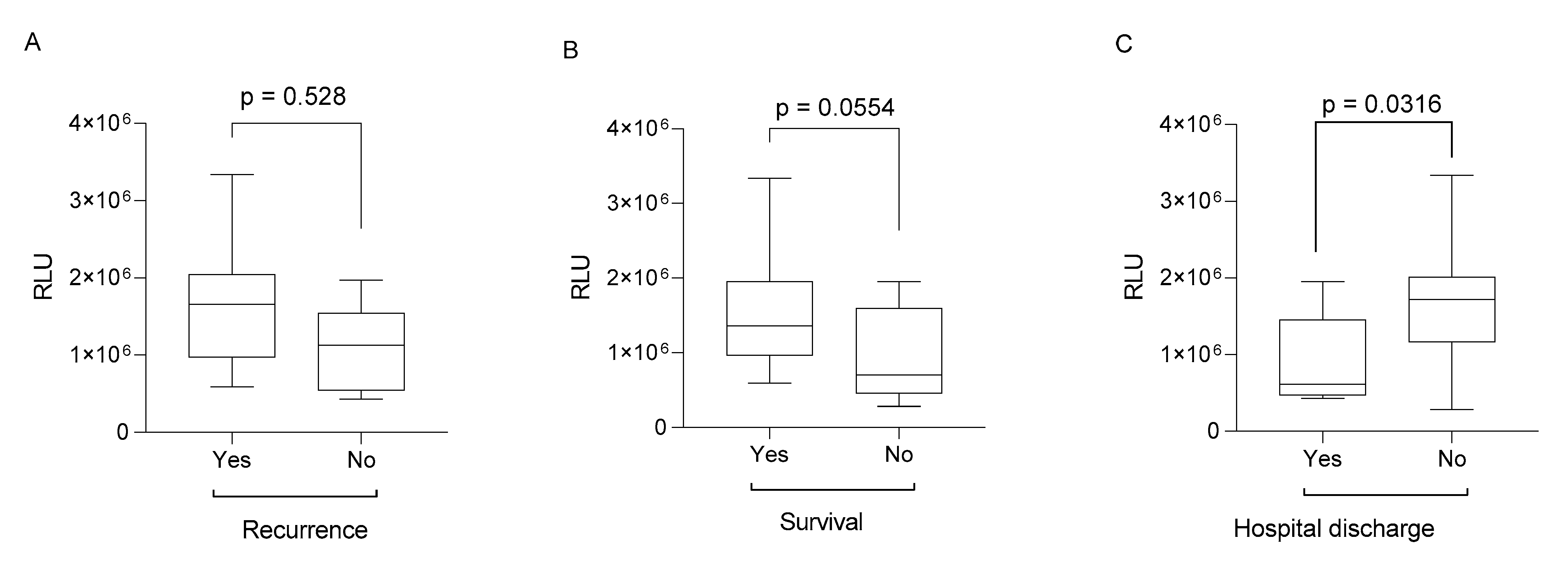
In patients who relapsed (median: 1.658995 × 10⁶ RLU, Figure 2A), died (median: 1.360012 × 10⁶ RLU, Figure 2B), or were not discharged from the hospital at the time of the study (median: 1.719426 × 10⁶ RLU, Figure 2C), the lipoperoxide levels were significantly higher than in those who did not relapse (median: 1.129336 × 10⁶ RLU), survived (median: 7.02951 × 10⁵ RLU), or were definitively discharged (median: 6.11824 × 10⁵ RLU), with p values of 0.0528, 0.0554, and 0.0316, respectively.
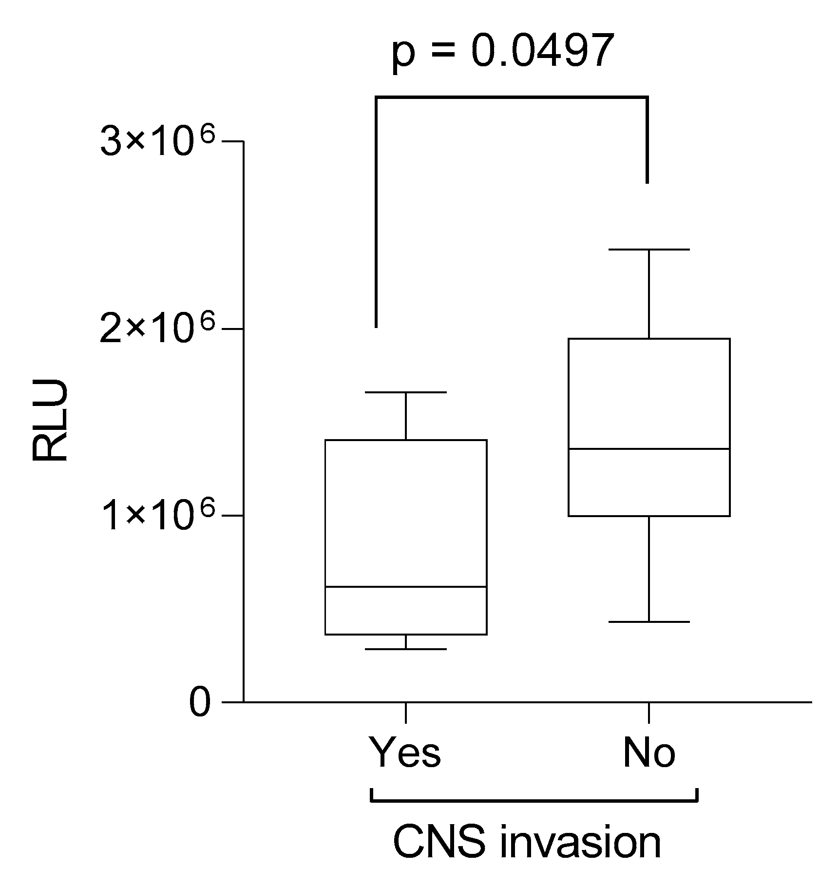
As shown in Figure 3, the lipoperoxide levels were higher in patients without central nervous system (CNS) involvement (median: 1.357068 × 10⁶ RLU) compared to those with CNS involvement (median: 6.18261 × 10⁵ RLU, p = 0.0497).
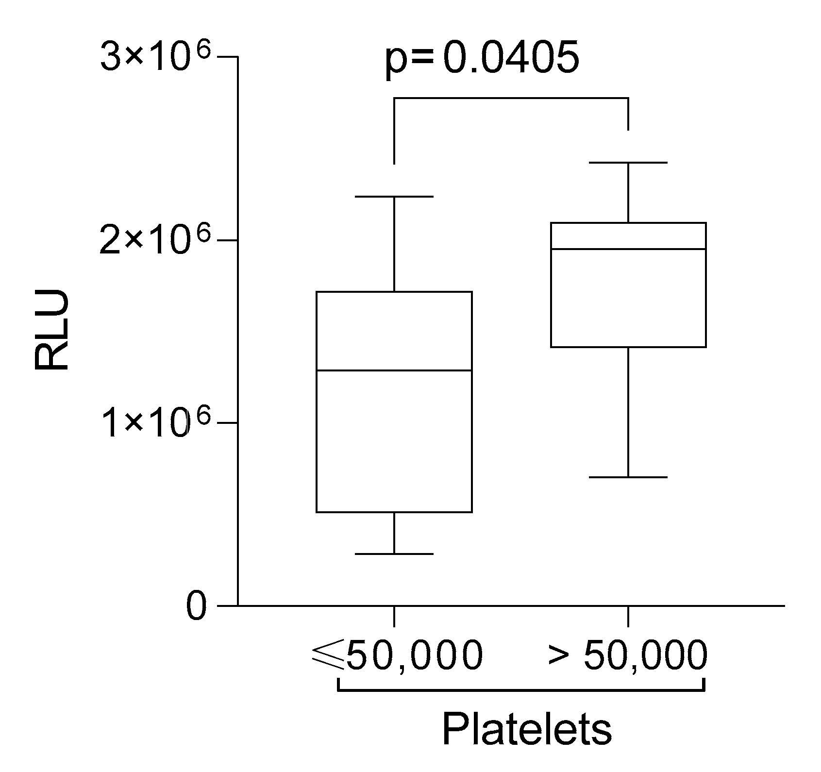
Lipid peroxidation levels were significantly higher in B-ALL patients undergoing induction chemotherapy with platelet counts exceeding 50,000/mm3 (median: 1.950910 × 10⁶ RLU) compared to those with platelet counts of 50,000/mm3 or lower (median: 1.287271 × 10⁶ RLU, p = 0.0405, Figure 4).
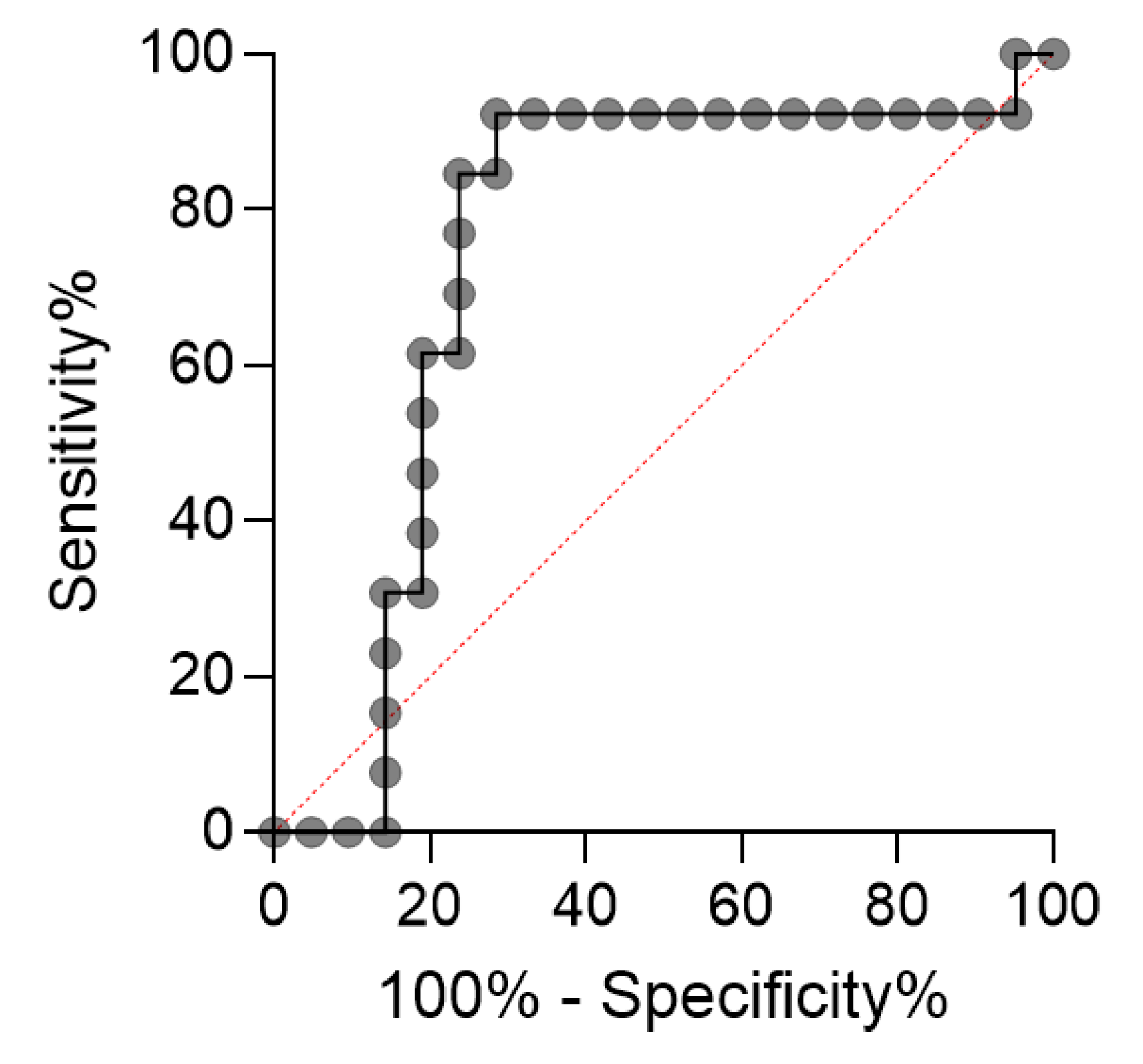
According to the ROC curve (Figure 5), lipoperoxide levels serve as a significant discriminator between B-ALL patients undergoing induction chemotherapy and those who are out of treatment (p = 0.0168). The analysis revealed a high sensitivity of 92.31%, indicating that lipoperoxide levels accurately identify most patients receiving chemotherapy. Additionally, the specificity of 71.43% suggests a reliable distinction between treated and untreated patients, minimizing false-positive classifications. Our analysis determined an optimal cutoff point of >1.282844 × 10⁶, established using the Youden index, and a positive likelihood ratio of 3.231, suggesting that lipid peroxide levels exceeding this are 3.23 times more likely to indicate that a patient is undergoing treatment.

3. Discussion

This study demonstrates that lipid peroxide levels display a significant predictive value in B-ALL patients undergoing chemotherapy. Our findings also show that lipid peroxidation levels vary considerably in peripheral plasma according to clinicopathological parameters that determine disease prognosis, effectively differentiating patients undergoing induction treatment from those not receiving chemotherapy. To the best of our knowledge, this is a novel finding that contributes to the existing literature on oxidative stress in B-ALL.
We observed that patients under 10 years old exhibited higher lipid peroxidation levels than did older patients. It is known that younger children diagnosed with ALL experience increased oxidative stress compared to older children, as evidenced by higher levels of oxidized phosphatidylcholine in the cerebrospinal fluid (CSF) of patients undergoing treatment [24]. These findings suggest that age is a critical factor influencing the severity of oxidative damage in pediatric ALL. The heightened oxidative stress observed in younger patients may also result from the hypermetabolic state induced by leukemia, particularly due to the elevated metabolic activity of neoplastic cells, which leads to excessive production of reactive oxygen species (ROS) [25,26].
Regarding patient outcomes, we observed that lipoperoxide levels were elevated in individuals with unfavorable prognoses, particularly those who experienced disease relapse or death. It is well established that ROS production can protect tumor cells from apoptosis while promoting proliferation, migration, metastasis, and resistance to drug treatment [27]. Additionally, elevated ROS levels and diminished antioxidant defenses have been linked to disease progression [28]. In B-ALL, relapse remains one of the most significant challenges in treatment, especially when it occurs early after initial therapy, as it is associated with poor prognosis. Leukemic cells can alter the bone marrow microenvironment, transforming it into a setting that supports leukemia progression while suppressing normal hematopoiesis. This modified microenvironment provides a sanctuary for leukemic cells, allowing them to evade the cytotoxic effects of pro-oxidant agents. The oxidative environment further facilitates metabolic and intracellular signaling alterations that enhance leukemic cell survival and proliferation. For instance, elevated ROS levels can activate pro-survival signaling pathways such as NF-κB and PI3K/AKT, which are associated with resistance to chemotherapy-induced apoptosis. These adaptive mechanisms contribute to the persistence of leukemic cells, rendering them resistant to pro-oxidant therapies and increasing the likelihood of relapse [29].
In addition to the mechanisms inherent in tumor biology, chemotherapy can influence the production of ROS, particularly with drugs from the anthracycline class, such as doxorubicin. These chemotherapeutic agents, commonly used in therapeutic protocols for B-ALL, induce oxidative stress through several mechanisms. They promote ROS generation by activating the mitochondrial apoptotic pathway in leukemic cells, altering mitochondrial dynamics (including fission and fusion), and forming complexes with iron ions, which generate highly reactive species that can damage lipids, proteins, and cellular DNA [30,31,32]. This oxidative damage extends to healthy tissues, contributing to systemic toxicity. Moreover, these processes can modulate the tumor microenvironment and impact the overall efficacy of treatment [32]. Therefore, part of the observed increase in lipoperoxide levels may be linked to the pharmacodynamics of these chemotherapeutic agents, emphasizing the need to consider this factor when interpreting biomarkers of oxidative stress during B-ALL therapy.
We also observed an inverse correlation between lipid peroxidation levels and the achievement of definitive hospital discharge or cure. Specifically, lower lipid peroxidation levels in patients who were definitively discharged may reflect a reduction in oxidative stress [11,12]. Furthermore, the decline in lipid peroxidation appears to be linked to the metabolic improvement of ALL patients [26], suggesting a more favorable prognosis. This observation reinforces the potential of lipid peroxidation as an indicator of treatment success and overall clinical outcome in B-ALL.
Regarding CNS invasion, we observed that higher lipid peroxide levels appeared to act as a protective factor. The brain is particularly susceptible to oxidative stress due to its limited antioxidant defenses, high energy demands, and substantial lipid content, including glycerophospholipids, which are essential for maintaining cell membrane integrity. Lipid peroxidation plays a significant role in this vulnerability [33]. CNS leukemia occurs when leukemic cells infiltrate the meninges, cranial nerves, brain, or spinal cord, representing a common and serious complication of ALL. This involvement is associated with poor prognosis and a high risk of relapse. In ALL patients, the CNS is the most frequently affected site of extramedullary involvement, increasing the risk of complications and mortality [34,35]. Blast infiltration typically occurs in the cerebrospinal fluid, leptomeninges, and bone marrow, particularly in pediatric cases, leading to the development of CNS prophylaxis strategies, such as intrathecal chemotherapy, to prevent relapse [7]. Although we did not assess the oxidative status of the cerebrospinal fluid, our findings suggest that elevated systemic lipid peroxidation levels may correlate with the absence of CNS invasion. However, based on our data, we cannot establish a definitive causal relationship between lipid peroxidation and CNS involvement.
Another key parameter in which lipid peroxidation levels showed significant variation was platelet count. A direct correlation was observed, with patients presenting higher platelet counts exhibiting increased lipid peroxidation levels. Platelet count is considered a potential prognostic factor in treatment protocols for childhood ALL, playing a crucial role in identifying patients at higher risk of treatment failure [36]. Both platelet levels at diagnosis and their fluctuations during treatment have been linked to event-free survival rates, emphasizing their relevance in patient management. Lipid hydroperoxides, such as HPETEs, are known to influence platelet function and arachidonic acid metabolism [37]. Although platelet counts above 50,000/mm3 are generally considered a favorable prognostic marker, the elevated lipid peroxidation observed in these patients may impact disease prognosis by modulating platelet-dependent coagulation mechanisms. Dysregulation in clot formation could, therefore, negatively influence treatment outcomes.
Since we observed significant variations in lipid peroxide levels during the induction phase of B-ALL treatment under specific clinicopathological conditions, we further explored their potential prognostic value as a treatment biomarker. To assess this, we conducted an ROC curve analysis comparing patients undergoing induction therapy with those out of treatment. Our analysis identified an optimal cutoff value of >1.282844 × 10⁶, determined by the Youden index, with a sensitivity of 92.31% and a specificity of 71.43%, demonstrating strong discriminatory power. Furthermore, the positive likelihood ratio (3.231) indicates that lipid peroxide levels above this threshold increase the probability of that a patient is undergoing treatment by 3.23 times. The success of the induction phase is a critical prognostic factor for patient outcomes in B-ALL [23]. Current biomarker research in ALL primarily focuses on soluble immune mediators [38,39], genetic alterations [40,41], and proteomic analyses [42]; however, no studies to date have directly linked lipoperoxides to specific B-ALL outcomes. Considering that lipoperoxides play a significant role in the carcinogenesis and progression of various cancers [43,44], including their impact on antitumor immune function [45,46] and normal hematopoiesis [47], our findings suggest that lipoperoxide levels can effectively differentiate between patients undergoing treatment and those who are not, positioning them as a promising biomarker for assessing treatment-related disease parameters.
Although this study did not assess antioxidant levels, a previous study from our group evaluated these levels in the same cohort of children [48]. The results showed a reduction in antioxidants in patients who died, suggesting that variations in total antioxidant capacity during chemotherapy correlate with disease prognosis in B-ALL. In the present study, we observed lower lipid peroxide levels in patients who died, indicating that antioxidant consumption may reflect the body’s effort to neutralize lipid peroxides and other reactive oxygen species (ROS). While specific antioxidants like glutathione, superoxide dismutase, or catalase were not assessed, the imbalance between antioxidants and lipid peroxidation appears to be significant, particularly in relation to poorer disease prognosis and survival outcomes.
This study has some limitations, including a modest sample size, the absence of antioxidant status evaluation, and the lack of healthy control individuals. Additionally, potential confounders such as nutrition, infection, and inflammation—which are known to influence lipid peroxide levels—were not fully assessed due to data constraints. Despite these limitations, our findings highlight the potential of lipid peroxidation as a biomarker for monitoring treatment response in pediatric B-ALL, offering a valuable tool for clinical management.

4. Materials and Methods

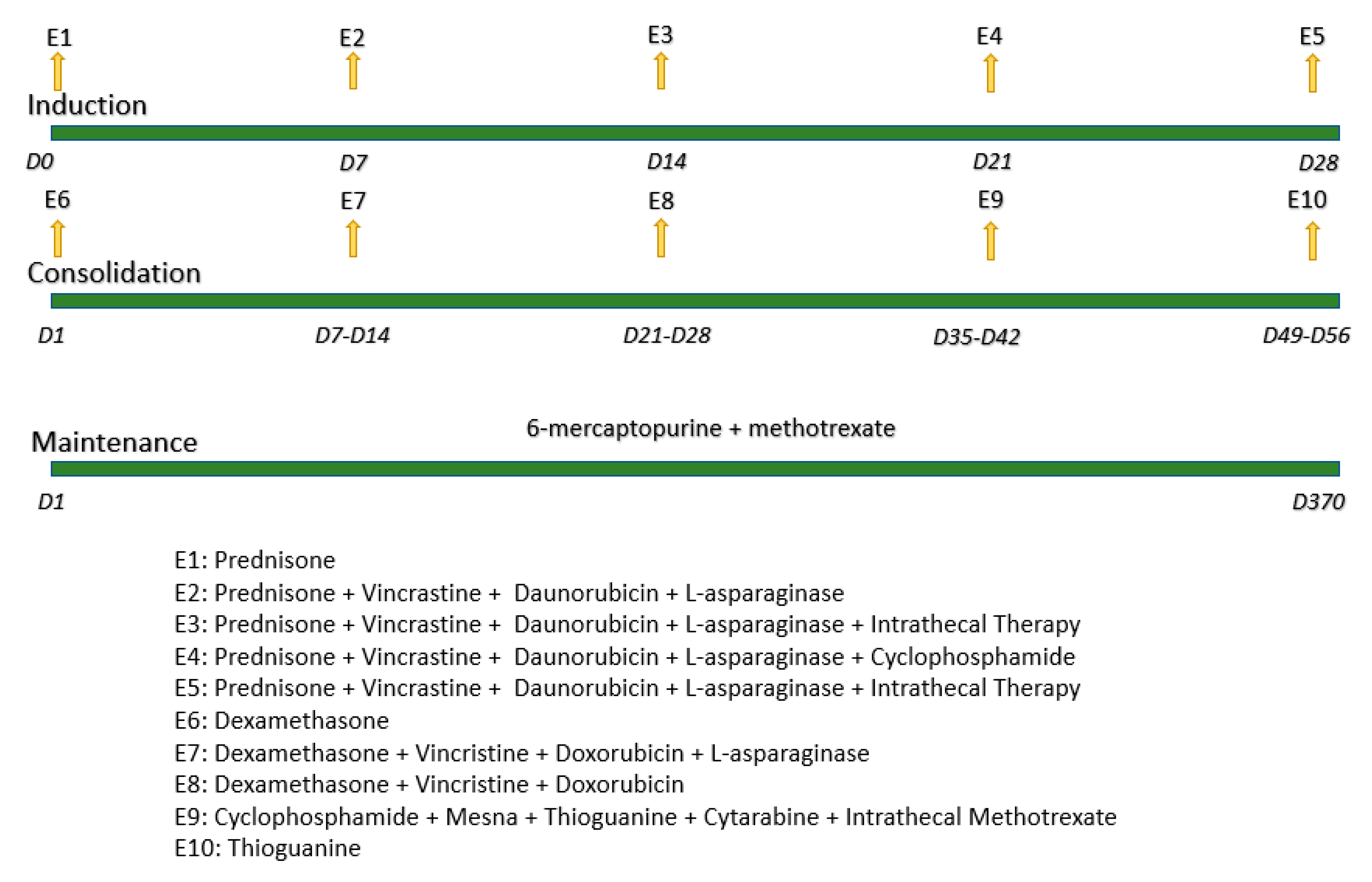
This study was approved by the institutional ethics committee (24498213.0.0000.5231), and informed consent was obtained from the guardians of all participants. A total of 40 patients diagnosed with B-ALL were selected based on the following criteria: guardian authorization for participation, age between 0 and 20 years, any sex, and confirmed diagnosis of B-ALL through myelogram and immunophenotyping. All patients were treated at a public pediatric cancer hospital in northern Paraná, Brazil. Exclusion criteria included a diagnosis of T-cell acute lymphoblastic leukemia and/or unconfirmed diagnosis by immunophenotyping. All participants underwent treatment according to the standardized GBTLI-LLA 2009 protocol [49], receiving the same drug combinations administered throughout the induction, consolidation, and maintenance phases, as illustrated in Figure 6.
Peripheral blood samples (5 mL in EDTA tubes) were collected from patients after informed consent was obtained from their legal guardians. The samples were centrifuged at 5000 rpm for 5 min to isolate plasma. Clinical data were retrieved from the patients’ medical records. Lipoperoxide levels were measured using a methodology standardized in our laboratory, based on the protocol published by Gonzalez-Flecha et al. (1991) [50]. Tert-butyl hydroperoxide, a potent peroxyl radical generator, was used to initiate lipid peroxidation. In biological membranes, these radicals attack lipids, generating lipoperoxides that further propagate lipid oxidation through chain reactions, emitting photons that can be quantified. For the assay, 125 µL of plasma was mixed with 865 µL of 10 mM monobasic phosphate buffer (pH 7.4) in 0.9% NaCl and incubated at 37 °C for 5 min. A 10 µL aliquot of the tert-butyl solution was then added to initiate the reaction. Photon emission was measured using a Glomax luminometer over 40 min, and the results were recorded in relative light units (RLU).
Statistical analyses were performed using GraphPad Prism 9.0 and OriginLab 9.0 software. Data distribution was assessed using the Shapiro–Wilk test. Parametric tests were applied to normally distributed variables, while nonparametric tests were used for non-normal distributions. Outliers were excluded to avoid distortion of the results. Lipoperoxide levels between patient groups were compared using the Student’s t-test, the Mann–Whitney test, or ANOVA followed by the Bonferroni post hoc test. A p-value ≤ 0.05 was considered statistically significant.
For ROC curve analysis, a control group was added which consisted of patients who were not undergoing treatment at the time of peripheral blood collection—those who had completed the three phases of the standard treatment for B-ALL, had been off treatment for at least two years, and were in the waiting period for definitive discharge. The ROC curve was constructed by comparing patients undergoing treatment with those who were not, plotting sensitivity on the y-axis and 1-specificity on the x-axis, using the Wilson/Brown statistical method with a 95% confidence interval. The optimal cutoff point was determined by calculating the Youden index [51] (sensitivity + specificity − 1).

5. Conclusions

This study demonstrates the potential of lipoperoxides as valuable prognostic biomarkers in pediatric B-ALL. The observed associations between lipoperoxide levels and key clinical outcomes, such as disease relapse, remission, and platelet counts, underscore their role in reflecting oxidative stress and treatment response. It further suggests that lipoperoxide levels could differentiate between patients undergoing chemotherapy and those not in treatment. These findings support lipoperoxides as potential biomarkers for monitoring B-ALL treatment and prognosis, offering a promising tool for enhancing patient management and predicting treatment outcomes.

Author Contributions

Conceptualization, C.P., F.C.T. and B.Y.K.; methodology, B.Y.K., M.R.S., C.C., A.L.G.S., A.W.R., M.B.L., M.E.P.S., V.d.M.T., S.T.L., G.E.B. and R.G.P.; formal analysis, C.P. and B.Y.K.; writing—original draft preparation, B.Y.K., M.R.S., C.C., A.L.G.S., A.W.R., M.B.L., M.E.P.S., V.d.M.T., S.T.L. and G.E.B.; writing—review and editing, B.Y.K., M.R.S., C.C., A.L.G.S., A.W.R., M.B.L., M.E.P.S., V.d.M.T., S.T.L., G.E.B., C.P. and F.C.T. All authors have read and agreed to the published version of the manuscript.

Funding

This research received no external funding.

Institutional Review Board Statement

The study was conducted in accordance with the Declaration of Helsinki, and approved by the Institutional Review Board of Londrina State University, protocol code number 24498213.0.0000.5231.

Informed Consent Statement

Informed consent was obtained from all subjects involved in the study.

Data Availability Statement

Data is unavailable due to privacy or ethical restrictions.

Conflicts of Interest

The authors declare no conflict of interest.

Abbreviations

The following abbreviations are used in this manuscript:
B-ALLB-type acute lymphoblastic leukemia
Nnumber of patients
CNScentral nervous system
ROCreceiver operating characteristic
ALLacute lymphoblastic leukemia
RLUrelative light units
CSFcerebrospinal fluid
ROSreactive oxygen species
EFSevent-free survival

References

  1. National Cancer Institute. Available online: https://www.gov.br/inca/pt-br/assuntos/cancer/numeros/estimativa/estado-capital/brasil/cancer-infantojuvenil (accessed on 16 April 2024).
  2. Brazilian Society of Pediatrics. Available online: https://www.sbp.com.br/fileadmin/user_upload/publicacoes/C-Doc-Cientifico-Oncologia-Epidemiol-30-mar-17.pdf (accessed on 16 April 2024).
  3. Pedrosa, F.; Lins, M. Acute lymphocytic leukemia: A curable disease. Rev. Bras. Saúde Materno Infantil. 2002, 2, 63–68. [Google Scholar] [CrossRef]
  4. Canto, M.E.B.S.; Ferreira, J.A.; Rosa, F.C.; Gomes, A.C.D.B.; Zveibil, G.S. Perfil epidemiológico da leucemia linfoide no Brasil. Hematology. Transfus. Cell Ther. 2023, 45 (Suppl. 4), S163. [Google Scholar] [CrossRef]
  5. Wang, L.; Yao, X.; Yang, L. Acute Lymphoblastic Leukemia Collaborators. (2025). Global, regional, and national burden of children and adolescents with acute lymphoblastic leukemia from 1990 to 2021: A systematic analysis for the Global Burden of Disease Study 2021. Front. Public Health 2021, 13, 1525751. [Google Scholar] [CrossRef]
  6. Associação Brasileira de Linfoma e Leucemia. Leucemia Linfoide Aguda. Available online: https://www.abrale.org.br/doencas/leucemia/lla/o-que-e/ (accessed on 16 April 2024).
  7. Zago, M.A.; Falcão, R.P.; Pasquini, R. Leucemia Linfoide da Criança e do Adolescente. In Tratado de Hematologia, 1st ed.; Atheneu: São Paulo, Brasil, 2013; pp. 391–401. [Google Scholar]
  8. Hong, Z.; Wei, Z.; Xie, T.; Fu, L.; Sun, J.; Zhou, F.; Jamal, M.; Zhang, Q.; Shao, L. Targeting chemokines for acute lymphoblastic leukemia therapy. J. Hemat. Oncol. 2021, 14, 48. [Google Scholar] [CrossRef]
  9. Cole, P.D.; Finkelstein, Y.; Stevenson, K.E.; Blonquist, T.M.; Vijayanathan, V.; Silverman, L.B.; Neuberg, D.S.; Sallan, S.E.; Robaey, P.; Waber, D.P. Polymorphisms in genes related to oxidative stress are associated with inferior cognitive function after therapy for childhood acute lymphoblastic leukemia. J. Clin. Oncol. 2015, 33, 2205–2211. [Google Scholar] [CrossRef]
  10. Clemente, S.M.; Martínez-Costa, O.H.; Monsalve, M.; Samhan-Arias, A.K. Targeting lipid peroxidation for cancer treatment. Molecules 2020, 25, 5144. [Google Scholar] [CrossRef]
  11. Hybertson, B.M.; Gao, B.; Bose, S.K.; McCord, J.M. Oxidative stress in health and disease: The therapeutic potential of Nrf2 activation. Mol. Asp. Med. 2011, 32, 234–246. [Google Scholar] [CrossRef]
  12. Ferreira, A.L.A.; Matsubara, L.S. Free radicals: Concepts, related diseases, defense system and oxidative stress. J. Braz. Med. Assoc. 1997, 43, 61–68. [Google Scholar] [CrossRef]
  13. Panis, C.; Victorino, V.J.; Herrera, A.C.; Cecchini, A.L.; Simão, A.N.; Tomita, L.Y.; Cecchini, R. Can breast tumors affect the oxidative status of the surrounding environment? A comparative analysis among cancerous breast, mammary Adjacent tissue, and plasma. Oxidative Med. Cell. Longev. 2015, 2015, 6429812. [Google Scholar] [CrossRef]
  14. Al-Tonbary, Y.; Al-Hasan, S.A.; Zaki, M.; Hammad, A.; Kandil, S.; Fouda, A. Impact of anti-oxidant status and apoptosis on the induction phase of chemotherapy in childhood acute lymphoblastic leukemia. Hematology 2011, 16, 14–19. [Google Scholar] [CrossRef]
  15. Babiak, R.M.V.; Campello, A.P.; Garnieri, E.G.S.; Oliveira, M.B.M. Metotrexate: Pentose cycle and oxidative stress. Cell Biochem. Funct. 1998, 16, 283–293. [Google Scholar] [CrossRef]
  16. Battisti, V.; Maders, L.D.K.; Bagatini, M.D.; Santos, K.F.; Spanevello, R.M.; Maldonado, P.A.; Brulé, A.O.; de Araújo, M.C.; Schetinger, M.A.; Morsch, V.M. Measurement of oxidative stress and antioxidant status in acute lymphoblastic leukemia patients. Clin. Biochem. 2008, 41, 511–518. [Google Scholar] [CrossRef] [PubMed]
  17. Broto, G.E.; Corrêa, S.; Trigo, F.C.; Santos, E.C.; Tomiotto-Pelissier, F.; Pavanelli, W.R.; Silveira, G.F.; Abdelhay, E.; Panis, C. Comparative analysis of systemic and tumor microenvironment proteomes from children with B-cell acute lymphocytic leukemia at diagnosis and after induction treatment. Front. Oncol. 2020, 10, 550213. [Google Scholar] [CrossRef] [PubMed]
  18. De Cavanagh, E.M.V.; Honegger, A.E.; Hofer, E.; Bordenave, R.H.; Bullorsky, E.O.; Chasseing, N.A.; Fraga, C. Higher oxidation and lower antioxidant levels in peripheral blood plasma and bone marrow plasma from advanced cancer patients. Cancer 2002, 94, 3247–3251. [Google Scholar] [CrossRef]
  19. Cramer, S.L.; Saha, A.; Liu, J.; Tadi, S.; Tiziani, S.; Yan, W.; Triplett, K.; Lamb, C.; Alters, S.E.; Rowlinson, S.; et al. Systemic depletion of L-cyst(e)ine with cyst(e)inase increases reactive oxygen species and suppresses tumor growth. Nat. Med. 2017, 23, 120–127. [Google Scholar] [CrossRef]
  20. Devi, G.S.; Prasad, M.H.; Saraswathi, I.; Raghu, D.; Rao, D.N.; Reddy, P.P. Free radicals, antioxidant enzymes, and lipid peroxidation in different types of leukemias. Clin. Chim. Acta 2000, 293, 53–62. [Google Scholar] [CrossRef]
  21. El-Sabagh, M.E.; Ramadan, K.S.; El-Slam, I.M.A.; Ibrahim, A.M. Antioxidants status in acute lymphoblastic leukemic patients. Am. J. Med. Med. Sci. 2011, 1, 1–6. Available online: http://article.sapub.org/10.5923.j.ajmms.20110101.01.html (accessed on 25 March 2025). [CrossRef]
  22. Er, T.-K.; Tsai, S.-M.; Wu, S.-H.; Chiang, W.; Lin, H.-C.; Lin, S.-F.; Wu, S.-H.; Tsai, L.-Y.; Liu, T.-Z. Antioxidant status and superoxide anion radical generation in acute myeloid leukemia. Clin. Biochem. 2007, 40, 1015–1019. [Google Scholar] [CrossRef]
  23. Oliveira, G.E.B. Impact of the Induction Phase of Chemotherapy on the Profile of Systemic Inflammatory Mediators and the Tumor Microenvironment in Childhood Acute Lymphocytic Leukemia. Ph.D. Thesis, State University of Londrina, Londrina, Brazil, 2021. [Google Scholar]
  24. Caron, J.E.; Krull, K.R.; Hockenberry, M.; Jain, N.; Kaemingk, K.; Moore, I.M. Oxidative stress and executive function in children receiving chemotherapy for acute lymphoblastic leukemia. Pediatr. Blood Cancer 2009, 53, 551–556. [Google Scholar] [CrossRef]
  25. Kien, C.L.; Camitta, B.M. Close association of accelerated rates of whole body protein turnover (synthesis and breakdown) and energy expenditure in children with newly diagnosed acute lymphocytic leukemia. J. Parenter. Enter. Nutr. 1987, 11, 129–134. [Google Scholar] [CrossRef]
  26. Chaudhary, P.; Kumari, S.; Dewan, P.; Gomber, S.; Ahmed, R.S.; Kotru, M. Chemotherapy-Induced Oxidative Stress in Pediatric Acute Lymphoblastic Leukemia. Cureus 2023, 15, e35968. [Google Scholar] [CrossRef] [PubMed]
  27. Udensi, U.K.; Tchounwou, P.B. Dual effect of oxidative stress in the induction and treatment of leukemia cancer. J. Exp. Clin. Cancer Res. 2014, 106, 3594–3601. [Google Scholar] [CrossRef] [PubMed]
  28. Ahmad, R.; Tripathi, A.K.; Tripathi, P.; Singh, R.; Singh, S.; Singh, R.K. Oxidative stress and antioxidant status in patients with chronic myeloid leukemia. Indian. J. Clin. Bioch. 2008, 23, 328–333. [Google Scholar] [CrossRef] [PubMed]
  29. Chen, Y.; Li, J.; Zhao, Z. Redox control in acute lymphoblastic leukemia: From physiology to pathology and therapeutic opportunities. Cells 2021, 10, 1218. [Google Scholar] [CrossRef]
  30. Minotti, G.; Menna, P.; Salvatorelli, E.; Cairo, G.; Gianni, L. Anthracyclines: Molecular advances and pharmacologic developments in antitumor activity and cardiotoxicity. Pharmacol. Rev. 2004, 56, 185–229. [Google Scholar] [CrossRef]
  31. Carvalho, C.; Santos, R.X.; Cardoso, S.; Correia, S.; Oliveira, P.J.; Santos, M.S.; Moreira, P.I. Doxorubicin: The good, the bad and the ugly effect. Curr. Med. Chem. 2009, 16, 3267–3285. [Google Scholar] [CrossRef]
  32. Wang, T.; Xing, G.; Fu, T.; Ma, Y.; Wang, Q.; Zhang, S.; Chang, X.; Tong, Y. Role of mitochondria in doxorubicin-mediated cardiotoxicity: From molecular mechanisms to therapeutic strategies. Cell Stress Chaperones 2024, 29, 349–357. [Google Scholar] [CrossRef]
  33. Moore, K.I.; Gundy, P.; Pasvogel, A.; Montgomery, D.W.; Taylor, O.A.; Koerner, K.M.; McCarthy, K.; Hockenberry, M.J. Increase in oxidative stress as measured by cerebrospinal fluid lipid peroxidation during treatment for childhood acute lymphoblastic leukemia. J. Pediatr. Hematol. Oncol. 2015, 37, 86–93. [Google Scholar] [CrossRef]
  34. Tang, X.; Mai, H.; Wang, L.; Chen, S.; Chen, F.; Li, T.; Liu, Y.; Zhou, G.; Liu, S.; Wang, Y.; et al. Diagnostic significance of cerebrospinal fluid flow cytometry in Chinese children with B lineage acute lymphoblastic leukemia. BMC Pediatr. 2024, 24, 204. [Google Scholar] [CrossRef]
  35. Xu, L.H.; Geng, X.; Liao, N.; Yang, L.H.; Mai, H.R.; Wan, W.Q.; Huang, L.B.; Zheng, M.C.; Tian, C.; Chen, H.Q.; et al. Prognostic significance of CNSL at diagnosis of childhood B-cell acute lymphoblastic leukemia: A report from the South China Children’s Leukemia Group. Front. Oncol. 2022, 12, 943761. [Google Scholar] [CrossRef]
  36. Zeidler, L.; Zimmermann, M.; Möricke, A.; Meissner, B.; Bartels, D.; Tschan, C.; Schrauder, A.; Cario, G.; Goudeva, L.; Jäger, S.; et al. Low platelet counts after induction therapy for childhood acute lymphoblastic leukemia are strongly associated with poor early response to treatment as measured by minimal residual disease and are prognostic for treatment outcome. Haematologica 2012, 97, 402–409. [Google Scholar] [CrossRef] [PubMed]
  37. Calzada, C.; Vericel, E.; Lagarde, M. Low concentrations of lipid hydroperoxides prime human platelet aggregation specifically via cyclo-oxygenase activation. Biochem. J. 1997, 325, 495–500. [Google Scholar] [CrossRef] [PubMed]
  38. Carvalho, M.P.S.S.; Magalhães-Gama, F.; Loiola, B.P.; Neves, J.C.F.; Araújo, N.D.; Silva, F.S.; Catão, C.L.S.; Alves, E.B.; Pimentel, J.P.D.; Barbosa, M.N.S.; et al. Systemic immunological profile of children with B-cell acute lymphoblastic leukemia: Performance of cell populations and soluble mediators as serum biomarkers. Front. Oncol. 2023, 13, 1290505. [Google Scholar] [CrossRef] [PubMed]
  39. Kerr, M.W.A.; Magalhães-Gama, F.; Ibiapina, H.N.S.; Hanna, F.S.A.; Xabregas, L.A.; Alves, E.B.; Pimentel, J.P.D.; Carvalho, M.P.S.S.; Tarragô, A.M.; Teixeira-Carvalho, A.; et al. Bone marrow soluble immunological mediators as clinical prognosis biomarkers in B-cell acute lymphoblastic leukemia patients undergoing induction therapy. Front. Oncol. 2021, 11, 696032. [Google Scholar] [CrossRef]
  40. Du, Y.; Zhang, X.; Sun, M.; Yang, L.; Long, F.; Qi, S.; Luo, L.; Lv, X.; Wang, C.; Wu, X.; et al. Molecular characterization and biomarker identification in paediatric B-cell acute lymphoblastic leukaemia. J. Cell. Mol. Med. 2024, 28, e70126. [Google Scholar] [CrossRef]
  41. Moorman, A.V. New and emerging prognostic and predictive genetic biomarkers in B-cell precursor acute lymphoblastic leukemia. Haematologica 2016, 101, 407–416. [Google Scholar] [CrossRef]
  42. Cavalcante, M.S.; Torres-Romero, J.C.; Lobo, M.D.; Moreno, F.B.; Bezerra, L.P.; Lima, D.S.; Matos, J.C.; Moreira, R.A.; Monteiro-Moreira, A.C. A panel of glycoproteins as candidate biomarkers for early diagnosis and treatment evaluation of B-cell acute lymphoblastic leukemia. Biomark. Res. 2016, 4, 1. [Google Scholar] [CrossRef]
  43. Hristozov, D.; Gadjeva, V.; Vlaykova, T.; Dimitrov, G. Evaluation of oxidative stress in patients with cancer. Arch. Physiol. Biochem. 2001, 109, 331–336. [Google Scholar] [CrossRef]
  44. Jelic, M.D.; Mandic, A.D.; Maricic, S.M.; Srdjenovic, B.U. Oxidative stress and its role in cancer. J. Cancer Res. Ther. 2021, 17, 22–28. [Google Scholar] [CrossRef]
  45. Kim, R.; Hashimoto, A.; Markosyan, N.; Tyurin, V.A.; Tyurina, Y.Y.; Kar, G.; Fu, S.; Sehgal, M.; Garcia-Gerique, L.; Kossenkov, A.; et al. Ferroptosis of tumour neutrophils causes immune suppression in cancer. Nature 2022, 612, 338–346. [Google Scholar] [CrossRef]
  46. Ma, X.; Xiao, L.; Liu, L.; Ye, L.; Su, P.; Bi, E.; Wang, Q.; Yang, M.; Qian, J.; Yi, Q. CD36-mediated ferroptosis dampens intratumoral CD8+ T cell effector function and impairs their antitumor ability. Cell Metab. 2021, 33, 1001–1012. [Google Scholar] [CrossRef] [PubMed]
  47. Rodríguez-García, A.; García-Vicente, R.; Morales, M.L.; Ortiz-Ruiz, A.; Martínez-López, J.; Linares, M. Protein carbonylation and lipid peroxidation in hematological malignancies. Antioxidants 2020, 9, 1212. [Google Scholar] [CrossRef] [PubMed]
  48. Garbim, M.R.; Broto, G.E.; Trigo, F.C.; Victorino, V.J.; Oliveira, S.T.; Barbosa, D.S.; Panis, C. Chemotherapy induces plasmatic antioxidant changes in pediatric patients with acute lymphoid leukemia B that correlate to disease prognosis. Curr. Res. Immunol. 2022, 3, 228–233. [Google Scholar] [CrossRef] [PubMed]
  49. Sociedade Brasileira de Oncologia Pediátrica. GBTLI-LLA. In Protocolo de Tratamento da Leucemia Linfoblástica Aguda na Infância; Sociedade Brasileira de Oncologia Pediátrica: São Paulo, Brazil, 2009; pp. 1–6. [Google Scholar]
  50. Gonzales-Flecha, B.; Lleswy, S.; Boveris, A. Hydroperoxide-initiated chemiluminescence: An assay for oxidative stress in biopsies of heart, liver and muscle. Free Radic. Biol. Med. 1991, 10, 93–100. [Google Scholar] [CrossRef]
  51. Martínez-Camblor, P.; Pardo-Fernández, J. The Youden Index in the Generalized Receiver Operating Characteristic Curve Context. Int. J. Biostat. 2019, 15, 20180060. [Google Scholar] [CrossRef]
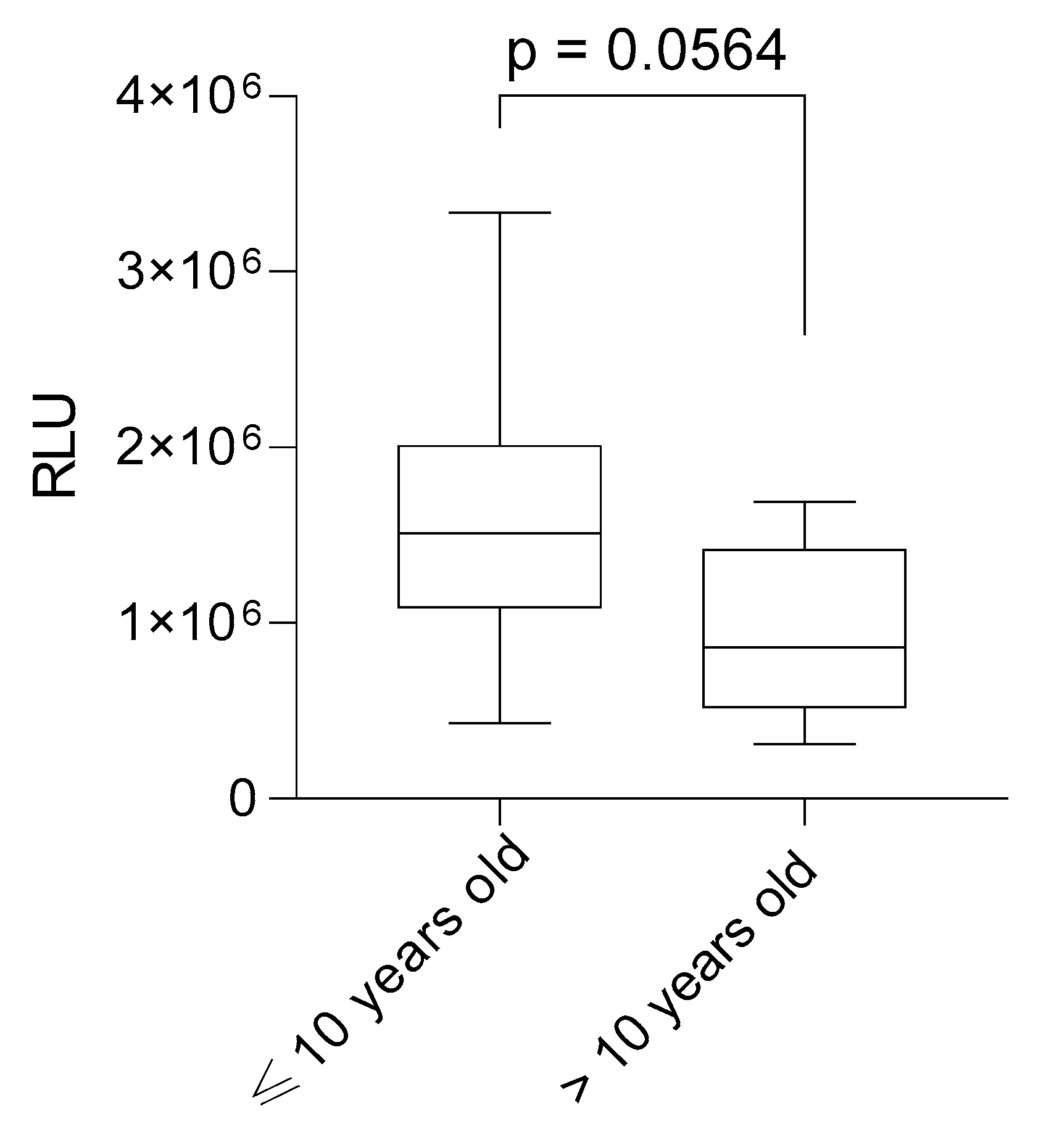
Figure 1. Lipid peroxide levels in plasma of B-ALL patients undergoing induction chemotherapy according to their age at diagnosis (N = 40). Data are represented as box-plots, and the lines inside represent the medians.
Lymphatics 03 00011 g001
Figure 2. Lipid peroxide levels in plasma of B-ALL patients ongoing induction chemotherapy according to their recurrence status—N = 40 (A), survival—N = 40 (B), and hospital definitive discharge—N = 40 (C). Data are represented as box-plots, and the lines inside represent the medians.
Lymphatics 03 00011 g002
Figure 3. Lipid peroxide levels in plasma of B-ALL patients undergoing induction chemotherapy according to the central nervous system commitment (CNS) (N = 40). Data are represented as box-plots, and the lines inside represent the medians.
Lymphatics 03 00011 g003
Figure 4. Lipid peroxide levels in plasma of B-ALL patients undergoing induction chemotherapy according to platelet levels (N = 27). Data are represented as box-plots, and the lines inside represent the medians.
Lymphatics 03 00011 g004
Figure 5. Receiver operating characteristic (ROC) curve based on lipid peroxide levels: off-treatment vs. induction. The red diagonal line represents a random classifier with no discriminative ability (AUC = 0.5).
Lymphatics 03 00011 g005
Figure 6. Design of the study. The chemotherapy protocol used for the treatment of B-ALL patients (GBTLI-LLA, 2009) consists of three phases. Induction involves prednisone (60 mg/m2 PO, D1–D7; 40 mg/m2 PO, D8–D29, tapering), vincristine (1.5 mg/m2 IV, max 2 mg, D8, 15, 22, 29), daunorubicin (40 mg/m2 IV, D8, 15, 22), L-asparaginase (10,000 IU/m2 IM/IV every 3 days from D8, 9 doses), cyclophosphamide (500 mg/m2 IV on D22, 23 for slow responders), and intrathecal therapy (methotrexate, Ara-C, dexamethasone, age-adjusted doses, D15, 29). Consolidation includes dexamethasone (10 mg/m2 PO, D1–D22, tapering), vincristine (1.5 mg/m2 IV), doxorubicin (30 mg/m2 IV, on D8, 15, 22, 29), L-asparaginase (10,000 IU/m2 IM, D8, 11, 15, 18), cyclophosphamide + mesna (1000 mg/m2 IV, D36), thioguanine (60 mg/m2 PO, D36–D49) with cytarabine (75 mg/m2 IV, D38–41, D45–48), and intrathecal methotrexate (age-adjusted, D38–D45). Maintenance lasts 24 months (104 weeks), with 6-mercaptopurine (50 mg/m2 PO, daily) and methotrexate (20 mg/m2 PO, weekly). Our study focused on the induction phase.
Lymphatics 03 00011 g006
Table 1. Clinicopathological data from patients.
ParameterNumber of Patients
Total number of patientsn = 40
Molecular profile
B cell typen = 40 (100%)
Gender
Femalen = 19 (47.5%)
Malen = 21 (52.5%)
Age at diagnosis
Age at diagnosis ≤ 10n = 24 (60%)
Age at diagnosis > 10n = 7 (17.5%)
No datan = 9 (22.5%)
Recurrencen = 10 (25%)
Deathn = 17 (42.5%)
Hospital dischargen = 8 (20%)
Leukometry, mm3
Leukometry > 10,000n = 10 (25%)
Leukometry ≤ 10,000n = 17 (42.5%)
No datan = 13 (32.5%)
Hemoglobin, g/dL
Hemoglobin > 8n = 10 (25%)
Hemoglobin ≤ 8n = 16 (40%)
No datan = 14 (35%)
Platelets, mm3
Platelet > 50,000n = 9 (22.5%)
Platelet ≤ 50,000n = 18 (45%)
No datan = 13 (32.5%)
CNS involvementn = 5 (12.5%)
Disclaimer/Publisher’s Note: The statements, opinions and data contained in all publications are solely those of the individual author(s) and contributor(s) and not of MDPI and/or the editor(s). MDPI and/or the editor(s) disclaim responsibility for any injury to people or property resulting from any ideas, methods, instructions or products referred to in the content.