Article

Uptake, Distribution, and Activity of Pluronic F68 Adjuvant in Wheat and Its Endophytic Bacillus Isolate

1
Department of Biological Engineering, Utah State University, Logan, UT 84322, USA
2
Department of Plants Soils and Climate, Utah State University, Logan, UT 84322, USA
3
Utah Water Research Laboratory, Utah State University, Logan, UT 84322, USA
*
Authors to whom correspondence should be addressed.
Agrochemicals 2025, 4(3), 12; https://doi.org/10.3390/agrochemicals4030012
Submission received: 7 June 2025 / Revised: 17 July 2025 / Accepted: 19 July 2025 / Published: 23 July 2025

Abstract

Surfactants are widely utilized in agriculture as emulsifying, dispersing, anti-foaming, and wetting agents. In these adjuvant roles, the inherent biological activity of the surfactant is secondary to the active ingredients. Here, the hydrophilic non-ionic surface-active tri-block copolymer Pluronic® F68 is investigated for direct biological activity in wheat. F68 binds to and inserts into lipid membranes, which may benefit crops under abiotic stress. F68’s interactions with Triticum aestivum (var Juniper) seedlings and a seed-borne Bacillus spp. endophyte are presented. At concentrations below 10 g/L, F68-primed wheat seeds exhibited unchanged emergence. Root-applied fluorescein-F68 (fF68) was internalized in root epidermal cells and concentrated in highly mobile endosomes. The potential benefit of F68 in droughted wheat was examined and contrasted with wheat treated with the osmolyte, glycine betaine (GB). Photosystem II activity of droughted plants dropped significantly below non-droughted controls, and no clear benefit of F68 (or GB) during drought or rehydration was observed. However, F68-treated wheat exhibited increased transpiration values (for watered plants only) and enhanced shoot dry mass (for watered and droughted plants), not observed for GB-treated or untreated plants. The release of seed-borne bacterial endophytes into the spermosphere of germinating seeds was not affected by F68 (for F68-primed seeds as well as F68 applied to roots), and the planktonic growth of a purified Bacillus spp. seed endophyte was not reduced by F68 applied below the critical micelle concentration. These studies demonstrated that F68 entered wheat root cells, concentrated in endosomes involved in transport, significantly promoted shoot growth, and showed no adverse effects to plant-associated bacteria.

1. Introduction

Climate extremes, diminishing soil health, and increasing global population impose a greater need to maximize crop productivity and quality. Adjuvants that aid in the delivery and utilization of nutrients provide an alternative to the traditional approach of increasing yield through increased fertilizer and pesticide use. Pluronics (also termed Poloxamers or Kolliphors) are non-ionic, ethoxylated triblock copolymers demonstrating adjuvant functionality [1]. These polymers are used in foods, cosmetics, and biotechnology, and exhibit unique membrane-protective/membrane-healing activity, as reviewed by Kwiatkowski et al. [2]. Pluronic® F68 (Poloxamer P188 or Kolliphor® P188) binds to and inserts into lipid bilayers, altering cell membrane microviscosity, and protecting cells from shear damage in suspended cell cultures and limiting electrolyte leakage in damaged membranes [3,4,5]. Pluronics are generally recognized as safe (GRAS) and the excipient and membrane-active functions of F68 are FDA-approved (FDA inactive ingredient database search “Poloxamer 188”, accessed 5/2025). For instance, in trials with humans, pain improvement in sickle-cell anemia is attributed to F68 coating of red blood cells [6,7]. F68’s affinity for plant and bacteria membranes is less studied.
Demonstrated lipid bilayer protection and membrane repair from F68 may translate into agriculture to protect drought-stressed plant cells where membrane integrity is compromised [8,9,10,11]. F68 also exhibits a metal-binding capacity that potentially shields F68-coated cells from toxic ion concentrations while retaining the metals for later utilization [12]; thus, F68 could be of value in the rhizosphere as a reservoir for essential elements. F68 and related Pluronics aid in solubilization and encapsulation of hydrophobic compounds, which may aid in the delivery of active ingredients. Pluronics and similar block copolymers with surfactant activity are present in soil-wetting agents; thus, a better understanding of interactions with crops and microbes is warranted.
Plant health depends on the cooperation of the plant with both epiphytic and endophytic microbes, which constitute the plant’s microbiome. One of the many roles of the microbiome is to confer protection to the host from drought and pathogen stresses [13,14,15,16], and a root-colonizing epiphyte, Pseudomonas chlororaphis isolate O6 (PcO6), is included here to serve in this role. A wheat seed endophyte, a Bacillus spp. labeled JunSE1, was also identified in the rhizosphere, and F68’s interactions with both JunSE1 and PcO6 were investigated.
The use of F68 as an agricultural adjuvant requires that this Pluronic must not have deleterious effects on the plant or its microbiome. F68 shows no adverse effects on PcO6, which does not utilize the ethoxylated polymer as a carbon source in planktonic growth experiments [17]. Investigations of F68 in plant cell culture and agriculture are limited. We have previously reported F68-treated wheat under non-stressed conditions exhibits increased shoot mass [18], possibly due to improved nutrient utilization, as suggested by Kok et al. for F68-treated rice [19,20].
In this paper, the interactions of F68 were examined in wheat and a subset of its microbiome, consisting of a Gram-negative PcO6 epiphyte and a Gram-positive JunSE1L endophyte. The fate and transport of fluorescently labeled F68 in the rhizoplane were monitored to assess F68 membrane affinity, microbiome interactions, and mobility in plant cells. F68’s biological activity in watered and droughted plants was compared to untreated controls or plants receiving the osmolyte glycine betaine (GB), utilized here as a known plant protective compound. These studies support the suitability of F68 as an agricultural adjuvant.

2. Materials and Methods

2.1. F68 Properties

Pluronic® F68, purchased from Sigma-Aldrich, St. Louis, MO, USA, consists of a hydrophobic polypropylene oxide (PPO) chain flanked by hydrophilic polyethylene oxide (PEO) chains with the following structure: HO-[PEO]76-[PPO]29-[PEO]76-OH. The high hydrophilic/lipophilic balance (HLB) value of 29 reduces the driving force for F68 micellization. Dynamic light scattering (DLS) (DynaPro Nanostar, Wyatt Technology, Santa Barabara, CA, USA) was used to measure the hydrodynamic radius of F68 unimers, micelles, and larger aggregates of 1 mM (8.4 g/L) F68 at 25 °C. F68’s self-assembly and effect on water surface tension were assessed with a wire probe tensiometer (Kibron Inc., Helsinki, Finland) using a range of F68 concentrations between 0 and 10 mg/L at 25 °C in double-distilled water (ddH2O). For DLS, 10 acquisitions per measurement with 4 separate measurements were conducted. Three surface tension acquisitions were made per F68 concentration.

2.2. F68 Effect on Seed Water Uptake, Germination, Emergence, and Endophyte Colonization of Spermosphere

Wheat seeds (Tritium aestivum, var. Juniper, Reg. No. CV-1021, PI 639951) of a hard red winter wheat developed by the Idaho Agriculture Experiment Station were used. F68-treated wheat seed water uptake, germination on Lysogeny Broth (LB) 2% agar plates, and emergence in sand was examined using seeds surface sterilized with 10% hydrogen peroxide (100 mL in a 250 mL beaker containing about 100 seeds) shaken at 120 RPM for 10 min followed by extensive washing in sterile double-distilled water. Quartz sand was utilized to provide the plants with a solid growth matrix without the complexities of whole soils while still supporting root hair development (in contrast to hydroponics) [18]. The seeds (five seeds per treatment with four replications) were treated with defined doses of F68 (0, 0.1, 1, 10, 100, 250 mg/mL) with 12 h immersion at room temperature before being transferred to LB agar plates [21] or planted at 5 mm depths in wetted and fluffed quartz sand [18]. Germination was noted as positive for seeds where the root grew to be as long as the seed by 48 h after transfer. Emergence of endophytic microbes around the germinating seeds was noted after 4 d of incubation of the seeds at room temperature under ambient lighting on sealed LB 2% agar plates.

2.3. Assessment of Drought Stress Responses in F68, GB, and F68 + GB Wheat Seedlings

Adjuvants effects on wheat drought stress responses were tested in seedlings grown for 14 d under sufficient watering, followed by 7 d of water withholding, followed by 7 d rehydration. Exogenous glycine betaine (GB) was examined in parallel to F68 as this osmolyte is produced by plants in response to abiotic stress such as drought [22]. The plants were grown from surface-sterilized seeds that were inoculated with PcO6 by immersion in a suspension of 104 colony-forming units [CFUs]/mL for 5 min and blotted dry before planting in sterile sand wetted with half strength Hoagland solution (see reference [18] for preparation) and fluffed with a sterile metal fork to create pore space in this growth matrix. The sand was previously washed with water purified by reverse osmosis, sieved to remove particles below 125 µm, and dried at 200 °C for sterilization. The inoculum of PcO6 was prepared from cells grown overnight with gentle shaking at room temperature in a minimal medium broth to late logarithmic phase from stocks stored at −70 °C in 15% glycerol. The cells were pelleted by centrifugation at 10,000× g for 10 min before suspension and dilution in sterile water to 104 colony forming units (CFUs)/mL, as determined by dilution plating and colony counting on LB agar plates. [18]
Nine seeds were planted per pot with six pots per treatment; the sand (300 g) was wetted with 150 mL half-strength Hoagland solution to provide complete mineral nutrition required for optimal plant health [18]. Glycine betaine (GB) (Sigma Aldrich) was investigated as a small molecule osmolyte with known benefit to plants under abiotic stress [22]. At day 14 prior to onset of drought, four treatments were applied: (1) Hoagland solution, (2) Hoagland + F68 (8.9 mg/L = 1.7 mg F68 per kg sand), (3) Hoagland + GB (61.3 mg/L = 11.5 mg GB per kg sand), and (4) Hoagland with F68 + GB. The concentrations were selected as correlating with field applications. An additional six pots that were not planted were treated with the same watering schedules to measure the loss of water due to evaporation only from the sand. White LED light 30 cm above the base of the pots were set to a 16 h on/8 h off cycle, providing a photon flux of ~650 μmol/m2/s and corresponding temperature of 25 °C at maximum shoot height at 22 d. Fans provided continuous air circulation, and pot locations below the lamps were randomized daily.
For the first 14 d, water was added daily to maintain the mass of each pot at 135 g above dry mass per pot. At day 14, the watering contained a second treatment of F68, GB or GB + F68 at the same doses as above/kg sand. However, to provide drought stress, the daily water at 14 d was reduced to 20 g above the dry mass for half of the pots for each treatment. After 8 d of reduced watering, the 22 d stressed seedlings were rehydrated for 6 d by adding water daily to 135 g above dry mass per pot. Seedlings from both continuously watered growth and drought stress growth were harvested at 28 d. The masses obtained daily for each pot enabled the calculation of water loss/pot due to evaporation from the sand plus loss due to plant transpiration.
Photosynthetic competency in the seedling leaves was examined starting at 14 d by examining three leaves from each pot using an LI-COR LI-6800 portable photosynthesis instrument (LI-COR Biosciences, Lincoln, NE, USA) to measure quantum yield (ΦPsII) and photosynthetic efficiency (Fv/Fm); the instrument operating conditions are provided in Table S1. The leaves used to measure ΦPsII had at least 2 h of light before sampling. The leaves used to assess Fv/Fm were without light for 8 h. These measurements were made every two days from 14 d until the day before the final harvest at 28 d.
To determine the growth effects of the treatments, the leaves of the 28-day-old seedlings were cut above the coleoptiles and oven-dried at 60 °C for 48 h before weighing to determine tissue dry mass. To establish that neither the F68/GB treatments nor the drought period had eliminated PcO6–root surface colonization, excised root tip sections from seedlings from each treatment were transferred to LB 2% agar. The growth of orange-colored bacterial cells from the surface of the root was indicative of colonization by PcO6 [18].

2.4. Fluorescent F68 (fF68) Labeling of Plant and Microbiome

Bacterial and plant cells were treated with a 0.1 mM solution of F68 (0.84 mg/L) containing 5 mol% fluorescein-labeled F68 (fF68). The fluorescein-labeled F68 (fF68) was prepared and handled according to the method of Kjar et al. [23]. The stability of the fluorescent probe was confirmed by passing the dye-conjugated F68 through a PD-10 size exclusion column (Cytiva, Wilmington, DE, USA) under UV illumination—no free fluorescein was observed at the top of the column, while a band of fluorescence corresponding to fF68 moved down the column when the elution buffer (ddH2O) was added to the column.

2.4.1. Interaction of fF68 with Root Epidermal Cells and Root-Epiphytic Bacteria

The effects of fF68 on root cells and root-epiphytic bacteria were examined using the roots of germinated, surface-sterilized seeds grown for 7 d on LB 2% agar first to confirm the absence of any seed surface epiphytes after they were surface-sterilized. The roots were immersed in fF68 for 3 h, rinsed in sterile water, and then transferred to a glass slide and sealed with a cover-glass. The root cells and bacteria in these samples were imaged immediately with a Nikon Eclipse TE2000 epifluorescence microscope (Melville, NY, USA) using a 10 MPixel camera. Time-lapse events for single epidermal root cells were captured with 10×, 40×, and 100× (NA 1.4) objective lenses and with camera integration times of 1 or 5 s.

2.4.2. Effect of F68 on JunSE1L: Fluorescent Labeling and Planktonic Growth

Bacterial colonies growing from the spermosphere of surface-sterilized wheat seeds as they germinated on LB 2% agar were purified by streaking onto LB agar and a single isolate was selected to be studied for this paper. This sporulating, Gram-positive isolate, termed JunSE1L, was in the Bacillus genus, based on several characteristics, including the Gram-positive staining of vegetative cells and sporulation (according to Wankhade et al. in preparation). The JunSE1L cells were grown for 18 h on LB and collected by centrifugation at 10,000× g for 10 min. After washing and suspension in sterile distilled water, the cells were treated with fF68 (0.1 mM F68 + 5 mol% fF68) for 3 h. The cells were examined for fluorescence with and without two washes in sterile water using the instrumentation described in 2.4.1.
To determine the sensitivity of JunSE1L to F68, the bacterial cells were cultured in a minimal medium broth [18] until the late-log phase. The cells were pelleted by centrifugation at 10,000× g for 10 min and washed twice with sterile distilled water before suspension to OD600nm = 0.5 (107 CFU/mL). This inoculum (100 µL) was added to 100 mL minimal medium broth, without amendment or with additions of 5 or 10 g/L F68. Three cultures for each treatment were incubated at 24 °C with rotary shaking at 140 rpm and the OD600nm was measured at defined times of growth up to 55 h to reveal growth as changes in optical density. This study was repeated twice.

2.5. Statistical Analyses

The software SAS on Demand for Academics version 9.4 M7 for MS Windows was used for analysis by ANOVA with Tukey’s HSD. The data for photosynthetic function measured with the LI-COR instrument involved a restricted maximum likelihood (REML) method along with ANOVA with Tukey’s HSD when a significance of p < 0.05 was revealed.

3. Results

3.1. F68 Characterization: Size and Surface Activity

The DLS analysis of 8.4 g/L F68 in water at 25 °C revealed ~5.4 nm diameter unimers and a range of larger sub-100 nm micelles and aggregates (Supplementary Figure S1), confirming the presence of nanosized F68 particles at that concentration. The DLS calculation of the F68 particle sizes does not provide information regarding shape, although a spherical or prolate geometry is generally expected for non-ionic block copolymers that self-assemble primarily through hydrophobic interactions. A multi-angle light scattering instrument would be required to resolve particle structure. Surface tension decreased more gradually to 45 mN/m but never transitioned to a lower surface tension plateau indicative of a critical micelle concentration (CMC). Using dye solubilization methods, Alexandridis et al. were likewise unable to determine a CMC for F68 below 40 °C [24], while Nakashima et al. reported a CMC of 100 g/L at 20 °C and 5 g/L at 50 °C [25].

3.2. F68 Effects on Seed Water Uptake, Germination, Emergence, and Endophyte Growth into the Spermosphere

There was no effect of F68 at doses of 10 g/L and below on seed water uptake and germination; however, at 100 and 250 g/L, water uptake and germination declined (Table 1), attributed to an osmotic imbalance between seed and F68 solution. Shoot emergence was not impeded by F68 seed priming for any of the investigated concentrations (Table 1). For all F68 seed priming levels, endophyte emerged into the seed’s spermosphere and grew as white, wrinkled colonies on LB 2% agar, consistent with Bacillus spp. An image of the bacterial growth typical of the wheat seed spermosphere microbes is shown in Supplementary Figure S3.

3.3. F68 Influence on Growth of PcO6—Inoculated Wheat Seedlings Under Drought Stress

Evapotranspiration (ET) trends for F68-, GB-, and F68 + GB-treated wheat during the 28 d growth period, with half the pots from each treatment droughted from days 14 to 21, and then rehydrated from days 22 to 27, are shown in Figure 1. Also shown are the evaporation rates in plant-free growth pots (dotted black line), which remain relatively steady at ~1 mL/h throughout this study, indicating that factors directly influencing ET such as temperature, convective air currents, and RH, which were not tightly controlled, were either constant, or the variations had a net neutral effect on evaporation.
During the initial 14-day full-watering period, the ET values for all treatments increased steadily and were statistically indistinguishable up to day 9, when the F68-treated plants exhibited statistically higher (>20%) ET than other treatments and untreated controls. At day 16, the F68 + GB-treated plants caught up to the ET rates of the F68-only-treated plants. The ET rates for these two treatments remained statistically higher than all other treatments until day 23. GB treatments alone did not increase ET in the continuously watered cohort, but the primary benefit of this osmolyte is anticipated during abiotic stress, which was initiated at day 14 in half the plants. During the droughting (days 15–21) and recovery (days 22–27) stages of the experiment, the ET values were statistically similar across all droughted plants receiving F68, GB, and F68 + GB treatments and the untreated droughted controls. Indeed, by day 27, all droughted plants across all treatments recovered, with ET rates converging on the watered controls that never experienced drought.
Measurement of the quantum yield for photosystem II (ΦPsII) (Figure 2) showed reduced function by 4 d of drought stress that further decreased by 6 d of drought, with no observable benefits from any of the treatments in maintaining higher photosynthetic activity than that of untreated droughted controls. Rehydration returned the values for all drought-stressed plants to those of the continuously watered plants, illustrating strong recovery in the photosynthetic function in 27 d seedlings. Fv/Fm (Supplementary Figure S4) demonstrated a similar pattern across all treatments and controls of decreased photosynthetic efficiency with drought, followed by recovery on rehydration.
At harvest, all the roots showed surface colonization by PcO6 upon their transfer to LB agar, indicating survival of this bacterium as a colonist through a drought stress period and exposure to F68 and GB. Shoot dry mass measurements (Figure 3) show two clear trends: First, drought stress significantly reduced shoot mass compared to watered controls across all treatments, aligned with the ΦPsII watered vs. droughted data in Figure 2. Second, the continuously watered plants receiving F68 and F68 + GB treatments had significantly higher dry mass than the untreated and GB-treated watered controls. Thus, exogenously applied GB did not provide any benefit to continuously watered or droughted plants, also suggesting that the increased dry mass for F68 + GB plants is due solely to F68. These shoot dry mass trends match the increased ET rates for F68- and F68 + GB-treated plants (Figure 1).

3.4. Fluorescent F68 (fF68) Labeling of Plant and Microbiome

3.4.1. Interaction of fF68 with Root Epidermal Cells and Root-Epiphytic Bacteria

We have previously observed that wheat roots treated with fF68 and washed with water resulted in the fluorescent labeling of root hairs and some organelles. Here, after 3 h of exposure of 7-day-old wheat seedling roots to fF68, labeling of highly mobile intracellular endosomes within root epidermal cells was observed (Figure 4A–C). This concentration of F68 in endosomes provides mechanistic insight into the uptake and transport of Pluronics in plant tissues. During the observation period, the “dancing endosomes” remained confined to a given cell.
When water washes from the roots of 7-day-old seedlings grown from surface-sterilized seeds were treated with fF68 for 3 h and examined using a fluorescent microscope, fluorescently labeled bacterial cells (presumably seed endophytes) were observed (Figure 5). Some of these bacterial cells were paired or were connected in chains (Figure 5A) and others had branched “V”- and “Y”-shaped structures (Figure 5B). Diploid, linear chained, and branched supramolecular structures have been observed for Bacillus subtills cells under different stresses [26]. Plate cultures of our surface-sterilized wheat seeds (e.g., Supplementary Figure S3) show colony morphologies consistent with Bacillus, and our purified endophyte isolate, JunSE1, also forms similar structures (Figure 6). However, multiple endophyte species may be present, including Coryneforms, which are also well known to form similar structures.

3.4.2. Effect of F68 on JunSE1L: Fluorescent Labeling and Planktonic Growth

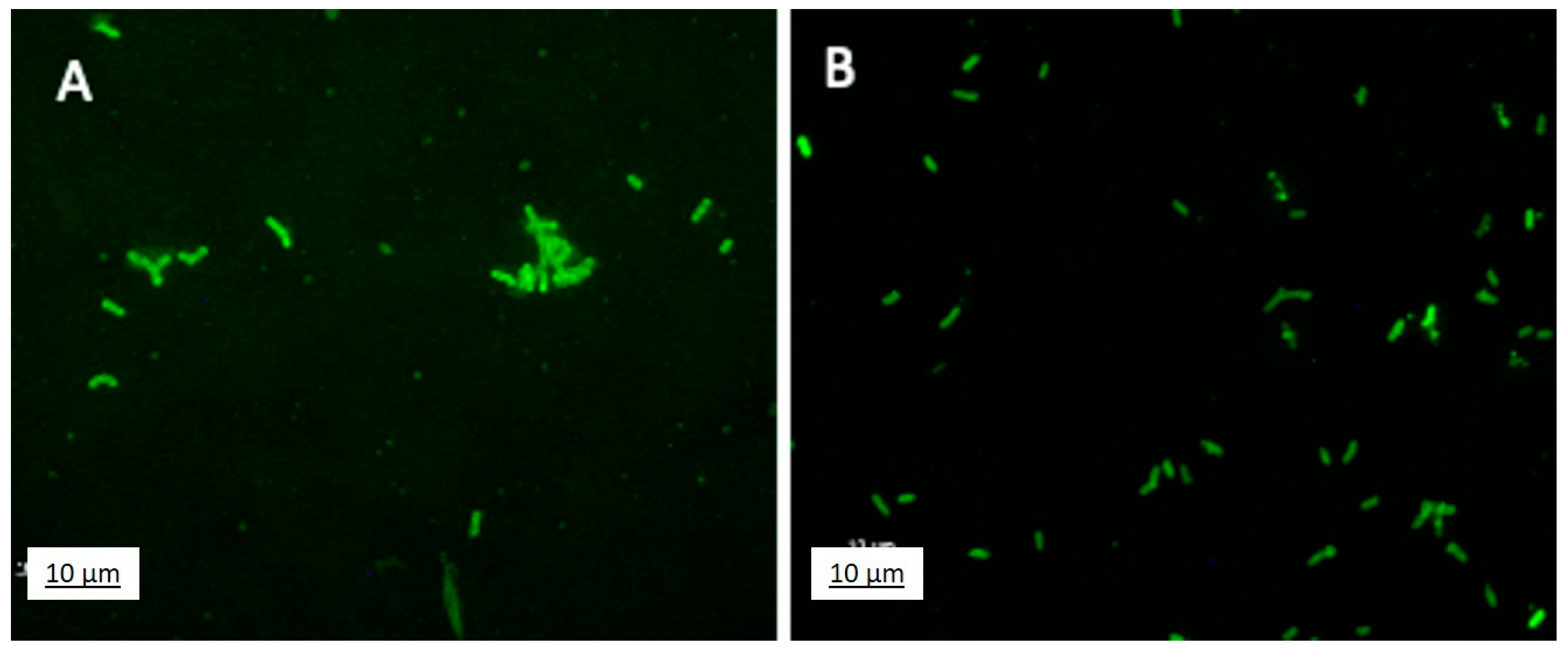
The spermosphere microbes, growing on LB agar as a white mass around the germinating wheat seeds, as shown in the Supplementary Material’s Figure S3, were streaked on LB agar to obtain single colonies. One of the purified isolates, termed JunSE1L, had characteristics consistent with the isolate, being within the genus Bacillus (Wankhade et al., in preparation). The cells of JunSE1L, when grown planktonically in LB for 18 h and then exposed to fF68, were fluorescently labeled (Figure 6A), with the fluorescence being stable to washing with water (Figure 6B). No fluorescence was observed for untreated JunSE1L cells or for cells treated with unlabeled F68, indicating an absence of intrinsic fluorescence under these imaging conditions. Some of these cells also had “Y” and “V” structures similar to those observed when cells associated with the surface of young seedling roots were examined (Figure 5). The planktonic growth rate of JunSE1L in minimal medium was not significantly affected by amendment with 5 g/L F68, as shown by the changes in OD600nm from 24, 32, 48, and 55 h in shake culture (Supplementary Figure S5). At a higher dose, 10 g/L, a significant decrease in OD600nm was observed early in growth, but by the stationary phase, there was no significant decrease (p = 0.05). No spores were observed at any of these growth times either with or without treatment with F68. Thus, the cause for the lower optical density in the exponential phase of growth with 10 g/L F68 does not relate to enhanced sporulation.

4. Discussion

From the water–drought–rehydrate cycle imposed on wheat over 28 d, there was no observable effect of any F68, GB, or combined treatments based on ET, shoot mass, or photosynthesis measurements. This absence of effects for the droughted plants may indicate that the treatment concentrations, wheat cultivar (bred for drought tolerance), age of wheat (seedlings), presence of microbiomes, and/or selected levels of drought/rehydration were not poised to reveal anticipated benefits from F68 (as a membrane repair agent) or GB (as a known plant protective osmolyte). F68 treatment did lead to increased ET and shoot mass for sufficiently watered plants. The enhanced shoot mass following F68 rhizosphere delivery may involve a greater efficacy in N utilization [27], as suggested by the increased production of glutamate synthase for rice callus treated with F68 [19,20]. Thus, this work with seedlings indicates that F68 benefits plant survival during establishment when field-grown wheat is sensitive to drought. The mechanisms for F68 growth improvement require further investigation.
There was no observable impact of F68 on the colonization of the wheat roots by the epiphyte PcO6. Our studies also demonstrated that the release of seed endophytes into the rhizosphere was not altered, nor was the planktonic growth a purified endophyte, a Bacillus spp., inhibited by F68. Consequently, the beneficial impacts of such microbiomes would likely be maintained in plant and soil treatments with F68. Mitigation of drought stress is reported from plant colonization with endophytic and epiphytic microbes, such as Bacillus spp. isolates [16,28,29] and PcO6 [30].
The higher foliar mass associated with F68 treatment occurred without improvements in photosynthetic ΦPsII quantum yield or the photosynthetic efficiency (Fv/Fm ratio), both of which dropped significantly during drought. We speculated that the membrane-protective effects of F68 [3,5,31,32,33] would reduce the drought-induced membrane damage and accompanying losses in photosynthetic yield and efficiency. Although there was no demonstration of buffered photosynthesis functions, the declines in photosynthetic activity caused by drought were restored upon rehydration, again suggesting that drought tolerance was already at its maximum because of the induced protection from PcO6 root colonization [30] and/or the protection offered by the seed endophytes. Bacillus spp. isolates associated with wheat are correlated with improved drought tolerance [16,28,29,34].
Fluorescein-conjugated F68 showed labeling of the perimeters of cells of the Gram-positive wheat seed endophyte, as previously observed with the Gram-negative cells of PcO6 [17]. Thus, although the cell walls of Gram-positive cells differ from those of Gram-negative cells, both were similarly labeled with the membrane-inserting polymer, F68. Currently, we do not know if F68 is internalized by the bacterial cells. Intracellular penetration was observed in the root epidermal cells, as evidenced by endosome labeling. It is possible that the nanosize of F68 unimers, about 5 nm, enabled its ready movement into and through the plant apoplast, followed by intracellular trafficking through the plant cells’ aquaporins. Intracellular movement between plant cells would readily occur through the plasmodesmatal channels with the nanosized unimers. The term “dancing endosomes” was used by other researchers for these mobile endosomes, here labeled fF68; these types of vesicles are proposed to carry cargo to and from the cell’s plasma membranes [35,36]. Our novel finding was that when roots were exposed to fF68, it was also shown that the rapid mobility of endosomes was not inhibited by exposure to the Pluronic. The finding of fluorescence in plant and both Gram-positive and Gram-negative bacterial cells upon exposure to fF68 suggests a broad affinity for a range of cell membranes that can be utilized to track fate and transport.
Further studies are needed to understand the mechanisms by which F68 could be beneficial in agricultural formulations. Examining wheat through grain filling is necessary to determine whether F68 becomes translocated into the seed (even carried by endophyte). The existence of F68 as nanosized structures may improve soil transport and be integral for its cellular associations, as revealed for plant and bacterial cells by use of the fluorescently labeled product in these studies. The unimer form of F68, with an average hydrodynamic diameter near 5.4 nm, is anticipated to shift to micelles and larger aggregates with increasing concentrations [37]. These agglomerates may not travel as well in the apoplast or symplastically in the plant. However, the average size of the F68 particles and polydispersity are highly dependent on temperature and solvent properties because micellization is driven by hydrophobic interactions. Other Pluronics with a greater hydrophobic content (i.e., lower hydrophilic to lipophilic balance, HLB) form micelles at much lower concentrations. If encapsulating, solubilizing, and delivering hydrophobic agrochemicals are the goals, then Pluronics with lower HLB and CMC values than F68 are recommended. Although the high HLB of F68 makes it a weak micelle-forming Pluronic, this property contributes to its biocompatibility observed in these studies, and its unique membrane-active properties [2].

5. Conclusions

The absence of any detrimental impact on wheat or its microbiome support the utilization of Pluronic® F68 as an agricultural adjuvant. In an adjuvant role, F68 may improve the solubilization and delivery of active ingredients, including improved root ingress, as demonstrated using fluorescein-F68. Whether F68 would transport encapsulated cargo into an intact root is unknown; however, that root-applied F68 increased shoot mass suggests that F68 improved nutrient acquisition in both sufficiently watered and drought-stressed wheat. It is not believed that the plant directly utilizes F68 as a nutrient; indeed, neither PcO6 nor JunSE1 are able to utilize F68 as a carbon source. Thus, the designation of F68 as an inert GRAS compound appears to extend to agricultural settings. With regard to its known role in cell membrane repair and anticipated benefits to plants under drought stress, where membrane leakage may occur, it was not possible to identify any benefit of F68 under the experimental conditions. This may reflect a need to increase the drought stress and/or F68-dosing levels. This is supported by the observation that root-applied glycine betaine, a protective osmolyte, did not benefit either watered or drought-stressed wheat under these experimental conditions. The transport of F68 and GB from roots to the shoots was not assessed, and considering the practicality of field-scale applications, wheat response following foliar delivery should be investigated. In sum, these observations of both the plant and their bacterial colonists support consideration of Pluronic® F68 as an adjuvant for agricultural formulations.

Supplementary Materials

The following supporting information can be downloaded at: https://www.mdpi.com/article/10.3390/agrochemicals4030012/s1. Table S1: LI-6800 chamber environmental parameters for photosynthesis measurements. Figure S1: DLS of F68 solutions. Figure S2: F68 surface tension effects. Figure S3: Fv/Fm photosynthetic efficiency. Figure S4: Germination of surface-sterilized wheat seeds on LB 2% agar showing release of endophytic bacteria into the spermosphere. Figure S5: Effect of F68 on planktonic growth of Bacillus sp. JunSE1L in minimal medium.

Author Contributions

Conceptualization, D.W.B. and A.J.A. methodology, A.C., M.Z., A.W., A.J.A. and D.W.B.; formal analysis, A.C., M.Z., A.W., A.J.A., A.J., J.E.M. and D.W.B.; investigation, A.C., M.Z. and A.W.; writing—original draft preparation, A.J.A. and D.W.B.; writing—review and editing, A.C., M.Z., A.W., A.J.A., A.J., J.E.M. and D.W.B.; visualization, A.C., M.Z., A.W., A.J.A., A.J., J.E.M. and D.W.B.; supervision, A.J.A. and D.W.B.; project administration, D.W.B.; funding acquisition, A.J.A., A.J., J.E.M. and D.W.B. All authors have read and agreed to the published version of the manuscript.

Funding

This work was supported with funding from Utah Agriculture Experiment Station, Projects 1581 and 1746, and by the Nanotechnology for Food and Agriculture Systems, project award no. 2024-67022-42830, from the U.S. Department of Agriculture’s National Institute of Food and Agriculture. Support to summer undergraduate researchers was provided from the National Science Foundation Research Experience for Undergraduate Program, Award 1950299.

Institutional Review Board Statement

Not relevant.

Informed Consent Statement

Not relevant.

Data Availability Statement

All data are available upon request to the authors.

Acknowledgments

Laboratory assistance from NSF REU Participants Amanda Streeter and Adrian Vigil is gratefully acknowledged.

Conflicts of Interest

The authors declare no conflicts of interest.

References

  1. Hazen, J.L. Adjuvants—Terminology, classification, and chemistry. Weed Technol. 2000, 14, 773–784. [Google Scholar] [CrossRef]
  2. Kwiatkowski, T.A.; Rose, A.L.; Jung, R.; Capati, A.; Hallak, D.; Yan, R.; Weisleder, N. Multiple poloxamers increase plasma membrane repair capacity in muscle and nonmuscle cells. Am. J. Physiol.-Cell Physiol. 2020, 318, C253–C262. [Google Scholar] [CrossRef] [PubMed]
  3. Lee, R.C.; River, L.P.; Pan, F.S.; Ji, L.; Wollmann, R.L. Surfactant-induced sealing of electropermeabilized skeletal muscle membranes in vivo. Proc. Natl. Acad. Sci. USA 1992, 89, 4524–4528. [Google Scholar] [CrossRef] [PubMed]
  4. Adhikari, U.; Goliaei, A.; Tsereteli, L.; Berkowitz, M.L. Properties of poloxamer molecules and poloxamer micelles dissolved in water and next to lipid bilayers: Results from computer simulations. J. Phys. Chem. B 2016, 120, 5823–5830. [Google Scholar] [CrossRef] [PubMed]
  5. Moloughney, J.; Weisleder, N. Poloxamer 188 (p188) as a membrane resealing reagent in biomedical applications. Recent Pat. Biotechnol. 2012, 6, 200–211. [Google Scholar] [CrossRef] [PubMed]
  6. Casella, J.F.; Kronsberg, S.S.; Gorney, R.T. Poloxamer 188 vs. placebo for painful vaso-occlusive episodes in children and adults with sickle cell disease—Reply. JAMA 2021, 326, 975–976. [Google Scholar] [CrossRef] [PubMed]
  7. Orringer, E.P.; Casella, J.F.; Ataga, K.I.; Koshy, M.; Adams-Graves, P.; Luchtman-Jones, L.; Wun, T.; Watanabe, M.; Shafer, F.; Kutlar, A.; et al. Purified poloxamer 188 for treatment of acute vaso-occlusive crisis of sickle cell disease: A randomized controlled trial. JAMA 2001, 286, 2099–2106. [Google Scholar] [CrossRef] [PubMed]
  8. Kirova, E.; Pecheva, D.; Simova-Stoilova, L. Drought response in winter wheat: Protection from oxidative stress and mutagenesis effect. Acta Physiol. Plant. 2021, 43, 8. [Google Scholar] [CrossRef]
  9. Tyagi, M.; Pandey, G.C. Physiology of heat and drought tolerance in wheat: An overview. J. Cereal Res. 2022, 14, 13–25. [Google Scholar] [CrossRef]
  10. Impa, S.M.; Nadaradjan, S.; Jagadish, S.V.K. Drought stress induced reactive oxygen species and anti-oxidants in plants. In Abiotic Stress Responses in Plants: Metabolism, Productivity and Sustainability; Springer: New York, NY, USA, 2012; pp. 131–147. [Google Scholar] [CrossRef]
  11. Kumar, M.; Lee, S.C.; Kim, J.Y.; Kim, S.J.; Aye, S.S.; Kim, S.R. Over-expression of dehydrin gene, OsDhn1, improves drought and salt stress tolerance through scavenging of reactive oxygen species in rice (Oryza sativa L.). J. Plant Biol. 2014, 57, 383–393. [Google Scholar] [CrossRef]
  12. Hellung-Larsen, P.; Assaad, F.; Pankratova, S.; Saietz, B.L.; Skovgaard, L.T. Effects of Pluronic F-68 on Tetrahymena cells: Protection against chemical and physical stress and prolongation of survival under toxic conditions. J. Biotechnol. 2000, 76, 185–195. [Google Scholar] [CrossRef] [PubMed]
  13. Trivedi, P.; Leach, J.E.; Tringe, S.G.; Sa, T.; Singh, B.K. Plant–microbiome interactions: From community assembly to plant health. Nat. Rev. Microbiol. 2020, 18, 607–621. [Google Scholar] [CrossRef] [PubMed]
  14. Camaille, M.; Fabre, N.; Clément, C.; Ait Barka, E. Advances in wheat physiology in response to drought and the role of plant growth promoting rhizobacteria to trigger drought tolerance. Microorganisms 2021, 9, 687. [Google Scholar] [CrossRef] [PubMed]
  15. Etesami, H.; Jeong, B.R.; Glick, B.R. Biocontrol of plant diseases by Bacillus spp. Physiol. Mol. Plant Pathol. 2023, 126, 102048. [Google Scholar] [CrossRef]
  16. Lastochkina, O.V. Adaptation and tolerance of wheat plants to drought mediated by natural growth regulators Bacillus spp.: Mechanisms and practical importance. Agric. Biol. 2021, 56, 843–867. [Google Scholar] [CrossRef]
  17. Streeter, A.R.; Cartwright, A.; Zargaran, M.; Wankhade, A.; Anderson, A.J.; Britt, D.W. Adjuvant Pluronic F68 is compatible with a plant root-colonizing probiotic, Pseudomonas chlororaphis O6. Agrochemicals 2023, 3, 1–11. [Google Scholar] [CrossRef]
  18. Cartwright, A. Surface-Functionalized Silica Nanocarriers for Mitigating Water Stress in Wheat and Benefiting the Root Microbiome. Master’s Thesis, Utah State University, Logan, UT, USA, 2023. Available online: https://digitalcommons.usu.edu/etd/8803 (accessed on 18 July 2025).
  19. Kok, A.D.X.; Wan Abdullah, W.M.A.N.; Tan, N.P.; Ong-Abdullah, J.; Sekeli, R.; Wee, C.Y.; Lai, K.S. Growth promoting effects of Pluronic F-68 on callus proliferation of recalcitrant rice cultivar. 3 Biotech 2020, 10, 116. [Google Scholar] [CrossRef] [PubMed]
  20. Kok, A.D.X.; Mohd Yusoff, N.F.; Sekeli, R.; Wee, C.Y.; Lamasudin, D.U.; Ong-Abdullah, J.; Lai, K.S. Pluronic F-68 improves callus proliferation of recalcitrant rice cultivar via enhanced carbon and nitrogen metabolism and nutrients uptake. Front. Plant Sci. 2021, 12, 667434. [Google Scholar] [CrossRef] [PubMed]
  21. Murungu, F.S. Effects of seed priming and water potential on seed germination and emergence of wheat (Triticum aestivum L.) varieties in laboratory assays and in the field. Afr. J. Biotechnol. 2011, 10, 4365–4371. [Google Scholar] [CrossRef]
  22. Wang, G.P.; Hui, Z.; Li, F.; Zhao, M.R.; Zhang, J.; Wang, W. Improvement of heat and drought photosynthetic tolerance in wheat by overaccumulation of glycine betaine. Plant Biotechnol. Rep. 2010, 4, 213–222. [Google Scholar] [CrossRef]
  23. Kjar, A.; Wadsworth, I.; Vargis, E.; Britt, D.W. Poloxamer 188–quercetin formulations amplify in vitro ganciclovir antiviral activity against cytomegalovirus. Antivir. Res. 2022, 204, 105362. [Google Scholar] [CrossRef] [PubMed]
  24. Alexandridis, P.; Holzwarth, J.F.; Hatton, T.A. Micellization of poly (ethylene oxide)-poly (propylene oxide)-poly (ethylene oxide) triblock copolymers in aqueous solutions: Thermodynamics of copolymer association. Macromolecules 1994, 27, 2414–2425. [Google Scholar] [CrossRef]
  25. Nakashima, K.; Anzai, T.; Fujimoto, Y. Fluorescence studies on the properties of a Pluronic F68 micelle. Langmuir 1994, 10, 658–661. [Google Scholar] [CrossRef]
  26. Mamou, G.; Mohan, G.B.M.; Rouvinski, A.; Rosenberg, A.; Ben-Yehuda, S. Early developmental program shapes colony morphology in bacteria. Cell Rep. 2016, 14, 1850–1857. [Google Scholar] [CrossRef] [PubMed]
  27. Chichkova, S.; Arellano, J.; Vance, C.P.; Hernández, G. Transgenic tobacco plants that overexpress alfalfa NADH-glutamate synthase have higher carbon and nitrogen content. J. Exp. Bot. 2001, 52, 2079–2087. [Google Scholar] [CrossRef] [PubMed]
  28. Lastochkina, O.; Garshina, D.; Ivanov, S.; Yuldashev, R.; Khafizova, R.; Allagulova, C.; Fedorova, K.; Avalbaev, A.; Maslennikova, D.; Bosacchi, M. Seed priming with endophytic Bacillus subtilis modulates physiological responses of two different Triticum aestivum L. cultivars under drought stress. Plants 2020, 9, 1810. [Google Scholar] [CrossRef] [PubMed]
  29. Lastochkina, O.; Yuldashev, R.; Avalbaev, A.; Allagulova, C.; Veselova, S. The contribution of hormonal changes to the protective effect of endophytic bacterium Bacillus subtilis on two wheat genotypes with contrasting drought sensitivities under osmotic stress. Microorganisms 2023, 11, 2955. [Google Scholar] [CrossRef] [PubMed]
  30. Cho, S.M.; Kang, B.R.; Han, S.H.; Anderson, A.J.; Park, J.Y.; Lee, Y.H.; Cho, B.H.; Yang, K.Y.; Ryu, C.M.; Kim, Y.C. 2R, 3R-butanediol, a bacterial volatile produced by Pseudomonas chlororaphis O6, is involved in induction of systemic tolerance to drought in Arabidopsis thaliana. Mol. Plant-Microbe Interact. 2008, 21, 1067–1075. [Google Scholar] [CrossRef] [PubMed]
  31. Sharma, V.; Stebe, K.; Murphy, J.C.; Tung, L. Poloxamer 188 decreases susceptibility of artificial lipid membranes to electroporation. Biophys. J. 1996, 71, 3229–3241. [Google Scholar] [CrossRef] [PubMed]
  32. Chen, H.; McFaul, C.; Titushkin, I.; Cho, M.; Lee, R. Surfactant copolymer annealing of chemically permeabilized cell membranes. Regen. Eng. Transl. Med. 2018, 4, 1–10. [Google Scholar] [CrossRef] [PubMed]
  33. Houang, E.M.; Bates, F.S.; Sham, Y.Y.; Metzger, J.M. All-atom molecular dynamics-based analysis of membrane-stabilizing copolymer interactions with lipid bilayers probed under constant surface tensions. J. Phys. Chem. B 2017, 121, 10657–10664. [Google Scholar] [CrossRef] [PubMed]
  34. Anderson, A.J.; Hortin, J.M.; Jacobson, A.R.; Britt, D.W.; McLean, J.E. Changes in metal-chelating metabolites induced by drought and a root microbiome in wheat. Plants 2023, 12, 1209. [Google Scholar] [CrossRef] [PubMed]
  35. Voigt, B.; Timmers, A.C.; Šamaj, J.; Hlavacka, A.; Ueda, T.; Preuss, M.; Nielsen, E.; Mathur, J.; Emans, N.; Stenmark, H.; et al. Actin-based motility of endosomes is linked to the polar tip growth of root hairs. Eur. J. Cell Biol. 2005, 84, 609–621. [Google Scholar] [CrossRef] [PubMed]
  36. Von Wangenheim, D.; Rosero, A.; Komis, G.; Šamajová, O.; Ovečka, M.; Voigt, B.; Šamaj, J. Endosomal interactions during root hair growth. Front. Plant Sci. 2016, 6, 1262. [Google Scholar] [CrossRef] [PubMed]
  37. Zhou, Z.; Chu, B. Light-scattering study on the association behavior of triblock polymers of ethylene oxide and propylene oxide in aqueous solution. J. Colloid Interface Sci. 1988, 126, 171–180. [Google Scholar] [CrossRef]
Figure 1. Water loss from plants continually watered (W) or exposed to drought (D). All plant roots were colonized with PcO6, and fertilized with half-strength Hoagland solution on days 0 and 14. After day 14, half the plants from each treatment were droughted and half continuously watered. After day 21, the drought-stressed plants were rehydrated for 6 days. F68, GB, and F68 + GB treatments were added at day 0 and day 14. Evaporation from unplanted boxes is shown by the black dashed line. The data are means and standard deviations of three separate growth boxes, each with nine plants per treatment. The statistics used to compare treatments on individual days were ANOVA with Tukey’s HSD for p < 0.05 (see text for discussion of statistics).
Agrochemicals 04 00012 g001
Figure 2. Evaluation of photosynthetic efficiency at days 4 (A) and 6 (B) of drought (D) contrasted with sufficiently watered (W) plants receiving the same treatments. All plant roots were colonized with PcO6. The data are means and standard errors from three separate growth boxes, each with nine plants per treatment. A restricted maximum likelihood (REML) method was used for statistical analysis using SAS. Values at day 0 of drought stress were used as baselines to address potential inherent variations without stress. The different letters above the bars show values with significant difference when analyzed using ANOVA with Tukey’s HSD adjustment, at p < 0.05.
Agrochemicals 04 00012 g002
Figure 3. F68 improves the dry mass of leaf tissue for 28 d seedlings. The wheat plants were grown with continuous watering or with drought for 8 d, followed by 6 d of rehydration. The plants were grown from PcO6-inoculated seeds and all plant roots independent of treatment were colonized by this bacterium at harvest. The different letters above the bars show values with significant difference when analyzed using ANOVA with Tukey’s HSD adjustment, at p < 0.05.
Agrochemicals 04 00012 g003
Figure 4. Exposure of intact 7 d wheat seedling roots to fF68 for 3 h, followed by rinsing reveals F68 internalization and concentration in endosomes within the epidermal cells. The red arrows indicate two pairs of fF68 fluorescently labeled endosomes whose intracellular movement is seen in the time course of images. (A) (t = 0), to (B) (t = 5 s), and to (C) at t = 15 s.
Agrochemicals 04 00012 g004
Figure 5. The bacteria in washes of intact root surfaces grown from surface-sterilized wheat seeds are fluorescent after exposure to fF68. Wheat seedlings for this assay were grown from surface-sterilized seeds for 7 d on LB agar before the roots were immersed for 3 h in fF68. The root tips were treated with sterile water to obtain the suspension used for imaging. At magnification (100× oil lens), the brightfield (A) and fluorescence (B) images reveal bacterial cells with different multicellular forms.
Agrochemicals 04 00012 g005
Figure 6. Cells of the wheat seed-borne endophyte, Bacillus JunSE1L, become fluorescent after exposure to fF68 for 3 h. Fluorescence before (A) and after (B) washing the cells with sterile distilled water.
Agrochemicals 04 00012 g006
Table 1. Effect of F68 treatments on wheat seeds for water uptake, germination, and emergence.
Treatments
12 h F68 (g/L)
H2O Imbibed
Per Seed (g)
% Germination
@ 48 h on LB Agar
% Emergence @ 5 d
Planted in Sand
00.018 ± 0.00280 ± 1392 ± 6
0.10.018 ± 0.00285 ± 992 ± 6
10.019 ± 0.00280 ± 1892 ± 6
100.019 ± 0.00280 ± 996 ± 6
1000.017 ± 0.00147 ± 896 ± 6
2500.015 ± 0.00157 ± 396 ± 6
All data are means ± standard deviation for four experimental replicates, each with nine seeds.
Disclaimer/Publisher’s Note: The statements, opinions and data contained in all publications are solely those of the individual author(s) and contributor(s) and not of MDPI and/or the editor(s). MDPI and/or the editor(s) disclaim responsibility for any injury to people or property resulting from any ideas, methods, instructions or products referred to in the content.