Review

Molecular Targets for Cannabinoids in Natural Killer Cells: Do They Modulate the Antitumor Activity?

by
Miguel Olivas-Aguirre
1,2,*,
Cecilia Gutiérrez-Iñiguez
3,
Igor Pottosin
3 and
Oxana Dobrovinskaya
3
1
Laboratory of Cancer Pathophysiology, University Center for Biomedical Research, University of Colima, Colima 28040, Mexico
2
Consejo Nacional de Humanidades, Ciencia y Tecnología (CONAHCYT), Programa de Investigadores e Investigadoras por México, Mexico City 03940, Mexico
3
Laboratory of Immunobiology and Ionic Transport Regulation, University Center for Biomedical Research, University of Colima, Colima 28040, Mexico
*
Author to whom correspondence should be addressed.
Receptors 2024, 3(2), 122-144; https://doi.org/10.3390/receptors3020007
Submission received: 15 January 2024 / Revised: 27 February 2024 / Accepted: 21 March 2024 / Published: 25 March 2024

Abstract

:
Recent research has emphasized the potential of natural and synthetic cannabinoids as anticancer agents. Yet it remains unclear whether and in which sense cannabinoids affect the anticancer activity of NK cells, an important branch of anticancer immunity. Similar uncertainty exists regarding NK cells-based immunotherapy. Here we presented an overview of multiple cannabinoid targets as canonical (mainly CB2) and non-canonical receptors, ion channels, transporters, and enzymes, expressed in NK cells, along with underlying molecular mechanisms. Through them, cannabinoids can affect viability, proliferation, migration, cytokine production, and the overall anticancer activity of NK cells. Respective holistic studies are limited, and, mostly, are phenomenological, not linking observed effects with certain molecular targets. Another problem of existing studies is the lack of standardisation, so that diverse cannabinoids at variable concentrations and ways of administration are applied, and often, instead of purified NK cells, the whole lymphocyte population is used. Therefore, there is an urgent need for more focused, systemic, and in-depth studies of the impact of the cannabinoid toolkit on NK cell function, to critically address the compatibility and potential synergies between NK activity and cannabinoid utilization in the realm of anticancer interventions.

1. Introduction

Natural Killer (NK) cells are a fundamental component of the innate immune system [1], serving as the frontline defense against infections and malignant transformations. Unlike other immune cells that require prior exposure to a specific pathogen, NK cells possess innate cytotoxicity, enabling them to rapidly identify and eliminate infected or abnormal cells without prior sensitization. This effector function of NK cells relies on the regulation of several activating and inhibitory receptors as well as co-stimulatory receptors [2]. NK cell activation involves a delicate balance between stimulatory and inhibitory signaling, triggering key steps such as the formation and stabilization of the immunological synapse (IS), movement of cytolytic granules (CGs) to the microtubule organizing center (MTOC), polarization, and the subsequent release of CGs’ cytotoxic content (degranulation) into the target cell. Notably, degranulation, a critical step in the process, is heavily dependent on intracellular calcium (Ca2+i). Human NK granules contain perforin, a pore-forming protein facilitating the delivery of granzymes (A, B, H, K, M) to target cells, which through their serine protease function activate caspases to promote target cell death [3,4].
Activating and inhibitory receptors in NK cells, as well as granule content, are a subject of dynamic regulation by multiple endogenous and exogenous stimuli. Additionally, the activity of NK cells can be potentiated through the engagement of death receptors, which include Fas ligand (FasL), TNF, and TRAIL receptors. These receptors bind to their respective counterparts on the surface of target cells, triggering a conformational change in the death receptors. This alteration leads to the recruitment of intracellular adaptor proteins, which in turn induce downstream signaling, ultimately resulting in the apoptotic induction of the target cells.
Nowadays, significant attention is directed towards a group of functionally related molecules collectively termed cannabinoids. This category encompasses endogenously produced cannabinoids (endocannabinoids), those found naturally in plants (e.g., Cannabis sativa, phytocannabinoids), and synthetic cannabinoids. In recent decades, there has been extensive interest in the anticancer properties of certain cannabinoids, particularly those lacking psychoactive effects. Cannabinoids demonstrate significant clinical potential in addressing cancer-related symptoms and managing pain, anorexia, neurological diseases, and sleep-related issues [5]. Furthermore, accumulating evidence from in vivo and in vitro studies supports the anticancer properties of cannabinoids against multiple cancer types [6,7,8,9].
Incorporation of cannabinoids into chemotherapeutic protocols, with a specific focus on non-psychoactive phytocannabinoids like CBD, has been recently considered by our research group [9]. Despite a lack of regulatory approval, many cancer patients turn to CBD products as dietary supplements, to alleviate complications such as pain, fatigue, and neurological disorders. Additionally, cannabinoids have been reported to possess a notable immunomodulatory capacity [10]. This raises questions about how cannabinoids influence the anticancer activity of NK cells and whether NK cell-based immunotherapy is compatible with cannabinoid treatments.
The effects of cannabinoids are not predictable across all cell types, primarily due to complex and diverse mechanisms of action that remain only partially understood. Additionally, there is a growing body of evidence highlighting the significant diversity and variability in the expression of multiple targets for cannabinoids within various cell types. This variability ultimately shapes the response of cells to cannabinoids. Notably, some cannabinoids exhibit hormetic responses, demonstrating opposing effects on cells, depending on the concentration of the cannabinoid.
Given these complexities, modulatory effects of cannabinoids in each specific cell model have to be rigorously evaluated, in order to responsibly assess the clinical potential of these compounds. This consideration is particularly pertinent to NK cells and NK-based anticancer therapies, where NK-specific evaluations are essential for a comprehensive understanding of cannabinoid effects in the context of cancer treatment.
In this review, we have collected and critically analyzed the available data regarding the expression of elements of the cannabinoid toolkit in NK cells and the effects of cannabinoids on NK cells, focusing on the mechanisms underlying the immunomodulatory potential of these compounds. The article starts with a summary of available cannabinoid types and the presence, location, and functionality of multiple cannabinoid receptors and additional targets in NK cells. Then, we summarize the reported effects of cannabinoids on NK cell function and discuss the current state of research in this field, to identify the underexplored issues and to determine the directions of future research.

2. Cannabinoids

Cannabinoids, a group of terpenophenolic compounds, are categorized into three groups: phytocannabinoids, coming from plant sources like Cannabis (e.g., Δ9-tetrahydrocannabinol, THC; cannabidiol, CBD); endocannabinoids, naturally produced in humans and animals (e.g., anandamide, AEA; 2-arachidonoylglycerol, 2-AG); and synthetic cannabinoids, chemically designed ligands for cannabinoid receptors (Figure 1).
Synthetic cannabinoids comprise the largest subfamily within the cannabinoid group, including over 280 members, often demonstrating higher potency compared to endocannabinoids or phytocannabinoids. These cannabinoids are typically named after the scientist or company responsible for their initial synthesis, such as AM for Alexandros Makriyannis, CP for Charles Pfizer, HU for Hebrew University, JWH for John W. Huffman, and WIN for Sterling Winthrop, among others, followed by a sequential experimental number. Common examples of frequently utilized synthetic cannabinoids include CP55-940, WIN55212-2, JWH133, AM251, AM630, etc. For detailed and updated information on each cannabinoid type, extensive reviews are available online [12,13,14].
While cannabinoids exhibit promising anticancer properties by promoting cancer cell death and influencing cancer cell behavior, their translation into effective drugs faces significant challenges due to inconsistent in vivo outcomes compared to in vitro observations. This discrepancy argues for the need of understanding the intrinsic molecular mechanisms underlying cannabinoid-mediated effects, especially concerning their diverse impact on different cancers. Efforts to elucidate these mechanisms are underway, emphasizing the necessity of exploring the signaling pathways involved in cannabinoid actions and the development of innovative formulations and combinations with conventional chemotherapeutics. This approach aims to optimize the potency, efficacy, and delivery of cannabinoids as potential anticancer agents [9,15,16].
Meanwhile, cannabinoids exert indirect anticancer effects by modulating the activity of the immune system, as extensively observed in immune cells such as B and T cells, and macrophages [17,18]. However, information specific to NK cells is limited, despite the fact that NK cells express canonical cannabinoid receptors and a diverse array of additional molecular targets for cannabinoids. To unlock the full potential of cannabinoids in NK cell function and therapeutic applications, a deeper understanding is required regarding which of these cannabinoid targets are effective and how the respective signaling pathways integrate, thereby influencing the overall immune response

3. Structural and Functional Diversity of the Cannabinoid Targets in NK Cells

3.1. Overview of the Cannabinoid Targets

Since the discovery of the endocannabinoid system in the mid-1980s, including the cannabinoid receptors CB1 and CB2, it has become evident that their distribution extends beyond the central nervous system. While CB1 is primarily localized in the cerebellum and cortex of the brain, with marginal presence in peripheral tissues, CB2 was initially identified within the immune system, exhibiting a notable expression in B lymphocytes and NK cells [19,20,21,22,23] (Supplementary Table S1).
Recent experimental data indicate that cannabinoids exert effects beyond classical targets, CB1 and CB2, prompting the exploration of new cannabinoid receptors (CBRs). NK cells exhibit numerous non-canonical CBRs, including enzymes (11), ion channels (9), G-protein coupled receptors (GPCRs; 7), and transporters (4; Figure 2). Expression and functional roles of these CBRs are tissue dependent. While their involvement in immune system function is extensively studied in various cell types, evidence for NK cells is limited. Subcellularly, most of the reported CBRs are localized in the plasma membrane (PM), followed by cytosol, mitochondria, and nucleus (Figure 2B). Notably, functional evidence is absent for many of the CBRs expressed in NK cells, leaving their roles elusive. Nonetheless, functionally explored CBRs exhibit diverse outcomes, influencing effector functions, intracellular signaling, and metabolism (Figure 2C; see also Section 4).

3.2. Canonical CB1 and CB2 Expression in NK Cells

Canonical CBRs, CB1 and CB2, have been extensively characterized in human and murine NK cells through multiple experimental approaches (Figure 2A, Supplementary Table S1). Notably, the preponderance of CB2 in NK cells is highlighted by a striking 100:1 ratio of CB2 to CB1 expression, as revealed by PCR analysis [21,23].
CB2 mRNA expression in NK cells is surpassed only by B cells among immune cell populations. Flow cytometry and fluorescent microscopy analysis confirmed that NK cells express high CB2 protein levels, albeit showing a considerable variability among donors [22]. A similarly high CB2/CB1 ratio was also observed in NK cells, infiltrating in human and murine lung cancer [25].
Single reports of CB2 expression are also available for uterine NK cells (uNK) [26] and KHYG-1, a cell line derived from a patient with aggressive NK leukemia [27], underscoring the widespread presence of CB2 not only in circulating primary NK and uNK cells but also in cancerous NK cell lines
Activation of NK cells is characterized by the overexpression of multiple genes. This transcriptional response extends to the cannabinoid receptors, particularly CB2. Notably, studies have revealed that a specific subset of NK cells, referred to as NKT cells, undergoes a significant increase in CB2 expression during activation induced by IL-2 [28,29]. However, it remains uncertain whether the increased CB2 expression, observed during IL-2-induced activation of NKT cells, is universally applicable to other activation mechanisms, such as chemical induction or recognition of target cells. Currently, there is a lack of evidence supporting similar dynamics in other subsets of NK cells and different activation scenarios, which should be addressed in future research.

3.3. Non-Canonical Cannabinoid Receptors in NK Cells

The orphan G protein-coupled receptors (GPCRs), GPR55 and GPR18, have been identified as key candidates, responsible for mediating the non-CB1/2 effects of cannabinoids. They are also expressed in NK cells (Supplementary Table S1). In one study, microarray analysis demonstrated a gradual increase in GPR18 expression during the differentiation of human CD34+ cells to NK cells from the umbilical cord under in vitro experimental conditions [30]. Another work reported a higher GPR55 protein expression in human monocytes and NK cells as compared to other immune cell populations, based on flow cytometry, western blot analysis, and confocal microscopy data [31].
Other CBRs belonging to the GPCRs family have been described in NK cells, including dopamine receptors (D2), serotonin receptors (5HT1A), and opioid receptors (δ, μ, ҝ). Their role in NK function has been linked to the regulation of antitumoral activity, by modulation of intracellular calcium (Ca2+i) levels, cytokine production, and NK granule content [32,33,34,35,36,37]. However, the actual evidence is very scarce, and this issue needs to be explored in more detail.

3.4. Ion Channels and Ca2+ Signaling

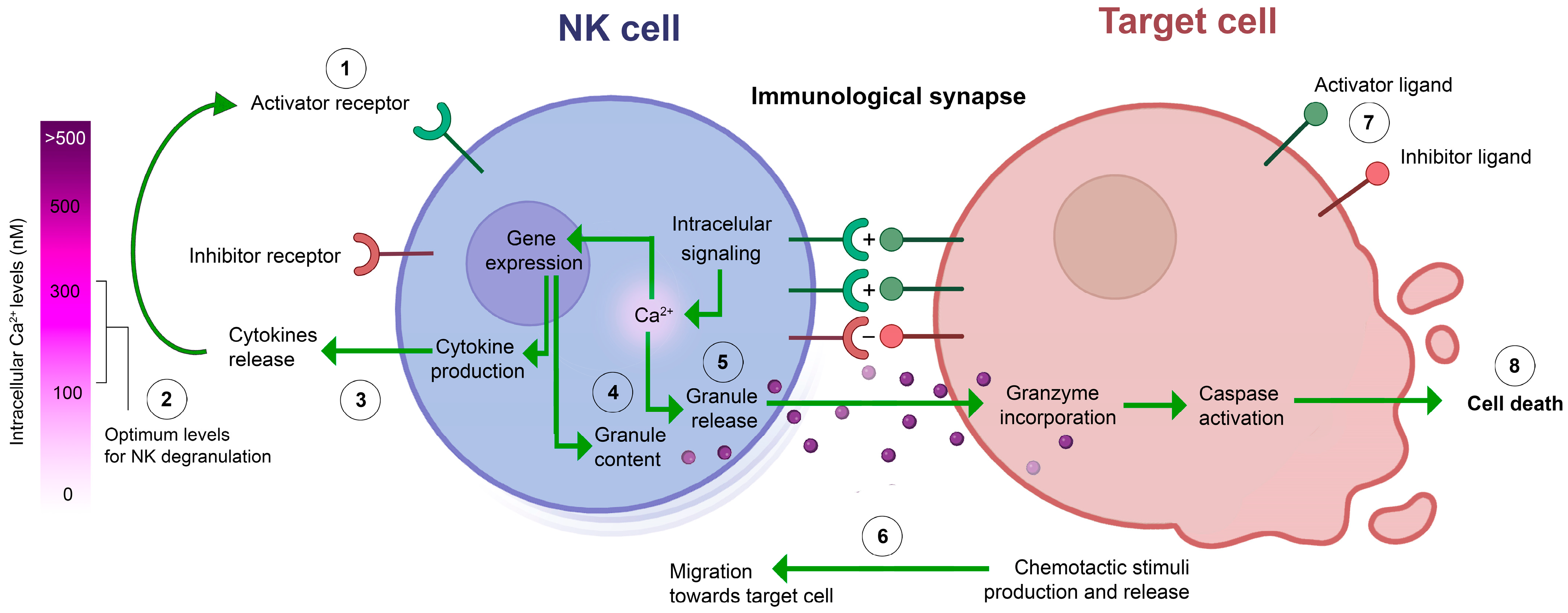
In non-excitable cells such as NK cells, generation and modulation of Ca2+ signals play a crucial role in gene transcription, proliferation, metabolism, cytokine secretion, cell death, and migration. Ca2+ influx in immune cells is primarily mediated by the store-operated Ca2+ entry (SOCE), mediated by Ca2+ release-activated channel (CRAC). This channel is composed of two Orai protein subunits, whose assembly is orchestrated by STIM (stromal interaction molecules) proteins, which undergo oligomerization in a response to endoplasmic reticulum (ER) Ca2+ depletion [38]. SOCE is required for NK cell degranulation and target killing [39]. Of note, degranulation of NK cells has a relatively low Ca2+ optimum level, which is slightly above the resting value (100 nM).
During the activation of NK cells, triggered by the recognition of target cells, multiple receptors are engaged, leading to an increase of Ca2+i. However, this signal is often exaggerated and not optimal for NK cell function. Thus, manipulations leading to a decrease of SOCE can improve the NK activity [40,41].
While there is no direct evidence indicating that cannabinoids directly impact CRAC, they target additional channels that modulate CRAC activity. Upon the formation of the immunological synapse (IS), both Orai and mitochondria relocate to the IS, where mitochondria absorb inflowing Ca2+, alleviating CRAC inactivation induced by local Ca2+i increases in the vicinity of the IS [42]. CBD targets a unique ion and metabolite exchange channel in the outer mitochondrial membrane, VDAC, inducing its highly Ca2+-permeable conformation. This results in mitochondrial Ca2+ overload and the formation of the stable permeation transition pore, collapsing all gradients across the inner mitochondrial membrane and rendering the mitochondria incapable of taking up Ca2+ [43,44,45]. Additionally, CBD induces Ca2+ release from the ER [44]. CBD-induced changes in Ca2+ handling by mitochondria and ER can eventually impact Ca2+-dependent degranulation by NK cells, which requires further investigation.
Inflowing across plasma membrane Ca2+ induces a depolarization, which reduces SOCE, acting as a feedback control [38]. This implies that the activity of other channels, functionally expressed in plasma membrane, can either repolarize or further depolarize the membrane, thus modulating SOCE. Besides, some of these channels can directly contribute to Ca2+ signal. In this regard, transient receptor potential (TRP) family members are vital. The activity of TRP channels, constitutive or stimulated, contributes to membrane potential depolarization, and exerts a significant influence on CRAC channel functionality [46].
The TRP superfamily members, TRPV1-4, TRPM8, and TRPA1, are direct targets for cannabinoids and are often named “ionotropic cannabinoid receptors” [47]. These channels are nonselective cation ones, conducting Ca2+ with a little preference over Na+. TRPV1 activation is influenced by endocannabinoids such as AEA (acting as a low-potency partial antagonist), phytocannabinoids like CBD (which desensitizes TRPV1 to its natural agonist capsaicin), and synthetic cannabinoids like arachidonoyl-2 chloroethanolamine (which also desensitizes TRPA1, forming a reciprocal relationship, where TRPA1-selective cannabinoids desensitize TRPV1) [48,49]. Notably, the synthetic cannabinoid analog WIN55212-2 exhibits dual effects on TRPV1: inhibition at low concentrations, peaking at 1 nM, and stimulation at concentrations above 1 μM [50]. While TRPV1 is functionally expressed in NK cells and contributes significantly to Ca2+ entry, its precise physiological role remains unclear [51].
Non-acidic phytocannabinoids are exclusive natural modulators of TRPV2. CBD and THC activate TRPV2 at low micromolar range, but at the same concentrations cause posterior TRPV2 desensitization [48]. Structure–function relationships, related to CBD binding in TRPV2, are well understood [52]. Recent studies revealed a distinct binding site for another phytocannabinoid (C16), which is under allosteric control by TRPV2 agonist probenecid [53]. TRPV2-mediated Ca2+ signaling plays multiple roles in different immune system cells, yet its role in NK cell remains unexplored, albeit its gene expression exceeds by more than one order of magnitude an average of that in innate and adaptive immunity cells and by two orders of magnitude of that in any other tissue [54]. Of note, CBD not only activates TRPV2 but also induces its relocation from internal compartments to PM [55]. In mast cells, TRPV2-mediated Ca2+ influx plays a key role in stimuli-induced degranulation [56]. Thus, it is tempting to test the role of TRPV2 in target cell-induced Ca2+ influx and degranulation in NK cells, along with the CBD modulation of these processes.
Phytocannabinoids and endocannabinoids exhibit a potent inhibition of TRPM8-mediated Ca2+ influx (IC50: 0.1 μM for CBD and THC) [47,48]. While the exact role of TRPM8 in immune cells remains elusive, unlike other members of the TRPM family (e.g., TRPM2 and TRPM3) that are functionally expressed in NK cells and contribute to cytotoxicity through Ca2+ signaling [57,58], the functional expression of TRPM8 has not been demonstrated yet. Indirect evidence supporting the potential significance of TRPM8 in NK cell function is derived from Marshall-Gradisnik and colleagues [59], who demonstrated that two single nucleotide polymorphisms in TRPM8 are associated with reduced efficiency in the killing of target cells by NK cells.
While TRPA1 was traditionally believed to be specifically expressed in sensory neurons, recent studies have unveiled its functional expression in immune cells, particularly in NK cells [60,61]. Intriguingly, TRPA1 expression is confined to the NK CD56dimCD16+ subset, recognized for its maximal cytotoxic potential against target cells. Notably, specific stimulation of TRPA1 with allyl isothiocyanate in this NK subset induces Ca2+ influx, enhancing granzyme production, degranulation, and target cell killing [61]. TRPA1 is activated and desensitized by phytocannabinoids like CBD and THC at submicromolar concentrations. In contrast, endocannabinoids and synthetic cannabinoids exhibit a lower potency in modulating TRPA1 [47,48].
There is abundant evidence that immune cells possess the complete cholinergic system, including enzymes for acetyl choline (Ach) synthesis and degradation, Ach transfer proteins, and various muscarinic and nicotinic receptors, which affects them in autocrine and paracrine ways [62]. Nicotinic acetylcholine receptors (nAChRs) are ionotropic, conducting small cations with low preference, mediating both Ca2+ and Na+ influx. In NK cells, the functional expression of α7 nAChR has been demonstrated. Stimulation of α7 nAChR induces intracellular Ca2+ release, resulting in the reduction of inflammatory cytokine production and decreased cytotoxicity against target cells [63,64,65]. The homopentameric channel formed by α7 nAChR subunits is inhibited by CBD (IC50 ~11 μM), whereas other cannabinoids, including THC, produced inefficient results [66]. The inhibitory effect of CBD on the nAChR channel is complex and likely arises from the stabilization of resting or desensitized conformational states of the nAChR complex [67]. Thus, CBD acts as an antagonist for nAChR, although, to our knowledge, there have been no attempts to reverse the reduction of NK cell cytotoxicity caused by nAChR agonists using CBD.
NK cells also possess GABA and glycine receptors, characterized by selectivity for small anions such as Cl. In lymphocytes, which are characterized by a relatively high internal Cl concentration, the activation of GABA and glycine receptor channels in the PM causes Cl efflux, which depolarizes the membrane potential, thus reducing SOCE [38].
GABA stimulation of intrinsic GABAA receptors, expressed in NK cells, results in reduced degranulation and cytotoxicity [68]. 2-AG and CBD act as allosteric agonists for GABAA receptors [69]. Notably, NK cells can produce, secrete, and respond to GABA [70].
The presence of glycine receptors (GlyRs), particularly the α subunits, has been demonstrated on the cell surface of NK cells [71]. While direct evidence of GlyR functionality in NK cells is currently lacking, downregulation of GLRA3 expression is associated with NK cell dysfunction in AML [72]. By drawing parallels with other immune cells, it is plausible to speculate that glycine, analogous to its effects in other contexts, might contribute to the modulation of Ca2+ influx and cytokine secretion in NK cells [71].
The presence of Nav channels in NK cells is based on membrane potential evaluation by fluorescent dyes and its modulation by Nav agonists and antagonists [73,74]. To our knowledge, Nav currents have never been measured in NK cells and the information available for other immune cells (T cells) is controversial [38]. Typical Cav3.1 channel activity has been directly demonstrated so far only in mouse T cells [75]. All attempts to detect depolarization-activated Ca2+ currents in human T cells have been unsuccessful. Although the mRNA of pore-forming α subunits of Cav3.2 and Cav3.3 has been found, these encode truncated proteins, which are likely unable to form functional voltage-dependent ion-conducting channels [76]. Given the uncertainty of the operation mode of Nav and Cav proteins in immune cells, we feel that the discussion of a possible impact of their modulation by cannabinoids would be premature.
Phytocannabinoids (particularly THC) are widely recognized as promoting phospholipase activity [77]. The consequent increase in the production of diacylglycerol (DAG) serves as an activator for several Ca2+-permeable transient receptor potential canonical (TRPC) channels. For NK cells, only TRPC3 expression has been demonstrated functionally where it mediates the Ca2+ response to haptens [78]. Therefore, it is plausible to hypothesize that specific cannabinoids, given their capacity to promote phospholipase activity, elevate DAG levels and activate Ca2+-permeable TRPC channels, which may influence NK cells activity via subsequent alterations in global calcium signaling. TRPC channels functionally interact with SOCE, by variating the membrane potential and by interacting with specific proteins in ER, which are essential for Ca2+ release, Ca2+ store, and CRAC assembly [79].
In summary, there are multiple channels or targets for cannabinoids, which are potentially able to shape Ca2+ signaling in NK cells (Figure 3). Although the effects of different cannabinoids on individual channels are relatively well understood, a prediction of their impact on NK cells’ Ca2+i global responses and function is very challenging. Apparently, the effects of the same cannabinoid, e.g., CBD, on different ion channels, can partly compensate each other. Also, the dual effect on TRPVs and TRPA1 on activation and desensitization, needs to be considered. One needs to establish first which of these channels contribute significantly to the response induced by a specific stimulus like target cell presentation, deciphering later on their specific roles in NK cell function.

3.5. Enzymes

Enzymes belonging to the cytochrome P450 superfamily (CYPs) are an emerging and abundant group of cannabinoid targets in NK cells. Although the level of CYP1B1 is low in healthy resting NK cells, in vitro NK expansion by IL-21 or IL-2 administration resulted in a robust (ten times) increase of CYP1B1 transcript. However, the implication of such change to the NK cell function has not been fully understood and the experimental evidence is limited to the observation that CYP1B1 antagonism did not alter NK cell viability [80].
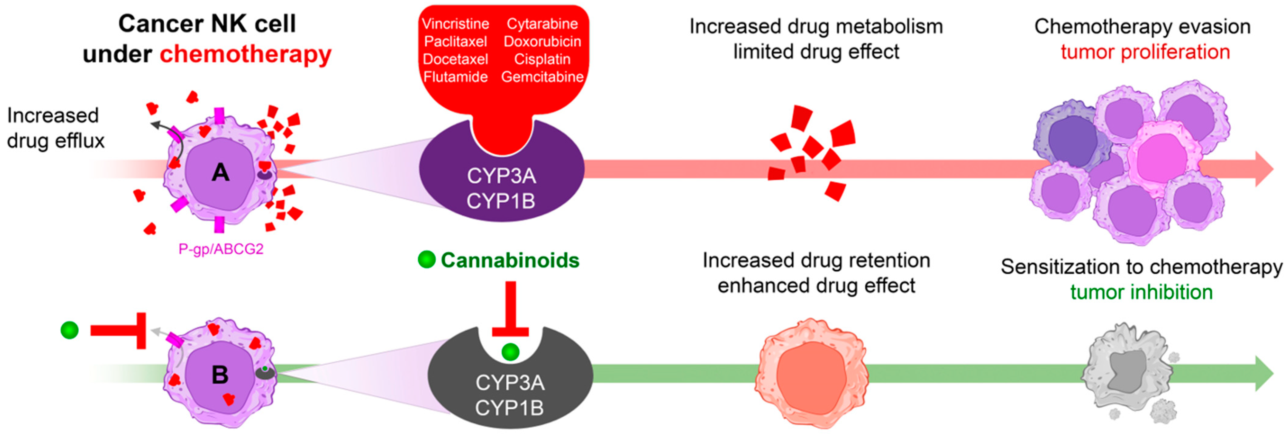
Interestingly, many other CYP3A isoforms are overexpressed in NK tumors with different occurrences: CYP3A4 (57%), CYP3A7 (29%), or CYP3A5 (14%) [81]. It is important to mention that CYP1B together with CYP3A are responsible for the inactivation of many anticancer drugs (e.g., flutamide, vincristine, paclitaxel, docetaxel). In this context, phytocannabinoids (e.g., Δ9-tetrahydrocannabidiol, THC; Cannabidiol, CBD; and Cannabinol CBN; 0–10 µM) act as CYP inhibitors [82]. Consequently, the administration of phytocannabinoids to CYP-overexpressing NK tumors may result in the improvement of chemotherapy (Figure 4). Of note, some CYP3A subfamily members, like CYP3A4, display only limited sensitivity to most of the phytocannabinoids [83].
Another important enzyme that acts as a target for cannabinoids is the phospholipase A2 (PLA2), which catalyzes the hydrolysis of membrane phospholipids to produce free fatty acids, including endocannabinoid production, e.g., arachidonic acid (AA) and lysophospholipids, which can be further metabolized to produce eicosanoids. The latter can alter the cytotoxic activity of NK cells against target cells [84,85]. The role of PLA2 in NK cells is evidenced as its inhibition reduced AA/lysophospholipids production and consequently cytotoxicity against K562. These alterations were reverted by the addition of lysophosphatidylcholine, suggesting that PLA2 activity is necessary for NK effector activity. Correspondingly, independent groups have demonstrated that multiple phytocannabinoids promote PLA2 activity [77,86]. Nonetheless, to date the effect of cannabinoid-mediated PLA2 activation and consequent increase of cytotoxicity against target cells have not been experimentally tested.
Cyclooxygenase 2 (COX-2) is another NK target that can be modulated by cannabinoids. It is responsible for the production of prostaglandins and eicosanoids from AA. Analysis of COX-2 in NK lymphoma demonstrated that up to 70% of the patients displayed COX-2 enrichment. However, its functional role in NK malignancies has not been elucidated yet [87]. Conversely, in non-oncological murine NK cells, COX-2 inhibition led to an enhanced cytotoxic activity against tumor target cells [88]. Interestingly, phytocannabinoids have been described as potent and selective COX-2 inhibitors [89,90]. However, whether cannabinoid administration modulates the cytotoxicity of healthy NK cells through COX-2 inhibition remains elusive.
Fatty acid amide hydrolase, FAAH, a crucial enzyme for the metabolism of endogenous cannabinoids, is also expressed in NK cells. FAAH-deficient mice do not exhibit any significant alteration in NK cytotoxic function. However, despite the unaltered cytotoxic activity of NK cells, these mice demonstrate exaggerated responses to endocannabinoids, including hypomotility, analgesia, and catalepsy [91]. Moreover, FAAH-deficient mice display a reduced cytokine production within NK cells. Additionally, it has been observed that FAAH silencing induces the redistribution of circulating NK cells, with a predominant re-localization to the spleen [92,93].
Grimaldi and colleagues’ discovery, elucidating estrogen’s regulation on FAAH expression [94], represents a significant advance in understanding of the interplay between hormones and the endocannabinoid system. Reinforcing this insight, Curran and co-workers demonstrated estrogen’s modulation of NK cell activity, even in the absence of estrogen receptors in knockout (KO) mice, suggesting alternative pathways for estrogen’s influence on NK cells [95].
Notably, in certain estrogen-regulated cancer types such as breast cancer, patients with low FAAH expression face a poor prognosis. The precise connection between NK estrogen regulation via FAAH expression and this clinical observation remains unclear but signifies a compelling area for future exploration.
Finally, the energy supply for NK cells relies mostly on glycolysis and oxidative phosphorylation (OXPHOS). Both processes are more pronounced in activated NK cells. OXPHOS is indeed a requisite for NK cell function and the inhibition of ETC complexes, e.g., inhibition of the F-ATP synthase by oligomycin or Complex I inhibitor rotenone limits NK cells IFN-γ and TNF-α production [96,97]. In this regard, multiple phytocannabinoids (CBD: 8.2 µM; THC: 36 µM) and endogenous cannabinoids (AEA: 43 µM) act as Complex I inhibitors. Additionally, phytocannabinoids and endocannabinoids act as Complex II (CBD: 19 µM; THC: 24 µM; AEA: 39 µM) and Complex IV (CBD: 18 µM; THC: 14 µM; AEA: 23 µM) inhibitors [98]. Due to the strong dependence of malignant NK cells on OXPHOS, its inhibition by cannabinoids can represent an effective approach for the treatment of NK malignancies.

3.6. Transporters

Early studies of glycoprotein P (P-gp) expression patterns demonstrated that among leukocytes, NK cells express the highest amount of P-gp. Interestingly the inhibition of P-gp by multiple pharmacological approaches limits the cytotoxic effects of NK cells against target cells in a dose-dependent manner [99,100,101]. The data from independent groups suggest that P-gp is critical for promoting NK function. However, the precise mechanism responsible for such NK-cytotoxicity improvement is still elusive.
P-gp is also present in NK leukemias, where it acts as a multidrug efflux system. A case-report of a patient with NK cell leukemia demonstrated increased levels of functional P-gp [102]. However, the implications of P-gp expression in NK cell chemoresistance or leukemic progression was not evaluated. Some phytocannabinoids (CBD, CBN, and THC) rapidly limit the P-gp-mediated drug extrusion. Additional independent evidence confirmed that long-exposure to cannabinoids (72 h) promotes P-gp downregulation [103,104]. Thus, suppression of P-gp by cannabinoids can be used as a tool against cancerous NK cells (Figure 2).
The ATP-binding cassette (ABC) family includes multiple proteins that extrude chemotherapeutic drugs. The subfamily G, member 2 protein (ABCG2) is expressed in oncological NK cells [105]. In these cells, ABCG2 expression confers resistance to multiple chemotherapeutics such as cytarabine, doxorubicin, cisplatin, or gemcitabine, in contrast to non-oncological cells. Therefore, in oncological NK cells, ABCG2 plays a pro-survival role. Of note, some cannabinoids (CBN, CBD, and THC) have strong inhibitory effects on ABCG2-expressing cancer cells [103]. However, their effect on NK lymphoma/leukemia has not been addressed yet.
A less-known cannabinoid target found in NK cells is the fatty-acid-binding protein 5 (FABP5). The role of FABP5 seems to depend on the cell type. A recent study with NK cells demonstrated that FABP5 is necessary for the NK control of tumor development. Specifically, FABP5 deficiency leads to impaired NK maturation, decreased granzyme content, limited IFNγ production, and consequent tumor progression [106]. It is known that endocannabinoids (2-AG, AEA) as well as phytocannabinoids (CBD, THC) target FABP5 [107,108]. Yet the impact of these interactions for the NK function is to be evaluated.
In this section we have described the total of CBRs found in NK cells and NK tumors, with the emphasis on those characterized functionally. The effects of different cannabinoids on the same CBR can differ. Apparently, some CBRs may have more influence on specific NK functional properties than others, and, eventually, the net effect of simultaneous hitting of multiple CBRs must be considered. Therefore, it is important to review global effects of cannabinoids on NK functions. Available data will be discussed next.

4. Biological Effects of Cannabinoids on NK Cells in Animal Models and In Vitro Studies

Original data discussed in this section are presented in more detail in Supplementary Table S2.

4.1. Functional Role of CB2 in NK Cells: Evidence from Murine CB2-Knockout Models

The role of the CB2 receptor in NK cell function has been explored using a CB2-knockout (CB2−/−) murine model, exposed to allergen-induced pulmonary inflammation. In this model, CB2−/− mice exhibited increased migratory capacity of pulmonary NK cells, leading to enhanced infiltration and accumulation in the airway’s microenvironment. CB2−/− mice were found to be resistant to the development of allergic airway disease, contrasting with wild type (WT) mice. Correspondingly, pharmacologic CB2 inhibition with AM251 in WT mice decreased peribronchial inflammation, while NK cell depletion in CB2−/− mice restored allergic inflammation. Transfer of CB2−/− NK cells into WT mice suppressed the allergic response. In vitro activation of CB2−/− NK cells resulted in a higher IFN-γ production compared to WT NK cells. The findings suggest that CB2 expression in lung NK cells is linked to allergic predisposition [109].
Independent work established a murine model of non-small cell lung cancer, using CB2−/− mice [25]. In this model, the leukocyte count remained unaffected in terms of viability and proliferation patterns. However, for NK cells, there was a notable increase in migratory capacity compared to WT NK cells. Additionally, CB2−/− NK cells exhibited enhanced degranulation capacity when subjected to an activating stimulus in vitro. These findings strongly suggest that CB2 plays a negative regulatory role in the migratory and antitumor capacities of NK cells.

4.2. Effects of Cannabinoids on NK Cell Viability and Proliferation

Cannabinoids have been shown to be cytotoxic against various types of tumors [15,44,110,111,112,113,114]. A non-psychotropic cannabinoid, CBD, was tested in most of these studies. For in vitro experiments, the cytotoxic effects of CBD were reported for micromolar concentrations, whereas a significant decrease of tumor growth in animal models was observed for 5–10 mg/kg doses [9,115]. There is multiple evidence that cannabinoids preferably affect cancer cells. However, CBD is almost equally toxic to healthy activated lymphocytes and T-ALL cells, whereas healthy resting lymphocytes are resistant. Also, many cancer types are much more resistant to CBD than T-ALL cells [44]. Thus, both tissue specificity and metabolic/physiological status matter.
The earliest work, studying the effect of Δ9-THC on NK cells, was performed on a population of cloned murine NK cells stimulated by IL-2 [116]. It was found that Δ9-THC in the concentration range of 2.5–10 μM drastically limited the incorporation of 3H thymidine, a marker of cell proliferation. This inhibitory effect was reversible.
CBD (2.5 or 5 mg/kg/day) induces lymphopenia in rats at 14 days of administration. However, this effect was restricted to T and B lymphocytes populations, but not to NK cells [117]. Moreover, the authors reported that lower doses of CBD (2.5 mg/kg/day) stimulated proliferation of NK and NKT cells. Thus, NK cells seem to be more resistant to CBD than other lymphocytes. Findings in animal models have been confirmed by some observations in humans (Section 5).
As mentioned above, the effects of cannabinoids on tumor and NK cells under similar experimental conditions reveal true differences in their sensitivity. In this regard, Garofano and Schmidt-Wolf tested the cytotoxicity of CBD in the range of 1–20 μM on multiple myeloma cell line KMS-12 PE and cytokine-induced CD3+CD56+ NKT cells [28]. They demonstrated that whereas CBD caused cytotoxicity against myeloma at high (15–20 μM) concentrations, it was protective at all tested concentrations against spontaneous in vitro lysis of primary NKT cells, and the absolute number of alive NKT cells was even increased at 1 μM CBD.
The data presented here suggest that NK cells may be less sensitive to cannabinoids than T and B cells and cancer cells. Yet we feel that the existing experimental evidence is insufficient to draw definitive conclusions. Additional comparative studies, using different types of tumors and NK cells, are needed to elucidate the differential effects of cannabinoids on tumor and NK cells. The emphasis should be on CBD, since this cannabinoid is the most likely candidate for the anticancer therapy, and it is the one most often consumed by cancer patients for palliative purposes.

4.3. Effects of Cannabinoids on NK Cell Migration and Cytokine Production

Increased level of cytokine production, in particular of IFNγ and migration capacities, are the main features of activated NK cells. Here we present some available data on how cannabinoids modulate these processes.
As it was mentioned above (Section 4.1), silencing of CB2 receptors in mice drastically increases the production of IFN-γ, migratory, and degranulation capacities of NK cells, in a response to activation stimuli [109]. One can assume from this data that endocannabinoids, through CB2 receptors, negatively regulate the NK cell activity. In this context, the long-term administration of the specific CB2 agonist JWH-133 (5 mg/kg) on spontaneous chronic colitis progression in IL-10−/− murine model decreases the migration of NK cell in vivo, confirming the assumption that CB2 negatively regulates NK cells’ migration [118].
KHYG-1 cell line is a popular in vitro model to study NK cells. It was used to explore and compare the effects of two main endocannabinoids (AEA and 2-AG) and Δ9-THC [27]. It was observed that 2-AG (1 μM), in contrast to AEA and Δ9-THC, induced the migration of KHYG-1 cells in a concentration-dependent manner. Similar data were obtained with human primary NK cells isolated from peripheral blood. The effect was abolished by CB2 receptor antagonist SR144528, suggesting the CB2 involvement in the 2-AG-induced NK cells’ migration. Interestingly, Δ9-THC also abolishes the 2-AG-induced migration, indicating an antagonistic effect of Δ9-THC on CB2 in NK cells. These results apparently contradict the data on the negative regulation of NK cell migration activity through the CB2 receptor [109]. However, the increased migration was observed at micromolar concentrations of 2-AG, whereas its serum concentrations in humans are lower, 10 to 500 nM [119]. Furthermore, KHYG-1 cell line was derived from an aggressive NK cell leukemia, whose features may differ from those of native NK cells.
When AEA at high (10 μM) concentration was added to the incubation medium of isolated uterine NK cells, they slightly increased IFNγ production [26]. Although this study demonstrated the expression of both CB1 and CB2 receptors on uterine NK cells, their role in the observed effect was not experimentally tested.
Indirect data, evidencing the inhibitory effect of Δ9-THC on IFNγ but not IL2 production by NK cells, were earlier reported by Massi and colleagues [120]. In this work, Δ9-THC and CB1/CB2 antagonists were administrated in vivo to mice. Cytokines’ production by the whole population of isolated splenocytes but not pure NK cells in response to Con A was tested in this work, limiting the data interpretation. Interestingly, that inhibitory effect of Δ9-THC on IFNγ production was completely reversed by both CB1 and CB2 antagonists.
As mentioned previously, expression of orphan cannabinoid receptor GPR55 is higher in human NK cells than in other populations of immune cells [31]. Correspondingly, GPR55 stimulation with O-1602, increases the expression of CD69 and production of granzyme B, IFNγ, and TNFα, which evidences possible involvement of GPR55/GPR18 receptors in NK cell activation and function [31].

4.4. Effect of Cannabinoids on Anticancer Activity of NK Cells

To functionally assess the anticancer capacity of NK cells, the standard practice involves conducting an in vitro cytotoxicity assay. NK cells are co-cultured with tumor target cells, and subsequent determination of target cell death is performed. Within the scope of this review, we present a comprehensive analysis of several reports elucidating the modulatory effects of cannabinoids on the cytotoxic activity of NK cells against target cells.
Sub-chronic treatment of Wistar rats with Δ9-THC (3 mg/kg, subcutaneously, 25 days) suppresses cytotoxicity of isolated splenocytes against target YAC cells, whereas acute injection was not effective [121]. However, a decreased cytolytic activity of splenocytes was reported also after acute Δ9-THC injection in mice, when the drug concentration was higher (15 mg/kg subcutaneously) [120]. This inhibitory activity of Δ9-THC was attributed to canonical CB1 and CB2 receptors, since in vivo pretreatment of animals with corresponding CB1/CB2 antagonists partially reversed the effect, and the CB1 antagonist was more effective. Intraperitoneal injection of high Δ9-THC concentration in mice (1 mg/mice ≈ 40 mg/kg) also significantly decreased the cytotoxic activity of splenocytes against target YAC-1 cells after 2 days of treatment [122].
An inhibitory effect of Δ9-THC (10–30 µM) was evident in in vitro experiments, when the drug was directly introduced into the cultured medium during cytotoxicity assays. These experiments were conducted using splenocytes isolated from murine and human peripheral blood lymphocytes against K562 cells [122,123,124,125,126]. Notably, the suppressive potency of 11-hydroxy-THC surpassed that of Δ9-THC. Similar results were observed for CBD at high concentrations (3–20 μM). While CBD demonstrates suppressive effects on the cytotoxicity of NKT cells against the myeloid cell line KMS-12 PE, the concentrations of 10–20 µM also exhibit toxicity towards NKT cells [28].
Regarding synthetic cannabinoids, studies with the non-selective CB1/2 agonist CP-55,940 (0.2–0.4 mg/kg) in murine models revealed a partial inhibition of the cytotoxic activity, exhibited by rat splenocytes against YAC cells, without concurrent adverse effects on NK cell viability [124,125]. Moreover, synthetic agonists for CB1 and CB2 receptors, ACEA and GW833972A, respectively, revealed distinct outcomes in the modulation of cytotoxic activity. While ACEA lacked any effect, the cytotoxicity of CD8+ cells was attenuated in the presence of GW833972A [126].
Finally, markers of the anticancer activity against target cells, such as enhanced levels of granzyme B and degranulation markers (e.g., CD107), were observed in purified human NK cells exposed to O-1602, a GPR55/GPR18 agonist, and these stimulatory effects were abolished in the presence of the GPR55 antagonist CBD [31].
The data presented here suggest that cannabinoids often display an inhibitory effect on the activation and functional activity of NK cells. This may call into question the compatibility of cannabinoids and immunotherapy. It should be noted, however, that there are few studies of the topic, and the range of studied concentrations is limited. Low concentrations that are achieved when consuming CBD-containing supplements have not been studied. The fact that functional tests were performed in most cases on a total population of splenocytes (in rodents) or human peripheral blood lymphocytes, rather than on a purified population of NK cells, complicates the interpretation.
Global effects of cannabinoids on NK cells are summarized in Figure 5.

4.5. Effect of the Cannabinoids in the Interaction between NK and Target Cells

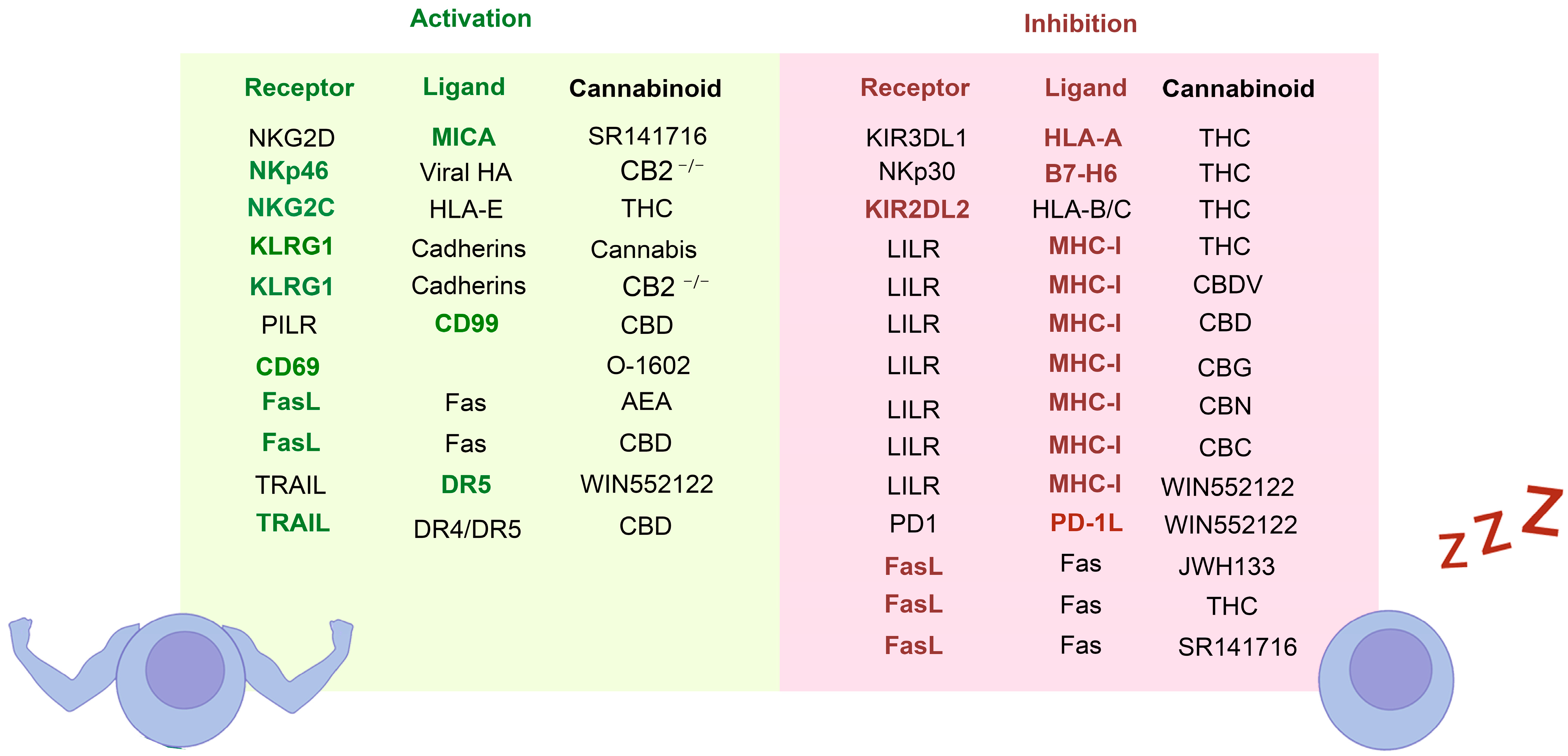
As previously discussed, NK cells’ effector function depends on the balance between inhibitory and activator receptors, which in turn is regulated by the interaction with respective ligands on the target cell surface (Supplementary Table S3). Most of the activator receptors act by employing conserved sequences (Immunoreceptor Tyrosine-based Activation Motifs; ITAMs). Downstream events result in the elevation of Ca2+i levels, which favors the transcription of cytokines and chemokines, as well as cytoskeleton reorganization, and stimulates cytotoxic granule release [2,127]. Another consequence of NK cell activation is the expression of death ligands, which, through death receptors, induce the regulated cell death. These death ligands include TNF-α, Fas ligand (FasL), and TRAIL, which bind their cognate receptors (TNFR1, Fas, and DR4/DR5, respectively) in the target cell, triggering cell death [128].
In this context, endocannabinoids, phytocannabinoids, and synthetic cannabinoids—through the modification in the activator receptors and ligands in the NK and target cells —change their balance, hence defining the activated or inhibited NK phenotype. Respective data can be found in Supplementary Table S3, with a summary presented in Figure 6.

5. Cannabis Effect on NK-Related Branch of Immunity in Clinical Reports

Available data suggest that NK cells are relatively resistant to cannabinoid-mediated cytotoxicity (Section 4). We searched for human studies on this topic and found that clinical reports on the effects of cannabinoids on immune function, and in particular NK cell status, are limited and have been conducted under variable conditions. Here we present some of the published results.
For example, the effects of cannabinoid ingestion (in the form of bhang, made from cannabis leaves boiled with water and sugar) on the immune system were studied in groups of high school and university students in Egypt [129]. In this study, a statistically significant reduction in the amount of NK cells in peripheral blood was observed in those individuals who consumed bhang for a period of up to 24 months, while for longer periods of use up to 36 months, the amount of NK was closer to the control value, with no statistical difference (each study group included 30 people). A similar study was conducted by Pacifici and colleagues to evaluate cell-mediated immune function in young cannabis users and compare them with non-users (20–30 participants in each group) [130]. They reported an approximately two-fold decrease in NK cell numbers. A significant limitation of both reports was that the composition and dose, frequency, and period of cannabis use were not controlled. It should be noted that the influence of additional factors that can affect the immune status in groups of cannabis users cannot be excluded, in particular specific lifestyle habits, including poor diet, tobacco and alcohol consumption, circadian rhythm disorders, deficiencies in medical care and hygiene, among others. For example, severe stress has been shown to cause a decrease in NK cell populations in asymptomatic human immunodeficiency virus (HIV)-positive homosexual men [131].
There is some research on the effects of cannabinoids on immune status in human immunodeficiency virus (HIV)-infected patient populations. Marijuana and THC (dronabinol, marinol) have been used to treat HIV-associated anorexia and weight loss. But few of these studies have specifically addressed the relationship between cannabis use and immune competence of NK cell populations. Bredt and colleagues designed a study to determine the safety/toxicity profile of THC in people with HIV infection on protease inhibitor-containing regimens [132]. The drug was consumed by smoking of cigarettes (0.9 mg, 3.95% THC, 3 times daily) or as dronabiol capsules. In this study, no differences in NK cell count and function were observed between marijuana smokers, dronabinol, and placebo groups (20 individuals in each group) after a short-term (21 days) protocol. Similarly, no differences in NK cell counts were detected in HIV-positive adolescents using marijuana, in comparison to non-users [133]. Interestingly, lytic activity per NK cell moderately enhanced and was associated with recent cannabis use in this report.
Non-psychotropic cannabinoid CBD seems to stimulate the lymphocyte proliferation at low concentrations (discussed in Section 4.2). These findings in animal models were confirmed by some observations in humans. When CBD was used as a daily supplement (50 mg/daily) in 530 healthy volunteers, the number of NK cells in peripheral blood was enhanced [134].
As for other cannabinoids, Siniscalco’s group reported that long-term oral administration of GPR55 endogenous agonist palmitoylethanolamide (PEA) in a dose up to 1200 mg daily by a 13-year-old male with allergic and asthmatic disorders resulted in the slight enrichment of NK cells in peripheral blood samples from 32 to 52 cells/mL (normal range 60–300 cells/mL) after one month of treatment [135]. However, the mechanisms by which PEA favors NK proliferation were not explored.

6. Conclusions and Perspectives

This review has been stimulated by a growing interest in cannabinoid use in anticancer therapies. This raises a very important question of how cannabinoids affect patients’ immune system, and, in particular, NK cells, which represent the important branch of natural anticancer immunity. Compatibility of cannabinoid use with NK-based immunotherapy for diverse cancer types remains a pivotal consideration for both palliative and anticancer treatments.
Nowadays we know that the signaling events, triggered by cannabinoids, are complex. These are not restricted to the action through canonical CB1 and CB2 receptors, of which CB2 is predominantly expressed in immune system cells. Cannabinoids act on multiple other targets, including non-canonical receptors, ion channels, transporters, and enzymes. Here we summarized the current knowledge on those expressed in NK cells, focusing on their function and the mechanisms of modulation by diverse cannabinoids. On the other hand, we discussed here global effects of cannabinoids on NK cell viability, proliferation, migration, cytokine production, and anticancer activity. Critical analysis reveals that the present state of research on these topics is rather incomplete. We are, as yet, unable to state which of the reported CBRs are more essential for each global function.
The growing interest in cannabinoid effects on NK cells contrasts with the current state of the literature, which remains notably fragmented. Studies exhibit discrepancies across experimental systems, clinical observations, cannabinoid types, concentrations, and administration routes, lacking a cohesive framework. Differential impact of various cannabinoids on common targets, along with dose- and use-dependent effects, emphasizes the urgency for more focused investigations. Moreover, methodological limitations, such as employing entire populations of rodent splenocytes or human peripheral lymphocytes in functional assays rather than purified NK cells, underscore the critical need for rigorously designed systematic studies. Thus, effects of cannabinoids need to be primarily evaluated on purified NK cells.
Remarkably, there is a very limited number of clinical studies in which the effects of cannabis use on the human immune system have been assessed. It should be noted that these studies were limited to measuring circulating leukocyte populations, without assessing cell function and reported conflicting results: increased, decreased, or no change in NK cell numbers for cannabis users. Our view aligns with the National Institutes of Health (NIH) committee report on cannabis health effects [136], emphasizing the urgent need for well-designed clinical research on this topic.

Supplementary Materials

The following supporting information can be downloaded at: https://www.mdpi.com/article/10.3390/receptors3020007/s1. Table S1: Expression of cannabinoids targets in NK cells; Table S2: Effect of cannabinoids on NK function; Table S3: Effect of the cannabinoids on the molecular entities, participating in the interaction between NK and target cells. References [137,138,139,140,141,142,143,144,145,146,147,148,149,150,151,152,153,154,155,156,157,158,159,160,161,162] are cited in the Supplementary Materials.

Author Contributions

Conceptualization, M.O.-A.; investigation, M.O.-A., C.G.-I., O.D. and I.P.; resources, O.D. and I.P.; data curation, M.O.-A., C.G.-I., O.D. and I.P.; writing—original draft preparation, M.O.-A., C.G.-I., O.D. and I.P.; writing—review and editing, M.O.-A., C.G.-I., O.D. and I.P.; visualization, M.O.-A.; supervision, M.O.-A., O.D. and I.P.; project administration, O.D. and I.P.; funding acquisition, O.D. and I.P. All authors have read and agreed to the published version of the manuscript.

Funding

This study was funded by the Mexican National Council of Humanities, Science, and Technology (CONAHCYT) programs (PRONACES #303072 to O.D., CF-2019 21887, FOP02-2022-02 321696 to I.P.) and a doctoral scholarship for C.G-I (CVU: 1044934).

Acknowledgments

M.O.-A. would like to thank CONAHCYT and University of Colima (UCOL) for the support received through the “Investigadoras e Investigadores por México” program, to which he belongs.

Conflicts of Interest

The authors declare no conflicts of interest.

References

  1. Mujal, A.M.; Delconte, R.B.; Sun, J.C. Natural Killer Cells: From Innate to Adaptive Features. Annu. Rev. Immunol. 2021, 39, 417–447. [Google Scholar] [CrossRef]
  2. Pegram, H.J.; Andrews, D.M.; Smyth, M.J.; Darcy, P.K.; Kershaw, M.H. Activating and inhibitory receptors of natural killer cells. Immunol. Cell Biol. 2011, 89, 216–224. [Google Scholar] [CrossRef] [PubMed]
  3. Guzman, L.G.M.; Keating, N.; Nicholson, S.E. Natural Killer Cells: Tumor Surveillance and Signaling. Cancers 2020, 12, 952. [Google Scholar] [CrossRef] [PubMed]
  4. Ham, H.; Medlyn, M.; Billadeau, D.D. Locked and Loaded: Mechanisms Regulating Natural Killer Cell Lytic Granule Biogenesis and Release. Front. Immunol. 2022, 13, 871106–871124. [Google Scholar] [CrossRef] [PubMed]
  5. Khoury, M.; Cohen, I.; Bar-Sela, G. “The Two Sides of the Same Coin”—Medical Cannabis, Cannabinoids, and Immunity: Pros and Cons Explained. Pharmaceutics 2022, 14, 389. [Google Scholar] [CrossRef] [PubMed]
  6. Massi, P.; Solinas, M.; Cinquina, V.; Parolaro, D. Cannabidiol as potential anticancer drug. Br. J. Clin. Pharmacol. 2013, 75, 303–312. [Google Scholar] [CrossRef]
  7. Seltzer, E.S.; Watters, A.K.; MacKenzie, D.; Granat, L.M.; Zhang, D. Cannabidiol (CBD) as a Promising Anti-Cancer Drug. Cancers 2020, 12, 3203. [Google Scholar] [CrossRef] [PubMed]
  8. Mangal, N.; Erridge, S.; Habib, N.; Sadanandam, A.; Reebye, V.; Sodergren, M.H. Cannabinoids in the landscape of cancer. J. Cancer Res. Clin. 2021, 147, 2507–2534. [Google Scholar] [CrossRef]
  9. Olivas-Aguirre, M.; Torres-López, L.; Villatoro-Gómez, K.; Perez-Tapia, S.M.; Pottosin, I.; Dobrovinskaya, O. Cannabidiol on the Path from the Lab to the Cancer Patient: Opportunities and Challenges. Pharmaceutics 2022, 15, 366. [Google Scholar] [CrossRef]
  10. Aziz, A.; Nguyen, L.C.; Oumeslakht, L.; Bensussan, A.; Mkaddem, S.B. Cannabinoids as Immune System Modulators: Cannabidiol Potential Therapeutic Approaches and Limitations. Cannabis Cannabinoid Res. 2023, 8, 254–269. [Google Scholar] [CrossRef]
  11. Kim, S.; Chen, J.; Cheng, T.; Gindulyte, A.; He, J.; He, S.; Li, Q.; Shoemaker, B.A.; Thiessen, P.A.; Yu, B.; et al. PubChem 2023 update. Nucleic Acids Res. 2023, 51, 1373–1380. [Google Scholar] [CrossRef]
  12. Rahaman, O.; Ganguly, D. Endocannabinoids in immune regulation and immunopathologies. Immunology 2021, 164, 242–252. [Google Scholar] [CrossRef]
  13. Gülck, T.; Møller, B.L. Phytocannabinoids: Origins and Biosynthesis. Trends Plant Sci. 2020, 25, 985–1004. [Google Scholar] [CrossRef] [PubMed]
  14. Alves, V.L.; Gonçalves, J.L.; Aguiar, J.; Teixeira, H.M.; Câmara, J.S. The synthetic cannabinoids phenomenon: From structure to toxicological properties. A review. Crit. Rev. Toxicol. 2020, 50, 359–382. [Google Scholar] [CrossRef] [PubMed]
  15. Velasco, G.; Hernández-Tiedra, S.; Dávila, D.; Lorente, M. The use of cannabinoids as anticancer agents. Prog. Neuro-Psychopharmacol. Biol. Psychiatry 2016, 64, 259–266. [Google Scholar] [CrossRef] [PubMed]
  16. Andradas, C.; Truong, A.; Byrne, J.; Endersby, R. The Role of Cannabinoids as Anticancer Agents in Pediatric Oncology. Cancers 2021, 13, 157. [Google Scholar] [CrossRef] [PubMed]
  17. Tanasescu, R.; Constantinescu, C.S. Cannabinoids and the immune system: An overview. Immunobiology 2010, 215, 588–597. [Google Scholar] [CrossRef]
  18. Parolaro, D.; Massi, P.; Rubino, T.; Monti, E. Endocannabinoids in the immune system and cancer. Prostaglandins Leukot. Essent. Fat. Acids (PLEFA) 2002, 66, 319–332. [Google Scholar] [CrossRef] [PubMed]
  19. Devane, W.A.; Dysarz, F.A.; Johnson, M.R.; Melvin, L.S.; Howlett, A.C. Determination and characterization of a cannabinoid receptor in rat brain. Mol. Pharmacol. 1988, 34, 605–613. [Google Scholar] [PubMed]
  20. Munro, S.; Thomas, K.L.; Abu-Shaar, M. Molecular characterization of a peripheral receptor for cannabinoids. Nature 1993, 365, 61–65. [Google Scholar] [CrossRef]
  21. Galiègue, S.; Mary, S.; Marchand, J.; Dussossoy, D.; Carrière, D.; Carayon, P.; Bouaboula, M.; Shire, D.; Fur, G.; Casellas, P. Expression of Central and Peripheral Cannabinoid Receptors in Human Immune Tissues and Leukocyte Subpopulations. Eur. J. Biochem. 1995, 232, 54–61. [Google Scholar] [CrossRef] [PubMed]
  22. Graham, E.S.; Angel, C.E.; Schwarcz, L.E.; Dunbar, P.R.; Glass, M. Detailed Characterisation of CB2 Receptor Protein Expression in Peripheral Blood Immune Cells from Healthy Human Volunteers Using Flow Cytometry. Int. J. Immunopathol. Pharmacol. 2009, 23, 25–34. [Google Scholar] [CrossRef]
  23. López, A.J.S.; Román-Vega, L.; Tojeiro, E.R.; Giuffrida, A.; García-Merino, A. Cannabinoids in treated multiple sclerosis. Clin. Exp. Immunol. 2015, 179, 119–127. [Google Scholar]
  24. Thul, P.J.; Åkesson, L.; Wiking, M.; Mahdessian, D.; Geladaki, A.; Blal, H.A.; Alm, T.; Asplund, A.; Björk, L.; Breckels, L.M.; et al. A subcellular map of the human proteome. Science 2017, 356, eaal3321. [Google Scholar] [CrossRef]
  25. Sarsembayeva, A.; Kienzl, M.; Gruden, E.; Ristic, D.; Maitz, K.; Valadez-Cosmes, P.; Santiso, A.; Hasenoehrl, C.; Brcic, L.; Lindenmann, J.; et al. Cannabinoid receptor 2 plays a pro-tumorigenic role in non-small cell lung cancer by limiting anti-tumor activity of CD8+ T and NK cells. Front. Immunol. 2023, 13, 997115. [Google Scholar] [CrossRef] [PubMed]
  26. Fonseca, B.M.; Cunha, S.C.; Gonçalves, D.; Mendes, A.; Braga, J.; Correia-da-Silva, G.; Teixeira, N.A. Decidual NK cell-derived conditioned medium from miscarriages affects endometrial stromal cell decidualisation: Endocannabinoid anandamide and tumour necrosis factor-α crosstalk. Hum. Reprod. 2020, 35, 265–274. [Google Scholar] [CrossRef]
  27. Kishimoto, S.; Muramatsu, M.; Gokoh, M.; Oka, S.; Waku, K.; Sugiura, T. Endogenous Cannabinoid Receptor Ligand Induces the Migration of Human Natural Killer Cells. J. Biochem. 2005, 137, 217–223. [Google Scholar] [CrossRef] [PubMed]
  28. Garofano, F.; Schmidt-Wolf, I.G.H. High Expression of Cannabinoid Receptor 2 on Cytokine-Induced Killer Cells and Multiple Myeloma Cells. Int. J. Mol. Sci. 2020, 21, 3800. [Google Scholar] [CrossRef]
  29. Garofano, F.; Sharma, A.; Abken, H.; Gonzalez-Carmona, M.A.; Schmidt-Wolf, I.G.H. A Low Dose of Pure Cannabidiol Is Sufficient to Stimulate the Cytotoxic Function of CIK Cells without Exerting the Downstream Mediators in Pancreatic Cancer Cells. Int. J. Mol. Sci. 2022, 23, 3783. [Google Scholar] [CrossRef]
  30. Wu, Y.; Li, Y.; Fu, B.; Jin, L.; Zheng, X.; Zhang, A.; Sun, R.; Tian, Z.; Wei, H. Programmed differentiated natural killer cells kill leukemia cells by engaging SLAM family receptors. Oncotarget 2017, 8, 57024–57038. [Google Scholar] [CrossRef]
  31. Chiurchiù, V.; Lanuti, M.; Bardi, M.D.; Battistini, L.; Maccarrone, M. The differential characterization of GPR55 receptor in human peripheral blood reveals a distinctive expression in monocytes and NK cells and a proinflammatory role in these innate cells. Int. Immunol. 2015, 27, 153–160. [Google Scholar] [CrossRef]
  32. Zhao, W.; Huang, Y.; Liu, Z.; Cao, B.-B.; Peng, Y.-P.; Qiu, Y.-H. Dopamine Receptors Modulate Cytotoxicity of Natural Killer Cells via cAMP-PKA-CREB Signaling Pathway. PLoS ONE 2013, 8, e65860. [Google Scholar] [CrossRef]
  33. Hellstrand, K.; Hermodsson, S. Serotonergic 5-HT1a Receptors Regulate a Cell Contact-Mediated Interaction between Natural Killer Cells and Monocytes. Scand. J. Immunol. 1993, 37, 7–18. [Google Scholar] [CrossRef]
  34. Frank, M.G.; Johnson, D.R.; Hendricks, S.E.; Frank, J.L.W. Monocyte 5-HT1A receptors mediate pindobind suppression of natural killer cell activity: Modulation by catalase. Int. Immunopharmacol. 2001, 1, 247–253. [Google Scholar] [CrossRef]
  35. Russo, E.B.; Burnett, A.; Hall, B.; Parker, K.K. Agonistic Properties of Cannabidiol at 5-HT1a Receptors. Neurochem. Res. 2005, 30, 1037–1043. [Google Scholar] [CrossRef]
  36. Maher, D.P.; Walia, D.; Heller, N.M. Suppression of Human Natural Killer Cells by Different Classes of Opioids. Anesthesia Analg. 2019, 128, 1013–1021. [Google Scholar] [CrossRef]
  37. Sarkar, D.K.; Sengupta, A.; Zhang, C.; Boyadjieva, N.; Murugan, S. Opiate Antagonist Prevents μ- and δ-Opiate Receptor Dimerization to Facilitate Ability of Agonist to Control Ethanol-altered Natural Killer Cell Functions and Mammary Tumor Growth*. J. Biol. Chem. 2012, 287, 16734–16747. [Google Scholar] [CrossRef]
  38. Feske, S.; Wulff, H.; Skolnik, E.Y. Ion Channels in Innate and Adaptive Immunity. Annu. Rev. Immunol. 2015, 33, 291–353. [Google Scholar] [CrossRef] [PubMed]
  39. Maul-Pavicic, A.; Chiang, S.C.C.; Rensing-Ehl, A.; Jessen, B.; Fauriat, C.; Wood, S.M.; Sjöqvist, S.; Hufnagel, M.; Schulze, I.; Bass, T.; et al. ORAI1-mediated calcium influx is required for human cytotoxic lymphocyte degranulation and target cell lysis. Proc. Natl. Acad. Sci. USA 2011, 108, 3324–3329. [Google Scholar] [CrossRef]
  40. Kaschek, L.; Zöphel, S.; Knörck, A.; Hoth, M. A calcium optimum for cytotoxic T lymphocyte and natural killer cell cytotoxicity. Semin. Cell Dev. Biol. 2021, 115, 10–18. [Google Scholar] [CrossRef] [PubMed]
  41. Olivas-Aguirre, M.; Cruz-Aguilar, L.H.; Pottosin, I.; Dobrovinskaya, O. Reduction of Ca2+ Entry by a Specific Block of KCa3.1 Channels Optimizes Cytotoxic Activity of NK Cells against T-ALL Jurkat Cells. Cells 2023, 12, 2065. [Google Scholar] [CrossRef]
  42. Schwindling, C.; Quintana, A.; Krause, E.; Hoth, M. Mitochondria Positioning Controls Local Calcium Influx in T Cells. J. Immunol. 2010, 184, 184–190. [Google Scholar] [CrossRef]
  43. Rimmerman, N.; Ben-Hail, D.; Porat, Z.; Juknat, A.; Kozela, E.; Daniels, M.P.; Connelly, P.S.; Leishman, E.; Bradshaw, H.B.; Shoshan-Barmatz, V.; et al. Direct modulation of the outer mitochondrial membrane channel, voltage-dependent anion channel 1 (VDAC1) by cannabidiol: A novel mechanism for cannabinoid-induced cell death. Cell Death Dis. 2013, 4, e949. [Google Scholar] [CrossRef]
  44. Olivas-Aguirre, M.; Torres-López, L.; Valle-Reyes, J.S.; Hernández-Cruz, A.; Pottosin, I.; Dobrovinskaya, O. Cannabidiol directly targets mitochondria and disturbs calcium homeostasis in acute lymphoblastic leukemia. Cell Death Dis. 2019, 10, 779–797. [Google Scholar] [CrossRef]
  45. Olivas-Aguirre, M.; Torres-López, L.; Pottosin, I.; Dobrovinskaya, O. Phenolic Compounds Cannabidiol, Curcumin and Quercetin Cause Mitochondrial Dysfunction and Suppress Acute Lymphoblastic Leukemia Cells. Int. J. Mol. Sci. 2020, 22, 204. [Google Scholar] [CrossRef] [PubMed]
  46. Saul, S.; Stanisz, H.; Backes, C.S.; Schwarz, E.C.; Hoth, M. How ORAI and TRP channels interfere with each other: Interaction models and examples from the immune system and the skin. Eur. J. Pharmacol. 2014, 739, 49–59. [Google Scholar] [CrossRef]
  47. Muller, C.; Morales, P.; Reggio, P.H. Cannabinoid Ligands Targeting TRP Channels. Front. Mol. Neurosci. 2019, 11, 487. [Google Scholar] [CrossRef]
  48. Petrocellis, L.D.; Ligresti, A.; Moriello, A.S.; Allarà, M.; Bisogno, T.; Petrosino, S.; Stott, C.G.; Marzo, V.D. Effects of cannabinoids and cannabinoid-enriched Cannabis extracts on TRP channels and endocannabinoid metabolic enzymes. Br. J. Pharmacol. 2011, 163, 1479–1494. [Google Scholar] [CrossRef] [PubMed]
  49. Storozhuk, M.V.; Zholos, A.V. TRP Channels as Novel Targets for Endogenous Ligands: Focus on Endocannabinoids and Nociceptive Signalling. Curr. Neuropharmacol. 2018, 16, 137–150. [Google Scholar] [CrossRef]
  50. Wang, W.; Cao, X.; Liu, C.; Liu, L. Cannabinoid WIN 55,212-2 inhibits TRPV1 in trigeminal ganglion neurons via PKA and PKC pathways. Neurol. Sci. 2012, 33, 79–85. [Google Scholar] [CrossRef]
  51. Kim, H.S.; Kwon, H.-J.; Kim, G.E.; Cho, M.-H.; Yoon, S.-Y.; Davies, A.J.; Oh, S.B.; Lee, H.; Cho, Y.K.; Joo, C.H.; et al. Attenuation of natural killer cell functions by capsaicin through a direct and TRPV1-independent mechanism. Carcinogenesis 2014, 35, 1652–1660. [Google Scholar] [CrossRef]
  52. Pumroy, R.A.; Samanta, A.; Liu, Y.; Hughes, T.E.; Zhao, S.; Yudin, Y.; Rohacs, T.; Han, S.; Moiseenkova-Bell, V.Y. Molecular mechanism of TRPV2 channel modulation by cannabidiol. eLife 2019, 8, e48792. [Google Scholar] [CrossRef]
  53. Zhang, L.; Simonsen, C.; Zimova, L.; Wang, K.; Moparthi, L.; Gaudet, R.; Ekoff, M.; Nilsson, G.; Hellmich, U.A.; Vlachova, V.; et al. Cannabinoid non-cannabidiol site modulation of TRPV2 structure and function. Nat. Commun. 2022, 13, 7483–7500. [Google Scholar] [CrossRef] [PubMed]
  54. Santoni, G.; Farfariello, V.; Liberati, S.; Morelli, M.B.; Nabissi, M.; Santoni, M.; Amantini, C. The role of transient receptor potential vanilloid type-2 ion channels in innate and adaptive immune responses. Front. Immunol. 2013, 4, 34–42. [Google Scholar] [CrossRef]
  55. Hassan, S.; Eldeeb, K.; Millns, P.J.; Bennett, A.J.; Alexander, S.P.H.; Kendall, D.A. Cannabidiol enhances microglial phagocytosis. Br. J. Pharmacol. 2014, 171, 2426–2439. [Google Scholar] [CrossRef]
  56. Zhang, D.; Spielman, A.; Wang, L.; Ding, G.; Huang, F.; Gu, Q.; Schwarz, W. Mast-Cell Degranulation Induced by Physical Stimuli Involves the Activation of Transient-Receptor-Potential Channel TRPV2. Physiol. Res. 2012, 61, 113–124. [Google Scholar] [CrossRef] [PubMed]
  57. Rah, S.-Y.; Kwak, J.-Y.; Chung, Y.-J.; Kim, U.-H. ADP-ribose/TRPM2-mediated Ca2+ signaling is essential for cytolytic degranulation and antitumor activity of natural killer cells. Sci. Rep. 2015, 5, 9482. [Google Scholar] [CrossRef] [PubMed]
  58. Cabanas, H.; Muraki, K.; Eaton, N.; Balinas, C.; Staines, D.; Marshall-Gradisnik, S. Loss of Transient Receptor Potential Melastatin 3 ion channel function in natural killer cells from Chronic Fatigue Syndrome/Myalgic Encephalomyelitis patients. Mol. Med. 2018, 24, 44. [Google Scholar] [CrossRef]
  59. Marshall-Gradisnik, S.; Huth, T.; Chacko, A.; Johnston, S.; Smith, P.; Staines, D. Natural killer cells and single nucleotide polymorphisms of specific ion channels and receptor genes in myalgic encephalomyelitis/chronic fatigue syndrome. Appl. Clin. Genet. 2016, 9, 39–47. [Google Scholar] [CrossRef]
  60. Froghi, S.; Grant, C.R.; Tandon, R.; Quaglia, A.; Davidson, B.; Fuller, B. New Insights on the Role of TRP Channels in Calcium Signalling and Immunomodulation: Review of Pathways and Implications for Clinical Practice. Clin. Rev. Allergy Immunol. 2021, 60, 271–292. [Google Scholar] [CrossRef]
  61. Scopelliti, F.; Dimartino, V.; Cattani, C.; Cavani, A. Functional TRPA1 Channels Regulate CD56dimCD16+ NK Cell Cytotoxicity against Tumor Cells. Int. J. Mol. Sci. 2023, 24, 14736. [Google Scholar] [CrossRef]
  62. Dobrovinskaya, O.; Valencia-Cruz, G.; Castro-Sánchez, L.; Bonales-Alatorre, E.O.; Liñan-Rico, L.; Pottosin, I. Cholinergic Machinery as Relevant Target in Acute Lymphoblastic T Leukemia. Front. Pharmacol. 2016, 7, 290. [Google Scholar] [CrossRef] [PubMed]
  63. Zanetti, S.R.; Ziblat, A.; Torres, N.I.; Zwirner, N.W.; Bouzat, C. Expression and Functional Role of α7 Nicotinic Receptor in Human Cytokine-stimulated Natural Killer (NK) Cells*. J. Biol. Chem. 2016, 291, 16541–16552. [Google Scholar] [CrossRef] [PubMed]
  64. Jiang, W.; Li, D.; Han, R.; Zhang, C.; Jin, W.-N.; Wood, K.; Liu, Q.; Shi, F.-D.; Hao, J. Acetylcholine-producing NK cells attenuate CNS inflammation via modulation of infiltrating monocytes/macrophages. Proc. Natl. Acad. Sci. USA 2017, 114, E6202–E6211. [Google Scholar] [CrossRef] [PubMed]
  65. Shelukhina, I.; Siniavin, A.; Kasheverov, I.; Ojomoko, L.; Tsetlin, V.; Utkin, Y. α7- and α9-Containing Nicotinic Acetylcholine Receptors in the Functioning of Immune System and in Pain. Int. J. Mol. Sci. 2023, 24, 6524. [Google Scholar] [CrossRef] [PubMed]
  66. Mahgoub, M.; Keun-Hang, S.Y.; Sydorenko, V.; Ashoor, A.; Kabbani, N.; Kury, L.A.; Sadek, B.; Howarth, C.F.; Isaev, D.; Galadari, S.; et al. Effects of cannabidiol on the function of α7-nicotinic acetylcholine receptors. Eur. J. Pharmacol. 2013, 720, 310–319. [Google Scholar] [CrossRef] [PubMed]
  67. Chrestia, J.F.; Esandi, M.d.C.; Bouzat, C. Cannabidiol as a modulator of α7 nicotinic receptors. Cell. Mol. Life Sci. 2022, 79, 564. [Google Scholar] [CrossRef]
  68. Bhandage, A.K.; Friedrich, L.M.; Kanatani, S.; Jakobsson-Björkén, S.; Escrig-Larena, J.I.; Wagner, A.K.; Chambers, B.J.; Barragan, A. GABAergic signaling in human and murine NK cells upon challenge with Toxoplasma gondii. J. Leukoc. Biol. 2021, 110, 617–628. [Google Scholar] [CrossRef]
  69. Cifelli, P.; Ruffolo, G.; Felice, E.D.; Alfano, V.; van Vliet, E.A.; Aronica, E.; Palma, E. Phytocannabinoids in Neurological Diseases: Could They Restore a Physiological GABAergic Transmission? Int. J. Mol. Sci. 2020, 21, 723. [Google Scholar] [CrossRef]
  70. Bhandage, A.K.; Barragan, A. GABAergic signaling by cells of the immune system: More the rule than the exception. Cell. Mol. Life Sci. 2021, 78, 5667–5679. [Google Scholar] [CrossRef]
  71. den Eynden, J.V.; Ali, S.S.; Horwood, N.; Carmans, S.; Brône, B.; Hellings, N.; Steels, P.; Harvey, R.J.; Rigo, J.-M. Glycine and Glycine Receptor Signalling in Non-Neuronal Cells. Front. Mol. Neurosci. 2009, 2, 9. [Google Scholar] [CrossRef]
  72. Turk, S.; Baesmat, A.S.; Yılmaz, A.; Turk, C.; Malkan, U.Y.; Ucar, G.; Haznedaroğlu, I.C. NK-cell dysfunction of acute myeloid leukemia in relation to the renin–angiotensin system and neurotransmitter genes. Open Med. 2022, 17, 1495–1506. [Google Scholar] [CrossRef]
  73. Mandler, R.N.; Seamer, L.C.; Whitlinger, D.; Lennon, M.; Rosenberg, E.; Bankhurst, A.D. Human natural killer cells express Na+ channels. A pharmacologic flow cytometric study. J. Immunol. 1990, 144, 2365–2370. [Google Scholar] [CrossRef]
  74. Djamgoz, M.B.A.; Firmenich, L. Chapter Four-Novel immunotherapeutic approaches to cancer: Voltage-gated sodium channel expression in immune cells and tumors. In Cancer Immunology and Immunotherapy; Elsevier: Amsterdam, The Netherlands, 2022; Volume 1, pp. 83–109. ISBN 9780128233979. [Google Scholar]
  75. Wang, H.; Zhang, X.; Xue, L.; Xing, J.; Jouvin, M.-H.; Putney, J.W.; Anderson, M.P.; Trebak, M.; Kinet, J.-P. Low-Voltage-Activated CaV3.1 Calcium Channels Shape T Helper Cell Cytokine Profiles. Immunity 2016, 44, 782–794. [Google Scholar] [CrossRef] [PubMed]
  76. Erdogmus, S.; Concepcion, A.R.; Yamashita, M.; Sidhu, I.; Tao, A.Y.; Li, W.; Rocha, P.P.; Huang, B.; Garippa, R.; Lee, B.; et al. Cavβ1 regulates T cell expansion and apoptosis independently of voltage-gated Ca2+ channel function. Nat. Commun. 2022, 13, 2033. [Google Scholar] [CrossRef] [PubMed]
  77. Burstein, S.; Budrow, J.; Debatis, M.; Hunter, S.A.; Subramanian, A. Phospholipase participation in cannabinoid-induced release of free arachidonic acid. Biochem. Pharmacol. 1994, 48, 1253–1264. [Google Scholar] [CrossRef] [PubMed]
  78. Grandclément, C.; Pick, H.; Vogel, H.; Held, W. NK Cells Respond to Haptens by the Activation of Calcium Permeable Plasma Membrane Channels. PLoS ONE 2016, 11, e0151031. [Google Scholar] [CrossRef] [PubMed]
  79. Wang, H.; Cheng, X.; Tian, J.; Xiao, Y.; Tian, T.; Xu, F.; Hong, X.; Zhu, M.X. TRPC channels: Structure, function, regulation and recent advances in small molecular probes. Pharmacol. Ther. 2020, 209, 107497. [Google Scholar] [CrossRef] [PubMed]
  80. Trikha, P.; Moseman, J.E.; Thakkar, A.; Campbell, A.R.; Elmas, E.; Foltz, J.A.; Chakravarti, N.; Fitch, J.R.; Mardis, E.R.; Lee, D.A. Defining the AHR-regulated transcriptome in NK cells reveals gene expression programs relevant to development and function. Blood Adv. 2021, 5, 4605–4618. [Google Scholar] [CrossRef]
  81. Rodríguez-Antona, C.; Leskelä, S.; Zajac, M.; Cuadros, M.; Alvés, J.; Moneo, M.V.; Martín, C.; Cigudosa, J.C.; Carnero, A.; Robledo, M.; et al. Expression of CYP3A4 as a predictor of response to chemotherapy in peripheral T-cell lymphomas. Blood 2007, 110, 3345–3351. [Google Scholar] [CrossRef] [PubMed]
  82. Yamaori, S.; Kushihara, M.; Yamamoto, I.; Watanabe, K. Characterization of major phytocannabinoids, cannabidiol and cannabinol, as isoform-selective and potent inhibitors of human CYP1 enzymes. Biochem. Pharmacol. 2010, 79, 1691–1698. [Google Scholar] [CrossRef]
  83. Doohan, P.T.; Oldfield, L.D.; Arnold, J.C.; Anderson, L.L. Cannabinoid Interactions with Cytochrome P450 Drug Metabolism: A Full-Spectrum Characterization. AAPS J. 2021, 23, 91. [Google Scholar] [CrossRef]
  84. Whalen, M.M.; Doshi, R.N.; Bader, B.W.; Bankhurst, A.D. Lysophosphatidylcholine and Arachidonic Acid Are Required in the Cytotoxic Response of Human Natural Killer Cells to Tumor Target Cells. Cell. Physiol. Biochem. 1999, 9, 297–309. [Google Scholar] [CrossRef]
  85. Hoffman, T.; Hirata, F.; Bougnoux, P.; Fraser, B.A.; Goldfarb, R.H.; Herberman, R.B.; Axelrod, J. Phospholipid methylation and phospholipase A2 activation in cytotoxicity by human natural killer cells. Proc. Natl. Acad. Sci. USA 1981, 78, 3839–3843. [Google Scholar] [CrossRef]
  86. Evans, A.T.; Formukong, E.; Evans, F.J. Activation of phospholipase A2 by cannabinoids. FEBS Lett. 1987, 211, 119–122. [Google Scholar] [CrossRef]
  87. Shim, S.J.; Yang, W.-I.; Shin, E.; Koom, W.S.; Kim, Y.B.; Cho, J.H.; Suh, C.O.; Kim, J.H.; Kim, G.E. Clinical significance of cyclooxygenase-2 expression in extranodal natural killer (NK)/T-cell lymphoma, nasal type. Int. J. Radiat. Oncol. Biol. Phys. 2007, 67, 31–38. [Google Scholar] [CrossRef]
  88. Benish, M.; Bartal, I.; Goldfarb, Y.; Levi, B.; Avraham, R.; Raz, A.; Ben-Eliyahu, S. Perioperative Use of β-blockers and COX-2 Inhibitors May Improve Immune Competence and Reduce the Risk of Tumor Metastasis. Ann. Surg. Oncol. 2008, 15, 2042–2052. [Google Scholar] [CrossRef]
  89. Takeda, S.; Misawa, K.; Yamamoto, I.; Watanabe, K. Cannabidiolic Acid as a Selective Cyclooxygenase-2 Inhibitory Component in Cannabis. Drug Metab. Dispos. 2008, 36, 1917–1921. [Google Scholar] [CrossRef] [PubMed]
  90. Ruhaak, L.R.; Felth, J.; Karlsson, P.C.; Rafter, J.J.; Verpoorte, R.; Bohlin, L. Evaluation of the Cyclooxygenase Inhibiting Effects of Six Major Cannabinoids Isolated from Cannabis sativa. Biol. Pharm. Bull. 2011, 34, 774–778. [Google Scholar] [CrossRef] [PubMed]
  91. Cravatt, B.F.; Demarest, K.; Patricelli, M.P.; Bracey, M.H.; Giang, D.K.; Martin, B.R.; Lichtman, A.H. Supersensitivity to anandamide and enhanced endogenous cannabinoid signaling in mice lacking fatty acid amide hydrolase. Proc. Natl. Acad. Sci. USA 2001, 98, 9371–9376. [Google Scholar] [CrossRef] [PubMed]
  92. Freigang, S.; Zadorozhny, V.; McKinney, M.K.; Krebs, P.; Herro, R.; Pawlak, J.; Kain, L.; Schrantz, N.; Masuda, K.; Liu, Y.; et al. Fatty acid amide hydrolase shapes NKT cell responses by influencing the serum transport of lipid antigen in mice. J. Clin. Investig. 2010, 120, 1873–1884. [Google Scholar] [CrossRef] [PubMed]
  93. Shamran, H.; Singh, N.P.; Zumbrun, E.E.; Murphy, A.; Taub, D.D.; Mishra, M.K.; Price, R.L.; Chatterjee, S.; Nagarkatti, M.; Nagarkatti, P.S.; et al. Fatty acid amide hydrolase (FAAH) blockade ameliorates experimental colitis by altering microRNA expression and suppressing inflammation. Brain Behav. Immun. 2017, 59, 10–20. [Google Scholar] [CrossRef] [PubMed]
  94. Grimaldi, P.; Pucci, M.; Siena, S.D.; Giacomo, D.D.; Pirazzi, V.; Geremia, R.; Maccarrone, M. The faah gene is the first direct target of estrogen in the testis: Role of histone demethylase LSD1. Cell. Mol. Life Sci. 2012, 69, 4177–4190. [Google Scholar] [CrossRef] [PubMed]
  95. Curran, E.M.; Berghaus, L.J.; Vernetti, N.J.; Saporita, A.J.; Lubahn, D.B.; Estes, D.M. Natural Killer Cells Express Estrogen Receptor-α and Estrogen Receptor-β and Can Respond to Estrogen Via a Non-Estrogen Receptor-α-Mediated Pathway. Cell. Immunol. 2001, 214, 12–20. [Google Scholar] [CrossRef]
  96. Surace, L.; Doisne, J.-M.; Escoll, P.; Marie, S.; Dardalhon, V.; Croft, C.; Thaller, A.; Topazio, D.; Sparaneo, A.; Cama, A.; et al. Polarized mitochondria as guardians of NK cell fitness. Blood Adv. 2021, 5, 26–38. [Google Scholar] [CrossRef]
  97. Keating, S.E.; Zaiatz-Bittencourt, V.; Loftus, R.M.; Keane, C.; Brennan, K.; Finlay, D.K.; Gardiner, C.M. Metabolic Reprogramming Supports IFN-γ Production by CD56bright NK Cells. J. Immunol. 2016, 196, 2552–2560. [Google Scholar] [CrossRef]
  98. Fišar, Z.; Singh, N.; Hroudová, J. Cannabinoid-induced changes in respiration of brain mitochondria. Toxicol. Lett. 2014, 231, 62–71. [Google Scholar] [CrossRef] [PubMed]
  99. Klimecki, W.T.; Taylor, C.W.; Dalton, W.S. Inhibition of cell-mediated cytolysis and P-glycoprotein function in natural killer cells by verapamil isomers and cyclosporine a analogs. J. Clin. Immunol. 1995, 15, 152–158. [Google Scholar] [CrossRef]
  100. Takahashi, M.; Misawa, Y.; Watanabe, N.; Kawanishi, T.; Tanaka, H.; Shigenobu, K.; Kobayashi, Y. Role of P-glycoprotein in Human Natural Killer-Like Cell Line-Mediated Cytotoxicity. Exp. Cell Res. 1999, 253, 396–402. [Google Scholar] [CrossRef]
  101. Wilisch, A.; Noller, A.; Handgretinger, R.; Weger, S.; Nüssler, V.; Niethammer, D.; Probst, H.; Gekeler, V. Mdr1P-glycoprotein expression in natural killer (NK) cells enriched from peripheral or umbilical cord blood. Cancer Lett. 1993, 69, 139–148. [Google Scholar] [CrossRef]
  102. Perkovic, S.; Basic-Kinda, S.; Gasparovic, V.; Krznaric, Z.; Babel, J.; Ilic, I.; Aurer, I.; Batinic, D. Epstein-Barr virus-negative aggressive natural killer-cell leukaemia with high P-glycoprotein activity and phosphorylated extracellular signal-regulated protein kinases 1 and 2. Hematol. Rep. 2012, 4, e16. [Google Scholar] [CrossRef]
  103. Holland, M.L.; Lau, D.T.T.; Allen, J.D.; Arnold, J.C. The multidrug transporter ABCG2 (BCRP) is inhibited by plant-derived cannabinoids. Br. J. Pharmacol. 2007, 152, 815–824. [Google Scholar] [CrossRef]
  104. Zhu, H.-J.; Wang, J.-S.; Markowitz, J.S.; Donovan, J.L.; Gibson, B.B.; Gefroh, H.A.; DeVane, C.L. Characterization of P-glycoprotein Inhibition by Major Cannabinoids from Marijuana. J. Pharmacol. Exp. Ther. 2006, 317, 850–857. [Google Scholar] [CrossRef]
  105. Wu, S.; Zhang, X.; Dong, M.; Yang, Z.; Zhang, M.; Chen, Q. sATP-binding cassette subfamily G member 2 enhances the multidrug resistance properties of human nasal natural killer/T cell lymphoma side population cells. Oncol. Rep. 2020, 44, 1467–1478. [Google Scholar] [CrossRef]
  106. Yang, S.; Kobayashi, S.; Sekino, K.; Kagawa, Y.; Miyazaki, H.; Shil, S.K.; Umaru, B.A.; Wannakul, T.; Owada, Y. Fatty acid-binding protein 5 controls lung tumor metastasis by regulating the maturation of natural killer cells in the lung. FEBS Lett. 2021, 595, 1797–1805. [Google Scholar] [CrossRef] [PubMed]
  107. Haj-Dahmane, S.; Shen, R.-Y.; Elmes, M.W.; Studholme, K.; Kanjiya, M.P.; Bogdan, D.; Thanos, P.K.; Miyauchi, J.T.; Tsirka, S.E.; Deutsch, D.G.; et al. Fatty-acid–binding protein 5 controls retrograde endocannabinoid signaling at central glutamate synapses. Proc. Natl. Acad. Sci. USA 2018, 115, 3482–3487. [Google Scholar] [CrossRef] [PubMed]
  108. Elmes, M.W.; Kaczocha, M.; Berger, W.T.; Leung, K.; Ralph, B.P.; Wang, L.; Sweeney, J.M.; Miyauchi, J.T.; Tsirka, S.E.; Ojima, I.; et al. Fatty Acid-binding Proteins (FABPs) Are Intracellular Carriers for Δ9-Tetrahydrocannabinol (THC) and Cannabidiol (CBD)*. J. Biol. Chem. 2015, 290, 8711–8721. [Google Scholar] [CrossRef]
  109. Ferrini, M.E.; Hong, S.; Stierle, A.; Stierle, D.; Stella, N.; Roberts, K.; Jaffar, Z. CB2 receptors regulate natural killer cells that limit allergic airway inflammation in a murine model of asthma. Allergy 2017, 72, 937–947. [Google Scholar] [CrossRef]
  110. Barchi, M.; Innocenzi, E.; Giannattasio, T.; Dolci, S.; Rossi, P.; Grimaldi, P. Cannabinoid receptors signaling in the development, epigenetics, and tumours of male germ cells. Int. J. Mol. Sci. 2020, 21, 25. [Google Scholar] [CrossRef] [PubMed]
  111. Hamad, H.; Olsen, B.B. Cannabidiol induces cell death in human lung cancer cells and cancer stem cells. Pharmaceuticals 2021, 14, 1169. [Google Scholar] [CrossRef]
  112. Bogdanović, V.; Mrdjanović, J.; Borišev, I. A Review of the Therapeutic Antitumor Potential of Cannabinoids. J. Altern. Complement. Med. 2017, 23, 831–836. [Google Scholar] [CrossRef]
  113. Tomko, A.M.; Whynot, E.G.; Ellis, L.D.; Dupré, D.J. Anti-Cancer Potential of Cannabinoids, Terpenes, and Flavonoids Present in Cannabis. Cancers 2020, 12, 1985. [Google Scholar] [CrossRef]
  114. Fraguas-Sánchez, A.I.; Fernández-Carballido, A.; Torres-Suárez, A.I. Phyto-, endo- and synthetic cannabinoids: Promising chemotherapeutic agents in the treatment of breast and prostate carcinomas. Expert Opin. Investig. Drugs 2016, 25, 1311–1323. [Google Scholar] [CrossRef]
  115. Kis, B.; Ifrim, F.C.; Buda, V.; Avram, S.; Pavel, I.Z.; Antal, D.; Paunescu, V.; Dehelean, C.A.; Ardelean, F.; Diaconeasa, Z.; et al. Cannabidiol—From Plant to Human Body: A Promising Bioactive Molecule with Multi-Target Effects in Cancer. Int. J. Mol. Sci. 2019, 20, 5905. [Google Scholar] [CrossRef]
  116. Kawakami, Y.; Klein, T.W.; Newton, C.; Djeu, J.Y.; Dennert, G.; Specter, S.; Friedman, H. Suppression by Cannabinoids of a Cloned Cell Line with Natural Killer Cell Activity. Proc. Soc. Exp. Biol. Med. 1988, 187, 355–359. [Google Scholar] [CrossRef]
  117. Ignatowska-Jankowska, B.; Jankowski, M.; Glac, W.; Swiergel, A.H. Cannabidiol-induced lymphopenia does not involve NKT and NK cells. J Physiol. Pharmacol. 2008, 60 (Suppl. 3), 99–103. [Google Scholar]
  118. Singh, U.P.; Singh, N.P.; Singh, B.; Price, R.L.; Nagarkatti, M.; Nagarkatti, P.S. Cannabinoid receptor-2 (CB2) agonist ameliorates colitis in IL-10−/− mice by attenuating the activation of T cells and promoting their apoptosis. Toxicol. Appl. Pharmacol. 2012, 258, 256–267. [Google Scholar] [CrossRef] [PubMed]
  119. Hillard, C.J. Circulating Endocannabinoids: From Whence Do They Come and Where are They Going? Neuropsychopharmacology 2018, 43, 155–172. [Google Scholar] [CrossRef] [PubMed]
  120. Massi, P.; Fuzio, D.; Viganò, D.; Sacerdote, P.; Parolaro, D. Relative involvement of cannabinoid CB1 and CB2 receptors in the Δ9-tetrahydrocannabinol-induced inhibition of natural killer activity. Eur. J. Pharmacol. 2000, 387, 343–347. [Google Scholar] [CrossRef] [PubMed]
  121. Patel, V.; Borysenko, M.; Kumar, M.S.A.; Millard, W.J. Effects of Acute and Subchronic Δ9-tetrahydrocannabinol Administration on the Plasma Catecholamine, β-Endorphin, and Corticosterone Levels and Splenic Natural Killer Cell Activity in Rats. Proc. Soc. Exp. Biol. Med. 1985, 180, 400–404. [Google Scholar] [CrossRef] [PubMed]
  122. Klein, T.W.; Newton, C.; Friedman, H. Inhibition of natural killer cell function by Marijuana components. J. Toxicol. Environ. Health 1987, 20, 321–332. [Google Scholar] [CrossRef]
  123. Specter, S.C.; Klein, T.W.; Newton, C.; Mondragon, M.; Widen, R.; Friedman, H. Marijuana effects on immunity: Suppression of human natural killer cell activity by delta-9-tetrahydrocannabinol. Int. J. Immunopharmacol. 1986, 8, 741–745. [Google Scholar] [CrossRef]
  124. Patrini, G.; Sacerdote, P.; Fuzio, D.; Manfredi, B.; Parolaro, D. Regulation of immune functions in rat splenocytes after acute and chronic in vivo treatment with CP-55,940, a synthetic cannabinoid compound. J. Neuroimmunol. 1997, 80, 143–148. [Google Scholar] [CrossRef]
  125. Massi, P.; Vaccani, A.; Romorini, S.; Parolaro, D. Comparative characterization in the rat of the interaction between cannabinoids and opiates for their immunosuppressive and analgesic effects. J. Neuroimmunol. 2001, 117, 116–124. [Google Scholar] [CrossRef]
  126. Takheaw, N.; Jindaphun, K.; Pata, S.; Laopajon, W.; Kasinrerk, W. Cannabinoid Receptor 1 Agonist ACEA and Cannabinoid Receptor 2 Agonist GW833972A Attenuates Cell-Mediated Immunity by Different Biological Mechanisms. Cells 2023, 12, 848. [Google Scholar] [CrossRef] [PubMed]
  127. Paul, S.; Lal, G. The Molecular Mechanism of Natural Killer Cells Function and Its Importance in Cancer Immunotherapy. Front. Immunol. 2017, 8, 1124. [Google Scholar] [CrossRef] [PubMed]
  128. Ramírez-Labrada, A.; Pesini, C.; Santiago, L.; Hidalgo, S.; Calvo-Pérez, A.; Oñate, C.; Andrés-Tovar, A.; Garzón-Tituaña, M.; Uranga-Murillo, I.; Arias, M.A.; et al. All About (NK Cell-Mediated) Death in Two Acts and an Unexpected Encore: Initiation, Execution and Activation of Adaptive Immunity. Front. Immunol. 2022, 13, 896228–896241. [Google Scholar] [CrossRef] [PubMed]
  129. EL–Gohary, M.; Eid, M.A. Effect of cannabinoid ingestion (in the form of bhang) on the immune system of high school and university students. Hum. Exp. Toxicol. 2004, 23, 149–156. [Google Scholar] [CrossRef]
  130. Pacifici, R.; Zuccaro, P.; Farré, M.; Poudevida, S.; Abanades, S.; Pichini, S.; Langohr, K.; Segura, J.; Torre, R.D.L. Combined immunomodulating properties of 3,4-methylenedioxymethamphetamine (MDMA) and cannabis in humans. Addiction 2007, 102, 931–936. [Google Scholar] [CrossRef]
  131. Evans, D.L.; Leserman, J.; Perkins, D.O.; Stern, R.A.; Murphy, C.; Tamul, K.; Liao, D.; van der Horst, C.M.; Hall, C.D.; Folds, J.D. Stress-associated reductions of cytotoxic T lymphocytes and natural killer cells in asymptomatic HIV infection. Am. J. Psychiatry 1995, 152, 543–550. [Google Scholar]
  132. Bredt, B.M.; Higuera-Alhino, D.; Shade, S.B.; Hebert, S.J.; McCune, J.M.; Abrams, D.I. Short-Term Effects of Cannabinoids on Immune Phenotype and Function in HIV-1-Infected Patients. J. Clin. Pharmacol. 2002, 42, 82S–89S. [Google Scholar] [CrossRef] [PubMed]
  133. Douglas, S.D.; Camarca, M.; Xu, J.; Durako, S.; Murphy, D.; Moscicki, B.; Wilson, C.M.; Network, R. for E. in A.C. and H.P., Adolescent Medicine HIV/AIDS Research the Relationships between Substance Abuse, Psychosocial Variables, and Natural Killer Cell Enumeration and Function in HIV-Infected and High-Risk Uninfected Adolescents. AIDS Res. Hum. Retroviruses 2003, 19, 399–408. [Google Scholar] [CrossRef] [PubMed]
  134. Kisiolek, J.N.; Flores, V.A.; Ramani, A.H.; Butler, B.; Haughian, J.M.; Stewart, L.K. Eight Weeks of Daily Cannabidiol Supplementation Improves Sleep Quality and Immune Cell Cytotoxicity. Nutrients 2023, 15, 4173. [Google Scholar] [CrossRef] [PubMed]
  135. Antonucci, N.; Cirillo, A.; Siniscalco, D. Beneficial Effects of Palmitoylethanolamide on Expressive Language, Cognition, and Behaviors in Autism: A Report of Two Cases. Case Rep. Psychiatry 2015, 2015, 325061. [Google Scholar] [CrossRef] [PubMed]
  136. National Academies of Sciences, Engineering, and Medicine. The Health Effects of Cannabis and Cannabinoids: The Current State of Evidence and Recommendations for Research, 1st ed.; The National Academies Press: Washington, DC, USA, 2017; pp. 199–216. [Google Scholar]
  137. Casey, T.M.; Meade, J.L.; Hewitt, E.W. Organelle Proteomics Identification of the Exocytic Machinery Associated with the Natural Killer Cell Secretory Lysosome*. Mol. Cell. Proteom. 2007, 6, 767–780. [Google Scholar] [CrossRef]
  138. Fernandes, E.S.; Brito, C.X.L.; Teixeira, S.A.; Barboza, R.; Reis, A.S.d.; Azevedo-Santos, A.P.S.; Muscará, M.; Costa, S.K.P.; Marinho, C.R.F.; Brain, S.D.; et al. TRPV1 Antagonism by Capsazepine Modulates Innate Immune Response in Mice Infected with Plasmodium berghei ANKA. Mediat. Inflamm. 2014, 2014, 506450. [Google Scholar] [CrossRef] [PubMed]
  139. Su, A.I.; Wiltshire, T.; Batalov, S.; Lapp, H.; Ching, K.A.; Block, D.; Zhang, J.; Soden, R.; Hayakawa, M.; Kreiman, G.; et al. A gene atlas of the mouse and human protein-encoding transcriptomes. Proc. Natl. Acad. Sci. USA 2004, 101, 6062–6067. [Google Scholar] [CrossRef] [PubMed]
  140. Deem, R.L.; Britvan, L.J.; Targan, S.R. Definition of a secondary target cell trigger during natural killer cell cytotoxicity: Possible role of phospholipase A2. Cell. Immunol. 1987, 110, 253–264. [Google Scholar] [CrossRef]
  141. Assmann, N.; O’Brien, K.L.; Donnelly, R.P.; Dyck, L.; Zaiatz-Bittencourt, V.; Loftus, R.M.; Heinrich, P.; Oefner, P.J.; Lynch, L.; Gardiner, C.M.; et al. Srebp-controlled glucose metabolism is essential for NK cell functional responses. Nat. Immunol. 2017, 18, 1197–1206. [Google Scholar] [CrossRef]
  142. Ziegler, S.; Weiss, E.; Schmitt, A.-L.; Schlegel, J.; Burgert, A.; Terpitz, U.; Sauer, M.; Moretta, L.; Sivori, S.; Leonhardt, I.; et al. CD56 Is a Pathogen Recognition Receptor on Human Natural Killer Cells. Sci. Rep. 2017, 7, 6138. [Google Scholar] [CrossRef]
  143. Mah-Som, A.Y.; Keppel, M.P.; Tobin, J.M.; Kolicheski, A.; Saucier, N.; Sexl, V.; French, A.R.; Wagner, J.A.; Fehniger, T.A.; Cooper, M.A. Reliance on Cox10 and oxidative metabolism for antigen-specific NK cell expansion. Cell Rep. 2021, 35, 109209–109225. [Google Scholar] [CrossRef]
  144. Zhao, H.B.; Zhang, X.F.; Shi, F.; Zhang, M.Z.; Xue, W.L. Comparison of the expression of human equilibrative nucleotide transporter 1 (hENT1) and ribonucleotide reductase subunit M1 (RRM1) genes in seven non-Hodgkin lymphoma cell lines. Genet. Mol. Res. 2016, 15, 220–226. [Google Scholar] [CrossRef] [PubMed]
  145. Kim, T.M.; Kim, S.; Ahn, Y.-O.; Lee, S.-H.; Kim, D.-W.; Heo, D.S. Anti-cancer activity of gemcitabine against natural killer cell leukemia/lymphoma. Leuk. Lymphoma 2014, 55, 940–943. [Google Scholar] [CrossRef]
  146. Chen, C.; Chang, Z.; Tsai, F.; Chen, S. Cannabinoid receptor type 1 antagonist inhibits progression of obesity-associated nonalcoholic steatohepatitis in a mouse model by remodulating immune system disturbances. Immun. Inflamm. Dis. 2020, 8, 544–558. [Google Scholar] [CrossRef] [PubMed]
  147. Kiran, S.; Rakib, A.; Moore, B.M.; Singh, U.P. Cannabinoid Receptor 2 (CB2) Inverse Agonist SMM-189 Induces Expression of Endogenous CB2 and Protein Kinase A That Differentially Modulates the Immune Response and Suppresses Experimental Colitis. Pharmaceutics 2022, 14, 936. [Google Scholar] [CrossRef]
  148. Ciaglia, E.; Torelli, G.; Pisanti, S.; Picardi, P.; D’Alessandro, A.; Laezza, C.; Malfitano, A.M.; Fiore, D.; Zottola, A.C.P.; Proto, M.C.; et al. Cannabinoid receptor CB1 regulates STAT3 activity and its expression dictates the responsiveness to SR141716 treatment in human glioma patients’ cells. Oncotarget 2015, 6, 15464–15481. [Google Scholar] [CrossRef]
  149. Ko, M.-W.; Breznik, B.; Senjor, E.; Jewett, A. Synthetic cannabinoid WIN 55,212–2 inhibits growth and induces cell death of oral and pancreatic stem-like/poorly differentiated tumor cells. Adv. Cancer Biol.-Metastasis 2022, 5, 100043–100052. [Google Scholar] [CrossRef]
  150. Hernández-Cervantes, R.; Pérez-Torres, A.; Prospéro-García, Ó.; Montor, J.M. Gestational exposure to the cannabinoid WIN 55,212-2 and its effect on the innate intestinal immune response. Sci. Rep. 2019, 9, 20340. [Google Scholar] [CrossRef]
  151. Hu, Y.; Ranganathan, M.; Shu, C.; Liang, X.; Ganesh, S.; Osafo-Addo, A.; Yan, C.; Zhang, X.; Aouizerat, B.E.; Krystal, J.H.; et al. Single-cell Transcriptome Mapping Identifies Common and Cell-type Specific Genes Affected by Acute Delta9-tetrahydrocannabinol in Humans. Sci. Rep. 2020, 10, 3450. [Google Scholar] [CrossRef]
  152. Karmaus, P.W.F.; Wagner, J.G.; Harkema, J.R.; Kaminski, N.E.; Kaplan, B.L.F. Cannabidiol (CBD) enhances lipopolysaccharide (LPS)-induced pulmonary inflammation in C57BL/6 mice. J. Immunotoxicol. 2013, 10, 321–328. [Google Scholar] [CrossRef]
  153. Dada, S.; Ellis, S.L.S.; Wood, C.; Nohara, L.L.; Dreier, C.; Garcia, N.H.; Saranchova, I.; Munro, L.; Pfeifer, C.G.; Eyford, B.A.; et al. Specific cannabinoids revive adaptive immunity by reversing immune evasion mechanisms in metastatic tumours. Front. Immunol. 2023, 13, 982082. [Google Scholar] [CrossRef]
  154. Hurrell, B.P.; Helou, D.G.; Shafiei-Jahani, P.; Howard, E.; Painter, J.D.; Quach, C.; Akbari, O. Cannabinoid receptor 2 engagement promotes group 2 innate lymphoid cell expansion and enhances airway hyperreactivity. J. Allergy Clin. Immunol. 2022, 149, 1628–1642.e10. [Google Scholar] [CrossRef]
  155. Falcinelli, S.D.; Cooper-Volkheimer, A.D.; Semenova, L.; Wu, E.; Richardson, A.; Ashokkumar, M.; Margolis, D.M.; Archin, N.M.; Rudin, C.D.; Murdoch, D.; et al. Impact of Cannabis Use on Immune Cell Populations and the Viral Reservoir in People with HIV on Suppressive Antiretroviral Therapy. J. Infect. Dis. 2023, 228, 1600–1609. [Google Scholar] [CrossRef]
  156. Libro, R.; Scionti, D.; Diomede, F.; Marchisio, M.; Grassi, G.; Pollastro, F.; Piattelli, A.; Bramanti, P.; Mazzon, E.; Trubiani, O. Cannabidiol Modulates the Immunophenotype and Inhibits the Activation of the Inflammasome in Human Gingival Mesenchymal Stem Cells. Front. Physiol. 2016, 7, 559. [Google Scholar] [CrossRef]
  157. Huang, L.; Ramirez, J.C.; Frampton, G.A.; Golden, L.E.; Quinn, M.A.; Pae, H.Y.; Horvat, D.; Liang, L.; DeMorrow, S. Anandamide exerts its antiproliferative actions on cholangiocarcinoma by activation of the GPR55 receptor. Lab. Investig. 2011, 91, 1007–1017. [Google Scholar] [CrossRef]
  158. Sun, X.; Zhou, L.; Wang, Y.; Deng, G.; Cao, X.; Ke, B.; Wu, X.; Gu, Y.; Cheng, H.; Xu, Q.; et al. Single-cell analyses reveal cannabidiol rewires tumor microenvironment via inhibiting alternative activation of macrophage and synergizes with anti-PD-1 in colon cancer. J. Pharm. Anal. 2023, 13, 726–744. [Google Scholar] [CrossRef]
  159. Sido, J.M.; Nagarkatti, P.S.; Nagarkatti, M. Δ9-Tetrahydrocannabinol attenuates allogeneic host-versus-graft response and delays skin graft rejection through activation of cannabinoid receptor 1 and induction of myeloid-derived suppressor cells. J. Leukoc. Biol. 2015, 98, 435–447. [Google Scholar] [CrossRef] [PubMed]
  160. Greiner, B.; Sommerfeld, M.; Kintscher, U.; Unger, T.; Kappert, K.; Kaschina, E. Differential Regulation of MMPs, Apoptosis and Cell Proliferation by the Cannabinoid Receptors CB1 and CB2 in Vascular Smooth Muscle Cells and Cardiac Myocytes. Biomedicines 2022, 10, 3271. [Google Scholar] [CrossRef] [PubMed]
  161. Pellerito, O.; Calvaruso, G.; Portanova, P.; Blasio, A.D.; Santulli, A.; Vento, R.; Tesoriere, G.; Giuliano, M. The Synthetic Cannabinoid WIN 55,212-2 Sensitizes Hepatocellular Carcinoma Cells to Tumor Necrosis Factor-Related Apoptosis-Inducing Ligand (TRAIL)-Induced Apoptosis by Activating p8/CCAAT/Enhancer Binding Protein Homologous Protein (CHOP)/Death Receptor 5 (DR5) Axis. Mol. Pharmacol. 2010, 77, 854–863. [Google Scholar]
  162. Ivanov, V.N.; Wu, J.; Hei, T.K. Regulation of human glioblastoma cell death by combined treatment of cannabidiol, γ-radiation and small molecule inhibitors of cell signaling pathways. Oncotarget 2017, 8, 74068–74095. [Google Scholar] [CrossRef] [PubMed]
Figure 1. Overview of the cannabinoid types. The structure of representative phytocannabinoids (green; left), endocannabinoids (orange, center), and synthetic cannabinoids (grey, right) are provided. Further details and additional characteristics for each cannabinoid can be accessed online through the PubChem database [11].
Receptors 03 00007 g001
Figure 2. Overview of the classical and non-classical CBRs in NK cells. (A) Classification of the proteins targeted by cannabinoids in NK cells. (B) Subcellular location of the CBRs in NK cells. (C) Processes regulated by the reported CBRs in NK cells. Color code: CBRs were colored only when their subcellular localization was appropriately validated for NK cells, whereas grey-coded CBRs represent functionally expressed ones with the suggested location, based on the data from the protein atlas [24] (see details in Supplementary Table S1).
Receptors 03 00007 g002
Figure 3. Role of the ion channels present in NK cells in the regulation of intracellular calcium and NK cell function. (1) NK cell activation results in the formation of the immunological synapse (IS), promoting the intracellular downstream signaling, which results in the production of IP3 and consequent Ca2+ release from the ER. (2) Depletion of ER Ca2+ induces STIM conformational change, resulting in the interaction of STIM with Orai subunits, to assemble the functional CRAC channel, which promotes Ca2+ entry (SOCE). (3) K+ channels functionally interact with CRAC by mediating K+ efflux and promoting hyperpolarization to sustain CRAC activity. (4) Mitochondria are recruited upon IS formation and contribute to preserve CRAC activity by taking up high amounts of Ca2+ to limit CRAC inactivation. Additionally, mitochondrial Ca2+ uptake favors the cell´s metabolism, necessary for its effector function (e.g., migration, degranulation). (5) Intracellular Ca2+ rise triggers the expression of multiple genes involved in NK cell activation, proliferation, and function. (6) The magnitude of intracellular Ca2+ rise determines the efficiency of the lytic granule release (see text for details). (7) The contribution of TRP family members, nAChR, and GABAA, to the global Ca2+ signal can impact the NK cells’ response to target cells. Blue circles depict Ca2+ ion. Yellow signs (!) indicate ion channels, expressed in NK cells, which are regulated by cannabinoids (see text for the details of regulation). Pink rectangles represent perforin, whereas yellow circles represent granzymes.
Receptors 03 00007 g003
Figure 4. Model for cannabinoid-induced sensitization of malignant NK cells to chemotherapy. (A) Tumoral NK cells exposed to chemotherapy overexpress proteins belonging to the cytochrome P450 superfamily (CYPs), which metabolize common chemotherapeutics to promote a pro-tumorigenic state. Additionally, they express high levels of P-gp and ABCG2, both acting as efflux systems for chemotherapeutics. (B) Cannabinoids, mainly from plant sources, act as P-gp, ABC2G, and CYP inhibitors, which enable maximum retention of chemotherapeutics in NK tumor cells, limiting tumor growth.
Receptors 03 00007 g004
Figure 5. NK cytotoxic activity against target cells and its modulation by cannabinoids. The effector response of NK cells is a multistep process. First, NK cells recognize the target cell through the interaction of surface molecules, forming the immunological synapse (IS). Protein clustering at the IS promotes the intracellular cell signaling that includes Ca2+i elevation, cytoskeleton reorganization, and gene expression. As an early response, lytic granules, containing granzymes and perforin, are released. A long-term response involves the production and release of cytokines and chemokines with autocrine and paracrine activities. Numbers indicate the steps at which cannabinoids have been demonstrated to act as regulators. (1) THC and O-1602 promote the expression of activation receptors in the target cell (detailed information can be found in Supplementary Table S3). (2) Most cannabinoids (CBD, THC, O-1602, AEA) have been shown to regulate intracellular Ca2+ levels in multiple cell types. (3) Cannabinoids have different effects on cytokine production and release. THC, CBD, and JWH133 decrease IFN-γ, IL-12, and TNF-α production, whereas AEA, AA, and O-1602 promote IL-12, IFN-γ, and TNF-α production. WIN55-212-2 and AEA promote cytokine production and release, whereas THC and JWH-133 inhibit cytokine production. (4) O-1602 enhances Granzyme B content in lytic granules, whereas CBD decreases Granzyme B content. (5) O-1602 favors NK degranulation. (6) CBD and THC have been shown to inhibit the NK chemotactic stimuli produced by target cells. (7) Cannabinoids modify the balance between activator and inhibitor proteins in target cells (further discussed in Section 4.5). (8) Cannabinoids exert direct cytotoxic effects on several cancer types [113,114].
Receptors 03 00007 g005
Figure 6. Activatory and inhibitory interactions, modulated by cannabinoids in NK and target cells. Green: modifications in these interactions result in activation of NK cells. Red: modifications in these interactions result in inhibition of NK cells. Names of altered proteins are set in colored and bold font.
Receptors 03 00007 g006
Disclaimer/Publisher’s Note: The statements, opinions and data contained in all publications are solely those of the individual author(s) and contributor(s) and not of MDPI and/or the editor(s). MDPI and/or the editor(s) disclaim responsibility for any injury to people or property resulting from any ideas, methods, instructions or products referred to in the content.