Review

Advancements in Peripheral Nerve Injury Research Using Lab Animals

1
F. Edward Hérbert School of Medicine, Uniformed Services University of the Health Sciences, Bethesda, MD 20814, USA
2
Department of Orthopaedics, University of Texas Health Science Center at San Antonio, 7703 Floyd Curl Drive, San Antonio, TX 78229, USA
3
Metis Foundation, 84 NE Interstate 410 Loop # 325, San Antonio, TX 78216, USA
4
Chief Scientist’s Office of Science and Technology, 59th Medical Wing, Joint Base San Antonio-Lackland, San Antonio, TX 78236, USA
*
Author to whom correspondence should be addressed.
Anatomia 2025, 4(2), 8; https://doi.org/10.3390/anatomia4020008
Submission received: 26 February 2025 / Revised: 30 April 2025 / Accepted: 14 May 2025 / Published: 23 May 2025

Abstract

:
Peripheral nerve injuries (PNIs) commonly result from trauma, compression, or iatrogenic causes, leading to functional deficits. Despite the peripheral nervous system’s regenerative capacity, current treatments yield inconsistent outcomes. Basic science and translational research supporting nerve repair remain underdeveloped, partly due to the absence of standardized protocols, limiting reproducibility. Animal models are essential for studying injury mechanisms, repair strategies, and therapeutic development. This review examines commonly used animal models in PNI research, from non-mammalian species to rodents and large mammals. We discuss the relevance of injury types, experimental variables (i.e., age, sex, nerve type), and study design elements (i.e., nerve gap size, injury induction methods). Assessing these models’ strengths and limitations, this review aims to guide researchers in selecting appropriate models that enhance preclinical relevance. It also addresses the need for standardized protocols and future directions for improving PNI research and patient outcomes. Various PNI treatments—including microsurgery, nerve grafts, scaffolds, stem cells, immunomodulators, nerve augmentation strategies, and polyethylene glycol-mediated fusion—have been developed using animal models. These models are essential for driving innovation and translating emerging therapies to improve outcomes across a broad range of peripheral nerve injuries.

1. Introduction

Peripheral nerve injury (PNI) is a common condition affecting an estimated 20 million people in the United States alone [1]. Trauma, compression, and iatrogenic injuries are common causes of PNIs. They pose a significant clinical challenge, accounting for 2–5% of all traumatic injuries [1]. While the peripheral nervous system has some regenerative capacity, severe PNI, especially involving mixed motor and sensory nerves, often leads to limited recovery and significant disability [1,2,3,4,5,6]. The complexity of PNIs and the lack of standardized treatment protocols contribute to inconsistent outcomes, emphasizing the need for advanced research and therapeutic strategies.
Despite advances in understanding nerve regeneration, PNI remains a significant clinical challenge, often resulting in suboptimal functional outcomes, chronic pain, and muscle atrophy [1,4,5,6]. Basic science and animal research are both crucial for developing better treatments. Representative animal models are essential for simulating these injuries, understanding nerve injury mechanisms, evaluating nerve repair and regeneration, and testing therapeutic interventions. However, the lack of standardized protocols in animal models can hinder research progress and affect patient outcomes. Physiological differences among these models limit their translatability to human conditions, highlighting the need for representative models to enhance clinical research and treatments.
Research on PNIs requires a diverse approach, using various animal models to better understand nerve injury mechanisms, evaluate nerve repair and regeneration, and test therapeutic interventions. Non-mammalian species provide unique insights into nerve regeneration and guide future treatment approaches [7,8,9,10]. For instance, since up to 65% of human disease genes have orthologs in Drosophila, it is widely used to model spinal cord and brain injuries, as well as neurodegenerative diseases such as ALS, sensory neuropathies, and dendrite degeneration [8]. Its genetic accessibility and conserved axon regeneration pathways make it an ideal model for studying mechanisms like Wallerian degeneration. These features can help reveal simplified pathways of axonal degeneration and repair, potentially facilitating therapeutic insights and identifying novel targets [7,11]. Rodent models are established models for PNI preclinical studies, offering advantages such as ease of handling, cost-effectiveness, and well-characterized genetics [2,3,5,12]. In contrast, larger mammals share more physiological features with humans and serve as advantageous models with a higher potential for translation to clinical practice [1,6,13,14,15,16,17,18,19,20,21,22,23,24,25]. These models are particularly valuable for studying complex nerve injuries and for testing surgical interventions that closely mimic clinical scenarios [13,14,15,26].
This review evaluates surgical animal models for PNIs to assess their efficacy and translatability to human injuries. It emphasizes standardized protocols for testing treatments to ensure reliable and reproducible results. Additionally, this review explores how these models can be used to understand nerve repair mechanisms, and the influence of factors such as sex and age on recovery. Finally, it discusses current advances, future directions, and emerging models, such as targeted muscle reinnervation, for optimizing PNI interventions and improving patient outcomes.

2. Selection of Peripheral Nerve Model in Animal Studies

The selection of an appropriate type of peripheral nerve (PN) to model in animal studies is crucial for accurately replicating human conditions and effectively evaluating therapeutic interventions. There are many types of PN that can be studied, each with unique anatomical and functional characteristics. The choice of nerve can impact the relevance and translatability of preclinical findings to clinical practice because different nerves vary in their anatomy, function, and response to injury and treatment. Selecting nerves that closely mimic human PNs in terms of size, fascicular structure, and sensorimotor function provides more clinically applicable results.

2.1. Sciatic Nerve

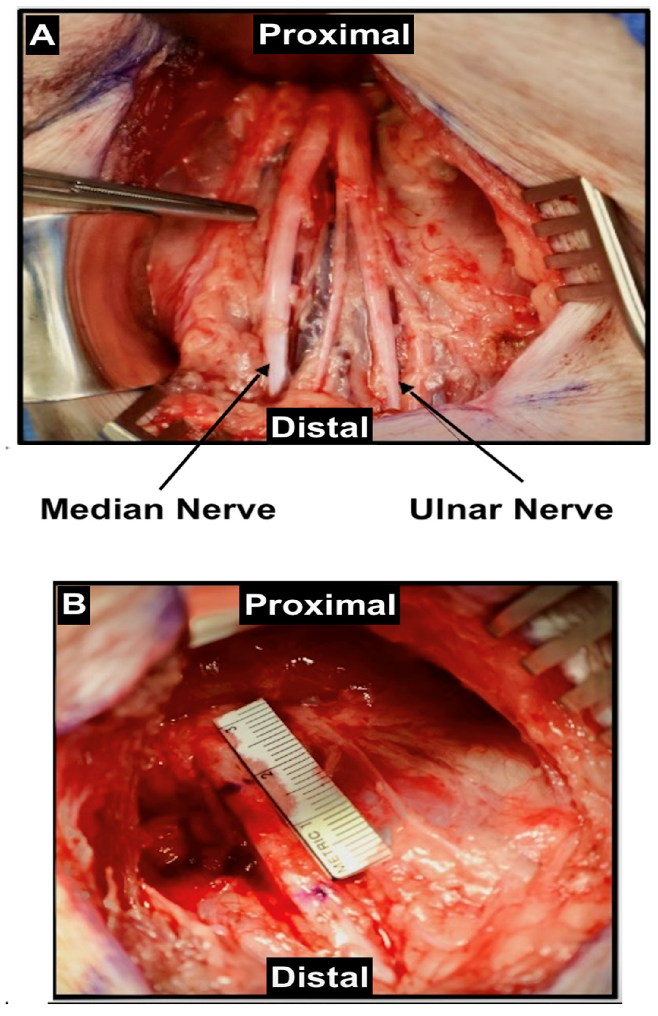
Approximately 90% of PNI models utilize rodents, particularly for crush and transection injuries [27]. The sciatic nerve is the most used nerve in these models due to its large size, surgical accessibility, and mixed sensorimotor function, which closely resembles human PNs (Figure 1).
Using the sciatic nerve allows researchers to study both motor and sensory nerve regeneration simultaneously. However, sciatic nerve injury can result in significant hindlimb paralysis due to neurotrophic changes and loss of joint function [3], which may subsequently lead to auto-mutilation behaviors such as toe-biting, thereby affecting animal well-being [26,28]. This raises ethical concerns and may introduce variability in experimental outcomes, potentially limiting the translatability of the findings.

2.2. Median Nerve

To mimic upper extremity injuries, the median nerve is frequently used in animal models (Figure 2).
The median nerve is a single-fascicle nerve that innervates muscles responsible for fine motor control in the forelimb, similar to its role in humans [26]. It enables simple behavioral tests, such as the grasping test, to evaluate function [26]. The ease of functional assessment with the median nerve enhances the relevance of the findings to human motor function recovery. Evaluations of the median nerve include nerve blood flow measurements, electrophysiology, morphometry, compound nerve action potentials, and muscle weight assessments [29]. Functional recovery can be tested using grasping tests, staircase tests, and electrodiagnostic evaluations [29]. The reduced severity of impairment minimizes self-injurious behavior, facilitating more thorough functional assessments.
In non-human primates, median nerve impairment leads to loss of thumb opposition and sensory deficits, closely mirroring human symptoms. Behavioral assessments such as fist clenching, the apple pinch test, and food gripping are used to evaluate functional recovery [28]. These assessments are analogous to clinical tests in humans, enhancing the relevance of the research. However, loss of function in the thumb, index, and middle fingers can lead to self-care difficulties and impaired recovery in primates. This must be considered when designing studies to ensure ethical treatment and accurate interpretation of results.

2.3. Radial Nerve

The radial nerve, although rarely used in PNI animal studies due to its small diameter, controls elbow and wrist extension and digital movement [28,30]. In rats its function can be evaluated with the Montoya staircase test, while in monkeys it can be evaluated via the wrist-extension angle test [30,31]. However, because animals may retain some function post-injury, it limits the model’s efficacy in assessing complete functional loss and recovery.

2.4. Ulnar Nerve

The ulnar nerve is responsible for finger and wrist flexion, as well as grasping in humans. It is relatively long and easily accessible in animal models (Figure 2) [32]. While functional outcomes related to this nerve are most appropriately assessed in non-human primates, they can also be evaluated in pigs, particularly when studying nerve regeneration across different diameters [32]. Functional assessments typically focus on finger and wrist flexion and grasping [28,33]. In some models, such as those described by Meyers et al., combined injuries to both the ulnar and median nerves have been used to investigate reinnervation through quantitative forelimb strength measurements and isometric pull tasks [28,34]. In rodent models, the ulnar nerve is also utilized for nerve transfer studies, such as ulnar-to-musculocutaneous transfers. In these cases, the Bertelli test is employed to evaluate recovery of elbow flexion, which reflects reinnervation of the musculocutaneous nerve rather than primary ulnar nerve function [28,35]. Despite anatomical similarities to humans and surgical accessibility, evaluating functional recovery of the ulnar nerve remains challenging due to limited assessment tools and behavioral complications, such as self-injury [28].

2.5. Tibial and Peroneal Nerve

The tibial nerve is occasionally used in mice to study the molecular mechanisms of motor axon excitability. Functional recovery following tibial nerve injury can be assessed through gait analysis, force threshold measurements, and gastrocnemius muscle testing [36]. However, injury to the tibial nerve may result in gait abnormalities, ulcerations, and failure to thrive, raising ethical concerns.
The common peroneal nerve, the most commonly injured nerve in the lower extremity, is commonly used to study Wallerian degeneration, particularly in gene and protein expression analyses. Functional assessments for common peroneal nerve injury include gait analysis and the toe-spreading reflex [28,36,37]. It should be noted that toe extension and spreading involve contributions from both the tibial and common peroneal nerves, and thus the toe-spreading reflex is not specific to peroneal function alone.
Compared to the tibial nerve, the common peroneal nerve exhibits inferior regenerative capacity following injury. Lin et al. employed proteomic assays and qRT-PCR to evaluate molecular responses during Wallerian degeneration, demonstrating that the common peroneal nerve has impaired protein synthesis and altered gene expression compared to the tibial nerve, with the exception of the first postoperative day [38]. In addition to these molecular differences, Zhang et al. reported that the common peroneal nerve shows lower compound muscle action potential (CMAP) amplitudes, reduced axonal density, and overall diminished recovery potential compared to the tibial nerve [39]. These molecular and electrophysiological disparities contribute to the poorer regeneration outcomes observed in the common peroneal nerve, making it a less optimal model for nerve regeneration studies.

2.6. Facial Nerve

The facial nerve is a cranial nerve commonly used in rat and sheep models due to its motor and sensory innervation, predictable anatomy, and easy accessibility. Outcomes are typically measured through electrophysiologic response, immunohistochemistry, fluorescent and confocal microscopy, and blood flow recordings [13,40]. Additionally, tracking the movement of the vibrissal pad, along with assessing eye closure, whisking, and eye-blinking, can help evaluate functional outcomes after repair [40,41]. Corneal reflex, facial symmetry, and snout movement can also be assessed [13].

2.7. Trigeminal Nerve

The trigeminal nerve is a complex cranial nerve that provides both motor and sensory functions. It has been studied in various animal models, including mice, rats, cats, monkeys, and horses. It is primarily used in research on trigeminal neuralgia, particularly with the crush injury model, but it has also been used in migraine studies to examine trigeminal sensory processing through stimulation [42,43].

2.8. Impact of Nerve Selection on Translatability

The choice of PN significantly impacts the translatability of preclinical findings to clinical practice for several reasons. (1) Selecting nerves that closely resemble human nerves in terms of anatomy and function ensures that the mechanisms of injury and regeneration observed are relevant to humans. This enhances the likelihood that therapeutic interventions effective in animal models will be effective in clinical settings. (2) Nerves that allow for precise and quantifiable functional assessments enable researchers to detect meaningful differences in regenerative outcomes. Functional tests that mimic clinical evaluations in humans improve the applicability of the findings. (3) Ethical considerations and animal welfare are important. Minimizing animal suffering and behavioral complications reduces confounding variables that can affect experimental results. Ethical research practices also enhance the validity of the study and its acceptance in the scientific community. (4) Using well-characterized nerve models with established assessment methods improves the consistency and reproducibility of studies. Consistent findings across different research groups strengthen the evidence base and facilitate the translation of research into clinical practice. By carefully selecting appropriate nerve models, researchers can design studies that yield clinically relevant data, ultimately contributing to the development of effective therapies for PNIs.

3. Species Selection for Peripheral Nerve Injury

Selecting the appropriate species is critical in PNI research to replicate human conditions accurately and evaluate therapeutic interventions effectively. Different species offer unique advantages and limitations.

3.1. Non-Mammalian Species

Non-mammalian species are utilized for high-throughput therapeutic screening and for elucidating mechanisms of nerve regeneration due to their genetic tractability, rapid development, and transparency.
Drosophila melanogaster (D. melanogaster, fruit fly) models lack myelin and associated inhibitory factors, making them valuable for studying axonal regeneration pathways [44]. The larval nerve crush model resembles the human sciatic nerve by encompassing motor and sensory functions, allowing real-time observation of regeneration through GFP expression in GAL4 genetic lines [7,8]. However, challenges include complex injury preparation and short study periods due to rapid maturation.
Danio rerio (D. rerio, zebrafish) are also utilized due to their physiological and genetic resemblance to mammals [45,46]. They facilitate real-time analysis of axonal transport, degeneration, and regeneration, especially in the posterior lateral line [45,46]. Their transparency and rapid development enable high-throughput screening of compounds that affect PN repair [45]. Caenorhabditis elegans (C. elegans, nematode) models are used for drug-induced peripheral neuropathy and provide insights into nervous system regeneration through analysis of axon regeneration mechanisms following laser-induced precision damage [47]. Rana pipiens (R. pipiens, the northern leopard frog) have been widely used to study optic nerve regeneration due to their exceptional regenerative capacity within the central nervous system, particularly in the optic nerve, which is well characterized and highly responsive to neurotrophic modulation. This model offers standardized methods, cost-efficiency, and translational value for evaluating the effects of factors such as ciliary neurotrophic factor and fibroblast growth factor on axonal regrowth [10,48].
While these models offer significant benefits for mechanistic studies and initial therapeutic screening, their phylogenetic distance from humans limits the direct translatability of findings due to differences in nervous system complexity and physiology. Nonetheless, they are invaluable for uncovering the fundamental principles of nerve regeneration that inform mammalian studies and guide the development of novel therapeutic strategies.

3.2. Small Animal Models

Small animal models are widely used in PNI research due to their practicality, cost-effectiveness, and the availability of genetic tools. They offer advantages like shorter lifespans, rapid gestation, faster reinnervation, reduced study times, and easier maintenance. However, they are prone to self-mutilation and may have limited utility in modeling long-gap nerve injuries seen in humans. The critical nerve defect, defined as the minimum nerve gap over which spontaneous regeneration without intervention does not occur, is approximately 1.3 cm in mice, 1.5 cm in rats, and 3.0 cm in rabbits, compared to 4 cm in pigs and humans [16,27]. These species-specific differences are essential when selecting a model to study long-gap nerve repair strategies. Despite their rapid regenerative capacity, small animals may not accurately replicate the biological and mechanical challenges of human nerve injuries [17,27]. A summary of key advantages and limitations is provided in Table 1.
Rats are commonly used due to their size, fascicular organization, and morphological similarities to humans [12,27]. The sciatic nerve is often selected for studies in rats due to its large size [26,27,49]. They are economical, easy to handle, resistant to infections, and suitable for large-scale studies [13,16,27]. Their well-studied anatomy with morphological similarities to humans supports standardization. However, the spontaneous regeneration seen in severed rat nerves limits the translation of therapeutic interventions to humans, highlighting a significant challenge in applying rat research to human applications [26]. Additionally, rats have fewer transgenic models compared to mice, which can limit mechanistic studies [16]. Notably, increased autophagy may enhance nerve regeneration [57]. Certain rat strains, such as Wistar rats, are believed to exhibit higher levels of autophagy, which may play a protective role against neuropathy [58].
Murine models offer benefits similar to rats, including cost-effectiveness, ease of handling, suitability for large-scale studies, and available genetic models [18,26,27,52]. The extensive range of transgenic and knockout mice allows for the study of specific genes involved in nerve regeneration. However, their small size limits axonal regeneration and vascular collaterals, and they exhibit mechanical allodynia with hypersensitivity to mechanical stimuli, which can complicate behavioral assessment [52].
Guinea pigs have larger nerve sizes compared to rats and are more cooperative and docile [19]. They enable complex behavioral analyses such as pain perception and neurological recovery. However, they are prone to ulcer formations, have increased costs, and there are fewer genetically modified strains and tools available for guinea pigs [19]. Adult rabbits have been used in PNI studies to model sciatic and median nerve injuries, benefiting from larger nerve diameters (2 to 4 mm) and the ability to accommodate gaps up to 30 mm [17]. They are also employed in simulations of blast injuries and hand/wrist surgery models [39]. However, rabbits can be more expensive, regenerate faster than humans, and are prone to self-mutilation. In a study, Merolli et al. report that about 50% exhibited post-PNI self-injury, with 18.75% experiencing ulcerative pododermatitis on the surgical limb [17]. Preventative measures, such as prophylactic bandaging, should be considered to mitigate self-injury and costs.

3.3. Large Animal Models

Large animal models, including non-human primates, sheep, pigs, and dogs, offer significant advantages in PNI research due to their greater resemblance to human neuroanatomy and the ability to accommodate larger nerve gaps essential for prosthesis development [1,12,14,15,23,59,60,61]. Electrophysiological assessments are also improved, with fewer electrical artifacts using transcutaneous and intraoperative stimulations compared to smaller models [1]. Primates have similar neuroanatomy, physiology, and nerve scaffolds, and, like humans, mild PNI can result in chronic morbidity and neuropathy. Larger animals feature longer gestational and reproductive cycles and lifespans, enabling extended observation for functional evaluation. However, ethical concerns have limited their use in preclinical research [16,60,62]. The advantages and disadvantages of large animal models are outlined in Table 2.
Sheep are particularly promising for preclinical PNI studies due to their low cost, wide availability, and comparable nerve anatomy, capable of accommodating segmental defects up to 10 cm [14]. The median nerve in sheep is similar to the human ulnar and median nerves, while the femoral nerve is comparable to the human ulnar nerve [13]. The larger nerve size and defect similarity increases the translational potential [66]. The common peroneal nerve is also studied, while the median nerve is frequently used because it does not affect limb function or weight-bearing ability [59]. To better align regenerative expectations across species, one-year-old sheep are considered age-equivalent to young adult humans, accounting for comparable regenerative capacity [13,28]. Sheep hindlimb and forelimb nerves exhibit similar dimensions, axon regeneration rates, and polyfascicular peripheral nerve (PN) histomorphology when compared to humans [14]. Their low rates of sepsis and hoof desensitization enhance the promise of this model [15]. However, studies in sheep typically last between 1 and 12 months due to the slow rate of nerve regeneration, approximately 1 to 2 mm per day, which is comparable to that in humans [28,37].
Porcine models are suitable for ultra-long segmental repair (over 15 cm), nerve transfers, and electrical stimulation for enhanced regeneration [1]. Despite behavioral studies being time and resource intensive, and swine potentially developing compensatory mechanisms, they serve as valuable models for examining various nerve modalities, including motor, sensory, and mixed compositions, as well as different gap lengths and regenerative distances [1,25,32]. Burrell et al. [1] developed a preclinical PNI model using porcine common and deep peroneal nerve injuries, demonstrating that swine have similar physiology and polyfascicular nerve architecture to human nerves. Notably, the sciatic nerve diameter in large pig breeds, such as the American Yorkshire and Wisconsin Miniature Swine, closely resembles the diameter of major human upper extremity nerves, including the median, ulnar, and radial nerves originating from the brachial plexus [25,67]. However, their motor and sensory conduction velocities are significantly higher than those observed in humans, likely due to differences in neuromuscular demands and fine sensorimotor function [67,68].
Dogs have an anatomy, physiology, and pathology comparable to humans, particularly in the context of large nerve gaps, nerve sizes, and nerve fibers [61]. For instance, Xue et al. demonstrated that autologous mesenchymal stem cells in a chitosan/PLGA scaffold can effectively model PNI repair in a 60 mm sciatic nerve gap in dogs [60]. Attar et al. utilized transected facial nerves in adult female dogs to compare epineurial sutures with fibrin glue for nerve repair [61]. The facial nerve was chosen due to its consistent anatomy and functional importance in motor nerve studies. Both repair methods yielded comparable histological outcomes, including axonal regeneration and fibrosis, but fibrin glue offered the advantage of reduced operative time and less tissue handling, suggesting it may be a viable alternative in specific clinical contexts. Yao et al. used the sciatic nerve in Beagle dogs to further demonstrate that combining collagen conduits with nerve growth factor within a neural tube enhanced nerve regeneration more effectively than either component alone, suggesting a potential alternative to autografting in large-gap repairs [63]. Nonetheless, the use of dogs in PNI research remains ethically sensitive due to their status as household pets. Similarly, although cats can accommodate nerve gaps of up to 50 mm, they are not commonly used in research owing to comparable ethical concerns [69].
Non-human primates (NHPs) are valuable models for studying PNIs, such as sciatic, median, ulnar, and radial nerve injuries, due to their neuroanatomical and physiological similarities to humans, including fine motor control-like thumb opposition and their ability to replicate neuropathic pain behaviors [28]. Guo et al. suggested that Cynomolgus monkeys are particularly well-suited for modeling chronic sciatic nerve neuropathy as their behavioral and physiological responses closely mimic human pathology and morbidity following nerve injury [15]. However, factors such as cost, housing and maintenance requirements, genetic variability, and ethical considerations limit their use. Compared to rodents, which exhibit rapid regeneration rates of 3–4 mm/day, non-human primates regenerate axons at approximately 1 mm/day, similar to humans [70,71,72,73]. However, NHPs also exhibit a prolonged lag phase before regeneration begins. This delay is primarily attributed to the longer persistence of intact distal axons following injury, which postpones the onset of Wallerian degeneration—a necessary precondition for axonal regrowth [28,74,75]. As a result, studies involving NHPs often require extended follow-up durations over several months to adequately assess regenerative outcomes, increasing both the cost and logistical burden.
Due to their anatomical and functional similarities to humans, Wang et al. used rhesus monkeys to demonstrate that acellular allografts combined with autologous bone marrow cells are superior to both acellular allografts alone and autografts, as evaluated by the wrist extension test [31]. Although rhesus monkeys share similar anatomy and physiology with humans, cadaveric studies have revealed variability in brachial plexus nerve trunks and inconsistent distal cord formation [28,64,74,76]. Therefore, Li et al. and Lu et al. recommend focusing on nerve roots, particularly in studies of brachial plexus injuries [28,64]. Nonetheless, promising approaches such as marrow mesenchymal stem cell-engineered nerve grafts have shown efficacy in repairing median nerve defects in this model, as demonstrated by Hu et al. [65].

4. Methods of Peripheral Nerve Induced Injury

The choice of injury model is crucial as it can significantly influence the interpretation of results and the applicability of findings to clinical settings. Different injury models simulate various types of nerve damage, each affecting nerve regeneration differently. By selecting an injury model that closely mimics real-world injuries, researchers can formulate more accurate hypotheses and enhance the translatability of their studies. This alignment between the experimental model and clinical scenarios ensures that the observed regenerative processes and therapeutic outcomes are relevant to human conditions.
PNI research employs various animal models to simulate different types of nerve damage. A significant challenge in this field is the absence of standardized protocols for inducing these injuries, which can affect the reproducibility and comparability of studies. Nonetheless, all models aim to induce nerve degeneration to study regeneration processes. Confirmation of successful injury induction typically involves histological assessment of Wallerian degeneration, along with functional and behavioral evaluations. General strengths and limitations of each model are outlined in Table 3 and Table 4, which summarize all published experimental studies that we reviewed.

4.1. Crush Injury Model

The crush model is a popular choice for PNI research due to its well-established advantages [2,5,6,13,19,59]. The crush injury model involves applying a controlled compressive force to a PN without severing it completely. This type of injury corresponds to axonotmesis and aligns with Sunderland grades II–IV, indicating incomplete nerve damage where the nerve’s connective tissues remain intact [5,20]. In crush injuries, key structures like the connective tissue, Schwann cell basal lamina, perineurium, and epineurium are preserved. This preservation facilitates Wallerian degeneration and subsequent regeneration, closely mirroring the natural healing process observed in clinical scenarios [5]. Functional and morphological improvements are generally observed around 21 days post-injury in Sprague Dawley rats when the injury is induced 1 cm from the sciatic notch using a 1.5 mm crush insult lasting 5 s [77].
The crush injury offers high reproducibility, ensuring consistent results, and provides clear insights into tissue changes at both macroscopic and molecular levels, simplifying analysis [2,5,13]. Unlike models that require microsurgery or suturing, the crush technique avoids complete nerve disruption, thus mimicking certain real-world injuries such as compression neuropathies or mild traumatic nerve compressions.
Despite its advantages, there is a lack of standardized sciatic nerve crush models and evaluation criteria. Researchers typically use non-serrated clamps, such as FST toothless forceps, applied at various intervals (e.g., three sequential 10 s crushes) or a dead-weight machine at a programmed weight for a set duration (e.g., 10 min) [2,5,50]. This variability poses challenges for standardization and comparability across studies.
To address these challenges, efforts have been made to improve the standardization of the crush injury model. Lee et al. proposed using an Arduino UNO microcontroller with a force-sensitive resistor for real-time pressure measurement due to its affordability and ease of construction [78]. Siwei et al. utilized a microneedle holder with micro-forceps to apply a controlled compression to the sciatic nerve for 30 s, disturbing axonal continuity while preserving the outer epineurial layer and overall nerve trunk structure [3]. This model avoids complete transection, enabling rudimentary reconnection and axonal regrowth across a small (1 mm) gap. Functional recovery depends on the extent of axonal injury and the distance to target tissues, with regeneration typically proceeding at 1–3 mm per day [66,79].
It is important to note that the controlled crush injuries used in research differ from most PNIs encountered in clinical settings. Clinically, injuries often involve partial transection or laceration, leading to significant fibrosis that hinders natural nerve regeneration. Consequently, surgical intervention is often needed to remove damaged tissue and bridge the gap with a conduit for nerve repair [20,26]. Crush injuries are typically assessed using a combination of methods, including electrophysiological examination, the sciatic nerve functional index, and morphological and histological analysis with the Sunderland classification system [2,5,6,13,19,59].
To further enhance the reproducibility and severity of the crush injury model, Umansky et al. propose a stretch–crush injury model, which they claim is more reproducible than the crush injury alone [53]. Stretch–crush models lead to more severe histomorphological injuries and have a lower weight ratio. However, both models yield similar results in terms of withdrawal response, hindlimb stance width, SFI scores, and nerve conduction velocity [53].

4.2. Transection Model

The transection injury model involves completely severing the nerve, representing neurotmesis and corresponding to Sunderland grade V injuries (Figure 1 and Figure 2) [20]. This model results in total disruption of nerve continuity and is used to study severe nerve damage which requires surgical repair. Transection injuries cause extensive Wallerian degeneration, and without surgical repair functional recovery is minimal. This model closely replicates severe clinical nerve injuries, such as lacerations from sharp objects, where complete nerve transection occurs and surgical intervention is necessary. Importantly, transection models alone without surgical repair are unlikely to achieve near-normal functional recovery (SFI), even if nerve regeneration is better than in crush models [12]. Fibrosis and collagen deposition contribute to this limited recovery potential [14].
These injuries typically lead to slower functional and behavioral recovery, as evidenced by histological findings of scar tissue formation and the degradation or disorganization of Büngner bands, which are longitudinal columns formed by aligned Schwann cells that guide axonal regrowth and are essential for effective nerve regeneration [1,49,80]. While transection models are valuable for studying regenerative mechanisms and informing clinical therapies, their dependence on microsurgical nerve repair (anastomosis) reduces reproducibility [12,26]. Despite these limitations, the transection model allows for the comprehensive examination of nerve regeneration, neurovascular reconstruction, and the efficacy of interventions like artificial nerve conduits or surgical anastomosis [20]. It also enables the investigation of scar formation and provides insight into severe nerve injuries.
To improve standardization, Merolli et al. introduced a disposable 3D-printed tool called the transection and suture consistency (TASC) device, which standardizes transection injuries with varying probe sizes to match nerve diameter [17]. The blade ensures a clean, transverse cut and facilitates the implantation of grafts ranging from 24 mm to 44 mm. This standardization allows researchers to consistently evaluate nerve regeneration and post-operative changes [17].
Comparative studies have further clarified the functional and histological differences between transection and other injury models. Wang et al. evaluated transection, clamping, epineurium preserving, and chemical injury models in rodents, demonstrating that transection produces the poorest outcomes in terms of sciatic functional index (SFI), compound action potential (CMAP), pain sensitivity, and nerve fiber count [3,20]. In a Sprague Dawley rat sciatic nerve model, Siwei et al. found that the chemical injury model yielded the best results, defined by superior recovery across multiple metrics, including the highest CMAP, SFI scores, nociceptive threshold, gastrocnemius muscle weight, and regenerated nerve fiber count [3]. Clamping and epineurium-preserving models showed intermediate recovery, with no significant differences between them in nerve fiber count, muscle histology, or functional metrics. In contrast, transection models consistently exhibited greater muscle fiber degeneration and reduced muscle mass compared to less severe injuries such as crush or chemical models [20].

4.3. Comparison Between Crush and Transection Models

Crush injuries correspond to Sunderland grades II–IV (incomplete nerve damage), while transection injuries correspond to Sunderland grade V (complete nerve severance) [5]. Both models induce Wallerian degeneration, but their recovery mechanisms differ significantly. In crush injuries in SD rats, preserved nerve structures such as the extracellular matrix (ECM), including basal lamina tubes and aligned Schwann cell scaffolds, facilitate a faster rate of regeneration. This structural integrity supports axonal guidance and remyelination, resulting in functional and behavioral improvements that are often evident by 21 days, compared to 28 days in the transection group, based on electrophysiological and histological assessments [20]. In contrast, transection injuries require surgical repair for functional recovery due to complete nerve disruption and generally show greater muscle fiber degeneration and lower muscle weight compared to crush injuries [20]. Studies, including Wang B et al.’s, indicate faster regeneration in the crush group, demonstrated by improved gait analysis, SFI scores, muscle function (CMAP), and nerve fiber regrowth [20]. This may be attributed to partial preservation of the outer vascular architecture in crush injuries, including the perivascular connective tissue and basal lamina of blood vessels, which can support revascularization and early axonal regrowth. In contrast, transection models require full re-establishment of blood supply, delaying regeneration [20,26].
When selecting between these models, researchers must consider their study objectives and the model’s applicability to human conditions. Crush models are ideal for studying milder nerve injuries and the inherent regenerative capacity of nerves, aligning with conditions like compressive neuropathies. However, significant variability due to force duration, lesion size, and force magnitude poses standardization challenges. Transection models, reflecting severe clinical scenarios requiring surgical intervention, offer insights into severe nerve injuries and allow evaluation of surgical repair methods, but they depend on surgical skill and have reduced reproducibility. They also reflect the clinical scenario of complete nerve severance requiring surgical intervention, making them valuable for studying the efficacy of grafts, conduits, biomaterials, and microsurgical techniques, as well as the investigation of scar formation.

4.4. Chronic Constriction/Ligation Model

The chronic constriction injury model involves applying loose ligatures around a PN, such as the sciatic nerve, to produce prolonged constriction. The chronic constriction injury model applies prolonged constriction to the sciatic nerve, enabling researchers to study pain mechanisms and the efficacy of analgesics for neuropathic pain [81]. Two popular models developed by Bennett and Xie induce symptoms such as mechanical allodynia, hyperalgesia, and dysesthesia [82]. Clinically, this model simulates conditions of chronic nerve compression seen in entrapment neuropathies like carpal tunnel syndrome. However, a significant limitation is the lack of standardized ligature tightness and constriction levels.
Researchers like Chen Wang et al. have addressed this by comparing the following different constriction levels: loose (freely sliding ligature), medium (causing brief muscle twitching), and tight (resulting in marked hindlimb twitch) [83]. Their findings indicate that medium and tight constrictions (around 14–15% of the original diameter) induce hyperalgesia and nerve deficits, potentially mimicking typical neuropathic pain symptoms rather than affecting blood supply as it is difficult to observe [20]. Another limitation is the reliance on qualitative assessments in most studies. While microscopic analysis indicates a correlation between increased constriction and demyelination, there is a need for more objective, semi-quantitative evaluation methods to enhance reproducibility and accuracy of findings.

4.5. Epineurium-Preserving Injury Model

Siwei et al. proposed a novel PNI model that involves slitting the sciatic nerve epineurium longitudinally and cutting the axons with micro-scissors [3]. Unlike the crush injury model, this approach preserves the epineurium but disrupts the endoneurium and perineurium. Their study found that grouped fascicular sutures had minimal clinical significance, with no statistically significant differences in myelinated nerve fibers, muscle fiber area, wet weight, or CMAP [3]. This model allows for the investigation of nerve regeneration while preserving certain structural components, providing insights into the role of nerve sheaths in the healing process. This method may simulate certain surgical nerve injuries where the epineurium is preserved, and understanding regeneration in this context can inform surgical repair techniques.

4.6. Chemical Injury Model

Chemical damage to PNs can be induced by injecting phospholipids and antibiotics through various, not fully understood mechanisms. For example, lysophosphatidylcholine (LPC) can cause demyelination, neuropathic pain, hyperalgesia, and plantar hypnoanalgesia [3]. Antibiotics are believed to mimic neuropathy, resulting in perineural inflammation [3]. These models replicate chemically induced neuropathies seen in conditions like chemotherapy-induced peripheral neuropathy. However, a major drawback of this model is the rapid recovery of animals due to the incomplete nature of the nerve damage, along with the inconsistent duration of effects, limiting the use of chemical injury models for studying sciatic nerve injuries.

4.7. Ischemia-Reperfusion Injury/Tourniquet-Induced Injury Models

Trauma and vascular injury can lead to ischemia-reperfusion, resulting in multiple organ dysfunction and systemic inflammation [18,84,85,86]. Ischemia is typically induced using invasive clamping in mouse hind limb models or inflatable tourniquets. Open surgical methods often involve clamping the common iliac artery; however, vascular collaterals can limit the efficacy of this approach [18].
Bonheur et al. propose a controlled tension tourniquet (CTT) model for evaluating hind limb ischemia-reperfusion in mice, highlighting its reproducibility and clinical relevance [55]. A study by Drysch et al. suggests that while both clamping methods reduce blood flow and oxygenation, the low-pressure tourniquet compresses not only arterial inflow but also venous outflow, leading to venous congestion, impaired clearance of metabolic waste, and increased interstitial fluid accumulation, factors that collectively contribute to greater tissue swelling [18]. This contrasts with femoral artery clamping, which restricts arterial flow but leaves venous drainage largely intact, thereby reducing edema formation. According to their findings, the low-pressure tourniquet resulted in a more pronounced reduction in blood perfusion, leading to 1.5 times more edema, measured by the wet-to-dry weight ratio of the entire leg, compared to the femoral clamping group. Notably, the femoral clamping group exhibited no significant edema formation, as confirmed by histological and immunohistochemical analyses, and showed no meaningful difference from the control group. Additionally, the tourniquet group exhibited higher levels of cell death and oxidative stress markers (Caspase-3, 3-Nitrotyrosine, and 4-Hydroxynonenal) than the clamping group, indicating more extensive tissue damage [18].
The low-pressure tourniquet method has limitations. Occluding both arteries and veins increases the risk of hemorrhagic infarction and nonspecific edema [18]. Selectively clamping only the artery is more invasive, may not accurately represent all open surgical procedures that cause ischemia-reperfusion injury, and it does not consider the influence of collateral blood flow [87]. Prolonged severe ischemia, regardless of the method, can lead to reperfusion injury, characterized by nerve fiber damage, disrupted nerve conduction, and breakdown of the blood–nerve barrier [18]. Clinically, ischemia-reperfusion injuries are relevant in situations such as tourniquet application during surgery or traumatic vascular occlusions, making these models pertinent for studying nerve damage associated with such conditions.

5. Evaluation of Peripheral Nerve Injuries in Animals

Accurate assessment of nerve injury and regeneration is important in PNI research. Clinically, most mixed PN repairs occur in the upper extremities, but standardization of evaluation methods remains challenging. Researchers employ a combination of objective techniques to assess functional recovery, nerve regeneration, and the development of neuropathic pain. Evaluation methods can be broadly categorized into histomorphological and microscopic assessments, neuromuscular functional assessments, advanced imaging techniques, and pain assessments.

5.1. Histomorphological and Microscopic Assessments

Histomorphological analysis examines structural changes in nerves following injury and during regeneration. Electron microscopy is commonly used to assess axonal regeneration by focusing on parameters such as axon count, density, diameter, and myelin sheath thickness [3,19,88]. Key metrics include the G-ratio (the ratio of the inner axonal diameter to the total outer fiber diameter), myelin thickness, and nerve fiber diameter [2,14]. Larger diameter fibers are usually affected first due to the loss of viscoelasticity [50,88].
Comparing wet muscle weight between injured and non-injured sides addresses muscle atrophy’s impact on function [3,16,19,83]. The gastrocnemius wet-weight ratio is calculated to quantify muscle atrophy resulting from denervation, providing an indirect measure of motor nerve regeneration and muscle reinnervation. Without nerve innervation, muscles will atrophy; thus, muscle weight can be indicative of nerve function restoration.
Techniques such as retrograde neuronal labeling with cholera toxin subunit-B conjugated to Alexa Fluor 555 (CTB-Alexa 555) and direct quantification of labeled neurons enhance the evaluation of nerve regeneration [20]. Masson’s trichrome staining provides insights into muscle fiber condition post-operatively, allowing for the assessment of muscle fibrosis and regeneration [24,89].
Histomorphological assessments often require invasive procedures and the sacrifice of animals for tissue collection, limiting the ability to perform longitudinal studies on the same subject. These techniques can be time-consuming and require specialized equipment and expertise. Additionally, histological evaluations provide end-point data rather than real-time insights into the dynamic process of nerve regeneration.

5.2. Neuromuscular Functional Assessments

Animals are observed both pre- and post-injury to assess various parameters, including electrophysiological assessments and gait function. Electrophysiological measurements are vital for assessing functional recovery. Researchers often measure compound muscle action potentials (CMAPs) and somatosensory evoked potentials (SSEPs) to determine motor nerve conduction velocity (NCV), as injury severity correlates with nerve conduction function [1,3,5,13,14,20,21,26,28,60]. Latency reflects remyelination, while amplitude indicates the number of surviving or regenerating axons [20]. Increased latency and decreased amplitude are indicative of nerve damage. In addition, forced grip strength in anesthetized animals with exogenous electrical stimuli is also employed to assess motor function and neuromuscular integrity [90].
Gait analysis evaluates locomotor recovery and is essential for assessing functional outcomes. Advanced gait analysis systems include the Visual Gait Lab with deep learning algorithms, the CatWalk XT system, DigiGait System, and the beam walking test [20,51,53,91,92]. These systems analyze parameters such as stride length, paw placement, inter-limb coordination, and gait symmetry. Walking tracks are used to calculate the sciatic functional index (SFI), providing a general assessment for crush and transected sciatic nerve injuries in rodents [3,16,87,93,94]. Values closer to 0 indicate normal motor function, while more negative values suggest greater impairment. Quantitative gait analysis, along with clinical signs such as “hoof drop”, aids in assessing functional recovery and the extent of nerve regeneration [1].
Neuromuscular functional assessments can be influenced by animal behavior, motivation, and stress levels, introducing variability. Electrophysiological tests require anesthesia and specialized equipment, which may not be available in all research settings. Additionally, some tests rely on subjective interpretation, which can affect reproducibility and comparability across studies.

5.3. Advanced Imaging Techniques

Advanced imaging techniques offer comprehensive, non-invasive methods to assess nerve injury and regeneration over time. Magnetic resonance imaging (MRI) is utilized to visualize nerve structures and monitor recovery [26,95,96,97]. Geuna et al. note that MRI, along with ultrasonography, is gaining interest for in vivo monitoring of peripheral nerve regeneration [26]. Magnetization transfer imaging allows for the calculation of the magnetization transfer ratio (MTR), a noninvasive and contrast-free method that correlates well with histological assessments and effectively monitors PN recovery [97]. MTR reflects changes in macromolecular content, such as myelin integrity.
Magnetic resonance diffusion tensor imaging (DTI) analyzes the diffusion of water molecules in tissues, providing information on nerve fiber integrity [81,85,98,99]. DTI metrics include fractional anisotropy (FA) and apparent diffusion coefficient (ADC), which have been linked to functional recovery and histology. DTI can detect microstructural changes in nerves that are not visible with conventional MRI. Diffusion tensor tractography reconstructs three-dimensional trajectories of nerve fibers based on DTI data [100], allowing for qualitative visualization of axonal outgrowth and regeneration.
High-resolution ultrasound imaging assesses nerve morphology and detects structural changes such as nerve swelling, fibrosis, and neuroma formation [96,101]. Ultrasound provides real-time imaging and is advantageous due to its portability, cost-effectiveness, and non-invasive nature.
Advanced imaging techniques require access to specialized equipment and expertise, which may not be feasible for all research facilities. MRI scans can be time-consuming and expensive. Imaging small PNs in rodents presents challenges due to resolution limitations. Additionally, interpretation of imaging data requires specialized training, and standardization across different imaging modalities remains a challenge.

5.4. Pain Assessments

Pain, encompassing both nociceptive and neuropathic dimensions, is a critical endpoint for evaluating sensory nerve function and maladaptive responses following PNI [3,19,20]. These assessments help researchers understand the extent of sensory recovery and the presence of maladaptive pain responses, which are important indicators of nerve regeneration quality.
Subjective evaluation techniques are used to assess pain-related behaviors in rodents and large animals such as sheep. Although these methods can provide valuable insights, they are inherently subjective and may lack reproducibility. Common behavioral pain assessments in rodents include grimace scales, which evaluate facial expressions associated with pain such as eye squinting, nose bulge, and changes in ear position [94,102]. Burrowing behavior, a natural activity in rodents, is diminished in the presence of pain [94,102]. In larger animals like sheep, weight-bearing assessments are used to monitor limb discomfort based on asymmetries in load distribution [14].
Objective pain assessments quantify pain-related reflexes and sensory thresholds. In rodents, thermal nociception is measured via the hot and cold plate tests, which record latency to paw withdrawal or licking [94]. Increased latency indicates sensory deficits, while decreased latency can suggest hyperalgesia. The Von Frey test assesses mechanical sensitivity using calibrated filaments that apply increasing pressure until a withdrawal response is elicited [3,5,94]. Changes in withdrawal thresholds indicate alterations in sensory nerve function [94]. The paw withdrawal latency measures the time taken for the animal to withdraw the paw upon stimulus application, providing additional data on sensory response speed [3,5,25]. The Randall–Selitto test, or paw pressure test, measures the mechanical pain threshold by applying increasing pressure to the paw until the rodent withdrawals or vocalizes [94].
These tools help detect both recovery of normal sensation and emergence of neuropathic phenomena such as hyperalgesia or allodynia, which can result from aberrant axonal regrowth or maladaptive plasticity in the nervous system.
Pain assessments can be influenced by various factors, including the animal’s stress levels, environmental conditions, and inter-observer variability. Standardization of protocols and careful strain selection, such as choosing Lewis rats to reduce confounding effects of autophagia, can improve reproducibility and interpretation of pain behavior data.

6. Influence of Age and Sex on Animal Models

Aging and sex can influence nerve recovery in animal models. It is believed that nerve regeneration is slower in aged rats [103]. Giorgetti et al. used MRI, electrophysiology, gene expression, and histological assessments to compare young and old male C57BL/6JRj mice and Wistar rats [97]. They found that older mice had lower MTR, indicating compromised axonal integrity and reduced myelin levels, which suggested slower recovery. Older mice displayed slower motor reflexes, decreased CMAP, and disparities in motor unit number estimation, with MRI revealing increased muscle atrophy. Maita et al. observed similar trends, noting that aged mice had a delayed acute immune response and a persistent state of chronic inflammation, termed “inflammaging” [104]. Low-dose acetylsalicylic acid therapy enhanced nerve regeneration. Moreover, aging was associated with decreased c-Jun levels in Schwann cells, NAD depletion affecting mitochondrial function, loss of P/Q-type voltage-gated calcium channels, and reduced Schwann cell plasticity [104]. Additionally, Hort-Legrand et al. demonstrate that sensory nerve conduction velocity data depend on a minipig’s age and follow a logarithmic regression [68]. However, the values stabilize around 14–16 months, likely due to maturation [68].
While differences in immunological function and nerve regeneration by sex are suspected, few studies have directly addressed these variations. Most research has focused on males, limiting the translatability of findings as androgens may accelerate axonal regeneration [49,105]. However, in renal ischemia-reperfusion injury, female hormones may be protective by regulating blood flood through increased nitric oxide and antioxidant signaling [106].

7. Therapeutic Strategies for Nerve Regeneration Informed by Animal Models

Building upon the comprehensive understanding of PNI mechanisms and the evaluation of diverse animal models, numerous nerve augmentation strategies and clinical therapies have been developed to enhance nerve regeneration and functional recovery. These treatment options are frequently tested and refined using the animal models discussed in the previous sections, with the goal of improving efficacy and informing future translation to human clinical scenarios, although many strategies remain at the preclinical stage. Various treatment options for PNI include microsurgery, nerve conduits, scaffolds, nerve autografts, decellularized allografts, stem cells, and immunomodulators like Tacrolimus as an adjunct [6,16,107,108,109]. In their systematic review, Lavarato et al. highlighted the promising results of various mesenchymal stem cells such as bone marrow-derived, adipose-derived, dental pulp-derived, and fetal stem cells for peripheral nerve regeneration in rats [108]. These findings were derived from rodent models, highlighting the utility of small animal studies in optimizing surgical techniques before clinical application. Animal models, particularly those that closely mimic human nerve injuries such as the transection and crush models in rodents and large animal models like pigs and sheep, have been pivotal in testing the efficacy of these treatments.
Nerve conduits, such as cylindrical guides, effectively treat individual nerve gap lesions. For example, Merolli et al. demonstrated that conduits could facilitate the regeneration of myelinated fibers across a 26 mm transverse sciatic nerve gap in New Zealand white rabbits, highlighting their potential for use in long-term-gap peripheral nerve repair [17]. Yao et al. evaluated the translational potential of longitudinally oriented collagen conduits combined with nerve growth factor (NGF) to bridge 35 mm gaps in the dog sciatic nerve. Their study, supported by electrophysiological and histochemical assessments, showed efficient regeneration, highlighting the promise of NGF-enhanced conduits as alternatives to autografts for large nerve defects [63]. Furthermore, Siemionow et al. used a sheep model to examine the ability of epineurial nerve conduits to repair 60 mm median nerve gaps, demonstrating that such conduits can support axonal regeneration and functional recovery over clinically relevant gap lengths. This study provided valuable insights into the feasibility of using nerve conduits as an alternative to autografts for large-gap peripheral nerve repairs in a clinically relevant model.
Clinically, the choice of treatment depends on the nerve gap and the time elapsed since the injury. For gaps over 3 cm, sural nerve autografts or decellularized cadaveric allografts often outperform a “watch and wait” approach [4]. Delayed repair beyond three months can lead to fewer viable Schwann cells, while decellularized allografts have been linked to significant pain reduction and better outcomes [4,13]. Orthodromic and antidromic grafts, nerve grafts oriented in the same (orthodromic) or opposite (antidromic) direction as natural axonal conduction, typically result in poorer outcomes compared to direct nerve repairs. This finding is supported by a preclinical study using the tibial nerve in adult rabbits, which showed inferior functional and histological recovery in both grafting orientations [110]. In cases of ischemia-reperfusion injury, delivering hydrogen sulfide either before ischemia or shortly after its onset is a novel approach that can reduce the apoptotic index [111,112]. This strategy helps protect skeletal muscle and limits cellular damage, likely by reducing neutrophil extravasation [113]. Lavarato et al. go a step further, suggesting that conduits should be multifunctional, not only bridging the nerve gap but also delivering therapeutic stem cells [108].
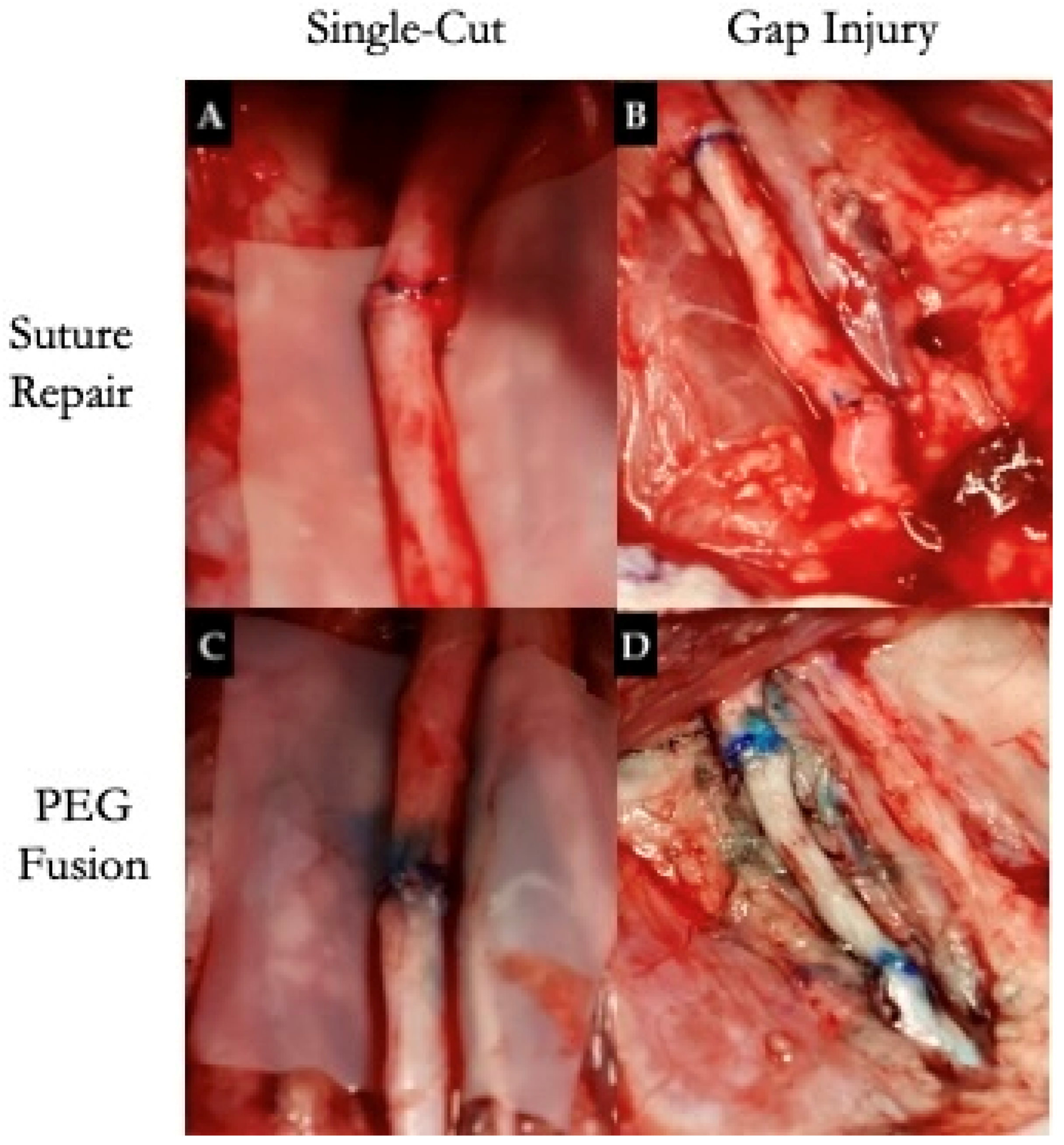
Recent innovations in nerve augmentation, such as targeted muscle reinnervation (TMR) and regenerative peripheral nerve interfaces (RPNIs), aim to enhance the functionality of limb prostheses [114]. Both techniques are designed to reduce neuroma formation and associated pain in extremity amputations, resulting in significantly improved prosthesis control and outcomes [114,115,116]. Emerging techniques, such as polyethylene glycol-mediated fusion (PEG fusion) of sciatic, facial, ulnar, and median nerves (Figure 3) and bio-scaffolds in various animals including rats and pigs, are under investigation [117,118]. PEG fusion has been applied following sharp nerve transection to promote axonal continuity by fusing severed axolemma, aiming to bypass Wallerian degeneration and accelerate recovery. Frost et al. demonstrated that PEG fusion combined with neurorrhaphy results in a faster functional return, higher CMAP, improved muscle strength, and enhanced behavioral recovery compared to standard repair in porcine median and ulnar nerve injury models [117].
Additionally, emerging biomimetic neural scaffolds are designed to more closely mimic native nerve microarchitecture and promote axonal regeneration [16,118,119]. These scaffolds are being investigated in rodent and swine models and often incorporate biomaterials, such as neural stem cells, nanofibers, and silk fibroin, which have shown promising results in supporting functional and histological regeneration. Techniques such as 3D printing, electrospinning, and molding are under active development to fabricate these constructs and optimize their biocompatibility and mechanical properties [119].
Regenerative gel (GRG) and antigliotic regenerative gel (AGRG) are being explored as fillers for nerve conduits with critical gaps of 25 mm, serving as alternatives to autologous nerve grafts [120]. Additionally, therapies involving stem cells, growth factors, nutritional support, acellular treatments with exosomes, and tissue engineering are also being reported [6,16,23,25,121].

8. Conclusions

Despite advancements in understanding nerve regeneration, PNI remains a significant clinical challenge. This review examined various preclinical animal models, highlighting their respective strengths and limitations. A major barrier to clinical translation is the lack of standardized protocols and the predominant reliance on rodent models, which may not fully replicate human pathophysiology.
By evaluating the advantages and drawbacks of each model, this review underscores the importance of selecting appropriate peripheral nerve types and animal species to enhance the relevance and effectiveness of emerging therapies. Preclinical studies have informed the development of promising intervention, including nerve conduits, cell-based treatments, targeted muscle reinnervation, and regenerative nerve interfaces, demonstrating the potential for significant clinical impact. However, the translation of these findings into clinical practice remains limited, in part due to inconsistencies in experimental design. Refining animal model selection and implementing standardized protocols are essential next steps. Undertaking this standardization will improve the reliability and comparability of preclinical data, accelerate the development of effective treatments, and ultimately improve outcomes for patients with PNIs.

Author Contributions

Conceptualization, C.M.S. and S.H.; methodology, N.A.P., M.G. and S.H.; validation, N.A.P., M.G., C.M.S. and S.H.; formal analysis, N.A.P.; investigation, N.A.P., M.G. and S.H.; data curation, N.A.P., M.G. and S.H.; writing—original draft preparation, N.A.P.; writing—review and editing, N.A.P., M.G., C.M.S. and S.H.; visualization, N.A.P.; supervision, C.M.S. and S.H. All authors have read and agreed to the published version of the manuscript.

Funding

This research received no external funding.

Institutional Review Board Statement

Not applicable.

Informed Consent Statement

Not applicable.

Data Availability Statement

No new data were created or analyzed in this study. Data sharing is not applicable to this article.

Conflicts of Interest

The authors declare no conflicts of interest.

Abbreviations

PNIPeripheral Nerve Injury
PNPeripheral nerve
C. elegans, nematodeCaenorhabditis elegans
R. pipiens, northern leopard frogRana pipiens
LPCLysophosphatidylcholine
CMAPsCompound muscle action potentials
NCVMotor nerve conduction velocity
NGFNerve growth factor
MRI Magnetic resonance imaging
MTRMagnetization transfer ratio
DTIMagnetic resonance diffusion tensor imaging
FAFractional anisotropy
ADCApparent diffusion coefficient
TMRTargeted muscle reinnervation
PEG fusionPolyethylene glycol-mediated fusion
GRGRegenerative gel
AGRGAntigliotic regenerative gel

References

  1. Burrell, J.C.; Browne, K.D.; Dutton, J.L.; Laimo, F.A.; Das, S.; Brown, D.P.; Roberts, S.; Petrov, D.; Ali, Z.; Ledebur, H.C.; et al. A Porcine Model of Peripheral Nerve Injury Enabling Ultra-Long Regenerative Distances: Surgical Approach, Recovery Kinetics, and Clinical Relevance. Neurosurgery 2020, 87, 833–846. [Google Scholar] [CrossRef] [PubMed]
  2. Muratori, L.; Ronchi, G.; Raimondo, S.; Giacobini-Robecchi, M.G.; Fornaro, M.; Geuna, S. Can Regenerated Nerve Fibers Return to Normal Size? A Long-term Post-traumatic Study of the Rat Median Nerve Crush Injury Model. Microsurgery 2012, 32, 383–387. [Google Scholar] [CrossRef] [PubMed]
  3. Siwei, Q.; Ma, N.; Wang, W.; Chen, S.; Wu, Q.; Li, Y.; Yang, Z. Construction and Effect Evaluation of Different Sciatic Nerve Injury Models in Rats. Transl. Neurosci. 2022, 13, 38–51. [Google Scholar] [CrossRef]
  4. Jones, P.E.; Meyer, R.M.; Faillace, W.J.; Landau, M.E.; Smith, J.K.; McKay, P.L.; Nesti, L.J. Combat Injury of the Sciatic Nerve–An Institutional Experience. Mil. Med. 2018, 183, e434–e441. [Google Scholar] [CrossRef]
  5. An, Y.; Yan, H.-X.; Zhao, J.-N.; Yang, X.-M.; Yan, J.-T. Evaluation Methods of a Rat Sciatic Nerve Crush Injury Model. J. Integr. Neurosci. 2022, 21, 91. [Google Scholar] [CrossRef]
  6. Casañas, J.; Torre, J.D.L.; Soler, F.; García, F.; Rodellar, C.; Pumarola, M.; Climent, J.; Soler, R.; Orozco, L. Peripheral Nerve Regeneration after Experimental Section in Ovine Radial and Tibial Nerves Using Synthetic Nerve Grafts, Including Expanded Bone Marrow Mesenchymal Cells: Morphological and Neurophysiological Results. Injury 2014, 45, S2–S6. [Google Scholar] [CrossRef]
  7. Fang, Y.; Bonini, N.M. Axon Degeneration and Regeneration: Insights from Drosophila Models of Nerve Injury. Annu. Rev. Cell Dev. Biol. 2012, 28, 575–597. [Google Scholar] [CrossRef]
  8. Bhattacharya, M.R.C. A Nerve-Wracking Buzz: Lessons from Drosophila Models of Peripheral Neuropathy and Axon Degeneration. Front. Aging Neurosci. 2023, 15, 1166146. [Google Scholar] [CrossRef]
  9. Vest, M.; Guida, A.; Colombini, C.; Cordes, K.; Pena, D.; Maki, M.; Briones, M.; Antonio, S.; Hollifield, C.; Tian, E.; et al. Closing the Gap Between Mammalian and Invertebrate Peripheral Nerve Injury: Protocol for a Novel Nerve Repair. JMIR Res. Protoc. 2020, 9, e18706. [Google Scholar] [CrossRef]
  10. Blanco, R.E.; Rosado, J.; Padilla, J.; Del Cueto, C. Ultrastructural Studies of Dorsal Root Axons Regenerating through Adult Frog Optic and Sciatic Nerves. Microsc. Res. Tech. 1999, 46, 310–318. [Google Scholar] [CrossRef]
  11. Soares, L.; Parisi, M.; Bonini, N.M. Axon Injury and Regeneration in the Adult Drosophila. Sci. Rep. 2014, 4, 6199. [Google Scholar] [CrossRef] [PubMed]
  12. DeLeonibus, A.; Rezaei, M.; Fahradyan, V.; Silver, J.; Rampazzo, A.; Bassiri Gharb, B. A META-ANALYSIS of Functional Outcomes in Rat Sciatic Nerve Injury Models. Microsurgery 2021, 41, 286–295. [Google Scholar] [CrossRef] [PubMed]
  13. Diogo, C.C.; Camassa, J.A.; Pereira, J.E.; Costa, L.M.D.; Filipe, V.; Couto, P.A.; Geuna, S.; Maurício, A.C.; Varejão, A.S. The Use of Sheep as a Model for Studying Peripheral Nerve Regeneration Following Nerve Injury: Review of the Literature. Neurol. Res. 2017, 39, 926–939. [Google Scholar] [CrossRef]
  14. Roballo, K.C.S.; Burns, D.T.; Ghnenis, A.B.; Osimanjiang, W.; Bushman, J.S. Long-term Neural Regeneration Following Injury to the Peroneal Branch of the Sciatic Nerve in Sheep. Eur. J. Neurosci. 2020, 52, 4385–4394. [Google Scholar] [CrossRef]
  15. Guo, N.; Gu, X. Sciatic Nerve Neuropathy in Cynomolgus Monkey Macaca Fascicularis: Altered Leg Usage and Peripheral Nerve Firing. J. Neurol. Neurophysiol. 2014, 05, 1000247. [Google Scholar] [CrossRef]
  16. Angius, D.; Wang, H.; Spinner, R.J.; Gutierrez-Cotto, Y.; Yaszemski, M.J.; Windebank, A.J. A Systematic Review of Animal Models Used to Study Nerve Regeneration in Tissue-Engineered Scaffolds. Biomaterials 2012, 33, 8034–8039. [Google Scholar] [CrossRef]
  17. Merolli, A.; Li, M.; Voronin, G.; Bright, L. A Sciatic Nerve Gap-Injury Model in the Rabbit. J. Mater. Sci. Mater. Med. 2022, 33, 14. [Google Scholar] [CrossRef]
  18. Drysch, M.; Wallner, C.; Schmidt, S.V.; Reinkemeier, F.; Wagner, J.M.; Lehnhardt, M.; Behr, B. An Optimized Low-Pressure Tourniquet Murine Hind Limb Ischemia Reperfusion Model: Inducing Acute Ischemia Reperfusion Injury in C57BL/6 Wild Type Mice. PLoS ONE 2019, 14, e0210961. [Google Scholar] [CrossRef]
  19. Rafee, M.; Amarpal; Kinjavdekar, P.; Aithal, H.; Wani, S.; Bhat, I. Guinea Pigs as an Animal Model for Sciatic Nerve Injury. Neural Regen. Res. 2017, 12, 452. [Google Scholar] [CrossRef]
  20. Wang, B.B.; Guo, C.; Sun, S.Q.; Zhang, X.N.; Li, Z.; Li, W.J.; Li, D.Z.; Schumacher, M.; Liu, S. Comparison of the Nerve Regeneration Capacity and Characteristics between Sciatic Nerve Crush and Transection Injury Models in Rats. Biomed. Environ. Sci. 2023, 36, 160–173. [Google Scholar] [CrossRef]
  21. Yayama, T.; Kobayashi, S.; Nakanishi, Y.; Uchida, K.; Kokubo, Y.; Miyazaki, T.; Takeno, K.; Awara, K.; Mwaka, E.S.; Iwamoto, Y.; et al. Effects of Graded Mechanical Compression of Rabbit Sciatic Nerve on Nerve Blood Flow and Electrophysiological Properties. J. Clin. Neurosci. 2010, 17, 501–505. [Google Scholar] [CrossRef] [PubMed]
  22. Rao, P.; Kotwal, P.P.; Farooque, M.; Dinda, A.K. Muscle Autografts in Nerve Gaps. Pattern of Regeneration and Myelination in Various Lengths of Graft: An Experimental Study in Guinea Pigs. J. Orthop. Sci. 2001, 6, 527–534. [Google Scholar] [CrossRef]
  23. Ding, F.; Wu, J.; Yang, Y.; Hu, W.; Zhu, Q.; Tang, X.; Liu, J.; Gu, X. Use of Tissue-Engineered Nerve Grafts Consisting of a Chitosan/Poly(Lactic- Co. -Glycolic Acid)-Based Scaffold Included with Bone Marrow Mesenchymal Cells for Bridging 50-Mm Dog Sciatic Nerve Gaps. Tissue Eng. Part A 2010, 16, 3779–3790. [Google Scholar] [CrossRef] [PubMed]
  24. Kaemmer, D.; Bozkurt, A.; Otto, J.; Junge, K.; Klink, C.; Weis, J.; Sellhaus, B.; O’Dey, D.M.; Pallua, N.; Jansen, M.; et al. Evaluation of Tissue Components in the Peripheral Nervous System Using Sirius Red Staining and Immunohistochemistry: A Comparative Study (Human, Pig, Rat). J. Neurosci. Methods 2010, 190, 112–116. [Google Scholar] [CrossRef]
  25. Zilic, L.; Garner, P.E.; Yu, T.; Roman, S.; Haycock, J.W.; Wilshaw, S. An Anatomical Study of Porcine Peripheral Nerve and Its Potential Use in Nerve Tissue Engineering. J. Anat. 2015, 227, 302–314. [Google Scholar] [CrossRef]
  26. Geuna, S. The Sciatic Nerve Injury Model in Pre-Clinical Research. J. Neurosci. Methods 2015, 243, 39–46. [Google Scholar] [CrossRef]
  27. Kaplan, H.M.; Mishra, P.; Kohn, J. The Overwhelming Use of Rat Models in Nerve Regeneration Research May Compromise Designs of Nerve Guidance Conduits for Humans. J. Mater. Sci. Mater. Med. 2015, 26, 226. [Google Scholar] [CrossRef]
  28. Li, A.; Pereira, C.; Hill, E.E.; Vukcevich, O.; Wang, A. In Vitro, In Vivo and Ex Vivo Models for Peripheral Nerve Injury andRegeneration. Curr. Neuropharmacol. 2022, 20, 344–361. [Google Scholar] [CrossRef] [PubMed]
  29. Ronchi, G.; Morano, M.; Fregnan, F.; Pugliese, P.; Crosio, A.; Tos, P.; Geuna, S.; Haastert-Talini, K.; Gambarotta, G. The Median Nerve Injury Model in Pre-Clinical Research–A Critical Review on Benefits and Limitations. Front. Cell. Neurosci. 2019, 13, 288. [Google Scholar] [CrossRef]
  30. O’Daly, A.; Rohde, C.; Brushart, T. The Topographic Specificity of Muscle Reinnervation Predicts Function. Eur. J. Neurosci. 2016, 43, 443–450. [Google Scholar] [CrossRef]
  31. Wang, D.; Huang, X.; Fu, G.; Gu, L.; Liu, X.; Wang, H.; Hu, J.; Yi, J.; Niu, X.; Zhu, Q. A Simple Model of Radial Nerve Injury in the Rhesus Monkey to Evaluate Peripheral Nerve Repair. Neural Regen. Res. 2014, 9, 1041–1046. [Google Scholar] [CrossRef]
  32. Scholz, T.; Pharaon, M.; Evans, G.R.D. Peripheral Nerve Anatomy for Regeneration Studies in Pigs: Feasibility of Large Animal Models. Ann. Plast. Surg. 2010, 65, 43–47. [Google Scholar] [CrossRef] [PubMed]
  33. Yu, F.; Yu, Y.-L.; Niu, S.-P.; Zhang, P.-X.; Yin, X.-F.; Han, N.; Zhang, Y.-J.; Zhang, D.-Y.; Kou, Y.-H.; Jiang, B.-G. Repair of Long Segmental Ulnar Nerve Defects in Rats by Several Different Kinds of Nerve Transposition. Neural Regen. Res. 2019, 14, 692–698. [Google Scholar] [CrossRef] [PubMed]
  34. Meyers, E.C.; Granja, R.; Solorzano, B.R.; Romero-Ortega, M.; Kilgard, M.P.; Rennaker, R.L.; Hays, S. Median and Ulnar Nerve Injuries Reduce Volitional Forelimb Strength in Rats. Muscle Nerve 2017, 56, 1149–1154. [Google Scholar] [CrossRef] [PubMed]
  35. Bergmeister, K.D.; Aman, M.; Riedl, O.; Manzano-Szalai, K.; Sporer, M.E.; Salminger, S.; Aszmann, O.C. Experimental Nerve Transfer Model in the Rat Forelimb. Eur. Surg. 2016, 48, 334–341. [Google Scholar] [CrossRef]
  36. Vela, F.; Martínez-Chacón, G.; Ballestín, A.; Campos, J.; Sánchez-Margallo, F.; Abellán, E. Animal Models Used to Study Direct Peripheral Nerve Repair: A Systematic Review. Neural Regen. Res. 2020, 15, 491–502. [Google Scholar] [CrossRef]
  37. Contreras, E.; Traserra, S.; Bolívar, S.; Forés, J.; Jose-Cunilleras, E.; Delgado-Martínez, I.; García, F.; Udina, E.; Navarro, X. Repair of Long Peripheral Nerve Defects in Sheep: A Translational Model for Nerve Regeneration. Int. J. Mol. Sci. 2023, 24, 1333. [Google Scholar] [CrossRef]
  38. Lin, H.; Chen, D.; Hou, C. Common Peroneal Nerve Grafting to Repair the Tibial Nerve as a Salvage Procedure in the Treatment of Sciatic Nerve Injury with Long-Segment Defects. J. Reconstr. Microsurg. Open 2018, 03, e41–e45. [Google Scholar] [CrossRef]
  39. Zhang, S.; Han, G.; Xiong, Y.; Wang, Z.; Wang, Z.; Lai, X. Characteristics and Mechanism of Lower Limb Injury Induced by Landmine Blast: A Research in a Rabbit Model. Ulus. Travma Acil Cerrahi Derg. 2023, 29, 1335–1343. [Google Scholar] [CrossRef]
  40. Ali, S.A.; Stebbins, A.W.; Hanks, J.E.; Kupfer, R.A.; Hogikyan, N.D.; Feldman, E.L.; Brenner, M.J. Facial Nerve Surgery in the Rat Model to Study Axonal Inhibition and Regeneration. JoVE 2020, 59224. [Google Scholar] [CrossRef]
  41. Hadlock, T.; Kowaleski, J.; Lo, D.; Bermejo, R.; Zeigler, H.P.; Mackinnon, S.; Heaton, J.T. Functional Assessments of the Rodent Facial Nerve: A Synkinesis Model. Laryngoscope 2008, 118, 1744–1749. [Google Scholar] [CrossRef] [PubMed]
  42. Luo, D.-S.; Zhang, T.; Zuo, C.-X.; Zuo, Z.-F.; Li, H.; Wu, S.-X.; Wang, W.; Li, Y.-Q. An Animal Model for Trigeminal Neuralgia by Compression of the Trigeminal Nerve Root. Pain. Physician 2012, 15, 187–196. [Google Scholar]
  43. Harriott, A.M.; Strother, L.C.; Vila-Pueyo, M.; Holland, P.R. Animal Models of Migraine and Experimental Techniques Used to Examine Trigeminal Sensory Processing. J. Headache Pain. 2019, 20, 91. [Google Scholar] [CrossRef]
  44. Brace, E.J.; DiAntonio, A. Models of Axon Regeneration in Drosophila. Exp. Neurol. 2017, 287 Pt 3, 310–317. [Google Scholar] [CrossRef]
  45. Bremer, J.; Skinner, J.; Granato, M. A Small Molecule Screen Identifies in Vivo Modulators of Peripheral Nerve Regeneration in Zebrafish. PLoS ONE 2017, 12, e0178854. [Google Scholar] [CrossRef]
  46. Stewart, A.M.; Braubach, O.; Spitsbergen, J.; Gerlai, R.; Kalueff, A.V. Zebrafish Models for Translational Neuroscience Research: From Tank to Bedside. Trends Neurosci. 2014, 37, 264–278. [Google Scholar] [CrossRef]
  47. Cirrincione, A.M.; Rieger, S. Analyzing Chemotherapy-Induced Peripheral Neuropathy in Vivo Using Non-Mammalian Animal Models. Exp. Neurol. 2020, 323, 113090. [Google Scholar] [CrossRef]
  48. Vega-Meléndez, G.S.; Blagburn, J.M.; Blanco, R.E. Ciliary Neurotrophic Factor and Fibroblast Growth Factor Increase the Speed and Number of Regenerating Axons after Optic Nerve Injury in Adult Rana Pipiens. J. Neurosci. Res. 2014, 92, 13–23. [Google Scholar] [CrossRef]
  49. Gordon, T.; Borschel, G.H. The Use of the Rat as a Model for Studying Peripheral Nerve Regeneration and Sprouting after Complete and Partial Nerve Injuries. Exp. Neurol. 2017, 287, 331–347. [Google Scholar] [CrossRef]
  50. Mazzer, P.Y.C.N.; Barbieri, C.H.; Mazzer, N.; Fazan, V.P.S. Morphologic and Morphometric Evaluation of Experimental Acute Crush Injuries of the Sciatic Nerve of Rats. J. Neurosci. Methods 2008, 173, 249–258. [Google Scholar] [CrossRef]
  51. Medeiros, P.; Dos Santos, I.R.; Júnior, I.M.; Palazzo, E.; Da Silva, J.A.; Machado, H.R.; Ferreira, S.H.; Maione, S.; Coimbra, N.C.; De Freitas, R.L. An Adapted Chronic Constriction Injury of the Sciatic Nerve Produces Sensory, Affective, and Cognitive Impairments: A Peripheral Mononeuropathy Model for the Study of Comorbid Neuropsychiatric Disorders Associated with Neuropathic Pain in Rats. Pain. Med. 2021, 22, 338–351. [Google Scholar] [CrossRef] [PubMed]
  52. Kim, H.W.; Shim, S.W.; Zhao, A.M.; Roh, D.; Han, H.M.; Middleton, S.J.; Kim, W.; Chung, S.; Johnson, E.; Prentice, J.; et al. Long-Term Tactile Hypersensitivity after Nerve Crush Injury in Mice Is Characterized by the Persistence of Intact Sensory Axons. Pain 2023, 164, 2327–2342. [Google Scholar] [CrossRef]
  53. Umansky, D.; Hagen, K.M.; Chu, T.H.; Pathiyil, R.K.; Alzahrani, S.; Ousman, S.S.; Midha, R. Functional Gait Assessment Using Manual, Semi-Automated and Deep Learning Approaches Following Standardized Models of Peripheral Nerve Injury in Mice. Biomolecules 2022, 12, 1355. [Google Scholar] [CrossRef]
  54. Hammers, D.W.; Matheny, R.W.; Sell, C.; Adamo, M.L.; Walters, T.J.; Estep, J.S.; Farrar, R.P. Impairment of IGF-I Expression and Anabolic Signaling Following Ischemia/Reperfusion in Skeletal Muscle of Old Mice. Exp. Gerontol. 2011, 46, 265–272. [Google Scholar] [CrossRef]
  55. Bonheur, J.A.; Albadawi, H.; Patton, G.M.; Watkins, M.T. A Noninvasive Murine Model of Hind Limb Ischemia-Reperfusion Injury. J. Surg. Res. 2004, 116, 55–63. [Google Scholar] [CrossRef] [PubMed]
  56. Wang, G.J.; Baugher, W.H.; Stamp, W.G. Epiphyseal Transplant in Amputations. Effects on Overgrowth in a Rabbit Model. Clin. Orthop. Relat. Res. 1978, 285–288. [Google Scholar]
  57. Huang, H.; Chen, L.; Zhang, H.; Li, S.; Liu, P.; Zhao, T.; Li, C. Autophagy Promotes Peripheral Nerve Regeneration and Motor Recovery Following Sciatic Nerve Crush Injury in Rats. J. Mol. Neurosci. 2016, 58, 416–423. [Google Scholar] [CrossRef]
  58. Kosacka, J.; Nowicki, M.; Blüher, M.; Baum, P.; Stockinger, M.; Toyka, K.V.; Klöting, I.; Stumvoll, M.; Serke, H.; Bechmann, I.; et al. Increased Autophagy in Peripheral Nerves May Protect Wistar Ottawa Karlsburg W Rats against Neuropathy. Exp. Neurol. 2013, 250, 125–135. [Google Scholar] [CrossRef]
  59. Alvites, R.; Lopes, B.; Sousa, P.; Sousa, A.C.; Coelho, A.; Moreira, A.; Rêma, A.; Atayde, L.; Mendonça, C.; Luís, A.L.; et al. Ultrasound Landmarks in the Approach to the Common Peroneal Nerve in a Sheep Model—Application in Peripheral Nerve Regeneration. Life 2023, 13, 1919. [Google Scholar] [CrossRef]
  60. Xue, C.; Hu, N.; Gu, Y.; Yang, Y.; Liu, Y.; Liu, J.; Ding, F.; Gu, X. Joint Use of a Chitosan/PLGA Scaffold and MSCs to Bridge an Extra Large Gap in Dog Sciatic Nerve. Neurorehabil Neural Repair. 2012, 26, 96–106. [Google Scholar] [CrossRef]
  61. Attar, B.M.; Zalzali, H.; Razavi, M.; Ghoreishian, M.; Rezaei, M. Effectiveness of Fibrin Adhesive in Facial Nerve Anastomosis in Dogs Compared With Standard Microsuturing Technique. J. Oral. Maxillofac. Surg. 2012, 70, 2427–2432. [Google Scholar] [CrossRef]
  62. Archibald, S.J.; Krarup, C.; Shefner, J.; Li, S.; Madison, R.D. A Collagen-based Nerve Guide Conduit for Peripheral Nerve Repair: An Electrophysiological Study of Nerve Regeneration in Rodents and Nonhuman Primates. J. Comp. Neurol. 1991, 306, 685–696. [Google Scholar] [CrossRef]
  63. Yao, Y.; Cui, Y.; Zhao, Y.; Xiao, Z.; Li, X.; Han, S.; Chen, B.; Fang, Y.; Wang, P.; Pan, J.; et al. Efect of Longitudinally Oriented Collagen Conduit Combined with Nerve Growth Factor on Nerve Regeneration after Dog Sciatic Nerve Injury. J. Biomed. Mater. Res. 2018, 106, 2131–2139. [Google Scholar] [CrossRef]
  64. Lu, Q.; Gu, L.; Jiang, L.; Qin, B.; Fu, G.; Li, X.; Yang, J.; Huang, X.; Yang, Y.; Zhu, Q.; et al. The Upper Brachial Plexus Defect Model in Rhesus Monkeys: A Cadaveric Feasibility Study. NeuroReport 2013, 24, 884–888. [Google Scholar] [CrossRef]
  65. Hu, N.; Wu, H.; Xue, C.; Gong, Y.; Wu, J.; Xiao, Z.; Yang, Y.; Ding, F.; Gu, X. Long-Term Outcome of the Repair of 50 Mm Long Median Nerve Defects in Rhesus Monkeys with Marrow Mesenchymal Stem Cells-Containing, Chitosan-Based Tissue Engineered Nerve Grafts. Biomaterials 2013, 34, 100–111. [Google Scholar] [CrossRef]
  66. Lopes, B.; Coelho, A.; Alvites, R.; Sousa, A.C.; Sousa, P.; Moreira, A.; Atayde, L.; Salgado, A.; Geuna, S.; Maurício, A.C. Animal Models in Peripheral Nerve Transection Studies: A Systematic Review on Study Design and Outcomes Assessment. Regen. Med. 2024, 19, 189–203. [Google Scholar] [CrossRef]
  67. Hanna, A.S.; Hellenbrand, D.J.; Schomberg, D.T.; Salamat, S.M.; Loh, M.; Wheeler, L.; Hanna, B.; Ozaydin, B.; Meudt, J.; Shanmuganayagam, D. Brachial Plexus Anatomy in the Miniature Swine as Compared to Human. J. Anat. 2022, 240, 172–181. [Google Scholar] [CrossRef] [PubMed]
  68. Hort-Legrand, C.; Noah, L.; Mériguet, E.; Mésangeau, D. Motor and Sensory Nerve Conduction Velocities in Yucatan Minipigs. Lab. Anim. 2006, 40, 53–57. [Google Scholar] [CrossRef]
  69. Sufan, W.; Suzuki, Y.; Tanihara, M.; Ohnishi, K.; Suzuki, K.; Endo, K.; Nishimura, Y. Sciatic Nerve Regeneration through Alginate with Tubulation or Nontubulation Repair in Cat. J. Neurotrauma 2001, 18, 329–338. [Google Scholar] [CrossRef]
  70. Bisby, M.A.; Pollock, B. Increased Regeneration Rate in Peripheral Nerve Axons Following Double Lesions: Enhancement of the Conditioning Lesion Phenomenon. J. Neurobiol. 1983, 14, 467–472. [Google Scholar] [CrossRef]
  71. Forman, D.S.; Wood, D.K.; DeSilva, S. Rate of Regeneration of Sensory Axons in Transected Rat Sciatic Nerve Repaired with Epineurial Sutures. J. Neurol. Sci. 1979, 44, 55–59. [Google Scholar] [CrossRef] [PubMed]
  72. Sjöberg, J.; Kanje, M. The Initial Period of Peripheral Nerve Regeneration and the Importance of the Local Environment for the Conditioning Lesion Effect. Brain Res. 1990, 529, 79–84. [Google Scholar] [CrossRef] [PubMed]
  73. Grinsell, D.; Keating, C.P. Peripheral Nerve Reconstruction after Injury: A Review of Clinical and Experimental Therapies. BioMed Res. Int. 2014, 2014, 1–13. [Google Scholar] [CrossRef]
  74. Mohanty, C.; Bhat, D.; Devi, B. Use of Animal Models in Peripheral Nerve Surgery and Research. Neurol. India 2019, 67, S100–S105. [Google Scholar] [CrossRef]
  75. Gaudet, A.D.; Popovich, P.G.; Ramer, M.S. Wallerian Degeneration: Gaining Perspective on Inflammatory Events after Peripheral Nerve Injury. J. Neuroinflamm. 2011, 8, 110. [Google Scholar] [CrossRef]
  76. Tajima, K.; Tohyama, K.; Ide, C.; Abe, M. Regeneration through Nerve Allografts in the Cynomolgus Monkey (Macaca Fascicularis). J. Bone Jt. Surg. Am. 1991, 73, 172–185. [Google Scholar] [CrossRef]
  77. Dash, H.; Kononov, A.; Prayson, R.A.; Petras, S.; Browne, E.Z. Evaluation of Nerve Recovery From Minimal-Duration Crush Injury. Ann. Plast. Surg. 1996, 37, 526–531. [Google Scholar] [CrossRef]
  78. Lee, J.I.; Wandling, G.; Talukder, M.A.H.; Elfar, J.; Govindappa, P.K. A Novel Standardized Peripheral Nerve Transection Method and a Novel Digital Pressure Sensor Device Construction for Peripheral Nerve Crush Injury. BIO-Protoc. 2022, 12, e4350. [Google Scholar] [CrossRef]
  79. Lopes, B.; Sousa, P.; Alvites, R.; Branquinho, M.; Sousa, A.C.; Mendonça, C.; Atayde, L.M.; Luís, A.L.; Varejão, A.S.P.; Maurício, A.C. Peripheral Nerve Injury Treatments and Advances: One Health Perspective. Int. J. Mol. Sci. 2022, 23, 918. [Google Scholar] [CrossRef]
  80. Jessen, K.R.; Mirsky, R.; Lloyd, A.C. Schwann Cells: Development and Role in Nerve Repair. Cold Spring Harb. Perspect. Biol. 2015, 7, a020487. [Google Scholar] [CrossRef]
  81. Wu, W.; Niu, Y.; Kong, X.; Liu, D.; Long, X.; Shu, S.; Su, X.; Wang, B.; Liu, X.; Ma, Y.; et al. Application of Diffusion Tensor Imaging in Quantitatively Monitoring Chronic Constriction Injury of Rabbit Sciatic Nerves: Correlation with Histological and Functional Changes. Br. J. Radiol. 2018, 91, 20170414. [Google Scholar] [CrossRef] [PubMed]
  82. Bennett, G.J.; Xie, Y.-K. A Peripheral Mononeuropathy in Rat That Produces Disorders of Pain Sensation like Those Seen in Man. Pain. 1988, 33, 87–107. [Google Scholar] [CrossRef]
  83. Wang, C.; Chen, P.; Lin, D.; Chen, Y.; Lv, B.; Zheng, K.; Lin, X.; Wu, Z. Effects of Varying Degrees of Ligation in a Neuropathic Pain Model Induced by Chronic Constriction Injury. Life Sci. 2021, 276, 119441. [Google Scholar] [CrossRef]
  84. Crawford, R.S.; Hashmi, F.F.; Jones, J.E.; Albadawi, H.; McCormack, M.; Eberlin, K.; Entabi, F.; Atkins, M.D.; Conrad, M.F.; Austen, W.G.; et al. A Novel Model of Acute Murine Hindlimb Ischemia. Am. J. Physiol.-Heart Circ. Physiol. 2007, 292, H830–H837. [Google Scholar] [CrossRef]
  85. Heemskerk, A.M.; Drost, M.R.; Van Bochove, G.S.; Van Oosterhout, M.F.M.; Nicolay, K.; Strijkers, G.J. DTI-based Assessment of Ischemia-reperfusion in Mouse Skeletal Muscle. Magn. Reson. Med. 2006, 56, 272–281. [Google Scholar] [CrossRef] [PubMed]
  86. Vignaud, A.; Hourde, C.; Medja, F.; Agbulut, O.; Butler-Browne, G.; Ferry, A. Impaired Skeletal Muscle Repair after Ischemia-Reperfusion Injury in Mice. J. Biomed. Biotechnol. 2010, 2010, 724914. [Google Scholar] [CrossRef]
  87. Bain, J.R.; Mackinnon, S.E.; Hunter, D.A. Functional Evaluation of Complete Sciatic, Peroneal, and Posterior Tibial Nerve Lesions in the Rat. Plast. Reconstr. Surg. 1989, 83, 129–136. [Google Scholar] [CrossRef]
  88. Wood, M.D.; Kemp, S.W.P.; Weber, C.; Borschel, G.H.; Gordon, T. Outcome Measures of Peripheral Nerve Regeneration. Ann. Anat.-Anat. Anz. 2011, 193, 321–333. [Google Scholar] [CrossRef]
  89. Di Scipio, F.; Raimondo, S.; Tos, P.; Geuna, S. A Simple Protocol for Paraffin-Embedded Myelin Sheath Staining with Osmium Tetroxide for Light Microscope Observation. Microsc. Res. Tech. 2008, 71, 497–502. [Google Scholar] [CrossRef]
  90. Wang, H.; Sorenson, E.J.; Spinner, R.J.; Windebank, A.J. Electrophysiologic Findings and Grip Strength after Nerve Injuries in the Rat Forelimb. Muscle Nerve 2008, 38, 1254–1265. [Google Scholar] [CrossRef]
  91. Heinzel, J.; Längle, G.; Oberhauser, V.; Hausner, T.; Kolbenschlag, J.; Prahm, C.; Grillari, J.; Hercher, D. Use of the CatWalk Gait Analysis System to Assess Functional Recovery in Rodent Models of Peripheral Nerve Injury–a Systematic Review. J. Neurosci. Methods 2020, 345, 108889. [Google Scholar] [CrossRef] [PubMed]
  92. Bozkurt, A.; Deumens, R.; Scheffel, J.; O’Dey, D.M.; Weis, J.; Joosten, E.A.; Führmann, T.; Brook, G.A.; Pallua, N. CatWalk Gait Analysis in Assessment of Functional Recovery after Sciatic Nerve Injury. J. Neurosci. Methods 2008, 173, 91–98. [Google Scholar] [CrossRef] [PubMed]
  93. De Medinaceli, L.; Freed, W.J.; Wyatt, R.J. An Index of the Functional Condition of Rat Sciatic Nerve Based on Measurements Made from Walking Tracks. Exp. Neurol. 1982, 77, 634–643. [Google Scholar] [CrossRef]
  94. Deuis, J.R.; Dvorakova, L.S.; Vetter, I. Methods Used to Evaluate Pain Behaviors in Rodents. Front. Mol. Neurosci. 2017, 10, 284. [Google Scholar] [CrossRef]
  95. Li, X.; Shen, J.; Chen, J.; Wang, X.; Liu, Q.; Liang, B. Magnetic Resonance Imaging Evaluation of Acute Crush Injury of Rabbit Sciatic Nerve: Correlation with Histology. Can. Assoc. Radiol. J. 2008, 59, 123–130. [Google Scholar]
  96. Zhu, Y.; Jin, Z.; Luo, Y.; Wang, Y.; Peng, N.; Peng, J.; Wang, Y.; Yu, B.; Lu, C.; Zhang, S. Evaluation of the Crushed Sciatic Nerve and Denervated Muscle with Multimodality Ultrasound Techniques: An Animal Study. Ultrasound Med. Biol. 2020, 46, 377–392. [Google Scholar] [CrossRef]
  97. Giorgetti, E.; Obrecht, M.; Ronco, M.; Panesar, M.; Lambert, C.; Accart, N.; Doelemeyer, A.; Nash, M.; Bidinosti, M.; Beckmann, N. Magnetic Resonance Imaging as a Biomarker in Rodent Peripheral Nerve Injury Models Reveals an Age-Related Impairment of Nerve Regeneration. Sci. Rep. 2019, 9, 13508. [Google Scholar] [CrossRef]
  98. Sun, C.; Hou, Z.; Hong, G.; Wan, Q.; Li, X. In Vivo Evaluation of Sciatic Nerve Crush Injury Using Diffusion Tensor Imaging: Correlation With Nerve Function and Histology. J. Comput. Assist. Tomogr. 2014, 38, 790–796. [Google Scholar] [CrossRef]
  99. Yamasaki, T.; Fujiwara, H.; Oda, R.; Mikami, Y.; Ikeda, T.; Nagae, M.; Shirai, T.; Morisaki, S.; Ikoma, K.; Masugi-Tokita, M.; et al. In Vivo Evaluation of Rabbit Sciatic Nerve Regeneration with Diffusion Tensor Imaging (DTI): Correlations with Histology and Behavior. Magn. Reson. Imaging 2015, 33, 95–101. [Google Scholar] [CrossRef]
  100. Farinas, A.F.; Pollins, A.C.; Stephanides, M.; O’Neill, D.; Al-Kassis, S.; Esteve, I.V.M.; Colazo, J.M.; Keller, P.R.; Rankin, T.; Wormer, B.A.; et al. Diffusion Tensor Tractography to Visualize Axonal Outgrowth and Regeneration in a 4-Cm Reverse Autograft Sciatic Nerve Rabbit Injury Model. Neurol. Res. 2019, 41, 257–264. [Google Scholar] [CrossRef]
  101. Ricci, V.; Ricci, C.; Cocco, G.; Gervasoni, F.; Donati, D.; Farì, G.; Özçakar, L. Histopathology and High-Resolution Ultrasound Imaging for Peripheral Nerve (Injuries). J. Neurol. 2022, 269, 3663–3675. [Google Scholar] [CrossRef] [PubMed]
  102. Langford, D.J.; Bailey, A.L.; Chanda, M.L.; Clarke, S.E.; Drummond, T.E.; Echols, S.; Glick, S.; Ingrao, J.; Klassen-Ross, T.; LaCroix-Fralish, M.L.; et al. Coding of Facial Expressions of Pain in the Laboratory Mouse. Nat. Methods 2010, 7, 447–449. [Google Scholar] [CrossRef] [PubMed]
  103. Kang, H.; Lichtman, J.W. Motor Axon Regeneration and Muscle Reinnervation in Young Adult and Aged Animals. J. Neurosci. 2013, 33, 19480–19491. [Google Scholar] [CrossRef]
  104. Maita, K.C.; Garcia, J.P.; Avila, F.R.; Torres-Guzman, R.A.; Ho, O.; Chini, C.C.S.; Chini, E.N.; Forte, A.J. Evaluation of the Aging Effect on Peripheral Nerve Regeneration: A Systematic Review. J. Surg. Res. 2023, 288, 329–340. [Google Scholar] [CrossRef]
  105. Brown, T.J.; Khan, T.; Jones, K.J. Androgen Induced Acceleration of Functional Recovery after Rat Sciatic Nerve Injury. Restor. Neurol. Neurosci. 1999, 15, 289–295. [Google Scholar] [CrossRef]
  106. Azarkish, F.; Armin, F.; Parvar, A.A.A.; Dehghani, A. The Influence of Renal Ischemia-Reperfusion Injury on Remote Organs: The Histological Brain Changes in Male and Female Rats. Brain Circ. 2021, 7, 194–200. [Google Scholar] [CrossRef]
  107. Syu, W.-Z.; Hueng, D.-Y.; Chen, W.-L.; Chan, J.Y.-H.; Chen, S.-G.; Huang, S.-M. Adipose-Derived Neural Stem Cells Combined with Acellular Dermal Matrix as a Neural Conduit Enhances Peripheral Nerve Repair. Cell Transplant. 2019, 28, 1220–1230. [Google Scholar] [CrossRef]
  108. Lavorato, A.; Raimondo, S.; Boido, M.; Muratori, L.; Durante, G.; Cofano, F.; Vincitorio, F.; Petrone, S.; Titolo, P.; Tartara, F.; et al. Mesenchymal Stem Cell Treatment Perspectives in Peripheral Nerve Regeneration: Systematic Review. Int. J. Mol. Sci. 2021, 22, 572. [Google Scholar] [CrossRef]
  109. Daeschler, S.C.; Feinberg, K.; Harhaus, L.; Kneser, U.; Gordon, T.; Borschel, G.H. Advancing Nerve Regeneration: Translational Perspectives of Tacrolimus (FK506). Int. J. Mol. Sci. 2023, 24, 12771. [Google Scholar] [CrossRef]
  110. Kim, J.; Choi, Y.E.; Kim, J.H.; Lee, S.H.; Oh, S.; Kim, S.H. Nerve Repair and Orthodromic and Antidromic Nerve Grafts: An Experimental Comparative Study in Rabbit. Biomed. Res. Int. 2020, 2020, 5046832. [Google Scholar] [CrossRef]
  111. Sun, X.; Wang, W.; Dai, J.; Jin, S.; Huang, J.; Guo, C.; Wang, C.; Pang, L.; Wang, Y. A Long-Term and Slow-Releasing Hydrogen Sulfide Donor Protects against Myocardial Ischemia/Reperfusion Injury. Sci. Rep. 2017, 7, 3541. [Google Scholar] [CrossRef] [PubMed]
  112. Wetzel, M.D.; Wenke, J.C. Mechanisms by Which Hydrogen Sulfide Attenuates Muscle Function Following Ischemia–Reperfusion Injury: Effects on Akt Signaling, Mitochondrial Function, and Apoptosis. J. Transl. Med. 2019, 17, 33. [Google Scholar] [CrossRef]
  113. Ball, C.J.; Reiffel, A.J.; Chintalapani, S.; Kim, M.; Spector, J.A.; King, M.R. Hydrogen Sulfide Reduces Neutrophil Recruitment in Hind-Limb Ischemia-Reperfusion Injury in an L-Selectin and ADAM-17–Dependent Manner. Plast. Reconstr. Surg. 2013, 131, 487–497. [Google Scholar] [CrossRef]
  114. Kurlander, D.E.; Wee, C.; Chepla, K.J.; Lineberry, K.D.; Long, T.C.; Gillis, J.A.; Valerio, I.L.; Khouri, J.S. TMRpni: Combining Two Peripheral Nerve Management Techniques. Plast. Reconstr. Surg.-Glob. Open 2020, 8, e3132. [Google Scholar] [CrossRef]
  115. Hooper, R.C.; Cederna, P.S.; Brown, D.L.; Haase, S.C.; Waljee, J.F.; Egeland, B.M.; Kelley, B.P.; Kung, T.A. Regenerative Peripheral Nerve Interfaces for the Management of Symptomatic Hand and Digital Neuromas. Plast. Reconstr. Surg.-Glob. Open 2020, 8, e2792. [Google Scholar] [CrossRef]
  116. Kuiken, T.A.; Barlow, A.K.; Hargrove, L.J.; Dumanian, G.A. Targeted Muscle Reinnervation for the Upper and Lower Extremity. Tech. Orthop. 2017, 32, 109–116. [Google Scholar] [CrossRef]
  117. Frost, C.; Salous, A.; Ketheeswaran, S.; Ngaage, L.M.; Hanwright, P.J.; Ghergherehchi, C.; Tuffaha, S.; Vaidya, D.; Bittner, G.D.; Brandacher, G.; et al. Polyethylene Glycol Fusion Restores Axonal Continuity and Improves Return of Function in a Rat Median Nerve Denervation Model. Plast. Reconstr. Surg. 2023. [Google Scholar] [CrossRef]
  118. Du, J.; Chen, H.; Qing, L.; Yang, X.; Jia, X. Biomimetic Neural Scaffolds: A Crucial Step towards Optimal Peripheral Nerve Regeneration. Biomater. Sci. 2018, 6, 1299–1311. [Google Scholar] [CrossRef] [PubMed]
  119. Mankavi, F.; Ibrahim, R.; Wang, H. Advances in Biomimetic Nerve Guidance Conduits for Peripheral Nerve Regeneration. Nanomaterials 2023, 13, 2528. [Google Scholar] [CrossRef]
  120. Rochkind, S.; Almog, M.; Meilin, S.; Nevo, Z. Reviving Matrix for Nerve Reconstruction in Rabbit Model of Chronic Peripheral Nerve Injury With Massive Loss Defect. Front. Surg. 2020, 7, 609638. [Google Scholar] [CrossRef]
  121. Martinez De Albornoz, P.; Delgado, P.J.; Forriol, F.; Maffulli, N. Non-Surgical Therapies for Peripheral Nerve Injury. Br. Med. Bull. 2011, 100, 73–100. [Google Scholar] [CrossRef]
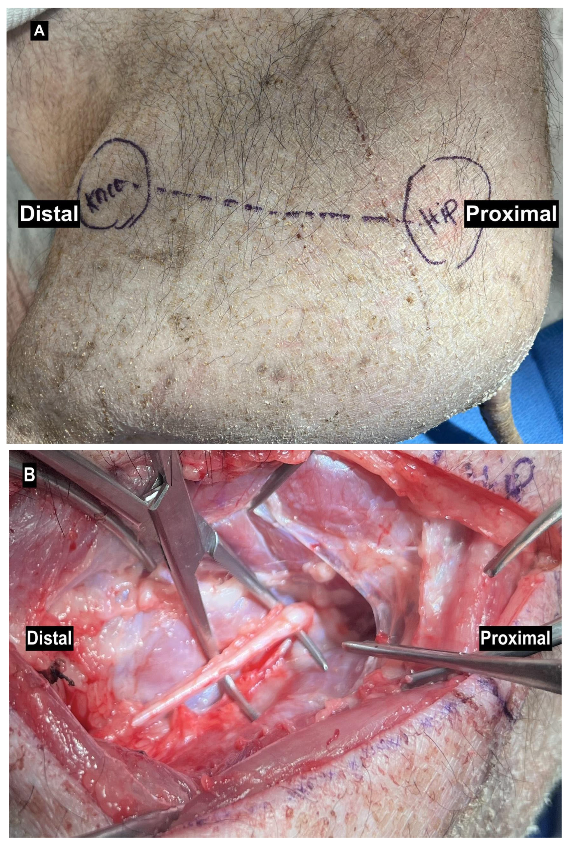
Figure 1. (A) Lateral surgical approach to expose the sciatic nerve of a Yucatan pig in a lateral decubitus position. (B) Exposed sciatic nerve in the same position.
Anatomia 04 00008 g001
Figure 2. (A) Exposure and identification of the median and ulnar nerves in a Yucatan pig. (B) Close-up of the median nerve trunk, highlighting the epineurial sheath that encloses and protects the underlying fascicles, similar to human nerve anatomy. A sterile ruler is included for scale.
Anatomia 04 00008 g002
Figure 3. Porcine median nerve injury model. (A) Single-cut injury with simple suture repair. (B) Segmental nerve defect repaired with standard neurorrhaphy. (C) Single-cut injury repaired with PEG fusion following epineurial repair. (D) Segmental reconstruction using an ipsilateral ulnar nerve autograft and PEG fusion.
Anatomia 04 00008 g003
Table 1. This table highlights the strengths and limitations of commonly used small animal models in nerve injury research.
Small AnimalsStrengthsLimitationsStudies
Rats
(Sprague Dawley, Wistar, Long Evans, and Lewis)
  • Similar size, fascicular organization, and morphology
  • Short nerve gaps
  • Economical
  • Easy to house and handle
  • Resistant to surgical infections
  • Can be studied in mass
  • Short reproductive and gestational cycles
  • Predominantly utilized sciatic nerve model
  • Increased regenerative capacity
  • Fewer transgenic models compared to mice
  • Spontaneous axonal regeneration
[2,3,5,12,20,26,27,49,50,51]
Mice
(C57BL/6J, C57/Bl6, B6, and 129SF2/J)
  • Genetically modified and transgenic technology
  • Easy to house and handle
  • Economical
  • Widely accepted as a research animal
  • Can be studied in mass
  • Short nerve gaps
  • Hypersensitivity to mechanical stimuli (Kim)
[18,52,53,54,55]
Guinea Pigs
(Cavia porcellus and Duncan-Hartley)
  • Cooperative, docile nature
  • Complex behavioral analysis
  • Easier to perform electrophysiological studies
  • Larger nerve size compared to rodents
  • Ulcer formation
  • Cost
  • Fewer genetically modified strains and tools
  • Not as widely accepted as a research animal
[19,22]
Rabbits
(New Zealand White and Japanese White)
  • Longer nerve gaps
  • Can be useful in artificial nerve guides
  • Similar neuroanatomy, physiology, and nerve scaffolds
  • Docile nature
  • Ease of handling and care
  • Complex behavioral analysis
  • Autophagia, self-mutilation, and pressure ulcers
  • Expensive, especially with post-surgical complications
[16,17,21,56]
Table 2. This table highlights the strengths and limitations of commonly used large animal models in nerve injury research.
Large AnimalsStrengthsLimitationsStudies
Sheep (Ovis aries, Merino, or Suffolk breed)
  • Known age equivalents
  • Similar nerve anatomy and regenerative capabilities to humans
  • Long nerve gaps
  • Accepted as a lab animal
  • Low infection rates and hoof desensitization
  • Low costs
  • Challenging surgical procedures
  • Lengthy study times
  • Husbandry requirements
  • Regulatory challenges
[6,13,14,59]
Porcine (Yucatan Minipigs, German House Pig, and Yorkshire Pigs)
  • Model complex nerve injuries
  • Flexibility in nerve gap lengths
  • Total regenerative distance analysis
  • Similar nerve anatomy and size to humans
  • Hoof drop is analogous to foot drop in humans
  • Established lab animal
  • Compensatory mechanisms
  • Cannot evaluate dexterous movement
  • Time and resource intensive
  • Costly
[1,24,25]
Canine
(Male Beagle Dogs and Dogs (18–24 kg; breed not specified)
  • Large nerve gaps
  • Comparable anatomy, physiology, and pathology to humans
  • Number of nerve fibers in each nerve is similar to humans
  • Ethical considerations
  • Costly
[23,60,61,63]
Monkey
(Macaca fascicularis or Cynomolgus Rhesus)
  • Similar neuroanatomy and physiology to humans
  • Neuropathic pain behaviors resemble human behavior
  • Nerve gaps up to 6 cm
  • Ethical considerations
  • Heterogeneity of genetic origins
  • Longer study periods
  • Husbandry and maintenance
  • Costly
[15,31,62,64,65]
Table 3. Summary of the strengths and limitations of various peripheral nerve injury models.
Peripheral Nerve Injury ModelsStrengthsLimitations
Crush
  • Reproducible.
  • Low skill requirement.
  • Easy visualization of changes grossly and molecularly.
  • Preserves tissue architecture; maintains connective tissue and nerve structures.
  • Analysis with electrophysiological examination and sciatic nerve functional index.
  • Limited clinical translatability.
  • Non-specific injury.
  • Difficulty in standardization.
  • Difficulty in functional recovery assessment.
Transection
  • Mimics chronic denervation and clinical injury models.
  • Sunderland grade V.
  • Analysis of regenerative mechanisms.
  • Stable.
  • Low and inconsistent reproducibility.
  • Slower functional and behavioral recovery.
  • Requires microsurgical repair.
Chronic Constriction
  • Reproduces hyperalgesia and nerve defects in neuropathic pain syndromes.
  • Qualitative assessment.
  • Varying limb circumferences.
  • Lack of standardization in tightness and constriction.
  • Collateral blood flow.
  • Decreasing tension over time.
  • Limited quantitative assessment.
Chemical Damage
  • Reproducible.
  • Potential for standardization.
  • Mimics neuropathy.
  • Incomplete damage.
  • Poor stability.
  • Quick recovery.
Ischemia-Reperfusion
  • Reliable.
  • Low cost.
  • Non-invasive.
  • Adjustable for various limb circumferences.
  • Reproducible.
  • Translatable.
  • Risk of hemorrhagic infection.
  • Blood flow collaterals.
  • Risk of nonspecific edema.
Table 4. A summary of published experimental studies on peripheral nerve injury models, including reference, title, animal model, technique, main outcome, and limitations. Systematic reviews, meta-analyses, and literature reviews were excluded.
ReferenceTitle Animal Model TechniqueMain Outcome
Burrell et al. (2020)
[1]
A Porcine Model of Peripheral Nerve Injury Enabling Ultra-Long Regenerative Distances: Surgical Approach, Recovery Kinetics, and Clinical Relevance (October 2020) Yucatan Minipigs Repair of common peroneal nerve (CPN) and deep peroneal nerve (DPN) long segmental defects and repair using a 5cm saphenous or sural nerve autograft.
  • Maximum repair length: 4–5 cm CPN, 10 cm DPN.
  • Maximum regenerative distance: up to 27 cm CPN, up to 20 cm DPN.
Siwei (2022)
[3]
Construction and Effect Evaluation of Different Sciatic Nerve Injury Models in Rats Sprague Dawley RatsTransverse, clamp, keep epineurium and axon cutting, and chemical damage.
  • Transverse injury models exhibit the most stable effects.
  • Chemical injury models recover quickly but cause incomplete damage with poor stability.
  • Clamp and epineurium injury models show medium stability, with no significant differences in many aspects.
Merolli (2022)
[17]
A sciatic nerve gap-injury model in the rabbitNew Zealand White RabbitsTransection with artificial nerve guides.
  • Documentation of nerve regeneration as long as 26 mm.
Muratori (2012)
[2]
Can Regenerated Nerve Fibers Return to Normal Size? A Long-Term Post-Traumatic Study of the Rat Median Nerve Crush Injury Model Female Wistar RatsCrush median nerve via non-serrated clamp.
  • Fiber number normalized by week 24.
  • Without adjunct treatment, full recovery does not occur spontaneously.
Drysch (2019)
[18]
An Optimized Low-Pressure Tourniquet Murine Hind Limb Ischemia Reperfusion Mode: Inducing Acute Ischemia Reperfusion Injury in C57BL/6 Wild Type MiceC57BL/6J mice Tourniquet and artery clamping.
  • The low-pressure tourniquet reliably produces reproducible ischemia-reperfusion injury.
An (2022)
[5]
Evaluation methods of a rat sciatic nerve crush injury model Sprague–Dawley (SD) male ratsCrush injury.
  • Neuroelectrophysiological examination reflected morphological changes in PNI.
Casañas (2014)
[6]
Peripheral nerve regeneration after experimental section in ovine radial and tibial nerves using synthetic nerve grafts, including expanded bone marrow mesenchymal cells: morphological and neurophysiological results Sheep Synthetic nerve grafts (bone marrow mesenchymal cells) using radial and tibial nerve.
  • MSC biodegradable scaffolds promote regeneration and functional recovery.
Roballo (2020)
[14]
Long-term neural regeneration following injury to the peroneal branch of the sciatic nerve in sheepMerino or Suffolk breed sheepBisection, 5 cm reverse autograft, and sham surgery.
  • Bisections lead to improved CMAPs, increased muscle mass, and reduced fibrosis compared to reverse autografts.
Guo (2014)
[15]
Sciatic Nerve Neuropathy in Cynomolgus Monkey Macaca Fascicularis: Altered Leg Usage and Peripheral Nerve Firing Macaca fascicularis Monkeys Mild injury to sciatic nerve via incomplete constriction with ligature.
  • Sciatic nerve injury resulted in hypersensitization, long-lasting malfunction, and neuropathy.
Alvites (2021)
[59]
Establishment of a Sheep Model for Hind Limb Peripheral Nerve Injury: Common Peroneal Nerve Ovis aries Sheep Surgical protocol for common peroneal nerve, including baseline controls using crush injuries and neurotmesis, with repair variables (end-to-end, nerve guidance conduit, and axonotmesis).
  • Sheep are a valid model for PNI at 12 and 24 weeks.
Rafee (2017)
[19]
Guinea Pigs as an Animal Model for Sciatic Nerve Injury Cavia porcellus Guinea Pigs Crush injury.
  • Guinea pigs could serve as an alternative model, with functional, macroscopic, microscopic, and ultrastructural changes observed in the sciatic nerve and gastrocnemius.
Yayama (2010)
[21]
Effect of Graded Mechanical Compression of Rabbit Sciatic Nerve on Nerve Blood Flow and Electrophysiological Properties Japanese white rabbits Clamped with a custom compressor to investigate the relationship between compressive force on the nerve and (i) intraneural blood flow and (ii) compound nerve action potentials.
  • Intraneural edema may impair nerve function.
Rao (2001)
[22]
Muscle autografts in nerve gaps. Pattern of regeneration and myelination in various lengths of graft: an experimental study in guinea pigsDuncan-Hartley guinea pigs Evaluated different autograft graft lengths.
  • The length of the graft is an important consideration, with shorter grafts enhancing myelination and axonal regeneration.
Ding (2010)
[23]
Use of Tissue-Engineered Nerve Grafts Consisting of a Chitosan/Poly (lactic-co-glycolic acid)-Based Scaffold Included with Bone Marrow Mesenchymal Cells for Bridging 50-mm Dog Sciatic Nerve Gaps Male Beagle dogs Chitosan/PLGA-based neural scaffold combined with autologous bone marrow mesenchymal stem cells (MSCs).
  • The addition of MSCs promoted sciatic nerve regeneration and functional recovery, demonstrating significant efficacy comparable to nerve autografting and superior to the scaffold alone.
Kaemmer (2010)
[24]
Evaluation of tissue components in the peripheral nervous system using Sirius red staining and immunohistochemistry: A comparative study (human, pig, rat) Human, Rat (Lewis inbred rats), Pig (German house)Evaluation of collagen (stroma) and nerve fibers (parenchyma) in different species.
  • Similar collagen type I and III ratios in the epineurium, with consistent neurofilament and S-100B staining; however, there were significantly different collagen compositions in the endoneurium.
  • Calculated collagen ratios may serve as objective diagnostic and prognostic markers for therapeutic approaches in peripheral nerve pathology.
Attar (2012)
[61]
Effectiveness of Fibrin Adhesive in Facial Nerve Anastomosis in Dogs Compared with Standard Microsuturing TechniqueDogs (18–24 kg; breed not specified)Fibrin glue for peripheral nerve anastomosis.
  • Similar efficacies of epineurial suturing and fibrin glue in peripheral nerve anastomosis.
Zilic (2015)
[25]
An Anatomical Study of Porcine Peripheral Nerve and Its Potential Use in Nerve Tissue Engineering Rats (Wistar) vs. Porcine (Yorkshire Pigs) Dissection and quantification of the ECM components.
  • Porcine nerves are more similar to human nerves than those of rats.
Wang (2014)
[31]
A Simple Model of Radial Nerve Injury in the Rhesus Monkey to Evaluate Peripheral Nerve Repair Rhesus Monkeys 2.5 cm radial nerve lesions.
  • Acellular allografts and stem cell-seeded allografts improved wrist-extension function compared to simple autografts. Stem cell-ladened allografts achieved regeneration quality similar to that of autografts.
Mazzer (2008)
[50]
Morphologic and morphometric evaluation of experimental acute crush injuries of the sciatic nerve of rats Wistar Rats Histological and morphometric analysis of a 5 mm intermediate segment after 10-min dead-weight machine application.
  • Nerve fiber damage began at 500 g and increased between 10,000 and 15,000 g, while external tissues and small fibers remained intact.
  • Axonotmesis was the predominant lesion.
Wang (2023)
[20]
Comparison of the Nerve Regeneration Capacity and Characteristics between Sciatic Nerve Crush and Transection Injury Models in Rats Sprague Dawley Rats Crush or transection injury followed by surgical repair.
  • Faster regeneration of nerve fibers in the crush injury group compared to the transection group.
Medeiros (2021)
[51]
An Adapted Chronic Constriction Injury of the Sciatic Nerve Produces Sensory, Affective, and Cognitive Impairments: A Peripheral Mononeuropathy Model for the Study of Comorbid Neuropsychiatric Disorders Associated with Neuropathic Pain in RatsWistar RatsChronic constriction injury (CCI) model with four loose ligatures vs. a single ligature.
  • A single ligature around the sciatic nerve induces sensory, affective, cognitive, and motor alterations comparable to the CCI model, without generating autotomy.
  • The single ligature may serve as a model for neuropathic pain.
Kim (2023)
[52]
Long-term tactile hypersensitivity after nerve crush injury in mice is characterized by the persistence of intact sensory axons C57BL/6J mice Complete or incomplete crush injury.
  • Partial crush injury resulted in the early return of pinprick sensitivity, followed by transient thermal and chronic tactile hypersensitivity.
  • Incomplete axonotmesis and the persistence of intact afferents are risk factors for persistent neuropathic pain.
Umansky (2022)
[53]
Functional Gait Assessment Using Manual, Semi-Automated and Deep Learning Approaches Following Standardized Models of Peripheral Nerve Injury in Mice C57/Bl6 mice Crush or stretch–crush injury.
  • Stretch–crush injury produced a more consistent Sunderland Grade 3 injury than crush-only, but both showed similar recovery kinetics in behavioral tests.
Bonheur (2004)
[55]
A Noninvasive Murine Model of Hind Limb Ischemia-Reperfusion Injury B6,129SF2/J mice Ischemia-reperfusion injury.
  • Blood flow decreased with ischemia duration, edema increased after 4 h, and neutrophil infiltration remained similar.
  • Tissue viability remained intact after 1 h of ischemia but dropped to 40% after 3–6 h.
Archibald (1991)
[62]
A Collagen-Based Nerve Guide Conduit for Peripheral Nerve Repair: An Electrophysiological Study of Nerve Regeneration in Rodents and Nonhuman PrimatesMacaca fascicularis monkeys, Long Evans rats Rats: Sciatic nerve transection and repair by (1) direct microsurgical suture, (2) 4 mm autograft, or (3) entubulation repair with collagen-based nerve guide conduits.Monkey: The median nerve was transected 2 cm above the wrist and repaired with either a 4 mm nerve autograft or a collagen-based nerve guide conduit, leaving a 4 mm gap between the nerve ends.
  • Collagen-based nerve guides are as effective as autografts for 4 mm gaps and support axon regeneration over 15 mm gaps.
Blanco (1999)
[10]
Ultrastructural Studies of Dorsal Root Axons Regenerating Through Adult Frog Optic and Sciatic Nerves R. pipiens frog Optic nerve grafts in frogs were used to test CNS glial permissiveness to sensory neurons, compared to sciatic nerve grafts.
  • Schwann cell-supported axon growth improved with the presence of blood vessels and cell dispersion.
  • Astrocytes from the optic nerve are not inhibitory to, and provide a suitable substrate for, regrowing sensory neurons.
Vega-Melendez (2014)
[48]
Ciliary Neurotrophic Factor and Fibroblast Growth Factor Increase the Speed and Number of Regenerating Axons After Optic Nerve Injury in Adult Rana pipiens R. pipiens frog Effect of neurotrophins on nerve regeneration after optic nerve crush injury.
  • Both ciliary neurotrophic factor (CNTF) and brain-derived neurotrophic factor (BDNF) significantly enhance axon regeneration in the frog optic nerve.
Luo (2022)
[42]
An animal model for trigeminal neuralgia by compression of the trigeminal nerve rootSprague Dawley rats Chronic compression of the trigeminal nerve.
  • Chronic compression of the trigeminal nerve root in rats successfully induced persistent orofacial neuropathic pain behaviors.
Disclaimer/Publisher’s Note: The statements, opinions and data contained in all publications are solely those of the individual author(s) and contributor(s) and not of MDPI and/or the editor(s). MDPI and/or the editor(s) disclaim responsibility for any injury to people or property resulting from any ideas, methods, instructions or products referred to in the content.