Article

Linear DNA–Chitosan Nanoparticles: Formulation Challenges and Transfection Efficiency in Lung Cell Line

1
Department of Pharmacy, University of Pisa, Via Bonanno 6, 56126 Pisa, Italy
2
Centre for Instrument Sharing of University of Pisa (CISUP), 56126 Pisa, Italy
*
Author to whom correspondence should be addressed.
Appl. Biosci. 2025, 4(2), 29; https://doi.org/10.3390/applbiosci4020029
Submission received: 26 February 2025 / Revised: 17 April 2025 / Accepted: 8 May 2025 / Published: 6 June 2025

Abstract

:
Linear DNA constructs are used in gene delivery and therapy application due to their capacity of integration into the mammalian genome, offering stable transgene expression. Compared to circular plasmids, linear DNA also has the advantage that its dimension and steric hindrance are directly correlated to the length of the nucleotide chain. These considerations make linear DNA an effective choice for gene delivery pilot studies, where formulations and transfection efficiency calculations are studied considering the nucleic acid dimensions. Meanwhile, the development of DNA–chitosan nanoparticles (NPs) has gained significant interest for their potential in nucleic acid delivery, especially as non-viral gene delivery systems and for embedding linear DNA fragments, as well as gene delivery to the lung. This study explored an easy polyelectrolyte complexing preparation of linear DNA-loaded chitosan nanoparticles. Among the different formulations of nanoparticles prepared, the optimal one exhibited a size of approximately 290 nm, an encapsulation efficiency of 86% and a zeta potential of 25 mV. Additionally, this study examined how the concentration of DNA in solution influenced nanoparticle formation, encapsulation efficiency and particle size. In particular, transient transfection of the chitosan–linear DNA fragment complex, encoding for green fluorescent protein (GFP), was conducted in human pulmonary distal lung cells (NCI-H441 cells), demonstrating successful cellular internalization and protein expression. These studies highlight the potential of DNA–chitosan NPs in nucleic acid delivery, particularly for pulmonary applications. Future works will focus on formulating the achieved carrier into an inhalable dosage form to improve its translational application.

1. Introduction

The advent of nanotechnology has profoundly impacted various scientific fields, particularly biotechnology and medicine, by enabling the development of sophisticated materials and delivery systems with unprecedented precision [1]. Nanomedicine, a branch of nanotechnology applied to medicine, has shown tremendous potential in various biomedical applications. The success of nanomedicine relies heavily on the ability to prepare nanoparticles with specific physicochemical properties, such as size, shape, surface charge and composition, which affect their interactions with biological systems [2]. Nanoparticle size influences biodistribution, cellular uptake and intracellular trafficking. Smaller nanoparticles often exhibit enhanced cellular uptake and can penetrate tissues more effectively, while larger particles may be cleared more rapidly by the body’s filtration systems. The surface charge of nanoparticles is another critical parameter, as it affects their stability, interactions with biological membranes and targeting capabilities. These properties are crucial for the rational design of nanocarriers for targeted and efficient delivery of therapeutic agents [3]. Recent studies have highlighted how nanoparticles have revolutionized drug delivery systems, diagnostic applications and gene therapy [4,5]. Gene therapy employs nucleic acids to treat various diseases by modulating gene expression at the cellular and tissue levels. It represents a cutting-edge research area focused on translating scientific discoveries into effective clinical therapies [6]. The primary objective of gene therapy was to aid genetic disorder by transfecting genetic material. This method offered numerous benefits over traditional protein-based treatments. By incorporating external nucleic acids into targeted cells, it became feasible to regulate and influence gene expression [7]. Ideally, this process would take place under more physiological conditions than those typically achieved through the conventional delivery of therapeutic proteins [8,9]. As a result, the straightforward strategy of utilizing therapeutic genes as a ‘pro-drug’ to treat patients could provide an alternative means to address the limitations associated with recombinant proteins [10]. Gene delivery strategies are classified as viral or non-viral. Viral vectors offer high transfection efficiency but face limitations like immune response and size restrictions [11]. Non-viral delivery methods based on lipids, polymers, peptides and inorganic compounds have been developed, offering greater safety and flexibility in gene delivery [12].
Among the diverse range of nanomaterials, nanoparticles composed of natural-based polymers, such as chitosan, have garnered considerable attention due to their unique properties and advantages [13,14,15]. Chitosan, a polymer derived from the deacetylation of chitin, has been extensively studied as a non-viral gene delivery polymer for over two decades [16,17]. Chitosan promotes intracellular delivery of nucleic acid by destabilizing cellular membranes, aiding in its passage through biological barriers and improving transfection efficiency. This non-viral gene delivery approach offers a more secure alternative to viral vectors, which are potent but often limited by immunogenicity and restricted cargo capacity [18,19]. Moreover, chitosan exhibits mucoadhesive properties, allowing it to adhere to mucosal surfaces, which can improve targeted delivery, bioavailability and therapeutic efficacy. Additionally, the biodegradability and biocompatibility of the polymer ensure minimal toxicity and safe elimination from the body after use, making it highly suitable for biomedical applications [20]. In this context, pulmonary drug delivery systems offer various benefits, such as controlled drug release, targeted delivery for localized treatment and improved patient compliance. However, the immune defense mechanisms of the respiratory tract can limit drug deposition in the lungs, thereby reducing drug absorption. While chitosan has been extensively explored for nucleic acid delivery, its application for linear DNA fragments, particularly for lung-targeted delivery, presents unique advantages and challenges. Linear DNA, unlike plasmid DNA, offers enhanced potential for stable gene integration [21], but its delivery can be more complex due to its structure and susceptibility to degradation. This study aims to address these challenges by investigating how the concentration of linear DNA influences nanoparticle formation and transfection efficiency, which is crucial for future therapeutic applications and is a relatively understudied aspect. Despite several researchers reporting the use of chitosan for gene delivery applications [22], the translation of chitosan-based nucleic acid delivery systems into clinical applications faces several challenges. These challenges include achieving consistent and scalable production of nanoparticles with desired characteristics (size, homogeneity and stability), ensuring efficient and targeted delivery to specific cells or tissues, controlling the release of the therapeutic payload and demonstrating long-term safety and efficacy in complex biological environments. As a result, while many in vitro and in vivo studies have shown promising results, the achievement of clinical trials for chitosan-based nucleic acid delivery formulations presents several challenges. This highlights the need for continued research focused on overcoming these limitations to fully harness the potential of chitosan in gene therapy.
In this sense, this study aims to explore the preparation through bulk polyelectrolyte complexing of chitosan nanoparticles bearing linear DNA, focusing on their physicochemical properties, encapsulation efficiency and in vitro transfection efficacy. This work investigates how keeping the N:P ratio constant, the concentration of the solution containing DNA, affects the formation of nanoparticles, leading to different encapsulation efficiencies and particle sizes. Indeed, chitosan-based systems can be prepared using three main techniques: polyelectrolyte complexation, ionic gelation with small crosslinkers and the adsorption of DNA/RNA onto the surface of preformed chitosan nanoparticles. These methods represent different approaches for incorporating genetic material into chitosan or its derivatives, primarily through encapsulation, adsorption or electrostatic interactions. Nanoparticles were formulated under bulk conditions using polyelectrolyte complexing, a method that does not require harsh conditions or toxic solvents, thereby preserving DNA while optimizing the production process. The use of chitosan in the preparation of chitosan–DNA nanoparticles lies in its positive charge, which arises from protonatable amino groups. These groups interact electrostatically with the negatively charged phosphate in the DNA, enabling the formation of stable complexes that protect DNA from enzymatic degradation and facilitate its efficient incorporation into nanoparticles [18,21,22]. Several synthetic polymers have been explored for this purpose, predominantly polyethylenimine (PEI) and poly-L-lysine (PLL), which create nanocarriers by forming polyelectrolyte complexes with DNA [23,24,25,26]. Nevertheless, their limited biocompatibility has restricted clinical investigation to only a handful of cases, primarily involving branched copolymers. In contrast, chitosan offers superior biocompatibility and demonstrates mucoadhesive properties, making it particularly advantageous for localized pulmonary administration. Moreover, studies have reported the preparation and characterization of DNA–chitosan nanoparticles [27], demonstrating the ability of nanoparticles to protect DNA from degradation by nucleases [28], and there are few studies on the effect of nanoparticle dimensions on the concentration and length of the DNA construct [29]. Therefore, the polymerase chain reaction (PCR) was employed in order to generate linear DNA molecules with different lengths to maximize the loading of nucleic acid and minimize the NP size. Although the majority of preclinical studies have focused on viral vectors, non-viral systems based on chitosan are beginning to show promising results in vitro, particularly in the context of pulmonary gene therapy for cystic fibrosis [30]. In this regard, biological investigations were performed with human distal lung cells (NCI-H441 cells), used as a cellular model to conduct preliminary viability studies, providing an initial assessment of nanoparticle performance. These findings offer valuable insights into the potential of DNA–chitosan nanoparticles for nucleic acid delivery and lay the groundwork for future research aimed at refining nanoparticle properties and enhancing their performance for advanced therapeutic applications [31,32].

2. Materials and Methods

2.1. Materials

Chitosan was provided by Giusto Faravelli (Milano, Italy) and was depolymerized as previously described, resulting in a molecular weight of 30 kDa [33]. Linear DNA constructs of different lengths were considered for the current study as follows: a double strand of 41 base pairs (bp) (Sigma Aldrich, St. Louis, MO, USA), a 720-base pair amplicon produced via the PCR of a GFP coding sequence inserted into the corresponding p-eGFP-N1 plasmid (Clontech, Philadelphia, PA, USA) as a template, a 1000-base pair amplicon produced via the PCR of a GFP coding sequence encompassing CMV and poly-A sequences and a double strand of a 5000-base pair sequence obtained via the linearization of the p-eGFP-N1 plasmid. The PCR mixture was composed of the plasmid DNA template (2 µg/50 µL of total volume), forward and reverse primers (Sigma Aldrich) and Phusion high-fidelity DNA polymerase (#F-531, Thermo Fisher Scientific, Waltham, MA, USA), setting a PCR program by following the manufacturer’s protocol. The plasmid linearization protocol was performed by using plasmid DNA template (4 µg/30 µL of total volume) and NotI-HF (#R3189, New England Biolabs, Ipswich, MA, USA) as the restriction enzyme, followed by treatment with Antarctic Phosphatase (#M0289, New England Biolabs, Ipswich, MA, USA), thus preventing re-ligation of linearized plasmid DNA.
The NCI-H441 epithelial cell line was obtained from the American Type Culture Collection (ATCC HTB-174) from LGC Standards (Milan, Italy) and was propagated according to the supplier’s instructions. Sodium tripolyphosphate (TPP), sodium acetate, acetic acid, fluorescein isothiocyanate and paraformaldehyde were purchased from Sigma-Aldrich, (Milan, Italy). L-glutamine, penicillin, streptomycin, phosphate-buffered saline, fetal bovine serum (FBS), RPMI 1640 cell culture medium, fluoroshield with 4′,6-diamidino-2-phenylindole (DAPI), lipofectamine and Optimem were purchased from Thermofisher (Milan, Italy). The cell proliferation reagent (WST-1) was provided by Roche diagnostic (Milan, Italy).

2.2. Preparation of Fluorescent Chitosan (FITC-CS)

The synthesis of FITC-CS was carried out following a modified procedure reported by Di Colo et al. [34]. Briefly, 1 mL of FITC solution in dimethyl sulfoxide (DMSO) at a concentration of 3 mg/mL was added to 4 mL of chitosan aqueous solution (25 mg/mL). The reaction mixture was incubated at room temperature for 20 h. To precipitate the FITC–chitosan conjugate, ethanol was added to the solution, and the mixture was maintained overnight at 4 °C. The resulting pellet was collected via centrifugation, washed with water to remove residual solvents and unreacted FITC and then dried under vacuum at 100 mbar and 37 °C for 12 h.

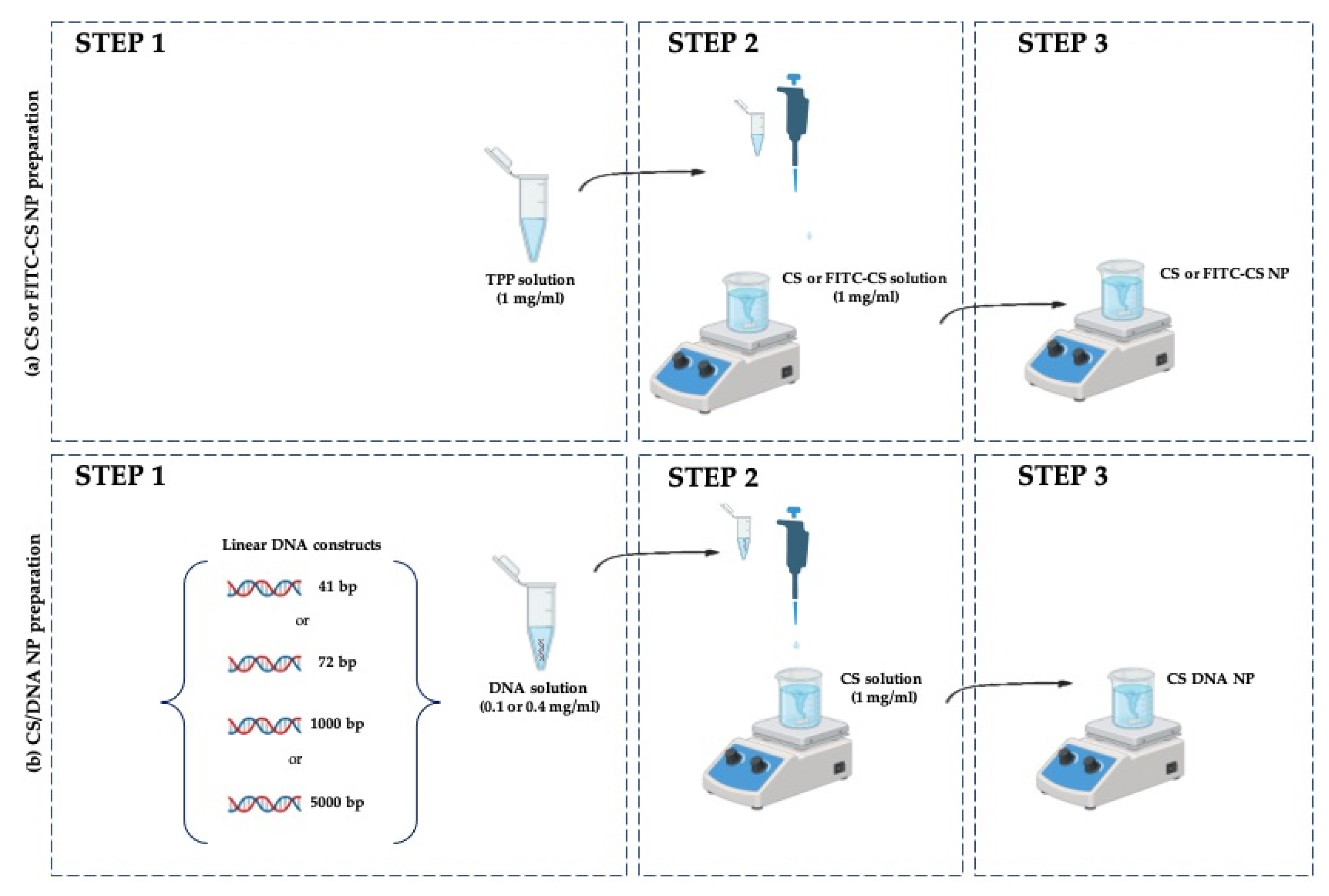
2.3. Preparation of Reference Blank (CS NP) and Chitosan–DNA Nanoparticles (CS/DNA NPs)

Blank CS NPs were prepared using the ionic gelation method. Chitosan was dissolved in a 1% (v/v) acetic acid solution at a concentration of 1 mg/mL. The solution was stirred at room temperature for 24 h and then filtered through a 0.22 μm membrane. TPP was dissolved in water at a concentration of 1 mg/mL. CS NPs were formed by adding 2 mL of a solution of TPP in water (concentration 1 mg/mL, filtered through a 0.22 μm membrane) dropwise to 5 mL of the chitosan solution under magnetic stirring, and the mixture was stirred at room temperature for an additional 2 h. The corresponding fluorescein labeled nanoparticles (FITC-CS NPs) were prepared using the FITC-CS conjugate at a weight ratio of 1:9 (FITC-CS to CS) (Figure 1a).
DNA-bearing nanoparticles (CS/DNA NPs) were prepared via polyelectrolyte complexing. DNA solutions were prepared by dissolving various DNA constructs, including a DNA sequence encoding green fluorescent protein (GFP), in deionized water at final concentrations of either 0.1 mg/mL or 0.4 mg/mL (Table 1). All particles were prepared at a N/P ratio of 5:1. The DNA solution was added dropwise to the chitosan solution under continuous stirring. The mixture was maintained at room temperature for 2 h (Figure 1b).
Both CS NPs and CS/DNA NPs were purified via centrifugation at 13,000 rpm for 30 min at 4 °C (Centrifughe Megafuge™ serie 16, Thermo Scientific, Milan, Italy), and the pellets were re-dispersed in 0.4 M phosphate buffer (pH 7.4).

2.4. Physical–Chemical Characterization of Nanoparticles

2.4.1. NP Size Distribution and Zeta Potential Analysis

The size distribution of the nanoparticles was measured using Dynamic Light Scattering (DLS) with a Zetasizer Nano ZS (Malvern, UK). Samples of CS/DNA NPs were diluted 1:10 in 0.4 M phosphate buffer (pH 7.4) and measured at 25 °C. Zeta potential values were determined at 25 °C using the same 0.4 M phosphate buffer (pH 7.4). All measurements were performed at least in triplicate.

2.4.2. Entrapment Efficiency (EE) of CS-DNA NPs

The entrapment efficiency (EE) of chitosan-based nanoparticles containing various DNA constructs was evaluated using an indirect method, with a NanoDrop One spectrophotometer (Thermofisher, Waltham, MA, USA). The EE was calculated as the difference between the total DNA added and the free DNA recovered in the supernatant after NP purification and expressed as a percentage of the total DNA. The formula used to calculate the EE is as follows:
EE (%) = ((DNAtotal − DNAfree)/(DNAtotal)) × 100
where DNAtotal is the total amount of DNA added to the nanoparticle suspension.
DNAfree is the amount of free DNA present in the supernatant after the nanoparticle purification step (determined by measuring the DNA concentration in the supernatant using the NanoDrop spectrophotometer).
An arbitrary experimental uncertainty of ±5% was assumed for the EE measurement, based on expected variations observed in comparable studies [35].

2.5. Biological Evaluation

2.5.1. Cell Culture Condition

NCI-H441 cells were grown in a CO2 incubator at 37 °C, 5% CO2 in Roswell Park Memorial Institute (RPMI-1640) medium supplemented with 2 mM L-glutamine, 1% penicillin/streptomycin and 10% fetal bovine serum (FBS).

2.5.2. Cell Viability of NPs

To evaluate the cytotoxicity of CS NPs, nanoparticles were purified via centrifugation (30 min, 13,400 rpm, 4 °C). The pellets were re-suspended in RPMI cell culture medium and used at various concentrations (1, 10, 50, 100 and 500 µg/mL) to treat NCI-H441 cells. NCI-H441 epithelial cells were seeded in 96-well culture plates at a concentration of 30 × 104 and 12 × 104 per well for the analysis of cell viability at 4 h and 24 h, respectively. Cells were incubated at 37 °C and 5% CO2 and left to proliferate for 24 h prior to the incubation with the samples. The culture medium from each well was removed and replaced with complete medium containing the sample diluted as previously described. After 4 h and 24 of incubation, cell viability was assessed using WST-1 tetrazolium salt reagent diluted to 1:10 and incubated for 4 h at 37 °C and 5% CO2. Measurements of formazan dye absorbance were carried out at 450 nm, with a reference wavelength of 655 nm, using a microplate reader (BioTek 800/TS, Thermo Scientific).

2.5.3. Uptake of Nanoparticles by Cells

For preliminary uptake studies, NCI-H441 cells were seeded into 8-well culture slides at a density of 1 × 10⁴ cells per well in 500 μL of cell medium one day prior to transfection and incubated in antibiotic-free medium. FITC-CS NPs were then added to the cells. The cells were incubated with the nanoparticles for 1 h, 2 h, 3 h and 24 h at 37 °C in 5% CO2. Following each incubation period, the samples were removed, and the cells were washed three times with phosphate-buffered saline (PBS). The cells were then fixed with paraformaldehyde (PFA) and visualized using a fluorescence optical microscope (Nikon Eclipse Ts2R, Tokyo, Japan) to assess the uptake of the FITC-labeled nanoparticles.

2.5.4. In Vitro Transfection Studies

NCI-H441 cells were seeded on 8-chamber slides with a density of 1 × 10⁴ one day prior to transfection and incubated overnight to allow for cell adhesion. The formulation “CS/DNA GFP (1000)b NP” was tested. Each sample contained 2 µg of loaded DNA. The nanoparticles were incubated with the cells for 3 h at 37 °C and 5% CO2. After incubation, the cells were washed with PBS and further incubated for 24 h to allow for protein expression. NCI-H441 cells were used as a negative control.

2.5.5. Confocal Microscopy

After the incubation with NPs, cells were fixed with 4% PFA and washed three times with PBS. The glass slides were then washed and mounted with DAPI fluorescent mounting medium for acquisition through a laser scanning confocal microscope (Nikon Eclipse Ti-A1MP) using a 60X/1.4NA oil objective and pinhole set to 1 Airy Unit. The 405 nm and 488 nm laser lines were used for imaging DAPI and GFP by using the 400–500 and 475–575 emission filter cubes, respectively. GFP signal quantification was performed using whole-field analysis, as previously reported [36].

2.6. Statistical Analysis

All data are expressed as the mean ± standard deviation of at least six independent replicates. Significant differences were determined via the t-test using GraphPad Prism 10.0 software. Differences were considered significant for p values lower than 0.05.

3. Results

3.1. NP Physical–Chemical Characterization

The size distribution of the nanoparticles under study was measured using Dynamic Light Scattering (DLS) and is shown in Table 2. The nanoparticles showed variations in their size, polydispersity index (PDI) and EE. Indeed, sizes as well as PDI index values ranged from 150 to 700 nm and from 0.2 to 0.8, respectively. These data indicated that the DNA construct concentration mostly influenced NP formation, with a minor effect due to the construct length. Regarding zeta potential values, all formulations showed a positive charge in accordance with the applied N/P ratio. Although the dimensions of all CS/DNA NP-containing samples ranged between 150 and 700 nm, no significant differences were observed between the different DNA constructs when used at the same concentration. The same observation applies to the PDI. Differently, the EE is drastically affected by the concentration of the DNA solution used during NP formation, showing higher values at increased concentrations. However, in this context, the EE is not influenced by the length of the applied construct. The CS/DNA(1000)b NP sample exhibited the most optimal characteristics in terms of size, zeta potential and encapsulation efficiency among all samples and was, therefore, chosen for subsequent studies.

3.2. Biological Evaluations

The cytotoxicity of CS NPs was assessed in NCI-H441 cells over two incubation periods, 4 h and 24 h, using nanoparticle concentrations ranging from 1 to 500 µg/mL. The results, shown in Figure 2, illustrated the concentration-dependent cytotoxicity of CS-TPP nanoparticles. After 4 h of exposure, cell viability was moderately affected, with a notable decrease at higher concentrations. Specifically, cell viability decreased from 83.3% at 1 µg/mL to 62.5% at 500 µg/mL. The cytotoxicity of CS NPs at the same concentration was more pronounced at 24 h, although at the highest concentrations tested (namely 100 and 500 µg/mL), no differences were observed between the two time points.
The preliminary qualitative uptake studies of FITC-CS NPs in NCI-H441 cells revealed that nanoparticle internalization increased over time (Figure 3). Qualitative assessment using fluorescence microscopy showed that a significant amount of FITC-CS NPs was internalized by the cells at each time point. However, qualitatively, the most substantial uptake was observed at 3 h. At this time point, fluorescence microscopy images indicated a pronounced accumulation of nanoparticles within the cells compared to 1 h and 2 h of incubation periods. This suggests that for the investigated incubation times, longer incubation enhanced the internalization of FITC-labeled nanoparticles by NCI-H441 cells without efflux or saturation effects.
Fluorescence microscopy analysis successfully revealed not only the internalization of the nanoparticles but also the expression of the fluorescent protein reporter (GFP) encoded by the 1000 bp DNA linear fragment (Figure 4). Indeed, GFP expression could be detected in cells treated with nanoparticles containing the encoding DNA construct (Figure 4a), and an average higher fluorescence in the GFP emission channel could be measured in NP-treated cells with respect to untreated cells (Figure 4b). The results indicated effective delivery and expression of the DNA constructs, with clear fluorescence in the treated cells. Furthermore, no qualitative differences in the morphology of the nuclei were observed between treated and untreated cells. This finding supported the notion that the nanoparticle formulation is non-toxic and does not cause cellular alterations.

4. Discussion

This study explores the development and characterization of chitosan-based nanoparticles loaded with linear DNA for nucleic acid delivery, investigating the behavior in the NCI-H441 lung cell line.
This research examined how varying DNA concentrations influence nanoparticle formation, leading to different encapsulation efficiencies and particle sizes. These findings offer insights into optimizing the physicochemical properties of chitosan–DNA nanoparticles for enhanced gene delivery and therapeutic potential.
Chitosan has been widely utilized in nanoparticle preparation due to its biocompatibility, biodegradability, non-toxicity, ease of preparation and effectiveness as a drug delivery system for gene therapy [16,17,18,19,20]. Nevertheless, it remained unclear which specific type of chitosan was most effective for various therapeutic nucleic acid (e.g., linear DNA, plasmid DNA, siRNA, miRNA, etc.) or how the nucleic acid conformation (e.g., single stranded versus double stranded) influenced the performance [37]. However, it has been already reported that the efficacy of transfection is dependent on the CS molecular weight, in particular that CS with a MW in the range 10–50 kDa are the best performing [38]. Conscious of that report, CS with an average MW of 30 kDa was applied in the present research. In this work, chitosan-based nanoparticles were prepared using various linear DNA constructs, maintaining a constant N:P ratio of 5:1, where N represents the amino groups from CS, and P represents the phosphate groups from DNA. The 5:1 N:P ratio was selected based on promising research reported in the literature [39,40,41], while the attention in this study was directed towards two specific DNA concentration: 0.1 mg/mL and 0.4 mg/mL.
These concentrations were chosen due to experimental limitations related to the use of nucleic acids. The preparation of nanoparticles incorporating DNA presents several challenges, as DNA is highly sensitive to different physical and chemical conditions, which can complicate the process [30,42]. Additionally, the high cost of in-lab preparation made it difficult to maintain consistent and reproducible concentrations, further restricting the ability to explore a broader range of concentrations. The difficulty recovering and homogenizing DNA, combined with the inherent variability in DNA amplification processes, also contributed to these limitations [43,44]. CS/DNA nanoparticles were prepared using polyelectrolyte complexing. The observed range of nanoparticle sizes (150–700 nm) and PDI values suggested a versatile formulation, with the ability to fine-tune particle characteristics for optimized drug delivery. Furthermore, the zeta potential of the nanoparticles was measured and found to be greater than +20 mV, indicating good colloidal stability and low aggregation, which is essential for maintaining effective delivery in biological systems [45]. The encapsulation efficiency (EE) of 20% to 86% reflects the capability of this method to achieve satisfactory loading of nucleic acids, a critical factor for effective gene delivery systems. The variations in nanoparticle size and EE could be attributed to differences in DNA concentrations. Indeed, CS/DNA NPs prepared with 0.4 mg/mL of DNA demonstrated better results in terms of size, ranging from 290 to 400 nm, with PDI ranging from 0.219 to 0.348, as well as for encapsulation efficiency, which ranged from 58 to 86%. The two employed concentrations represented a balance between ensuring sufficient DNA for encapsulation and maintaining the feasibility of nanoparticle preparation while considering the practical constraints associated with nucleic acid processing. However, from the results obtained, it can be hypothesized that the lowest DNA concentration tested, 0.1 mg/mL, was too low to allow for an effective entropic interaction between the two macromolecular components of the nanoparticles (CS and DNA). At such low concentrations, the interactions between DNA and chitosan might be insufficient to form stable and well-characterized nanoparticles [46].
Several previous studies have reported the use of chitosan nanoparticles for the transport of nucleic acids, primarily focusing on circular plasmids rather than linear DNA [41] Fernández-Paz et al. [47] utilized a plasmid and a low-molecular-weight CS to develop nanoparticles, achieving an encapsulation efficiency exceeding 90%. Nanoparticles prepared using low-molecular-weight chitosan (CS) and a GFP-tagged plasmid through a complex coacervation method had an approximate size of 260 nm [48]. However, linear DNA constructs, specifically, offer several key advantages in the preparation of gene delivery systems, particularly when used in non-viral gene therapy applications [27]. Linear DNA, as opposed to circular plasmids, tends to integrate into the mammalian genome at higher rates, which is beneficial for stable transgene expression. From the literature, it is suggested that linear DNA formats, such as those with exposed 5′ and 3′ ends and Closed-End Linear DNAs (CELiDs) with thioester loops, present favorable integration characteristics, reducing the risks of unwanted integration events associated with circular plasmids. The linear forms have shown higher stability and more consistent expression profiles in comparison to other vectors. This makes linear DNA constructs a valuable option for gene delivery systems, offering the potential for sustained and controlled gene expression while minimizing the risks associated with random chromosomal integrations, as demonstrated by the reduced frequency of integration into tumor suppressor genes [21].
Although the present study does not directly address RNA, the structural similarities between dsRNA and linear DNA suggest that similar factors may be involved in CS nanoparticles loaded with either linear DNA or RNA. In fact, the data presented here are consistent with recent studies on the use of CS for the delivery of double-stranded RNA [28], where 35 kDa chitosan and dsRNA nanoparticles were prepared via polyelectrolyte complexing, exhibiting sizes around 300 nm, a positive, stable zeta potential >20 mV and high encapsulation efficiency. Also, Scarpin et al. [49] documented high encapsulation efficiencies for chitosan-based nanoparticles carrying double-stranded RNA, with values falling within a similar range. Most preclinical studies have primarily concentrated on viral vectors, and non-viral systems utilizing chitosan are showing promising in vitro results, especially in the context of pulmonary gene therapy for cystic fibrosis [31,37,50,51].
In this regard, biological evaluations were conducted using human distal lung cells (NCI-H441) to assess the efficacy and cellular response to the treatment. Pulmonary drug delivery systems offer several advantages, e.g., controlled drug release, targeted delivery for local treatment and enhanced patient compliance [52]. However, the immunological defense of the respiratory tract can limit drug deposition into the lung, thus reducing drug absorption. In this regard, nanoparticulate systems, thanks to their small dimension size, avoid lung clearance, providing effective drug absorption into the lung epithelium. Among various nanoparticle types, the chitosan-based ones have demonstrated numerous advantages for local drug delivery into the lungs [53].
To achieve effective gene therapy, the polymeric vectors have to be non-cytotoxic. Cytotoxicity tests on NCI-H441 cells, a model lung epithelial cell line, demonstrated that CS/NPs did not induce significant toxic effects at low concentrations, but the cell viability decreased in a dose-dependent manner at higher concentrations. This observation was important, as it suggested that although CS/NPs were generally biocompatible, their dose must be carefully regulated to avoid cellular damage during therapeutic treatments [54].
Fluorescein isothiocyanate (FITC)-labeled nanoparticle uptake experiments revealed that incorporation into cells significantly increased over time. These results could be related to the strong positive surface charge of the particles, as previously described [33]. It is suggested that while nanoparticles may be rapidly absorbed, extended incubation periods enable efficient intracellular accumulation. Additionally, using DNA encoding a fluorescent protein, such as GFP, allows for the observation of protein expression in treated cells, confirming the effectiveness of the transfection technology. Gene transfection analysis showed promising results, with linear GFP protein expression observed in NCI-H441 cells treated with DNA-loaded nanoparticles. The effective DNA transfection and protein production were confirmed using images obtained via confocal microscopy, which revealed strong fluorescence in the treated cells. Indeed, the fluorescence intensity was significantly higher in the cells treated with NP-EGFP than the corresponding fluorescence in the control corresponding to the background fluorescence of the cells, which is not influenced by external treatment.
This study provided a starting point for investigating linear DNA-loaded CS nanoparticles via simple in-bulk preparation through polyelectrolyte complexing. In summary, chitosan nanoparticles present a viable method for the delivery of linear DNA fragments, offering protection and facilitating cellular uptake. However, several areas require further investigation. Firstly, optimizing the nanoparticle size is essential, since smaller nanoparticles may facilitate greater cellular uptake, while larger particles could be less efficient. Future applications could benefit from more in-depth preclinical studies, including animal models, to test their therapeutic efficacy in vivo.

5. Conclusions

The present work highlighted the potential of linear DNA-loaded CS nanoparticles in gene delivery to the lung. The combination of chitosan with DNA of varying lengths allows for the development of nanoparticles with favorable physicochemical properties and effective transfection capabilities. Further optimization of their preparation and characterization conditions is necessary to enhance the delivery efficiency. However, regarding the optimization of the formulation and particle size reduction, these efforts do not indicate an inherent deficiency but rather an opportunity to further enhance the system’s efficiency. Nevertheless, this study contributes to the groundwork for future advances in non-viral gene therapies by providing insights into the potential of chitosan–DNA nanoparticles. Future research should also focus on translating the formulation to a potential therapeutic sequence and conducting stability studies and process scalability to facilitate preclinical investigations.

Author Contributions

Conceptualization, A.M.P., A.F., Y.Z., C.M. (Claudia Martini) and C.M. (Chiara Migone); methodology, C.M. (Chiara Migone) and R.P.; software, C.M. (Chiara Migone) and L.M.; validation, A.M.P., A.F., Y.Z., R.P., L.M., C.G. and C.M. (Chiara Migone); formal analysis, R.P., L.M. and C.M. (Chiara Migone); investigation, C.M. (Chiara Migone); resources, A.M.P., A.F. and C.M. (Claudia Martini); data curation, A.M.P., A.F., Y.Z., R.P., L.M., C.G. and C.M. (Chiara Migone); writing—original draft preparation, C.M. (Chiara Migone); writing—review and editing, A.M.P., A.F., Y.Z., L.M. and C.G.; visualization, A.M.P.; supervision, A.M.P. and Y.Z.; project administration, A.M.P., L.M. and C.G.; funding acquisition, A.M.P., C.M. (Claudia Martini), L.M. and C.G. All authors have read and agreed to the published version of the manuscript.

Funding

This research was funded by the European Union—Next Generation EU_Italian Ministry of University and Research_Soggetto attuatore Fondazione “Centro Nazionale di Ricerca- Sviluppo di terapia genica e farmaci con tecnologia a RNA” ITALIA DOMANI PIANO NAZIONALE RIPRESA E RESILIENZA- CN00000041 CUP—I53C22000710007.

Institutional Review Board Statement

Not applicable.

Informed Consent Statement

Not applicable.

Data Availability Statement

The raw data supporting the conclusions of this article will be made available by the authors on request.

Conflicts of Interest

The authors declare no conflicts of interest. The funders had no role in the design of the study; in the collection, analyses or interpretation of data; in the writing of the manuscript or in the decision to publish the results.

Abbreviations

The following abbreviations are used in this manuscript:
CSChitosan
TPPSodium tripolyphosphate
NPsNanoparticles
FITCFluorescein isothiocyanate
CS NPsReference chitosan blank nanoparticles
FITC-CS NPs Fluorescent chitosan nanoparticles
bpBase pairs
CS/DNA NPsChitosan–DNA nanoparticles
EEEncapsulation efficiency
PFAParaformaldehyde
PBSPhosphate-buffered saline
DMSODimethyl sulfoxide
GFPGreen fluorescence protein
CELiDsClosed-End Linear DNAs

References

  1. Karahmet Sher, E.; Alebić, M.; Marković Boras, M.; Boškailo, E.; Karahmet Farhat, E.; Karahmet, A.; Pavlović, B.; Sher, F.; Lekić, L. Nanotechnology in medicine revolutionizing drug delivery for cancer and viral infection treatments. Int. J. Pharm. 2024, 660, 1–22. [Google Scholar] [CrossRef] [PubMed]
  2. Nagpal, S.; Palaniappan, T.; Wang, J.W.; Wacker, M.G. Revisiting nanomedicine design strategies for follow-on products: A model-informed approach to optimize performance. J. Control Release 2024, 376, 1251–1270. [Google Scholar] [CrossRef]
  3. Mitchell, M.J.; Billingsley, M.M.; Haley, R.M.; Wechsler, M.E.; Peppas, N.A.; Langer, R. Engineering precision nanoparticles for drug delivery. Nat. Rev. Drug Discov. 2021, 20, 101–124. [Google Scholar] [CrossRef] [PubMed]
  4. Mendes, B.B.; Conniot, J.; Avita, L. Nanodelivery of nucleic acids. Nat. Rev. Methods Primers 2022, 2, 24. [Google Scholar] [CrossRef]
  5. Sung, Y.K.; Kim, S.W. Recent advances in the development of gene delivery systems. Biomater. Res. 2019, 23, 8. [Google Scholar] [CrossRef]
  6. Kurul, F.; Turkmen, H.; Cetin, A.E.; Topkaya, S.N. Nanomedicine: How nanomaterials are transforming drug delivery, bio-imaging, and diagnosis. Next Nanotechnol. 2025, 7, 100129. [Google Scholar] [CrossRef]
  7. Pan, X.; Veroniaina, H.; Su, N.; Sha, K.; Jiang, F.; Wu, Z.; Qi, X. Applications and developments of gene therapy drug delivery systems for genetic diseases. Asian J. Pharm. Sci. 2021, 16, 687–703. [Google Scholar] [CrossRef]
  8. Patra, J.K.; Das, G.; Fraceto, L.F.; Ramos Campos, E.V.; Rodriguez-Torres, M.P.; Acosta-Torres, L.S.; Diaz-Torres, L.A.; Grillo, R.; Kumara Swamy, M.; Sharma, S.; et al. Nano based drug delivery systems: Recent developments and future prospects. J. Nanobiotechnol. 2018, 16, 71. [Google Scholar] [CrossRef]
  9. Valatabar, N.; Oroojalian, F.; Kazemzadeh, M.; Mokhtarzadeh, A.A.; Safaralizadeh, R.; Sahebkar, A. Recent advances in gene delivery nanoplatforms based on spherical nucleic acids. J. Nanobiotechnol. 2024, 22, 386. [Google Scholar] [CrossRef]
  10. Taghdiri, M.; Mussolino, C. Viral and Non-Viral Systems to Deliver Gene Therapeutics to Clinical Targets. Int. J. Mol. Sci. 2024, 25, 7333. [Google Scholar] [CrossRef]
  11. Patil, S.; Gao, Y.G.; Lin, X.; Li, Y.; Dang, K.; Tian, Y.; Zhang, W.J.; Jiang, S.F.; Qadir, A.; Qian, A.R. The Development of Functional Non-Viral Vectors for Gene Delivery. Int. J. Mol. Sci. 2019, 20, 5491. [Google Scholar] [CrossRef] [PubMed]
  12. Baylot, V.; Le, T.K.; Taïeb, D.; Rocchi, P.; Colleaux, L. Between hope and reality: Treatment of genetic diseases through nucleic acid-based drugs. Commun. Biol. 2024, 7, 489. [Google Scholar] [CrossRef]
  13. Migone, C.; Mattii, L.; Giannasi, M.; Moscato, S.; Cesari, A.; Zambito, Y.; Piras, A.M. Nanoparticles Based on Quaternary Ammonium Chitosan-methyl--cyclodextrin Conjugate for the Neuropeptide Dalargin Delivery to the Central Nervous System: An In Vitro Study. Pharmaceutics 2021, 13, 5. [Google Scholar] [CrossRef] [PubMed]
  14. Jafernik, K.; Ładniak, A.; Blicharska, E.; Czarnek, K.; Ekiert, H.; Wiącek, A.E.; Szopa, A. Chitosan-Based Nanoparticles as Effective Drug Delivery Systems—A review. Molecules 2023, 28, 1963. [Google Scholar] [CrossRef]
  15. Haider, A.; Khan, S.; Iqbal, D.N.; Shrahili, M.; Haider, S.; Mohammad, K.; Mohammad, A.; Rizwan, M.; Kanwal, Q.; Mustafa, G. Advances in chitosan-based drug delivery systems: A comprehensive review for therapeutic applications. Eur. Polym. J. 2024, 210, 1–23. [Google Scholar] [CrossRef]
  16. Chuan, D.; Jin, T.; Fan, R.; Zhou, L.; Guo, G. Chitosan for gene delivery: Methods for improvement and applications. Adv. Colloid. Interface Sci. 2019, 268, 25–38. [Google Scholar] [CrossRef]
  17. Genedy, H.H.; Delair, T.; Montembault, A. Chitosan Based MicroRNA Nanocarriers. Pharmaceuticals 2022, 15, 1036. [Google Scholar] [CrossRef]
  18. Antoniou, V.; Mourelatou, E.A.; Galatou, E.; Avgoustakis, K.; Hatziantoniou, S. Gene Therapy with Chitosan Nanoparticles: Modern Formulation Strategies for Enhancing Cancer Cell Transfection. Pharmaceutics 2024, 16, 868. [Google Scholar] [CrossRef]
  19. Karayianni, M.; Sentoukas, T.; Skandalis, A.; Pippa, N.; Pispas, S. Chitosan-Based Nanoparticles for Nucleic Acid Delivery: Technological Aspects, Applications, and Future Perspectives. Pharmaceutics 2023, 15, 1849. [Google Scholar] [CrossRef]
  20. Desai, N.; Rana, D.; Salave, S.; Gupta, R.; Patel, P.; Karunakaran, B.; Sharma, A.; Giri, J.; Benival, D.; Kommineni, N. Chitosan: A Potential Biopolymer in Drug Delivery and Biomedical Applications. Pharmaceutics 2023, 15, 1313. [Google Scholar] [CrossRef]
  21. Lim, S.; Yocum, R.R.; Silver, P.A.; Way, J.C. High spontaneous integration rates of end-modified linear DNAs upon mammalian cell transfection. Sci. Rep. 2023, 26, 6835. [Google Scholar] [CrossRef] [PubMed]
  22. Naghib, S.M.; Ahmadi, B.; Mikaeeli Kangarshahi, B.; Mozafari, M.R. Chitosan-based smart stimuli-responsive nanoparticles for gene delivery and gene therapy: Recent progresses on cancer therapy. Int. J. Biol. Macromol. 2024, 278, 134542. [Google Scholar] [CrossRef] [PubMed]
  23. Gonçalves, M.M.; Maluf, D.F.; Pontarolo, R.; Saul, C.K.; Almouazen, E.; Chevalier, Y. Negatively charged chitosan nanoparticles prepared by ionotropic gelation for encapsulation of positively charged proteins. Int. J. Pharm. 2023, 642, 123164. [Google Scholar] [CrossRef] [PubMed]
  24. Casper, J.; Schenk, S.H.; Parhizkar, E.; Detampel, P.; Dehshahri, A.; Huwy, J. Polyethylenimine (PEI) in gene therapy: Current status and clinical applications. J. Control Release 2023, 362, 667–691. [Google Scholar] [CrossRef]
  25. Souto, E.B.; Blanco-Llamero, C.; Krambeck, K.; Kiran, N.S.; Yashaswini, C.; Postwala, H.; Severino, P.; Priefer, R.; Prajapati, B.G.; Maheshwari, R. Regulatory insights into nanomedicine and gene vaccine innovation: Safety assessment, challenges, and regulatory perspectives. Acta Biomater. 2024, 180, 1–17. [Google Scholar] [CrossRef]
  26. Tasset, A.; Bellamkonda, A.; Wang, W.; Pyatnitskiy, I.; Ward, D.; Peppas, N.; Wang, H. Overcoming barriers in non-viral gene delivery for neurological applications. Nanoscale 2022, 10, 3698–3719. [Google Scholar] [CrossRef]
  27. Wang, C.; Pan, C.; Yong, H.; Wang, F.; Bo, T.; Zhao, Y.; Ma, B.; He, W.; Li, M. Emerging non-viral vectors for gene delivery. J. Nanobiotechnol. 2023, 21, 272. [Google Scholar] [CrossRef]
  28. Tran, T.-T.; Amalina, N.; Cheow, W.S.; Hadinoto, K. Effects of storage on the stability and aerosolization efficiency of dry powder inhaler formulation of plasmid DNA-Chitosan nanoparticles. J. Drug Deliv. Technol. 2020, 59, 101866. [Google Scholar] [CrossRef]
  29. Bivas-Benita, M.; van Meijgaarden, K.E.; Franken, K.L.M.C.; Junginger, H.E.; Borchard, G.; Ottenhoff, T.H.M.; Geluk, A. Pulmonary delivery of chitosan-DNA nanoparticles enhances the immunogenicity of a DNA vaccine encoding HLA-A*0201-restricted T-cell epitopes of Mycobacterium tuberculosis. Vaccine 2004, 22, 1609–1615. [Google Scholar] [CrossRef]
  30. Ma, W.; Zhan, Y.; Zhang, Y.; Mao, C.; Xie, X.; Lin, Y. The biological applications of DNA nanomaterials: Current challenges and future directions. Signal Transduct. Target. Ther. 2021, 6, 351. [Google Scholar] [CrossRef]
  31. Kolonko, A.K.; Bangel-Ruland, N.; Goycoolea, F.M.; Weber, W.M. Chitosan Nanocomplexes for the Delivery of ENaC Antisense Oligonucleotides to Airway Epithelial Cells. Biomolecules 2020, 10, 553. [Google Scholar] [CrossRef] [PubMed]
  32. Plasschaert, L.W.; MacDonald, K.D.; Moffit, J.S. Current landscape of cystic fibrosis gene therapy. Front. Pharmacol. 2024, 15, 1476331. [Google Scholar] [CrossRef]
  33. Zambito, Y.; Felice, F.; Fabiano, A.; Di Stefano, R.; Di Colo, G. Mucoadhesive nanoparticles made of thiolated quaternary chitosan crosslinked with hyaluronan. Carbohydr. Polym. 2013, 92, 33–39. [Google Scholar] [CrossRef]
  34. Di Colo, G.; Zambito, Y.; Zaino, C.; Sansò, M. Selected polysaccharides at comparison for their mucoadhesiveness and effect on precorneal residence of different drugs in the rabbit model. Drug Dev. Ind. Pharm. 2009, 35, 941–949. [Google Scholar] [CrossRef]
  35. Lv, Y.; He, H.; Qi, J.; Lu, Y.; Zhao, W.; Dong, X.; Wu, W. Visual validation of the measurement of entrapment efficiency of drugnanocarriers. Int. J. Pharm. 2018, 547, 395–403. [Google Scholar] [CrossRef]
  36. Piccarducci, R.; Germelli, L.; Falleni, A.; Luisotti, L.; Masciulli, B.; Signore, G.; Migone, C.; Fabiano, A.; Bizzarri, R.; Piras, A.M.; et al. GFP Farnesylation as a Suitable Strategy for Selectively Tagging Exosomes. ACS Appl. Bio Mater. 2024, 7, 8305–8318. [Google Scholar] [CrossRef]
  37. Santos-Carballal, B.; Fernández Fernández, E.; Goycoolea, F.M. Chitosan in Non-Viral Gene Delivery: Role of Structure, Characterization Methods, and Insights in Cancer and Rare Diseases Therapies. Polymers 2018, 10, 444. [Google Scholar] [CrossRef]
  38. Sato, T.; Ishii, T.; Okahata, Y. In vitro gene delivery mediated by chitosan: Effect of pH, serum, and molecular mass of chitosan on the transfection efficiency. Biomaterials 2001, 22, 2075–2080. [Google Scholar] [CrossRef]
  39. Lin, G.; Huang, J.; Zhang, M.; Chen, S.; Zhang, M. Chitosan-Crosslinked Low Molecular Weight PEI-Conjugated Iron Oxide Nanoparticle for Safe and Effective DNA Delivery to Breast Cancer Cells. Nanomaterials 2022, 12, 584. [Google Scholar] [CrossRef]
  40. Lebre, F.; Borchard, G.; Faneca, H.; Pedroso de Lima, M.; Borges, O. Intranasal administration of novel chitosan nanoparticle/DNA complexes induces antibody response to hepatitis B surface antigen in mice. Mol. Pharm. 2016, 13, 472–482. [Google Scholar] [CrossRef]
  41. Lavertu, M.; Methot, S.; Tran-Khanh, N.; Buschmann, M.D. High efficiency gene transfer using chitosan/DNA nanoparticles with specific combinations of molecular weight and degree of deacetylation. Biomaterials 2006, 27, 4815–4824. [Google Scholar] [CrossRef] [PubMed]
  42. Seeman, N.; Sleiman, H. DNA nanotechnology. Nat. Rev. Mater. 2018, 3, 17068. [Google Scholar] [CrossRef]
  43. Xu, F.; Xia, Q.; Wang, P. Rationally Designed DNA Nanostructures for Drug Delivery. Front. Chem. 2020, 8, 751. [Google Scholar] [CrossRef]
  44. Keller, A.; Linko, V. Challenges and Perspectives of DNA Nanostructures in Biomedicine. Angew. Chem. Int. 2020, 59, 15818–15833. [Google Scholar] [CrossRef]
  45. Azeez, Y.; Almotairy, A.R.Z.; Henidi, H.; Alshehri, O.Y.; Aldughaim, M.S. Nanoparticles as drug delivery systems: A review of the implication of nanoparticles’ physicochemical properties on responses in biological systems. Polymers 2023, 15, 1596. [Google Scholar] [CrossRef]
  46. Dey, G.R.; McCormick, C.R.; Soliman, S.S.; Darling, A.J.; Schaak, R.E. Chemical Insights into the Formation of Colloidal High Entropy Alloy Nanoparticles. ACS Nano 2023, 17, 5943–5955. [Google Scholar] [CrossRef]
  47. Fernández-Paz, E.; Feijoo-Siota, L.; Gaspar, M.M.; Csaba, N.; Remuñán-López, C. Microencapsulated Chitosan-Based Nanocap- sules: A New Platform for Pulmonary Gene Delivery. Pharmaceutics 2021, 13, 1377. [Google Scholar] [CrossRef]
  48. Khan, I.N.; Navaid, S.; Waqar, W.; Hussein, D.; Ullah, N.; Khan, M.U.A.; Hussain, Z.; Javed, A. Chitosan-Based Polymeric Nanoparticles as an Efficient Gene Delivery System to Cross Blood Brain Barrier: In Vitro and In Vivo Evaluations. Pharmaceuticals 2024, 17, 169. [Google Scholar] [CrossRef]
  49. Scarpin, D.; Nerva, L.; Chitarra, W.; Moffa, L.; D’Este, F.; Vuerich, M.; Filippi, A.; Braidot, E.; Petrussa, E. Characterisation and functionalisation of chitosan nanoparticles as carriers for double-stranded RNA (dsRNA) molecules towards sustainable crop protection. Biosci Rep. 2023, 43, BSR20230817. [Google Scholar] [CrossRef]
  50. Kolonko, A.K.; Efing, J.; González-Espinosa, Y.; Bangel-Ruland, N.; van Driessche, W.; Goycoolea, F.M.; Weber, W.M. Capsaicin-Loaded Chitosan Nanocapsules for wtCFTR-mRNA Delivery to a Cystic Fibrosis Cell Line. Biomedicines 2020, 8, 364. [Google Scholar] [CrossRef]
  51. Fernández Fernández, E.; Santos-Carballal, B.; Weber, W.M.; Goycoolea, F.M. Chitosan as a non-viral co-transfection system in a cystic fibrosis cell line. Int. J. Pharm. 2016, 50, 1–9. [Google Scholar] [CrossRef] [PubMed]
  52. Fei, Q.; Bentley, I.; Ghadiali, S.N.; Englert, J.A. Pulmonary drug delivery for acute respiratory distress syndrome. Pulm. Pharmacol. Ther. 2023, 79, 102196. [Google Scholar] [CrossRef] [PubMed]
  53. Zacaron, T.M.; Silva, M.L.S.e.; Costa, M.P.; Silva, D.M.e.; Silva, A.C.; Apolônio, A.C.M.; Fabri, R.L.; Pittella, F.; Rocha, H.V.A.; Tavares, G.D. Advancements in Chitosan-Based Nanoparticles for Pulmonary Drug Delivery. Polymers 2023, 15, 3849. [Google Scholar] [CrossRef] [PubMed]
  54. Salomon, J.J.; Muchitsch, V.E.; Gausterer, J.C.; Schwagerus, E.; Huwer, H.; Daum, N.; Lehr, C.M.; Ehrhardt, C. The cell line NCl-H441 is a useful in vitro model for transport studies of human distal lung epithelial barrier. Mol. Pharm. 2014, 3, 995–1006. [Google Scholar] [CrossRef]
Figure 1. Schematic illustration of the steps for the synthesis of (a) CS and FITC-CS NPs and (b) CS/DNA NPs.
Applbiosci 04 00029 g001
Figure 2. In vitro cytotoxicity of CS NPs on the NCI-H441 cell line. The data represent the percentage of cell viability after treatment with different concentrations of CS NPs after 4 h and 24 h of incubation. Means ± SD, n = 6.
Applbiosci 04 00029 g002
Figure 3. Representative optical fluorescence microscopy images showing the FITC-CS NP uptake by NCI-H441 cells at different time points: (a) 1 h, (b) 2 h and (c) 3 h; scale bar: 10 mm.
Applbiosci 04 00029 g003
Figure 4. Confocal fluorescence studies. (a) Representative confocal fluorescence microscopy images of NCI-H441 cells transfected with control NP (CTRL, upper row) and CS-NP-DNA-GFP (NP-EGFP, bottom row). For each sample, typical DAPI, EGFP and Merge channels are shown. Scale bar: 10 mm. (b) Scatter plot of fluorescence intensity quantification of the analyzed fields for CTRL (n = 7) and NP-EGFP (n = 9). The horizontal line is the mean value, and the error bar is the SD. ** p < 0.01 according to an unpaired T-test, two-tailed.
Applbiosci 04 00029 g004
Table 1. Composition of in vitro tested samples. All samples contained CS at a concentration of 1 mg/mL. For FITC-CS NPs, FITC-CS was applied at a 1:9 ratio with respect to CS.
Sample NameDNA Construct
Base Pair (bp)Solution Concentration (mg/mL)
CS NP--
FITC-CS NP--
CS/DNA(41)a NP410.1
CS/DNA(41)b NP41 0.4
CS/DNA(720)a NP720 0.1
CS/DNA(720)b NP720 0.4
CS/DNA GFP(1000)a NP1000 0.1
CS/DNA GFP (1000)b NP1000 0.4
CS/DNA(5000)a NP5000 0.1
CS/DNA(5000)b NP5000 0.4
Table 2. NP size, polydispersity index (PDI) and entrapment efficiency (EE).
Sample NameNP Size (nm) PDIEE (%)Zeta Potential (z)
CS NP150.2 ± 3.60.289 ± 0.018/+21 ± 2.17
FITC-CS NP182.4 ± 2.40.301 ± 0.032/+20.0 ± 1.90
CS/DNA(41)a NP323.3 ± 8.30.447 ± 0.01620+21.0 ± 2.17
CS/DNA(41)b NP442.6 ± 2.60.348 ± 0.06078+21.0 ± 2.31
CS/DNA(720)a NP 500.4 ± 1.80.429 ± 0.04338+20.0 ± 3.90
CS/DNA(720)b NP340.4 ± 17.50.298 ± 0.06258+22.0 ± 1.78
CS/DNA-GFP(1000)a NP720.1 ± 8.70.775 ± 0.72040+23.90 ± 1.56
CS/DNA-GFP(1000)b NP290.7 ± 2.30.190 ± 0.03286+25.50 ± 2.09
CS/DNA(5000)a NP374.4 ± 24.20.445 ± 0.01143+23.0 ± 2.11
CS/DNA(5000)b NP308.8 ± 1.20.219 ± 0.04280+28.0 ± 2.45
Disclaimer/Publisher’s Note: The statements, opinions and data contained in all publications are solely those of the individual author(s) and contributor(s) and not of MDPI and/or the editor(s). MDPI and/or the editor(s) disclaim responsibility for any injury to people or property resulting from any ideas, methods, instructions or products referred to in the content.

