Article

AG1®, a Novel Synbiotic, Demonstrates Superior Mineral Bioaccessibility and Bioavailability Compared to a Tablet Multivitamin and Mineral Supplement Using an In Vitro Model of the Upper Gastrointestinal Tract

1
Research, Nutrition, and Innovation, Athletic Greens International, Carson City, NV 89701, USA
2
Health & Human Performance, Concordia University Chicago, River Forest, IL 60305, USA
3
ProDigest BVBA, 9052 Ghent, Belgium
4
Center of Microbial Ecology and Technology (CMET), Ghent University, 9000 Ghent, Belgium
5
Department of Nutrition, Food Studies, and Public Health, New York University-Steinhardt, New York, NY 10003, USA
*
Author to whom correspondence should be addressed.
This paper is an extended version of our published paper Sapp, P.A.; Townsend, J.R.; Kirby, T.O.; Govaert, M.; Duysburgh, C.; Marzorati, M.; Marshall, T.M.; Esposito R. AG1, A Novel Foundational Nutrition Supplement, Demonstrates Increased Bioaccessibility and Bioavailability of Minerals Compared to a Tablet Multivitamin in vitro. In Proceedings of the 3rd International Electronic Conference on Nutrients (IECN 2023), online, 1–15 November 2023. https://doi.org/10.3390/IECN2023-15523.
Appl. Biosci. 2023, 2(4), 656-667; https://doi.org/10.3390/applbiosci2040041
Submission received: 13 September 2023 / Revised: 10 November 2023 / Accepted: 29 November 2023 / Published: 1 December 2023

Abstract

:
While traditional multivitamin and mineral (MVM) supplements generally come in tablet form, new powder forms of MVM supplements are available with theoretically higher bioavailability relative to tablet MVM supplements. The purpose of this study was to assess the bioaccessibility and bioavailability of minerals (magnesium (Mg), zinc (Zn), calcium (Ca), and potassium (K)) in a tablet MVM supplement compared to a novel powder Foundational Nutrition supplement (AG1®), containing minerals, vitamins, phytochemicals, and pre-/probiotics, in the upper gastrointestinal tract. The tablet MVM supplement was specifically formulated for this study, with matched mineral contents and identical chemical structures. The adapted Simulator of the Human Intestinal Microbial Ecosystem (SHIME®) model was used to assess the bioaccessibility and bioavailability of soluble minerals using a simulated upper gastrointestinal tract and dialysis membrane to mimic human digestion and absorption. The bioaccessibility was assessed at the end of the stomach and duodenum. The bioaccessibility and bioavailability were assessed at 1, 2, and 3 h following dialysis. The preliminary soluble mineral analysis of the tablet (crushed to a powder) and AG1 powder demonstrated significantly higher (p < 0.05) soluble fractions of Zn and Ca, but lower Mg in the AG1 powder vs. the tablet. The total soluble mineral percentages at the stomach and duodenum end were all significantly higher for the AG1 powder vs. the tablet (p < 0.05). Mg, Ca, and Zn were more (p < 0.05) bioaccessible and bioavailable in the powder compared to the tablet during the small intestine simulation. The bioaccessible fraction of K was higher (p < 0.05) only at 3 h for the tablet vs. the powder. These preclinical data demonstrate that the AG1 powder has superior dissolution and disintegration characteristics compared to the tablet, leading to increased bioaccessibility and bioavailability in vitro.

Graphical Abstract

1. Introduction

Suboptimal diet quality represents a substantial concern in the United States (US), contributing to the largest proportion of deaths annually [1]. The American diet consists primarily of energy-dense, nutrient-poor foods, characterized by high intakes of sodium, saturated fat, and refined grains, with low intakes of fruits, vegetables, and whole grains [2]. Utilizing the Healthy Eating Index-2020 score (scale: 0–100), the majority of US adults consume a diet low in quality, which scores between 55.3 and 59.5 [2]. These dietary patterns are accompanied by the low intake of several micronutrients, with greater than 40% of individuals over 1-year-old consuming amounts of vitamin D, magnesium, and calcium that are below the estimated average requirements (EARs) [3].
Due to these micronutrient shortcomings, many adults consume dietary supplements intending to fill these nutritional gaps [4] and the Dietary Guidelines for Americans 2020–2025 recognizes that MVMs may be useful in supplementing nutrients [5]. Recent analysis by the National Health and Nutrition Examination Survey (NHANES) revealed that over 57% of US adults over the age of 20 have taken a supplement in the last 30 days, with multivitamin and mineral (MVM) supplements accounting for the highest proportion consumed [6]. Traditionally, MVM supplements are consumed daily in tablet form. However, contemporary MVM supplements are often sold in a powder format for a variety of reasons [7].
A salient feature of a powder MVM supplement is the expectation that the powder delivery format will possess a higher level of bioavailability and bioaccessibility [8]. Given the prolonged (>20 min for a significant proportion of mass-produced MVM tablets [9]) disintegration rate of conventional MVM tablets, a limitation to this delivery format is the possibility that the vitamins and minerals will not be liberated from the tablet matrix upon entering the small intestine for absorption [10,11,12]. Conversely, as powders forego the disintegration requirement, they may have increased overall bioavailability compared to tablet MVM supplements. There is some clinical evidence showing increased efficacy or absorption of single-ingredient supplements or pharmaceuticals when consumed as a powder compared to a tablet [8,13,14]. To the best of our knowledge, only one study has assessed the bioavailability of different forms of MVM supplements, comparing an MVM in tablet form versus a crushed form. Interestingly, the data revealed increased absorption of vitamin B12 for the crushed tablet, increased absorption of iron for the formed tablet, and variable results for other minerals that could not be explained by the authors [15]. As the powdered MVM supplements currently on the market are not provided simply as crushed versions of MVM tablets (i.e., unique chemical formulations), more data is needed to understand the differences in bioavailability and bioaccessibility among the MVM delivery formats.
A novel Foundational Nutrition supplement, AG1®, provides minerals and vitamins, in addition to prebiotics, probiotics, and phytonutrients in a powder form. The purpose of this study was to compare the mineral bioavailability and bioaccessibility of AG1® powder with a tablet MVM supplement in the upper gastrointestinal tract (UGIT), using the adapted Simulator of the Human Intestinal Microbial Ecosystem (SHIME®) model. The tablet was formulated with matched mineral contents and identical chemical structures to AG1, but devoid of prebiotics, probiotics, and phytonutrients. This model was chosen due to a recent study assessing magnesium supplements employing the SHIME model of the UGIT paired with a human trial, which demonstrated similar bioavailability results between the in vitro and clinical data [16]. Our hypothesis was that this in vitro model would demonstrate superior bioavailability and bioaccessibility of the minerals present in a powder multivitamin (AG1) compared to the traditional tablet form of a chemically similar MVM supplement in the UGIT.

2. Materials and Methods

2.1. Study Protocol

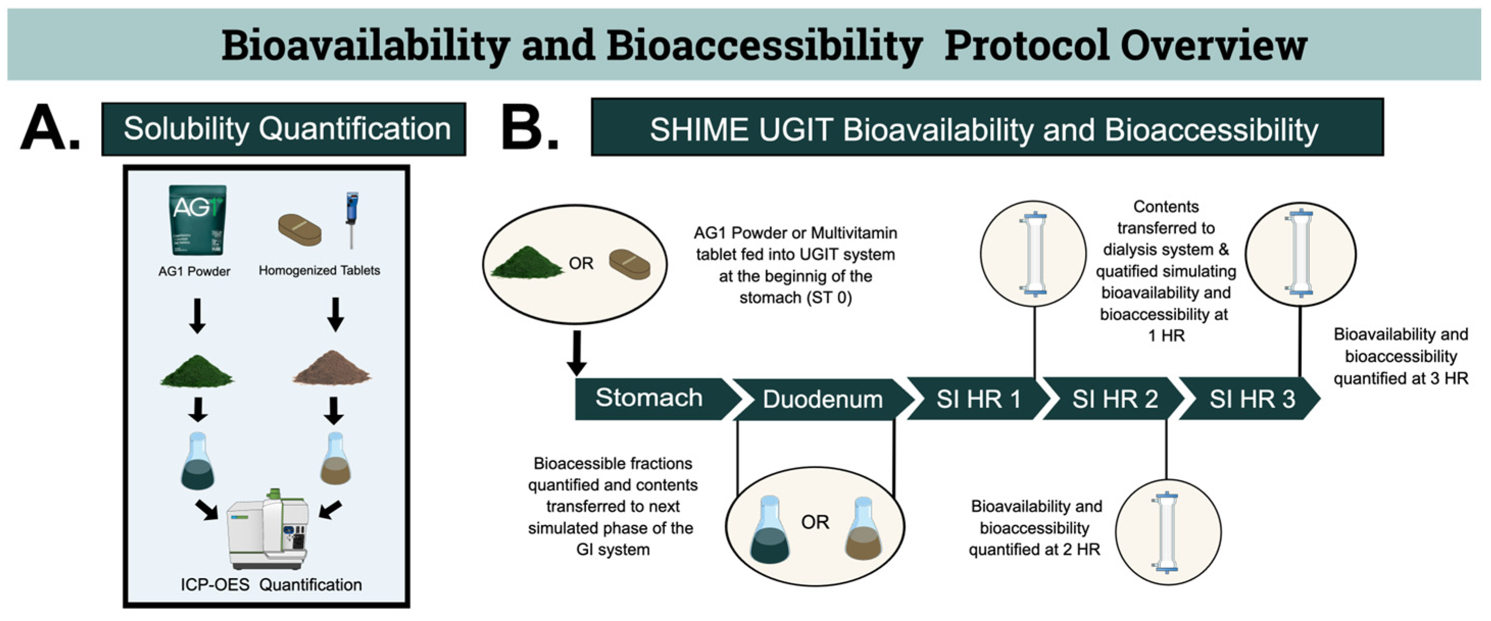
2.1.1. Solubility Quantification before the SHIME Model

Quantification of the soluble fractions of the powder and tablet were determined, before the test products entered the SHIME® upper gastrointestinal tract (UGIT) model (Figure 1, Panel A), to determine the exact amount of soluble minerals in each test product. The tablet formulation was crushed to ascertain the absolute soluble mineral quantities that might not have arisen through normal disintegration (further discussed in Section 2.3.1). This was completed in triplicate. It is important to note that this was the only portion of the experiment where the tablet was crushed.

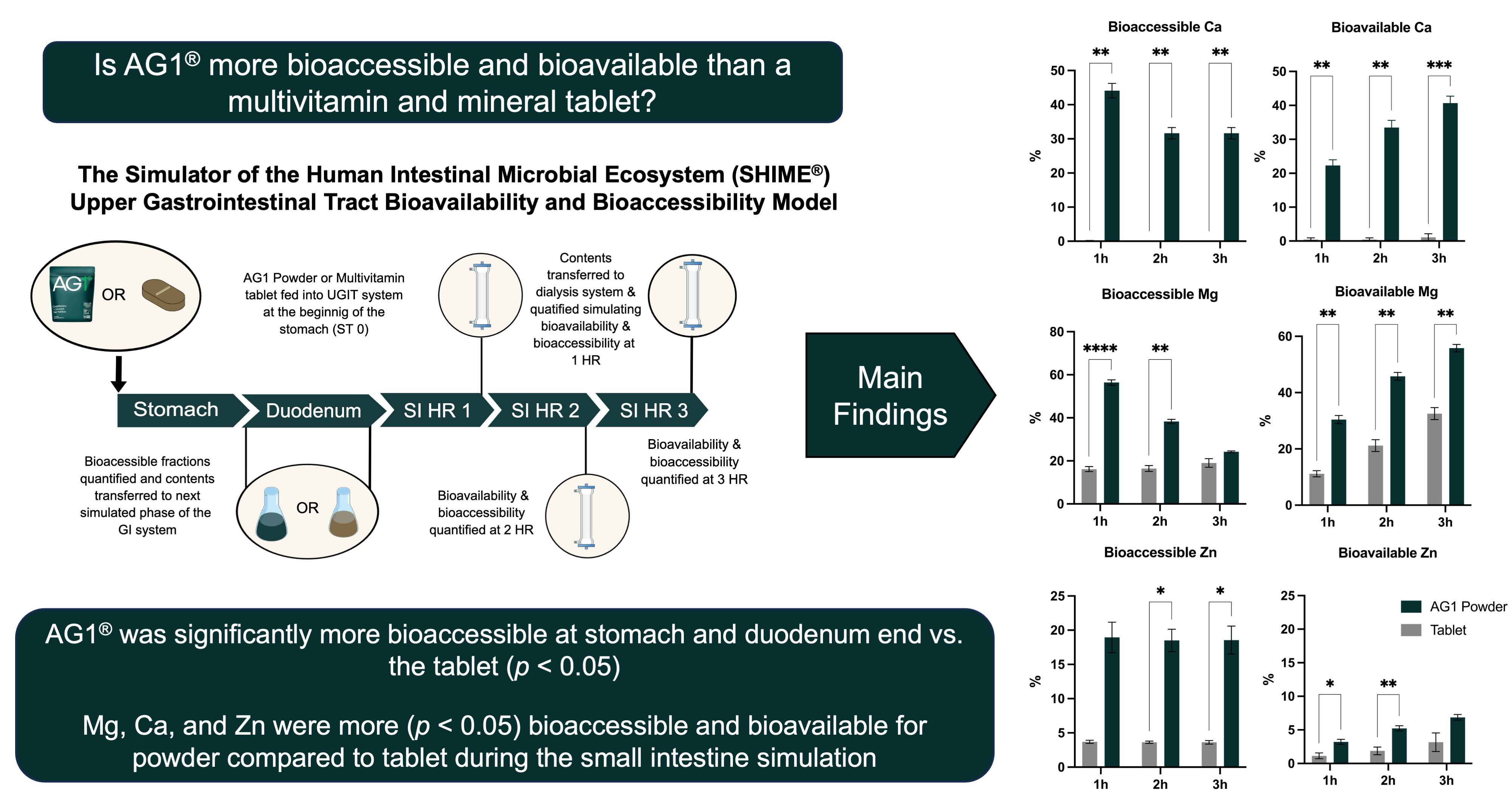
2.1.2. SHIME UGIT Bioavailability and Bioaccessibility

The bioaccessibility and bioavailability of the tablet and powder were assessed using the SHIME UGIT model (Figure 1, Panel B), discussed in detail below. Briefly, the tablet and powder were individually fed into the SHIME bioreactor, which mimics human digestion in the stomach, duodenum, and the absorption in the distal small intestine phase using a dialysis membrane (SI). Each product was tested in triplicate.

2.2. Test Products

The powder supplement investigated in this study was AG1® (AG1; Athletic Greens International, Carson City, NV, USA), which provides minerals and vitamins, in addition to prebiotics, probiotics, and phytonutrients in a powder form. The recommended dose of AG1 is 12 g per serving. Due to the potential physical complications that could impact the biological and mechanical factors of the SHIME® model, a dose of 6 g/bioreactor was chosen. The comparator group received a tablet MVM supplement formulated with the same amount of all the vitamins and minerals as in AG1, but only magnesium (Mg), zinc (Zn), calcium (Ca), and potassium (K) were investigated in this study. The nutrition facts label for AG1 is publicly available [17] and has undergone evaluation and verification via NSF testing (Ann Arbor, MI, USA) to ensure the product meets strict purity, safety, label, quality, and accuracy standards [18]. Prior to the investigation, the quantities of all the vitamins and minerals in AG1 were quantified by the manufacturer, and identical tablets were made with the same amounts of vitamins and minerals in tablet form. The full list of ingredients for the tablet MVM supplement is available in Supplementary Table S1. The forms of the minerals in the AG1 powder and tablet were dipotassium phosphate, magnesium glycinate, calcium (as calcium citrate, calcium carbonate, and calcium phosphate), and zinc citrate. During the manufacturing process of AG1, all the ingredients were added in a dry powdered form and subsequently mixed to create the final powder. Similarly, all the ingredients for the tablet were added in a dry powdered form, mixed, and pressed.

2.3. Determination of Initial Soluble Fraction, Subsequent Mineral Analysis, and Definitions

2.3.1. Determination of Absolute Initial Soluble Amounts and Fraction

The absolute amounts of each constituent in the powder and tablet were provided by the manufacturer. To measure the soluble fraction (Figure 1, Panel A) before utilizing the SHIME model, the inductively coupled plasma optical emission spectroscopy (ICP-OES) methodology was employed in triplicate. To expedite the process, six individual tablets were weighed to determine the average weight of the test dose (i.e., one tablet), the six tablets were crushed into a powder with a mortar and pestle, the exact average weight of one dose was collected from the crushed tablets and added to 80 mL of Milli-Q water prior to baseline product testing. The powder AG1 and powdered tablet were individually homogenized in water suspension using a vortex mixer to make a homogenous solution and 5 mL samples were used for the mineral analysis. The samples then underwent deconstruction (acidification with HNO3 (67%), followed by destruction for 2 h at 110 °C) to liberate the minerals from the matrices. The absolute initial soluble amounts and the absolute amount provided by the manufacturer were used to calculate the initial soluble fraction (Table 1).

2.3.2. Subsequent Mineral Analysis

The AG1 powder was fed into the system as a powder, whereas the tablet was not crushed and allowed to disintegrate naturally (Figure 1, Panel B) in the test solution, but underwent the same deconstruction process (discussed in Section 2.3.1), and the ICP-OES was used to assess the soluble fraction. This was completed in triplicate. The results from the baseline testing (initial soluble fraction before using the SHIME model) were used as a theoretical maximal soluble fraction and were used to calculate the % soluble fraction to determine the theoretical bioaccessibility (assessed throughout the whole model) and bioavailability (assessed only during the dialysis phase) at each phase in the GI tract simulation.

2.3.3. Definitions

In this experiment, bioaccessibility was defined as the soluble fraction still within the dialysis membrane and as an estimation of the unabsorbed luminal fraction (bioaccessibility = soluble minerals at the stomach end, duodenum end, and in the dialysis membrane/initial soluble fraction concentration). Bioavailability was defined as the soluble fraction that was able to diffuse across the dialysis membrane and as an estimation of the absorbed fraction able to enter the intestinal periphery (bioavailability = soluble minerals in the intestinal periphery/initial soluble concentration). Additionally, we calculated the maximum concentration (Cmax), which was defined as the total amount of an elemental mineral that diffused across the membrane during the dialysis phase (Cmax = highest observed bioavailable mineral amount/initial soluble fraction concentration).
We employed the SHIME model adapted from Molly et al., 1993, utilizing one reactor which simulates the stomach and small intestine [19]. The reactor was used to mimic the physiological conditions in the stomach and small intestine. A specific gastric suspension, followed by an enzyme solution and standardized bile acids were added to the reactor over time to simulate and maintain fasted state conditions in the human small intestine. Specific pH levels and incubation durations were predetermined to replicate the in vivo conditions corresponding to each segment of the human gastrointestinal tract referenced [20]. This experimental setup was run in three, independent, parallel reactors per treatment.

2.4. Test Gastrointestinal Tract System

2.4.1. Gastric Phase

The incubation (45 min) for each sample occurred at 37 °C. During the incubation period, stirring was utilized to continuously mix the solution, while a constant pH of 2.0 was maintained. Pepsin was introduced, and its activity was standardized by assessing the increase in absorbance at 280 nm of the TCA-soluble substances generated during the digestion of hemoglobin. Phosphatidylcholine and pepsin (0.02 mM and 1000 U/mL, respectively) were added [21]. The base medium employed consisted solely of salts and mucins, as suggested by the consensus method, with NaCl and KCl achieving concentrations of approximately 50 mM and 7 mM, respectively [20]. Luminal content samples were collected at the end of the gastric phase (stomach end) to assess the bioaccessible fraction for each mineral.

2.4.2. Small Intestine Phase

The gastric phase contents were mixed (via stirring), and the pH was automatically raised from 2.0 to 6.5 during the duodenal phase. Stirring took place for 27 min at a consistent pH of 6.5. Following the conclusion of the duodenal phase, a simulated absorptive process was implemented using a dialysis approach to replicate the environment of the jejunum and ileum. The combined jejunal and ileal phase lasted for 3 h, maintaining a constant pH of 7.0 at 37 °C. Following the start of the small intestine phase and before the dialysis phase, luminal content samples were collected at 27 min to measure the bioaccessible fraction at the duodenum end (Figure 1, Panel B). The dialysis method employed a methylcellulose membrane, with a cutoff of 14 kDa. The complete luminal content was placed into the dialysis membrane and immersed in dialysis fluid, with the solution refreshed every hour. In the small intestine phase, the pancreatic enzymes employed were derived from a raw animal pancreatic extract (pancreatin) that contained all the pertinent enzymes in a specific ratio. The normalization for the specific activity was achieved by measuring the trypsin activity (TAME assay), with the activity level set at 1.12 TAME U/mL [21]. Defined ratios of specific enzymes were used, with the activity set at 3.1 TAME U/mL for trypsin and 0.76 BTEE U/mL for chymotrypsin [21]. The bile salts employed in the small intestine phase originated from bovine bile, which is a closer resemblance to human bile than porcine, particularly concerning tauro- and glycocholate. Following the approach outlined by Riethorst et al., 2016, the concentration of bile salts was reduced by a factor of 3, and an overall amount of 3.33 mM of bovine bile extract was added. The bioaccessible and bioavailable fractions of the luminal content and dialysis solution were measured at 1, 2, and 3 h during the dialysis phase, according to physiological processes (Figure 1, Panel B) [22]. The bioaccessible fraction is representative of the minerals in the luminal content that remain within the dialysis membrane and the bioavailable fraction is the mineral content that diffused across the membrane into the dialysis fluid.

2.5. Statistics

All the statistical analyses and subsequent graphs were performed using GraphPad Prism (version 10.0.0 for Windows, GraphPad Software, Boston, MA, USA, www.graphpad.com (accessed on 10 September 2023).) Due to variation in the total mineral amounts added to the SHIME model, all the values are presented as a percentage unless otherwise stated. A two-way ANOVA with repeated measures was employed to evaluate the changes in the bioaccessible % and bioavailable % for Mg, Zn, Ca, and K. The variables included in the analysis included supplement form, time, and the interaction between the two variables. The data was matched to account for the same reactor being used across various timepoints. A multiple comparisons test with a Sidak correction was used to evaluate the differences between the form of the supplement at each timepoint. Unpaired parametric t-tests were used to evaluate the differences in the % maximal concentration (Cmax) for Mg, Zn, Ca, and K. Welch’s correction was applied to each test due to the variability in the disintegration of a powder versus a tablet being assumed and the standard deviation was predicted not to be equal between the two supplement forms.

3. Results

3.1. Mineral Amounts Present at Stomach and Duodenum End

The bioaccessible fractions for all the minerals at the end of the stomach and duodenum are presented in Table 2. The bioaccessible fraction of Mg, K, Ca, and Zn in the luminal contents, relative to the soluble amounts of both test products, was significantly higher (p < 0.05) for the AG1 powder compared to the tablet at the stomach end. Similarly, at the end of the duodenum all the minerals had higher bioaccessible fractions for the AG1 powder compared to the tablet (p < 0.05).

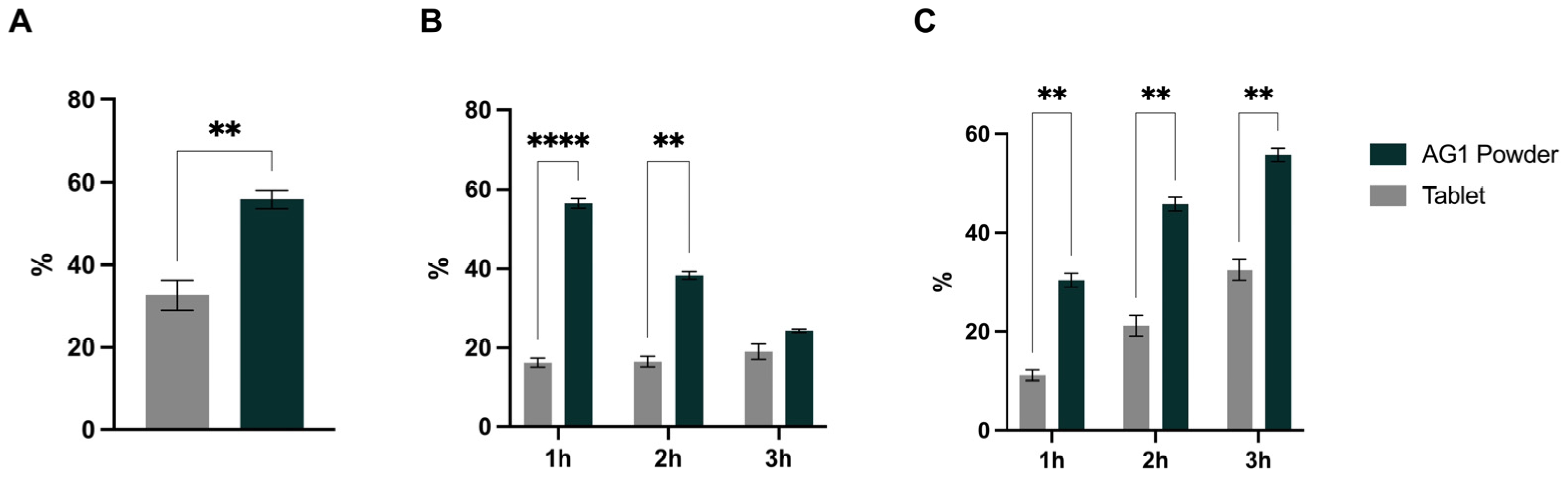
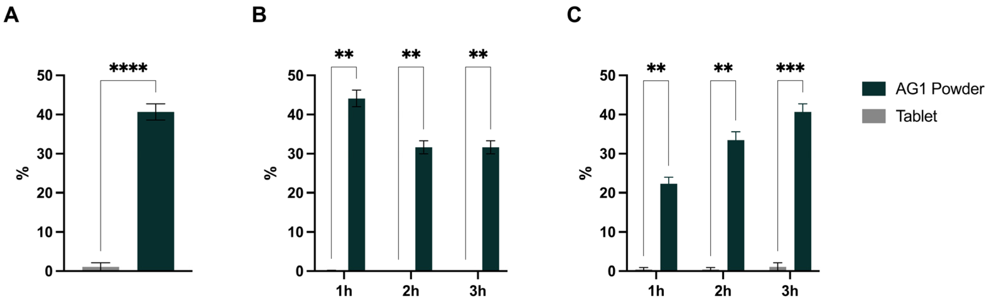
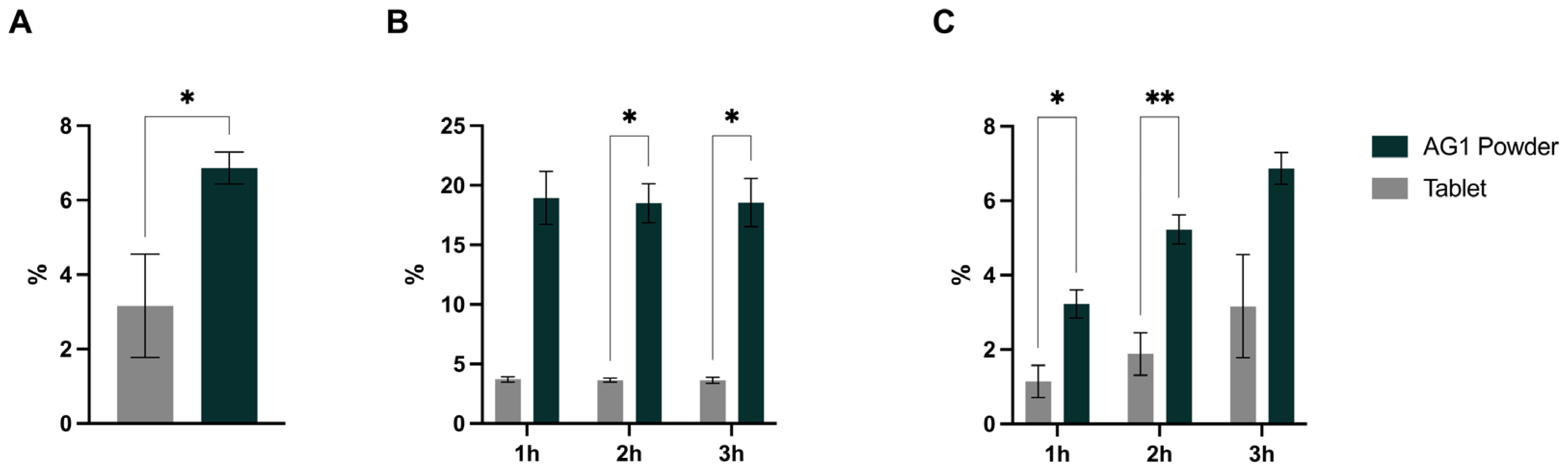
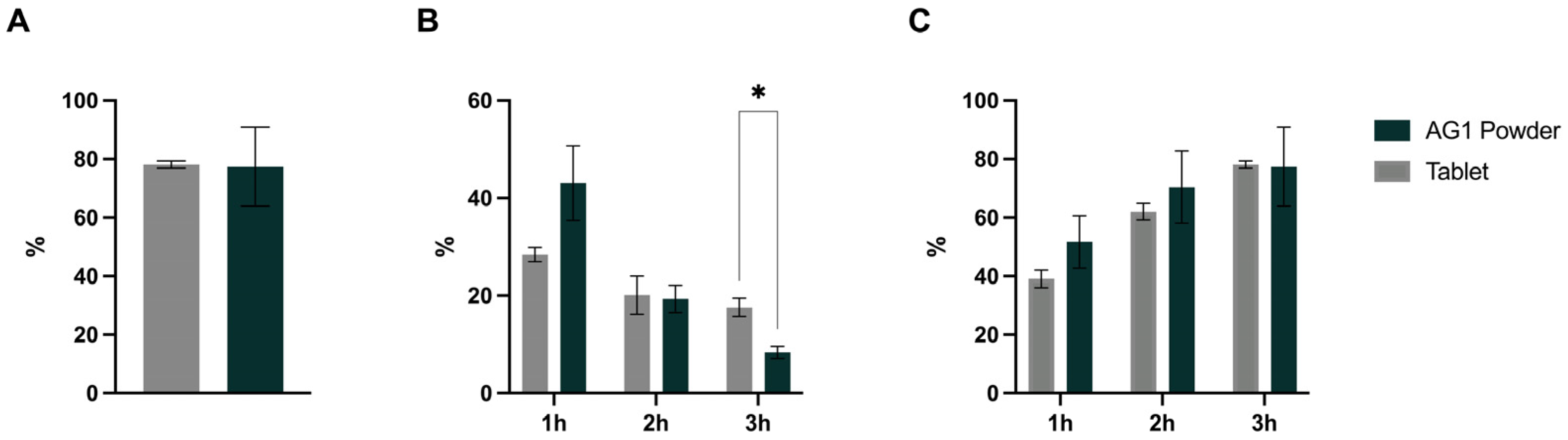
3.2. Bioaccessibility, Bioavailability, and Cmax

The results for Mg are presented in Figure 2. The soluble % Cmax was significantly higher (p = 0.0016) for the AG1 powder vs. the tablet (MD: 23.3% (95% CI: 15.8, 30.8)) (Figure 2, Panel A). A significant effect (p < 0.001) of time, form, and time x form interaction was observed for the bioaccessible fraction of Mg, with higher values for the AG1 powder vs. the tablet at 1 and 2 h timepoints (mean difference (MD): 40.2% (95% CI: 33.5, 46.9) and 21.8% (95% CI: 14.7, 28.9), respectively) (Figure 2, Panel B). Additionally, there was a significant effect (p < 0.05) of time, form, and time x form interaction for the bioavailable fraction of Mg, with significantly higher percentages at 1, 2, and 3 h timepoints (MD: 19.3% (95% CI: 11.8, 26.8), 24.6% (95% CI: 13.8, 35.4), and 23.3% (95% CI: 12.2, 34.3), respectively) (Figure 2, Panel C).
The results for Zn are presented in Figure 3. The soluble % Cmax was significantly higher (p = 0.0344) for the AG1 powder vs. the tablet (MD: 3.7% (95% CI: 0.6, 6.8)) (Figure 3, Panel A). No significant effect of time or time x form interaction was observed for the bioaccessible fraction of Zn, but there was a significant effect of form (p = 0.0016) (Figure 3, Panel B). Post-hoc testing revealed higher values for the AG1 powder vs. the tablet at the 2 and 3 h timepoints (MD: 14.9 (95% CI: 2.7, 27.0) and 14.9 (95% CI: 0.1, 29.7), respectively). Additionally, there was a significant effect (p < 0.05) of time, form, and time x form interaction for the bioavailable fraction of Zn, with significantly higher percentages at 1 and 2 h timepoints (MD: 2.1% (95% CI: 0.8, 3.4) and 3.3% (95% CI: 1.6, 5.1), respectively) (Figure 3, Panel C).
The results for Ca are presented in Figure 4. The soluble % Cmax for Ca was significantly higher (p < 0.0001) for the AG1 powder vs. the tablet (MD: 39.6% (95% CI: 35.3, 43.8)) (Figure 4, Panel A). A significant effect (p < 0.0001) of time, form, and time x form interaction was observed for the bioaccessible fraction of Ca, with post-hoc tests revealing higher values for the AG1 powder vs. the tablet at 1, 2, and 3 h timepoints (MD: 44.0% (95% CI: 34.9, 53.1), 31.6% (24.3, 39.0), and 31.6% (95% CI: 24.3, 39.0), respectively) (Figure 4, Panel B). Similarly, a significant effect (p < 0.0001) of time, form, and time x form interaction for the bioavailable fraction of Ca was observed. Significantly higher bioavailable percentages at 1, 2, and 3 h timepoints (MD: 21.9% (95% CI: 15.7, 28.0), 33.0% (95% CI: 24.8, 41.3), and 39.6% (95% CI: 33.0, 46.1), respectively) were observed (Figure 4, Panel C).
The results for K are presented in Figure 5. There were no differences in % Cmax for K (Figure 5, Panel A). A significant effect (p < 0.001) of time and time x form interaction was observed for the bioaccessible fraction of K, but no significant effect of form occurred (Figure 5, Panel B). Post-hoc tests showed that the bioaccessible fraction at the 3 h timepoint was significantly lower for the AG1 powder vs. the tablet (MD: −9.2% (95% CI: −14.8, −3.6)). Additionally, there was a significant effect (p < 0.01) of time and time x form interaction for the bioavailable fraction of K, with no independent effects of form, and no significant differences at any timepoint (Figure 5, Panel C).

4. Discussion

The aim of the current study was to assess the bioaccessibility and bioavailability of minerals from a novel synbiotic product in a powder form with a tablet MVM supplement formulated with matched mineral (chemically identical form to the powder) and vitamin contents using an in vitro model simulating the human UGIT. The results indicate that mechanical crushing of the tablet into a powder form, similar to the AG1 powder, leads to close but statistically significant differences in the theoretical soluble fraction of Mg, Ca, and Zn. When the whole tablet and powder were fed into the SHIME system, the powder demonstrated superior bioaccessibility for all minerals at the end of the stomach and the duodenum. Lastly, the powder demonstrated significantly higher bioaccessibility and bioavailability for Mg, Ca, and Zn, with no differences in bioavailability for K.
Despite the fact that the actual input of mineral salts was identical, our data indicates differing theoretical soluble fractions for the powder form versus the tablet. While each test product was formulated with the same input in regard to the mineral salts, we did not observe similar maximal soluble fractions between AG1 and the tablet. The baseline concentrations of the minerals were determined by crushing the tablet into a powder with a similar particle size to the AG1 powder. Although both were made to have similar surface areas, it is possible that the existence of excipients in the tablet still caused significant amounts of the mineral salts to remain insoluble. Excipients can change the solubility of active pharmaceutical ingredients and, thus, their bioavailability, with some tablets formulated with specific excipients to strategically increase the bioavailability of the active compound [23]. However, many excipients commonly included in MVM tablets have been shown to impede solubility, like cellulose [24]. Since the MVM tablet in the present study contained microcrystalline cellulose, among other excipients, it is likely their presence may have impacted the overall solubility of the mineral salts. AG1 does not contain microcrystalline cellulose and, therefore, the exact effect of the excipients and how they might have impacted solubility is not fully understood in the current study. However, given the differences in solubility of the minerals between AG1 and the tablet, even following full mechanical homogenization, it is reasonable to assume that excipients added to the decrease in the MVM supplement’s solubility and subsequent bioaccessibility.
The chyme exiting the stomach, as well as the luminal content exiting the duodenum, contained a higher soluble fraction of all the minerals for the powder vs. the tablet. This may be partially explained by the lower exposed surface area in the tablet relative to the powder form. The surface area of the MVM form can alter the ionization of the mineral by impacting how much water can directly access the salts [25,26,27]. Powders have an increased surface area relative to tablets leading to increased rates of disintegration [28] and velocity of dissolution [29]. By increasing the rate of ionization, there is a theoretical increase in bioavailability due to increased absorption. The microcrystalline cellulose excipient frequently utilized in MVM tablets to make tablets more compressible is highly hygroscopic [30]. Hygroscopicity has been known to influence solubility [31,32], and there appears to be a relationship between the hygroscopicity of tablets and the efficiency in disintegration rates [33]. Thus, a higher overall composite hygroscopicity of a tablet results in a lower rate of disintegration. This is likely due to competition for local water molecules, leading to impeded hydration and, thus, disintegration (i.e., the “competition for water” hypothesis) [34]. When taken together, a decreased surface area and increased competition for water results in lower solubility for the mineral salts, which was observed in the luminal contents of the stomach and duodenum end in the current study.
To then assess bioavailability, the luminal content leaving the duodenum was placed into a dialysis system [16]. The bioavailable fraction of Mg, Zn, and Ca were significantly higher for the powder vs. the tablet, with no differences between forms for K. The SHIME model employs a dialysis method, using a methylcellulose membrane to simulate absorption via passive diffusion [16]. Therefore, the disintegration of the tablet and the eventual dissolution of the mineral salts is critical for the passing of ionized minerals through the membrane. As expected, demonstrated by the bioaccessibility at the end of the duodenum, the tablet failed to reach similar rates of dissolution compared to the powder. This can be seen when looking at the significantly higher Cmax values for Mg, Ca, and Zn. Future research using Caco-2 cells to account for other forms of transport is necessary to confirm the differences in the Cmax values between the tablet and the powder.
This study had several strengths. To the best of our knowledge, this is the first study to use the SHIME model to assess the bioaccessibility and bioavailability of a powder vs. a tablet MVM supplement. In the current study, two chemically similar MVM supplements with matched mineral contents were used. This is compared to the one study that assessed powder vs. tablet supplements, but used different physical forms, different amounts, and different mineral salts, making it difficult to interpret their results [8]. Further, the model of the upper gastrointestinal tract accurately mimics human digestion and allows for the assessment of the bioaccessible fraction of minerals in the luminal content, whereas these data are not feasible in human clinical trials. Measurement of contents in the stomach and small intestine in humans is not possible without invasive methods.
There are some limitations to this study as well. An in vitro model does not account for all the factors that influence bioavailability in humans (e.g., other forms of absorption, the effect of metabolism, the distribution of the minerals, excretion). Despite this, SHIME has been shown to be a good predictor of mineral bioavailability in humans [16]. That being said, human studies are necessary to confirm the findings from this study. Further, it is important to note that that AG1 contains other ingredients that could possibly alter mineral absorption (e.g., inulin, phytonutrients). However, these chemicals would theoretically decrease mineral absorption [35,36] and, thus, if they were included in the tablet, it is likely we would have observed an even higher bioavailability and bioaccessibility in the AG1 powder compared to the tablet form. Although out of the scope of this acute dosing paradigm in the UGIT, it is important to note that the probiotics in AG1 may have an impact on mineral absorption during chronic dosing paradigms [37,38]. Finally, this was a proof-of-concept study to provide preliminary data; larger sample sizes are needed to understand the variability we observed within this study.

5. Conclusions

Using MVM powder (AG1) and a tablet MVM supplement formulated to contain matched mineral contents with identical chemical structures, the overall bioavailability of minerals was higher for the powdered form. The significant difference in the bioaccessibility and bioavailability between the tablet and AG1 powder is likely driven by differences in the physical properties and additives leading to altered disintegration and dissolution rates, which ultimately altered the mineral salt solubility. The physical form and use of specific excipients should be considered when designing MVM supplements.

Supplementary Materials

The following supporting information can be downloaded at: https://www.mdpi.com/article/10.3390/applbiosci2040041/s1, Table S1: Ingredient list for tablet multivitamin and mineral supplement formulated for this study.

Author Contributions

Conceptualization, P.A.S., J.R.T., T.O.K., T.M.M. and R.E.; investigation, M.G., C.D. and M.M.; writing—original draft preparation, P.A.S., J.R.T. and T.O.K.; writing—review and editing, P.A.S., J.R.T., T.O.K., M.G., T.M.M. and R.E. All authors have read and agreed to the published version of the manuscript.

Funding

This research was funded by Athletic Greens (AG; Athletic Greens International, Carson City, NV).

Institutional Review Board Statement

Not applicable.

Informed Consent Statement

Not applicable.

Data Availability Statement

Upon reasonable request, data from the corresponding author are made available; however, certain data may not be made available owing to privacy issues.

Conflicts of Interest

J.R.T. and P.A.S. have conducted sponsored research on nutritional supplements. J.R.T., T.O.K., P.A.S., T.M.M. and R.E. are employees of Athletic Greens (AG). M.G., M.M. and C.D. are employees of ProDigest BVBA. The authors declare that this study received funding from AG. The funders were involved in the design of the study, in the writing of the manuscript, and in the decision to publish the results.

References

  1. US Burden of Disease Collaborators; Mokdad, A.H.; Ballestros, K.; Echko, M.; Glenn, S.; Olsen, H.E.; Mullany, E.; Lee, A.; Khan, A.R.; Ahmadi, A.; et al. The State of US Health, 1990–2016: Burden of Diseases, Injuries, and Risk Factors Among US States. JAMA 2018, 319, 1444–1472. [Google Scholar] [CrossRef]
  2. Shams-White, M.M.; Pannucci, T.E.; Lerman, J.L.; Herrick, K.A.; Zimmer, M.; Meyers Mathieu, K.; Stoody, E.E.; Reedy, J. Healthy Eating Index-2020: Review and Update Process to Reflect the Dietary Guidelines for Americans, 2020–2025. J. Acad. Nutr. Diet. 2023, 123, 1280–1288. [Google Scholar] [CrossRef]
  3. USDA, Agricultural Research Service. Usual Nutrient Intake from Food and Beverages, by Gender and Age, What We Eat in America, NHANES 2017–March 2020 Prepandemic. 2023. Available online: http://www.ars.usda.gov/nea/bhnrc/fsrg (accessed on 10 September 2023).
  4. Dickinson, A.; Blatman, J.; El-Dash, N.; Franco, J.C. Consumer Usage and Reasons for Using Dietary Supplements: Report of a Series of Surveys. J. Am. Coll. Nutr. 2014, 33, 176–182. [Google Scholar] [CrossRef]
  5. U.S. Department of Agriculture; U.S. Department of Health and Human Service. Dietary Guidelines for Americans, 2020–2025, 9th ed.; U.S. Department of Agriculture: Washington, DC, USA, 2020. Available online: https://www.DietaryGuidelines.gov (accessed on 10 September 2023).
  6. Mishra, S.; Stierman, B.; Gahche, J.J.; Potischman, N. Data Brief 399: Dietary Supplement Use among Adults: United States, 2017–2018; National Center for Health Statistics: Hyattsville, MD, USA, 2021. [Google Scholar]
  7. Fact.MR. Powder Dietary Supplements Market by Ingredient (Vitamins, Botanicals, Proteins & Amino Acids, Fibers & Specialty Carbohydrates, Omega Fatty Acids), by Application (Bone & Joint Health, Immunity, Cardiac Health), by End User, by Region—Global Insights 2022–2032. 2022. Available online: https://www.factmr.com/report/48/powder-dietary-supplements-market (accessed on 10 September 2023).
  8. Wang, H.; Bua, P.; Capodice, J. A Comparative Study of Calcium Absorption Following a Single Serving Administration of Calcium Carbonate Powder versus Calcium Citrate Tablets in Healthy Premenopausal Women. Food Nutr. Res. 2014, 58. [Google Scholar] [CrossRef] [PubMed]
  9. Löbenberg, R.; Steinke, W. Investigation of Vitamin and Mineral Tablets and Capsules on the Canadian Market. J. Pharm. Pharm. Sci. 2006, 9, 40–49. [Google Scholar] [PubMed]
  10. Thakker, K.M.; Sitren, H.S.; Gregory, J.F.; Schmidt, G.L.; Baumgartner, T.G. Dosage Form and Formulation Effects on the Bioavailability of Vitamin E, Riboflavin, and Vitamin B-6 from Multivitamin Preparations. Am. J. Clin. Nutr. 1987, 45, 1472–1479. [Google Scholar] [CrossRef] [PubMed]
  11. Oner, L.; Arcasoy, A.; Kas, H.S.; Hincal, A.A. Studies on Zinc Sulphate Microcapsules: (III) in Vivo Evaluation. Eur. J. Drug Metab. Pharmacokinet. 1989, 14, 107–110. [Google Scholar] [CrossRef]
  12. Bhagavan, H.N.; Wolkoff, B.I. Correlation between the Disintegration Time and the Bioavailability of Vitamin C Tablets. Pharm. Res. 1993, 10, 239–242. [Google Scholar] [CrossRef]
  13. Sakurada, T.; Oishi, D.; Shibagaki, Y.; Yasuda, T.; Kimura, K. Efficacy of Oral Powder Compared with Chewable Tablets for Lanthanum Carbonate Administration in Hemodialysis Patients. Hemodial. Int. 2013, 17, S2–S6. [Google Scholar] [CrossRef]
  14. Bende, G.; Biswal, S.; Bhad, P.; Chen, Y.; Salunke, A.; Winter, S.; Wagner, R.; Sunkara, G. Relative Bioavailability of Diclofenac Potassium from Softgel Capsule versus Powder for Oral Solution and Immediate-Release Tablet Formulation. Clin. Pharmacol. Drug Dev. 2016, 5, 76–82. [Google Scholar] [CrossRef]
  15. Navarro, M.; Wood, R.J. Plasma Changes in Micronutrients Following a Multivitamin and Mineral Supplement in Healthy Adults. J. Am. Coll. Nutr. 2003, 22, 124–132. [Google Scholar] [CrossRef]
  16. Blancquaert, L.; Vervaet, C.; Derave, W. Predicting and Testing Bioavailability of Magnesium Supplements. Nutrients 2019, 11, 1663. [Google Scholar] [CrossRef]
  17. AG1 Supplement Facts. Available online: https://drinkag1.com/ingredients/en (accessed on 24 October 2023).
  18. Travis, J.; Lattimore, L.G.; Harvey, M.; Frey, T. NSF International’s Role in the Dietary Supplements and Nutraceuticals Industries. In Nutraceutical and Functional Food Regulations in the United States and around the World; Elsevier: Amsterdam, The Netherlands, 2019; pp. 147–158. [Google Scholar]
  19. Molly, K.; Vande Woestyne, M.; Verstraete, W. Development of a 5-Step Multi-Chamber Reactor as a Simulation of the Human Intestinal Microbial Ecosystem. Appl. Microbiol. Biotechnol. 1993, 39, 254–258. [Google Scholar] [CrossRef] [PubMed]
  20. Mackie, A.; Rigby, N. InfoGest Consensus Method. In The Impact of Food Bioactives on Health; Springer International Publishing: Cham, Switzerland, 2015; pp. 13–22. [Google Scholar]
  21. Riethorst, D.; Mols, R.; Duchateau, G.; Tack, J.; Brouwers, J.; Augustijns, P. Characterization of Human Duodenal Fluids in Fasted and Fed State Conditions. J. Pharm. Sci. 2016, 105, 673–681. [Google Scholar] [CrossRef]
  22. Lee, Y.Y.; Erdogan, A.; Rao, S.S.C. How to Assess Regional and Whole Gut Transit Time with Wireless Motility Capsule. J. Neurogastroenterol. Motil. 2014, 20, 265–270. [Google Scholar] [CrossRef] [PubMed]
  23. Patel, R.; Barker, J.; ElShaer, A. Pharmaceutical Excipients and Drug Metabolism: A Mini-Review. Int. J. Mol. Sci. 2020, 21, 8224. [Google Scholar] [CrossRef] [PubMed]
  24. Gupta, D.; Bhatia, D.; Dave, V.; Sutariya, V.; Varghese Gupta, S. Salts of Therapeutic Agents: Chemical, Physicochemical, and Biological Considerations. Molecules 2018, 23, 1719. [Google Scholar] [CrossRef]
  25. Sun, H.; Wang, X.; Wang, J.; Shi, G.; Chen, L. Influence of the Formula on the Properties of a Fast Dispersible Fruit Tablet Made from Mango, Chlorella, and Cactus Powder. Food Sci. Nutr. 2020, 8, 479–488. [Google Scholar] [CrossRef]
  26. CK-12 Foundation. 16.2: Rate of Dissolution. In Introductory, Conceptual, and GOB Chemistry; LibreTexts: Davis, CA, USA, 2023. [Google Scholar]
  27. Bergstrom, G. 2.4: Water Chemistry. In Cell and Molecular Biology (Bergstrom); LibreTexts: Davis, CA, USA, 2023. [Google Scholar]
  28. Molavi, F.; Hamishehkar, H.; Nokhodchi, A. Impact of Tablet Shape on Drug Dissolution Rate Through Immediate Released Tablets. Adv. Pharm. Bull. 2020, 10, 656–661. [Google Scholar] [CrossRef]
  29. Babu, V.R.; Areefulla, S.H.; Mallikarjun, V. Solubility and Dissolution Enhancement: An Overview. J. Pharm. Res. 2010, 3, 141–145. [Google Scholar]
  30. Yassin, S.; Goodwin, D.J.; Anderson, A.; Sibik, J.; Ian Wilson, D.; Gladden, L.F.; Axel Zeitler, J. The Disintegration Process in Microcrystalline Cellulose Based Tablets, Part 1: Influence of Temperature, Porosity and Superdisintegrants. J. Pharm. Sci. 2015, 104, 3440–3450. [Google Scholar] [CrossRef]
  31. Han, S.; Hong, J.; Luo, Q.; Xu, H.; Tan, H.; Wang, Q.; Tao, J.; Zhou, Y.; Peng, L.; He, Y.; et al. Hygroscopicity of Organic Compounds as a Function of Organic Functionality, Water Solubility, Molecular Weight and Oxidation Level. Atmos. Chem. Phys. Discuss. 2022, 22, 2004–3985. [Google Scholar] [CrossRef]
  32. Maclean, N.; Khadra, I.; Mann, J.; Williams, H.; Abbott, A.; Mead, H.; Markl, D. Investigating the Role of Excipients on the Physical Stability of Directly Compressed Tablets. Int. J. Pharm. X 2022, 4, 100106. [Google Scholar] [CrossRef] [PubMed]
  33. Johnson, J.R.; Wang, L.H.; Gordon, M.S.; Chowhan, Z.T. Effect of Formulation Solubility and Hygroscopicity on Disintegrant Efficiency in Tablets Prepared by Wet Granulation, in Terms of Dissolution. J. Pharm. Sci. 1991, 80, 469–471. [Google Scholar] [CrossRef] [PubMed]
  34. Ekmekciyan, N.; Tuglu, T.; El-Saleh, F.; Muehlenfeld, C.; Stoyanov, E.; Quodbach, J. Competing for Water: A New Approach to Understand Disintegrant Performance. Int. J. Pharm. 2018, 548, 491–499. [Google Scholar] [CrossRef] [PubMed]
  35. Baye, K.; Guyot, J.-P.; Mouquet-Rivier, C. The Unresolved Role of Dietary Fibers on Mineral Absorption. Crit. Rev. Food Sci. Nutr. 2017, 57, 949–957. [Google Scholar] [CrossRef] [PubMed]
  36. Speer, H.; D’Cunha, N.M.; Botek, M.; McKune, A.J.; Sergi, D.; Georgousopoulou, E.; Mellor, D.D.; Naumovski, N. The Effects of Dietary Polyphenols on Circulating Cardiovascular Disease Biomarkers and Iron Status: A Systematic Review. Nutr. Metab. Insights 2019, 12, 1178638819882739. [Google Scholar] [CrossRef] [PubMed]
  37. Barone, M.; D’Amico, F.; Brigidi, P.; Turroni, S. Gut Microbiome-Micronutrient Interaction: The Key to Controlling the Bioavailability of Minerals and Vitamins? Biofactors 2022, 48, 307–314. [Google Scholar] [CrossRef]
  38. Bielik, V.; Kolisek, M. Bioaccessibility and Bioavailability of Minerals in Relation to a Healthy Gut Microbiome. Int. J. Mol. Sci. 2021, 22, 6803. [Google Scholar] [CrossRef]
Figure 1. Study design schematic for initial soluble fraction quantification (Panel A) and upper gastrointestinal tract Simulator of the Human Intestinal Microbial Ecosystem (SHIME®) model (Panel B). Abbreviations: gastrointestinal, GI; hour, HR; inductively coupled plasma optical emission spectroscopy, ICP-OES; small intestine, SI; upper gastrointestinal tract, UGIT.
Applbiosci 02 00041 g001
Figure 2. Cmax (A), bioaccessibility (B), and bioavailability (C) of magnesium for the AG1 powder vs. the tablet during the dialysis phase (n = 3 per treatment). All data are presented as a percentage unless otherwise stated. Statistical analysis included a two-way repeated measures ANOVA with post-hoc testing (Sidak correction) for bioaccessibility and bioavailability and Welch’s t-test for % Cmax. Data are shown as mean and standard error of the mean. ** p < 0.01, **** p < 0.0001. Abbreviations: maximum concentration, Cmax; and magnesium, Mg.
Applbiosci 02 00041 g002
Figure 3. Cmax (A), bioaccessibility (B), and bioavailability (C) of zinc for the AG1 powder vs. the tablet during the dialysis phase (n = 3 per treatment). All data are presented as a percentage unless otherwise stated. Statistical analysis included a two-way repeated measures ANOVA with post-hoc testing (Sidak correction) for bioaccessibility and bioavailability and Welch’s t-test for % Cmax. Data are shown as mean and standard error of the mean. * p < 0.05 and ** p < 0.01. Abbreviations: maximum concentration, Cmax; and zinc, Zn.
Applbiosci 02 00041 g003
Figure 4. Cmax (A), bioaccessibility (B), and bioavailability (C) of calcium for the AG1 powder vs. the tablet during the dialysis phase (n = 3 per treatment). All data are presented as a percentage unless otherwise stated. Statistical analysis included a two-way repeated measures ANOVA with post-hoc testing (Sidak correction) for bioaccessibility and bioavailability and Welch’s t-test % Cmax. Data are shown as mean and standard error of the mean. ** p < 0.01, *** p < 0.001, and **** p < 0.0001. Abbreviations: calcium, Ca; and maximum concentration, Cmax.
Applbiosci 02 00041 g004
Figure 5. Cmax (A), bioaccessibility (B), and bioavailability (C) of potassium for the AG1 powder vs. the tablet during the dialysis phase (n = 3 per treatment). All data are presented as a percentage unless otherwise stated. Statistical analysis included a two-way repeated measures ANOVA with post-hoc testing (Sidak correction) for bioaccessibility and bioavailability and Welch’s t-test for % Cmax. Data are shown as mean and standard error of the mean. * p < 0.05. Abbreviations: maximum concentration, Cmax; and potassium, K.
Applbiosci 02 00041 g005
Table 1. Maximal soluble fraction for both test products.
AG1 PowderTabletp-Value
% soluble 1
Magnesium79.37 ± 0.4483.03 ± 1.200.0236
Potassium100.4 ± 11.3094.34 ± 1.840.4489
Calcium93.32 ± 3.77 *36.06 ± 6.960.0010
Zinc84.30 ± 0.39 *56.25 ± 2.650.0025
Values are presented as mean ± SD. Welch’s t-test was conducted to assess the difference between the powder and tablet. These data are based on both products being tested in triplicate. 1 Relative amounts (percentage of the soluble mineral present relative to the inputs at the time of manufacture). * Significantly higher in the AG1 powder.
Table 2. Relative bioaccessible amounts of minerals present at stomach and duodenum end in the SHIME model prior to the dialysis phase.
AG1 PowderTablet 2p-Value
% Bioaccessible at stomach end 1
Magnesium102.2 ± 1.57 *9.3 ± 6.480.0010
Potassium106.5 ± 18.8 *24.1 ± 15.90.0048
Calcium100.1 ± 6.51 *0.7 ± 0.400.0014
Zinc106.5 ± 3.91 *5.1 ± 3.57<0.0001
% Bioaccessible at duodenum end 1
Magnesium94.4 ± 2.57 *12.7 ± 7.610.0012
Potassium108.2 ± 18.3 *35.2 ± 19.660.0094
Calcium73.9 ± 0.11 *0.7 ± 0.62<0.0001
Zinc25.9 ± 4.59 *3.7 ± 2.320.0052
Values are presented as mean ± SD. Welch’s t-test was conducted to assess the difference between the powder and tablet. These data are based on both products being tested in triplicate. 1 Relative amounts (percentage of the soluble mineral present relative to the total amount added at the baseline) presented due to differences in the total soluble amounts of minerals in the powder vs. the tablet based on the pre-test soluble mineral content. 2 Tablet was not crushed when fed into the SHIME model. * Significantly higher in the AG1 powder.
Disclaimer/Publisher’s Note: The statements, opinions and data contained in all publications are solely those of the individual author(s) and contributor(s) and not of MDPI and/or the editor(s). MDPI and/or the editor(s) disclaim responsibility for any injury to people or property resulting from any ideas, methods, instructions or products referred to in the content.

