Review

Ruthenium Complexes, an Emerging Class of Leishmanicidal Drug Candidates

by
Susana Santos Braga
LAQV/REQUIMTE, Department of Chemistry, University of Aveiro, Campus of Santiago, 3810-193 Aveiro, Portugal
Appl. Biosci. 2022, 1(2), 129-142; https://doi.org/10.3390/applbiosci1020009
Submission received: 13 April 2022 / Revised: 6 June 2022 / Accepted: 4 July 2022 / Published: 5 July 2022

Abstract

:
This review addresses the search for activity enhancement of leishmanicidal organic compounds through their coordination chemistry with ruthenium. In an introduction to leishmaniasis, its clinical manifestations, geographical distribution, available forms of treatment, and challenges to disease management are presented. Ruthenium complexes, owing to their physico-chemical and biological properties, are introduced as a suitable molecular library from which to find alternatives to current medicines. The main sections of the review describe complexes reported in the literature, organised into two main groups: organometallics and inorganic complexes. The activity of the ruthenium complexes is presented compared with that of the ligands for a critical assessment of their utility in future clinical application.

1. Introduction

1.1. Leishmaniasis, a Growing Concern

Human leishmaniasis is an infectious disease caused by over 20 different species of the Leishmania genus. Reported in 98 countries and affecting, in 2017, a number of people estimated between 50,000 and 90,000, leishmaniasis is considered as a major public health issue [1,2]. Its transmission to humans originates from dogs or small mammal reservoirs, such as rats, mice, and other rodents. Human to human transmission is also known. The parasite is inoculated into the patient during the hematophagy of female sandflies of the Phlebotomus (Old World) and Lutzomyia (New World) genera [3]. Since these insects grow preferentially in warm climates, human leishmaniasis has a higher incidence in tropical and warm climates [4].
Typically called a tropical disease, leishmaniasis is now growing beyond the tropics, as climate changes are expanding the habitat of its vectors northwards to the USA and Europe [5]. Two studies published in the early 2010s decade used mathematical models to postulate that the global warming effect would expand the habitat of sand flies northwards to reach Canada [6], Germany, and Poland [7] in thirty to forty years. The expansion, however, is already happening. A study conducted in Germany in 2020 revealed the presence of sand flies in areas where they had not been detected before, with fifteen new sites in Southwest Germany having been identified as positive. Once more, global warming was proposed as a leading cause for expanding the distribution area of sand flies, with consequent increase in the risk for sand fly-borne infections in Southwest Germany [8].
Other factors contributing to expansion of the disease are globalisation and geopolitics. In the last two decades, the number of global travellers, by airplane travel alone, has more than doubled, and travelling strongly contributes to the spread of infectious diseases (as recently demonstrated by the current SARS-CoV-2 pandemic). Geopolitical factors refer to the various armed conflicts in sensitive areas that have caused numbers to almost double in countries like Syria, Libya, and Yemen [9]. In association with armed conflicts, the low socio-economic conditions in African and Middle Eastern countries that have caused a large number of refugees and unprecedented migration waves. The agglomeration of these people in refugee camps, along with the poor nutritional and sanitary resources therein, resulted in several leishmaniasis outbreaks [10]. Adding to these concerns, challenges to leishmaniasis management include the growing number of drug-resistant strains and the lack of drugs with a good tolerability profile, adequate storage time and, more importantly, the possibility of oral administration, which allows for ambulatory treatment in areas with limited hospital capacity (or where hospitals are completely absent).

1.2. Clinical Forms and Treatment

Human leishmaniasis has various clinical forms: cutaneous (CL), a polymorphic skin disease with a single or multiple ulcerating lesions; mucocutaneous (MCL), producing destructive lesions in the skin, mucosa, and cartilage, typically around the nose and mouth and eventually leading to necrosis of the nose; and visceral (VL), characterised by an evolution towards chronic disease with systemic spreading and infection of visceral organs that appear dark and necrotised, resulting, when untreated, in a 90% fatality rate, that is, roughly 70,000 deaths every year [4]. The available medication is limited to aggressive drugs as Sb(V) salts (e.g., meglumine antimoniate), amphotericin B (AmB), miltefosine, pentamidine and some azole compounds for CL (e.g., ketoconazole), as described in the following paragraphs. These drugs frequently cause severe adverse effects, sometimes even death, and their efficacy is reduced by frequent resistance development [11,12].
Pentavalent antimony salts, sodium stibogluconate and meglumine antimoniate, are first-line medicines for the treatment of leishmaniasis. Developed in the 1920s, these salts remain in use mainly due to their broad span of action, affordability, and lack of alternative drugs with equivalent activity and potency. They inhibit ADP phosphorylation, lowering intracellular ATP levels and thus limiting the ability of the parasite to produce the energy needed for its survival. Pentavalent antimony salts also bind to the ribose moiety of DNA to form stable complexes with adenine nucleosides that inhibit parasitic purine transporter proteins [13]. Nausea, vomiting, diarrhoea, abdominal cramps, weakness, hepatotoxicity, and cardiotoxicity are the main adverse effects of these drugs [14].
Amphotericin B acts by binding to sterols in the cell membrane of the parasite’s cells (note that the parasite’s membrane contains higher amounts of ergosterol, unlike the host cell membrane, which has cholesterol), resulting in the formation of pores that leak water, potassium, and small molecules. Amphotericyn B is also an ergosterol disruptor, i.e., it is able to block the sterol biosynthesis pathway and to cause ergosterol depletion in parasitic cells. Low ergosterol production results in further membrane disfunction, reducing the parasite’s viability [12]. The most common adverse effects are nephrotoxicity, hypocalcemia, anemia, fever, and malaise; anaphylasis may also occur, albeit it is less frequent [15].
Miltefosine is the only leishmanicidal drug approved for oral intake. It is a phospholipid analogue that accumulates inside cells by interacting first with the outer membrane, and then folding inwards; once in the inner leaflet of the plasma membrane, miltefosine can detach and balance with the membranes of the organelles [16]. The biological activity of miltefosine involves interaction with membrane lipids and the inhibition of cytochrome c oxidase, leading to apoptosis [17]. Limitations to its use include teratogenicity, gastrointestinal disturbances such as nausea, vomiting, or diarrhoea, and a moderate risk (2–3%) of severe kidney and liver toxicity.
Pentamidine isothionate is an orphan drug approved in the USA for the treatment of Pneumocistus carinii pneumonia that is also active against trypanosomatids, being recommended by the World Health Organization (WHO) for the treatment of leishmaniasis and being part of this organisation’s list of essential drugs [18]. While its mode of action is not fully known, some targets of action are already identified: pentamidine has been shown to hamper nucleobase synthesis [19] and to inhibit the transmembranar protein aguaglyceroporin-2, interfering with the parasite’s osmoregulation [20]. It is used as a first-line treatment for Leishmania aethiopica infections and as a second-line medicine in the treatment of VL and CL with other sensitive species. It has been reported to cause irreversible toxicity in some patients, which poses limitations to its use.
Azole drugs approved for the treatment of CL comprise ketoconazole, an imidazol derivative, and two trizoles, itraconazol and fluconazole. Their common mechanism of action is the inhibition of the enzyme lanosterol 14-α-demetylase, which is involved in the biosynthesis of ergosterol, the main cell membrane sterol in both yeasts and leishmania parasites. They are usually given per orum, but for some of these drugs there are also injectable forms. Itraconazole has no major adverse effects [21], posing as a good alternative for patients who do not respond to first-line drugs [22]. Ketoconazole and fluconazole can cause a few reversible adverse effects, such as elevated liver enzymes, but they offer the advantage of a CL healing rate comparable to that of first-line drugs [23].
Paromomycin, a repurposed aminoglycoside antibiotic, is employed in the treatment of VL by intravenous administration [24] being also used for treating CL, typically in the form of an ointment and in association with other anti-infectious drugs [2].
The discovery of a novel universal therapy that causes less adverse effects and is less prone to resistance is much needed. It is, however, a very challenging matter due to the complex pathophysiology of the parasite and of the varied strategies it possesses for adapting to its host. Parasites infect immunocompetent cells, such as macrophages, neutrophils, and monocytes, and hamper their normal behaviour in order to escape the body’s defences. In turn, exaggerated inflammatory responses are often observed in MCL and some disseminated forms of CL. In these forms, the parasites migrate to secondary sites and there is the formation of metastatic lesions that have very low parasite numbers but suffer, in contrast, from a concomitant hyper-inflammatory state. This is what ultimately causes the destruction of the tissues and the typical lesions observed in MCL [25]. This way, novel therapeutic agents should ideally combine leishmanicidal and anti-inflammatory properties, helping to mitigate infection as well as the exacerbated immune response of the host. Various strategies have been adopted in the search for new lead compounds, from natural product screening to the development of new libraries of compounds based on known molecular targets of the parasite.
Another approach is to use combined therapy, associating immunotherapeutics with classic drugs to improve patient outcomes [26,27]. A few clinical trials, conducted in the last three decades, have shown positive results, with combined immunotherapy reducing the healing time for patients, but more studies are needed before these combinations receive market approval [27].

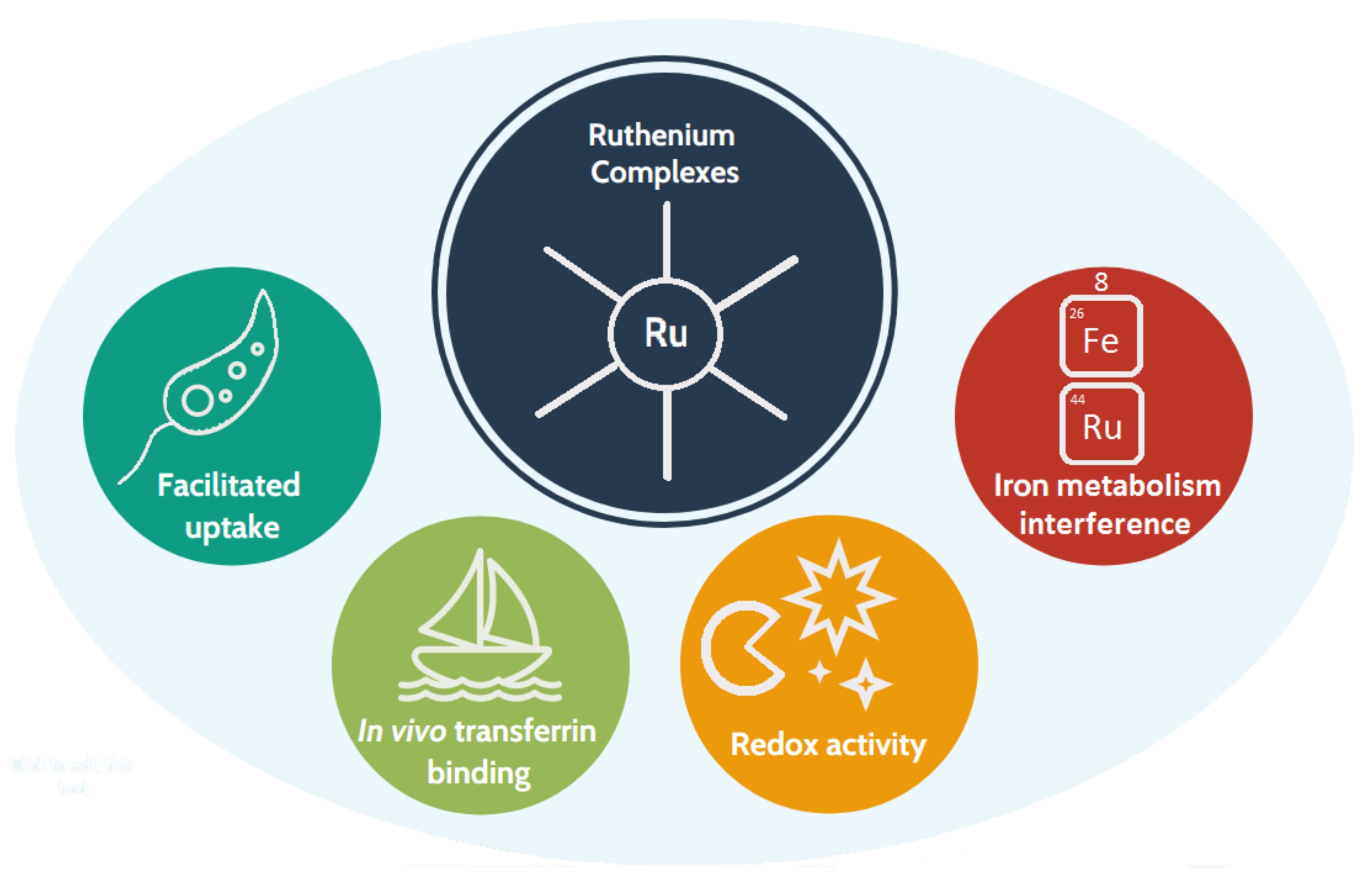
1.3. Why Ruthenium-Based Leishmanicidal Drugs?

Metals contribute strongly towards the activity of a compound. In biocidal activity, the role of the metal is particularly important, its relevance demonstrated by the use of metal compounds in disinfection since the dawn of Medicine. In Leishmania parasites, iron is known to be vital for the metabolism and the reproductive cycle, because infected patients often have iron deficiency or even anaemia [28]. The parasite’s dependency on iron led it to develop privileged uptake pathways to satisfy the demand for this metal, with a heme uptake path having already been observed in L. infantum [29]. Ruthenium, a transition metal belonging to group 8 of the periodic table, same as iron, has the ability to interfere with some of the biological processes developed for handling iron in vivo, including transport, cell uptake, and metabolism. Ruthenium complexes are an emerging class of metallopharmaceuticals for the treatment of parasitic infections [30,31]. Indeed, they gather a quite unique set of properties that makes them the best drug candidates for the treatment of leishmaniasis (Figure 1):
  • The redox properties of the Ru centre [32] are postulated to interfere with the parasite’s defence mechanisms against oxidation;
  • Ru is less toxic than Sb(V);
  • Ruthenium complexes can bind to transferrin [33,34] and be transported through the organism in this way;
  • In human cells, some ruthenium complexes are taken up and metabolised through iron-related pathways [34]; thus, they are likely to enter parasitic cells by the aforementioned heme pathway.
Novel ruthenium complexes developed as drug candidates for parasitic infections often aim at achieving the paradigm of ‘metal-to-ligand synergy’ [35]: a ligand is carefully selected according to its intrinsic anti-parasitic activity, and binding of the active ligand to ruthenium is expected to generate a complex with increased activity. This principle has been employed to design both organometallic and inorganic ruthenium complexes. These two main classes of leishmanicidal ruthenium complexes are described in detail in the two following sections.

2. Organometallic Ruthenium Complexes

Leishmanicidal organometallic ruthenium complexes typically have a geometry resembling a piano stool, in which the stool seat is the organometallic fragment, usually benzene or p-cymene, and the legs are the active ligand as well as spectator ligands such as chloride, ethylenediamine (en), acetylacetonate (acac), 2,2′-bipyridine (bpy); the organometallic part conveys some degree of lipophilicity to these molecules, which allows them to cross cell membranes. Lipophilicity may be further increased by adding apolar ligands such as bpy or phenylphosphines.

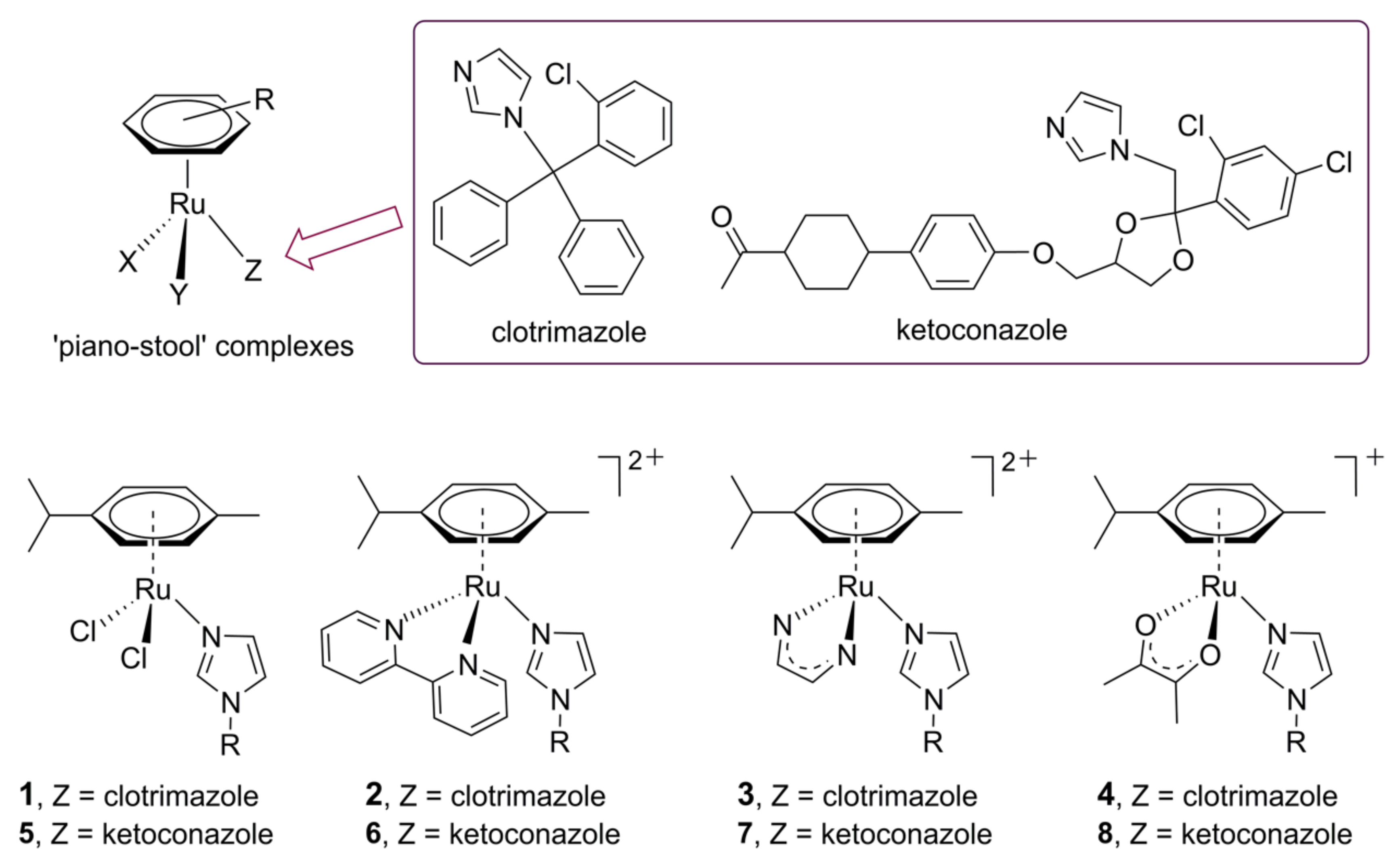
2.1. Ruthenium Azole Complexes

The azole drugs that have a well-known leishmanicidal activity can be used as ligands for organometallic ruthenium complexes, in what comprises one of the most successful approaches to achieve metal-to-ligand synergy. The effect, is not, however, universal, and the medicinal effect of each drug is better potentiated by a particular scaffold of the ruthenium complex, as well as the overall charge of the complex formed, as illustrated by the literature reports described below.
In a family of four p-cymene complexes with ketoconazole (KTZ), [RuII(p-cymene)(KTZ)Cl2] (1), [RuII(p-cymene)(en)(KTZ)]2+ (2), [RuII(p-cymene)(bpy)(KTZ)]2+ (3), and [RuII(p-cymene)(acac)(KTZ)]+ (4), the first three complexes showed increased anti-promastigote activity regarding pure ketoconazole, at rates of >240%, 160%, and 120%, respectively; complex 4, having one acac ligand, was less active (only 20% of the potency of KTZ) [35]. The complexes were quite safe, with 2 and 3 having no cytotoxicity against human fibroblast cells (Hs27 cell line) at 120 μM, the highest tested concentration, and 1 causing 47% inhibition at the same concentration. Given that the LD50 values against promastigotes of complexes 13 are quite low (0.8 ± 1.32, 1.16 ± 0.52, and 1.53 ± 0.84 μM, respectively), the corresponding selectivity indexes are quite high, within 78–150 folds. Complexes 13 were also tested against intracellular parasites in macrophage cells, with some inhibitory activity observed at concentrations around 0.25–1.0 μM; however, IC50 values were not determined as a dose-dependent action could not be observed.
In a follow-up study, complexes 1 and 3 were evaluated in vivo against L. major metacyclic promastigotes that were injected into the paw of BALB/c mice and allowed to incubate for two weeks [36]. The mice were then treated for another two weeks with an intra-peritoneal injection of the test compounds at a daily dose of 4 mg/kg. Compared to negative control, the skin lesion reducing action was 26% for complex 1 and 68% for complex 3 (compared to negative control). Moreover, 3 was slightly more effective than pure clotrimazole in reducing CL skin lesions. The authors concluded that 3 has moderate in vivo activity against L. major mice infection, with a parasite load reduction of 50% or more and no significant toxicity.
A similar family of organometallic ruthenium azole complexes was reported, differing only by having clotrimazole (CTZ) as the active ligand [37]. Complexes, depicted in Figure 2, are [RuII(p-cymene)(CTZ)Cl2] (5), [RuII(p-cymene)(bpy)(CTZ)]2+ (6), [RuII(p-cymene)(en)(CTZ)]2+ (7), and [RuII(p-cymene)(acac)(CTZ)]+ (8), and they were tested against Leishmania major promastigotes. Similarly to the results obtained with the first family, the neutral complex, [RuII(p-cymene)(CTZ)Cl2] (5), was the most active, with an IC50 of 15 ± 8 nM, already within the nanomolar range; it was almost one hundred times more potent than pure clotrimazole (IC50 = 1.6 ± 0.5 μM). The second most active complex was the monocationic one, [RuII(p-cymene)(acac)(CTZ)]+ (8), with an IC50 of 0.45 ± 0.15 μM. The dicationic complexes 6 and 7 have IC50 values of 1.4 ± 0.6 and 1.32 ± 0.69 μM, respectively, being thus fairly within the same potency as pure clotrimazole. Tests against intra-macrophage amastigotes, conducted with complexes 5 and 8, showed the high potency of complex 5, with an IC70 value of 29 nM, while complex 8 was able to inhibit 40% of the intracellular parasites at the highest concentration tested, 1 μM; note that at the same concentration, pure clotrimazole displayed no significant activity.
In another study, the two abovementioned families of ruthenium p-cymene complexes with KTZ and CTZ were expanded to include complexes bearing triphenilphosphine (PPh3) as ligands, [Ru(p-cymene)(KTZ)(PPh3)Cl]+ (9) and [Ru(p-cymene)(CTZ)(PPh3)Cl]+ (10) [38]. The complexes exhibited ligand-to-metal synergy when tested against Leishmania amazonensis promastigotes: IC50 values at 24h of incubation were 0.08 ± 2.62 μM for complex 9 and 0.24 ± 1.65 μM for complex 10 while the pure drugs ketoconazole and clotrimazole had IC50 > 3.0 μM and 0.82 ± 1.12 μM, respectively (at the same incubation time). The complexes were also very potent against intramacrophage amastigotes, with IC50 values in the nanomolar range: 15.53 ± 1.30 nM for complex 9 and 25.80 ± 1.09 nM for complex 10. Moreover, they presented a good safety profile on healthy macrophages, with no cytotoxicity observed at 24 h of incubation and only some growth inhibition occurring with complex 10 after 48 h of incubation at the highest tested concentration, 1 μM.

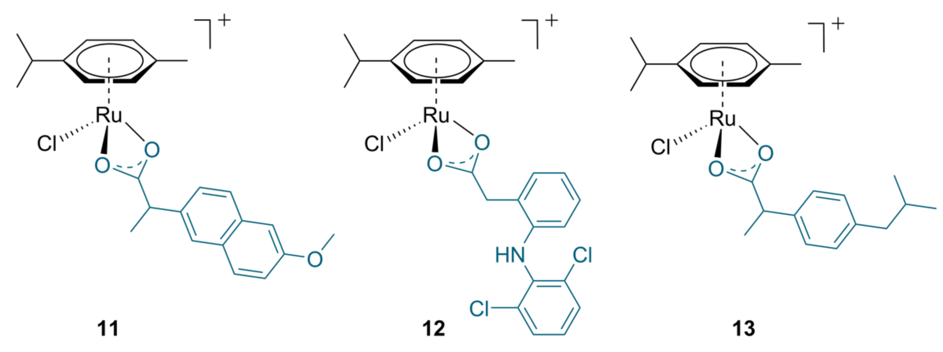
2.2. Ruthenium Complexes with Non-Steroidal Anti-Inflammatory Agents

The exacerbated inflammation response of the patient during infection with leishmaniasis often is the cause of the disfiguring lesions observed in MCL [25], as described previously in Section 1.2. It may thus be useful to convey anti-inflammatory drugs to the patient during the treatment, in order to mitigate the inflammatory response and prevent widespread tissue damage.
A family of ruthenium p-cymene complexes with NSAIDs (non-steroid anti-inflammatory drugs having the general formulae [RuII(p-cymene)(NSAID)Cl]+, where NSAID = naproxen (11), diclofenac (12) and ibuprofen (13), was recently reported (Figure 3) [39]. Activity screening on promastigote forms of Leishmania infantum (MCER/BR/79/M6445 strain) and L. amazonensis (IFLA/BR/67/PH8 strain) showed that binding naproxen and diclofenac to the ruthenium scaffold to form complexes 11 and 12 was able to imbue them with leishmanicidal action, even though these drugs do not have such effect in their pure form. Complex 11 displayed IC50 values of 42.3 ± 16.5 μM against L. infantum and 23.6 ± 1.0 μM against L. amazonensis, while for complex 12 values were 8.6 ± 0.4 μM and 7.4 ± 2.7 μM, respectively; the pure drugs and complex 13 did not display any activity at the highest concentration tested (200 μM). The partition coefficient was calculated for the complexes, showing values of 7.1, 7.3, and 7.7 (for 11, 12, and 13, respectively). This indicates an increase in lipophilicity in regard to the pure drugs, naproxen (log P = 3.2), diclofenac (log P = 4.5), and ibuprofen (log P = 4.0).

3. Inorganic Ruthenium Complexes

Inorganic ruthenium complexes feature an overall broader structural diversity than their organometallic counterparts. Besides the various active ligands, the coordination sphere in most complexes is comprised of kinetically inert ligands, usually of the polypyridyl or of the phenylphosphine kind. A few complexes, present, in alternative, labile ligands (e.g., Cl or DMSO), which render them more reactive but also less stable.

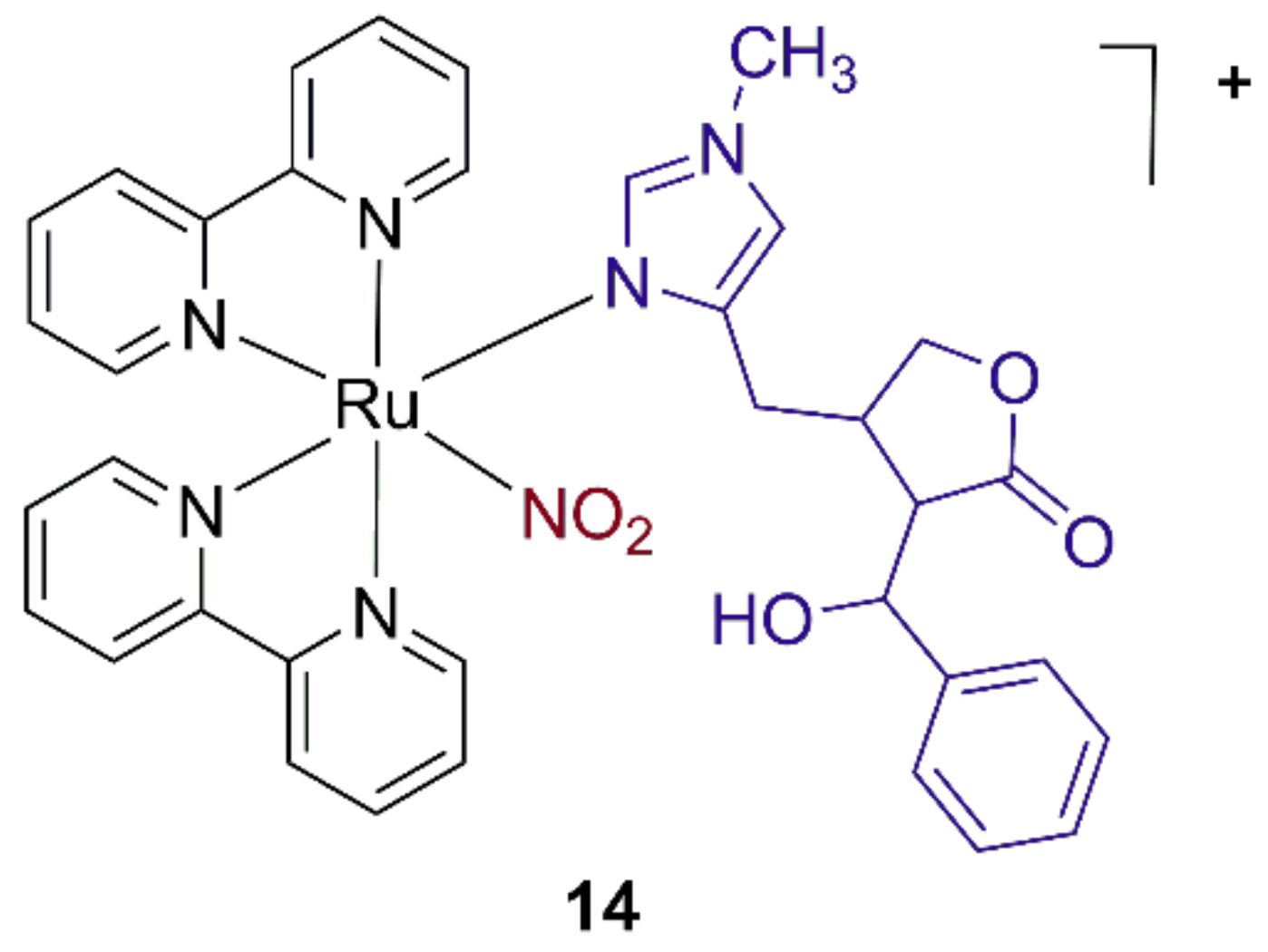
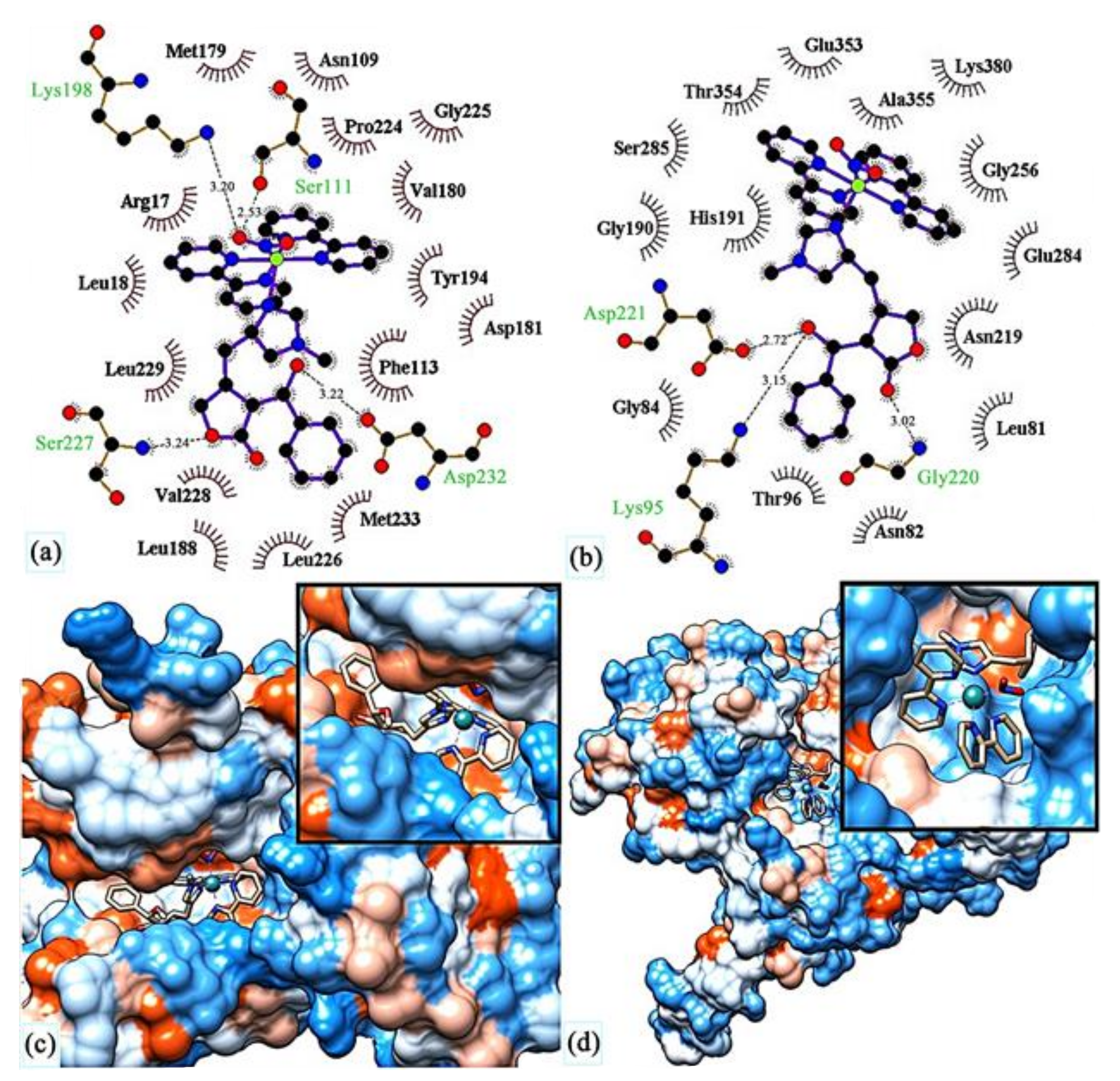
3.1. Ruthenium Complexes with Plant Alkaloids

Plant alkaloids are a vast class of metabolites that typically serve as defence mechanisms against sources of biotic stress, such as environmental factors, parasites, or even to protect against extensive culling by herbivores. Alkaloids are, thus, a vast and excellent natural source of cytotoxic agents. Epiisopiloturine (EPI) is an imidazole alkaloid abundant in the leaves of Pilocarpus microphyllus, a plant popularly known as ‘jaborandi do maranhão’. Having already demonstrated pharmacologic activity as an anti-inflammatory [40,41], anti-nociceptive [41] and anti-schisostomiasis [42,43] agent, EPI is a very promising ligand to combine with a ruthenium scaffold to form a new molecule targeting CL and MCL, because besides inhibiting the parasite it may also contribute to reduce the inflammation typically associated with these lesions.
The complex [Ru(bpy)2(EPI)NO2]+ (14), represented in Figure 4, was recently reported to inhibit L. major promastigotes (MHOM/IL/80/Friedlin strain) with an IC50 of 1.07 μM [44].
Molecular docking studies were conducted on complex 14 and target proteins of leishmania. Figure 5 shows the results for pteridine reductase (1e7w) and UDP-glucose pyrophosphorylase (5nzg), two of the proteins that formed hydrogen bonds with the complex.
The strongest binding affinity was calculated to occur with protein 1e7w (inhibition constant of 14.8 nM), indicating that complex 14 may act though interruption of the growth of the parasite (for which 1e7w is essential). Complex 14 also showed excellent molecular affinity towards the target protein 5nzg of L. major (inhibition constant of 19.74 nM), through the formation of three hydrogen bonds. This protein is vital for the production of cell surface glycans, and is also involved in other processes that generate parasite pathogenicity [44].

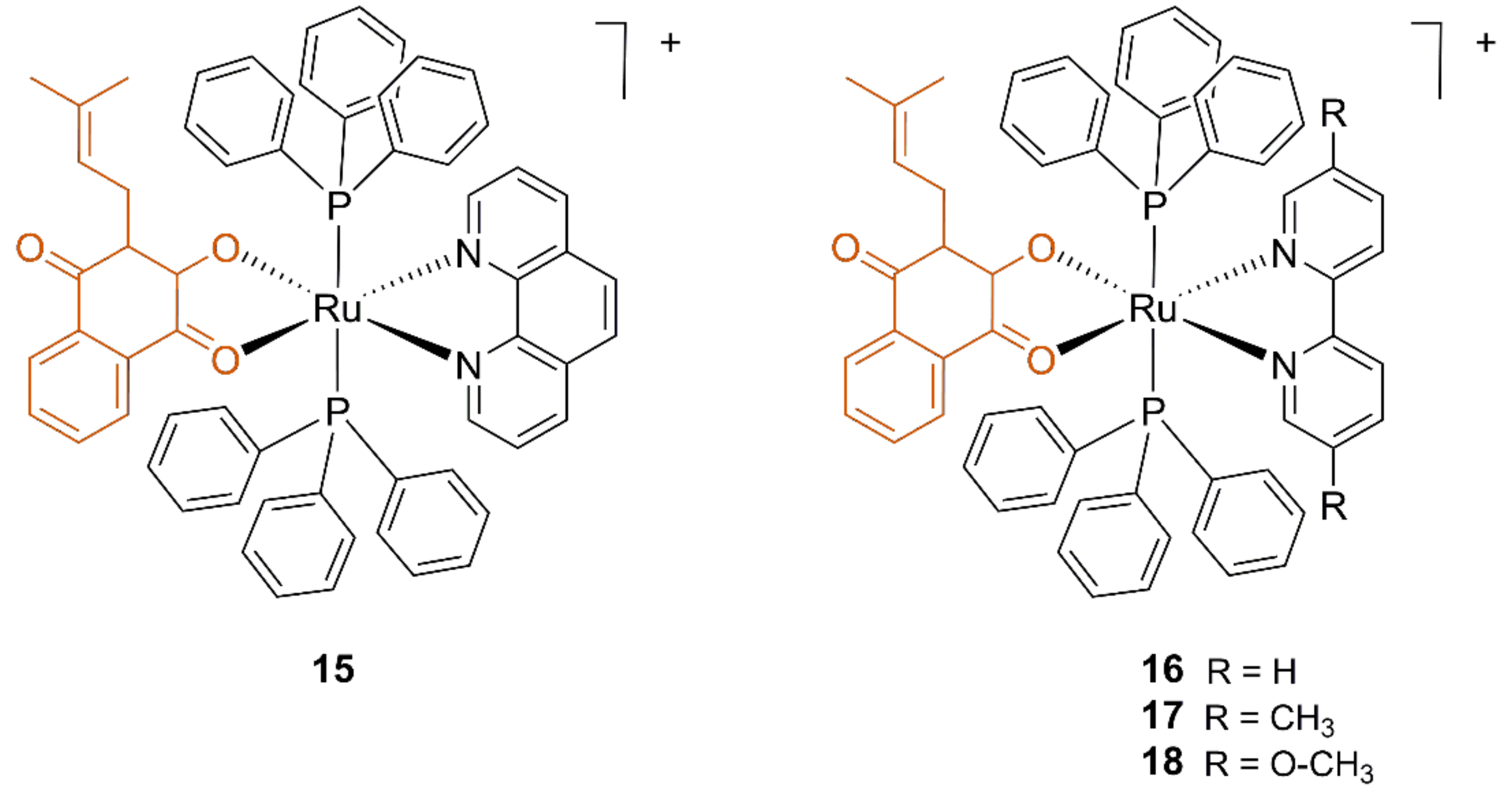
3.2. Ruthenium Lapachol Complexes

Lapachol is a natural biocidal compound isolated from the bark of Handroanthus impetiginosus, the lapacho tree. Traditionally used to treat a variety of bacterial and parasitic infections, lapachol also possesses leishmanicidal activity. Combining lapachol with an inorganic ruthenium scaffold is a paradigmatic example in metal-to-ligand synergy [45]. The complexes are depicted in Figure 6, and they can be described by the general formula [RuII(N,N′-lig)(lapachol)(PPh3)2]+, where the N,N′-lig is 1,10-phenanthroline (phen), bpy or its derivatives, 4,4′-dimethylbypiridine and 4,4′-dimethoxybypiridine. Besides the active ligand, lapachol, the remaining ligands were chosen due to their high inertia towards ligand substitution, that is, because they are very little labile.
The molecular design strategy allowed obtaining complexes that surpass the potency of pure lapachol, both against the promastigote form and the macrophage-internalised amastigote form of L. amazonensis. However, the complexes were also toxic to non-infected mouse macrophages (J744 line), as indicated by the relatively low IC50 values obtained against these cells (Table 1) [45].

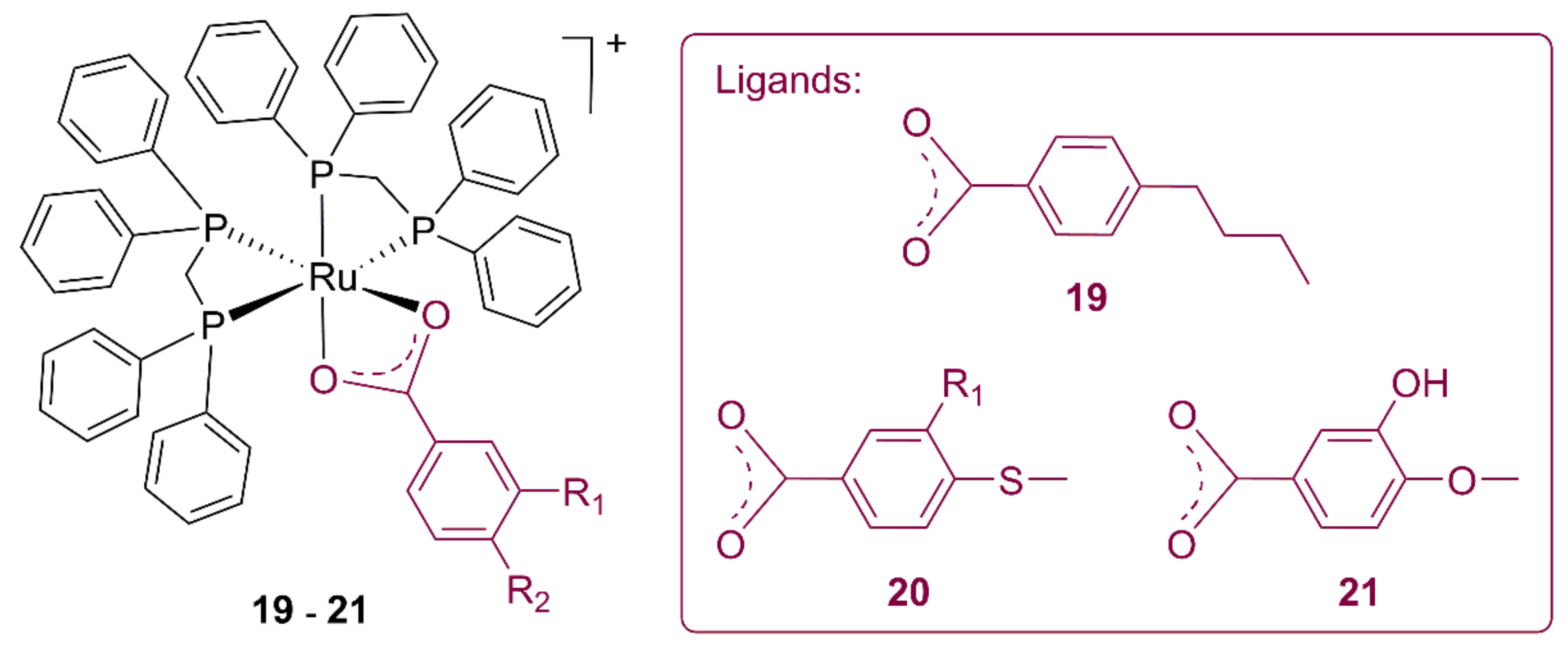
3.3. Ruthenium Alkylbenzoate Complexes

Alkylbenzoates are skin bleaching compounds acting by inhibition of tyrosinase [46], an enzyme that, besides being involved in the production of melanin in healthy humans, features strongly increased expression in leishmaniasis patients [47].
Complexes 19–21, having three distinct alkylbenzoate derivatives as active ligands and a bridged phenylphosphine inert ligand (Figure 7), had good leishmanicidal activity against promastigote forms of L. brasiliensis, L. amazonensis, and L. infantum, with IC50 values in the low micromolar or, for a few of them, in the nanomolar range (Table 2) [48]. However, the complexes also exhibited some toxicity against non-infected mouse macrophages (RAW 264 line), thus evidencing that complexation was unable to ensure selectivity of action towards infected immune cells.
In a follow-up study, complex 21 was investigated regarding the ability to generate reactive oxygen species (ROS), DNA fragmentation, and alterations to the cell cycle of L. amazonensis promastigotes [49]. The anti-proliferative effect of the complex was shown to involve cell cycle arrest in the sub-G1 phase and death by apoptosis associated with DNA fragmentation. Moreover, the complex was shown to increase the intracellular levels of ROS, with possible repercussions on the mitochondria, because depolarisation of the mitochondrial membrane was observed concomitantly.

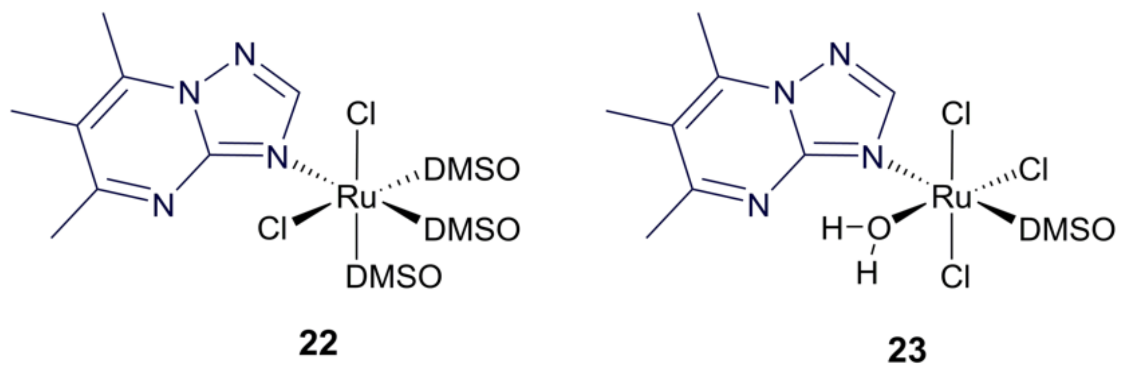
3.4. Ruthenium Purine Complexes

Ruthenium complexes with a purine derivative as the active ligand and the remaining coordination sphere composed of labile ligands, such as DMSO and chloride, presented metal-to-ligand synergy, translated as a higher activity against promastigote forms of L. infantum, L. brasiliensis, and L. Donovani [50]. Importantly, the two most potent complexes, 22 and 23 (Figure 8) have different oxidation states: Ru(II) for complex 22 and Ru(III) for complex 23. Complex 23 hydrolysed rapidly in pure water. Regarding complex 22, rapid hydrolysis was achieved by adding sodium chloride to the aqueous medium. This implies that both complexes can be expected to have similar hydrolytical profiles in vivo. Complex 22 was the most potent, with IC50 values ranging from 9.2 ± 0.7 to 41.8 ± 3.3 μM (according to the Leishmania species tested), while IC50 values for 23 laid between 46.1 ± 3.7 and 62.0 ± 4.9 μM. A strong advantage of these complexes is their in vitro safe profile on macrophages and kidney epithelial cells (IC50 > 300 μM). Nevertheless, the presence of labile ligands with foreseeably fast in vivo hydrolysis may cause undesired interactions with a vast number of host targets, leading to partial loss of activity and eventual unwanted toxic effects. Future studies should include the evaluation, in animal models, of the effective dose and safety of these complexes, to better understand their viability as new therapeutic agents.

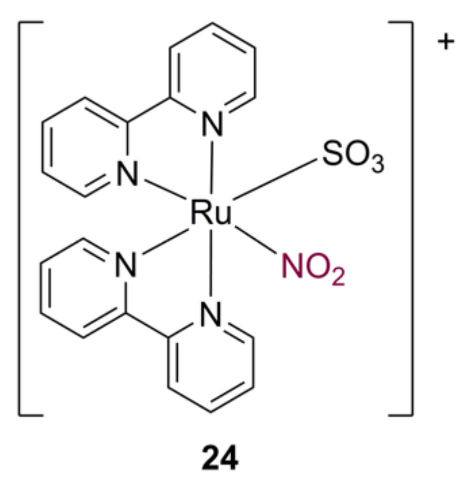
3.5. Ruthenium Nitrosyl Complexes

Nitric oxide is a small biosignalling molecule that in humans helps regulate vasodilation, inflammation, and even neuronal responses. It is also the main molecule mediating macrophage anti-leishmania activity during the course an infection. For this reason, NO is considered as an active ligand by some researchers, who have developed NO-releasing complexes as potential new drugs. While most of the complexes were designed for anti-tumour action [51], their role as leishmanicidal agents has also been investigated. An example is complex 24 (Figure 9), shown to generate NO with a dissociation constant of 2.0 × 10⁻³ s−1 and to resist hydroxide disruption up to pH 9 [52]. Follow-up biological activity studies with complex 24 showed growth inhibition of L. braziliensis parasites that were internalised in dermal fibroblasts (IC50 < 10 μM) and no toxicity towards the non-infected dermal fibroblasts; in vivo studies on hamsters showed that a dose of 0.3 mg/kg/day of 24 produced 99.9% reduction of parasitic load in the lesion, a feat that for glucantime required a dose of 60 mg/kg/day to be matched [53].

4. Conclusions and Outlook

The last two decades have witnessed a growth in research targeted at finding novel therapeutic agents for neglected tropical diseases. A good part of these research efforts have focused on leishmaniasis, a pathology with a large range of clinical manifestations and with tendency to expand beyond its traditional endemic tropical areas as a result of global warming and widespread global travel. In the wide range of possible new treatments, ruthenium metal complexes have gained interest owing to their unique set of properties and biocompatibility. As demonstrated throughout this article, ruthenium complexes offer a broad panorama of structures from which it is possible to identify promising leishmanicidal agents with increased activity regarding the corresponding organic ligands that can pose as new leads for leishmanicidal activity. Highlight should be given to the complexes 9, 14, and 15 in particular, for their activity in the low micromolar or even nanomolar range in tandem with the good safety profile observed in vitro. The phenanthroline lapachol complex, 15, was one of the most striking examples of metal-to-ligand synergy, being more than 140 times more potent than pure lapachol.
The main limitation of the presently available literature studies is the lack of in vivo studies to verify both the activity of the complexes and their safety profile. Future perspectives can rely on the optimisation of the lead structures herein identified, as well as the investigation of their activity in animal models of the disease.

Funding

Thanks are due to University of Aveiro and FCT/MCTES (Fundação para a Ciência e a Tecnologia, Ministério da Ciência, da Tecnologia e do Ensino Superior) for financial support to LAQV-REQUIMTE (Ref. UIDB/50006/2020) through national founds (PIDDAC) and, where applicable, co-financed by the European Regional Development Fund (FEDER), within the PT2020 Partnership Agreement.

Conflicts of Interest

The author declares no conflict of interest.

References

  1. Leishmaniasis: Situation and Trends. Available online: http://www.who.int/gho/neglected_diseases/leishmaniasis/en/ (accessed on 13 April 2022).
  2. Burza, S.; Croft, S.L.; Boelaert, M. Leishmaniasis. Lancet 2018, 392, 951–970. [Google Scholar] [CrossRef]
  3. Rodrigues, I.A.; Mazotto, A.M.; Cardoso, V.; Alves, R.L.; Amaral, A.C.F.; Silva, J.R.A.; Pinheiro, A.S.; Vermelho, A.B. Natural products: Insights into leishmaniasis inflammatory response. Mediat. Inflam. 2015, 2015, 835910. [Google Scholar] [CrossRef] [Green Version]
  4. Torres-Guerrero, E.; Quintanilla-Cedillo, M.R.; Ruiz-Esmenjaud, J.; Arenas, R. Leishmaniasis: A review. F1000Research 2017, 6, 750. [Google Scholar] [CrossRef]
  5. Available online: https://ecdc.europa.eu/en/disease-vectors/facts/phlebotomine-sand-flies (accessed on 13 April 2022).
  6. González, C.; Wang, O.; Strutz, S.E.; González-Salazar, C.; Sánchez-Cordero, V.; Sarkar, S. Climate change and risk of leishmaniasis in North America: Predictions from ecological niche models of vector and reservoir species. PLoS Negl. Trop. Dis. 2010, 4, e585. [Google Scholar] [CrossRef] [Green Version]
  7. Trájer, A.J.; Bede-Fazekas, Á.; Hufnagel, L.; Horváth, L.; Bobvos, J. The effect of climate change on the potential distribution of the European Phlebotomus species. Appl. Ecol. Environ. Res. 2013, 11, 189–208. [Google Scholar] [CrossRef]
  8. Oerther, S.; Jöst, H.; Heitmann, A.; Lühken, R.; Krüger, A.; Steinhausen, I.; Brinker, C.; Lorentz, S.; Marx, M.; Schmidt-Chanasit, J.; et al. Phlebotomine sand flies in Southwest Germany: An update with records in new locations. Parasit. Vectors 2020, 13, 173. [Google Scholar] [CrossRef] [Green Version]
  9. Steverding, D. The history of leishmaniasis. Parasit. Vectors 2017, 10, 82. [Google Scholar] [CrossRef] [Green Version]
  10. Du, R.; Hotez, P.J.; Al-Salem, W.S.; Costa-Serrano, A.A. Old world cutaneous leishmaniasis and refugee crises in the Middle East and North Africa. PLoS Negl. Trop. Dis. 2016, 10, e0004545. [Google Scholar] [CrossRef] [Green Version]
  11. Ponte-Sucre, A.; Gamarro, F.; Dujardin, J.C.; Barrett, M.P.; López-Vélez, R.; García-Hernández, R.; Pountain, A.W.; Mwenechanya, R.; Papadopoulou, B. Drug resistance and treatment failure in leishmaniasis: A 21st century challenge. PLoS Negl. Trop. Dis. 2017, 11, e0006052. [Google Scholar] [CrossRef] [PubMed]
  12. Braga, S.S. Multi-target drugs active against leishmaniasis: A paradigm of drug repurposing. Eur. J. Med. Chem. 2019, 183, 111660. [Google Scholar] [CrossRef] [PubMed]
  13. Mishra, J.; Saxena, A.; Singh, S. Chemotherapy of leishmaniasis: Past, present and future. Curr. Med. Chem. 2007, 14, 1153–1169. [Google Scholar] [CrossRef]
  14. Frézard, F.; Demicheli, C.; Ribeiro, R.R. Pentavalent antimonials: New perspectives for old drugs. Molecules 2009, 14, 2317–2336. [Google Scholar] [CrossRef] [Green Version]
  15. Laniado-Laborín, R.; Cabrales-Vargas, M.N. Amphotericin B: Side effects and toxicity. Rev. Iberoam. Micol. 2009, 26, 223–227. [Google Scholar] [CrossRef]
  16. Javier, F.P.; Seifert, K.; Croft, S.L.; Sundar, S.; Castanys, S.; Gamarro, F. Mechanisms of experimental resistance of Leishmania to miltefosine: Implications for clinical use. Drug Resist. Updates 2006, 9, 26–39. [Google Scholar] [CrossRef]
  17. Berman, J.J. Treatment of leishmaniasis with miltefosine: 2008 status. Expert Opin. Drug Metab. Toxicol. 2008, 4, 1209–1216. [Google Scholar] [CrossRef]
  18. World Health Organization. Model List of Essential Medicines: 21st List 2019. Available online: https://apps.who.int/iris/handle/10665/325771 (accessed on 31 March 2020).
  19. Bornstein, R.S.; Yarbro, J.W. An evaluation of the mechanism of action of pentamidine isethionate. J. Surg. Oncol. 1970, 2, 393–398. [Google Scholar] [CrossRef]
  20. Song, J.; Baker, N.; Rothert, M.; Henke, B.; Jeacock, L.; Horn, D.; Beitz, E. Pentamidine is not a permeant but a nanomolar inhibitor of the Trypanosoma brucei aquaglyceroporin-2. PLoS Pathog. 2016, 12, e1005436. [Google Scholar] [CrossRef] [Green Version]
  21. Dogra, J.; Saxena, V.N. Itraconazole and leishmaniasis: A randomised double-blind trial in cutaneous disease. Int. J. Parasitol. 1996, 26, 1413–1415. [Google Scholar] [CrossRef]
  22. Consigli, J.; Danielo, C.; Gallerano, V.; Papa, M.; Guidi, A. Cutaneous leishmaniasis: Successful treatment with itraconazole. Int. J. Dermatol. 2006, 45, 46–49. [Google Scholar] [CrossRef] [PubMed]
  23. Alrajhi, A.A.; Ibrahim, E.A.; De Vol, E.B.; Khairat, M.; Faris, R.M.; Maguire, J.H. Fluconazole for the treatment of cutaneous leishmaniasis caused by Leishmania major. N. Engl. J. Med. 2002, 346, 891–895. [Google Scholar] [CrossRef] [PubMed]
  24. Pokharel, P.; Ghimire, R.; Lamichhane, P. Efficacy and safety of paromomycin for visceral leishmaniasis: A systematic review. J. Trop. Med. 2021, 2021, 8629039. [Google Scholar] [CrossRef] [PubMed]
  25. Rossi, M.; Fasel, N. How to master the host immune system? Leishmania parasites have the solutions! Int. Immunol. 2017, 30, 103–111. [Google Scholar] [CrossRef] [PubMed] [Green Version]
  26. Akbari, M.; Orian, A.; Hatam, G. Immunotherapy in treatment of leishmaniasis. Immunol. Lett. 2021, 233, 80–86. [Google Scholar] [CrossRef] [PubMed]
  27. Mota, C.M.; Oyama, J.; Monich, M.S.T.; Brustolin, A.A.; de Souza, J.V.P.; Murase, L.S.; Lopes, L.D.G.; Santos, T.S.; Teixeira, J.J.V.; Silveira, T.G.V. Three decades of clinical trials on immunotherapy for human leishmaniases: A systematic review and meta-analysis. Immunotherapy 2021, 13, 693–721. [Google Scholar] [CrossRef]
  28. Goto, Y.; Cheng, J.; Omachi, S.; Morimoto, A. Prevalence, severity, and pathogeneses of anemia in visceral leishmaniasis. Parasitol. Res. 2017, 116, 457–464. [Google Scholar] [CrossRef]
  29. Carvalho, S.; Crus, T.; Santarém, N.; Castro, H.; Costa, V.; Tomás, A.M. Heme as a source of iron to Leishmania infantum amastigotes. Acta Trop. 2009, 109, 131. [Google Scholar] [CrossRef]
  30. Braga, S.S.; Lysenko, K.; Santos, N.E. Ruthenophenanthroline complexes active against tripanosomids. In A Closer Look at Phenanthroline, 1st ed.; Amies, W., Ed.; NovaScience Publishing: Hauppage, NY, USA, 2020. [Google Scholar]
  31. Britten, N.S.; Butler, J.A. Ruthenium metallotherapeutics: Novel approaches to combating parasitic infections. Curr. Med. Chem. 2022, 29, 1–20. [Google Scholar] [CrossRef]
  32. Pongratz, M.; Schluga, P.; Jakupec, M.A.; Arion, V.A.; Hartinger, C.G.; Allmaierb, G.; Keppler, B.K. Transferrin binding and transferrin-mediated cellular uptake of the ruthenium coordination compound KP1019, studied by means of AAS, ESI-MS and CD spectroscopy. J. Anal. At. Spectrom. 2004, 19, 46. [Google Scholar] [CrossRef]
  33. Alessio, E. Thirty years of the drug candidate NAMI-A and the myths in the field of ruthenium anticancer compounds: A personal perspective. Eur. J. Inorg. Chem. 2017, 12, 1549–1560. [Google Scholar] [CrossRef]
  34. Aitken, J.B.; Antony, S.; Weekley, C.M.; Lai, B.; Spiccia, L.; Harris, H.H. Distinct cellular fates for KP1019 and NAMI-A determined by X-ray fluorescence imaging of single cells. Metallomics 2012, 4, 1051–1056. [Google Scholar] [CrossRef]
  35. Inigues, E.; Sánchez, A.; Vasquez, M.A.; Martínez, A.; Olivas, J.; Sattler, A.; Sánchez-Delgado, R.A.; Maldonado, R.A. Metal–drug synergy: New ruthenium(II) complexes of ketoconazole are highly active against Leishmania major and Trypanosoma cruzi and nontoxic to human or murine normal cells. J. Biol. Inorg. Chem. 2013, 18, 779–790. [Google Scholar] [CrossRef] [PubMed] [Green Version]
  36. Iniguez, E.; Varela-Ramirez, A.; Martínez, A.; Torres, C.L.; Sánchez-Delgado, R.A.; Maldonado, R.A. Ruthenium-Clotrimazole complex has significant efficacy in the murine model of cutaneous leishmaniasis. Acta Trop. 2016, 164, 402–410. [Google Scholar] [CrossRef] [PubMed] [Green Version]
  37. Martínez, A.; Carreon, T.; Iniguez, E.; Anzellotti, A.; Sánchez, A.; Tyan, M.; Sattler, A.; Herrera, L.; Maldonado, R.A.; Sánchez-Delgado, R.A. Searching for new chemotherapies for tropical diseases: Ruthenium–clotrimazole complexes display high in vitro activity against Leishmania major and Trypanosoma cruzi and low toxicity toward normal mammalian cells. J. Med. Chem. 2012, 55, 3867–3877. [Google Scholar] [CrossRef] [PubMed] [Green Version]
  38. Colina-Vegas, L.; Godinho, J.L.P.; Coutinho, T.; Correa, R.S.; de Souza, W.; Rodrigues, J.C.F.; Batista, A.A.; Navarro, M. Antiparasitic activity and ultrastructural alterations provoked by organoruthenium complexes against Leishmania amazonensis. New J. Chem. 2019, 43, 1431–1439. [Google Scholar] [CrossRef]
  39. Miranda, V.M.; Costa, M.S.; Guilardi, S.; Machado, A.E.H.; Ellena, J.A.; Tudini, K.A.G.; Von Poelhsitz, G. In vitro leishmanicidal activity and theoretical insights into biological action of ruthenium(II) organometallic complexes containing anti-inflammatories. Biometals 2018, 31, 1003–1017. [Google Scholar] [CrossRef]
  40. de Carvalho, L.C.; de Brito, T.V.; Júnior, J.S.D.C.; Dias Júnior, G.J.; Magalhães, D.A.; Sousa, S.G.; Silva, R.O.; da Silva, F.R.P.; Vasconcelos, D.F.P.; Véras, L.M.C.; et al. Epiisopiloturine, an imidazole alkaloid, reverses inflammation and lipid peroxidation parameters in the Crohn disease model induced by trinitrobenzenosulfonic acid in Wistar rats. Biomed. Pharmacother. 2018, 102, 278–285. [Google Scholar] [CrossRef]
  41. Silva, V.G.; Silva, R.O.; Damasceno, S.R.B.; Carvalho, N.S.; Prudêncio, R.S.; Aragão, K.S.; Guimarães, M.A.; Campos, S.A.; Véras, L.M.C.; Godejohann, M.; et al. Anti-inflammatory and antinociceptive activity of epiisopiloturine, an imidazole alkaloid isolated from Pilocarpus microphyllus. J. Nat. Prod. 2013, 76, 1071–1077. [Google Scholar] [CrossRef]
  42. Véras, L.M.; Guimarães, M.A.; Campelo, Y.D.; Vieira, M.M.; Nascimento, C.; Lima, D.F.; Vasconcelos, L.; Nakano, E.; Kuckelhaus, S.S.; Batista, M.C.; et al. Activity of epiisopiloturine against Schistosoma mansoni. Curr. Med. Chem. 2012, 19, 2051–2058. [Google Scholar] [CrossRef]
  43. Véras, L.M.; Cunha, V.R.; Lima, F.C.; Guimarães, M.A.; Vieira, M.M.; Campelo, Y.D.; Sakai, V.Y.; Lima, D.F.; Carvalho, P.S., Jr.; Ellena, J.A.; et al. Industrial scale isolation, structural and spectroscopic characterization of epiisopiloturine from Pilocarpus microphyllus Stapf leaves: A promising alkaloid against schistosomiasis. PLoS ONE 2013, 8, e66702. [Google Scholar] [CrossRef]
  44. Araújo, J.; Bastos, R.; Santos, G.; de Moraes Alves, M.; Figueiredo, K.; de Sousa, L.; Passos, I.; de Amorim Carvalho, F.; das Chagas Alves Lima, F.; Rocha, J. Molecular docking and evaluation of antileishmania activity of a ruthenium complex with epiisopiloturine and nitric oxide. J. Biosci. Med. 2020, 8, 42–53. [Google Scholar] [CrossRef]
  45. Barbosa, M.I.F.; Corrêa, R.S.; De Oliveira, K.M.; Rodrigues, C.; Ellena, J.; Nascimento, O.R.; Rocha, V.P.C.; Nonato, F.R.; Macedo, T.S.; Barbosa-Filho, J.M.; et al. Antiparasitic activities of novel ruthenium/lapachol complexes. J. Inorg. Biochem. 2014, 136, 33–39. [Google Scholar] [CrossRef]
  46. Huang, X.H.; Chen, Q.X.; Wang, Q.; Song, K.K.; Wang, J.; Sha, L.; Guan, X. Inhibition of the activity of mushroom tyrosinase by alkylbenzoic acids. Food Chem. 2006, 94, 1–6. [Google Scholar] [CrossRef]
  47. Dogra, N.; Warburton, C.; McMaster, W.R. Leishmania major abrogates gamma interferon-induced gene expression in human macrophages from a global perspective. Infect. Immun. 2007, 75, 3506–3515. [Google Scholar] [CrossRef] [Green Version]
  48. Costa, M.S.; Gonçalves, Y.G.; Nunes, D.C.O.; Napolitano, D.R.; Maia, P.I.S.; Rodrigues, R.S.; Rodrigues, V.M.; Von Poelhsitz, G.; Yoneyama, K.A.G. Anti-leishmania activity of new ruthenium(II) complexes: Effect on parasite-host interaction. J. Inorg. Biochem. 2017, 175, 225–231. [Google Scholar] [CrossRef]
  49. Costa, M.S.; Gonçalves, Y.G.; Teixeira, S.C.; Nunes, D.C.O.; Lopes, D.S.; da Silva, C.V.; Borges, B.C.; Silva, M.J.B.; Rodrigues, R.S.; Rodrigues, V.M.; et al. Increased ROS generation causes apoptosis-like death: Mechanistic insights into the anti-Leishmania activity of a potent ruthenium(II) complex. J. Inorg. Biochem. 2019, 195, 1–12. [Google Scholar] [CrossRef]
  50. Fandzloch, M.; Arriaga, J.M.M.; Sánchez-Moreno, M.; Wojtczak, A.; Jezierska, J.; Sitkowski, J.; Wiśniewska, J.; Salas, J.M.; Łakomska, I. Strategies for overcoming tropical disease by ruthenium complexes with purine analog: Application against Leishmania spp. and Trypanosoma cruzi. J. Inorg. Biochem. 2017, 176, 144–155. [Google Scholar] [CrossRef]
  51. Negri, L.B.; Martins, T.J.; Ramos, L.C.B.; da Silva, R.S. Nitric oxide derivative ruthenium compounds as NO-based chemotherapeutic and phototherapeutic agents Nitric Oxide Donors. In Novel Biomedical Applications and Perspectives, 1st ed.; Seabra, A.B., Ed.; Academic Press: Cambridge, MA, USA, 2017. [Google Scholar]
  52. Da Silva, F.O.N.; Araújo, S.X.B.; Holanda, A.K.M.; Meyer, E.; Sales, F.A.M.; Diógenes, I.C.N.; Carvalho, I.M.M.; Moreira, I.S.; Lopes, L.G.F. Synthesis, characterization, and NO release study of the cis- and trans-[Ru(Bpy)2(SO3)(NO)]+ complexes. Eur. J. Inorg. Chem. 2006, 2006, 2020–2026. [Google Scholar] [CrossRef]
  53. Do Nascimento, N.R.F.; Aguiar, F.L.N.; Santos, C.F.; Costa, A.M.L.; Hardoim, D.J.; Calabrese, K.S.; Almeida-Souza, F.; de Sousa, E.H.S.; Lopes, L.G.F.; Teixeira, M.J.; et al. In vitro and in vivo leishmanicidal activity of a ruthenium nitrosyl complex against Leishmania (Viannia) braziliensis. Acta Trop. 2019, 192, 61–65. [Google Scholar] [CrossRef]
Figure 1. Overview of the advantages of ruthenium complexes as anti-leishmania medicines.
Applbiosci 01 00009 g001
Figure 2. Structural representation of ruthenium p-cymene complexes with azole ligands (18) having ‘piano-stool’ geometry.
Applbiosci 01 00009 g002
Figure 3. Structural representation of ruthenium p-cymene complexes with NSAIDs (1113).
Applbiosci 01 00009 g003
Figure 4. Structural representation of the ruthenium epiisopiloturine complex (14).
Applbiosci 01 00009 g004
Figure 5. Molecular docking between complex 14 and the target proteins 1e7w (a) and 5nzg (b); the bottom images show the three-dimensional details of the molecular interaction between complex 20 and the proteins 1e7w (c) and 5nzg (d). Reproduced from Ref. [44].
Applbiosci 01 00009 g005
Figure 6. Structural representation of the ruthenium lapachol complexes (15–18).
Applbiosci 01 00009 g006
Figure 7. Structural representation of the ruthenium complexes with alkylbenzoate ligands (19–21).
Applbiosci 01 00009 g007
Figure 8. Structural representation of ruthenium chloride dimethylsolfoxide (DMSO) complexes with the active ligand 5,6,7-trimethyl-1,2,4-triazolo[1,5-a]-pyrimidine (22 and 23).
Applbiosci 01 00009 g008
Figure 9. Structural representation of a ruthenium bipyridine complex with a nitrosyl ligand (24).
Applbiosci 01 00009 g009
Table 1. Leishmanicidal activities at 72 h of incubation of lapachol and its corresponding ruthenium (II) complexes, 15–18 [45].
CompoundL. amazonensis IC50 (μM)Healthy Mouse J774
Macrophagues IC50 (μM)
PromastigotesAmastigotes
15>100.07 ± 0.0020.33 ± 0.08
160.18 ± 0.040.17 ± 0.011.0 ± 0.46
170.42 ± 0.03>106.7 ± 1.3
181.60 ± 0.44n.d.1.9 ± 1.3
Lapachol12.4 ± 0.69>10>10
Note: IC50 values are expressed as mean ± s.d.; n.d. indicates “not determined”.
Table 2. Leishmanicidal activities at 24 h of incubation with promastigote forms of Leishmania spp. of alkylbenzoate complexes 19–21, in comparison with that of their precursor, [Ru(Ph2P-CH2-PPh2)2Cl2] [47].
CompoundL. Amazonensis IC50 (μM)L. Braziliensis IC50 (μM)L. Infantum IC50 (μM)Healthy Mouse RAW 264 Macrophagues IC50 (μM)
197.529.0912.598.73
200.703.283.171.85
210.520.861.752.14
Ru precursor15.483.9319.4662.24
Publisher’s Note: MDPI stays neutral with regard to jurisdictional claims in published maps and institutional affiliations.

