Article

Immune Responses Against West Nile Virus and Mosquito Salivary Proteins in Wild Birds from St. Tammany Parish, Louisiana

1
Department of Tropical Medicine and Infectious Disease, Celia Scott Weatherhead School of Public Health and Tropical Medicine, Tulane University, New Orleans, LA 70112, USA
2
St. Tammany Parish Mosquito Abatement District, Slidell, LA 70460, USA
*
Author to whom correspondence should be addressed.
Zoonotic Dis. 2025, 5(2), 11; https://doi.org/10.3390/zoonoticdis5020011
Submission received: 22 January 2025 / Revised: 1 April 2025 / Accepted: 22 April 2025 / Published: 6 May 2025

Simple Summary

West Nile virus is the most broadly disseminated mosquito-transmitted pathogen in the world. However, accurate prediction of outbreaks is complicated by the fact that current surveillance techniques are fallible, multiple mosquito genera serve as competent vectors, and numerous vertebrates are susceptible to the virus. Here, we evaluated the antibody responses of Northern cardinals in St. Tammany Parish, Louisiana to the salivary proteins of two local mosquito vectors, in addition to the viral pathogen. Utilizing the avian immunologic response as a method to determine the intensity of vector and pathogen exposure allows for the assessment of local transmission dynamics which may be particularly useful in developing more comprehensive local West Nile virus surveillance strategies.

Abstract

Though a variety of methods are used to conduct West Nile virus (WNV) surveillance, accurate prediction and prevention of outbreaks remain a global challenge. Previous studies have established that the concentration of antibodies to mosquito saliva is directly related to the intensity of exposure to mosquito bites and can be a good proxy to determine risk of infection in human populations. To assess exposure characteristics and transmission dynamics among avian communities, we tested the levels of IgY antibodies against whole salivary glands of Aedes albopictus and Culex quinquefasciatus, as well as WNV antigen, in 300 Northern cardinals sampled from April 2019 to October 2019 in St. Tammany Parish, Louisiana. Though there were no significant differences in antibody responses among sex or age groups, exposure to Ae. albopictus bites was more positively associated with exposure to WNV compared with Cx. quinquefasciatus exposure (ρ = 0.2525, p < 0.001; ρ = 0.1752, p = 0.02437). This association was more pronounced among female birds (ρ = 0.3004, p = 0.0075), while no significant relationship existed between exposure to either mosquito vector and WNV among male birds in the study. In general, two seasonal trends in exposure were found, noting that exposure to Ae. albopictus becomes less intense throughout the season (ρ = −0.1529, p = 0.04984), while recaptured birds in the study were found to have increased exposure to Cx. quinquefasciatus by the end of the season (ρ = 0.277, p = 0.0468). Additionally, we report the identification of several immunogenic salivary proteins, including D7 family proteins, from both mosquito vectors among the birds. Our results suggest that Ae. albopictus may have a role in early-season transmission of WNV, particularly among brooding females and hatchling cardinals. However, bloodmeal analysis was not included in this work and further studies are needed to verify this assumption. Yet, broad circulation of WNV in nesting avian communities could enhance risk of infection among Cx. quinquefasciatus mosquitoes in the late season, with the potential to contribute to human disease incidence and epizootic spillover in the environment.

1. Introduction

In the 25 years since its emergence in the United States, West Nile virus (WNV) has become the most frequent mosquito-transmitted pathogen in the country and the most widely distributed arboviral disease in the world [1]. Transmission of WNV occurs primarily through the bite of infected Culex female mosquitoes; however, the predominant vector species varies regionally across the continental United States [2]. Birds, particularly Passeriformes (songbirds) of the Corvidae (crows, jays, magpies, etc.) and Cardinalidae (cardinals) families are the main vertebrate reservoir hosts [2,3]. A previous study where North American birds were experimentally infected with WNV suggests that passerine bird species, particularly those in the Corvidae family, develop and sustain the highest viremia levels throughout the course of infection in comparison to bird species from nine other orders [3]. They were also more likely to develop clinical signs of infection and were more susceptible to mortality than any other bird species included in the study [3]. Previous studies have demonstrated host competency as an important factor contributing to an individual species role as amplifying hosts in enzootic and epizootic WNV transmission cycles, as is the case with several passerine species [4]. Conversely, studies suggest that Northern cardinals display low to moderate host competence in comparison to other passerine species due to the development of mild viremia [4]. However, these levels are strong enough to infect mosquito vectors, such that this species is important in sustaining enzootic WNV cycles but likely have limited influence in epizootic transmission of the virus [4].
As a result of their ornithophagic preferences and competency to transmit the virus, Culex mosquitoes are the most common vectors of WNV [5]. However, Aedes albopictus demonstrates less rigid preferences in its feeding behavior as these mosquitoes will readily feed from both mammalian and avian hosts, making it a potential bridge vector to consider in epizootic WNV transmission [5,6,7]. As a result, previous serological testing suggests WNV infection in a wide variety of mammals including deer, dogs, and rodents, as well as horses and humans, which are considered incidental hosts [2]. Though humans do develop clinical signs and symptoms, most cases are subclinical and remain undetected because of the asymptomology [2]. Approximately 25% of cases develop a mild self-limiting febrile illness, West Nile fever, characterized by a papular rash [8,9]. Unfortunately, approximately 1% of symptomatic cases progress to the more severe form of neurologic disease, commonly referred to as West Nile neuroinvasive disease [9].
In Louisiana, Culex quinquefasciatus is the primary mosquito vector implicated in WNV transmission, although mosquitoes from other genera also demonstrate vector competence, including several species of Anopheles, Culiseta, and Psorophora [2,10,11]. West Nile virus is endemic to all parishes within Louisiana but, historically, Caddo, East Baton Rouge, and St. Tammany parishes have reported the highest number of human cases [10,12]. Previously, surveillance of sentinel chicken flocks and testing of dead birds has been performed to predict and monitor WNV outbreaks as a measure of risk of transmission to humans in the state [13]. Due to the endemicity of the virus, testing dead birds for the presence of the virus is no longer conducted as a surveillance method for WNV unless requested by local mosquito abatement districts as part of a more comprehensive mosquito control and surveillance program [12]. Thus, current WNV surveillance relies primarily on molecular testing of pooled mosquito surveillance samples and the passive health-seeking behaviors of patients with symptomatic infections [12]. However, veterinary detection of equine infections and screening of blood center donations supplement these measures [12]. Though some human cases of WNV may be identified and allow for the estimation of local WNV transmission, many asymptomatic infections remain undetected, leading to the gross underestimations of WNV disease incidence in Louisiana [14].
Since WNV is mainly transmitted to humans through mosquito bite, current research is also focused on understanding the role of mosquito factors in the establishment of infection [15]. In this regard, several studies point to a major role of mosquito saliva in enhancement of not only viral replication but also enhancement of clinical characteristics of disease [15]. During the blood feeding process, mosquitoes introduce saliva into the vertebrate host, eliciting an antibody response that can be measured through immunologic assays, including enzyme-linked immunosorbent assays (ELISA) [16,17]. Previous studies have established that these antibodies can effectively measure intensity of vector–host interaction and can be a suitable proxy in determining risk of disease transmission in the human population [16,17,18]. Similar to the mammalian immune system, birds produce antibodies in response to these salivary antigens [19]. Immunoglobulin (Ig)Y is the primary antibody isotype found in amphibian, reptile, and avian species [19,20,21]. These antibodies have shared structural and functional homology with mammalian IgG and IgE and are primarily responsible for neutralization of antigens, though they also participate in anaphylactic host responses [19,20,21].
In this study, we evaluated the avian IgY antibody responses of wild Northern cardinals (Cardinalis cardinalis) from St. Tammany Parish to Cx. quinquefasciatus and Ae. albopictus whole salivary gland extract (SGE) to measure the intensity of biting exposure and to identify the most immunogenic proteins in the saliva of these mosquito vectors. We also tested the IgY antibodies against WNV whole-cell lysate antigen among the sampled birds. This study aims to determine the intensity of mosquito and virus exposure among wild-caught cardinals from St. Tammany Parish with the goal of improving the understanding of regional WNV transmission dynamics to inform and strengthen local mosquito surveillance and control measures.

2. Materials and Methods

2.1. Sample Collection

A total of 200 μL of whole blood was collected by jugular venipuncture from 300 Northern cardinals captured via mist nets at 3 sites in St. Tammany Parish, Louisiana during a six-month period from April 2019 to October 2019 as part of local WNV surveillance efforts. Samples were collected on a biweekly basis in the initial four months of sample collection (April 2019–August 2019), after which they were collected weekly for the remainder of the sample collection period (August 2019–October 2019). All birds were banded for identification and released back to the area where they were trapped after sample collection (USFWS Permit Number MB679047-0; USGS Permit Number 24238). Complete epidemiological information was only available for 165 birds and 26 birds included in this study were recaptured during the sampling period. Baseline and recapture serum samples for each bird were tested and included in both aggregate and separate analyses. Available characteristics of the sampled birds are presented in Table 1. Data for PCR detection of current WNV infection were not available for these samples.

2.2. Aedes albopictus and Culex quinquefasciatus Mosquito Rearing and Salivary Gland Extract (SGE) Preparation

Aedes albopictus (Gainesville strain) and Cx. quinquefasciatus (Sebring strain) mosquitoes were reared at Tulane University’s Health Science campus in New Orleans, Louisiana. In this process, Ae. albopictus and Cx. quinquefasciatus eggs were hatched and emerging larvae were kept at insectary conditions of room temperature at 25–27 °C, relative humidity 75–80%, and a 16:8 L:D cycle. Mosquitoes were allowed unrestricted access to a 10% sucrose solution throughout adult stages [22]. Female mosquitoes were cold anesthetized, washed in 70% ethanol, and placed in 1X phosphate buffered saline (PBS) for salivary gland dissection. Salivary glands were placed and kept in 1X PBS, frozen at −80 °C and thawed at 4 °C for three cycles to promote cell rupture and release of proteins. The resulting SGE was kept at −80 °C until use. Protein concentration was quantified using an Implen N50 Nanophotometer (Implen, Westlake Village, CA, USA) [23,24].

2.3. ELISA Testing Against Culex quinquefasciatus and Aedes albopictus Salivary Gland Extract

ELISA conditions were standardized as published elsewhere [23]. The ELISA tests were used to measure total IgY antibody titers against whole SGE from Cx. quinquefasciatus and Ae. albopictus mosquitoes. 96-well ELISA plates (High Binding Multiwell ELISA Microplates—UltraCruz®) (Santa Cruz Biotechnology, Dallas, TX, USA) were coated with 50 μL/well of 1 μg/mL of either Cx. quinquefasciatus or Ae. albopictus SGE in 1X PBS and incubated overnight at 4 °C. Following overnight incubation, plates were washed three times with wash buffer (1X PBS and 0.1% Tween) and blocked using a 2% milk solution for 30 min at room temperature. Blocked plates were incubated with 50 μL/well of 1:100 serum dilution in blocking buffer overnight at 4 °C. After overnight incubation, plates were washed three times with wash buffer and incubated with 50 μL/well of horseradish peroxidase (HRP)-conjugated goat anti-bird IgY antibody (Abcam, Cambridge, UK) diluted at 1:1000 in blocking buffer and incubated for 2 h at 37 °C on a plate shaker. Colorimetric development was achieved by using tetra-methyl-benzidine (TMB) (Abcam, Cambridge, UK) as substrate and was stopped with 1 M H2SO4. Plates were measured at 450 nm absorbance. Each sample was tested in duplicate. Additionally, three controls were included on each plate: (1) a control blank consisting of two wells without antigen or sample as a control for nonspecific color development for any of the reagents used in the assays; (2) a negative control consisting of two wells with antigen and without sample as a control for nonspecific binding of the detection antibody used in the assays; and (3) a positive control on each plate to evaluate plate-to-plate variation and to normalize OD (optical density) values. The positive control used throughout all assays was a serum sample from rooster blood in ethylenediaminetetraacetic acid (EDTA) (Lampire Biologicals, Pipersville, PA, USA).

2.4. ELISA Testing Against West Nile Virus Whole-Cell Lysate Antigen

96-well ELISA plates (High Binding Multiwell ELISA Microplates—UltraCruz®) were coated with 50 μL/well of Concanavalin A (MP Biomedicals, Santa Ana, CA, USA) prepared at 25 μg/mL in 10 mM Hepes (Thermofisher Scientific, Waltham, MA, USA) in deionized water, adapted from previous methods described by Robinson et al. [25]. Plates were then washed three times with wash buffer and coated with 50 μL/well of WNV cell lysate (ZeptoMatrix, LLC, Buffalo, NY, USA) at 0.5 μg/mL in 1X PBS and incubated overnight at 4 °C. Plates were washed three times with wash buffer and blocked using a 2% milk solution for 30 min at room temperature. Blocked plates were incubated with 50 μL/well of 1:100 serum dilution in blocking buffer overnight at 4 °C. After overnight incubation, plates were washed three times with wash buffer and incubated with 50 μL/well of HRP-conjugated goat anti-bird IgY antibody (Abcam, Cambridge, UK) diluted at 1:1000 in blocking buffer and incubated for 2 h at 37 °C on a plate shaker. Colorimetric development was achieved by using TMB (Abcam, Cambridge, UK) as substrate and was stopped with 1 M H2SO4. Plates were measured at 450 nm absorbance. Each sample was tested in duplicate. Controls were included as described above.

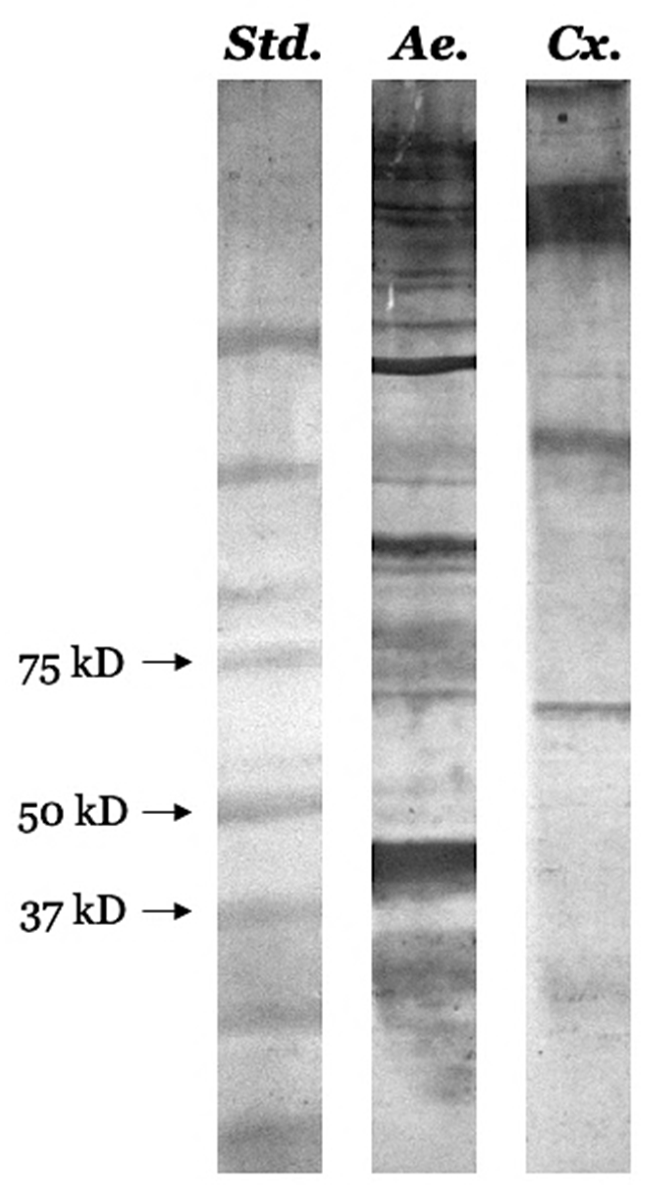
2.5. Mosquito SGE Protein Electrophoresis and Immunoblotting

Following our previous protocols for sodium dodecyl sulfate-polyacrylamide gel electrophoresis (SDS-PAGE) and immunoblotting, equal amounts (10 μg) of Ae. albopictus and Cx. quinquefasciatus SGE were loaded into miniprotean TGX 4–15% polyacrylamide gels (BioRad, Hercules, CA, USA) [18,26]. A pre-stained protein marker (Precision Plus Protein™ (10–250 kDa) Dual Color) was used as a molecular weight marker and the gel was run at 120 V for 75 min. The gel was then fixed in an acetic acid solution and silver stained using the Silver Stain Plus™ kit (BioRad, Hercules, CA, USA) according to the manufacturer’s instructions for visualization of separated proteins. An additional gel was used to transfer salivary proteins to a PVDF membrane. The PVDF membrane containing mosquito salivary proteins was blocked for one hour in a 2% milk solution and incubated overnight on a plate shaker at 4 °C in a 1:1000 serum dilution made using serum from the positive control in ELISA blocking buffer. Following overnight incubation, the membrane was washed three times using the ELISA wash buffer and incubated in a 1:1000 HRP-conjugated goat anti-bird IgY antibody (Abcam, Cambridge, UK) dilution in blocking buffer for 2 h at room temperature. Then the membrane was washed three times in 1X PBS and incubated in 1-step™ Ultra TMB-Blotting Solution (Thermofisher Scientific, Waltham, MA, USA) until desired development was achieved. The reaction was stopped using deionized water. Reactive proteins were visualized on a GelDocXR + (BioRad, Hercules, CA, USA).

2.6. In-Gel Digestion and Liquid Chromatography-Mass Spectrometry (LCMS) Preparation

Immunogenic bands in the Western blot from Ae. albopictus and Cx. quinquefasciatus SGE were sent for sequencing at the Tulane School of Medicine Proteomics Core Facility [27]. Two independent bands were sent for analysis from each mosquito vector. Individual protein bands were excised from the polyacrylamide gel in a fume hood using a surgical knife prior to destaining. Excised gel samples were destained by washing twice in deionized water followed by three washes with 25 mM ammonium bicarbonate (ABC) and 50% acetonitrile (ACN). Destained gel samples were digested by adding 100% ACN, vortexing for five minutes, and drying the gel samples at 60 °C for two minutes. 25 mM dithiothreitol in 25 mM ABC was added to the dried gels and incubated for one hour at room temperature. After incubation, 55 mM indole-3-acetic acid (IAA) in 25 mM ABC was added to the gel samples and incubated in a dark room for one hour, then the gel samples were washed using deionized water for five minutes to remove any remaining IAA. Samples were dehydrated by adding 25 mM ABC in 50% ACN and vortexing for five minutes. This was repeated using 100% ACN and samples were allowed to dry afterward. 200 μL 25 mM ABC and 1.5 μL trypsin were added to the dried samples and incubated at 37 °C overnight. The following day, digested peptides were extracted, and digestion was stopped using 20% ferulic acid (FA). The peptide solution was dried completely at 60 °C for 1–2 h. The dried peptide solution was reconstituted in LCMS buffer (2% ACN/0.1% FA) and centrifuged for 20 min at room temperature prior to performing LCMS analysis.

2.7. Protein Identification

To increase the number of identified proteins, the results of the LCMS analysis were referenced against all reviewed mosquito databases in UniProt for identification. Proteins identified in these analyses were filtered based on expected molecular weight and percent coverage. Filtered results were searched by ID code in the UniProt database for protein and molecular weight confirmation and amino acid sequence accession. A protein BLAST (pBLAST) analysis was performed on the sequences of uncharacterized proteins to determine proteins with similar sequence identities and to infer possible identity and function of the immunogenic proteins among our samples.

2.8. Data Analysis

Antibody concentrations used in our analyses are expressed as the calculated adjusted optical density (OD), as has been described by Londoño-Renteria et al. previously [17,28]. In brief, adjusted OD values were determined by subtracting the average optical density of the negative control and blank wells from the average OD value of the duplicate values, providing the ΔOD value for each sample. For each antigen, the positive control wells of each plate were averaged and divided by the average OD of all the positive control averages to attain a normalization factor for each corresponding plate. Each plate normalization factor was multiplied by plate sample ΔOD to obtain the normalized ΔODs that were used in statistical analyses. Inter- and intra-assay variation was assessed by calculating the coefficient of variation among duplicate samples. Samples demonstrating a coefficient of variation ≥20% were retested before inclusion in the study. Shapiro–Wilkes tests were performed on all variables to determine normality at an α level of 0.05. All variables were found to be significant (p < 0.05) by the Shapiro–Wilkes test, indicating non-parametric data. As such, Mann–Whitney tests were used to test for differences among two groups, including analyses by sex and age group. Matched pairs analyses were completed using the Wilcoxon matched pairs test to compare antibody responses among individual recaptured birds. Spearman correlation coefficients were calculated to determine the strength of association between two variables. Factors contributing to seroprevalence were assessed by multiple linear regression using antibody response to each antigen as a response variable and including age, week of sample collection, sex, site of collection, antibody response to the other two antigens, and the interaction of antibody response between those two antigens as predictor variables. All continuous variables were log-transformed before inclusion in the models to improve performance metrics. All statistical testing was performed using R statistical software (R Foundation for Statistical Computing, Vienna, Austria) version 4.3.3 at an α level of 0.05 [29].

3. Results

3.1. Description of the Study Population

The main characteristics of the study population are provided in Table 1. Throughout the 28-week sampling period, serum samples and epidemiologic data were collected from 165 birds. These birds were nearly equal by sex, with 78 (47.3%) female birds and 80 (48.5%) males (information was not available for 7 (4.2%) birds). By age, birds were less evenly split, with 64 (38.8%) hatch-year (juvenile) birds compared with 101 (61.2%) after-hatch-year (adult) birds collected. Sample collection by site was strongly biased: 116 (70.3%) samples were collected from Site 1, 38 (23%) from Site 3, and the remaining 11 (6.7%) samples from Site 2. Due to the overrepresentation of one site, statistical testing was not conducted by site of sample collection. However, even with the unequal sample size by site, the average IgY response to each antigen was consistent among all three locations, as well as every other variable included in this study (Table S1). Of the 165 observations used in the analyses, 30 samples were taken from 26 recaptured birds throughout the sampling period, with only two birds having been recaptured more than once, the highest number of recaptures for a single bird at three. Samples were collected at initial capture and every subsequent capture throughout the study. Using the IgY response of the initial and final captures for the recaptured birds, Wilcoxon signed rank tests were performed to test for the presence of any influential differences among the matched pairs in their response to Cx. quinquefasciatus, Ae. albopictus, and WNV before including these results in further analyses (Figure 1). No significant differences were found among the matched pair samples to any of the assayed antigens (p = 0.80, 0.94, and 0.72, respectively) (Figure 1). Wilcoxon signed rank matched pairs tests were also performed for the recaptured female, male, hatch-year, and after-hatch-year birds against Cx. quinquefasciatus, Ae. albopictus, and WNV. No significant results were found in any comparison among any group to any antigen (Figures S1 and S2). It is important to note that elapsed time between initial and final captures vary among individual birds.

3.2. WNV-Infected Mosquito Pools

In 2019, St. Tammany submitted 4227 mosquito pools for testing and 43 pools tested positive for WNV, representing four different mosquito species (33 Cx. quinquefasciatus, five Cx. nigripalpus, three Ae. albopictus, and two Cx. salinarius). The first positive pool was collected in late May (Cx. quinquefasciatus), while the first WNV-positive Ae. albopictus were not detected until July. In this year, Ae. albopictus had the highest WNV infection rate among all the mosquitoes sampled, with a bias-corrected maximum likelihood estimate (MLE) of estimated infection rate of 1.41 (95% CI, 0.37–3.78) per 1000 mosquitoes tested. The WNV infection rate among Cx. quinquefasciatus was the second-highest recorded in 2019 but was nearly half of the Ae. albopictus infection rate at 0.75 (95% CI, 0.52–1.04) (Table 2). Since sampling was conducted as part of routine arboviral surveillance for WNV and was not completed as part of this study, sampling methods are biased towards Culex spp. and therefore other mosquito species are not frequently sampled in numbers high enough to determine an accurate estimate of WNV positivity among other mosquito species present in the area.

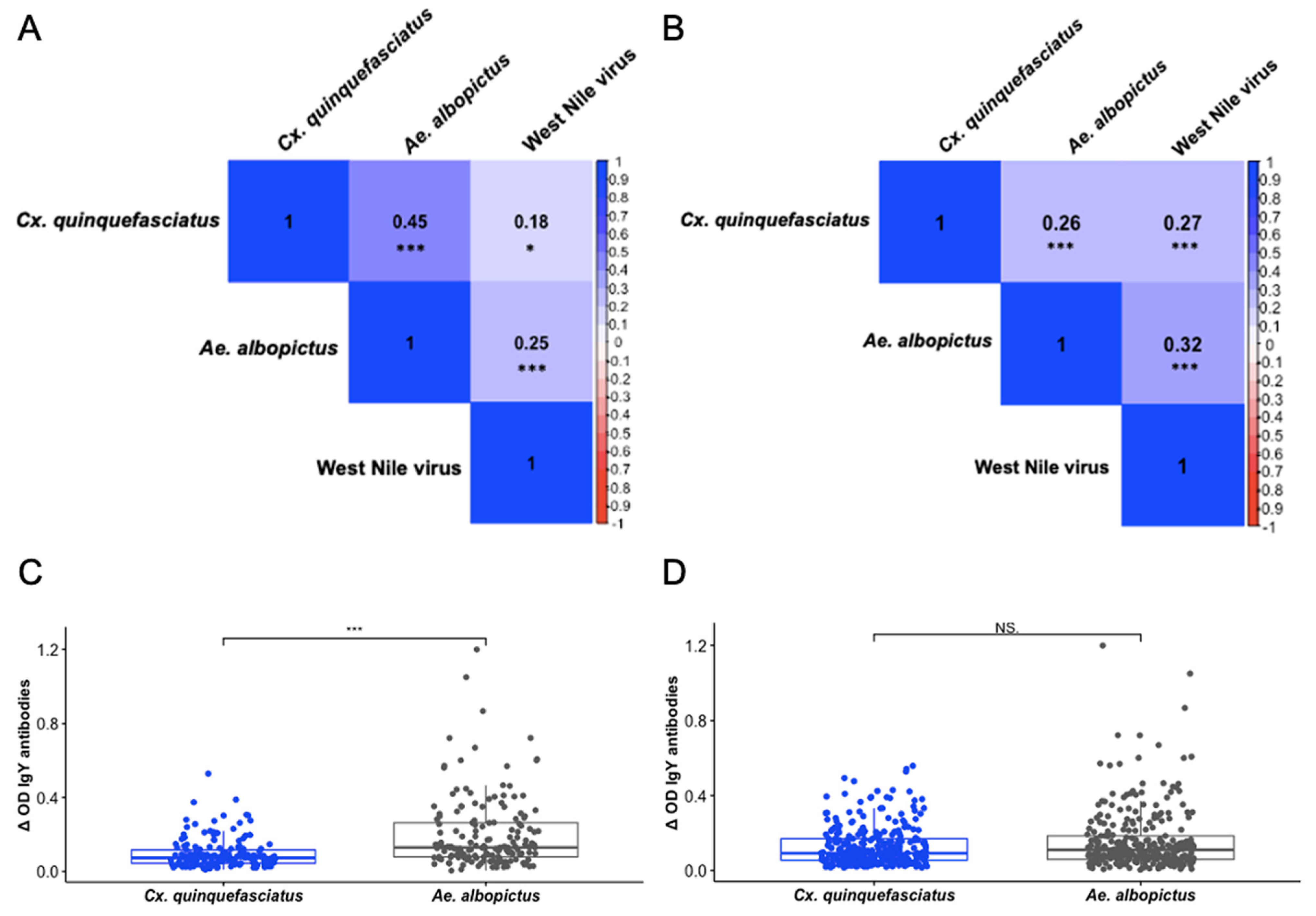
3.3. IgY Responses Are Associated Among Culex quinquefasciatus, Aedes albopictus, and West Nile Virus

Individual IgY responses were tested to determine if associations between exposure to WNV and the mosquito vectors were present in our study population. Initially, we analyzed results on 165 birds for which corresponding epidemiologic variables were available (Figure 2A). As expected, there were positive associations between exposure to Cx. quinquefasciatus and Ae. albopictus (ρ = 0.4471, p < 0.001), as well as exposure to Cx. quinquefasciatus and WNV (ρ = 0.1752, p = 0.02437). However, exposure to Ae. albopictus and WNV among these birds yielded a stronger positive association than was demonstrated between Cx. quinquefasciatus and WNV (ρ = 0.2525, p < 0.001) (Figure 2A). Subsequently, we compared the IgY response to Cx. quinquefasciatus against the IgY response to Ae. albopictus among the same 165 birds and found a significantly higher IgY response to Ae. albopictus (p < 0.001) (Figure 2C). Considering that the significant difference in IgY response between the mosquito vectors may have influenced the results of our correlations, we performed the same analyses on all 300 samples we assayed (Figure 2B,D). The results of these correlations reveal similar relationships among Cx. quinquefasciatus, Ae. albopictus, and WNV while no significant difference was present in the IgY response to Cx. quinquefasciatus and Ae. albopictus (p > 0.05). Though the relationship between Cx. quinquefasciatus and WNV was much stronger among all 300 samples (ρ = 0.2714, p < 0.001), a noticeably stronger association remained between Ae. albopictus and WNV (ρ = 0.3196, p < 0.001). Additionally, the association of exposure between the mosquito vectors was substantially diminished (ρ = 0.2562, p < 0.001) compared to the relationship found among the initial test using only 165 birds.

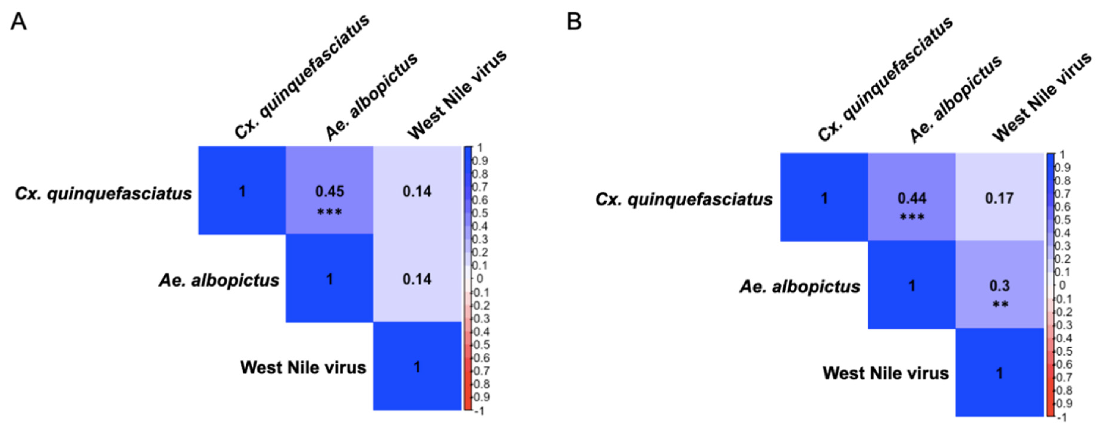
3.4. Presence of Sex-Specific Associations of IgY Response to Aedes albopictus and West Nile Virus

When testing the same associations between antigens among male and female bird groups, sex-specific differences emerged (Figure 3). Parallel to our analyses in the entire study population, strong positive associations were present between exposure to the mosquito vectors in both male and female birds (ρ = 0.4534, p < 0.001; ρ = 0.4433, p < 0.001, respectively). Spearman correlations of IgY response between either Cx. quinquefasciatus or Ae. albopictus and WNV among male birds were weak and not statistically significant, though positively associated (ρ = 0.1427, p = 0.2065; ρ = 0.1358, p = 0.2297, respectively). In female birds, the positive association between Cx. quinquefasciatus and WNV was comparable to the equivalent association in their male counterparts (ρ = 0.1661, p = 0.146), while the positive association between Ae. albopictus and WNV in this group was the only significant association among either mosquito vector with WNV in either sex category (ρ = 0.3004, p = 0.0075).

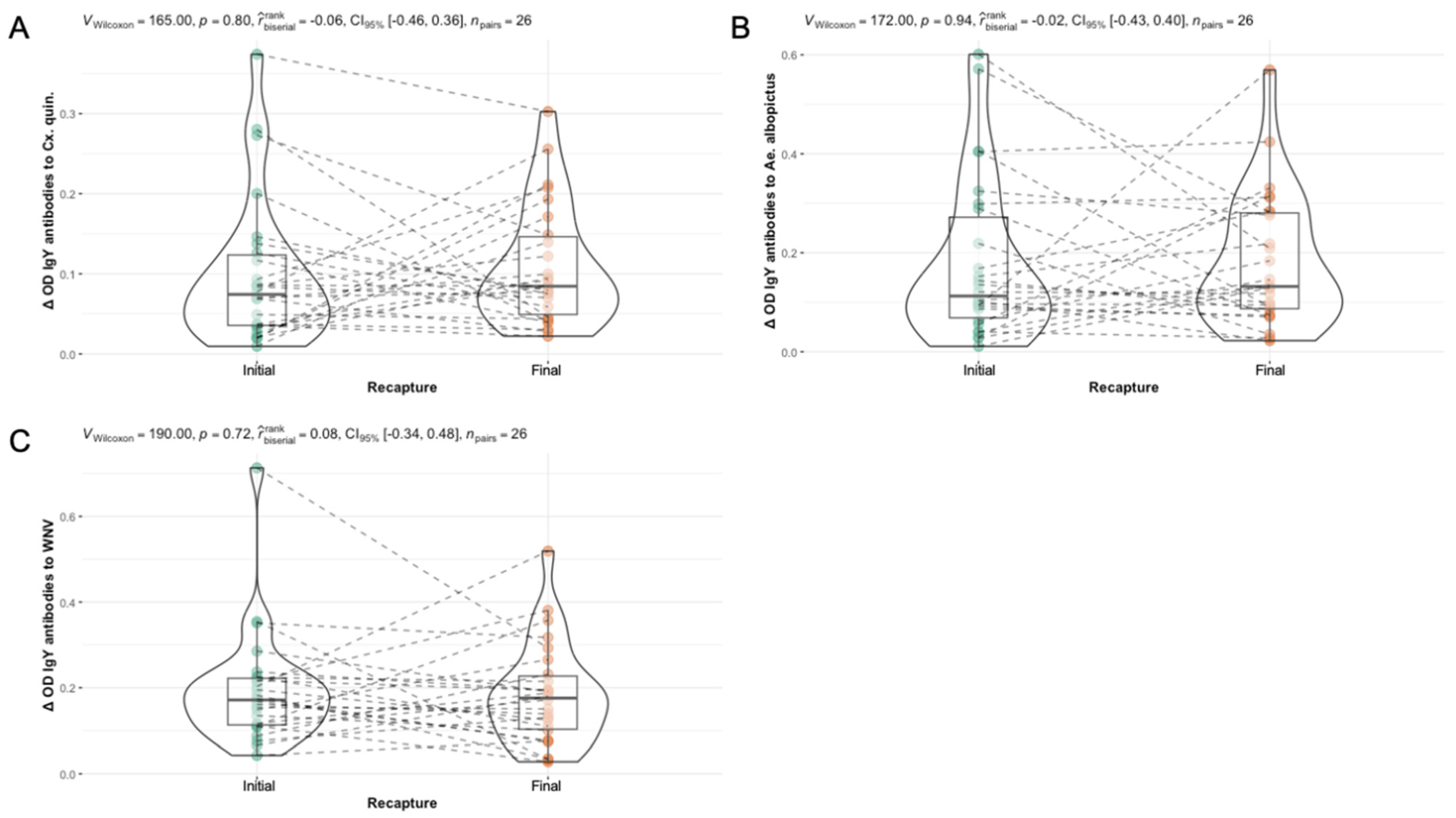
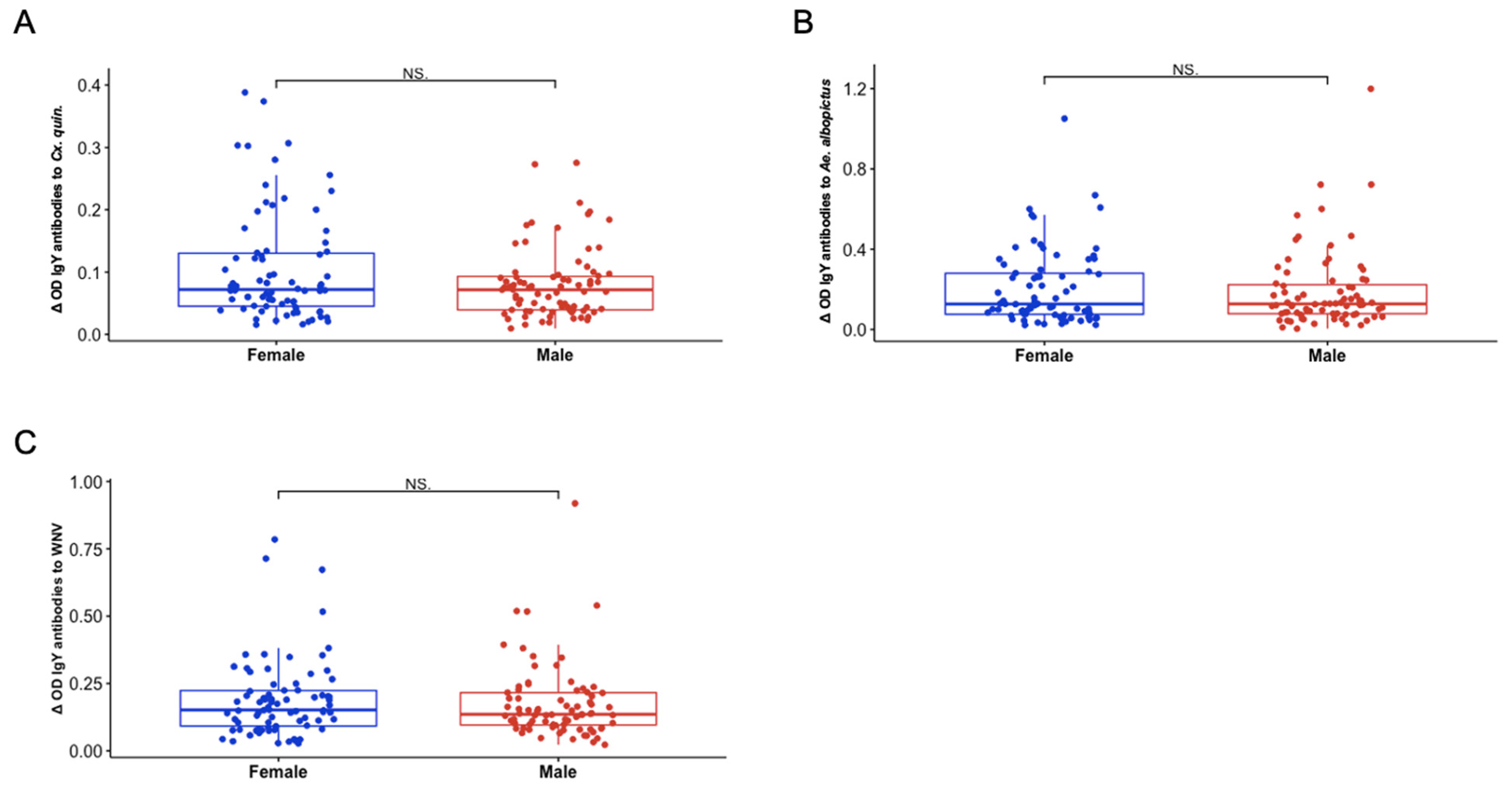
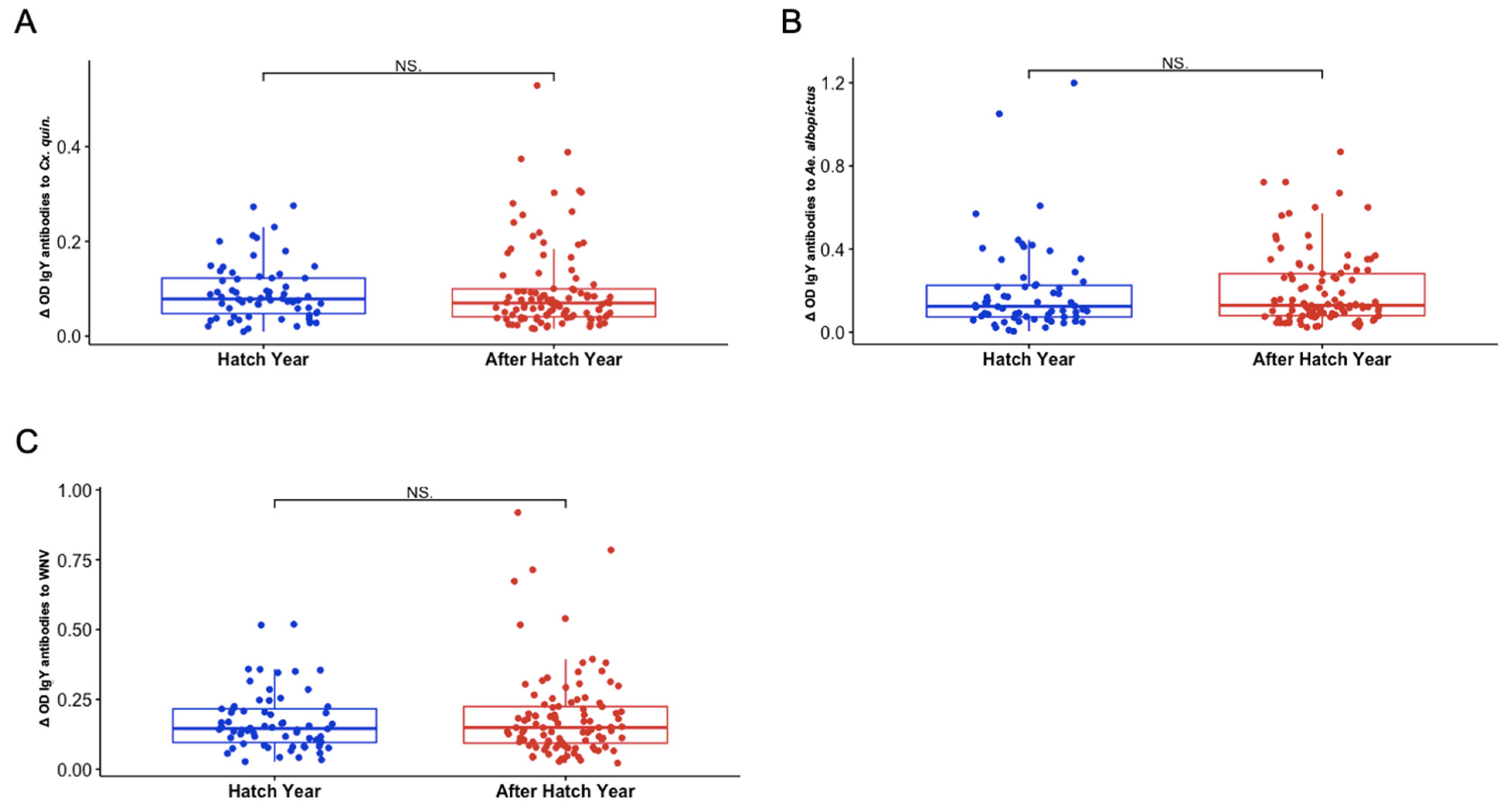
3.5. Sex and Hatch Year Are Not Important Variables Defining Exposure to Aedes albopictus or Culex quinquefasciatus Mosquito Bites Among Northern Cardinals

We observed no significant differences in IgY response by sex to Cx. quinquefasciatus SGE, Ae. albopictus SGE, and WNV antigen (p = 0.2904, 0.7543, and 0.3884, respectively) (Figure 4). Similarly, there were no significant differences in IgY response by age group to Cx. quinquefasciatus SGE, Ae. albopictus SGE, and WNV antigen (p = 0.3846, 0.4773, and 0.8976, respectively) (Figure 5). Consistent with our other findings, the highest average IgY response across both sex and age group categories was to Ae. albopictus SGE, while the IgY response to Cx. quinquefasciatus SGE was the weakest among all groups.

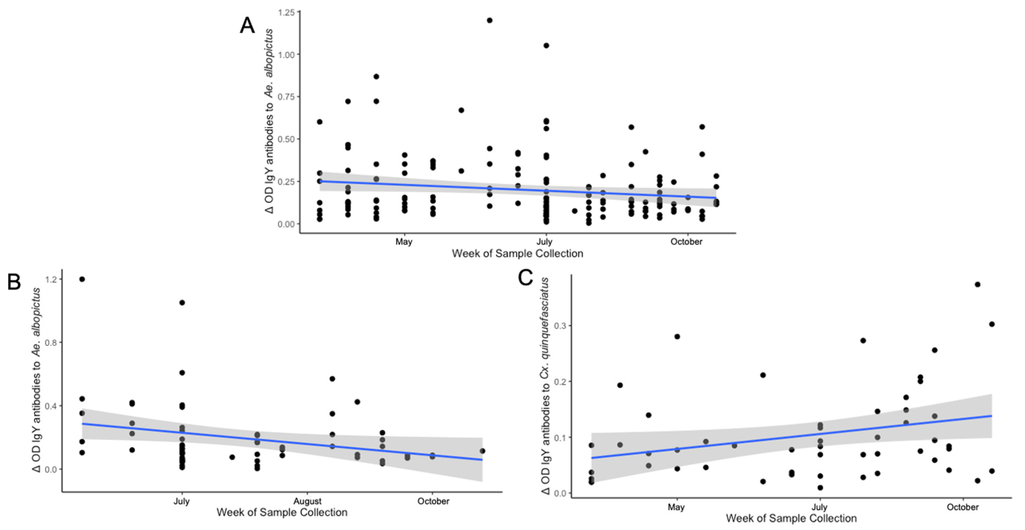
3.6. Differences in Seasonal Exposure to Aedes albopictus and Culex quinquefasciatus Among Northern Cardinals

This study yielded three significant temporal findings among IgY responses throughout the sampling period. The results demonstrate an overall negative association in IgY response to Ae. albopictus by week of sample collection (ρ = −0.1529, p = 0.04984) (Figure 6A). When examining the same association by age group, the negative temporality of IgY response to Ae. albopictus is only present in hatch-year birds (ρ = −0.2482, p = 0.04799) and was not observed in the after-hatch-year birds. (Figure 6B). Samples from juvenile birds collected earlier in the calendar year generally displayed stronger IgY responses to Ae. albopictus than samples collected later in the calendar year. The same relationship was not present among any other antigen in the hatch-year birds or with any antigen in the samples collected from after-hatch-year birds (Table 3). However, when determining exposure temporality by using the initial and final serum samples from the 26 recaptured birds in our study (n = 52 samples), only a positive association of IgY response to Cx. quinquefasciatus by week of sample collection exists (ρ = 0.277, p = 0.0468) (Figure 6). No other significant temporal associations were found among these birds. Linear regression models examining factors contributing to vector or virus exposure are available as Supplementary Information in Tables S2–S4. However, model performance was insufficient, limiting the ability to draw reliable conclusions from their results.

3.7. Identification of Several Pharmacologically Active Immunogenic Proteins

The immunoblot results of testing the reactivity of bird serum against whole salivary gland extract from Ae. albopictus and Cx. quinquefasciatus reveal stronger reactivity to a greater number of salivary proteins from Ae. albopictus SGE compared to those revealed among Cx. quinquefasciatus SGE (Figure 7). Mass spectrometry identified 2251 proteins among all four bands. Both structural and secreted proteins were found among the analyzed samples, since our study used whole salivary gland extract rather than saliva alone. After filtering the results by corresponding molecular weight and those with percent coverage greater than 25%, 60 total proteins were identified. The list provided in Table 4 has been filtered by genus to remove any redundant proteins from other mosquito genera. Any duplicate proteins within a genus were removed manually to include proteins identified to the species of interest only.
Mass spectrometry and sequencing of the proteins in the band at ~38–44 kDa from the Ae. albopictus sample identified six proteins corresponding to the Ae. albopictus proteome. Of the identified proteins, three displayed enzymatic function, two were uncharacterized, and one was found to have binding activity. A pBLAST analysis of both uncharacterized proteins (A0AAB0A6X6 and A0A182G0N4) showed strong identities (≥95%) with the D7L1 salivary protein of Ae. albopictus and the Aed a 2-like 37 kDa salivary gland allergen. The Ae. albopictus band identified at ~26–34 kDa revealed 13 proteins with identities to the Ae. albopictus proteome. Five of these were related to enzymatic functions, while another five were structural proteins, and three of the proteins were uncharacterized. A pBLAST analysis of the uncharacterized protein, A0A182GDW0, confirmed a 100% identity to tropomyosin-1 in the Ae. albopictus proteome. The other uncharacterized proteins from this band all had strong identities (>97%) to enzymatic proteins, including a dehydrogenase (A0A182H2F8) and a reductase (A0A182G1S0) from the Ae. albopictus proteome.
Analysis of the protein band at ~68–70 kDa in the Cx. quinquefasciatus sample resulted in the identification of three proteins, including one secreted protein (A0A8D8JS99), an enzyme (A0A8D8DSN3), and a molecular chaperone (B0X501). Of these three proteins, one was identified from the Cx. quinquefasciatus (B0X501) proteome and two from the Cx. pipiens proteome. The ~30–36 kDa band from the Cx. quinquefasciatus SGE yielded seven proteins, including three proteins with enzymatic activity, three structural proteins, and one secreted protein. None of the final proteins identified in the Cx. quinquefasciatus SGE were uncharacterized. A full list of the identified secreted proteins before coverage and genus filtering is available in Table S5.

4. Discussion

The use of avian serology to monitor West Nile virus in the environment is a well-established method of WNV surveillance, having been performed since its emergence in the United States, though avian serological studies have not explored IgY responses to arthropod saliva in wild bird populations. In our study, we describe the dynamics of IgY responses among Northern cardinals in St. Tammany Parish, Louisiana against salivary proteins of two local WNV vectors, Ae. albopictus and Cx. quinquefasciatus, as well as WNV whole-cell lysate antigen during a single transmission season.
Ae. albopictus was first identified in St. Tammany parish in the spring of 1986 [31]. While its involvement in enzootic and epizootic WNV transmission remains poorly understood, prior research in the region has hypothesized its involvement, especially during periods when Cx. quinquefasciatus abundance is reduced [32]. Although Cx. quinquefasciatus are active in Louisiana year-round, albeit with reduced activity in the fall and winter seasons, Ae. albopictus frequently emerge in the spring and are most abundant throughout the summer before populations decline in September [33,34]. The seasonality of Ae. albopictus occurrence in southern Louisiana is determined by the critical photoperiod (13–14 h) in the region, similar to patterns reported in Asian settings of comparable latitudes [33,35,36]. As the days shorten in the fall and winter months, Ae. albopictus females lay diapausing eggs that remain dormant until the following spring [33]. Emerging adults have a short flight range and usually remain close to the ground (~1 m; 3.3 ft), even when searching for hosts [37,38]. In contrast, Cx. quinquefasciatus exhibits quiescent behavior in the winter and, though they are less active during this time, they are not fully dormant throughout parts of the year, such that vertebrate hosts are likely to experience more consistent exposure to these mosquitoes [39]. Like Ae. albopictus, they show a distinct host-seeking height preference, as multiple studies have indicated greater quantities of Culex spp. mosquitoes captured at heights above 5 m (16.4 ft), suggesting that these mosquitoes occupy a unique ecological niche separate from Ae. albopictus [6,37,40,41]. These distinctions are especially important when considering enzootic WNV transmission dynamics and the serological trends presented in this study.
Likewise, Northern cardinals also exhibit distinct behavioral patterns based on breeding status that are likely to influence seasonal exposure to both Ae. albopictus and Cx. quinquefasciatus [42]. Northern cardinals raise one to four broods annually, occurring in the spring and summer months, generally March through August, although this range varies marginally by region [42]. Consequently, most of the sample collection for our study took place during breeding season as it falls within the six-month sampling period between April and October [42]. During this season, cardinals often select nesting sites outdoors, usually in shrubs or vines, but not within birdhouses [42,43]. On average, nests are constructed at an altitude of 1–2 m (3.3–6.6 ft) [42,43]. Moreover, while brooding their young, female cardinals develop incubation patches—bare sections of skin (~33 mm; 1.2 in in diameter) on their underside to facilitate more efficient incubation of the eggs [42]. Hatchling cardinals lack feathers, though they do develop down feathers within days of emerging from the egg [42]. However, even fledglings (juveniles) will undergo several molts, usually lasting several months after birth, before they acquire adult feathers [42]. Though male cardinals may occasionally sit on the nest for a brief time period, they are relatively uninvolved in incubation, with their primary responsibility being to provide food for the female and young [42]. Collectively, these behaviors, especially the low nesting altitude and temporary absence of feathers, in the females and young favor exposure to Ae. albopictus bites throughout the breeding season. At the conclusion of breeding, cardinals become more mobile and return to higher resting altitudes which favors exposure to Cx. quinquefasciatus bites in the late season.
Our findings reveal that Northern cardinals generally had higher IgY titers to salivary proteins from Ae. albopictus than Cx. quinquefasciatus. The high immunogenicity of Ae. albopictus SGE, in addition to the identification of a greater number of immunogenic salivary proteins compared to Cx. quinquefasciatus in our immunoblot, suggests a more novel exposure to Ae. albopictus in the tested birds. We suspect that several mechanisms are simultaneously contributing to these outcomes, the first of these being that consistent exposure to avian bloodmeals has influenced the adaptation of less immunogenic saliva in Cx. quinquefasciatus as vertebrate blood has been shown to effect immunologic selection pressure on arthropod saliva, noting that vectors more frequently exposed to a host develop less inflammatory salivary profiles over time to facilitate successful acquisition of a bloodmeal [44,45]. Secondly, the seasonal reemergence of Ae. albopictus in the spring after overwintering in egg diapause is contributing to the difference in IgY responses among the mosquito vectors, as it generates a prolonged period of nonexposure between Ae. albopictus and local bird species, preventing both the mosquito and the host from becoming more immunologically adapted to one another [33,39,46,47,48]. This hypothesis is further evidenced by the negative temporal associations reported among the cardinals in this study between IgY response to Ae. albopictus SGE and date of sample collection, suggesting that local bird populations develop less immunologic tolerance to Ae. albopictus saliva throughout winter months when vector–host contact is minimal, but become less antigenically stimulated by it throughout the summer months when exposure is more consistent [33]. By contrast, cardinals are exposed to Cx. quinquefasciatus bites year-round as these mosquitoes do not lay diapausing eggs and are much more abundant in the local environment than Ae. albopictus [39,49]. This continual exposure may provide a possible explanation for the absence of this association and the reduced IgY response among the birds included in this study to their saliva [39].
Further examination of this temporal trend among age groups demonstrates a more pronounced negative association between IgY response to Ae. albopictus SGE and date of sample collection in hatch-year birds. This elucidates an additional component to consider in the early-season involvement of Ae. albopictus in which hatch-year birds may have a disproportionate role as amplifying hosts compared to their adult counterparts, which has been postulated previously [50]. However, if this were true, we would have expected to find a corresponding association in hatch-year birds between IgY response to WNV and date of sample collection, as well as differences in IgY response between adult and hatch-year birds. The absence of these findings leads us to doubt a role favoring hatch-year birds as amplifying WNV hosts in our study area, though it is important to acknowledge that this may fluctuate annually as environmental conditions for transmission vary. Our observations in the hatch-year cardinals align with previous reports from red-footed falcons in central Europe, where higher seroprevalence was observed in nestling blood samples [51]. In that study, hatching order was negatively associated with antibody levels among broods with seropositive hatchlings, although active WNV infection was not detected in any of the nestlings [51]. The absence of IgY response differences among sex groups is not surprising given that previous work has noted no observable differences in avian immunity by sex; however, avian immune profiles have been reported to vary seasonally by breeding status in both male and female birds [52,53]. Similar to our analyses by sex, the absence of IgY response differences among age groups are supported by a previous study published in 2013 which noted no differences in WNV seropositivity among 1st year hatchling and adult Northern cardinals in east Illinois, though adults of other epidemiologically relevant bird species were found to have higher antibody responses than juvenile birds [54]. Several earlier studies have reported higher antibody titers among adult birds of other species in comparison to first year hatchlings of the same species [54,55]. We suspect that the temporal trends observed in hatch-year birds result from a combination of behavioral and habitat changes among cardinals as they mature into fledglings, including gaining mobility, developing adult plumage prior to winter, and residing at higher altitudes, characteristics that are unfavorable for exposure to Ae. albopictus [42]. Beyond this, other important factors to consider in explaining the seasonal decline of IgY titers to Ae. albopictus saliva among hatch-year birds are neutralizing antibody decay and transfer of maternal antibodies. Several studies have found that hatch-year birds have higher rates of WNV antibody decay compared to adults of the same species, but this alone still does not hold as an explanation for the absence of any association to WNV exposure in this group [56,57]. Without re-exposure, antibodies to WNV become undetectable within approximately two years; however, studies in humans report antibodies to mosquito saliva to be far shorter lived, suggesting their use as short-term markers of exposure, though the rate of antibody decay to arthropod saliva has yet to be evaluated among avian species [17,58]. In addition, the exact age of the birds in this study was not available, so we must also consider that maternal antibody acquisition may be influencing the results from hatch-year birds, especially those captured within 1 month of hatching, and may not be representative of true exposure [51,56,59].
Our study found a stronger relationship between IgY response to WNV and Ae. albopictus SGE rather than the established local WNV vector, Cx. quinquefasciatus, among Northern cardinals in the area. It is possible that the identification of two immunogenic D7-like secreted proteins in the Ae. albopictus sample compared to a single D7 protein from the Cx. quinquefasciatus sample is contributing to these observations. A previous study utilizing a recombinant D7 protein vaccine noted enhanced WNV pathogenesis in mice vaccinated with this protein [15]. The presence of more of these immunogenic proteins from Ae. albopictus could promote more intense WNV viremia and immunologic response among birds who were infected by Ae. albopictus bite. However, a previous study investigating vector competence of three WNV vectors, including Cx. pipiens and Ae. albopictus but excluding Cx. quinquefasciatus, found that Ae. albopictus required higher virus titers to become infected than was necessary for Cx. pipiens [60]. Though mammalian species have been documented with viremic titers higher than expected, no mammal has demonstrated viremia as strong as those observed in avian hosts, such that it is more probable that Ae. albopictus in the area are becoming infected with WNV predominantly through avian bloodmeals rather than a variety of mammalian hosts [61]. A study published in 2014 hypothesized the early-season (April–June) involvement of Cx. restuans in restarting seasonal WNV transmission in the Northeast, prior to the late-season (June–October) involvement and viral amplification commonly demonstrated by Cx. pipiens in the region [62]. Though this hypothesis was unsupported by the fact that Cx. restuans was found to frequently feed on avian hosts of poor WNV competency rather than known amplifying hosts, we believe it is an important consideration for the role of Ae. albopictus in the WNV transmission cycle of our study area, as a previous study in St. Tammany Parish reported Northern cardinals as the most abundant resident bird species in neighborhoods of previous focal WNV transmission and found high prevalence of WNV seropositivity and neutralizing antibodies in this species [63]. It remains unclear whether mammals are capable of developing viremia high enough to infect mosquito vectors, especially Ae. albopictus, though we believe it is reasonable to conclude these results indicate Ae. albopictus vectors WNV among avian communities more often than has been previously understood in southeast Louisiana, and their role as early-season enzootic vectors of WNV should be investigated further.
Given that maternal antibody acquisition cannot be completely separated from the results reported in the hatch-year birds, we examined IgY response associations between antigens by sex category, though we found no temporal associations of note among either group. However, when we observed associations between the IgY responses to each antigen among the two sex groups, the relationship between IgY response to WNV and Ae. albopictus SGE only existed among female birds. The same study in which the early-season involvement of Cx. restuans was hypothesized also reported an increase in bloodmeals on female birds while brooding their young [62]. We believe these exposure characteristics in female birds strongly reflect Northern cardinal brooding behaviors, which favor exposure to Ae. albopictus bites rather than Cx. quinquefasciatus, as Northern cardinals build outdoor nests at low altitudes and lack complete plumage [37,42]. Additionally, females lack plumage on their abdomen during breeding season to aid in successful egg incubation and become relatively immobile as they rely on the male to provide food [42]. A study among crows in Davis, California observed similar vector–host interactions among female and hatchling birds, demonstrating that Culex spp. bloodmeals were more frequently genotyped to nestling and female American crows, additionally noting that the likelihood of a crow bloodmeal increased within 10 m (32.8 ft) of an active nest [64]. The authors of this study predicted that American crows, particularly brooding females and newly emerged hatchlings, were important hosts for mosquitoes in the early breeding season [64]. The observation of early-season Culex feeding in crows underscores the role of brooding females and hatchling birds as vulnerable hosts for mosquitoes but has limited bearing on our own findings between Ae. albopictus and Northern cardinals, as the nesting behaviors of these two avian species differ greatly. While Northern cardinals frequently nest at low altitudes, American crows nest at much higher altitudes, often in tree canopies, with an earlier study reporting an average nest height of 9.7 m (31.8 ft), out of range for regular Ae. albopictus exposure to occur [42,43,65]. These findings, in conjunction with our own regarding the temporality of Ae. albopictus exposure, further substantiate the thought that Ae. albopictus has a crucial role in maintaining WNV sylvatic cycles, particularly vectoring the pathogen among brooding females and their offspring, before bloodmeals by Cx. quinquefasciatus become more intense in the late season [66].
The positive association to Cx. quinquefasciatus saliva in the recaptured birds demonstrates the intensification of Culex feeding behaviors in the later summer months (August–October). Although this finding has been reported among other studies, it is important to confirm its occurrence in our study area. In a study examining the temporal effects of Culex feeding behaviors on WNV transmissibility, researchers found that Culex spp. mosquitoes in Georgia exhibited a pronounced shift in host preference late in the summer season from feeding on American Robins (Turdus migratorius) to Northern cardinals, which made up the majority of Culex bloodmeals in the latter half of the season (August–October) [66]. We believe the increased frequency of exposure to Cx. quinquefasciatus later in the summer is also a reflection of the breeding behaviors of Northern cardinals, which end their breeding season in August, when they become more mobile and return to higher altitudes after brooding their young [42]. These behaviors make them less accessible to Ae. albopictus and more likely to be exposed to the bites of Cx. quinquefasciatus [6,37,40]. Unsurprisingly, the increase in exposure to Cx. quinquefasciatus among Northern cardinals corresponds to the seasonal patterns of human WNV cases in southeast Louisiana, likely due to their enhanced competency to amplify WNV compared to Ae. albopictus [60].
Taken together, our results indicate the possible role of Ae. albopictus as an early-season enzootic vector of WNV in southeast Louisiana, particularly vectoring WNV among brooding female Northern cardinals in the environment as they are relatively immobile and residing at a low altitude favorable for Ae. albopictus feeding. The broad circulation of WNV among these birds could favor transmission into the Cx. quinquefasciatus population as birds become more mobile and return to higher altitudes in the late summer after the end of their breeding season. Transmission to Cx. quinquefasciatus allows for rapid amplification in the environment and increases the likelihood of human infections, particularly symptomatic infection, corresponding to previous seasonal trends of human WNV case detection established in the area [67].
The primary strength of our study is the selective use of Northern cardinals, rather than other bird species in the area, to survey vector and pathogen exposure. Northern cardinals are a non-migratory species, corroborating the notion that exposure to both mosquito vectors and WNV occurred within the region from which the birds were sampled and limits the concern that the results of this study are due to exposure elsewhere [42]. Use of immunologic techniques to evaluate mosquito and pathogen exposure among wild-caught birds yields a more representative depiction of the transmission dynamics and mosquito–pathogen interface in southeast Louisiana than has been previously allowed by surveillance of sentinel bird flocks, equine testing, and monitoring of symptomatic human WNV cases.
However, the study is limited in three ways which are important to consider when interpreting the results presented in this work. The first limitation is that the information presented in the study only represents live birds. Dead birds were not sampled throughout the study period, such that the results of the study only represent immunologic characteristics of birds that had limited exposure to the vectors of interest in the study, were unexposed to WNV, or survived WNV infection. Additionally, the use of mist nets in sample collection procedures bias sampling as it excludes infected passerines which are likely to have impaired flight and activity patterns due to WNV infection. The exclusion of dead birds and the sampling bias could result in an underrepresentation of vector and pathogen exposure compared to what would be observed in the total population. Finally, the absence of PCR confirmation of active WNV infection in the birds limits the ability of the study to distinguish the vector–pathogen exposure characteristics of infected birds and uninfected birds.

5. Conclusions

West Nile virus poses a significant public health threat, not only to human health but also veterinary health in the U.S. and abroad. However, implementation of robust surveillance methods for the prediction and prevention of WNV outbreaks is challenging due to the complex relationships of vectors, amplifying reservoir hosts, and the environmental conditions influencing virus transmission. Routine surveillance of WNV is presently conducted primarily by mosquito pool testing and passive reporting of symptomatic equine and human infections; however, both methods are strongly biased and incomplete, which has major consequences on conclusions and practices determined from their interpretation. While four vaccines have been approved and licensed for equine prevention of WNV, notwithstanding their own limitations, there is no approved medical countermeasure to prevent or treat WNV infection in humans [68]. Additionally, standard clinical management strategies are palliative, further highlighting the value of reliable and holistic surveillance techniques to inform accurate predictions of epizootic WNV outbreaks. To achieve this, several studies have previously surveyed wild birds for the presence of IgY antibodies to WNV [69,70,71]. However, this study is the first to assess vector exposure by measuring IgY antibody titers against mosquito SGE in wild birds, in addition to pathogen exposure.
While the results presented here may not be applicable across all ecological contexts, they emphasize the contribution of avian serology against mosquito salivary proteins in evaluating local virus transmission and vector exposure. In our study area, they emphasize the critical need for integrating Ae. albopictus targeted vector surveillance in West Nile virus monitoring practices. Although widespread adoption of this avian serological surveillance technique as standard practice may be impractical, incorporating intermittent avian testing to assess regional vector exposure and WNV transmission patterns could provide valuable insights for improving routine surveillance practices in endemic areas.

Supplementary Materials

The following supporting information can be downloaded at: https://www.mdpi.com/article/10.3390/zoonoticdis5020011/s1, Figure S1: Wilcoxon signed ranks matched pairs tests among recaptured female (L; n = 12) and male (R; n = 14) birds using initial and final IgY antibody titers against Cx. quinquefasciatus SGE (A and D), Ae. albopictus SGE (B and E), and WNV antigen (C and F) [30]; Figure S2: Wilcoxon signed ranks matched pairs tests among recaptured hatch-year (L; n = 8) and after-hatch-year (R; n = 12) birds using initial and final IgY antibody titers against Cx. quinquefasciatus SGE (A and D), Ae. albopictus SGE (B and E), and WNV antigen (C and F) [30]; Table S1: Average IgY antibody titers measured in Δ in optical density (OD) values across all variables among 165 birds; Table S2: Effects of demographic and immunologic variables on IgY response to Cx. quinquefasciatus in the total study population; Table S3: Effects of demographic and immunologic variables on IgY response to Ae. albopictus in the total study population; Table S4: Effects of demographic and immunologic variables on IgY response to West Nile virus in the total study population; Table S5: Complete list of secreted proteins identified by mass spectrometry in immunogenic bands from Cx. quinquefasciatus and Ae. albopictus whole salivary gland extract.

Author Contributions

Conceptualization, B.L.-R. and K.A.C.; Methodology, A.R.S. and B.L.-R.; Formal analysis, A.R.S. and B.L.-R.; Investigation, A.R.S., S.H., Z.J. and B.L.-R.; Resources, D.M.W., J.d.V., K.A.C., S.B.J. and B.L.-R.; Writing—original draft preparation, A.R.S., K.A.C., S.R.M. and B.L.-R.; Writing—review and editing, A.R.S., S.H., Z.J., J.d.V., S.B.J., D.M.W., K.A.C., S.R.M. and B.L.-R.; Visualization, A.R.S.; Supervision, B.L.-R. All authors have read and agreed to the published version of the manuscript.

Funding

This research was funded by the Lavin-Bernick grants program at Tulane University.

Institutional Review Board Statement

Not applicable.

Informed Consent Statement

Not applicable.

Data Availability Statement

The raw data supporting the conclusions of this article will be made available by the authors on request.

Acknowledgments

We would like to thank the members of the Fan laboratory for their expertise, efforts, and dedication throughout the protein identification portion of this project, in addition to the members of the Robinson laboratory for providing valuable insights and guidance related to the ELISA methodologies used in this work. We also acknowledge and appreciate the St. Tammany Parish Mosquito Abatement team, particularly Benjamin Duckworth, for the collection of the avian blood samples. The authors acknowledge support from the NIH S10 High-End Instrumentation Award (1S10OD032453), which funded the acquisition of the Orbitrap Eclipse Tribrid Mass Spectrometer housed in the Tulane Proteomics Core. The following reagent was obtained through BEI Resources, NIAID, NIH: Aedes albopictus, Strain Gainesville, MRA-804, contributed by Sandra A. Allan. Culex quinquefasciatus (Sebring) colony material was kindly provided by Nicole Foley and Sean Masters from the Division of Vector-Borne Diseases at the Centers for Disease Control and Prevention in Fort Collins, CO.

Conflicts of Interest

The authors declare no conflicts of interest.

Abbreviations

The following abbreviations are used in this manuscript:
ABCAmmonium bicarbonate
ACNAcetonitrile
Ae.Aedes
Cx.Culex
ELISAEnzyme-linked immunosorbent assay
FAFerulic acid
IAAIndole-3-acetic acid
IgYImmunoglobulin Y
LCMSLiquid chromatography–mass spectrometry
ODOptical density
PBSPhosphate buffered saline
PVDFPolyvinylidene fluoride
SDS-PAGESodium dodecyl sulfate-polyacrylamide gel electrophoresis
SGESalivary gland extract
WNVWest Nile virus

References

  1. Fay, R.L.; Keyel, A.C.; Ciota, A.T. Chapter Three—West Nile virus and climate change. In Advances in Virus Research; Roossinck, M.J., Ed.; Academic Press: Cambridge, MA, USA, 2022; Volume 114, pp. 147–193. [Google Scholar] [CrossRef]
  2. Hayes, E.B.; Komar, N.; Nasci, R.S.; Montgomery, S.P.; O’Leary, D.R.; Campbell, G.L. Epidemiology and Transmission Dynamics of West Nile Virus Disease. Emerg. Infect. Dis. 2005, 11, 1167–1173. [Google Scholar] [CrossRef] [PubMed]
  3. Komar, N.; Langevin, S.; Hinten, S.; Nemeth, N.; Edwards, E.; Hettler, D.; Davis, B.; Bowen, R.; Bunning, M. Experimental Infection of North American Birds with the New York 1999 Strain of West Nile Virus. Emerg. Infect. Dis. 2003, 9, 311–322. [Google Scholar] [CrossRef] [PubMed]
  4. Levine, R.S.; Mead, D.G.; Kitron, U.D. Limited Spillover to Humans from West Nile Virus Viremic Birds in Atlanta, Georgia. Vector Borne Zoonotic Dis. 2013, 13, 812–817. [Google Scholar] [CrossRef]
  5. Faraji, A.; Egizi, A.; Fonseca, D.M.; Unlu, I.; Crepeau, T.; Healy, S.P.; Gaugler, R. Comparative Host Feeding Patterns of the Asian Tiger Mosquito, Aedes albopictus, in Urban and Suburban Northeastern USA and Implications for Disease Transmission. PLoS Neglected Trop. Dis. 2014, 8, e3037. [Google Scholar] [CrossRef] [PubMed]
  6. Hutchinson, R.A.; West, P.A.; Lindsay, S.W. Suitability of two carbon dioxide-baited traps for mosquito surveillance in the United Kingdom. Bull. Entomol. Res. 2007, 97, 591–597. [Google Scholar] [CrossRef]
  7. Savage, H.M.; Niebylski, M.L.; Smith, G.C.; Mitchell, C.J.; Craig, G.B., Jr. Host-Feeding Patterns of Aedes albopictus (Diptera: Culicidae) at a Temperate North American Site. J. Med. Entomol. 1993, 30, 27–34. [Google Scholar] [CrossRef]
  8. Vouillon, A.; Barthelemy, J.; Lebeau, L.; Nisole, S.; Savini, G.; Lévêque, N.; Simonin, Y.; Garcia, M.; Bodet, C. Skin tropism during Usutu virus and West Nile virus infection: An amplifying and immunological role. J. Virol. 2023, 98, e01830-23. [Google Scholar] [CrossRef]
  9. Rossi, S.L.; Ross, T.M.; Evans, J.D. West Nile Virus. Clin. Lab. Med. 2010, 30, 47–65. [Google Scholar] [CrossRef]
  10. Ratard, R. West Nile Virus Annual Report. 2021. Available online: https://ldh.la.gov/assets/oph/Center-PHCH/Center-CH/infectious-epi/Annuals/2021/EncephalitisWNV_LaIDAnnual_2021.pdf (accessed on 16 May 2024).
  11. Rochlin, I.; Faraji, A.; Healy, K.; Andreadis, T.G. West Nile Virus Mosquito Vectors in North America. J. Med. Entomol. 2019, 56, 1475–1490. [Google Scholar] [CrossRef]
  12. West Nile Virus|La Dept. of Health. Available online: https://ldh.la.gov/page/west-nile-virus (accessed on 16 May 2024).
  13. Louisiana Arbovirus Surveillance Summary 2020. 2021. Available online: https://ldh.la.gov/assets/oph/Center-PHCH/Center-CH/infectious-epi/Arboviral/arboweekly/2020_wnv_reports/ARBO_2049.pdf (accessed on 16 May 2024).
  14. Louisiana Arbovirus Surveillance Summary 2023. 2024. Available online: https://ldh.la.gov/assets/oph/Center-PHCH/Center-CH/infectious-epi/Arboviral/arboweekly/2023_WNV_Reports/ARBO_2352.pdf (accessed on 16 May 2024).
  15. Reagan, K.L.; Machain-Williams, C.; Wang, T.; Blair, C.D. Immunization of Mice with Recombinant Mosquito Salivary Protein D7 Enhances Mortality from Subsequent West Nile Virus Infection via Mosquito Bite. PLoS Neglected Trop. Dis. 2012, 6, e1935. [Google Scholar] [CrossRef]
  16. Londono-Renteria, B.L.; Eisele, T.P.; Keating, J.; James, M.A.; Wesson, D.M. Antibody Response Against Anopheles albimanus (Diptera: Culicidae) Salivary Protein as a Measure of Mosquito Bite Exposure in Haiti. J. Med. Entomol. 2010, 47, 1156–1163. [Google Scholar] [CrossRef]
  17. Londono-Renteria, B.; Cardenas, J.C.; Cardenas, L.D.; Christofferson, R.C.; Chisenhall, D.M.; Wesson, D.M.; McCracken, M.K.; Carvajal, D.; Mores, C.N. Use of Anti-Aedes aegypti Salivary Extract Antibody Concentration to Correlate Risk of Vector Exposure and Dengue Transmission Risk in Colombia. PLoS ONE 2013, 8, e81211. [Google Scholar] [CrossRef]
  18. Maldonado-Ruiz, L.P.; Montenegro-Cadena, L.; Blattner, B.; Menghwar, S.; Zurek, L.; Londono-Renteria, B. Differential Tick Salivary Protein Profiles and Human Immune Responses to Lone Star Ticks (Amblyomma americanum) From the Wild vs. A Laboratory Colony. Front. Immunol. 2019, 10, 1996. [Google Scholar] [CrossRef] [PubMed]
  19. Taylor, A.I.; Gould, H.J.; Sutton, B.J.; Calvert, R.A. Avian IgY Binds to a Monocyte Receptor with IgG-like Kinetics Despite an IgE-like Structure. J. Biol. Chem. 2008, 283, 16384–16390. [Google Scholar] [CrossRef]
  20. Lee, W.; Syed Atif, A.; Tan, S.C.; Leow, C.H. Insights into the chicken IgY with emphasis on the generation and applications of chicken recombinant monoclonal antibodies. J. Immunol. Methods 2017, 447, 71–85. [Google Scholar] [CrossRef] [PubMed]
  21. Pacheco, B.L.B.; Nogueira, C.P.; Venancio, E.J. IgY Antibodies from Birds: A Review on Affinity and Avidity. Animals 2023, 13, 3130. [Google Scholar] [CrossRef] [PubMed]
  22. Ponnusamy, L.; Schal, C.; Wesson, D.M.; Arellano, C.; Apperson, C.S. Oviposition responses of Aedes mosquitoes to bacterial isolates from attractive bamboo infusions. Parasites Vectors 2015, 8, 486. [Google Scholar] [CrossRef]
  23. Londono-Renteria, B.; Patel, J.C.; Vaughn, M.; Funkhauser, S.; Ponnusamy, L.; Grippin, C.; Jameson, S.B.; Apperson, C.; Mores, C.N.; Wesson, D.M.; et al. Long-Lasting Permethrin-Impregnated Clothing Protects Against Mosquito Bites in Outdoor Workers. Am. J. Trop. Med. Hyg. 2015, 93, 869–874. [Google Scholar] [CrossRef]
  24. Cardenas, J.C.; Drame, P.M.; Luque-Burgos, K.A.; Berrio, J.D.; Entrena-Mutis, E.; González, M.U.; Carvajal, D.J.; Gutiérrez-Silva, L.Y.; Cardenas, L.D.; Colpitts, T.M.; et al. IgG1 and IgG4 antibodies against Aedes aegypti salivary proteins and risk for dengue infections. PLoS ONE 2019, 14, e0208455. [Google Scholar] [CrossRef]
  25. Robinson, J.E.; Holton, D.; Liu, J.; McMurdo, H.; Murciano, A.; Gohd, R. A novel enzyme-linked immunosorbent assay (ELISA) for the detection of antibodies to HIV-1 envelope glycoproteins based on immobilization of viral glycoproteins in microtiter wells coated with concanavalin A. J. Immunol. Methods 1990, 132, 63–71. [Google Scholar] [CrossRef]
  26. Londono-Renteria, B.; Drame, P.M.; Montiel, J.; Vasquez, A.M.; Tobón-Castaño, A.; Taylor, M.; Vizcaino, L.; Lenhart, A.E. Identification and Pilot Evaluation of Salivary Peptides from Anopheles albimanus as Biomarkers for Bite Exposure and Malaria Infection in Colombia. Int. J. Mol. Sci. 2020, 21, 691. [Google Scholar] [CrossRef] [PubMed]
  27. Maity, S.; Huang, Y.; Kilgore, M.D.; Thurmon, A.N.; Vaasjo, L.O.; Galazo, M.J.; Xu, X.; Cao, J.; Wang, X.; Ning, B.; et al. Mapping dynamic molecular changes in hippocampal subregions after traumatic brain injury through spatial proteomics. Clin. Proteom. 2024, 21, 32. [Google Scholar] [CrossRef] [PubMed]
  28. Londono-Renteria, B.; Drame, P.M.; Weitzel, T.; Rosas, R.; Gripping, C.; Cardenas, J.C.; Alvares, M.; Wesson, D.M.; Poinsignon, A.; Remoue, F.; et al. An gambiae gSG6-P1 evaluation as a proxy for human-vector contact in the Americas: A pilot study. Parasites Vectors 2015, 8, 533. [Google Scholar] [CrossRef]
  29. R Core Team. R: A Language and Environment for Statistical Computing; R Foundation for Statistical Computing: Vienna, Austria, 2024. [Google Scholar]
  30. Patil, I. Visualizations with statistical details: The ‘ggstatsplot’ approach. J. Open Source Softw. 2021, 6, 3167. [Google Scholar] [CrossRef]
  31. Moore, C.G.; Francy, D.B.; Eliason, D.A.; Monath, T.P. Aedes albopictus in the United States: Rapid spread of a potential disease vector. J. Am. Mosq. Control Assoc. 1988, 4, 356–361. [Google Scholar] [PubMed]
  32. Palmisano, C.T.; Taylor, V.; Caillouet, K.; Byrd, B.; Wesson, D.M. Impact of West Nile virus outbreak upon St. Tammany Parish Mosquito Abatement District. J. Am. Mosq. Control Assoc. 2005, 21, 33–38. [Google Scholar] [CrossRef]
  33. Willis, F.S.; Nasci, R.S. Aedes albopictus (Diptera: Culicidae) Population Density and Structure in Southwest Louisiana. J. Med. Entomol. 1994, 31, 594–599. [Google Scholar] [CrossRef]
  34. Tesh, R.B.; Parsons, R.; Siirin, M.; Randle, Y.; Sargent, C.; Guzman, H.; Wuithiranyagool, T.; Higgs, S.; Vanlandingham, D.L.; Bala, A.A.; et al. Year-round West Nile Virus Activity, Gulf Coast Region, Texas and Louisiana. Emerg. Infect. Dis. 2004, 10, 1649–1652. [Google Scholar] [CrossRef]
  35. Armbruster, P.A. Photoperiodic Diapause and the Establishment of Aedes albopictus (Diptera: Culicidae) in North America. J. Med. Entomol. 2016, 53, 1013–1023. [Google Scholar] [CrossRef]
  36. Hawley, W.A. The biology of Aedes albopictus. J. Am. Mosq. Control. Assoc. 1988, 1, 1–39. [Google Scholar]
  37. Obenauer, P.J.; Kaufman, P.E.; Allan, S.A.; Kline, D.L. Host-Seeking Height Preferences of Aedes albopictus (Diptera: Culicidae) in North Central Florida Suburban and Sylvatic Locales. J. Med. Entomol. 2009, 46, 900–908. [Google Scholar] [CrossRef] [PubMed]
  38. Nava, M.R.; Debboun, M. Chapter 2—Mosquito Species of Texas. In Mosquitoes, Communities, and Public Health in Texas; Debboun, M., Nava, M.R., Rueda, L.M., Eds.; Academic Press: Cambridge, MA, USA, 2020; pp. 9–167. [Google Scholar] [CrossRef]
  39. Diniz, D.F.A.; de Albuquerque, C.M.R.; Oliva, L.O.; de Melo-Santos, M.A.V.; Ayres, C.F.J. Diapause and quiescence: Dormancy mechanisms that contribute to the geographical expansion of mosquitoes and their evolutionary success. Parasites Vectors 2017, 10, 310. [Google Scholar] [CrossRef]
  40. Savage, H.M.; Anderson, M.; Gordon, E.; Mcmillen, L.; Colton, L.; Delorey, M.; Sutherland, G.; Aspen, S.; Charnetzky, D.; Burkhalter, K.; et al. Host-Seeking Heights, Host-Seeking Activity Patterns, and West Nile Virus Infection Rates for Members of the Culex pipiens Complex at Different Habitat Types Within the Hybrid Zone, Shelby County, TN, 2002 (Diptera: Culicidae). J. Med. Entomol. 2008, 45, 276–288. [Google Scholar] [CrossRef] [PubMed]
  41. Darbro, J.M.; Harrington, L.C. Bird-baited traps for surveillance of West Nile mosquito vectors: Effect of bird species, trap height, and mosquito escape rates. J. Med. Entomol. 2006, 43, 83–92. [Google Scholar] [CrossRef] [PubMed]
  42. Halkin, S.L.; Linville, S.U. Northern cardinal: (Cardinalis cardinalis). In The Birds of North America, No. 440; The Birds of North America, Inc.: Philadelphia, PA, USA, 1999; pp. 1–32. [Google Scholar]
  43. Laskey, A.R. A Study of the Cardinal in Tennessee. Wilson Bull. 1944, 56, 27–44. [Google Scholar]
  44. Arcà, B.; Struchiner, C.J.; Pham, V.M.; Sferra, G.; Lombardo, F.; Pombi, M.; Ribeiro, J.M.C. Positive selection drives accelerated evolution of mosquito salivary genes associated with blood-feeding. Insect Mol. Biol. 2014, 23, 122–131. [Google Scholar] [CrossRef]
  45. Ribeiro, J.M.C. Blood-feeding in mosquitoes: Probing time and salivary gland anti-haemostatic activities in representatives of three genera (Aedes, Anopheles, Culex). Med. Vet. Entomol. 2000, 14, 142–148. [Google Scholar] [CrossRef]
  46. Doucoure, S.; Mouchet, F.; Cournil, A.; Goff, G.L.; Cornelie, S.; Roca, Y.; Giraldez, M.G.; Simon, Z.B.; Loayza, R.; Misse, D.; et al. Human Antibody Response to Aedes aegypti Saliva in an Urban Population in Bolivia: A New Biomarker of Exposure to Dengue Vector Bites. Am. J. Trop. Med. Hyg. 2012, 87, 504–510. [Google Scholar] [CrossRef]
  47. Montiel, J.; Carbal, L.F.; Tobón-Castaño, A.; Vásquez, G.M.; Fisher, M.L.; Londono-Rentería, B. IgG antibody response against Anopheles salivary gland proteins in asymptomatic Plasmodium infections in Narino, Colombia. Malar. J. 2020, 19, 42. [Google Scholar] [CrossRef]
  48. Buezo Montero, S.; Gabrieli, P.; Montarsi, F.; Borean, A.; Capelli, S.; De Silvestro, G.; Forneris, F.; Pombi, M.; Breda, A.; Capelli, G.; et al. IgG Antibody Responses to the Aedes albopictus 34k2 Salivary Protein as Novel Candidate Marker of Human Exposure to the Tiger Mosquito. Front. Cell. Infect. Microbiol. 2020, 10, 377. [Google Scholar] [CrossRef]
  49. Moise, I.K.; Riegel, C.; Muturi, E.J. Environmental and social-demographic predictors of the southern house mosquito Culex quinquefasciatus in New Orleans, Louisiana. Parasites Vectors 2018, 11, 249. [Google Scholar] [CrossRef]
  50. Hamer, G.L.; Walker, E.D.; Brawn, J.D.; Loss, S.R.; Ruiz, M.O.; Goldberg, T.L.; Schotthoefer, A.M.; Brown, W.M.; Wheeler, E.; Kitron, U.D. Rapid amplification of West Nile virus: The role of hatch-year birds. Vector Borne Zoonotic Dis. 2008, 8, 57–67. [Google Scholar] [CrossRef] [PubMed]
  51. Soltész, Z.; Erdélyi, K.; Bakonyi, T.; Barna, M.; Szentpáli-Gavallér, K.; Solt, S.; Horváth, É.; Palatitz, P.; Kotymán, L.; Dán, Á.; et al. West Nile virus host-vector-pathogen interactions in a colonial raptor. Parasites Vectors 2017, 10, 449. [Google Scholar] [CrossRef] [PubMed]
  52. Valdebenito, J.O.; Halimubieke, N.; Lendvai, Á.Z.; Figuerola, J.; Eichhorn, G.; Székely, T. Seasonal variation in sex-specific immunity in wild birds. Sci. Rep. 2021, 11, 1349. [Google Scholar] [CrossRef]
  53. Owen, J.C.; Nakamura, A.; Coon, C.A.; Martin, L.B. The effect of exogenous corticosterone on West Nile virus infection in Northern Cardinals (Cardinalis cardinalis). Vet. Res. 2012, 43, 34. [Google Scholar] [CrossRef] [PubMed]
  54. Lampman, R.L.; Krasavin, N.M.; Ward, M.P.; Beveroth, T.A.; Lankau, E.W.; Alto, B.W.; Muturi, E.; Novak, R.J. West Nile Virus Infection Rates and Avian Serology in East-Central Illinois. J. Am. Mosq. Control Assoc. 2013, 29, 108–122. [Google Scholar] [CrossRef]
  55. Gibbs, S.E.J.; Allison, A.B.; Yabsley, M.J.; Mead, D.G.; Wilcox, B.R.; Stallknecht, D.E. West Nile Virus Antibodies in Avian Species of Georgia, USA: 2000–2004. Vector-Borne Zoonotic Dis. 2006, 6, 57–72. [Google Scholar] [CrossRef]
  56. Lamb, J.S.; Tornos, J.; Lejeune, M.; Boulinier, T. Rapid loss of maternal immunity and increase in environmentally mediated antibody generation in urban gulls. Sci. Rep. 2024, 14, 4357. [Google Scholar] [CrossRef]
  57. McKee, E.M.; Walker, E.D.; Anderson, T.K.; Kitron, U.D.; Brawn, J.D.; Krebs, B.L.; Newman, C.; Ruiz, M.O.; Levine, R.S.; Carrington, M.E.; et al. West Nile virus antibody decay rate in free-ranging birds. J. Wildl. Dis. 2015, 51, 601–608. [Google Scholar] [CrossRef]
  58. Orlandi-Pradines, E.; Almeras, L.; Denis de Senneville, L.; Barbe, S.; Remoué, F.; Villard, C.; Cornelie, S.; Penhoat, K.; Pascual, A.; Bourgouin, C.; et al. Antibody response against saliva antigens of Anopheles gambiae and Aedes aegypti in travellers in tropical Africa. Microbes Infect. 2007, 9, 1454–1462. [Google Scholar] [CrossRef]
  59. Kaspers, B.; Göbel, T.W.; Schat, K.A.; Vervelde, L. Avian Immunology, 3rd ed.; Academic Press: Cambridge, MA, USA, 2022. [Google Scholar]
  60. Tiawsirisup, S.; Platt, K.B.; Evans, R.B.; Rowley, W.A. A Comparision of West Nile Virus Transmission by Ochlerotatus trivittatus (COQ.), Culex pipiens (L.), and Aedes albopictus (Skuse). Vector-Borne Zoonotic Dis. 2005, 5, 40–47. [Google Scholar] [CrossRef] [PubMed]
  61. Root, J.J.; Bosco-Lauth, A.M. West Nile Virus Associations in Wild Mammals: An Update. Viruses 2019, 11, 459. [Google Scholar] [CrossRef] [PubMed]
  62. Egizi, A.M.; Farajollahi, A.; Fonseca, D.M. Diverse Host Feeding on Nesting Birds May Limit Early-Season West Nile Virus Amplification. Vector-Borne Zoonotic Dis. 2014, 14, 447–453. [Google Scholar] [CrossRef]
  63. Komar, N.; Panella, N.A.; Langevin, S.A.; Brault, A.C.; Amador, M.; Edwards, E.; Owen, J.C. Avian Hosts for West Nile Virus in St. Tammany Parish, Louisiana, 2002. Am. J. Trop. Med. Hyg. 2005, 73, 1031–1037. [Google Scholar] [CrossRef]
  64. Wheeler, S.S.; Taff, C.C.; Reisen, W.K.; Townsend, A.K. Mosquito blood-feeding patterns and nesting behavior of American crows, an amplifying host of West Nile virus. Parasites Vectors 2021, 14, 331. [Google Scholar] [CrossRef] [PubMed]
  65. Chamberlain-Auger, J.A.; Auger, P.J.; Strauss, E.G. Breeding Biology of American Crows. Wilson Bull. 1990, 102, 615–622. [Google Scholar]
  66. Levine, R.S.; Mead, D.G.; Hamer, G.L.; Brosi, B.J.; Hedeen, D.L.; Hedeen, M.W.; McMillan, J.R.; Bisanzio, D.; Kitron, U.D. Supersuppression: Reservoir Competency and Timing of Mosquito Host Shifts Combine to Reduce Spillover of West Nile Virus. Am. J. Trop. Med. Hyg. 2016, 95, 1174–1184. [Google Scholar] [CrossRef]
  67. Louisiana Arbovirus Surveillance Summary 2021. Available online: https://ldh.la.gov/assets/oph/Center-PHCH/Center-CH/infectious-epi/Arboviral/arboweekly/2021_WNV_Reports/ARBO_2152.pdf (accessed on 1 January 2022).
  68. Cendejas, P.M.; Goodman, A.G. Vaccination and Control Methods of West Nile Virus Infection in Equids and Humans. Vaccines 2024, 12, 485. [Google Scholar] [CrossRef]
  69. McMillan, J.R.; Hamer, G.L.; Levine, R.S.; Mead, D.G.; Waller, L.A.; Goldberg, T.L.; Walker, E.D.; Brawn, J.D.; Ruiz, M.O.; Kitron, U.; et al. Multi-Year Comparison of Community- and Species-Level West Nile Virus Antibody Prevalence in Birds from Atlanta, Georgia and Chicago, Illinois, 2005–2016. Am. J. Trop. Med. Hyg. 2023, 108, 366–376. [Google Scholar] [CrossRef]
  70. Magallanes, S.; Llorente, F.; Ruiz-López, M.J.; la Puente, J.M.; Ferraguti, M.; Gutiérrez-López, R.; Soriguer, R.; Aguilera-Sepúlveda, P.; Fernández-Delgado, R.; Jímenez-Clavero, M.Á.; et al. Warm winters are associated to more intense West Nile virus circulation in southern Spain. Emerg. Microbes Infect. 2024, 13, 2348510. [Google Scholar] [CrossRef]
  71. Pedersen, K.; Marks, D.R.; Wang, E.; Eastwood, G.; Weaver, S.C.; Goldstein, S.M.; Sinnett, D.R.; DeLiberto, T.J. Widespread Detection of Antibodies to Eastern Equine Encephalitis, West Nile, St. Louis Encephalitis, and Turlock Viruses in Various Species of Wild Birds from Across the United States. Am. J. Trop. Med. Hyg. 2016, 95, 206–211. [Google Scholar] [CrossRef] [PubMed]
Figure 1. Wilcoxon signed ranks matched pairs tests among all recaptured birds using initial and final IgY antibody titers against (A) Cx. quinquefasciatus SGE, (B) Ae. albopictus SGE, and (C) WNV antigen. Dotted lines correspond to individual matched pairs across time points [30].
Zoonoticdis 05 00011 g001
Figure 2. (A) Correlogram of Spearman correlation of IgY antibody titers in subset of samples for which epidemiological variables were available (n = 165) against Cx. quinquefasciatus and Ae. albopictus mosquito SGE and WNV antigen. (B) Correlogram of Spearman correlation of IgY antibody titers among all samples that were tested (n = 300) against Cx. quinquefasciatus and Ae. albopictus mosquito SGE and WNV antigen. Positive correlations are indicated in gradient blue according to strength of association (darker blue represents a stronger positive correlation) while negative correlations are indicated in gradient red according to strength of association (darker red represents a stronger negative correlation). (C) Mann–Whitney U test comparing IgY antibody titers in subset of samples for which epidemiological variables were available (n = 165) between Cx. quinquefasciatus and Ae. albopictus. (D) Mann–Whitney U test comparing IgY antibody titers among all samples that were tested (n = 300) between Cx. quinquefasciatus and Ae. albopictus. IgY response measured in Δ in optical density (OD) values. p-values are denoted as * (p < 0.05), *** (p < 0.001). NS: not significant.
Zoonoticdis 05 00011 g002
Figure 3. (A) Correlogram of Spearman correlation of IgY antibody titers in samples from male birds (n = 80) against Cx. quinquefasciatus and Ae. albopictus mosquito SGE and WNV antigen. (B) Correlogram of Spearman correlation of IgY antibody titers in samples from female birds (n = 78) against Cx. quinquefasciatus and Ae. albopictus mosquito SGE and WNV antigen. Positive correlations are indicated in gradient blue according to strength of association (darker blue represents a stronger positive correlation) while negative correlations are indicated in gradient red according to strength of association (darker red represents a stronger negative correlation). p-values are denoted as ** (p < 0.01), *** (p < 0.001).
Zoonoticdis 05 00011 g003
Figure 4. Mann–Whitney tests by sex. (A) IgY responses to Cx. quinquefasciatus SGE, (B) IgY responses to Ae. albopictus SGE, (C) IgY responses to WNV. Results from female (n = 78) and male (n = 80) birds. Data for sex was not available for 7 samples, which were not included in these analyses. IgY responses measured in Δ in optical density (OD) values. NS: not significant.
Zoonoticdis 05 00011 g004
Figure 5. Mann–Whitney tests by age. (A) IgY responses to Cx. quinquefasciatus SGE, (B) IgY responses to Ae. albopictus SGE, (C) IgY responses to WNV. Results from hatch-year (n = 64) and after-hatch-year (n = 101) birds. IgY responses measured in Δ in OD values. NS: not significant.
Zoonoticdis 05 00011 g005
Figure 6. Results of Spearman correlations by week of sample collection. (A) IgY responses in total study population (n = 165) against Ae. albopictus SGE. (B) IgY responses in hatch-year birds (n = 64) against Ae. albopictus SGE. (C) IgY responses in recaptured birds (n = 52) against Cx. quinquefasciatus SGE. Grey shaded areas depict standard error range.
Zoonoticdis 05 00011 g006
Figure 7. Aedes albopictus and Culex quinquefasciatus immunoblot results. Please find the original figure in Figure S3.
Zoonoticdis 05 00011 g007
Table 1. Characteristics of the study population.
Site
n (%)
123
116 (70.3)11 (6.7)38 (23)
Sex
n (%)
MaleFemaleNot available
80 (48.5)78 (47.3)7 (4.2)
Age
n (%)
Hatch YearAfter Hatch YearNot available
64 (38.8)101 (61.2)0 (0)
Mass
(g)
AverageMinimumMaximum
39.022346
Table 2. WNV-positive mosquito pools from St. Tammany Parish in 2019. Maximum likelihood estimate (MLE) of WNV per 1000 mosquitoes tested. MLE bias was corrected when pools were positive.
SpeciesNumber of MosquitoesNumber Positive/Number of Pools TestedMLE95% CI
Aedes albopictus21333/1691.410.37–3.78
Culex quinquefasciatus45,15233/11550.750.52–1.04
Culex nigripalpus18,8875/5800.270.10–0.59
Culex salinarius60,8242/12810.030.01–0.11
Aedes vexans70310/37400–0.54
Culex erraticus14,8240/57100–0.26
Culex restuans3110/2600–10.78
Culiseta inornata330/300–62.88
Culiseta melanura60/100–231.16
Ochlerotatus triseriatus300/100–51.22
Table 3. Results of all Spearman correlations of IgY antibody titers in the total study population, hatch-year, after-hatch-year, female, male, and recaptured birds against Cx. quinquefasciatus and Ae. albopictus SGE, as well as WNV by week of sample collection. p-values are denoted as * (p < 0.05).
Cx. quinquefasciatusAe. albopictusWest Nile Virus
All Birds (n = 165)0.0797
p = 0.3087
−0.1529
p = 0.04984 *
0.0606
p = 0.4394
After Hatch Year (n = 101)0.0599
p = 0.5513
−0.0826
p = 0.4117
0.1215
p = 0.2262
Hatch Year (n = 64)0.1546
p = 0.2226
−0.2482
p = 0.04799 *
−0.0277
p = 0.828
Male (n = 80)−0.0277
p = 0.828
−0.1709
p = 0.1295
0.0093
p = 0.9349
Female (n = 78)0.1592
p = 0.1639
−0.1185
p = 0.3015
0.1099
p = 0.3377
Recaptured Birds (n = 52) 10.2770
p = 0.0468 *
−0.0132
p = 0.9259
0.1126
p = 0.4267
1 Comparisons among 26 recaptured birds include baseline and final serum samples of each recaptured bird during the study period.
Table 4. List of proteins with greater than 25% peptide coverage identified by mass spectrometry in immunogenic bands from Culex quinquefasciatus and Aedes albopictus whole salivary gland extract.
Protein NameIDMW (Da)
Aedes albopictus
~38–44 kDa band
Uncharacterized protein (Aedes albopictus)A0AAB0A6X638,495
Uncharacterized protein (Aedes albopictus)A0A182G0N441,526
Pyruvate dehydrogenase E1 component subunit betaA0A023ERL538,453
Fructose-bisphosphate aldolaseA0A023EQM639,152
Glycerol-3-phosphate dehydrogenase [NAD(+)]A0A182GGV838,604
Putative actin filament-coating protein tropomyosinA0A023ETF043,859
Aedes albopictus
~26–34 kDa band
14-3-3 protein epsilonA0A023ENU329,418
Enoyl-CoA hydratase, mitochondrialA0A023EPZ231,511
ATP synthase subunit gammaA0A023ENY532,711
Uncharacterized protein (Aedes albopictus)A0A182GDW029,466
ADP/ATP translocase A0A023EP2432,904
Proteasome subunit alpha typeA0A023ENX828,843
Regulator of microtubule dynamics protein 1A0A182GHN226,069
Electron transfer flavoprotein subunit betaA0A023ENM927,466
N-acetyltransferase domain-containing proteinA0A023EKP027,121
Proteasome subunit alpha typeA0A023EL0727,671
Putative 11beta-hydroxysteroid dehydrogenase type 1A0A023ENR727,211
Uncharacterized protein (Aedes albopictus)A0A182H2F826,847
Uncharacterized protein (Aedes albopictus)A0A182G1S027,190
Culex quinquefasciatus
~68–70 kDa band
H(+)-transporting two-sector ATPaseA0A8D8DSN368,188
Transmembrane protease serine 9A0A8D8JS9970,089
Heat shock protein 70 B2B0X50169,919
Culex quinquefasciatus
~30–36 kDa band
RegucalcinA0A8D8BZB533,670
Long-form salivary protein D7L2B0X6Z136,051
Tropomyosin-2A0A8D8A96035,025
Probable elongation factor 1-betaA0A8D8KQV631,741
Enoyl-CoA hydratase ECHA12B0W6D433,912
Malate dehydrogenaseA0A1Q3FI3235,186
ADP/ATP translocaseB0WFA532,972
Disclaimer/Publisher’s Note: The statements, opinions and data contained in all publications are solely those of the individual author(s) and contributor(s) and not of MDPI and/or the editor(s). MDPI and/or the editor(s) disclaim responsibility for any injury to people or property resulting from any ideas, methods, instructions or products referred to in the content.

