Simple Summary
Leishmaniasis is a human and animal disease caused by the protozoan parasites Leishmania spp. Visceral leishmaniasis is a very serious form of this disease and occurs only through infection by the species Leishmania infantum. Furthermore, it is a zoonotic disease transmitted by vectors, with a complex transmission cycle involving numerous reservoirs of domestic and wild animals, such as dogs, cats, and foxes. L. infantum detection in animals is necessary to prevent outbreaks of canine and human visceral leishmaniasis (CanL/HVL). Increasing knowledge of diagnostic techniques and the main clinical manifestations of CanL is also essential to diagnose and treat dogs with this disease.
Abstract
Leishmania infantum is a hemopathogen of importance for the health of domestic dogs (Canis lupus familiaris), causing canine leishmaniasis (CanL), and it is also the etiological agent of human visceral leishmaniasis (HVL). This parasite was not reported in southern Brazil until the early 2000s, but CanL and HVL were increasingly reported in the last 15 years, mainly in cities bordering Argentina. The present study aimed to detect L. infantum in domestic dogs and to determine the main clinical manifestations in infected animals from Uruguaiana, a city with a high incidence of CanL. Fifty-one dogs suspected of having CanL in the urban perimeter of the city were clinically examined by veterinarians and investigated for the occurrence of L. infantum with two immunoassays (rapid chromatography test and ELISA) and real-time PCR (polymerase chain reaction). Clinical signs were compared in positive and negative L. infantum animals. A total of 31 dogs (60.8%) were infected with L. infantum. The main clinical manifestations associated with CanL dogs were onychogryphosis and peeling (p < 0.05). L. infantum was frequently detected in urban dogs from Uruguaiana, highlighting the concerning situation regarding health in this city. The occurrence of some clinical signs (onychogryphosis/peeling) could help to detect CanL more frequently in the canine population.
1. Introduction
Hemopathogens are relatively frequent in dogs (Canis lupus familiaris), causing different infectious diseases with variable clinical signs and outcomes. Many hemopathogens are protozoa transmitted by vectors (fleas, ticks, and insects). Among them, Leishmania infantum (Kinetoplastida: Trypanosomatidae) is an important hemopathogenic protozoan that causes canine leishmaniasis (CanL). Infected dogs are reservoirs of L. infantum that can be transmitted to humans and cause visceral leishmaniasis (HVL, human visceral leishmaniasis) [1,2].
The transmission of this parasite occurs through the sandfly Lutzomyia longipalpis, which is endemic in central and northern parts of Brazil, while it occurs sporadically/focally in the south [3]. Until the early 2000s, few reports of CanL and HVL in southern Brazil were considered imported cases due to the absence of the leishmaniasis vector Lu. Longipalpis. The first CanL autochthonous case was reported in São Borja, a city bordering the province of Corrientes, Argentina, in 2008 [4]. Other studies detected CanL in geographical regions of Argentina bordering Brazil as well as in cities from the Brazilian southernmost states (Rio Grande do Sul and Santa Catarina) a few years later [5,6,7,8]. The recent widespread dispersion of Lu. longipalpis in south Brazil was attributed to its great capacity to adapt to different ecological niches, highlighting urban environments [3]. More recent studies are also raising the hypothesis that other insects could be vectoring L. infantum in Brazil [9,10].
As a result of the higher density of dogs and humans, the zoonotic role of dogs with CanL is far more concerning in urban areas. Amastigotes from this protozoon present in the skin of infected dogs can be transmitted first to sandflies vectors and then to humans [11,12]. In many Brazilian cities, CanL outbreaks have been reported to precede the onset of HVL cases [13,14].
CanL can range from a total absence of symptoms to a severe clinical syndrome [15]. The most frequent clinical manifestations are cutaneous (alopecia, dermatitis, onychogryphosis) and may be seen along with other clinical symptoms or pathological abnormalities. CanL signs unrelated to cutaneous lesions include ocular alterations, epistaxis, lameness, and vascular and neurological disorders [16]. In the final stage of the disease, many organs are affected, most animals present cachexia, and death is the result of renal or hepatic failure [15].
L. infantum infection has been routinely diagnosed using serological tests recommended by the Brazilian Ministry of Health in the Visceral Leishmaniasis Control Program (PCLV, Programa de Controle da Leishmaniose Visceral) [17]. Currently, the complete diagnosis procedure includes a rapid chromatographic immunoassay (dual-path platform—DPP®) as a screening test, and the enzyme-linked immunosorbent assay (ELISA) is used as a confirmatory test. As serological tests have been questioned, especially regarding accuracy, PCR-based assays have also been included for a more comprehensive diagnosis [18].
Several CanL autochthonous cases have been detected in Uruguaiana, a city located on the border with Argentina in the southernmost Brazilian state (Rio Grande do Sul) in the last ten years. The aims of this study were to report the emergence of CanL in the urban area of the city, as well as to investigate the main clinical signs of this disease in infected dogs.
2. Materials and Methods
2.1. Study Area
The study was conducted in the urban perimeter of Uruguaiana (latitude −29.7495, longitude −57.0882, 29°44′58″ south, 57°5′18″ west), Rio Grande do Sul state, south Brazil. It is a city located in the province of Corrientes close to the Brazil/Argentina border (Figure 1). The municipality occupies an area of 5702.1 km2 with a total population of 117,210 inhabitants (approximately 94% living in the urban area) according to the latest estimate by the Brazilian Institute of Geography and Statistic [19].
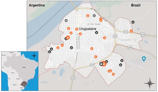
Figure 1.
Map of the sample collection site in the city of Uruguaiana (orange dots: positive dogs; black dots: negative dogs).
2.2. Animal Sampling
The population of this study was a convenience sample from a total of approximately 500 dogs (owned and free-ranged, with no record of previous anti-Leishmania immunization) living in the urban area of the city of Uruguaiana (Rio Grande do Sul, Brazil) and who were treated by veterinarians between August 2019 and February 2020. Demographic and general information were also provided by the pet’s tutors, answering an epidemiological survey form.
The whole study was approved by the Ethics Committee on the Use of Animals (CEUA) of the Lutheran University of Brazil (CEUA-ULBRA nº 2017/355).
2.3. Laboratory Analysis
Blood samples from the 51 selected dogs were collected by venipuncture and stored in two aliquots of around 1 mL, one in a tube without anticoagulant (serum) for the immunological tests and another in a sterile tube containing EDTA (whole blood) for real-time PCR (qPCR). The tubes for the immunological tests were kept refrigerated until processing, and the others (for DNA analyses) were stored at −20 °C until the molecular detection procedure.
The rapid screening test Immunochromatographic TR DPP® (DPP) was performed according to the manufacturer’s instructions (Biomanguinhos-FIOCRUZ, Rio de Janeiro, Brazil). The DPP test consists of a device impregnated with recombinant antigen rK28 (a chimaera combining antigens K9, K26, and K39) to detect the presence of IgG antibodies recognizing these specific L. infantum antigens. Positive samples in the DPP test were sent to a confirmatory ELISA assay (Biomanguinhos-FIOCRUZ, Rio de Janeiro, Brazil) performed at a reference laboratory (Laboratorio Central do Rio Grande do Sul/LACEN-RS, Porto Alegre, RS, Brazil).
The qPCR detection of L. infantum was carried out with commercial reagents. DNA was extracted by silica adsorption methodology using the NewGene® Prep and PreAmp commercial reagents according to the manufacturer’s instructions (Simbios Biotecnologia, Cachoeirinha, RS, Brazil). L. infantum DNA was specifically detected by qPCR with LVCAmp NewGene® reagents (Simbios Biotecnologia, Cachoeirinha, RS, Brazil). Control samples were used in all PCR assays, including water and DNAs extracted from healthy dogs (negative controls), as well as synthetic DNA fragments with oligonucleotide sequences hybridizing to the LVCAmp-specific target gene (gBlock, IDT, Coralville, IA, USA) and DNAs extracted from dogs with CanL (positive controls). All reactions were performed on the StepOnePlus™ real-time PCR system (Applied Biosystems, Palo Alto, CA, USA) with the following amplification conditions: a cycle of 95 °C for 3 min, followed by 40 cycles at 95 °C for 15 s and 60 °C for 1 min. Negative and positive DNA L. infantum controls were used in all runs. A standard curve with 10-fold dilutions of the DNA-positive control was carried out in some analyses to estimate the parasitemia. The positive result values were expressed in cycle thresholds (Cts) and parasites/mL.
2.4. Statistical Analysis
All data analysis was conducted with IBM SPSS (version 23.0) and RStudio (version 4.2.3 version). The demographic data and clinical signs were represented by their frequencies (absolute and relative). For continuous data, normality was assessed using the Anderson–Darling test. Bivariate analyzes were performed to evaluate the association between categorical variables and the outcome. Absolute and relative frequencies were estimated for categorical data using Pearson’s chi-square test or Fisher’s exact test. The pre-established significance level for the 5% alpha error, bilateral, and p < 0.05 were considered significant.
3. Results
3.1. Overall CanL Prevalence and Epidemiological Data
A total of 51 dogs, 10.2% of the approximately 500 dogs attended by veterinarians, were considered suspect to have CanL in the clinical exam in the period of the study. These suspect CanL-positive dogs were obtained in several locations in the city (Figure 1). Laboratorial tests demonstrated that 31 out of the 51 dogs were positive for L. infantum in at least one laboratorial detection method. Among them, a total of 22 (43.1%) dogs were positive in both rapid DPP and qPCR methods, though 6 (11.8%) were positive only in the DPP test and 3 (5.9%) were only positive in the qPCR test (Table 1). All six positive samples in the DPP test were also positive in the confirmatory ELISA. L. infantum parasitemia was evaluated in the 25 (49.0%) qPCR-positive samples, ranging from 3 to 52,700 parasites/mL of blood.

Table 1.
Comparative analysis of IC rapid test and qPCR.
The general epidemiological data demonstrated the occurrence of males (n = 29; 56.9%) and females (n = 22; 43.1%), pure (n = 9; 17.6%) and mixed (n = 41; 80.4%) breeds, and young (n = 23; 45.1%), adult (n = 15; 29.4%), and elderly (n = 11; 21.6%) dogs in all the examined animals. Among the 31 animals positive for CanL, 18 (58.1%) were males and 13 (41.9%) were females. In addition, 12 (38.7%) were puppies (0–2 years old), 11 (35.5%) were adults (3–5 years old), and 6 (19.4%) were elderly dogs (>6 years old). Further, animals with short fur and that live with other dogs represented 90.3% (28) of those positive for CanL (Table 2).
Table 2.
General demographic characteristics of the animals according to whether they were positive or negative for L. infantum.
3.2. Clinical Signs
Different clinical signs were observed in the dogs, including lymphadenopathy (n = 26), dull hair coat (n = 26), alopecia (n = 22), desquamation (n = 20), eye lesion (n = 20), loss (n = 17), onychogryphosis (n = 16), muscle atrophy (n = 10), hyperkeratosis (n = 10), skin ulcer (n = 10), apathy (n = 6), hepatomegaly (n = 5), and diarrhea (n = 2). Among the clinical signs commonly observed in dogs with visceral leishmaniasis, desquamation and onychogryphosis were frequently observed (in 64.5% and 51.6% of positive animals, respectively), with a p value < 0.05 (Table 3).
Table 3.
Clinical variables according to cases and controls dogs, respectively.
4. Discussion
Visceral leishmaniasis is considered a neglected disease endemic in many Brazilian regions. This situation is still the subject of discussion in southern Brazil, as L. infantum has not yet been detected in several cities. In the urban perimeters of certain cities, dogs are the main reservoirs of the etiological protozoan L. infantum. Furthermore, these animals can present CanL and play an important role in the epidemiology of this zoonosis which is very concerning to health [13].
In the present study, 51 dogs were suspected of having CanL in a clinical exam by veterinarians in the period of seven months in Uruguaiana, a city bordering Argentina. The convenience sampling obtained here presented different genders, breeds, and ages of dogs, and it also included animals living in different neighborhoods in the urban perimeter of the city. A total of 31 (60.8%) of the 51 animals with clinical manifestations were confirmed to be positive for L. infantum. If we consider only these animals as positive, we would have a prevalence of approximately 6.2% (31/500) in the total domestic dogs population. But there are probably many other positive animals that did not show clinical signs; therefore, the prevalence of dogs with present or past infection with L. infantum must be even higher. In a previous survey including 965 domestic dogs from this city, 4.5% of the animal samples presented antibodies against L. infantum between 2009 and 2010 [20]. In view of this concerning epidemiological situation with an increasing number of dogs with CanL, other studies have detected more animal species with L. infantum, including wild and domestic mammals [21,22].
The present study also highlights the importance of using different L. infantum detection methods in diagnosing CanL in domestic dogs. Indirect immunological methods evaluate the occurrence of antibodies (mainly IgG) against this parasite in animal body fluids, while direct tests detect L. infantum nucleic acids (mainly DNA). The use of these different assays (immunological and molecular) is still not officially recommended, but this rule must be revised. A more accurate diagnosis could be reached by evaluating the antigenic response (detecting antibodies with rapid test and/or ELISA) and the presence of the parasite by PCR [17]. Noteworthy, the whole diagnostic performance is influenced by the time of infection, sample collection, vaccination status, immunosuppressing diseases, and other factors. Therefore, the use of two or more methods, including PCR, would be very welcome for a definitive diagnostic [18,23]. In addition, qPCR is a useful method to report the parasites load. This information has been proved to be necessary to monitor dogs with CanL, as well as to evaluate the response to medicaments [24,25,26].
Also, the clinical exam is very important to identify CanL in the dogs in an endemic region. The main clinical signs of the infected animals observed here were enlarged lymph nodes (82.4%) and dull coat (64.7%), but weight loss and skin diseases were also important clinical manifestations. Skin lesions were detected mainly in the ears (where there is less hair), being preferable for mosquitoes [15,16]. Skin peeling and onychogryphosis were, respectively, observed 3.4 and 4.3 times more often in dogs with leishmaniasis than in uninfected animals. Onychogryphosis is a clinical manifestation highly correlated with CanL, especially when associated with other cutaneous findings such as exfoliative dermatitis, as observed in this study. Although approximately half of the dogs with leishmaniasis presented onychogryphosis in this study, this clinical sign has already been described in up to 70% of dogs with this disease [27,28,29,30,31]. The complete pathogenesis is not completely known, but excessive nail growth has been associated with more severe parasitism [30,32,33]. The frequent observation of cutaneous findings (onychogriphosis and skin scaling) in this study highlights that the observation of these main clinical manifestations are strong indications of CanL in this endemic region. Veterinarians should pay attention to these specific clinical signs to diagnose new cases.
The results of the present study also reinforce the high prevalence of L. infantum in southern Brazil, on the border with Argentina. Previous diagnoses of CanL in south Brazil were preceded by cases of this same disease in nearby tropical regions of this neighboring country [34,35]. This international border region has been considered the entry point for L. infantum in southern Brazil, which is now spreading to other cities in the state of Rio Grande do Sul. HVL cases were already detected in the Rio Grande do Sul state in recent years [36]. Therefore, more effective prevention methods are necessary to control the dissemination of L. infantum to human and animals in all southernmost states from Brazil.
5. Conclusions
CanL was detected in 31 out of 51 (60.8%) domestic dogs, with the clinical signs being suggestive of this disease in the urban area of Uruguaiana city. These data demonstrate the high frequency of L. infantum in urban dogs from this city in recent years, reinforcing the introduction of this parasite in southern Brazil by the Argentinian border. It also highlights the importance of detecting dogs with CanL by clinical and laboratorial evaluation to prevent more widespread L. infantum dissemination in dogs, humans, and even other animals.
Author Contributions
Conceptualization, A.P.d.F., P.d.F.S. and V.R.L.; methodology, A.P.d.F., V.P.d.S. and F.G.d.O.G.; software, V.R.Z.B.P.; validation, L.S.d.M. and V.R.Z.B.P.; formal analysis, A.P.d.F., V.P.d.S. and V.R.L.; investigation, A.P.d.F., V.P.d.S., F.G.d.O.G. and N.I.; resources, A.F.S. and A.S.K.F.; data curation, L.S.d.M., N.I. and V.R.L.; writing—original draft preparation, A.P.d.F., V.P.d.S. and V.R.L.; writing—review and editing, L.S.d.M., V.R.Z.B.P. and V.R.L.; supervision, A.F.S. and A.S.K.F.; project administration, V.R.L.; funding acquisition, A.S.K.F. and V.R.L. All authors have read and agreed to the published version of the manuscript.
Funding
This study was financed by Simbios Biotecnologia, FAPERGS (Fundação de Amparo à Pesquisa do Rio Grande do Sul) and FINEP (Financiadora de Estudos e Projetos). V.R.L. was also financially supported by the National Council for Scientific and Technological Development from Brazil (CNPq—Conselho Nacional de Desenvolvimento Científico e Tecnológico; process numbers 311010/2017-2; 308445/2020-1). V.P.S. was supported by the Coordenação de Aperfeiçoamento de Pessoal de Nível Superior—Brasil (CAPES)—Finance Code 001.
Institutional Review Board Statement
The study was conducted in accordance with the Declaration of Helsinki and approved by the Ethics Committee on the Use of Animals (CEUA) of the Lutheran University of Brazil (CEUA-ULBRA nº 2017/355).
Informed Consent Statement
Not applicable.
Data Availability Statement
Data are contained within the article; further inquiries can be directed to the corresponding authors.
Conflicts of Interest
The authors declare no conflicts of interest.
References
- Dantas-Torres, F. Canine vector-borne diseases in Brazil. Parasites Vectors 2008, 1, 25. [Google Scholar] [CrossRef] [PubMed]
- Travi, B.L.; Cordeiro-da-Silva, A.; Dantas-Torres, F.; Miró, G. Canine visceral leishmaniasis: Diagnosis and management of the reservoir living among us. PLoS Negl. Trop. Dis. 2018, 12, e0006082. [Google Scholar] [CrossRef] [PubMed]
- Andrade-Filho, J.D.; Scholte, R.G.C.; Amaral, A.L.G.; Shimabukuro, P.H.F.; Carvalho, O.S.; Caldeira, R.L. Occurrence and probability maps of Lutzomyia longipalpis and Lutzomyia cruzi (Diptera: Psychodidae: Phlebotominae) in Brazil. J. Med. Entomol. 2017, 54, 1430–1434. [Google Scholar] [CrossRef] [PubMed]
- Azevedo, J.S.C.; Esmeraldino, A.T.; Ávila, V.P.F.; Witz, M.I.; Fischer, C.D.B.; Tartarotti, A.L. Leishmaniose visceral canina autóctone no município de São Borja, Rio Grande do Sul, Brasil: Relato de caso. Veterinária Em Foco 2009, 7, 52–61. [Google Scholar]
- Acardi, S.A.; Liotta, D.J.; Santini, M.S.; Romagosa, C.M.; Salomón, O.D. Detection of Leishmania infantum in naturally infected Lutzomyia longipalpis (Diptera: Psychodidae: Phlebotominae) and Canis familiaris in Misiones, Argentina: The first report of a PCR-RFLP and sequencing-based confirmation assay. Mem. Inst. Oswaldo Cruz. 2010, 105, 796–799. [Google Scholar] [CrossRef] [PubMed]
- Acosta, L.; Díaz, R.; Torres, P.; Silva, G.; Ramos, M.; Fattore, G.; Bornay-Llinares, F.J. Identification of Leishmania infantum in Puerto Iguazú, Misiones, Argentina. Rev. Inst. Med. Trop. São Paulo 2015, 57, 175–176. [Google Scholar] [CrossRef]
- Rêgo, F.D.; Souza, G.D.; Miranda, J.B.; Peixoto, L.V.; Andrade-Filho, J.D. Potential Vectors of Leishmania Parasites in a Recent Focus of Visceral Leishmaniasis in Neighborhoods of Porto Alegre, State of Rio Grande do Sul, Brazil. J. Med. Entomol. 2020, 57, 1286–1292. [Google Scholar] [CrossRef] [PubMed]
- Pinto, A.O.; Carvalho, D.; Frizzo, C.; Lopes, K.; Tessari, G.B.; Catecati, T.; Dhom-Lemos, L.C.; Pasquali, A.K.S.; Quaresma, P.F.; Stoco, P.H. First case of canine visceral leishmaniasis in the midwestern of Santa Catarina State, Brazil. Braz. J. Biol. 2021, 82, e241162. [Google Scholar] [CrossRef] [PubMed]
- Guimarães, V.C.; Pruzinova, K.; Sadlova, J.; Volfova, V.; Myskova, J.; Filho, S.P.; Volf, P. Lutzomyia migonei is a permissive vector competent for Leishmania infantum. Parasites Vectors 2016, 9, 159. [Google Scholar] [CrossRef]
- Galvis-Ovallos, F.; Ueta, A.E.; Marques, G.O.; Sarmento, A.M.C.; Araujo, G.; Sandoval, C.; Dhom-Lemos, L.C.; Pasquali, A.K.S.; Quaresma, P.F.; Stoco, P.H. Detection of Pintomyia fischeri (Diptera: Psychodidae) with Leishmania infantum (Trypanosomatida: Trypanosomatidae) Promastigotes in a Focus of Visceral Leishmaniasis in Brazil. J. Med. Entomol. 2021, 58, 830–836. [Google Scholar] [CrossRef]
- Carvalho Junior, C.G.; Teixeira Neto, R.G.; Lopes, V.V.; Belo, V.S.; Alves, N.R.; de Paula, T.B.; Ribeiro, R.I.M.A.; Silva, E.S. Parasitism and inflammation in ear skin and in genital tissues of symptomatic and asymptomatic male dogs with visceral leishmaniasis. Parasitol. Res. 2017, 116, 987–995. [Google Scholar] [CrossRef] [PubMed]
- Lopes, J.V.; Michalsky, É.M.; Pereira, N.C.L.; Paula, A.J.V.; Souza, A.G.M.; Pinheiro, L.C.; Lima, A.C.V.M.d.R.; de Avelar, D.M.; França-Silva, J.C.; Lanzetta, V.A.S.; et al. Canine visceral leishmaniasis in area with recent Leishmania transmission: Prevalence, diagnosis, and molecular identification of the infecting species. Rev. Soc. Bras. Med. Trop. 2020, 53, e20200141. [Google Scholar] [CrossRef] [PubMed]
- De Araújo, V.E.; Pinheiro, L.C.; Almeida, M.C.; de Menezes, F.C.; Morais, M.H.; Reis, I.A.; Carneiro, M. Relative risk of visceral leishmaniasis in Brazil: A spatial analysis in urban area. PLoS Negl. Trop. Dis. 2013, 7, e2540. [Google Scholar] [CrossRef] [PubMed]
- Belo, V.S.; Werneck, G.L.; Barbosa, D.S.; Simões, T.C.; Nascimento, B.W.; da Silva, E.S.; Struchiner, C.J. Factors associated with visceral leishmaniasis in the americas: A systematic review and meta-analysis. PLoS Negl. Trop. Dis. 2013, 7, e2182. [Google Scholar] [CrossRef]
- Alvar, J.; Cañavate, C.; Molina, R.; Moreno, J.; Nieto, J. Canine leishmaniasis. Adv. Parasitol. 2004, 57, 1–88. [Google Scholar] [PubMed]
- Pennisi, M.G.; Cardoso, L.; Baneth, G.; Bourdeau, P.; Koutinas, A.; Miró, G.; Oliva, G.; Solano-Gallego, L. LeishVet update and recommendations on feline leishmaniosis. Parasites Vectors 2015, 8, 302. [Google Scholar] [CrossRef] [PubMed]
- Brasil—Ministério da Saúde (MS). Manual de Vigilância e Controle da Leishmaniose Visceral; Brasil—Ministério da Saúde (MS): Uruguaiana, Brazil, 2014; 122p, (accessed on 27 December 2023).
- Sevá, A.D.P.; Brandão, A.P.D.; Godoy, S.N.; Soares, R.M.; Langoni, H.; Rodrigues, B.C.; Zanotto, P.F.d.C.; Jimenez-Villegas, T.; Hiramoto, R. Investigation of canine visceral leishmaniasis in a non-endemic area in Brazil and the comparison of serological and molecular diagnostic tests. Rev. Soc. Bras. Med. Trop. 2021, 54, e01822021. [Google Scholar] [CrossRef] [PubMed]
- IBGE—Instituto Brasileiro de Geografia e Estatística. Uruguaiana. 2023. Available online: https://www.ibge.gov.br/cidades-e-estados/rs/uruguaiana.html (accessed on 8 April 2023).
- Massia, L.I.; Lamadril, R.D.Q.; Wellicks, J.R.; Bittencourt, R.A.; Bittencourt, D.G.; Marques, G.D.; Celis, E.L.H.; Pellegrini, D.D.C.P. Leishmaniose visceral canina em três bairros de Uruguaiana—RS. Vigil. Sanit. Debate. 2016, 4, 113–119. [Google Scholar] [CrossRef]
- Escobar, T.A.; Dowich, G.; Dos Santos, T.P.; Zuravski, L.; Duarte, C.A.; Lübeck, I.; Manfredini, V. Assessment of Leishmania infantum infection in equine populations in a canine visceral leishmaniosis transmission area. BMC Vet. Res. 2019, 15, 381. [Google Scholar] [CrossRef]
- Pradella, G.D.; Escobar, T.A.; Santos, T.P.D.; Vargas, R.C.; Góss, G.C.; Ferrareze, P.A.G.; Zuravski, L.; Pereira, K.B.; Duarte, C.A. PCR-RLFP characterization of Leishmania spp. in domestic animals from the south-western border of Brazil. Rev. Bras. Parasitol. Vet. 2022, 31, e005222. [Google Scholar] [CrossRef]
- Garay, A.F.G.; Fraenkel, S.; Diaz, J.J.A.R.; Recalde, O.D.S.; Gómez, M.C.V.; Riquelme, J.A.M.; Arze, P.V.; Centurión, G.N.R.; Britos, M.; Rolón, M. Sensitivity comparison for the Leishmania spp. detection in different canine tissues using PCR-HRM. Rev. Soc. Bras. Med. Trop. 2022, 55, e0069-2022. [Google Scholar] [CrossRef] [PubMed]
- Paltrinieri, S.; Gradoni, L.; Roura, X.; Zatelli, A.; Zini, E. Laboratory tests for diagnosing and monitoring canine leishmaniasis. Vet. Clin. Pathol. 2016, 45, 552–578. [Google Scholar] [CrossRef] [PubMed]
- Solano-Gallego, L.; Di Filippo, L.; Ordeix, L.; Planellas, M.; Roura, X.; Altet, L.; Montserrat, S. Early reduction of Leishmania infantum-specific antibodies and blood parasitemia during treatment in dogs with moderate or severe disease. Parasites Vectors 2016, 9, 235. [Google Scholar] [CrossRef] [PubMed]
- Pereira, D.C.A.; Teixeira-Neto, R.G.; Lopes, V.V.; Pena, H.P.; Paz, G.F.; Custodio, C.H.X.; Belo, V.S.; Júnior, A.A.d.F.; da Silva, E.S. Development of quantitative PCR and digital PCR for the quantification of Leishmania infantum in dogs. Mol. Cell. Biochem. 2023, 478, 1–6. [Google Scholar] [CrossRef] [PubMed]
- Teixeira Neto, R.G.; Giunchetti, R.C.; Carneiro, C.M.; de Almeida, R.W.V.; Coura-Vital, W.; Quaresma, P.F.; Ker, H.G.; de Melo, L.A.; Gontijo, C.M.F.; Reis, A.B. Relationship of Leishmania-specific IgG levels and IgG avidity with parasite density and clinical signs in canine leishmaniasis. Vet. Parasitol. 2010, 169, 248–257. [Google Scholar] [CrossRef] [PubMed]
- Galán-Relaño, Á.; Maldonado, A.; Gómez-Gascón, L.; Tarradas, C.; Astorga, R.J.; Luque, I.; Huerta, B. Pre-test probability and likelihood ratios for clinical findings in canine leishmaniasis. Transbound Emerg. Dis. 2022, 69, 3540–3547. [Google Scholar] [CrossRef] [PubMed]
- Oliveira, M.R.; Neto, M.B.O.; Bezerra, T.L.; Da Silva, W.S.I.; da Paz, W.S.; dos Santos, I.G.; Bezerra-Santos, M.; Lima, V.F.S. Canine leishmaniasis in an endemic region, Northeastern Brazil: A comparative study with four groups of animals. Parasitol. Res. 2021, 120, 3915–3923. [Google Scholar] [CrossRef] [PubMed]
- Silva, K.R.D.; Mendonça, V.R.R.D.; Silva, K.M.; Nascimento, L.F.M.D.; Mendes-Sousa, A.F.; Pinho, F.A.D.; Barral-Netto, M.; Barral, A.M.P.; e Cruz, M.D.S.P. Scoring clinical signs can help diagnose canine visceral leishmaniasis in a highly endemic area in Brazil. Mem. Inst. Oswaldo Cruz. 2017, 112, 53–63. [Google Scholar] [CrossRef]
- Koutinas, A.F.; Carlotti, D.N.; Koutinas, C.; Papadogiannakis, E.I.; Spanakos, G.K.; Saridomichelakis, M.N. Claw histopathology and parasitic load in natural cases of canine leishmaniosis associated with Leishmania infantum. Vet. Dermatol. 2010, 21, 572–577. [Google Scholar] [CrossRef]
- Koutinas, A.F.; Koutinas, C.K. Pathologic mechanisms underlying the clinical findings in canine leishmaniosis due to Leishmania infantum/chagasi. Vet. Parasitol. 2014, 51, 527–538. [Google Scholar]
- LeishVet—LeishVet Guidelines for the Practical Management of Canine and Feline Leishmaniosis: A Brief for the Practicing Veterinarian. 2023. Available online: https://www.leishvet.org/wp-content/uploads/2023/01/ALIVE-dec22-web-EN.pdf (accessed on 27 December 2023).
- Diaz, R.G.; Salvatierra, K.A.; Silva, G.A.; Deschutter, E.J.; Bornay-Llinares, F.J.; Acosta, L. First molecular characterization of Leishmania infantum species in patients infected with visceral leishmaniasis in Misiones province, Argentina. Biomedica 2019, 39, 405–414. [Google Scholar] [CrossRef] [PubMed]
- Lamattina, D.; Berrozpe, P.E.; Casas, N.; Moya, S.L.; Giuliani, M.G.; Costa, S.A.; Arrabal, J.P.; Martínez, M.F.; Rivero, M.R.; Salas, M.; et al. Twice upon a time: The progression of canine visceral leishmaniasis in an Argentinean city. PLoS ONE 2019, 14, e0219395. [Google Scholar] [CrossRef] [PubMed]
- Rio Grande do Sul—Secretaria da Saúde (MS). Leishmaniose Visceral Humana—Situação Epidemiológica/Dados 2023. Available online: https://www.cevs.rs.gov.br/lvh-situacao-epidemiologica-dados (accessed on 27 December 2023).

Disclaimer/Publisher’s Note: The statements, opinions and data contained in all publications are solely those of the individual author(s) and contributor(s) and not of MDPI and/or the editor(s). MDPI and/or the editor(s) disclaim responsibility for any injury to people or property resulting from any ideas, methods, instructions or products referred to in the content. |
© 2024 by the authors. Licensee MDPI, Basel, Switzerland. This article is an open access article distributed under the terms and conditions of the Creative Commons Attribution (CC BY) license (https://creativecommons.org/licenses/by/4.0/).