Article

Exploring Pathogenic and Zoonotic Bacteria from Wild Rodents, Dogs, and Humans of the Ngorongoro District in Tanzania Using Metagenomics Next-Generation Sequencing

by
Amina Ramadhani Issae
1,2,3,*,
Abdul Selemani Katakweba
1,3,
Rose Peter Kicheleri
2,
Augustino Alfred Chengula
4,
Marco van Zwetselaar
5 and
Christopher Jacob Kasanga
4
1
African Centre of Excellence for Innovative Rodent Pest Management and Biosensor Technology Development, Sokoine University of Agriculture, Morogoro P.O. Box 3110, Tanzania
2
Department of Wildlife Management, Sokoine University of Agriculture, Morogoro P.O. Box 3073, Tanzania
3
Institute of Pest Management, Sokoine University of Agriculture, Morogoro P.O. Box 3110, Tanzania
4
Department of Microbiology, Parasitology and Biotechnology, Sokoine University of Agriculture, Morogoro P.O. Box 3019, Tanzania
5
Kilimanjaro Clinical Research Institute (KCRI), Kilimanjaro Christian Medical University, Moshi P.O. Box 2236, Tanzania
*
Author to whom correspondence should be addressed.
Zoonotic Dis. 2023, 3(3), 226-242; https://doi.org/10.3390/zoonoticdis3030019
Submission received: 18 July 2023 / Revised: 15 August 2023 / Accepted: 29 August 2023 / Published: 1 September 2023

Abstract

:

Simple Summary

This study explored pathogenic and zoonotic bacteria in blood samples of wild rodents, domestic dogs, and humans in the Ngorongoro District in Tanzania. The district is inhabited by wildlife, domestic animals, and humans. Previous studies carried out on the livestock documented the existence of zoonotic bacterial diseases in the district. The role played by wild rodents and domestic dogs in the transmission of pathogenic and zoonotic bacteria was unknown. Therefore, the objective of this study was the detection and identification of pathogenic and zoonotic bacteria circulating among wild rodents, domestic dogs, and humans. The study concluded that a variety of zoonotic bacteria are present in wild rodents, domestic dogs, and humans sharing the same environment. Wild rodents carried numerous pathogenic and zoonotic bacteria compared to domestic dogs and humans. These results emphasize the importance of sustained investigations and unified health efforts to alleviate zoonotic disease transmission in this ecosystem.

Abstract

Globally, zoonoses have serious consequences due to their socioeconomic impacts. Ngorongoro District is home to a diverse range of wildlife and domestic animals, including rodents and dogs, which often coexist in close proximity with humans. The aim of the study was to identify the zoonotic bacteria present in wild rodents, domestic dogs, and humans using metagenomics next-generation sequencing technology. A cross-sectional study was conducted in 2022. This study used both Illumina and Oxford Nanopore sequencing technologies to identify bacteria in 530 blood samples collected from humans (n = 200), wild rodents (n = 230), and dogs (n = 100). Several zoonotic airborne/contagious bacteria, including Mycobacterium spp., Mycoplasma spp., Bordetella spp., and Legionella spp., were detected in wild rodents, domestic dogs, and humans. Arthropod-borne zoonotic bacteria such as Bartonella spp., Borrelia spp., and Rickettsia spp. were detected in all three hosts, while Orientia spp. was found in wild rodents and domestic dogs. Yersinia pestis, Streptobacillus spp. and Anaplasma spp. were found only in wild rodents. Other zoonotic bacteria found shared among wild rodents, domestic dogs, and humans are Leptospira spp., Brucella spp., and Salmonella spp. Generally, wild rodents had the highest prevalence of zoonotic bacterial species when compared to domestic dogs and humans. The detection of zoonotic bacteria in rodents, dogs, and humans supports the hypothesis that infections can spread between animals and humans sharing the same environment.

Graphical Abstract

1. Introduction

Zoonotic diseases have a significant socioeconomic impact globally [1]. Wildlife populations, by forming the reservoirs from which zoonotic agents can arise, have long been considered a link in the chain of pathogen emergence [2]. Approximately 75% of emerging infectious diseases affecting humans are zoonoses of animal origin [3]. Rodents are important reservoirs of numerous pathogenic and zoonotic bacteria including Leptospira [4], Bartonella [5], Mycobacteria [6], and Campylobacter [7]. It has been found that activities like crop cultivation and livestock farming bring wild rodents into close contact with humans [8]. Studies have documented that wild rodents are usually attracted to crops and stored grains, which can increase the interaction with humans and domestic dogs [8]. Additionally, improper waste disposal practices attract wild rodents to human settings [9]. These activities can facilitate the cross-species transmission of pathogenic bacteria [9]. In addition, many African countries, including Tanzania, are reporting a growing proportion of cases of fever of unknown origin [10,11]. Probably, some of these cases are associated with rodent-borne infections, which are under-reported in Tanzania. Therefore, surveillance studies are important in the determination and justification of the socioeconomic impact of rodent-borne diseases in Tanzania.
Keeping dogs in underdeveloped countries, such as Tanzania, can present unique challenges due to limited resources and infrastructure [12]. However, with proper planning and care, it is possible to maintain a healthy and safe environment for dogs. This study constitutes a comprehensive overview of the dog-keeping system in underdeveloped countries, focusing on Tanzania. In Tanzania, dog ownership is often influenced by cultural and traditional practices. Dogs are kept for various purposes such as security, herding, hunting, companionship, and even as status symbols [12]. There is limited awareness about responsible dog ownership including proper healthcare, a routine feeding system, and basic housing [12]. Tanzania’s dogs cannot access conventional dog houses or dedicated shelters. Instead, they live in makeshift shelters, such as small huts, outdoor enclosures, and open spaces, or live as stray dogs [12]. Stray dogs can play a significant role in the transmission of bacterial diseases [13]. Due to a lack of proper veterinary care, stray dogs are more susceptible to infections, and their scavenging and roaming behavior can contribute to the spread of bacterial pathogens to humans through direct contact or a contaminated environment. Some bacterial diseases commonly associated with stray dogs are Leptospirosis [13], Salmonellosis, Campylobacteriosis, Pasteurellosis, and a strain of Staphylococcus aureus bacteria that has developed resistance to several antibiotics [14]. Despite the large population of domesticated dogs in the Ngorongoro District, no study has been conducted to evaluate the health status and pathogenic bacteria of dogs.
Previous studies conducted in the Ngorongoro District documented the occurrences of bacterial diseases in livestock, including Anthrax [15], Bovine Tuberculosis [16], Leptospirosis [17], and Brucellosis [18]. On the human side, the studies documented seroprevalence of Brucella infection in pregnant women receiving antenatal care [19] and the genetic diversity of Mycobacterium tuberculosis from TB patients attending health facilities in the Serengeti ecosystem [20] bordering the Ngorongoro District. The above-mentioned studies were performed around human–livestock–wildlife interfaces. It has been found that areas of interfaces generate unique hotspots of numerous infectious diseases including bacterial infections [21].
Ngorongoro District in Tanzania is home to a diverse range of wildlife and domestic animals, including rodents and dogs, which often coexist in close proximity with humans [22]. While previous studies have examined the transmission of pathogens in livestock and hospital-based research in humans within the district, the specific role played by rodents and dogs in the transmission of pathogens remains largely unknown. This research gap highlights the need for a comprehensive investigation into the contribution of rodents and dogs in pathogen transmission in the Ngorongoro District, allowing for a more comprehensive understanding of disease dynamics and potential risks to public health.
High throughput methods, such as metagenomics, can analyze multiple genomes of bacterial species [23]. This allows the identification of bacteria genomes directly from samples and can reveal information related to the diversity of microbes that circulate among different hosts in the communities [24,25]. The main objective of the study was to identify pathogenic and zoonotic bacteria present in wild rodents, domesticated dogs, and humans of the Ngorongoro District by using metagenomics next-generation sequencing techniques.

2. Materials and Methods

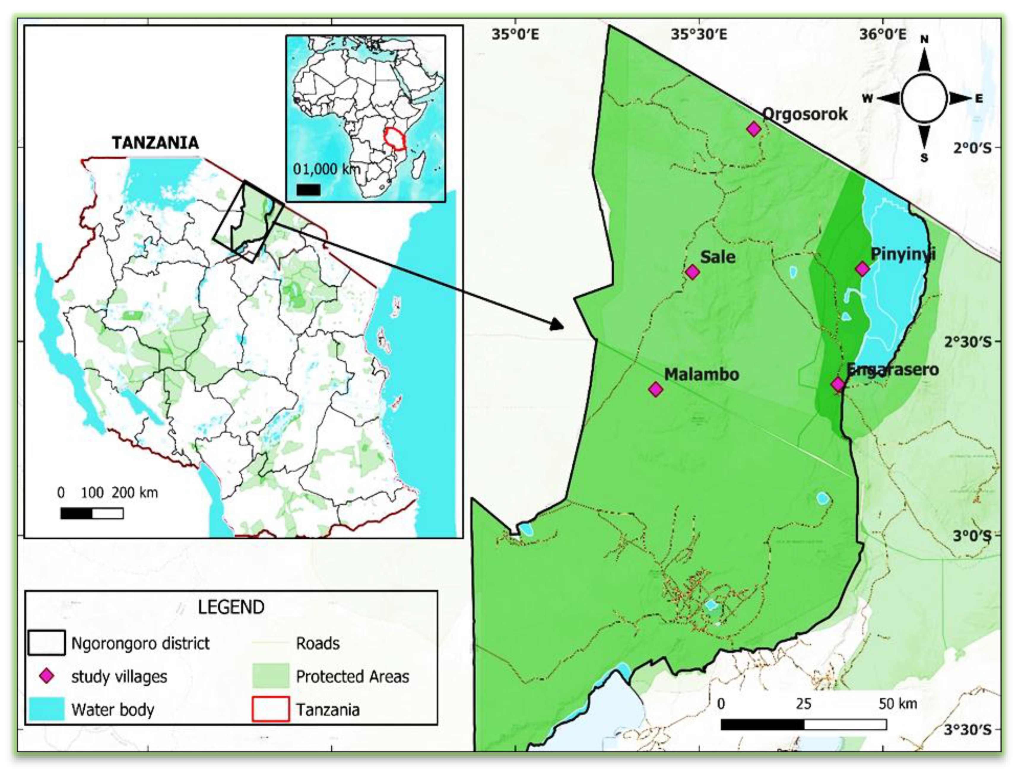
2.1. Description of the Study Area

This study was conducted in the Ngorongoro District (Figure 1). The district was selected based on areas of the interface of wild animals, domestic animals, and humans, as well as the previous seroprevalence studies of bacterial infection in livestock. Ngorongoro District is located in Arusha Region, northern Tanzania. It is bordered by the Manyara region to the West, the Karatu district to the south, and the Monduli district to the east. It has an area of 14,036 square kilometers, is located between latitudes 30.30′ S and longitudes 35.42′ E, and it is between 1009 and 3645 m above sea level [26]. The district has a population of 174,278 as of the 2012 Tanzania National Census [27]. Ngorongoro, Loliondo, and Sale are the 3 administrative divisions of the district, together with 28 wards and 65 villages. The district experiences tropical weather with moderate temperatures and an average rainfall of 800 to 1000 mm. The predominant vegetation in the study area is grass and bushes of several acacia species, as well as open, dense forests.

2.2. Study Design and Sampling Procedures

A cross-sectional study was conducted in the Ngorongoro District in 2022 to explore bacteria of public health importance found in wild rodents, domestic dogs, and humans. The study population was made up of all the households in the selected villages, and the sample frame was a list of households in each village.
The study villages were selected intentionally based on the availability of domesticated animals, including dogs and wild animals, and accessible areas. Five villages (Orgosorok, Malambo, Sale, Engarasero, and Pinyinyi) were included in this study. A purposeful sampling method was used in the selection of households based on the willingness of individuals and the availability of wild rodents, domestic dogs, and other animals.
The selection of participants was based on voluntary willingness and adult humans of 18 years and older. Adults were selected because they have a longer history of exposure to various environments and animals, which can provide more comprehensive insights into the transmission of zoonotic pathogens. Also, dogs of 6 months and above were selected for the study because, at this age, dogs do not have maternal antibodies which could prevent infections. Before starting the sampling of humans and domestic dogs and the trapping of wild rodents, written consent from the head of the household was sought.

2.3. Trapping of Rodents

Live rodents were captured using Sherman LFA live traps (HB Sherman Traps, Inc., Tallahassee, FL, USA) and wire cage traps baited with peanut butter mixed with maize bran and sardines [4]. Trapping was carried out in specifically defined places such as areas surrounding livestock farms, fallow land around houses, as well as areas with green vegetation and marshes near homes. In each study village, 30 to 50 houses were selected to set traps indoors and in their surroundings. Based on the size of the household, 2 to 4 modified wire cage traps were placed in each house for the purpose of trapping indoor rodents. Depending on rodents’ activities, 2 to 6 Sherman traps were set for peri domestic purposes. In each village, a total of 30 modified wire cage traps and 70 Sherman traps were used indoors and in the surroundings, respectively. For the remaining habitats (crop fields and grass-covered vegetation), the maximum number of Sherman traps set was 30–70 depending on the size of the selected habitat; thus, a total of 100 traps were used in each village. All traps were baited and set for 5 to 7 days in each village. Traps were set at 5 pm and checked in the morning at 8 am every day.

2.4. Collection and Handling of Samples (from Wild Rodents)

Trapped rodents were anaesthetized and humanely killed using isoflurane (volatile inhalation agent). The rodents were placed into an anesthetic chamber with cotton wool soaked in isoflurane, as described in the previous study. The animal was removed from the chamber after cessation of respiration and heartbeats, and morphometric characteristics of rodents were recorded prior to dissection. The ventral surfaces of the rodents were disinfected using 70% methylated spirit to kill external germs. Using proper protecting gears to safeguard the health of the researcher, almost 1 ml of blood samples was collected from the rodent’s ventral surface using hypodermic needles and syringes. The cardiac puncture technique was used in blood sample collection [10]. The blood sample was mixed with 2 mL of DNA/RNA shield reagents in cryogenic tubes, labelled, and kept in liquid nitrogen. Samples were transported to the Sokoine University of Agriculture in the Department of Veterinary Microbiology, Parasitology and Biotechnology Laboratory for detailed analysis. DNA/RNA shield reagent was used to maintain the integrity of the nucleic acids due to inhibition of DNase and RNase activities.

2.5. Collection of Blood Samples from Humans and Domestic Dogs

Before taking a blood sample from humans and dogs, the human laboratory scientist and veterinary officer thoroughly washed their hands with soap and water, and alcohol rub, for at least 30 s [28]. To prevent infections, the personnel put on safety gloves after cleansing his/her hands. The skin of the individual and domestic dog was disinfected using 70% alcohol, beginning at the needle-insertion site and making several outwardly expanding circles [28]. The cephalic vein was used for blood collection [29]. A blood sample of 1 mL was taken using a 21-gauge needle. To avoid contaminations that could lead to infections, the needle entry site was wrapped with gauze and sellotape immediately after sample collection. The obtained blood samples were mixed with 2 mL of DNA/RNA shield reagents in cryogenic tubes. All blood tubes were labelled, transported to SUA in liquid nitrogen, and stored at −80 °C until further analysis.

2.6. Preparation of Pools of Blood Samples

A total of 200 blood samples from human subjects were collected and then pooled into 22 pools and grouped by sexes [30]. Likewise, 230 blood samples were collected from rodents and then pooled into 16 pools [6], as shown in Table 1 below. Similarly, a total of 100 blood samples were collected from domestic dogs and then grouped into 10 pools based on their sexes [31] (Table 1). The study involved 5 villages and, in each village, 46 samples of rodents, 40 samples of humans, and 20 samples of domestic dogs were collected.

2.7. Nucleic Acids Extraction, Libraries Preparation, and Sequencing

The QIAamp® RNA blood Mini Kit and QIAamp® DNA blood Mini Kit were used for the purification of RNA and DNA, respectively, as per the manufacturer’s instructions (Qiagen, Valencia, CA, USA). The extracted RNA genomes were converted into complementary DNA (cDNA) using Omniscript RT Kit based on the supplier’s protocol (Qiagen, Valencia, CA, USA).
Two methods of next-generation sequencing (Illumina and Nanopore) were employed in this work: the MiSeq sequencing platform (Illumina) and MinION sequencing technology (Nanopore). The MinION sequencing libraries were generated by using the PCR-cDNA sequencing-barcoding kit (SQK-PCB109-Oxford Nanopore Technologies, Cambridge, UK) following the manufacturer’s protocol.
The Illumina NexteraTM XT DNA Library Prep Kit (Illumina, San Diego, CA, USA) was used to prepare sequencing libraries for the MiSeq platform following the manufacturer’s protocol. The quality of the libraries generated was assessed by using a qubit high-sensitivity quantification assay, following the manufacturer’s protocol (Thermo Fisher Scientific technology, Waltham, MA, USA). Subsequently, the nucleic acids were pooled in equimolar amounts, and the resulting libraries were sequenced on a single lane (paired-end, 151 bp read-length) on an Illumina MiSeqTM machine (Illumina, San Diego, CA, USA).

2.8. Bioinformatics Analysis
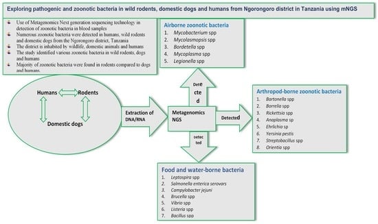
The sequencing data obtained from MinION were processed using ONT Guppy version 6.4.2 and the 9.4.1 450 bps SUP model was used for base calling. The reads that were demultiplexed were identified by ONT Guppy barcoder version 6.4.2. Unclassified reads from each run were saved as distinct pseudo-samples. FastQ Screen version 0.14.1 with GRCh38 and UniVec Core was used to screen reads for human and vector contamination [32]. The reads underwent quality filtering and trimming using fastp version 0.20.1 using settings -5 -3 -M 8 -q 6 -e 10 -l 64 [33]. Fundamental quality control measurements (read counts, base counts, and quality scores) were obtained using fastq-stats from fastq-utils 1.3.0 [33].
MiSeq reads were base called and demultiplexed with Illumina BCL Convert 3.9.3. Reads that were not classified were reserved as a separate pseudo-sample. The FastQ Screen version 0.14.1 was used with GRCh38 and UniVecCore to screen the reads for any contamination from human or vector sources [32]. Reads were trimmed with fastp 0.20.1 using default settings plus front and tail trimming (-5 -3). Important quality control measurements were acquired through the utilization of the fastq-stats function from fastq-utils version 1.3.0 [33].
The process of assigning taxonomy was accomplished by employing Kraken2 version 2.1.2 [34] using the Kraken2 standard databases plus fungi, constructed from NCBI Reference Sequence data. The analyses were carried out on 2 separate occasions. The first analysis categorized the quality-filtered and trimmed MiSeq and MinION reads using Kraken2′s paired-end mode. The second analysis classified the merged MiSeq and MinION reads for each sample, which was quality-filtered and trimmed, using Kraken2’s default mode.

3. Results

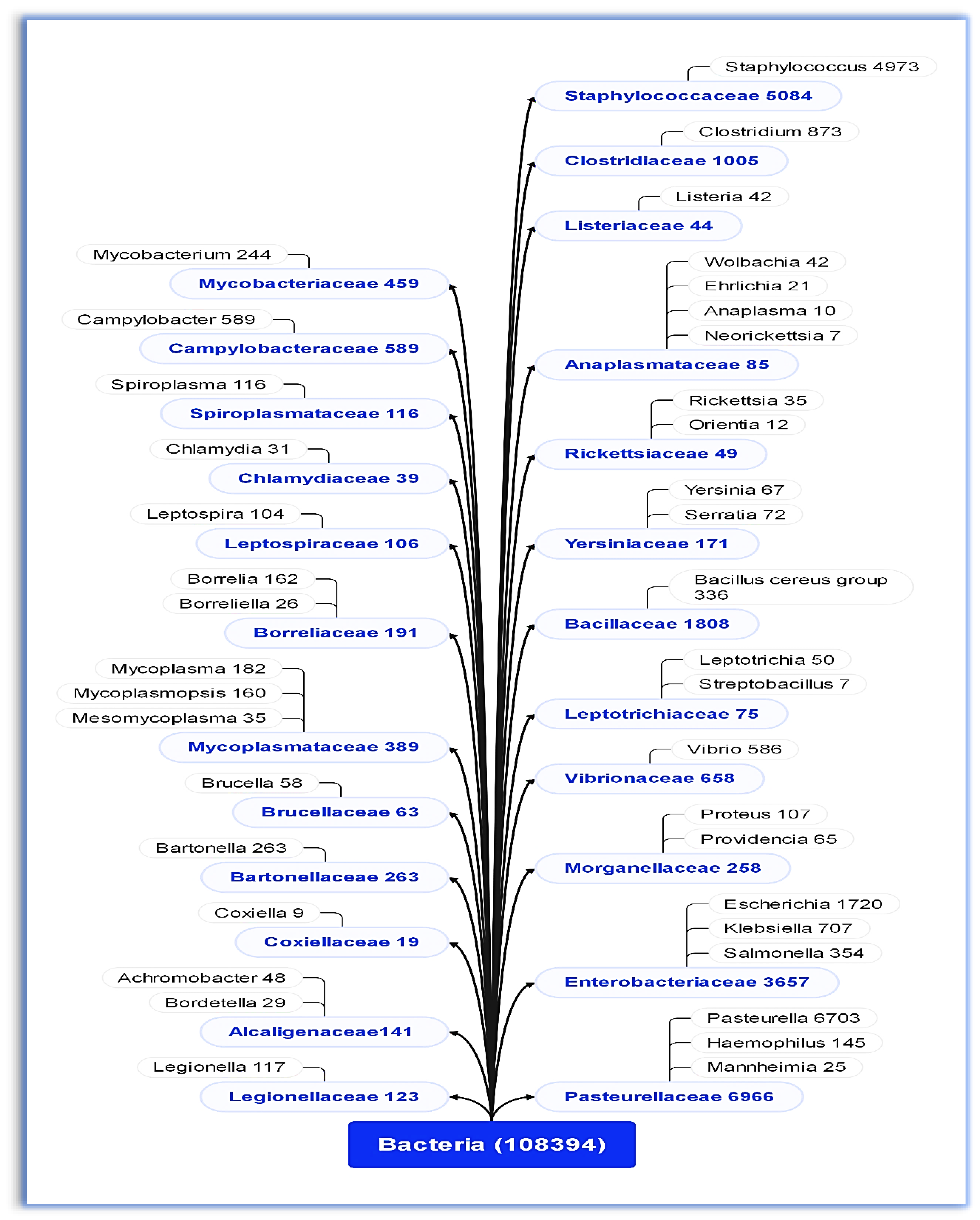
3.1. Bacterial Families and Genera Identified

This study detected 24 families of potentially pathogenic and zoonotic bacteria in wild rodents, domestic dogs, and humans (Figure 2).

3.2. Airborne, Contagious, and Arthropod-Borne Zoonotic Bacteria

The study detected five and nine genera of airborne and arthropod-borne zoonotic bacteria species, respectively (Table 2). Among the five genera of airborne bacteria species, Mycobacterium species were detected in high proportion in wild rodents (56.25%) (Table 2). Among the arthropod-borne bacteria detected, Bartonella species were found in high proportion in wild rodents (68.75%) compared to domestic dogs and humans. Among the nine arthropod-borne bacteria, three genera (Borrelia, Bartonella, and Rickettsia) were found in humans, wild rodents, and domestic dogs (Table 2). Generally, wild rodents have the highest proportion of zoonotic bacterial species, followed by domestic dogs, and then humans (Table 2). Various airborne and arthropod bacterial species identified in wild rodents, domestic dogs, and humans are presented in Table 3 and Table 4, respectively.

3.3. Pathogenic and Zoonotic Bacteria Detected in Humans, Wild Rodents, and Domestic Dogs

The results have shown that ten genera of zoonotic bacteria species were identified in humans and domestic dogs while nine genera were found in wild rodents (Table 5 and Table 6). Some of the pathogenic and zoonotic bacteria species are presented in Table 6.

4. Discussion

This study focused on the identification of various pathogenic and zoonotic bacteria circulating among wild rodents, domestic dogs, and humans in the Ngorongoro District. Numerous zoonotic bacteria that pose a threat to public and animal health were found, whereby some species were detected in either one, two, or three hosts involved in the study. This showed the possibility of cross-species transmission of different bacterial species in the study area.
Several airborne and contagious zoonotic bacteria species were found in wild rodents, domestic dogs, and humans in this study. Most of them are transmitted from one host to another through inhalation of infected aerosol droplets or through direct contact with infected animals or contaminated surfaces [6,35,36]. The current study found several species of Mycobacteria and Bordetella in domestic dogs, wild rodents, and humans. This indicated that the interaction between humans, wild rodents, and domestic dogs increases the chance of cross-transmission of pathogens among different host species. The importance of the Mycobacterium species for public health is based on its capacity to cause tuberculosis, leprosy, and ulcerations in humans [35]. A previous study carried out in Morogoro, Tanzania, also reported findings on the occurrence of nontuberculous Mycobacteria species in wild rodents and Crocidura species [6]. Most human cases caused by Bordetella spp. were documented in immunocompromised patients and presented in a variety of ways, from moderate coughing and tracheobronchitis to sepsis and death [36]. These results necessitate unified health surveillance of pathogens among communities in order to safeguard public health.
Bordetella species found in this study were previously linked with pulmonary infection in humans. B. bronchiseptica and B. pertussis are the causative agents of pneumonia and whooping cough in humans [37]. This study revealed the occurrence of B. bronchiseptica infection in wild rodents, domestic dogs, and humans and B. genomosp, B. flabilis, and B. trematum infection in both domestic dogs and humans. Contrary to the case in rodents and humans, most Bordetella species were detected in domestic dogs. This suggests that domestic dogs can play a role in the transmission of Bordetella spp. infection to humans and other domestic and wild mammals. The occurrences of Bordetella species in more than one host justify the possibility of cross-species transmission of these pathogens in the study area.
Additionally, some of the airborne and arthropod-borne Mycoplasma species were also identified in wild rodents, domestic dogs, and humans. M. pneumoniae, M. crocodyli, M. suis, and M. tauri species were all found in both humans and wild rodents in this study. In contrast to humans and dogs, the majority of Mycoplasma species were identified in wild rodents. And most of the species discovered in this work have been isolated in other animals, including cattle (M. tauri), goats (M. mycoides), pigs (M. suis, M. hyopneumoniae), and crocodiles (M. crocodyli) [38,39,40,41]. Some of the Mycoplasma species identified in this study have been reported to cause numerous animal fatalities and huge economic losses globally. For instance, the swine sector suffers financial losses because of hemolytic anemia and swine enzootic pneumonia caused by M. suis and M. hyopneumoniae, respectively [39,40]. Moreover, M. mycoides subsp. Capri caused a severe mortality outbreak of respiratory mycoplasmosis in goats in Mexico [42]. In general, these findings showed the possibility of the occurrence of inter-species cross-transmission of Mycoplasma in the study area. Moreover, this study recognized Legionella species that cause fatal pneumonia (Legionnaires’ disease) in humans after inhalation of airborne droplets containing viable bacteria (Cunha et al., 2016). The aforementioned Legionella species were found in wild rodents, domestic dogs, and humans. Perhaps the infections were acquired from contaminated natural water or aquatic environments. It was reported that water is the major natural reservoir for Legionella species [43].
This study presents the first report of Borrelia miyamotoi infection among wild rodents, domestic dogs, and humans in Tanzania. A high proportion of infection was found in rodents, followed by domestic dogs and, lastly, humans. This indicates that wild rodents are the main reservoirs of Borrelia spp. in Tanzania. Most of the identified arthropod-borne bacteria have been widely reported to cause infections in humans and animals [44,45,46,47,48]. For example, Borrelia spp. causes Lyme borreliosis in humans [45,46]. These pathogens use both rodents and ticks as reservoirs and vectors, respectively [46]. Bartonella species linked to human illnesses were found, including B. tribocorum, B. elizabethae, B. grahamii, and B. taylorii. Bilateral retinal branch occlusions or neuro retinitis have been linked to Bartonella grahamii [49]. Bartonella elizabethae was isolated in individuals with endocarditis illness [49]. Bartonella tribocorum was revealed in patients with fever in Thailand [50]. The discovery of these zoonotic Bartonella species in wild rodents, domestic dogs, and humans calls for increased awareness of these infections among healthcare professionals, particularly in cases of unexplained febrile illness.
Yersinia pestis, a causative agent of plague (a zoonotic disease which has stable foci throughout Africa, America, and Eurasia) was detected in wild rodents [47]. The main mode of transmission from one host to another is by infected flea bites that result in painful, swollen lymph nodes known as buboes and septicemia [47]. A bacterium Orientia tsutsugamushi was found in wild rodents in this study. This causes scrub typhus (tsutsugamushi sickness), an acute infectious disease in humans [51,52]. Streptobacillus moniliformis responsible for rat-bite fever was also found in wild rodents and domestic dogs in this study. Based on these findings, education on rodent management is important in order to protect the community from zoonotic bacteria.
Furthermore, the Rickettsia typhus group (TG) and Rickettsia spotted fever group (SFG) were identified in wild rodents, domestic dogs, and humans in this study. Eight species of Rickettsia were detected, including R. australis (a causative agent of Queensland tick typhus), R. typhii (a causative agent of murine typhus), and R. prowazekii (a causative agent of epidemic typhus) [53]. The rickettsia species discovered in this work had previously been found in ticks, dogs, and humans in Brazil [54,55], Wisconsin [44], and Australia [53]. The genus Anaplasma comprises different zoonotic species which cause diseases in animals and humans [56]. Anaplasma marginale cause bovine anaplasmosis in tropical and subtropical regions and other areas globally [56]. Anaplasma phagocytophilum cause animal and human granulocytic anaplasmosis [48]. Anaplasma platys have been reported to cause febrile illness associated with headache and fever in humans in Venezuela [57]. These pathogens parasitize red blood cells in susceptible hosts and are transmitted by ticks and biting insects [56]. This study documents, for the first time, the occurrence of these three Anaplasma species in wild rodents in Tanzania. Additionally, tick-borne bacteria, such as Ehrlichia canis and Ehrlichia muris, which cause life-threatening diseases, including canine ehrlichiosis in dogs, were observed in this study. These two species of Ehrlichia are zoonotic bacteria and were mainly detected in domestic dogs [44,58].
This study detected zoonotic Leptospira spp. shared among wild rodents, domestic dogs, and humans. Leptospira interrogans was found in all three hosts, while Leptospira kobayashii was detected in domestic dogs and humans and Leptospira santarosai, Leptospira kmetyi, and Leptospira weilii were identified in wild rodents and domestic dogs. The majority of Leptospira species were identified in domestic dogs and wild rodents compared to humans. This supports the possibility that dogs and rodents in the study area are the sources of human leptospirosis. Humans acquire the infection through direct contact with the urine of infected animals or a contaminated environment [59]. Human infection is associated with various symptoms ranging from asymptomatic fever to complex illnesses with significant morbidity and mortality rate, like Weil’s disease [13]. The current study revealed the occurrences of Brucella spp. among wild rodents, domestic dogs, and humans. B. anthropic, an emerging, opportunistic, nosocomial human pathogen [60,61], was found in humans and wild rodents. Moreover, the detection of B. suis in wild rodents and domestic dogs indicates the possibility of occurrences of cross-species transmission in the study area. Dogs that had been pig-hunting and those fed raw, feral pig meat were both confirmed to contract Brucellosis from B. sui [62]. Brucella is mostly transmitted through contact with contaminated fetal tissues, body fluids, and consumption of raw milk/blood [61].
Additionally, our study identified a number of zoonotic bacteria linked to gastrointestinal diseases that cause diarrhea and human mortality. These included Campylobacter jejuni (rodents and dogs), Salmonella enterica subsp. Enterica (rodents, dogs, and humans), Listeria monocytogenes (rodents and humans), and Clostridium botulinum (rodents, dogs, and humans). The main route of transmission of these pathogens is through contaminated food and water and via direct contact with animals or contaminated environments [63]. It has been found that Campylobacter jejuni and Salmonella are among the leading causes of foodborne bacterial illness worldwide [63]. Listeria monocytogenes is a causative agent of listeriosis, a foodborne illness with a mortality rate of 20% to 30% in immunocompromised individuals [64]. Worldwide, the incidence of foodborne botulism continues to increase more than the incidence of any other type of botulism [65]. Therefore, the identification of these pathogenic bacterial species in wild rodents supports the probability of rodents being the source of transmitting infections to humans and domestic dogs.
Livestock such as ruminants, pigs, and poultry carry and shed bacteria such as Salmonella spp., Campylobacter spp., Vibrio spp., and E. coli which can contaminate the environment, including feed and water sources [66]. Irrigated vegetable gardens provide a suitable environment for microbial growth. In general, bacteria thrive in moist environments, and regular irrigation can provide the necessary moisture for their growth [66]. Rodents that have access to contaminated environments can easily become carriers of bacteria and transmit them to other areas, including human environments.

Authors’ Reflection Based on the Finding

The presence of genetic material in a host does not mean that the host will transmit the disease immediately. Some pathogens can be present in a host without causing disease or being transmissible to others. Other factors, such as the host’s immune response and the pathogen’s ability to replicate and spread, also play a role in determining whether a pathogen can be transmitted to other hosts.

5. Conclusions

The finding of genetic material of several zoonotic bacteria in rodents, dogs, and humans sharing the same environment allows the hypothesis that infections may spread between species. Zoonotic airborne bacteria, including Mycobacterium spp., Mycoplasma spp., Bordetella spp., and Legionella spp., were found in rodents, dogs, and humans. Arthropod-borne zoonotic bacteria, such as Bartonella spp., Borrelia spp., and Rickettsia spp., were detected in all three hosts, while Orientia spp. was found in rodents and dogs. Yersinia pestis, Streptobacillus spp., and Anaplasma spp. were found in rodents. Other zoonotic bacteria found in both wild rodents, domestic dogs, and humans are Leptospira spp., Brucella spp., and Salmonella spp. Generally, wild rodents harbored more zoonotic bacteria species compared to dogs and humans. Hence, a unified, multidisciplinary health care approach is recommended in order to safeguard public health and animal health from acquiring zoonoses. Additional research should be carried out to investigate the presence of antibacterial resistance and virulence genes and their distribution in all observed pathogens in various animal species and environments. Lastly, studies pertaining to the identification of zoonotic bacteria in livestock (cattle, sheep, goats, cats, and donkeys) should be carried out in order to identify those carrying infectious agents. The government and private sectors are requested to increase the allocation of research funds for sustained surveillance and management of zoonotic diseases for the well-being of humans and animals.

Author Contributions

A.R.I. designed the study methodology, collected data, and wrote the first draft of the manuscript. A.S.K., R.P.K., A.A.C., M.v.Z. and C.J.K. improved the study methods and reviewed the manuscript. All authors revised the final version of the manuscript and accepted the submission for publication.

Funding

The study was funded by the African Centre of Excellence for Innovative Rodent Pest Management and Biosensor Technology Development (ACE IRPM &BTD) ACE II-Credit number 5799-TZ at Sokoine University of Agriculture, Morogoro, Tanzania.

Institutional Review Board Statement

The proposal of this study was revised by the Ethical Review Committee of the Tanzania Medical for Research Institute (NIMR) (Ref. No. NIMR/HQ/R.8a/Vol. IX/3676; 19 May 2021). The Tanzania Commission of Science and Technology (COSTECH) issued the research permit after Tanzania Wildlife Research Institute (TAWIRI) approved the proposal (Ref. No. 2023-38-NA-2022-480). Also, Sokoine University of Agriculture provided the permission letter for carrying out the study (Ref. No. SUA/ADM/R.1/8A/718; 3 February 2021). Likewise, the local administrative authorities of Arusha region (Ref. No. FA.132/95/01/38; 12 February 2021) and Ngorongoro District (Ref. No. AB.114/354/01/134; 1 April 2021) provided consent. Prior to the start of rodent trapping and blood sample collection from dogs and humans, the household heads provided written informed consent. In case he or she was unable to write and read, verbal assent was obtained. Additionally, participation in the study was voluntary.

Informed Consent Statement

Informed consent was obtained from all subjects involved in the study.

Data Availability Statement

The data presented in this study are available on request from the corresponding author.

Acknowledgments

The authors gratefully acknowledge funding assistance for this scientific study from the African Centre of Excellence for Innovative Rodent Pest Management and Biosensor Technology Development (ACEII-IRPM and BTD), the Sokoine University of Agriculture, Morogoro, Tanzania. The Ngorongoro District’s local authority leaders, communities, government officials, and staff at the Kilimanjaro Clinical Research Institute (KCRI) biotechnology laboratory have all provided helpful support and assistance to the authors throughout this study, and the authors are grateful for this.

Conflicts of Interest

The authors declare no conflict of interest.

References

  1. White, R.J.; Razgour, O. Emerging zoonotic diseases originating in mammals: A systematic review of effects of anthropogenic land-use change. Mammal Rev. 2020, 50, 336–352. [Google Scholar] [CrossRef] [PubMed]
  2. Daszak, P.; Cunningham, A.A.; Hyatt, A.D. Anthropogenic environmental change and the emergence of infectious diseases in wildlife. Acta Trop. 2001, 78, 103–116. [Google Scholar] [CrossRef]
  3. Rahman, M.T.; Sobur, M.A.; Islam, M.S.; Ievy, S.; Hossain, M.J.; El Zowalaty, M.E.; Rahman, A.T.; Ashour, H.M. Zoonotic diseases: Etiology, impact, and control. Microorganisms 2020, 8, 1405. [Google Scholar] [CrossRef]
  4. Mgode, G.F.; Katakweba, A.S.; Mhamphi, G.G.; Fwalo, F.; Bahari, M.; Mdangi, M.; Kilonzo, B.S.; Mulungu, L.S. Prevalence of leptospirosis and toxoplasmosis: A study of rodents and shrews in cultivated and fallow land, Morogoro rural district, Tanzania. Tanzan. J. Health Res. 2014, 16, 3. [Google Scholar] [CrossRef]
  5. Theonest, N.O.; Carter, R.W.; Amani, N.; Doherty, S.L.; Hugho, E.; Keyyu, J.D.; Mable, B.K.; Shirima, G.M.; Tarimo, R.; Thomas, K.M.; et al. Molecular detection and genetic characterization of Bartonella species from rodents and their associated ectoparasites from northern Tanzania. PLoS ONE 2019, 14, e0223667. [Google Scholar] [CrossRef]
  6. Durnez, L.; Eddyani, M.; Mgode, G.F.; Katakweba, A.; Katholi, C.R.; Machang’u, R.R.; Kazwala, R.R.; Portaels, F.; Leirs, H. First detection of mycobacteria in African rodents and insectivores, using stratified pool screening. Appl. Environ. Microbiol. 2008, 74, 768–773. [Google Scholar] [CrossRef]
  7. Song, H.; Kim, J.; Guk, J.H.; Kim, W.H.; Nam, H.; Suh, J.G.; Seong, J.K.; Cho, S. Metagenomic analysis of the gut microbiota of wild mice, a newly identified reservoir of Campylobacter. Front. Cell. Infect. Microbiol. 2021, 10, 596149. [Google Scholar] [CrossRef]
  8. Hieronimo, P.; Kimaro, D.N.; Kihupi, N.I.; Gulinck, H.; Mulungu, L.S.; Msanya, B.M.; Leirs, H.; Deckers, J.A. Land use determinants of small mammals’ abundance and distribution in a plague endemic area of Lushoto District, Tanzania. Tanzan. J. Health Res. 2014, 16, 3. [Google Scholar] [CrossRef]
  9. McCauley, D.J.; Salkeld, D.J.; Young, H.S.; Makundi, R.; Dirzo, R.; Eckerlin, R.P.; Lambin, E.F.; Gaffikin, L.; Barry, M.; Helgen, K.M. Effects of land use on plague (Yersinia pestis) activity in rodents in Tanzania. Am. J. Trop. Med. Hyg. 2015, 92, 776. [Google Scholar] [CrossRef]
  10. Katakweba, A.A.; Mulungu, L.S.; Eiseb, S.J.; Mahlaba, T.A.; Makundi, R.H.; Massawe, A.W.; Borremans, B.; Belmain, S.R. Prevalence of haemoparasites, leptospires and coccobacilli with potential for human infection in the blood of rodents and shrews from selected localities in Tanzania, Namibia and Swaziland. Afr. Zool. 2012, 47, 119–127. [Google Scholar]
  11. Chipwaza, B.; Sumaye, R.D.; Weisser, M.; Gingo, W.; Yeo, N.K.; Amrun, S.N.; Okumu, F.O.; Ng, L.F. Occurrence of 4 dengue virus serotypes and chikungunya virus in Kilombero Valley, Tanzania, during the dengue outbreak in 2018. Open Forum Infect. Dis. 2021, 8, 626. [Google Scholar] [CrossRef]
  12. Issae, A.M. Community Knowledge, Attitudes and Practices on Dog Management and Epidemiology of Parasitic Infestations in Dogs of Mvomero District and Morogoro Municipality, Tanzania. Ph.D. Thesis, Sokoine University of Agriculture, Morogoro, Tanzania, 2018. Available online: https://www.suaire.sua.ac.tz/handle/123456789/2742 (accessed on 12 June 2023).
  13. De Vries, S.G.; Visser, B.J.; Nagel, I.M.; Goris, M.G.; Hartskeerl, R.A.; Grobusch, M.P. Leptospirosis in Sub-Saharan Africa: A systematic review. Int. J. Infect. Dis. 2014, 28, 47–64. [Google Scholar] [CrossRef]
  14. Yaovi, A.B.; Sessou, P.; Tonouhewa, A.B.; Hounmanou, G.Y.; Thomson, D.; Pelle, R.; Farougou, S.; Mitra, A. Prevalence of antibiotic-resistant bacteria amongst dogs in Africa: A meta-analysis review. Onderstepoort J. Vet. Res. 2022, 89, 1–12. [Google Scholar] [CrossRef]
  15. Mwakapeje, E.R.; Høgset, S.; Fyumagwa, R. Anthrax outbreaks in the humans—Livestock and wildlife interface areas of Northern Tanzania: A retrospective record review 2006–2016. BMC Public Health 2018, 18, 106. [Google Scholar] [CrossRef]
  16. Katale, B.Z.; Mbugi, E.V.; Karimuribo, E.D.; Keyyu, J.D.; Kendall, S.; Kibiki, G.S.; Godfrey-Faussett, P.; Michel, A.L.; Kazwala, R.R.; van Helden, P.; et al. Prevalence and risk factors for infection of bovine tuberculosis in indigenous cattle in the Serengeti ecosystem, Tanzania. BMC Vet Res. 2013, 9, 267. [Google Scholar] [CrossRef]
  17. Motto, S.K.; Shirima, G.M.; de Clare Bronsvoort, B.M.; Cook, E.A.J. Epidemiology of leptospirosis in Tanzania: A review of the current status, serogroup diversity and reservoirs. PLoS Negl. Trop. Dis. 2021, 15, e0009918. [Google Scholar] [CrossRef]
  18. Mellau, L.S.; Kuya, S.L.; Wambura, P.N. Seroprevalence of brucellosis in domestic ruminants in livestock-wildlife interface: A case study of Ngorongoro Conservation Area, Arusha, Tanzania. Tanzan. Vet. J. 2009, 26, 44–50. [Google Scholar] [CrossRef]
  19. Makala, R.; Majigo, M.V.; Bwire, G.M.; Kibwana, U.; Mirambo, M.M.; Joachim, A. Seroprevalence of Brucella infection and associated factors among pregnant women receiving antenatal care around human, wildlife and livestock interface in Ngorongoro ecosystem, Northern Tanzania. A cross-sectional study. BMC Infect. Dis. 2020, 20, 152. [Google Scholar] [CrossRef]
  20. Mbugi, E.V.; Katale, B.Z.; Siame, K.K.; Keyyu, J.D.; Kendall, S.L.; Dockrell, H.M.; Streicher, E.M.; Michel, A.L.; Rweyemamu, M.M.; Warren, R.M.; et al. Genetic diversity of Mycobacterium tuberculosis isolated from tuberculosis patients in the Serengeti ecosystem in Tanzania. Tuberculosis 2015, 95, 170–178. [Google Scholar] [CrossRef]
  21. Swai, E.S.; Mkumbukwa, A.J.; Chaula, S.L.; Leba, B.G. Epidemiological Investigation of Bovine Brucellosis in Indigenous Cattle Herds in Kasulu District of Tanzania. Yale J. Biol. Med. 2021, 94, 285–296. [Google Scholar]
  22. Niboye, E.P. Vegetation cover changes in Ngorongoro Conservation Area from 1975 to 2000: The importance of remote sensing images. Open Geogr. J. 2010, 3, 15–27. [Google Scholar] [CrossRef]
  23. Nielsen, H.B.; Almeida, M.; Juncker, A.S.; Rasmussen, S.; Li, J.; Sunagawa, S.; Plichta, D.R.; Gautier, L.; Pedersen, A.G.; Le Chatelier, E. Identification and Assembly of Genomes and Genetic Elements in Complex Metagenomic Samples without Using Reference Genomes. Nat. Biotechnol. 2014, 32, 822–828. [Google Scholar] [CrossRef] [PubMed]
  24. Amrane, S.; Lagier, J.C. Metagenomic and clinical microbiology. Hum. Microbiome J. 2018, 9, 1–6. [Google Scholar] [CrossRef]
  25. Nayfach, S.; Roux, S.; Seshadri, R.; Udwary, D.; Varghese, N.; Schulz, F.; Wu, D.; Paez-Espino, D.; Chen, I.M.; Huntemann, M.; et al. A genomic catalog of Earth’s microbiomes. Nat. Biotechnol. 2021, 39, 499–509. [Google Scholar] [CrossRef]
  26. Mweya, C.N.; Holst, N.; Mboera, L.E.; Kimera, S.I. Simulation modelling of population dynamics of mosquito vectors for Rift Valley fever virus in a disease epidemic setting. PLoS ONE 2014, 9, e108430. [Google Scholar] [CrossRef] [PubMed]
  27. Tanzania Ministry of Finance. 2012 Population and Housing Census General Report; National Bureau of Statistics: Dar es Salaam, Tanzania, 2023; 264p. [Google Scholar]
  28. World Health Organization. WHO Guidelines on Drawing Blood: Best Practices in Phlebotomy; World Health Organization: Rome, Italy, 2010.
  29. Lima-Oliveira, G.; Lippi, G.; Salvagno, G.L.; Picheth, G.; Guidi, G.C. Laboratory Diagnostics and Quality of Blood Collection. J. Med. Biochem. 2015, 34, 288–294. [Google Scholar] [CrossRef]
  30. McKernan, K.; Spangler, J.; Helbert, Y.; Lynch, R.C.; Devitt-Lee, A.; Zhang, L.; Orphe, W.; Warner, J.; Foss, T.; Hudalla, C.J.; et al. Metagenomic analysis of medicinal Cannabis samples; pathogenic bacteria, toxigenic fungi, and beneficial microbes grow in culture-based yeast and mold tests. F1000Research 2016, 5, 2471. [Google Scholar] [CrossRef]
  31. Weber, M.N.; Cibulski, S.P.; Olegario, J.C.; Da Silva, M.S.; Puhl, D.E.; Mosena, A.C.; Alves, C.D.; Paim, W.P.; Baumbach, L.F.; Mayer, F.Q.; et al. Characterization of dog serum virome from Northeastern Brazil. Virology 2018, 525, 192–199. [Google Scholar] [CrossRef]
  32. Wingett, S.W.; Andrews, S. FastQ Screen: A tool for multi-genome mapping and quality control. F1000Research 2018, 7, 1338. [Google Scholar] [CrossRef]
  33. Chen, S.; Zhou, Y.; Chen, Y.; Gu, J. fastp: An ultra-fast all-in-one FASTQ preprocessor. Bioinformatics 2018, 34, i884–i890. [Google Scholar] [CrossRef]
  34. Wood, D.E.; Lu, J.; Langmead, B. Improved metagenomic analysis with Kraken 2. Genome Biol. 2019, 20, 257. [Google Scholar] [CrossRef] [PubMed]
  35. Katale, B.Z.; Mbugi, E.V.; Keyyu, J.D.; Fyumagwa, R.D.; Rweyemamu, M.M.; Van Helden, P.D.; Dockrell, H.M.; Matee, M.I. One Health approach in the prevention and control of mycobacterial infections in Tanzania: Lessons learnt and future perspectives. One Health Outlook 2019, 1, 2. [Google Scholar] [CrossRef] [PubMed]
  36. Gupta, S.; Goyal, P.; Mattana, J. Bordetella bronchiseptica pneumonia a thread in the diagnosis of human immunodeficiency virus infection. ID Cases 2019, 15, e00509. [Google Scholar] [CrossRef] [PubMed]
  37. Trainor, E.A.; Nicholson, T.L.; Merkel, T.J. Bordetella pertussis transmission. Pathog. Dis. 2015, 73, ftv068. [Google Scholar] [CrossRef]
  38. Huchzermeyer, F.W. Diseases of farmed crocodiles and ostriches. Rev. Sci. Tech. Off. Int. Épizooties 2002, 21, 265–276. [Google Scholar] [CrossRef]
  39. Simionatto, S.; Marchioro, S.B.; Maes, D.; Dellagostin, O.A. Mycoplasma hyopneumoniae: From disease to vaccine development. Vet. Microbiol. 2013, 165, 234–242. [Google Scholar] [CrossRef]
  40. Hoelzle, L.E.; Zeder, M.; Felder, K.M.; Hoelzle, K. Pathobiology of Mycoplasma suis. Vet. J. 2014, 202, 20–25. [Google Scholar] [CrossRef]
  41. Acosta, D.B.; Ruiz, M.; Sanchez, J.P. First molecular detection of Mycoplasma suis in the pig louse Haematopinus suis (Phthiraptera: Anoplura) from Argentina. Acta Trop. 2019, 194, 165–168. [Google Scholar] [CrossRef]
  42. Hernandez, L.; Lopez, J.; St-Jacques, M.; Ontiveros, L.; Acosta, J.; Handel, K. Mycoplasma mycoides subsp. capri associated with goat respiratory disease and high flock mortality. Can. Vet. J. 2006, 47, 366. [Google Scholar]
  43. Cunha, B.A.; Burillo, A.; Bouza, E. Legionnaires’ disease. Lancet 2016, 387, 376–385. [Google Scholar] [CrossRef]
  44. Murphy, D.S.; Lee, X.; Larson, S.R.; Johnson, D.K.; Loo, T.; Paskewitz, S.M. Prevalence and distribution of human and tick infections with the Ehrlichia muris-like agent and Anaplasma phagocytophilum in Wisconsin, 2009–2015. Vector-Borne Zoonotic Dis. 2017, 17, 229–236. [Google Scholar] [CrossRef] [PubMed]
  45. Talagrand-Reboul, E.; Boyer, P.H.; Bergström, S.; Vial, L.; Boulanger, N. Relapsing fevers: Neglected tick-borne diseases. Front. Cell. Infect. Microbiol. 2018, 8, 98. [Google Scholar] [CrossRef] [PubMed]
  46. Sánchez, R.S.; Santodomingo, A.M.; Muñoz-Leal, S.; Silva-de la Fuente, M.C.; Llanos-Soto, S.; Salas, L.M.; González-Acuña, D. Rodents as potential reservoirs for Borrelia spp. in northern Chile. Rev. Bras. Parasitol. Vet. 2020, 29. [Google Scholar] [CrossRef] [PubMed]
  47. Barbieri, R.; Signoli, M.; Chevé, D.; Costedoat, C.; Tzortzis, S.; Aboudharam, G.; Raoult, D.; Drancourt, M. Yersinia pestis: The natural history of plague. Clin. Microbiol. Rev. 2020, 34, e00044-19. [Google Scholar] [CrossRef] [PubMed]
  48. Kolo, A.O.; Collins, N.E.; Brayton, K.A.; Chaisi, M.; Blumberg, L.; Frean, J.; Gall, C.A.; Wentzel, J.M.; Wills-Berriman, S.; de Boni, L.; et al. Anaplasma phagocytophilum and other anaplasma spp. in various hosts in the Mnisi community, Mpumalanga Province, South Africa. Microorganisms 2020, 8, 1812. [Google Scholar] [CrossRef]
  49. Kamani, J.; Morick, D.; Mumcuoglu, K.Y.; Harrus, S. Prevalence and diversity of Bartonella species in commensal rodents and ectoparasites from Nigeria, West Africa. PLoS Negl. Trop. Dis. 2013, 7, e2246. [Google Scholar] [CrossRef]
  50. Kosoy, M.; Bai, Y.; Sheff, K.; Morway, C.; Baggett, H.; Maloney, S.A.; Boonmar, S.; Bhengsri, S.; Dowell, S.F.; Sitdhirasdr, A.; et al. Identification of Bartonella infections in febrile human patients from Thailand and their potential animal reservoirs. Am. J. Trop. Med. Hyg. 2010, 82, 1140. [Google Scholar] [CrossRef]
  51. Chung, M.H.; Lee, J.S.; Baek, J.H.; Kim, M.; Kang, J.S. Persistence of Orientia tsutsugamushi in humans. J. Korean Med. Sci. 2012, 27, 231–235. [Google Scholar] [CrossRef]
  52. Madhubashini, M.; George, S.; Chandrasekaran, S. Streptobacillus moniliformis endocarditis: Case report and review of literature. Indian Heart J. 2013, 65, 442–446. [Google Scholar] [CrossRef]
  53. Stewart, A.G.; Stewart, A.G. An update on the laboratory diagnosis of Rickettsia spp. infection. Pathogens 2021, 10, 1319. [Google Scholar] [CrossRef]
  54. Pinter, A.; Horta, M.C.; Pacheco, R.C.; Moraes-Filho, J.; Labruna, M.B. Serosurvey of Rickettsia spp. in dogs and humans from an endemic area for Brazilian spotted fever in the State of São Paulo, Brazil. Cad. Saude Publica 2008, 24, 247–252. [Google Scholar] [CrossRef]
  55. Blanda, V.; Torina, A.; La Russa, F.; D’Agostino, R.; Randazzo, K.; Scimeca, S.; Giudice, E.; Caracappa, S.; Cascio, A.; de la Fuente, J. A retrospective study of the characterization of Rickettsia species in ticks collected from humans. Ticks Tick-Borne Dis. 2017, 8, 610–614. [Google Scholar] [CrossRef]
  56. Moraga Fernández, A.; Ortiz, J.A.; Jabbar, A.; Ghafar, A.; Cabezas-Cruz, A.; de la Fuente, G.; de la Fuente, J.; Fernández de Mera, I.G. Fatal cases of bovine anaplasmosis in a herd infected with different Anaplasma marginale genotypes in southern Spain. Ticks Tick-Borne Dis. 2022, 13, 101864. [Google Scholar] [CrossRef] [PubMed]
  57. Arraga-Alvarado, C.M.; Qurollo, B.A.; Parra, O.C.; Berrueta, M.A.; Hegarty, B.C.; Breitschwerdt, E.B. Case report: Molecular evidence of Anaplasma platys infection in two women from Venezuela. Am. J. Trop. Med. Hyg. 2014, 91, 1161. [Google Scholar] [CrossRef]
  58. Nzalawahe, J.S.; Komba, E.V.; Lupindu, A.M.; Materu, A.E.; Katakweba, A.S.; Mnyone, L.L. Canine Ehrlichiosis in Africa: Epidemiology, Diagnosis, and Control. In Combating and Controlling Nagana and Tick-Borne Diseases in Livestock; IGI Global: Hershey, PA, USA, 2020; pp. 288–310. [Google Scholar]
  59. Topazio, J.; Tonin, A.A.; Machado, G.; Noll, J.C.; Ribeiro, A.; Moura, A.B.; Carmo, G.M.; Grosskopf, H.M.; Martins, J.L.; Badke, M.R.; et al. Antibodies to Leptospira interrogans in goats and risk factors of the disease in Santa Catarina (West side), Brazil. Res. Vet. Sci. 2015, 99, 53–57. [Google Scholar] [CrossRef]
  60. Lee, S.A.; Sang, M.K.; Song, J.; Kwon, S.W.; Weon, H.Y. Complete genome sequence of Brucella anthropic strain T16R-87 isolated from tomato (Solanum lycopersicum L.) rhizosphere. Microbiol. Soc. Korea 2020, 56, 430–432. [Google Scholar]
  61. Lama, M.; Chanakya, P.P.; Khamari, B.; Peketi, A.S.; Kumar, P.; Muddu, G.K.; Nagaraja, V.; Bulagonda, E.P. Genomic analysis of a multidrug-resistant Brucella anthropi strain isolated from a 4-day-old neonatal sepsis patient. J. Glob. Antimicrob. Resist. 2021, 26, 227–229. [Google Scholar] [CrossRef] [PubMed]
  62. Mor, S.M.; Wiethoelter, A.K.; Lee, A.; Moloney, B.; James, D.R.; Malik, R. Emergence of Brucella suis in dogs in New South Wales, Australia: Clinical findings and implications for zoonotic transmission. BMC Vet. Res. 2016, 12, 199. [Google Scholar] [CrossRef]
  63. Young, K.T.; Davis, L.M.; DiRita, V.J. Campylobacter jejuni: Molecular biology and pathogenesis. Nat. Rev. Microbiol. 2007, 5, 665–679. [Google Scholar] [CrossRef] [PubMed]
  64. Jordan, K.; McAuliffe, O. Listeria monocytogenes in foods. Adv. Food Nutr. Res. 2018, 86, 181–213. [Google Scholar] [PubMed]
  65. Dodds, K.L. Clostridium botulinum in foods. In Clostridium botulinum; CRC Press: Boca Raton, FL, USA, 2018; pp. 52–68. [Google Scholar]
  66. Cabral, J.P. Water microbiology. Bacterial pathogens and water. Int. J. Environ. Res. Public Health 2010, 7, 3657–3703. [Google Scholar] [CrossRef] [PubMed]
Figure 1. The map of Tanzania and the Ngorongoro District showing the study villages. The map was developed using QGIS software version 3.26.1 and shapefiles from DIVA-GIS and The Humanitarian Data Exchange (HDX), freely accessible at https://www.diva-gis.org/datadown (accessed on 3 July 2023) and https://data.humdata.org/dataset/cod-ab-tza (accessed on 3 July 2023), respectively.
Zoonoticdis 03 00019 g001
Figure 2. Tree diagram indicating the names of the bacterial families as well as genera and their overall reads (abundances).
Zoonoticdis 03 00019 g002
Table 1. Summary of the sample’s description, sample size, and procedures for pooling of blood samples.
Sample TypeSexNumber of Samples StudiedNumber of PoolsNumber of Samples (s) per PoolPooling
Volume (μL) per Sample
Total Volume (mL) per Pool
HumanFemale13013101001
Male7097–81000.7–0.8
Total20022
Wild RodentsTotal2301614–15801.12–1.2
Domestic DogsFemale575 11–121001.1–1.2
Male435 8–101000.8–1
Total10010
Table 2. Positive pools for airborne, contagious, and arthropod-borne zoonotic bacteria detected in humans, wild rodents, and domestic dogs.
HostsPositive Pools for Airborne and Contagious Bacteria SpeciesPositive Pools for Arthropod-Borne Bacteria Species
Mycobacterium sp.Mycoplasma sp.Mycoplasmopsis sp.Bordetella sp.Legionella sp.Borrelia
sp.
Borreliella sp.Bartonella sp.Yersinia pestisOrientia sp.Streptobacillus sp.Rickettsia sp.Anaplasma sp.Ehrlichia sp.
Humans11 pools2 pools04 pools2 pools1 pool0 1 pool0002 pools00
n = 22 pools(50%)(9.09%)(0.0%)(18.2%)(9.09%)(4.54%)(0.0%)(4.54%)(0.0%)(0.0%)(0.0%)(9.09%)(0.0%)(0.0%)
Rodents9 pools7 pools8 pools3 pools7 pools6 pools5 pools11 pools1 pool2 pools1 pool3 pools1 pool0
n = 16 pools(56.25%)(43.75%)(50.0%)(18.75%)(43.75%)(37.5%)(31.25%)(68.75%)(6.25%)(12.5%)(6.25%)(18.75%)(6.25%)(0.0%)
Dogs3 pools4 pools3 pools4 pools3 pools3 pools05 pools0 1 pool0 2 pools02 pools
n = 10 pools(30.0%)(40.0%)(30.0%)(40.0%)(30%)(30.0%)(0.0%)(50.0%)(0.0%)(10.0%)(0.0%)(20.0%)(0.0%)(20.0%)
Table 3. Airborne and contagious zoonotic bacteria species found in humans, rodents, and domestic dogs.
NaGenusIsolated SpeciesHost
1Mycobacterium avium complexM. avium subsp. Paratuberculosis,
M. intracellulare subsp. chimaera
Rodents, humans, and dogs
M. avium subsp. Hominissuis, M. kansasii, M. koreense, M. diernhoferi, M. paragordonae, and M. manteniiRodents
Mycobacterium tuberculosis complexM. canettii and M. tuberculosisRodents, dogs, and humans
M. grossiae, M. colombiense, M. mantenii, M. virginiense, M. basiliense, M. paragordonae, M. diernhoferi, M. marseillense, and M. senrienseRodents
Mycobacterium simiae complexM. simiae, and M. rufumRodents and humans
M. kubicae, M. lentiflavum, and M. saskatchewanenseRodents
Mycobacterium ulcerans groupM. ulcerans subsp. Shinshuense. M. spongiae
M. paraseoulense, M. dioxanotrophicus, M. shinjukuense, M. ostraviense, M. kansasii, M. holsaticum, M. leprae, and M. goodii
Rodents
M. seoulense, M. lacus, and M. cookiiRodents and humans
M. xenopiRodents, dogs, and humans
M. virginiense and M. heidelbergenseHumans
2MycoplasmaM. miroungigenitalium, M. fastidiosum, M. hyopneumoniae
M. putrefaciens, M. haemofelis, M. wenyonii, M. parvum, and M. iguanae
Rodents
M. crocodyli, M. pneumoniae, M. suis, and M. tauriRodents and humans
M. mycoides subsp. Capri and M. haemocanisDogs
3MycoplasmopsisM. argininiRodents and dogs
M. bovirhinis, M. gallopavonis, M. agalactiae
M. synoviae, M. felis, M. equigenitalium, and M. meleagridis
Rodents
M. glycophila, M. canis, M. bovis, and M. gallinaceaDogs
4BordetellaB. bronchisepticaRodents, dogs, and humans
B. bronchialis, B. parapertussis, B. avium, and B. pseudohinziiDogs
B. genomosp. 6, B. flabilis, and B. trematumDogs and humans
B. hinziiHumans
5LegionellaL. pneumophila and L. sainthelensiRodent and humans
L. antarctica and L. lyticaDogs
Table 4. Arthropod-borne zoonotic bacteria species found in humans, wild rodents, and domestic dogs.
NaGeneraSpeciesHosts
1Bartonella
(21 species)
B. krasnovii and B. tribocorumRodents and dogs
B. tayloriiRodents and humans
B. quintana, B. ancashensis, B. henselae, B. machadoae, B. clarridgeiae, B. vinsonii, B. bovis, B. birtlesii, B. elizabethae, B. taylorii, B. alsatica, B. bacilliformis, B. harrusi, B. grahamii, B. australis, B. schoenbuchensis, B. kosoyi, and B. apihabitansRodents
2Borrelia
(6 species)
Borrelia miyamotoi, Rodents, humans, and dogs
B. turcica, B. parkeri, B. anserina
B. coriaceae, and B. crocidurae
Rodents
3Borreliella
(5 species)
B. burgdorferi, B. afzelii, B. bissettiae
B. valaisiana, and B. mayonii
Rodents
4StreptobacillusS. moniliformisRodents and dogs
5RickettsiaR. rhipicephaliRodents and humans
R. typhi and R. prowazekiiRodents and dogs
R. tillamookensis, R. asiatica, R. slovaca
R. australis, and R. bellii
Rodents
6SpiroplasmaS. corruscaeRodents, dogs, and humans
S. cantharicolaHumans
7MycoplasmaM. suisHumans
8AnaplasmaA. platys, A. phagocytophilum, and A. marginaleRodents
9EhrlichiaE. canis and E. muris Dogs
10YersiniaY. pestis subsp. PestisRodents
11OrientiaO. tsutsugamushiRodents
Table 5. Positive pools for pathogenic and zoonotic bacteria detected in humans, wild rodents, and domestic dogs.
HostsBacteria Genera
LeptospiraBrucellaBacillusVibrioListeriaCampylobacterSalmonellaClostridiumPasteurellaChlamydia
Humans4 pools2 pools7 pools6 pools1 pool3 pools5 pools6 pools9 pools2 pools
n = 22 pools(18.18%)(9.09%)(31.81%)(27.27%)(4.54%)(13.63%)(22.72%)(27.27%)(40.9%)(9.09%)
Wild rodents6 pools7 pools3 pools2 pools8 pools4 pools7 pools 9 pools11 pools0
n = 16 pools(37.5%)(43.75%)(18.75%)(12.5%)(50%)(25%)(43.75%)(56.25%)(68.75%)(0.0%)
Domestic dogs3 pools2 pools6 pools5 pools4 pools4 pools3 pools4 pools4 pools3 pools
n = 10 pools(30%)(20%)(60%)(50%)(40%)(40%)(30%)(40%)(40%)(30%)
Table 6. Pathogenic and zoonotic bacteria species detected in humans, wild rodents, and domestic dogs that can spread via contaminated fomites, food, and water.
Bacterial Communities
NaGenusSpeciesHost
1LeptospiraL. santarosai, L. kmetyi, and L. weiliiRodents and dogs
L. interrogansRodents, dogs, and humans
L. kobayashiiRodents and humans
L. kirschneri, L. mayottensis, L. borgpeterseni, L. tipperaryensis, and L. noguchiiRodents
2BrucellaB. anthropiRodents and humans
B. suisRodents and dogs
B. pseudogrignonensisDogs
3BacillusB. cereusHumans, rodents, and dogs
B. cytotoxicus and Rodents and dogs
4VibrioV. anguillarumHumans, rodents, and dogs
V. vulnificusRodents
5ListeriaL. monocytogenesRodents and humans
6CampylobacterC. jejuniRodents and dogs
7SalmonellaS. enterica subsp. EntericaHumans, rodents, and dogs
8ClostridiumC. botulinumRodents, dogs, and humans
9PasteurellaP. multocida subsp. multocidaHumans, rodents, and dogs
10ChlamydiaC. crocodile and C. abortusHumans
C. gallinacean, C. trachomatis, C. pecorum, and C. felis. C. aviumDogs
Disclaimer/Publisher’s Note: The statements, opinions and data contained in all publications are solely those of the individual author(s) and contributor(s) and not of MDPI and/or the editor(s). MDPI and/or the editor(s) disclaim responsibility for any injury to people or property resulting from any ideas, methods, instructions or products referred to in the content.

