Case Report

Bone Lesions in a Young Dog and a NEEM (Azadirachta indica) Spray as the Only Preventive Measure against Leishmaniasis: A Case Report

1
Veterinary Teaching Hospital “Mario Modenato”, Department of Veterinary Sciences, University of Pisa, Via Livornese (Lato Monte) 1, San Piero a Grado, 56122 Pisa, Italy
2
Clinica Veterinaria Colombo, VetPartners Italia, Lido di Camaiore, 55041 Lucca, Italy
3
Canine Leishmaniasis Working Group, Seravezza, 55047 Lucca, Italy
*
Author to whom correspondence should be addressed.
Zoonotic Dis. 2022, 2(3), 95-110; https://doi.org/10.3390/zoonoticdis2030010
Submission received: 13 June 2022 / Revised: 19 July 2022 / Accepted: 25 July 2022 / Published: 29 July 2022
(This article belongs to the Special Issue Feature Papers of Zoonotic Diseases 2021–2022)

Abstract

:

Simple Summary

Canine leishmaniasis (CanL) is a serious, zoonotic, protozoan disease. CanL is transmitted from infected dogs to other dogs and to humans after a blood meal taken by sandflies. Prevention mainly relies on the protection from sandfly bites. Many synthetic pyrethroids are commercially available as repellents. However, owners and practitioners are often interested in natural herbal products, which are considered safer and with low environmental impact. This case reports the lack of effectiveness of a commercial Neem-oil based product, which has long been used alone, for CanL prevention in a young French bulldog. Neem oil is a plant extract that repels insects due to one of its components, azadirachtin. Neem-based products are quite frequently used for prevention of CanL. However, data on their repellent efficacy against sandfly bites are scarce. Based on the findings of this case report, the topical application of a spray formulation of Neem oil as the sole product used for prevention of CanL is not recommended.

Abstract

As the spread of canine leishmaniasis (CanL) is increasing throughout the world, the need for effective agents to prevent its transmission has intensified. In this case report, an intact 1.5-year-old male French bulldog was presented for treatment of severe, sudden, and constant lameness on his right hindlimb, which had started approximately four months previously and was unresponsive to routine nonsteroidal anti-inflammatory drugs. A Neem oil-based product was sprayed three times a week on the dog’s coat for about fourteen months as the only prophylactic measure against CanL. The orthopedic examination revealed grade 3–4 lameness and marked atrophy of the thigh muscles with swollen and painful right stifle joint. The radiological investigation showed polyostotic periosteal proliferation at both hindlimbs. The diagnosis of CanL was established by examination of fine-needle aspiration of lymph nodes (left prescapular, right and left popliteal) and immunofluorescence antibody testing. A leishmanicidal therapeutic protocol was prescribed. Within ten days of starting the therapy, the dog was significantly less lame, and eight months later radiographic examination revealed complete regression of the bone lesions. Some owners resort to a naturalistic approach for CanL prevention, also using products that have not been clinically evaluated. Neem oil is thought to prevent sandfly bites in dogs. Some laboratory and field studies have identified Neem oil as a possible alternative herbal drug that is repellent to sandflies. However, the clinical, laboratory, and radiographic findings clearly show that the Neem oil spray formulation used in this case report was not an effective means of CanL prevention. There is no clinical evidence in support of Neem oil-based products for the protection of dogs against CanL transmission. As Neem oil has previously been shown to be somewhat volatile, this case report suggests that even though it is a very effective repellent against sandflies, in practice, its effect on the dogs’ coat was only short-lived.

1. Introduction

Canine leishmaniasis (CanL) is a parasite infection in dogs caused by a flagellate protozoan belonging to the species Leishmania infantum. Dogs are considered to be a natural reservoir of the infection, though a number of other mammalian species (vertebrate hosts) can be infected [1,2]. CanL is endemic in areas of the countries bordering the Mediterranean Sea and is transmitted by the bite of infected female sandflies acting as invertebrate hosts. In Italy, these include Phlebotomus perniciosus, Phlebotomus papatasi, Phlebotomus perfiliewi, Sergentomia minuta, and other Phlebotomus species (Diptera; Psycodidae; Phlebotominae) [3,4]. Hematophagous female sandflies ingest Leishmania amastigotes (aflagellated form of the parasite) within macrophages during the blood meal on an infected vertebrate host (mostly dogs). After ingestion, amastigotes are released within their midgut and transform into flagellated promastigotes. These latter then multiply by binary fission and move to the insect vector’s proboscis. When sandflies harboring Leishmania promastigotes have the next blood meal on a suitable host, they release promastigotes into the new host. Promastigotes are phagocytized by macrophagic cells of the skin. Within macrophages promastigotes lose their flagellum, become amastigotes, and multiply until the host cell disintegrates. Amastigotes then penetrate new macrophagic cells and the massive multiplication process is repeated, gradually invading the reticuloendothelial system of the skin, lymph nodes, spleen, liver, bone marrow, and other body sites.
Other means of transmission include sexual and transplacental transmission, blood transfusion or blood derivatives from infected donors, organ transplantation, sharing of contaminated needles, and direct dog-to-dog transmission by wounds or bites [2]. CanL can be asymptomatic or show a wide range of unspecific clinical signs. If left untreated, it can lead to serious illness, even death, mostly because of severe kidney injuries [5]. L. infantum is a zoonotic agent causing visceral and cutaneous leishmaniasis in humans [6,7]. Due to climate change and the consequent occurrence of suitable vector hosts in new geographical regions, leishmaniasis is considered an expanding infection [8,9]. Prevention against CanL mainly relies on protecting against the bite of its insect vectors [10].
Azaridachta indica, commonly called Neem, has been known since ancient times because every part of the tree (leaves, bark, flowers, seeds) has innumerable medicinal properties [11,12]. In addition, Neem oil is used as a repellent and insecticide/acaricide for the control or treatment of a wide variety of parasitic arthropods of veterinary interest [13,14,15,16,17,18,19,20,21,22,23,24,25].
The results of some experimental and field studies have shown the repellent activity of A. indica oil against sandflies [26,27,28,29,30] and some veterinary practitioners in Italy prescribe the exclusive use of natural products such as Neem oil for the prevention of CanL [31]. However, a search of the literature databases revealed a lack of controlled clinical trials on the effectiveness of Neem oil for the prevention of CanL in field conditions. In order to fill this gap, this paper reports a clinical case where bone lesions were observed in a young dog diagnosed with leishmaniasis and a Neem-based commercial product had been regularly used for approximately fourteen months as the only prophylactic measure against CanL.

2. Case Report

2.1. Case History

In mid-December 2020, an intact 1.5-year-old male French bulldog was referred to our Veterinary Hospital due to severe and persistent lameness of his right hindlimb that had appeared suddenly about four months earlier. From the very beginning, the lameness had been unsuccessfully treated with different nonsteroidal anti-inflammatory drugs, the last of which was robenacoxib (1 mg/kg once daily PO). The dog lived and still lives in Tuscany but had been purchased from a breeder located in Sicily at the age of four months. The attending veterinarian had prescribed ivermectin and pyrantel pamoate monthly for the prevention of heartworms and intestinal roundworms, and had applied Neem oil for protection from any kind of ectoparasites (fleas, ticks, mange) and insect bites (mosquitoes, sandflies). Since the dog’s arrival in Tuscany (about 14 months earlier), the owner had sprayed its coat three times a week with a commercially available Neem oil product licensed for use in dogs. The product was regularly purchased from a pet shop. The product contained water, isopropyl alcohol, sodium lauryl ether sulfate, oat amino acids, A. indica seed oil, phenoxyethanol, Gaultheria procumbens extract, and ethylhexylglycerin. The owner reported that the attending veterinarian had advised the use of a natural product, such as Neem oil, as a treatment for parasitic prophylaxis because the dog was periodically affected by atopic dermatitis.

2.2. Orthopedic Examination and Radiological Findings

The first examination was performed at the orthopedic section of our hospital. Orthopedic examination showed grade 3–4 lameness (mainly in the right hindlimb) and marked atrophy of the thigh muscles, plus a swollen and painful right stifle joint. In addition, mucous membranes were slightly pale and a generalized mild/moderate lymphadenopathy (involving submandibular, prescapular and popliteal lymph nodes) was detected. A radiological investigation of both hindlimbs was undertaken, with the patient under general anesthesia. Venous blood gas analysis (Stat Profile Prime PlusTM VET, Nova Biomedical, Lainate, Milano, Italy) for preanesthetic assessment only showed mild sodium and anion gap reduction. For general anesthesia, the dog received premedication with dexmedetomidine (1 mcg/kg IV) and methadone (0.2 mg/kg IM), induction with propofol (2 mg/kg IV), and maintenance with isofluorane. The radiological investigation revealed an irregular and continuous periosteal and intramedullary reaction in the proximal and distal metaphysis of the tibia, more evident on the right than in the left hindlimb, and in the tarsal bones, mainly in the calcaneus. A similar reaction was evident at the proximal ulnar metaphysis. Joint effusion and periarticular swelling were observed in the stifle and tarsus joint, bilaterally. The radiographic pattern was therefore compatible with a prevalently osteoproliferative, polyostotic and symmetrical pathology, associated with non-erosive polyarthritis, which in turn was suggestive of a systematic inflammatory pathology due to protozoan or bacterial infections (leishmaniasis, hepatozoonosis, bacterial osteomyelitis of hematogenous origin) (Figure 1 and Figure 2).

2.3. Clinical Findings

Once the diagnosis of polyostotic periosteal proliferation at both hindlimbs was made, the dog was referred to the internal medicine section. The medical history reported additional data, such as postponed routine booster vaccinations for distemper, hepatitis, parvovirus, and leptospirosis (vaccinations should have been done two months earlier), normal appetite and stools, no polyuria/polydipsia, and a few episodes of vomiting during robenacoxib therapy.
The complex clinical picture included mild hyperthermia (39.6 °C), heart rate (HR) of 130 beats per minutes (bpm), respiratory rate (RR) of 44 breaths per minute (brpm), body condition score (BCS) of 3/9, muscle condition score (MCS) of 1/3, severe generalized muscle hypotonia especially on the head (bilateral temporal muscle atrophy), bodyweight (BW) of 12.2 kg, pale pink mucous membranes, hyperkeratosis of the nose and paw pads, onychogryphosis, interdigital erythema, mild to moderate generalized lymphadenomegaly of the readily palpable lymph nodes, severe bilateral blepharitis, and finally enlarged and painful joint of the right stifle (as previously observed).

2.4. Laboratory Findings

The diagnostic plan included complete blood count (CBC) (Idexx ProCyte Dx laser cell counter, Idexx Laboratories, Westbrook, ME, USA) with peripheral blood smears stained by May Grundwald Giemsa (Aerospray 7150, Delcon, Grassobbio, BG, Italy), serum biochemical profile (Liquid chemistry, SAT 450 instrument and dedicated reagents kits, Assel, Rome, Italy), urinalysis (Idexx VetLab UA) with urine sediment examination, coagulation profile (STA Compact DOS Stago, with dedicated reagents, Stago, Milan, Italy), and serum protein electrophoresis (Minicap Flex Piercing, capillary zone electrophoresis, Sebia, Florence, Italy). Fine-needle aspiration (FNA) of lymph nodes as well as serological tests for L. infantum (immunofluorescence antibody test: IFAT) and other vector-borne pathogens such as Ehrlichia canis, Anaplasma spp, Dirofilaria immitis, and Borrelia burgdorferi (SNAP 4Dx, IDEXX) were also performed. The IFAT for Leishmania was performed as described by the World Organization for Animal Health in the Manual of Diagnostic Tests and Vaccines for Terrestrial Animals, using a cut-off of 1:40 [32].
Results of the CBC revealed normocytic, normochromic, non-regenerative anemia while the peripheral blood smear showed many erythrocyte rouleaux, some toxic neutrophils, many reactive lymphocytes, some activated monocytes, and an adequate platelet estimate. Results of CBC and blood smears are summarized in Table 1.
The serum biochemical profile showed a high concentration of total proteins and C-reactive protein. The albumin level was low, while globulin levels were high (Table 2).
Only a mild increase in urinary protein/creatinine ratio and a few hyaline casts were noted in the urine sediment (Table 3) and a moderate increase in fibrinogen was found in the coagulation profile (Table 4).
A moderate decrease in albumin and a marked increase in gamma globulin fraction were observed in the serum protein electrophoresis (Table 5, Figure 3a).
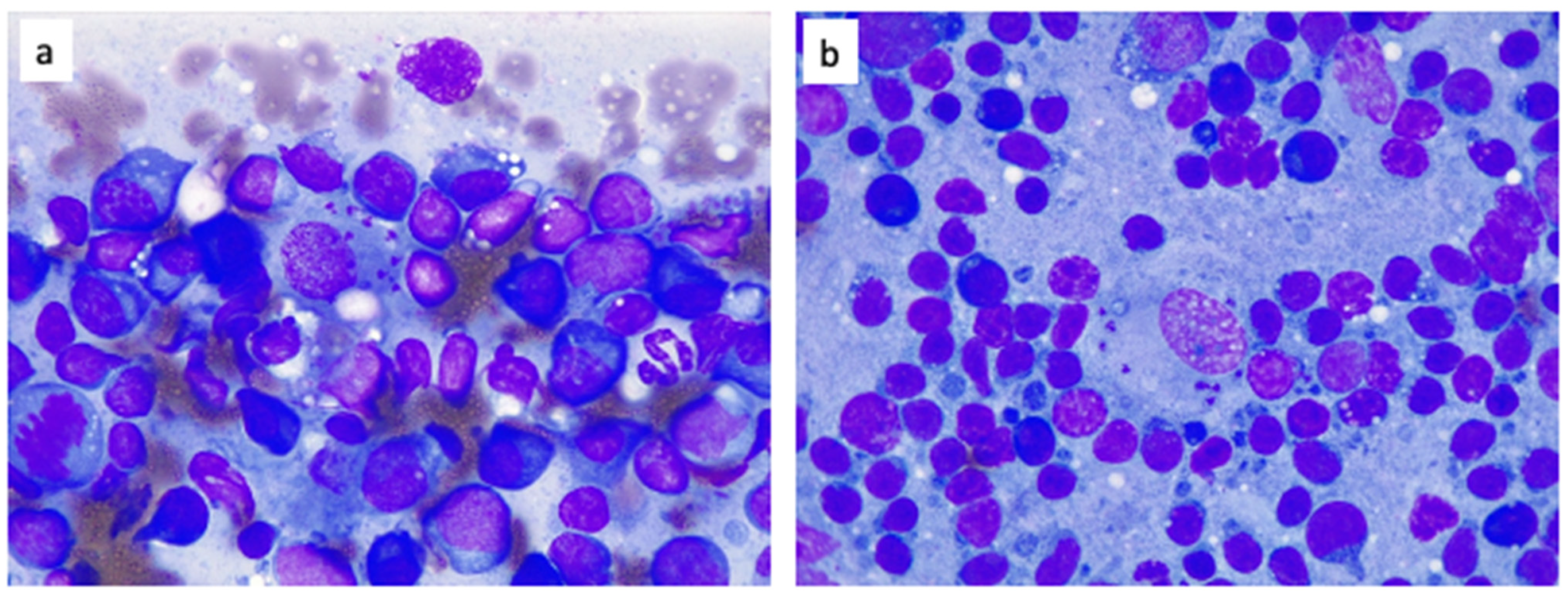
Examination of FNA from the left prescapular lymph node and both the popliteal lymph nodes showed marked lymphoplasmacellular hyperplasia along with many macrophages phagocyting bodies compatible with amastygotes of L. infantum (Figure 4).
The IFAT was markedly positive with an antibody titer of 1:1280. Serological tests for other vector borne diseases were negative. A definitive diagnosis of CanL was therefore made by combining clinical and clinico-pathological signs with results of the FNA and serological tests.

2.5. Ophthalmological Findings

An ophthalmologist consultation was also arranged. In the eye annexes, severe blepharitis was detected with multiple small nodules in the palpebral rims, palpebral oedema, hyperemic conjunctiva, and distichiasis. In both corneas, small central opacity with instability of the lacrimal film was found.

2.6. Treatment

Treatment for Leishmania infection was commenced with meglumine antimoniate (50 mg/kg once daily for the initial 10 days given subcutaneously (SC), then 50 mg/kg every 12 h for a further 20 days SC) and allopurinol (at approximately 10 mg/kg every 12 h PO). The dosage of meglumine antimoniate was initially kept low because of possible side effects. For the management of joint pain, gabapentin (about 10 mg/kg PO) was administered as follows: initially every 12 h for three weeks, then once daily for one week, and finally every other day for another week. A low gabapentin dose frequency was chosen because for a long time the dog had previously been treated with NSAIDs. In addition, an ointment with fatty acid was applied every eight hours to the eyelid of both eyes to provide relief and resolve clinical signs of blepharitis. A few days after the start of treatment, the owner was consulted by phone. Since no side effects of meglumine antimoniate were reported, the dosage was raised to the maximum allowed for the drug. In addition, the efficacy of gabapentin to reduce the pain was assessed; the dog did not need NSAIDs or other ongoing analgesia.

2.7. First Follow-Up Visit

After the definitive diagnosis of CanL was made (D0), subsequent periodic check-up visits were planned: D10, D31, and D89.
At D10, the owner reported a significant improvement in the dog’s lameness, liveliness and appetite, normal stools, and some episodes of fasting vomiting in the morning. The clinical findings were BCS of 3/9, MCS of 1/3, BW of 11.2 kg (mild decrease), slight pallor of the mucous membranes, mild hyperthermia (39.3 °C), HR of 120 bpm, RR of 35 brpm. The hyperkeratosis of nose and paw pads, onicogryphosis, alopecic skin nodule of about 1 cm at the base of the left ear pinna, and blepharitis remained unchanged. The generalized lymphadenomegaly was reduced. The swelling of the right knee joint was unchanged, but the pain was less severe than it was before.
Blood parameters were evaluated. The CBC showed a slightly worsened normocytic, normochromic, non-regenerative anemia and reduced PLT count. Peripheral blood smear revealed many RBC rouleaux, some reactive lymphocytes and activated monocytes, and confirmed an inadequate PLT number (Table 1).
The initial treatment plan with allopurinol was maintained and the dose remained unchanged, however omeprazole (about 1 mg/kg PO once daily) was added to treat occasional episodes of fasting vomiting.

2.8. Second Follow-Up Visit

At D31 (three days after the end of antimonials treatment), the owner reported a further improvement: the dog appeared more alert, playful, and no longer lame with a good appetite and normal stools.
The most striking clinical findings were an increase in BCS (5/9), MCS (2/3) and BW (12.8 kg). Temperature, HR, and RR were within reference intervals (WRI). Mucous membranes were pink. The hyperkeratosis of the nose and paw pads, onicogryphosis, blepharitis and interdigital erythema had improved. Lymph nodes remained palpable but were no longer enlarged and the right stifle joint was normal.
The complete diagnostic plan was performed to monitor blood and urinary parameters in response to the 28-day treatment with antimonials. The CBC showed an improvement in comparison to the previous blood collection (Table 1), with mild normocytic, normochromic, non-regenerative anemia and normal PLT count. The peripheral blood smear revealed that RBC rouleaux had disappeared while some reactive lymphocytes and activated monocytes were still present.
The serum biochemical profile showed an increased concentration of total proteins and C reactive protein, with a slight increase in amylase and creatine phosphokinase. The albumin concentration had returned to WRI and there was still a highly increased globulin concentration (Table 2).
The results of urinalysis were unremarkable (Table 3) and the results of coagulation profile were WRI (Table 4).
Serum protein electrophoresis revealed a moderate decrease in albumin but there was still an increase in gamma globulin fraction (Table 5, Figure 3b).
The result of L. infantum serology by IFAT was still positive with a titer of 1:640.
Allopurinol and omeprazole therapy remained unchanged. The analgesic treatment with gabapentin was continued, but was administered every other day.

2.9. Third Follow-Up Visit

At D89 (approximately two months after the end of antimonial therapy), overall the dog was doing very well, and was alert and lively. The lameness had disappeared (even without analgesic therapy) and the vomiting episodes had disappeared since the dog was now being fed three times a day.
The most striking clinical findings were an increase in MCS (3/3) and BW (14.4 kg). The temperature, HR, and RR were WRI and the BCS was still 5/9. The mucous membranes were pink, the hyperkeratosis of nose and paw pads, onychogryphosis, interdigital erythema and bilateral blepharitis had resolved, and the lymph nodes were normal. The complete diagnostic plan was repeated to monitor the blood and urinary parameters. The CBC (Table 1), urinalysis (Table 3), and serum protein electrophoresis (Table 5, Figure 3c) were all WRI, while the serum biochemical profile showed only a slight increase in glucose and urea concentrations of no clinical significance (Table 2).
The antibody titer to L. infantum by IFAT was still 1:640.
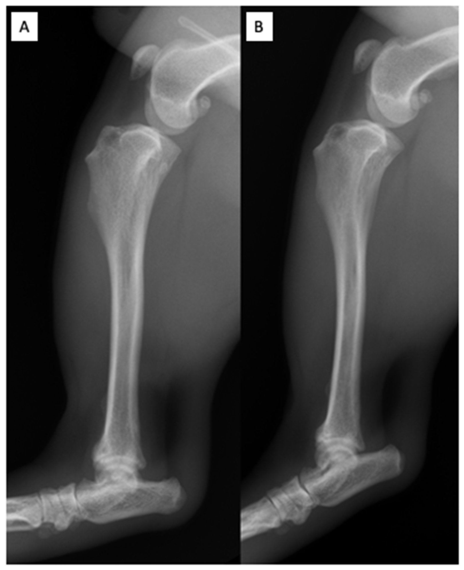
At this time, a radiological investigation of the bone pathologies was arranged (Figure 5). Radiological findings after three months of therapy revealed complete disappearance of the bone and joint radiographic lesions previously detected, with complete regression of the skeletal involvement.
Therapeutic treatment of the dog was maintained with allopurinol alone (approximately 10 mg/kg every 12 h PO). In addition, prophylaxis with permethrin spot-on plus oral afoxolaner and milbemycin was prescribed from March to November (i.e., during the sandfly activity season). A further follow-up visit was scheduled 3 months later. However, as this case report was focused on the use of a commercial Neem oil-based product in a dog for the prevention of CanL, further follow-ups were not included.

3. Discussion

To date more than 800 sandfly species have been identified worldwide [33]. Depending on the species, they can be found in tropical or subtropical areas of the New and the Old World, including the Mediterranean basin [33]. Both male and female sandflies feed on nectar and plant-derived juices. However, females need a blood meal on a vertebrate as a source of protein for egg production. The sandfly life cycle takes place in soils rich in organic matter, and passes through distinct stages (egg, four larval stages, pupa, adult), requiring a minimum of six weeks.
The activity period of the adults, and consequently CanL transmission, is typically seasonal and nocturnal. In Europe, it spans from April to November, depending on the latitude. Warmer regions have longer breeding seasons as there are up to three complete life cycles, i.e., three sandfly generations, between May and September [33,34]. Eggs and larvae may overwinter even in cold winters, remaining for many months in the diapause state [35,36]. Most species have a nocturnal circadian activity, or are crepuscular, i.e., they have peaks of host seeking activity soon after sunset and before dawn [35,37]. During the day, adult sandflies rest in cool, dark and humid places [36,38]. Species belonging to the genera Phlebotomus and Lutzomya are responsible for the transmission of CanL in the Old World and the New World, respectively: thirty-one Phlebotomus species are vectors of L. infantum [33].
Sandflies are gradually expanding their range northwards in Europe as global warming affects their distribution and abundance. As a consequence, new areas of CanL have been reported in more northerly regions where a suitable vector is present, probably in connection with the movement of L. infantum infected dogs [39,40,41]. This case report may be of particular educational value for small animal practitioners who work in areas where CanL is not endemic or has a low endemicity as they may be unfamiliar with the clinical signs of the disease, its correct diagnostic and therapeutic approach, and the most effective preventive measures.
CanL is endemic in tropical and subtropical regions of the world, including approximately 50 countries in Europe (especially in the Mediterranean area), Africa, Asia, South and Central America [1]. A high general prevalence of CanL has been reported in Italy, with the highest prevalence rate (29.6%) in central regions, followed by southern regions and islands (28.2%), and with an increasing number of cases (21.6%) in northern areas [42].
The dog in this case report was born in Sicily where he was living up to the age of 4 months, and was then transferred to Tuscany where he had lived up to the time of the veterinary visit (1.5-year-old). Tuscany and Sicily are traditionally known as regions of central and southern Italy which are endemic and highly endemic for L. infantum infection, respectively [43,44]. It is therefore not surprising that the dog was exposed to the risk of CanL transmission. It is very likely that the dog acquired CanL in Tuscany as it had spent most of its life in this region, resulting in a much longer exposure to the risk of infected sandfly bites. Nonetheless, considering the long incubation period of CanL, the infection could also have occurred in Sicily and the possibility of a congenital infection cannot be ruled out either. In addition, dogs older than one year of age have been reported to have a higher prevalence of L. infantum infection [45].
Classically, CanL leads to a high prevalence of subclinical infections [5]. When the infection results in disease, clinical signs are not specific. The most common clinical signs include generalized lymphadenopathy, weight loss, splenomegaly, polyuria/polydipsia, fever, vomiting, diarrhea, chronic colitis, and cutaneous lesions, such as nonpruritic exfoliative dermatitis, with or without alopecia [5] and ulceration, often located on several bony prominences [2]. These are generally associated with ocular lesions, epistaxis, hyperglobulinemia, hypoalbuminemia, and mild to moderate non-regenerative anemia. However, kidney disease may be the only clinical manifestation. When it occurs, it can progress from mild proteinuria to nephrotic syndrome or end-stage renal disease, and chronic renal failure is the most common cause of death in CanL [5]. Systemic disease can have both joint and bone localization, and be characterized by proliferative bone changes [46]. In Leishmania infected dogs, joint lesions have not been reported frequently, and lameness with associated polyarthritis [47,48,49,50,51,52] or synovitis [53] have been reported even less frequently. When the joint localization coincides with polyarthritis, this has been reported as being either non-erosive or erosive [46], is associated with soft tissue swelling [46,54,55], and with a tendency for a bilateral localization [46,55]. Bone involvement has only been reported in a limited number of cases. When they were reported, lesions were mostly characterized by periosteal proliferation, which can be solid and smooth or irregular, and by an increase of the intramedullary opacity, generally occurring close to the nutrient foramen [46,55,56]. Bone involvement can also be associated with cortical and/or medullary osteolysis [47,56,57,58]. Both joint and bone lesions have been reported to mostly involve the appendicular skeleton, although localization on the pelvis and occipital bone has been described [46,55].
This report describes a case of CanL in a young (1.5-year-old) dog, which was presented for further evaluation of long-lasting lameness with osteoarticular involvement and polyostotic periosteal proliferation. CanL should therefore be considered as part of the differential diagnosis of persistent lameness that is unresponsive to routine therapy, particularly in dogs that live or travel in areas endemic for CanL. The present case report may also help small animal veterinarians in the diagnosis and treatment of joint and bone lesions that can sometimes appear in dogs with leishmaniasis.
The first objective of CanL control is to interrupt L. infantum transmission from infected to uninfected dogs. As a general rule, in endemic areas, sandfly bites are prevented by applying repellents/insecticides regularly. These are usually in the form of impregnated collars, spot-on and spray formulations which are applied during the sandflies’ activity season (from April to November). This is currently the most effective preventive strategy against CanL. Pyrethroids are commercially available in different formulations in Italy and are licensed for topical use in dogs. These include permethrin, deltamethrin, and flumethrin, which are sold in various associations with imidacloprid, fipronil, dinotefuran, or pyriproxyfen [59]. A number of studies have assessed the efficacy of pyrethroids to prevent sandfly bites, thus reducing the risk of CanL transmission [60,61,62,63,64]. Recently, the insecticidal efficacy of a single oral administration of fluralaner against P. perniciosus has been reported [65].
An immune prevention strategy has also been investigated, and four CanL vaccines have been commercialized. Their reported efficacy in the prevention of active infection ranges from 68.4% to 80%, and the protection against clinical disease varies from 92.7% to 95%. This means that the use of these vaccines reduces the appearance and severity of clinical signs; however, the level of protection conferred by vaccination is not satisfactory [66]. Moreover, commercially approved vaccines can serologically mask dogs that have been infected after vaccination and in any case every single infected dog represents a potential source of L. infantum infection to other dogs and to humans.
To date, the most effective way to control CanL consists of the simultaneous application of topical pyrethroids combined with a vaccination that does not interfere with the most widely used serological diagnostic tests [66]. Additional measures to reduce CanL transmission include keeping dogs indoors from dusk to dawn during the whole sandfly activity season [5], the use of long-lasting insecticidal sprays within houses and animal shelters [67], the protection of doors and windows by nets with a small mesh (2–3 mm) [68], and the use of insecticide-treated mosquito-nets [67]. Furthermore, reducing microhabitats favorable to sandflies, such as piles of wood and stones, is also recommended in the vicinity of houses and in other locations where dogs spend time [5].
A report has been published detailing 47 populations of sandflies with confirmed resistance and 28 populations considered tolerant to one or more insecticides worldwide, including DDT in addition to dieldrin, methoxychlor, propoxur, lambdacyhalothrin, malathion, benzene hexachloride, permethrin, deltamethrin, and bendiocarb [69]. With the spread of insecticide resistance, managing the risk of CanL transmission by synthetic repellents and insecticides will become increasingly problematic.
There is therefore increasing interest in finding new, safe, and effective insecticides. A possible approach consists of focusing on the repellent and insecticide effects of plant-derived products to control the risk of sandfly bites. Some researchers have reported that Neem oil has repellent activity against sandflies [26,27,28,29,30]. Moreover, results of a survey on best practices and guideline awareness for diagnosis, treatment, follow-up, and prevention of CanL among 456 Italian veterinary practitioners reported that some practitioners (1.7%) exclusively prescribe natural products such as Neem oil for the prevention of CanL [31]. On the one hand, the results of all the above experimental and field studies uniformly show that the repellent activity of single applications of 2% and 5% Neem oil against sandflies is high. However, these results also suggest that, unfortunately, Neem oil is somewhat volatile, thus the persistence of its repellent effect is limited to a duration of a few hours. As a consequence, it can be assumed that any repellent effect exerted by a Neem oil-based product will likely be short-lived after application on dogs.
Neem-oil based products are sold at different concentrations and in different formulations (spray, spot-on, collar, shampoo, ear drops, mosquito coil) in pet shops and on the internet for the prevention or treatment of parasitic infections. All are sold with claims of efficacy, although scientifically sound data on their efficacy have never been published. There is also considerable variation in the treatment regimens for these products even though they have never been tested clinically. This suggests that the efficacy claimed is only intended to make these products appear more attractive to consumers. Consequently, although Neem oil seems to be effective in repelling sandflies [26,27,28,29,30], small animal veterinarians should not be recommending its sole use for the prevention of CanL.
Based on the main findings of this clinical case report, at present, veterinarians should only be recommending the use of Neem-oil as an adjuvant treatment in combination with other traditional repellents and insecticides that have been shown to be effective in reducing the risk of CanL transmission [58,59,60,61,62,63,64].

4. Conclusions

Consumers are using more and more herbal products as it is believed, often erroneously, that these products are more natural and therefore safer. This belief is of concern as regards the use of phytotherapeutic drugs licensed for use in veterinary medicine and commercially available as over-the-counter products.
This case report shows that owners’ awareness regarding the possible limitations of herbal therapies, especially as far as antiparasitic and repellent products, might be lacking and may often be overlooked by veterinary practitioners. This case report suggests that there may be a great need to educate owners on the reliable use of Neem oil and natural repellents, in order to improve the awareness of the limitations of their prophylactic role and the potential risks of their use for CanL prevention. It is the responsibility of veterinarians to provide dog owners with comprehensive advice regarding the use of phytotherapy and to ensure it is appropriate and effective. Currently, before commercial Neem oil-based products can be effectively used for the prevention of CanL, further studies are needed to definitely assess (prospectively or retrospectively) their protection rate and the duration of this protection against sandfly bites when applied on dogs’ coats.

Author Contributions

Conceptualization, G.D.F., G.L., S.C. and C.P.; methodology, G.D.F., G.L., S.C. and C.P.; investigation, G.D.F., G.L., S.C. and C.P.; resources, G.D.F., G.L., S.C. and C.P.; data curation, G.L.; writing—original draft preparation, R.A.P.; writing—review and editing, R.A.P.; visualization, G.L. and C.P.; supervision, G.L.; funding acquisition, R.A.P. All authors have read and agreed to the published version of the manuscript.

Funding

The publication of the study was financially supported by the budget “Fondi di Ateneo 2022” allocated by the University of Pisa and assigned to prof. Roberto Amerigo Papini.

Institutional Review Board Statement

The client-owned dog was treated by leishmanicidal drugs after informing the owner of the inherent risks. The owner consented and signed a release form to allow the treatments.

Informed Consent Statement

Informed consent was obtained from all subjects involved in the study.

Data Availability Statement

The raw data from this case report are available upon request to prof. George Lubas.

Conflicts of Interest

The authors declare no conflict of interest.

References

  1. Ayele, A.; Seyoum, Z. Review on canine leishmaniasis; etiology, clinical sign, pathogenesis, treatment and control methods. Glob. Vet. 2016, 17, 343–352. [Google Scholar] [CrossRef]
  2. Ribeiro, R.R.; Michalick, M.S.M.; Da Silva, M.E.; Dos Santos, C.C.P.; Frézard, F.; Da Silva, S.M. Canine Leishmaniasis: An Overview of the Current Status and Strategies for Control. BioMed. Res. Int. 2018, 2018, 3296893. [Google Scholar] [CrossRef] [PubMed]
  3. Bongiorno, G.; Habluetzel, A.; Khoury, C.; Maroli, M. Host preferences of phlebotomine sand flies at a hypoendemic focus of canine leishmaniasis in central Italy. Acta Trop. 2003, 88, 109–116. [Google Scholar] [CrossRef]
  4. Manzillo, V.F.; Gizzarelli, M.; Vitale, F.; Montagnaro, S.; Torina, A.; Sotera, S.; Oliva, G. Serological and entomological survey of canine leishmaniasis in Lampedusa island, Italy. BMC Vet. Res. 2018, 14, 286. [Google Scholar] [CrossRef]
  5. Solano-Gallego, L.; Miró, G.; Koutinas, A.; Cardoso, L.; Pennisi, M.G.; Ferrer, L.; Bourdeau, P.; Oliva, G.; Baneth, G. LeishVet guidelines for the practical management of canine leishmaniosis. Parasites Vectors 2011, 4, 86. [Google Scholar] [CrossRef] [Green Version]
  6. Martín-Sánchez, J.; Navarro-Mari, J.M.; Pasquau-Liaño, J.; Salomón, O.D.; Morillas-Márquez, F. Visceral leishmaniasis caused by Leishmania infantum in a Spanish patient in Argentina: What is the origin of the infection? Case report. BMC Infect. Dis. 2004, 4, 20. [Google Scholar] [CrossRef] [PubMed] [Green Version]
  7. Ben-Shimol, S.; Sagi, O.; Horev, A.; Avni, Y.S.; Ziv, M.; Riesenberg, K. Cutaneous leishmaniasis caused by Leishmania infantum in Southern Israel. Acta Parasitol. 2016, 61, 855–858. [Google Scholar] [CrossRef] [PubMed]
  8. Salomon, O.D.; Quintana, M.G.; Mastrangelo, A.V.; Fernández, M.S. Leishmaniasis and Climate Change—Case Study: Argentina. J. Trop. Med. 2012, 2012, 601242. [Google Scholar] [CrossRef] [PubMed]
  9. Kholoud, K.; Denis, S.; Lahouari, B.; El Hidan, M.A.; Souad, B. Management of Leishmaniases in the Era of Climate Change in Morocco. Int. J. Environ. Res. Public Health 2018, 15, 1542. [Google Scholar] [CrossRef] [PubMed] [Green Version]
  10. Gálvez, R.; Montoya, A.; Fontal, F.; De Murguía, L.M.; Miró, G. Controlling phlebotomine sand flies to prevent canine Leishmania infantum infection: A case of knowing your enemy. Res. Vet. Sci. 2018, 121, 94–103. [Google Scholar] [CrossRef] [PubMed]
  11. Alzohairy, M.A. Therapeutics Role ofAzadirachta indica(Neem) and Their Active Constituents in Diseases Prevention and Treatment. Evid. Based Complement. Altern. Med. 2016, 2016, 7382506. [Google Scholar] [CrossRef] [PubMed] [Green Version]
  12. Islas, J.F.; Acosta, E.; G.-Buentello, Z.; Delgado-Gallegos, J.L.; Moreno-Treviño, M.G.; Escalante, B.; Moreno-Cuevas, J.E. An overview of Neem (Azadirachta indica) and its potential impact on health. J. Funct. Foods 2020, 74, 104171. [Google Scholar] [CrossRef]
  13. Guerrini, V.; Kriticos, C. Effects of azadirachtin on Ctenocephalides felis in the dog and the cat. Vet. Parasitol. 1998, 74, 289–297. [Google Scholar] [CrossRef]
  14. Kilonzo, B.S.; Ngomuo, A.J.; Sabuni, C.A.; Mgode, G.F. Effects of Azadirachta indica (Neem) Extract on Livestock Fleas in Morogoro District, Tanzania. Int. J. Trop. Insect Sci. 2001, 21, 89–92. [Google Scholar] [CrossRef] [Green Version]
  15. Srivastava, R.; Ghosh, S.; Mandal, D.B.; Azhahianambi, P.; Singhal, P.S.; Pandey, N.N.; Swarup, D. Efficacy of Azadirachta indica extracts against Boophilus microplus. Parasitol. Res. 2008, 104, 149–153. [Google Scholar] [CrossRef] [PubMed]
  16. Webb, E.; David, M. The efficacy of neem seed extract (Azadirachta indica) to control tick infestation in Tswana, Simmentaler and Brahman cattle. South Afr. J. Anim. Sci. 2002, 32, 1–6. [Google Scholar] [CrossRef] [Green Version]
  17. Mehlhorn, H.; Walldorf, V.; Abdel-Ghaffar, F.; Al-Quraishy, S.; Al-Rasheid, K.A.S.; Mehlhorn, J. Biting and bloodsucking lice of dogs—treatment by means of a neem seed extract (MiteStop®, Wash Away Dog). Parasitol. Res. 2011, 110, 769–773. [Google Scholar] [CrossRef] [PubMed]
  18. Al-Quraishy, S.; Abdel-Ghaffar, F.; Al-Rasheid, K.A.S.; Mehlhorn, J.; Mehlhorn, H. Effects of a neem seed extract (MiteStop®) on mallophages (featherlings) of chicken: In vivo and in vitro studies. Parasitol. Res. 2011, 110, 617–622. [Google Scholar] [CrossRef] [PubMed]
  19. Valle Salazar, E.; Marín Rodriguez, C.; Morales, G. Control de piojos (Anoplura) con aceite de nim (Azadirachta indica a. Juss) en caprinos del estado Falcón, Venezuela. Arch. Latinoam. Prod. Anim. 2016, 24, 59–67. Available online: https://ojs.alpa.uy/index.php/ojs_files/article/view/2398/941 (accessed on 28 July 2022).
  20. Benelli, G.; Canale, A.; Toniolo, C.; Higuchi, A.; Murugan, K.; Pavela, R.; Nicoletti, M. Neem (Azadirachta indica): Towards the ideal insecticide? Nat. Prod. Res. 2016, 31, 369–386. [Google Scholar] [CrossRef] [PubMed]
  21. Showler, A. Botanically Based Repellent and Insecticidal Effects Against Horn Flies and Stable Flies (Diptera: Muscidae). J. Integr. Pest Manag. 2017, 8, 15. [Google Scholar] [CrossRef] [Green Version]
  22. Camarda, A.; Pugliese, N.; Bevilacqua, A.; Circella, E.; Gradoni, L.; George, D.; Sparagano, O.; Giangaspero, A. Efficacy of a novel neem oil formulation (RP03™) to control the poultry red mite Dermanyssus gallinae. Med. Vet. Èntomol. 2018, 32, 290–297. [Google Scholar] [CrossRef] [PubMed] [Green Version]
  23. Suraj, R.; Rambarran, R.; Ali, K.; Harbajan, D.; Charles, R.; Sant, C.; Georges, K.; Suepaul, S. A comparison of the efficacy of two commercial acaricides (fipronil and amitraz) with Azadirachta indica (neem) on the brown dog tick (Rhipicephalus sanguineus) from canines in Trinidad. Transbound. Emerg. Dis. 2020, 67 (Suppl. S2), 142–148. [Google Scholar] [CrossRef] [PubMed]
  24. Abdel-Ghaffar, F.; Al-Quraishy, S.; Sobhy, H.; Semmler, M. Neem seed extract shampoo, Wash Away Louse®, an effective plant agent against Sarcoptes scabiei mites infesting dogs in Egypt. Parasitol. Res. 2008, 104, 145–148. [Google Scholar] [CrossRef]
  25. Pasipanodya, C.N.; Tekedza, T.T.; Chatiza, F.P.; Gororo, E. Efficacy of neem (Azadirachta indica) aqueous fruit extracts against Sarcoptes scabiei var. suis in grower pigs. Trop. Anim. Health Prod. 2021, 53, 135. [Google Scholar] [CrossRef] [PubMed]
  26. Sharma, V.P.; Dhiman, R.C. Neem oil as a sand fly (Diptera: Psychodidae) repellent. J. Am. Mosq. Control. Assoc. 1993, 9, 364–366. Available online: https://www.biodiversitylibrary.org/content/part/JAMCA/JAMCA_V09_N3_P364-366.pdf (accessed on 28 July 2022). [PubMed]
  27. Dhiman, R.C.; Sharma, V.P. Evaluation of neem oil as sandfly, Phlebotomus papatasi (Scopoli) repellent in an Oriental sore endemic area in Rajasthan. Southeast Asian J. Trop. Med. Public Health 1994, 25, 608–610. Available online: https://www.tm.mahidol.ac.th/seameo/1994-25-3/1994-25-3-608.pdf (accessed on 28 July 2022).
  28. Srinivasan, R.; Kalyanasundaram, M. Relative efficacy of DEPA and neem oil for repellent activity against Phlebotomus papatasi, the vector of leishmaniasis. J. Commun. Dis. 2001, 33, 180–184. [Google Scholar]
  29. Marghinotti, M.; Bongiorno, G.; Bianchi, R.; Maroli, M. Evaluation of NeemAzal®, Neem oil and picaridin (KBR 3023) against the sandfly bites (Diptera: Psychodidae). Parassitologia 2004, 45 (Suppl. S1), 92. [Google Scholar]
  30. Kebede, Y.; Gebre-Michael, T.; Balkew, M. Laboratory and field evaluation of neem (Azadirachta indica A. Juss) and Chinaberry (Melia azedarach L.) oils as repellents against Phlebotomus orientalis and P. bergeroti (Diptera: Psychodidae) in Ethiopia. Acta Trop. 2010, 113, 145–150. [Google Scholar] [CrossRef]
  31. Vismarra, A.; Kramer, L.H.; Moschi, A.; Ciuca, L.; Genchi, M. A survey on canine leishmaniosis: Best practice and guideline awareness among Italian veterinary practitioners. Prev. Vet. Med. 2021, 195, 105450. [Google Scholar] [CrossRef] [PubMed]
  32. World Organization for Animal Health. Leishmaniosis. In Manual of Diagnostic Tests and Vaccines for Terrestrial Animals 2021; World Organization for Animal Health: Paris, France, 2021; Available online: https://www.woah.org/fileadmin/Home/eng/Health_standards/tahm/3.01.11_LEISHMANIOSIS.pdf (accessed on 28 July 2022).
  33. Akhoundi, M.; Kuhls, K.; Cannet, A.; Votypka, J.; Marty, P.; Delaunay, P.; Sereno, D. A Historical Overview of the Classification, Evolution, and Dispersion of Leishmania Parasites and Sandflies. PLoS Neglected Trop. Dis. 2016, 10, e0004349. [Google Scholar] [CrossRef]
  34. Alten, B.; Maia, C.; Afonso, M.O.; Campino, L.; Jiménez, M.; González, E.; Molina, R.; Bañuls, A.L.; Prudhomme, J.; Vergnes, B.; et al. Seasonal Dynamics of Phlebotomine Sand Fly Species Proven Vectors of Mediterranean Leishmaniasis Caused by Leishmania infantum. PLoS Neglected Trop. Dis. 2016, 10, e0004458. [Google Scholar] [CrossRef] [PubMed]
  35. Lewis, D.J. Phlebotomid sandflies. Bull. World Health Organ. 1971, 44, 535–551. Available online: https://www.ncbi.nlm.nih.gov/pmc/articles/PMC2427821/pdf/bullwho00201-0064.pdf (accessed on 28 July 2022). [PubMed]
  36. Alexander, B. Sampling methods for phlebotomine sandflies. Med. Vet. Èntomol. 2000, 14, 109–122. [Google Scholar] [CrossRef] [PubMed]
  37. Kravchenko, V.; Wasserberg, G.; Warburg, A. Bionomics of phlebotomine sandflies in the Galilee focus of cutaneous leishmaniasis in northern Israel. Med. Vet. Èntomol. 2004, 18, 418–428. [Google Scholar] [CrossRef] [PubMed]
  38. Alten, B.; Ozbel, Y.; Ergunay, K.; Kasap, O.; Cull, B.; Antoniou, M.; Velo, E.; Prudhomme, J.; Molina, R.; Bañuls, A.-L.; et al. Sampling strategies for phlebotomine sand flies (Diptera: Psychodidae) in Europe. Bull. Èntomol. Res. 2015, 105, 664–678. [Google Scholar] [CrossRef] [PubMed]
  39. Naucke, T.J.; Menn, B.; Massberg, D.; Lorentz, S. Sandflies and leishmaniasis in Germany. Parasitol. Res. 2008, 103, 65–68. [Google Scholar] [CrossRef]
  40. Tánczos, B.; Balogh, N.; Király, L.; Biksi, I.; Szeredi, L.; Gyurkovsky, M.; Scalone, A.; Fiorentino, E.; Gramiccia, M.; Farkas, R. First Record of Autochthonous Canine Leishmaniasis in Hungary. Vector Borne Zoonotic Dis. 2012, 12, 588–594. [Google Scholar] [CrossRef] [Green Version]
  41. Morosetti, G.; Toson, M.; Trevisiol, K.; Idrizi, I.; Natale, A.; Lucchese, L.; Michelutti, A.; Ceschi, P.; Lorenzi, G.; Piffer, C.; et al. Canine leishmaniosis in the Italian northeastern Alps: A survey to assess serological prevalence in dogs and distribution of phlebotomine sand flies in the Autonomous Province of Bolzano—South Tyrol, Italy. Vet. Parasitol. Reg. Stud. Rep. 2020, 21, 100432. [Google Scholar] [CrossRef] [PubMed]
  42. Mendoza-Roldan, J.; Benelli, G.; Panarese, R.; Iatta, R.; Furlanello, T.; Beugnet, F.; Zatelli, A.; Otranto, D. Leishmania infantum and Dirofilaria immitis infections in Italy, 2009–2019: Changing distribution patterns. Parasites Vectors 2020, 13, 193. [Google Scholar] [CrossRef] [PubMed] [Green Version]
  43. Gradoni, L.; Bettini, S.; Pozio, E. Leishmaniasis in Tuscany (Italy): VI. Canine leishmaniasis in the focus of Monte Argentario (Grosseto). Acta Trop. 1981, 38, 383–393. [Google Scholar] [CrossRef]
  44. Orndorff, G.R.; Cooper, B.A.; Smith, W.; Ryan, J.R. Canine Visceral Leishmaniasis in Sicily. Mil. Med. 2000, 165, 29–32. [Google Scholar] [CrossRef] [Green Version]
  45. Shang, L.-M.; Peng, W.-P.; Jin, H.-T.; Xu, D.; Zhong, N.-N.; Wang, W.-L.; Wu, Y.-X.; Liu, Q. The prevalence of canine Leishmania infantum infection in Sichuan Province, southwestern China detected by real time PCR. Parasites Vectors 2011, 4, 173. [Google Scholar] [CrossRef] [PubMed] [Green Version]
  46. Agut, A.; Corzo, N.; Murciano, J.; Laredo, F.; Soler, M. Clinical and radiographic study of bone and joint lesions in 26 dogs with leishmaniasis. Vet. Rec. 2003, 153, 648–652. [Google Scholar] [CrossRef]
  47. Spreng, D. Leishmanial polyarthritis in two dogs. J. Small Anim. Pract. 1993, 34, 559–563. [Google Scholar] [CrossRef]
  48. Wolschrijn, C.F.; Meyer, H.P.; Hazewinkel, H.A.W.; Wolvekamp, W.T.C. Destructive polyarthritis in a dog with leishmaniasis. J. Small Anim. Pract. 1996, 37, 601–603. [Google Scholar] [CrossRef] [PubMed]
  49. Koutinas, A.F.; Polizopoulou, Z.; Saridomichelakis, M.N.; Argyriadis, D.; Fytianou, A.; Plevraki, K.G. Clinical considerations on canine visceral leishmaniasis in Greece: A retrospective study of 158 cases (1989–1996). J. Am. Anim. Hosp. Assoc. 1999, 35, 376–383. [Google Scholar] [CrossRef] [PubMed]
  50. McConkey, S.E.; López, A.; Shaw, D.; Calder, J. Leishmanial polyarthritis in a dog. Can. Vet. J. 2002, 43, 607–609. Available online: https://www.ncbi.nlm.nih.gov/pmc/articles/PMC339396/pdf/20020800s00025p607.pdf (accessed on 28 July 2022). [PubMed]
  51. Santos, M.; Marcos, R.; Assunção, M.; Matos, A.J. Polyarthritis associated with visceral leishmaniasis in a juvenile dog. Vet. Parasitol. 2006, 141, 340–344. [Google Scholar] [CrossRef] [PubMed]
  52. Sbrana, S.; Marchetti, V.; Mancianti, F.; Guidi, G.; Bennett, D. Retrospective study of 14 cases of canine arthritis secondary to Leishmania infection. J. Small Anim. Pract. 2014, 55, 309–313. [Google Scholar] [CrossRef]
  53. Yamaguchi, R.A.; French, T.W.; Simpson, C.F.; Harvey, J.W. Leishmania donovani in the synovial fluid of a dog with visceral leishmaniasis. J. Am. Anim. Hosp. Assoc. 1983, 19, 723–726. [Google Scholar]
  54. Söffler, C.; Winkels, P.; Hess, M.; Engelhardt, P.; Wallborn, F. Leishmania-infantum-bedingte Knochenläsionen bei einem Hund. Tierarz. Praxis Klient. 2016, 44, 278–282. [Google Scholar] [CrossRef] [PubMed]
  55. Turrel, J.M.; Pool, R.R. Bone lesions in four dogs with visceral leishmaniasis. Vet. Radiol. 1982, 23, 243–249. [Google Scholar] [CrossRef]
  56. Buracco, P.; Abate, O.; Guglielmino, R.; Morello, E. Osteomyelitis and arthrosynovitis associated with Leishmania donovani infection in a dog. J. Small Anim. Pract. 1997, 38, 29–30. [Google Scholar] [CrossRef]
  57. Franch, J.; Lafuente, P.; Diaz-Bertrana, M.C.; Munilla, A.; Durall, I.; Pastor, J.; Torrent, E. Management of leishmanial osteolytic lesions in a hypothyroid dog by partial tarsal arthrodesis. Vet. Rec. 2004, 155, 559–562. [Google Scholar] [CrossRef] [PubMed]
  58. De Souza, A.I.; Juliano, R.S.; Gomes, T.S.; Diniz, S.D.A.; Borges, M.; Tafuri, W.L.; Santos, R.D.L. Osteolytic osteomyelitis associated with visceral leishmaniasis in a dog. Vet. Parasitol. 2005, 129, 51–54. [Google Scholar] [CrossRef] [PubMed]
  59. Fondati, A.; Gradoni, L.; Lubas, G.; Paltrinieri, S.; Roura, X.; Zatelli, A.; Zini, E.; Maroli, M. Prevenzione della leishmaniosi canina: Cosa è utile sapere prima di raccomandare un prodotto. Veterinaria 2018, 32, 101–112. Available online: https://veterinaria.scivac.org/2018/year-32-n-2-april-2018/prevenzione-della-leishmaniosi-canina-cosa-e-utile-sapere-prima-di-raccomandare-un-prodotto-topico-attivo-contro-la-puntura-dei-flebotomi.html (accessed on 28 July 2022).
  60. Maroli, M.; Mizzoni, V.; Siragusa, C.; D’Orazi, A.; Gradoni, L. Evidence for an impact on the incidence of canine leishmaniasis by the mass use of deltamethrin-impregnated dog collars in southern Italy. Med. Vet. Èntomol. 2001, 15, 358–363. [Google Scholar] [CrossRef]
  61. Brianti, E.; Gaglio, G.; Napoli, E.; Falsone, L.; Prudente, C.; Basano, F.S.; Latrofa, M.S.; Tarallo, V.D.; Dantas-Torres, F.; Capelli, G.; et al. Efficacy of a slow-release imidacloprid (10%)/flumethrin (4.5%) collar for the prevention of canine leishmaniosis. Parasites Vectors 2014, 7, 327. [Google Scholar] [CrossRef] [Green Version]
  62. Coura-Vital, W.; Leal, G.G.D.A.; Marques, L.A.; Pinheiro, A.D.C.; Carneiro, M.; Reis, A.B. Effectiveness of deltamethrin-impregnated dog collars on the incidence of canine infection by Leishmania infantum: A large scale intervention study in an endemic area in Brazil. PLoS ONE 2018, 13, e0208613. [Google Scholar] [CrossRef] [Green Version]
  63. Lopes, E.G.; Sevá, A.P.; Ferreira, F.; Nunes, C.M.; Keid, L.B.; Hiramoto, R.M.; Ferreira, H.L.; Oliveira, T.M.F.S.; Ovallos, F.G.; Galati, E.A.B.; et al. Vaccine effectiveness and use of collar impregnated with insecticide for reducing incidence of Leishmania infection in dogs in an endemic region for visceral leishmaniasis, in Brazil. Epidemiol. Infect. 2018, 146, 401–406. [Google Scholar] [CrossRef] [PubMed] [Green Version]
  64. Courtenay, O.; Dilger, E.; Calvo-Bado, L.A.; Kravar-Garde, L.; Carter, V.; Bell, M.J.; Alves, G.B.; Goncalves, R.; Makhdoomi, M.M.; González, M.A.; et al. Sand fly synthetic sex-aggregation pheromone co-located with insecticide reduces the incidence of infection in the canine reservoir of visceral leishmaniasis: A stratified cluster randomised trial. PLoS Negl. Trop. Dis. 2019, 13, e0007767. [Google Scholar] [CrossRef] [PubMed]
  65. Bongiorno, G.; Meyer, L.; Evans, A.; Lekouch, N.; Doherty, P.; Chiummo, R.; Gradoni, L. Insecticidal efficacy against Phlebotomus perniciosus in dogs treated orally with fluralaner in two different parallel-group, negative-control, random and masked trials. Parasites Vectors 2022, 15, 18. [Google Scholar] [CrossRef]
  66. Velez, R.; Gállego, M. Commercially approved vaccines for canine leishmaniosis: A review of available data on their safety and efficacy. Trop. Med. Int. Health 2020, 25, 540–557. [Google Scholar] [CrossRef] [Green Version]
  67. Alexander, B.; Maroli, M. Control of phlebotomine sandflies. Med. Vet. Èntomol. 2003, 17, 1–18. [Google Scholar] [CrossRef]
  68. Maroli, M.; Gradoni, L.; Oliva, G.; Castagnaro, M.; Crotti, A.; Lubas, G.; Paltrinieri, S.; Roura, X.; Zini, E.; Zatelli, A. Guidelines for prevention of leishmaniasis in dogs. J. Am. Vet. Med. Assoc. 2010, 236, 1200–1206. [Google Scholar] [CrossRef] [PubMed]
  69. Rocha, D.D.A.; da Costa, L.M.; Pessoa, G.D.C.; Obara, M.T. Methods for detecting insecticide resistance in sand flies: A systematic review. Acta Trop. 2020, 213, 105747. [Google Scholar] [CrossRef]
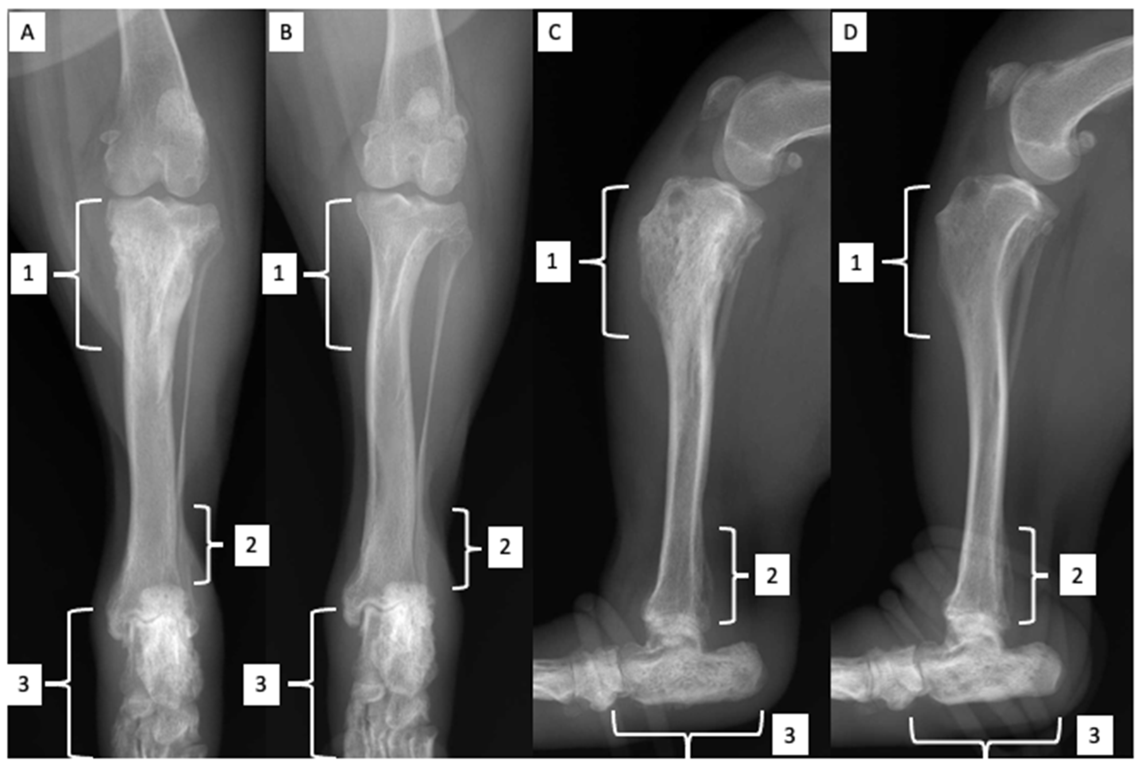
Figure 1. Caudocranial (A,B) and mediolateral (C,D) radiographs of the right (A,C) and left (B,D) crus, including the stifle joint and the tarsus. Note an irregular and continuous periosteal reaction in the proximal (1) and distal (2) metaphysis of the tibia, more evident on the right, and in tarsus bones, mostly calcaneus (3). Joint effusion and periarticular swelling of the stifle and tarsus can also be seen (3).
Zoonoticdis 02 00010 g001
Figure 2. Mediolateral radiographs of right (A) and left (B) antebrachium, including the elbow joint and the carpus. Note a mild increase and inhomogeneity of the medullary radiopacity in the proximal ulnar metaphysis and olecranon (1).
Zoonoticdis 02 00010 g002
Figure 3. Serum protein electrophoresis graph. From left to right results are presented starting at the time of the diagnosis of CanL (D0) (a), at an intermediate point after the first course of antimonials (D31) (b) to the last check-up (D89) (c).
Zoonoticdis 02 00010 g003
Figure 4. Right popliteal lymph node. Marked lymphoplasmacellular hyperplasia along with many macrophages phagocyting bodies compatible with amastygotes of L. infantum. Left, ×1000 magnification (a); Right, ×400 magnification (b) (May Grundwald Giemsa stain).
Zoonoticdis 02 00010 g004
Figure 5. Mediolateral radiographs of right (A) and left (B) crus, including the stifle joint and the tarsus. A complete disappearance of the bone and articular radiographic lesions previously described can be seen.
Zoonoticdis 02 00010 g005
Table 1. Complete blood count. Results are presented starting from the time of the diagnosis of CanL to the last check-up.
Parameters & UnitsReference IntervalD0 *D10 ^D31D89
RBC M/μL5.65–8.874.654.314.806.05
HCT %37.3–61.731.428.932.843.0
HGB g/dL13.1–20.511.210.511.815.4
MCV fL61.6–73.567.067.168.371.1
MCH pg21.2–25.923.924.424.625.5
MCHC g/dL32.0–37.935.736.336.635.8
RDW %13.6–21.714.715.216.314.3
Retics K/μL10.0–110.027.724.638.475.0
Retic-HGB pg22.3–29.626.925.925.825.7
WBC K/μL5.05–16.7614.7411.3510.7212.70
NEU seg K/μL3.7–11.911.29.317.298.38
NEU band K/μL0.0–0.30.00.00.00.0
EOS K/μL0.1–1.351.030.230.640.76
BAS K/μL0.0–0.10.00.00.110.0
LYM K/μL0.7–5.11.180.912.043.18
MON K/μL0.2–1.51.330.910.640.38
PLT K/μL148–484213144192145
MPV fL8.7–13.212.913.012.714.4
PDW fL9.1–19.414.216.416.219.6
PCT %0.14–0.460.270.190.240.21
PLT estimateadequateadequateinadequateadequateadequate
Notes on blood smear evaluation
RBC rouleauxAbsent++++++AbsentAbsent
PolychromasiaAbsent/+AbsentAbsentAbsent+
Reactive LYMAbsent++++++++Absent
Activated MONAbsent++++Absent
Toxic NEUAbsent+AbsentAbsentAbsent
* D0: day of CanL diagnosis, ^ D10, D31, and D89: days after D0; marks + to +++ indicate the estimated amount of alterations observed by an experienced clinical pathologist; italics and bold indicate values outside the reference intervals.
Table 2. Serum biochemical profile. Results are presented starting from the time of the diagnosis of CanL to the last check-up.
Parameters & UnitsReference IntervalD0 *D31 ^D89
Creatin phosphokinase IU/L40–185 236
Lactate dehydrogenase IU/L20–160 49
Aspartate transaminase IU/L15–40 60
Alanine transaminase IU/L20–70313746
Alkaline phosphatase IU/L45–25011111595
Gamma glutamyl transferase IU/L2–110.81.51.6
Amylase IU/L400–1500 1673
Bilirubin Total mg/dL0.07–0.3 0.10
Glucose mg/dL80–125102115129
Cholesterol mg/dL120–280134181253
Triglyceride mg/dL25–90 67
Urea mg/dL15–55403757
Creatinine mg/dL0.6–1.51.11.10.9
Total Protein g/dL5.8–7.810.88.47.1
Albumin g/dL2.6–4.12.12.64.0
Globulin g/dL2.5–4.58.75.83.1
A/G ratio0.6–1.30.240.451.3
Fructosamine μmol/L170–430 255
C-reactive protein mg/L0.0–0.32.00.4
Calcium mg/dL8.7–11.210.911.211.6
Phosphate mg/dL2.5–5.0 5.74.8
Na mEq/L146–156142146
K mEq/L3.9–5.54.34.0
Na/K ratio26.5–403337
Cl mEq/L10–122117108
HCO3 mEq/L21–312525
Anion Gap mEq/L12–244.317
Mg mg/dL1.6–2.72.01.7
Iron mcg/dL80–190 132
Serum aspectClearClearClearMild lipemia & hemolysis
* D0: day of CanL diagnosis; ^ D31 and D89: days after D0; italics and bold indicate values outside the reference intervals.
Table 3. Urinalysis. Results are presented starting from the time of the diagnosis of CanL to the last check-up.
Parameters & UnitsReference IntervalD0 *D31 ^D89
Sampling Free catchFree catchFree catch
ColorYellowDark yellowYellowLight yellow
AspectClearClearCloudyClear
Specific gravity1015–1045104910381058
pH5.0–7.57.06.05.0
GlucoseNeg.Neg.Neg.Neg.
KetonesNeg.Neg.Neg.Neg.
HGB-RBC/mcLNeg.250Neg.Neg.
Bilirubin mg/dLNeg.1Neg.Neg.
Urobilinogen mg/dLNeg.1Neg.Neg.
Protein mg/dL50100Neg.Neg.
UPC ratio< 0.50.620.060.23
Sediment
RBC/HPF0–51000
WBC/HPF0–5000
Epithelial cells/HPF0-rare011
Casts/LPF01 hyaline00
Crystals0-rare000
BacteriaAbsentAbsentAbsentAbsent
* D0: day of CanL diagnosis, ^ D31 and D89: days after D0; italics and bold indicate values outside the reference intervals.
Table 4. Citrated plasma coagulation profile. Results are presented at the time of the diagnosis of CanL and at the second follow-up visit.
Parameters & UnitsReference IntervalD0 *D31 ^
Prothrombin time s5.5–11.48.27.9
Activated partial thromboplastin time s10.6–19.916.214.8
Fibrinogen mg/dL125–335527263
D-dimer mcg/mL0.1–0.35 0.11
* D0: day of CanL diagnosis, ^ D31: days after D0.
Table 5. Serum protein electrophoresis. Results are presented starting from the time of the diagnosis of CanL to the last check-up.
Parameters & UnitsReference IntervalD0 *D31 ^D89
Albumin %51–6520.131.850.3
α1 globulin %2–64.74.04.8
α2 globulin %7–1511.76.710.4
β1 globulin %2–55.13.43.3
β2 globulin %2–92.67.08.8
β3 globulin %6–124.14.78.3
γ globulin %5–1551.742.414.1
Albumin g/dL3.0–4.22.02.73.6
α1 globulin g/dL0.2–0.30.50.30.3
α2 globulin g/dL0.6–0.81.20.60.7
β1 globulin g/dL0.2–0.30.50.30.2
β2 globulin g/dL0.3–0.40.30.60.6
β3 globulin g/dL0.5–0.74.10.40.6
γ globulin g/dL0.6–0.85.23.61.0
A/G ratio0.6–1.30.250.471.0
Total Protein g/dL5.5–7.610.18.47.1
* D0: day of CanL diagnosis; ^ D31 and D89: days after D0; italics and bold indicate values outside the reference intervals.
Publisher’s Note: MDPI stays neutral with regard to jurisdictional claims in published maps and institutional affiliations.