Article

Isolation and Characterization of Lytic Bacteriophages Specific for Campylobacter jejuni and Campylobacter coli

by
Abdul-Raouf Al-Mohammadi
1,
Gamal El-Didamony
2,
Mohamed S. Abd El Moneem
2,
Ibrahim M. Elshorbagy
3,
Ahmed Askora
2,* and
Gamal Enan
2,*
1
Department of Sciences, King Khalid Military Academy, Riyadh 11459, Saudi Arabia
2
Department of Microbiology and Botany, Faculty of Science, Zagazig University, Zagazig 44519, Egypt
3
Agriculture Research Center, Animal Health Research Institute, Cairo 12618, Egypt
*
Authors to whom correspondence should be addressed.
Zoonotic Dis. 2022, 2(2), 59-72; https://doi.org/10.3390/zoonoticdis2020007
Submission received: 24 March 2022 / Revised: 30 April 2022 / Accepted: 3 May 2022 / Published: 6 May 2022
(This article belongs to the Special Issue Food-Borne Pathogens in Livestock)

Abstract

:
In this study, two lytic bacteriophages designated as vB_CjP and vB_CcM were isolated and evaluated for their ability to combat multidrug-resistant bacteria Campylobacter jejuni and Campylobacter coli, respectively. A morphological analysis of these phages by transmission electron microscopy revealed that the vB-CjP bacteriophage had a mean head dimension of 66.6 ± 2.1 nm and a short non-contractile tail and belongs to the Podoviridae family, whereas vB_CcM had a mean head dimension of 80 ± 3.2 nm, a contractile tail, and a length calculated to be 60 ± 2.5 nm and belongs to the Myoviridae family. The results of the host range assay showed that vB_CjP could infect 5 of 10 C. jejuni isolates, whereas vB_CcM could infect 4 of 10 C. coli isolates. Both phages were thermostable and did not lose their infectivity and ability to lyse their host following exposure to 60 °C for 10 min; furthermore, phage particles were relatively stable within a pH range of 6–8. A one-step growth curve indicated that the phages produced estimated burst sizes of 110 and 120 PFU per infected cell with latent periods of 10 and 15 min, for vB-CjP and vB-CcM, respectively. The lytic activity of these phages against planktonic Campylobacter showed that these phages were able to control the growth of Campylobacter in vitro. These results suggest that these phages have a high potential for phage applications and can reduce significantly the counts of Campylobacter spp. The lytic activity of vB-CjP and vB-CcM phages at different (MOIs) against multidrug resistance Campylobacter strains was evaluated. The bacterial growth was slightly delayed by both phages, and the highest efficiency of both phages was observed when MOI = 1 was applied.

1. Introduction

Campylobacter spp. are versatile microorganisms that are recognized as food-spoilage bacteria [1]. Intestinal tracts of domestic livestock such as poultry, sheep, goat cattle, and pigs are considered the potential reservoir for Campylobacter species [2]. Campylobacter is regarded as a major food contaminant that can cause human diseases through ingestion of food that is contaminated or inadequately cooked [3]. Furthermore, water sources and direct contact with animals and pets are considered other sources for infection of humans with Campylobacter spp. [4]. In recent years, Campylobacter causes severe infections such as enteric diseases due to its ability to resist a broad range of antibiotics and its capacity to acquire a high resistance to the most effective antibiotics [5]. The misuse and overuse of antimicrobial agents in food animals leads to the overwhelming emergence of antibiotic-resistant Campylobacter, which consequently influences on the food safety and causes a considerable health concern of infections with antibiotic-resistant bacteria [6]. Among different Campylobacter spp., the most causative agents of human enteric infections are C. jejuni and C. coli [7]. Moreover, bacteria residing in biofilms are difficult to treat with antibacterial agents and are involved in the failure of the treatment [8]. C. jejuni has been found in preformed biofilms of other bacterial species [9]. Additionally, they can effectively attach to the surface and form a biofilm, and their aggregation increases their resistance to environmental stress and the most commonly used antibiotics [10]. Food can be treated with different bactericidal means such as sodium hypochlorite, which is used at slaughterhouses for decontamination of chicken carcass, and food such as meat can be treated with heat in order to control Campylobacter sp. [11]. Unfortunately, Campylobacter has developed resistance to these treatments, and there has been insufficient evidence to limit their viability [12]. Additionally, the emerging public health threat of bacterial antimicrobial resistance (AMR) has promoted the re-examination of using bacteriophages as an alternative to control multidrug-resistant (MDR) bacteria [13,14]. Bacteriophages are viruses that specifically infect and kill bacteria [15]. Phages have many advantages over the traditional antimicrobials; they selectively kill the pathogenic bacteria without deteriorating the beneficial normal flora, and they are able to destroy bacterial biofilms that play a significant role in the persistence and virulence of pathogenic bacteria [16]. The quest for new phages is urgent in overcoming the increasing mortalities caused by MDR bacteria [17]. The need for an additional tool for ensuring food safety and management of bacterial contamination is urgent [18,19,20,21]. Phages offer promising alternative antimicrobials, and the research on them has been renewed since the emergence of AMR [22]. The effectiveness of Campylobacter phages against Campylobacter in different food sources such as broiler chicken and chicken skin was reported in several studies [22]. Therefore, the aim of this study was to isolate, characterize, and evaluate the lytic activity of bacteriophages specific to multidrug-resistant C. jejuni and C. coli in order to be used in various ready-to-eat foods.

2. Materials and Methods

2.1. Bacterial Strains and Growth Conditions

Different C. jejuni and C. coli strains were originally isolated from different retail poultry meat samples and human stool samples, as follow: chicken samples (n = 400): (feces = 50, cecal part = 100, cloaca swabs = 50, fresh gizzard = 50, fresh liver = 50, frozen gizzard = 50, and frozen liver = 50), duck samples (n = 151): (feces = 30, cecal part = 25, cloacal swabs = 24, fresh gizzard = 18, fresh liver = 17, frozen gizzard = 17, and frozen liver = 20), and human stool samples (n= 100): (children diarrhea = 50 and adult diarrhea = 50). All samples were taken using sterile stick swab. For isolation of Campylobacter, 25 g of each samples was homogenized in 225 mL of the Preston enrichment broth medium; after that, 0.1 mL of the enriched samples was streaked onto the a modified cefoperazone charcoal deoxycholate agar (mCCDA) (Oxoid), and then, the plates were incubated under microaerophilic conditions using a gas jar containing Campylobacter gas pack systems and maintained at 42 °C for 48 h according to [23,24]. After incubation, suspected colonies of Campylobacter were tested using Gram staining, a catalase reaction, the oxidase test, the motility test, hipuurate hydrolysis, blood hemolysis, growth at 25 °C and 42 °C, and resistance to nalidixic acid (30 μg) and cephaolthin (30 μg). Positive isolates were Gram-negative, S-shaped, and motile with darting screw type motility and showed oxidase and catalase positive reactions. Campylobacter isolates were positive for hippurate hydrolysis by showing a deep blue color. The presumptive Campylobacter colonies were preserved in FBP medium broth [25] supplemented with 15% (v/v) glycerol 0.1% (w/v) yeast extract and stored at −20 °C for further analysis.

2.2. PCR Analysis for Detection of Virulence Genes mapA and ceuE in Campylobacter Strains

Presumptive Campylobacter strains were checked for the presence of mapA and ceuE genes by PCR assay. PCR with two sets of primers specifically designed for mapA and ceuE based on the specific sequence of C. jejuni and C. coli were used. PCR amplification was performed as described previously [26,27] using the forward primer 5′- AAT TGA AAA TTG CTC CAA CTA TG-3′ and reverse primer 5′- TGA TTT TAT TAT TTG TAG CAG CG-3′ (Midland Certified Reagent Company, Midland, TX, USA) to amplify the ceuE gene of C. coli (462 bp) and forward primer 5′- CTA TTT TAT TTT TGA GTG CTT GTG-3′ and reverse primer 5′- GCT TTA TTT GCC ATT TGT TTT ATT A-3′ (Midland Certified Reagent Company, USA) to amplify the mapA gene of C. jejuni (589 bp). The primer sequences and expected amplicon sizes are listed in Table 1. Amplification was carried out in a 25 μL reaction containing 12.5 μL of EmeraldAmp Max PCR Master Mix (Takara, Tokyo, Japan), 1 μL of each primer (20 pmol), 7.5 μL of water, and 3 μL of DNA template (100 ng), and an Applied Biosystems 2720 thermal cycler (Biometra, Göttingen, Germany). The thermal cycle conditions consisted of initial denaturation at 94 °C for 5 min followed by secondary denaturation at 94 °C for 30 s, annealing at 58 °C for 45 s and extension at 72 °C for 45 s, and with a final extension at 72 °C for 10 min. Twenty microliters of each PCR product were separated by electrophoresis at 100 V in a 1.5% agarose gel (1× TAE buffer) stained with 0.5 μg/mL ethidium bromide. The fragment sizes were determined using a Gelpilot 1000 bp Ladder (Qiagen GmbH, Hilden, Germany). The results were analyzed using associated software.

2.3. Susceptibility to Antibiotics

Antimicrobial susceptibility of isolated Campylobacter to 15 antibiotics (Oxide Ltd., Hampshire, UK)—ampicillin (10 µg), cephalothin (30 µg), cephradine (30 µg), nalidixic acid (30 µg), ciprofloxacin (5 µg), norfloxacin (10 µg), pefloxacin (5 µg), levofloxacin (5 µg), streptomycin (10 µg), gentamicin (10 µg), tobramycin (10 µg), neomycin (30 µg), tetracycline (30 µg), erythromycin (15 µg), and nitrofurantoin (300 µg)—was performed using the disk diffusion susceptibility method [28]. Briefly, overnight cultures were grown in Preston broth and adjusted to an optical density equal to 0.5 McFarland standards. Then, 0.1 mL of bacterial suspension was spread by sterile swaps on Muller Hinton agar plates (Sigma-Aldrich, Saint Louis, MO, USA). The antibiotic discs were placed on the agar plates using sterile forceps to apply the discs that were 2 cm apart from each other, and then, the plates were incubated for 16–18 h at 37 °C. After incubation, inhibition zones were visible and the diameter of the zone was measured with a ruler [28].

2.4. Isolation of Bacteriophages Specific for C. jejuni and C. Coli

Bacteriophages specific to MDR C. jejuni and C. coli were isolated from four sewage samples collected from Zagazig University Hospital, Sharkia Province, Egypt. Sewage samples were clarified through centrifugation at 6000× g for 20 min to remove debris particles, and the supernatant was filtered with a 0.45 μm-pore size membrane filter (Steradisc, Kurabo Co., Ltd. Osaka, Japan). The filtrate stocks were spotted on twenty Campylobacter isolates as indicator strains using the double agar overlay technique as described by Adams [29]. The positive and negative responses were examined.

2.5. Bacteriophage Purification and Propagation

Propagation and purification of bacteriophages obtained from single-plaque isolates were carried out as previously described [13]. Briefly, a single plaque was picked by using a sterile Pasteur pipette three times successively in order to purify phages. Then, plaques were put into 5.0 mL Preston broth containing Campylobacter and incubated at 37 °C under shaking. After incubation, the mixtures were centrifuged at 10,000× g for 20 min, the supernatants were filtrated through a sterilized 0.22 μm Millipore filter, and the purified phages were stored at 4 °C.

2.6. Morphological Characteristics (Electron Microscopy)

The morphological characters of isolated phages were shown by transmission electron microscopy (TEM) as previously described [30]. Briefly, 4 μL drops of purified phage particles (1011 PFU/mL) in SM buffer were deposited on 200 mesh carbon-coated copper grids with formvar films (Sigma-Aldrich, Saint Louis, MO, USA) and allowed to adsorb for 1 min, followed by staining with 2% phosphotungstic acid for 30 s (Sigma-Aldrich). Then, the samples were examined with a Hitachi H600A electron microscope at the Electron Microscopy Unit in the Faculty of Agriculture, Mansoura University. Pictures of phages were taken using a digital camera and digital micrograph software, and the dimensions of phage particles were measured in order to calculate the average and standard error values.

2.7. Phages Adsorption Experiment and One-Step Growth Curve

Phages lysates (1010 PFU/mL) were added to a bacterial culture of Campylobacter (1.8 × 10¹⁰ CFU/mL) at a multiplicity of infection (MOI) = 1.0 and incubated by shaking, and the samples were collected every 1 min during a total period of 20 min and then collected every 5 min for 35 min. The samples were immediately centrifuged at 10,000 rpm for 10 min, diluted, and titrated by the double-layer agar technique. After incubation, the plaques were counted, and the adsorption rate was calculated as previously described [31]. One-step growth curve was obtained as previously described [32], Briefly, the C. jejuni and C. coli strains were grown at OD = 0.2 (~108 CFU/mL) and infected with phages at an MOI of 1, and then, the mixtures were allowed to adsorb for 10 min at room temperature. After that, the mixtures were centrifuged and the obtained pellets were re-suspended in 10 mL of Preston broth medium and incubated at 37 °C. Then, 200 μL of each samples was diluted and immediately plated for phage titration in order to calculate the released phage particles from the infected bacterial cells, while chloroform was added to other sample at a concentration of 1% (v/v) to release the intracellular phages in order to determine the eclipse period.

2.8. Determination of Phage Host Range

The host range of isolated phages was investigated using the spot test technique. We tested the host range of these phages using bacterial lawns of twenty isolated Campylobacter isolates and other clinical pathogenic isolates: Listeria monocytogenes, Pseudomonas aeruginosa, Staphylococcus aureus, Escherichia coli, and Bacillus cereus.

2.9. Effect of Temperature and pH on the Stability of Phages

The stability of isolated phages at different temperatures and pH values was investigated. Each phage suspension was incubated for 10 min at different temperatures: 40 °C, 50 °C, 60 °C, 70 °C, 80 °C, 90 °C, and 100 °C. Phage survival was determined by the plaque assay technique. The sensitivity of phages to different pH values was determined by incubating the phages (vB_CjP and vB-Ccp) in Preston broth adjusted to pH 2–12 using 1 M HCl or 1 M NaOH solutions according to [32]. The mixtures were incubated overnight at 4 °C, and the activity of residual phage was evaluated by the plaque assay technique.

2.10. Bacterial Cell Lysis Assay

To evaluate the bacteriolytic efficacy of two phages (vB_CjP and vB-Ccp), Campylobacter strains were grown in Preston broth at 37 °C for 19 h. Briefly, about 0.1 mL of overnight culture was inoculated into 10 mL of fresh broth to obtain 108 CFU/mL and then phages were used to inoculate the broth at an MOI of <0.1, 0.1 and 1 and incubated under vigorous shaking, and then, the OD600 was measured at 0, 1, 3, 5, 7, 9, 12, and 24 h. All tests were performed in triplicate [33].

2.11. Statistical Analysis

In all data sets, the test and control sets were compared using Student’s t-test. A significance level of 0.05 was applied in all cases. Analytical statistics were undertaken using GraphPad PRISM version 7.00 for Windows (GraphPad Software, La Jolla, CA, USA). All experiments in this study were performed in triplicate.

3. Results

3.1. Prevalence of Campylobacter Species (C. jejuni and C. coli) in Collected Samples

Out of the total of 661 collected samples, 149 (22.5%) were positive for the two Campylobacter spp., 78 (11.8%) were positive for C. jejuni, and 71 (10.7%) were positive for C. coli. Among 400 poultry (chicken) samples tested, 55 (13.75%) and 49 (12.25%) were positive for C. jejuni and C. coli, respectively, and for the duck samples, out of 151 tested samples, 16 (10.59%) were positive for C. jejuni and 15 (9.93%) were positive for C. coli. Among 100 human feces samples, equal percentages (7%) were recorded for both species, whereas Campylobacter spp. was not detected in sewage samples (Table 1).

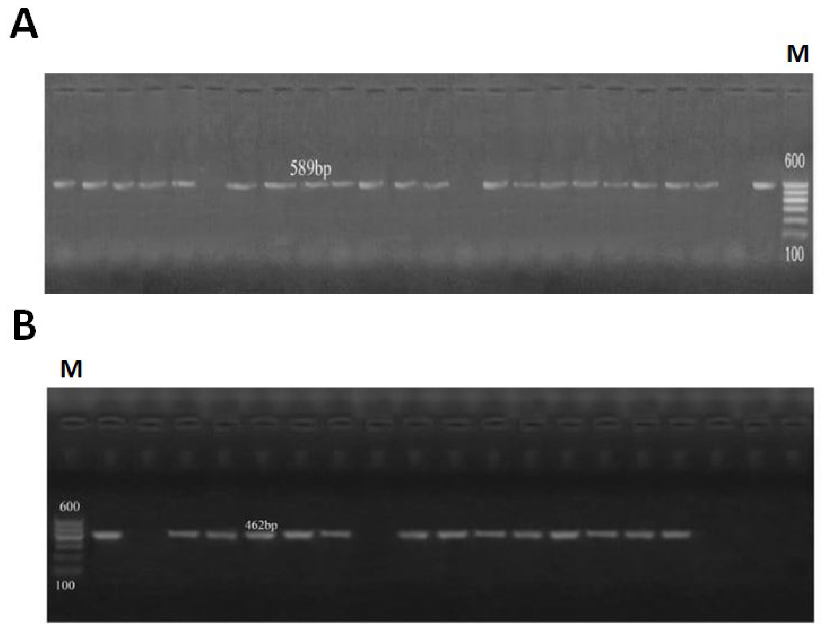
3.2. PCR Analysis for Detection of Virulence Genes mapA and ceuE

A PCR analysis for the presence of virulence genes mapA and ceuE in C. jejuni and C. coli isolates was conducted. The mapA gene was detected from 87.5% (21/24) of the C. jejuni isolates, and the ceuE gene was detected from 79.1% (19/24) of the C. coli isolates. The PCR products of each gene were identical for each isolate. For C. jejuni, it was 589 bp; for C. coli, it was about 462 bp (Figure 1).

3.3. Antimicrobial Susceptibility of C. jejuni and C. coli Isolates

The antibiotic sensitivity of both C. jejuni and C. coli isolates was tested using the disc diffusion method. C. jejuni and C. coli isolates were resistant to more than two of the tested antibiotics (Table 2). All isolates (100%) were resistant to cephalothin, cephradine, and nalidixic acid. About 45% and 25% of C. jejuni and C. coli were resistant to ciprofloxacin, respectively. Both C. jejuni (100%) and C. coli (89%) showed high levels of resistance against erythromycin. About 91% of C. jejuni isolates were resistance to ampicillin, while 82% of C. coli was resistant to it. C. jejuni and C. coli were resistant to norfloxacin (42% and 34%), pefloxacin (59% and 49%), streptomycin (60% and 39%), gentamicin (55% and 41%), tobramycin (49% and 34%), neomycin (53% and 44%), tetracycline (77% and 56%), and nitrofurantoin (33% and 25%). The highest levels of sensitivity of both C. jejuni (67%) and C. coli (85%) were observed against levofloxacin (Table 2).

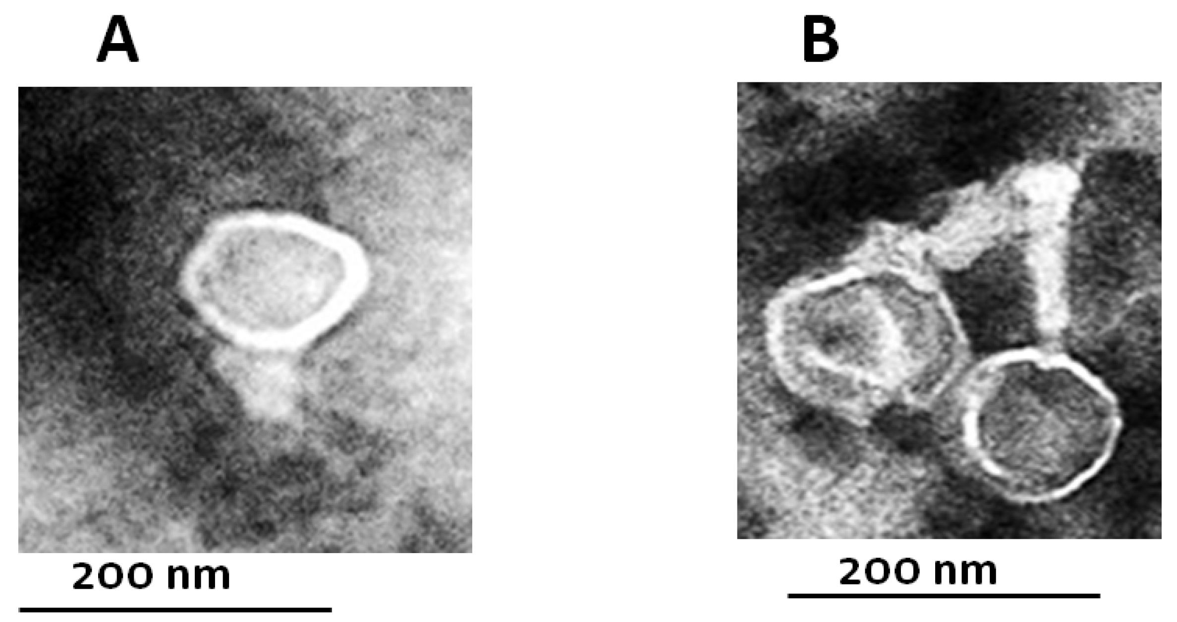
3.4. Phage Isolation and Characterization

Bacteriophages specific to C. jejuni and C. coli were isolated from enrichment culture containing C. jejuni and C. coli. Phages were detected by the spot and plaque assay method. Three different single plaques with different lysis pattern based on plaque morphology size and clarity designated vB-CjP for C. jejuni and vB-CcM for C. coli were picked up for further purification and characterization. Electron microscopy of the isolated phages particles revealed that, the vB-CjP bacteriophage had a mean head dimension of 66.6 nm ± 2.1 nm and a short non-contractile tail, and this isolated phage belongs to the Podoviridae family, whereas vB_CcM had a mean head dimension of 80 nm ± 3.2 nm, a contractile tail, and a length calculated to be 60 nm ± 2.5 nm, and this phage belongs to the Myoviridae family (Figure 2).

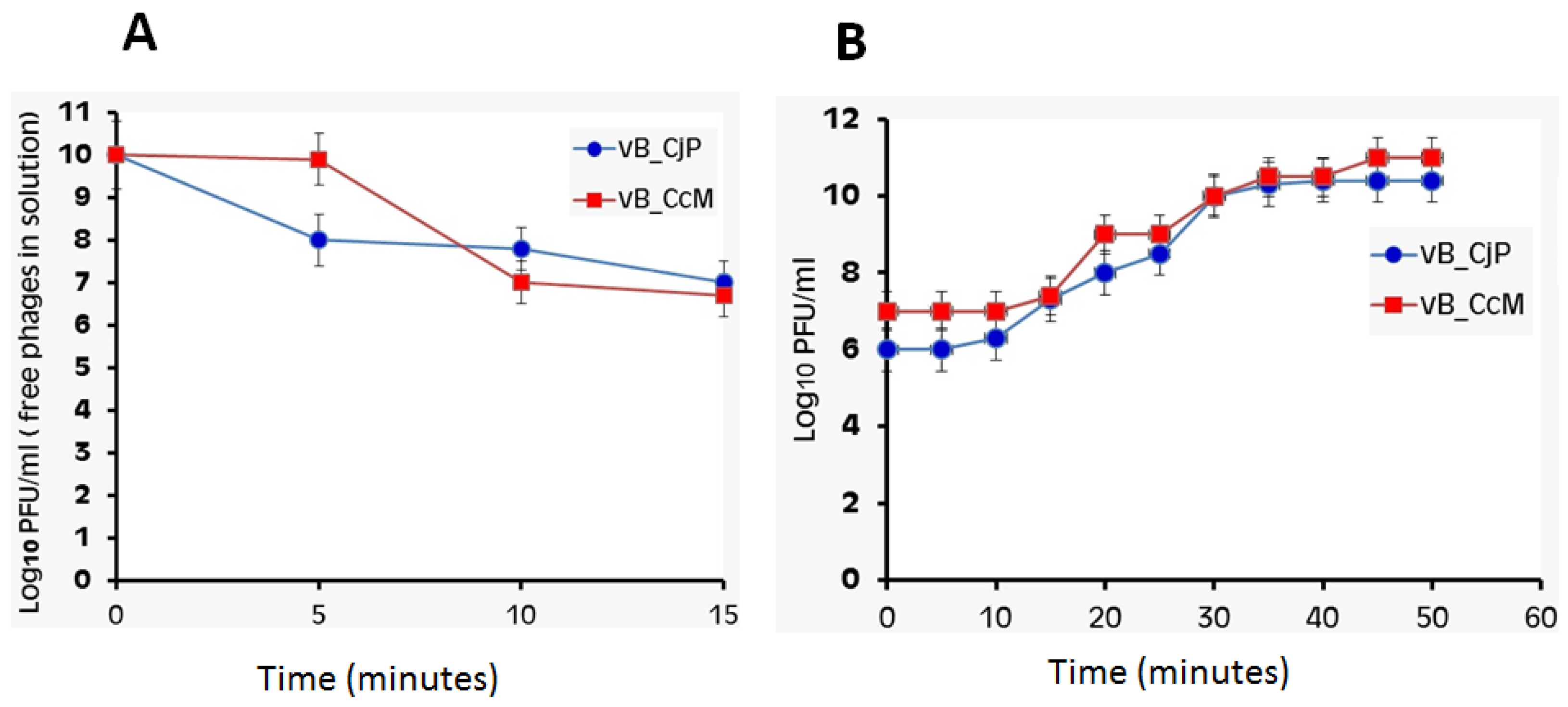
3.5. Phage Adsorption Rate and One-Step Growth Curve of Campylobacter Phages

The two isolated phages were adsorbed on bacterial cells (MOI = 1), and the phage titer decreased significantly with time until the minimum phage free in liquid medium at which the maximum adsorption times were recorded and used for calculation of adsorption rate constant was reached. The results showed that the maximum phage adsorption of vB-CjP was after 5 min with K = 7.7 × 10⁻⁹. The maximum phage adsorption of vB-CcM was after 10 min with k = 6.3 × 10⁻¹⁰ (Figure 3A). One-step growth experiment aimed to find out the following parameters: the latent period, generation time, as well as the burst size. Burst sizes and latent periods were determined for vB-CjP and vB-CcM phages. The phages produced estimated burst sizes of 110 and 120 PFU per infected cell with latent periods of 10 and 15 min for vB-CjP and vB-CcM, respectively (Figure 3B).

3.6. Host Range of Isolated Phages

The lytic ability of isolated phages was examined against the highest multidrug-resistant C. jejuni and C. coli strains and five different bacterial strains (Table 3). Phages displayed a strong lytic activity against C. jejuni and C. coli. The results showed that five strains of C. jejuni (50%) were lysed by the vB-CjP phage, while five strains (40%) of C. coli were lysed by the vB-CcM phage. The other tested bacterial strains were resistant to infection by isolated phages (Table 3).

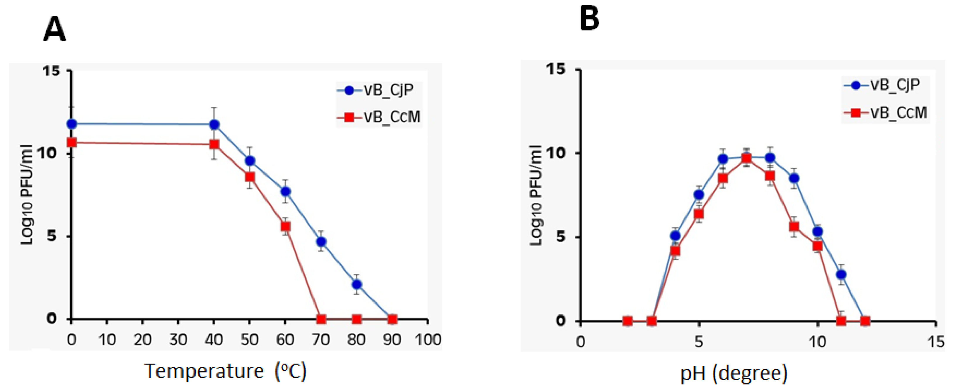
3.7. Effect of Temperature and pH on Bacteriophage Stability

The stability of the isolated two phages vB-CjP and vB_CcM at different temperatures and pH values was investigated. The results in Figure 4A show that the infectivity of all phages was not or was slightly affected by increases in temperature, especially after exposure to 60 °C. Isolated phages exhibited a survival rate reaching 50–80%, indicating that these phages are considered thermostable and did not lose their infectivity and ability to lyse Campylobacter strains following exposure to 60 °C for 10 min. Furthermore, the phage particles were relatively stable within a pH range of 6–8 (Figure 4B). All phages completely lost infectivity at pH 10 or higher and at pH 5 or lower. Campylobacter phages vB-CjP and vB_CcM displayed greater stability at pH 7.

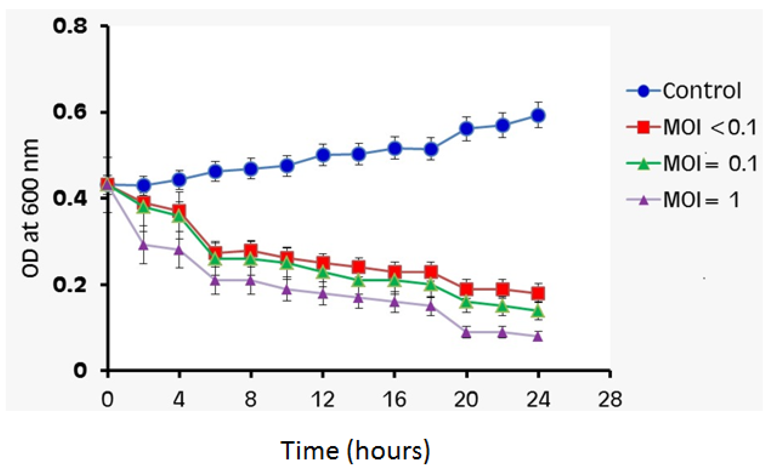
3.8. Bacterial Cell Lysis Assay

The lytic activity of isolated phages at different (MOIs) against multidrug-resistant Campylobacter strains as host strain was evaluated (Figure 5). There was a significant decrease in the growth of Campylobacter host strains infected with vB-CjP and vB_CcM at different MOIs. The efficacy of phages vB-CjP and vB_CcM against Campylobacter host strains was highest at an MOI of 1, and the OD decreased significantly from 0.4 to less than 0.1 (Figure 5).

4. Discussion

The rapid emergence of antibiotic resistance among Campylobacter spp., the most important zoonotic bacterial pathogen, is considered a major public health threat affecting the poultry industry and humans in Egypt and worldwide [34]. Therefore, improved management and prevention strategies should be introduced to control infections caused by multidrug-resistant Campylobacter spp. Several studies have proposed the use of bacteriophages as an alternative to antibiotics to combat bacterial infections in poultry and, thus, for food safety and public health [35,36,37,38]. Thus, this study aimed to evaluate the antibiotic susceptibility of Campylobacter spp. strains isolated from various sources and then to isolate and characterize bacteriophages with lytic activity against multidrug-resistant Campylobacter spp. In the present study, a total of 661 samples were taken from different sources (poultry, specifically chicken and duck, and human stool) in order to evaluate the presence of most frequently isolated species of Campylobacter. The results showed that 149 out of 661 were confirmed to be Campylobacter spp., and the prevalence of C. jejuni (11.8%) was higher than the presence of C. coli (10.7%). The highest prevalence of Campylobacter sp. was detected among the samples that were taken from chicken, followed by duck samples, and finally human stool samples. Similar results were found in several previous studies that revealed the prevalence of C. jejuni being higher than the prevalence of C. coli with different percentages [39,40], and our results indicated that poultry was the most important source for Campylobacter and that chicken carried the highest number of two Campylobacter spp. in their intestine compared with other sources [41]. The susceptibility of isolated Campylobacter spp. was further tested against 15 antibiotics, and the results showed that they were highly resistance to antibiotics. A high percentage of resistant strains were observed in the cases of cephalothin, cephradine, and nalidixic acid at a level of 100% for C. jejuni and for C. coli. On the other hand, the resistance rate of erythromycin was found to be 100% for C. jejuni and 89% for C. coli. These results were similar to those found by Nowaczek et al. (2019) [38], who have confirmed a high rate of resistance to erythromycin, ranging from 51.6% for C. jejuni to 64.7% for C. coli. Interestingly, the resistance rates of tetracycline were found to be 60% for C. jejuni and 56% for C. coli, which was close to the findings of Wozniak and Wieliczko (2011) [42], who found resistance to tetracycline in over 55% of all Campylobacter strains. In the case of other antibiotics, our results demonstrated a low rate of resistance to Ciprofloxacin ranging from 45% for C. jejuni to 25% for C. coli, which differed somewhat from the results demonstrated by Nowaczek et al. (2019) [38], who confirmed a high rate of resistance to this antibiotic, ranging from 80.6% for C. jejuni to 88.2% for C. coli. The isolated Campylobacter spp. in this study demonstrated high resistance to antibiotics routinely used in poultry production to reduce bacterial infections. The acquisition of several determinants of resistance may result in the emergence and spread of antibiotic resistance among pathogenic bacteria, and the mechanisms of genetic resistance can occur either by mutations or by acquisition of resistance conferring genes via horizontal gene transfer, which is thought to be the most important factor in antimicrobial resistance dissemination in microbial ecosystems [43]. Several studies have addressed the efficacy of bacteriophages as an alternative strategy to antibiotics for poultry and, thus, for food safety and public health. In the current study, two phages infecting multidrug-resistant C. jejuni and C. coli, named vB_CjP and vB_CcM, respectively, were isolated from four different sewage water samples obtained from Sharkia Governorate, Egypt. These phages were further characterized for morphology, phage stability, and burst size and evaluated for their lytic activity against multidrug-resistant Campylobacter strains. The isolated phages vB_CjP and vB_CcM were examined by electron microscopy for their morphological characteristics. Electron microscopy showed that the phages belong to the order Caudovirales (Podoviridae and Myoviridae), which are considered preferable for application treatments [44]. Similar results were found by Carvalho et al. (2010) [45], who isolated and characterized phages belonging to the family Myoviridae of infectious strains of C. coli and C. jejuni in chickens. Moreover, many phages that belong to different families, such as Siphoviridae [38] and Myoviridae [46], have been shown to be involved in the biocontrol of Campylobacter spp. The most common characterization of a phage is determining its lytic activity, which can help determine whether the phage should be further examined and what application of phages with broad lytic spectra are very useful for biocontrol Campylobacter spp. [35]. The host range of the isolated bacteriophages was tested against 20 Campylobacter isolates and other bacteria including L. monocytogenes, P. aeruginosa, S. aureus, E. coli, and B. cereus. Our results indicated that vB_CjP and vB_CcM phages displayed a strong lytic activity against C. jejuni, C. coli, where five strains of C. jejuni (50%) were lysed by the vB-CjP phage while four strains (40%) of C. coli were lysed by the vB-CcM phage. The other tested bacteria were resistant to infection, indicating that these phages have a specific host range only for Campylobacter strains. These results were similar to those found by Nowaczek et al. (2019) [38], who isolated four bacteriophages that showed lytic activity against 12 of the 48 Campylobacter spp. test strains. Our future research will be to expand the host range and the use of many phage mixtures to cover a wider inhibitory spectrum and to avoid the development of bacterial resistance. Different strategies have been undertaken to expand the host range of phages to combat bacterial resistance. One solution would be to engineer bacteriophages to target multiple hosts [47]. The specificity of each phage is a major challenge of bacteriophage-based therapy; therefore, targeting several bacteria in an infection at the same time at the same time would require a cocktail of bacteriophages [48]. Therefore, developing targeted and high-throughput methodologies to rapidly extend the phage host range and to overcome bacterial resistance could pave the way for next-generation phage technology. In addition, the infection dynamics of isolated phages were examined. The isolated phages exhibited different adsorption rates. Adsorption experiments for vB_CjP revealed a linear curve up 5 min, and the percentage of adsorbed phage reached 99.9%, and the adsorption rate for vB_CcM was after 10 min. Furthermore, isolated phages had growth profiles, with the latent time periods calculated to be 10 and 15 min for vB_CjP and vB_CcM, respectively, followed by a rising period that lasted for 35 min for both phages. The burst sizes were found to be 110 and 120 PFU per infected cell for vB_CjP and vB_CcM, respectively. The phages were capable of rapid and productive replication, characteristics that make them suitable for phage biocontrol applications. The determination of bacteriophage stability at various temperatures and pH is considered an important parameter for phage treatment application [49]. Our results demonstrated that phages vB_CcM and vB_CjP were thermostable and did not lose their infectivity or ability to lyse Campylobacter after exposure to 60 °C for 10 min, while their activity was completely lost at 70 and 80 °C, respectively. Similar stabilities of Campylobacter phages were reported by Steffan et al. (2021) [37], who reported that the infectivity of four Campylobacter phages CP1-4, CP1-5, CP74-2c1, and CP132-3c were stable in the range between 20 and 60 °C and that their infectivity was completely lost at 70 and 80 °C after 15 min. Furthermore, the stability of isolated phages was investigated at different pH. The phages (vB_CcM and vB_CjP) completely lost infectivity at pH 11 or higher and at pH 4 or lower, while they remained active after being exposed to pH values ranging from 5 to 10. This result was in agreement with to that of Hammerl et al. (2014) [24], which found that the Campylobacter phages CP14, CP81 and CP68 remained fully active at pH values between pH 5 and pH 9. Campylobacter phage-based biocontrol strategies have received an increasing amount of interest owing to their efficacy, practicability, safety, and specificity [35]. A number of in vitro studies have shown that bacteriophages have the potential to lyse targeted bacterial pathogens [36,37,38]. In the current research, the ability of the vB_CcM and vB_CjP phages to lyse Campylobacter spp. was characterized at different MOIs. There was a significant decrease the in viability of Campylobacter spp. infected with vB_CcM and vB_CjP compared to the untreated control. The strongest reduction in Campylobacter spp. was observed when the bacterial host was treated with an MOI = 1 of both phages. These findings were consistent with those of Loc Carrillo et al. (2005) [36], who demonstrated a significant decrease in the viability of C. jejuni infected with phages CP8 and CP34 at different MOIs (<0.1, 1, and >10). The current results are similar to those of a recent study by Steffan et al. (2021) [37], which reported that Campylobacter phages CP1-4, CP1-5, CP74-2c1, and CP132-3c were shown to be highly effective at reducing Campylobacter growth in vitro at different phage MOIs (0.001, 0.01, 0.1, 1 and 10) and suggested that these phages are promising candidates for the reduction the multidrug-resistant Campylobacter spp. This research showed promising in vitro results for phage stability and the capacity to inhibit Campylobacter spp. growth, suggesting that they could be used as biocontrol agents in treatment applications.

5. Conclusions

In conclusion, this study provides two specific lytic bacteriophages (vB_CjP and vB_CcM), and the lytic bacteriophages were isolated and evaluated for their ability to combat multidrug-resistant C. jejuni and C. coli. The use of Campylobacter bacteriophages in poultry hosts could be a potential strategy for reducing the number of Campylobacter bacteria in the food chain, which has a significant impact on human public health. Our future experiments will therefore determine the ability of phage cocktails to remove mixed C. jejuni and C. coli biofilms that occur with other normal microflora on food contact surfaces. Moreover, a focus of our future research will be to identify both the nucleotide sequence and protein of isolated phages and to ascertain the in vivo efficacy of phage mixtures.

Author Contributions

M.S.A.E.M., G.E.-D. and A.-R.A.-M. carried out the experiments and analyzed the data. G.E., I.M.E. and A.A. designed the experiments and wrote the paper. A.-R.A.-M., G.E., G.E.-D., I.M.E., M.S.A.E.M. and A.A. discussed the results and commented on the manuscript. All authors have read and agreed to the published version of the manuscript.

Funding

King Khalid Military Academy, Riyadh 11459, Saudi Arabia was responsible for paying the publication fees.

Data Availability Statement

The data presented in this study are available in the article.

Acknowledgments

The authors are indebted to Zagazig University, Egypt for the practical facilities and to King Khalid Military Academy for the financial support for the publication fees.

Conflicts of Interest

The authors declare no potential conflict of interest.

References

  1. Hlashwayo, D.F.; Sigaúque, B.; Bila, C.G. Epidemiology and antimicrobial resistance of Campylobacter spp. in animals in Sub-Saharan Africa: A systematic review. Heliyon 2020, 6, e03537. [Google Scholar] [CrossRef] [PubMed]
  2. Orhan, S.; Yaeger, M.; Zuowei, W.U.; Zhang, Q. Campylobacter-associated diseases in animals. Annu. Rev. Anim. Biosci. 2017, 5, 21–42. [Google Scholar]
  3. Sheppard, S.K.; Dallas, J.F.; Strachan, N.J.; MacRae, M.; McCarthy, N.D.; Wilson, D.J.; Gormley, F.J.; Falush, D.; Ogden, I.D.; Maiden, M.C.; et al. Campylobacter genotyping to determine the source of human infection. Clin. Infect. Dis. 2009, 48, 1072–1078. [Google Scholar]
  4. Goossens, H.; Vlaes, L.; De Boeck, M.; Levy, J.; De Mol, P.; Butzler, J.P.; Kersters, K.; Pot, B.; Vandamme, P. Is “Campylobacter upsaliensis” an unrecognised cause of human diarrhoea? Lancet 1990, 335, 584–586. [Google Scholar] [CrossRef]
  5. Isenbarger, D.W.; Hoge, C.W.; Srijan, A.; Pitarangsi, C.; Vithayasai, N.; Bodhidatta, L.; Hickey, K.W.; Cam, P.D. Comparative antibiotic resistance of diarrheal pathogens from Vietnam and Thailand, 1996–1999. Emerg. Infect. Dis. 2002, 8, 175. [Google Scholar] [CrossRef] [PubMed]
  6. Engberg, J.; Neimann, J.; Nielsen, E.M.; Aarestrup, F.M.; Fussing, V. Quinolone-resistant Campylobacter infections: Risk factors and clinical consequences. Emerg. Infect. Dis. 2004, 10, 1056. [Google Scholar] [CrossRef] [PubMed]
  7. Alam, K.; Lastovica, A.J.; le Roux, E.; Hossain, M.A.; Islam, M.N.; Sen, S.K.; Sur, G.C.; Nair, G.B.; Sack, D.A. Clinical characteristics and serotype distribution of Campylobacter jejuni and Campylobacter coli isolated from diarrhoeic patients in Dhaka, Bangladesh, and Cape Town, South Africa. Bangladesh J. Microbiol. 2006, 23, 121–124. [Google Scholar] [CrossRef] [Green Version]
  8. Hall-Stoodley, L.; Costerton, J.W.; Stoodley, P. Bacterial biofilms: From the natural environment to infectious diseases. Nat. Rev. Microbiol. 2004, 2, 95–108. [Google Scholar] [CrossRef]
  9. Trachoo, N.; Frank, J.F.; Stern, N.J. Survival of Campylobacter jejuni in biofilms isolated from chicken houses. J. Food Prot. 2002, 65, 1110–1116. [Google Scholar] [CrossRef]
  10. Joshua, G.P.; Guthrie-Irons, C.; Karlyshev, A.V.; Wren, B.W. Biofilm formation in Campylobacter jejuni. Microbiology 2006, 152, 387–396. [Google Scholar] [CrossRef] [Green Version]
  11. Sasaki, Y.; Maruyama, N.; Zou, B.; Haruna, M.; Kusukawa, M.; Murakami, M.; Asai, T.; Tsujiyama, Y.; Yamada, Y. Campylobacter cross-contamination of chicken products at an abattoir. Zoonoses Public Health 2013, 60, 134–140. [Google Scholar] [CrossRef] [PubMed]
  12. Furuta, M.; Nasu, T.; Umeki, K.; Minh, D.H.; Honjoh, K.I.; Miyamoto, T. Characterization and application of lytic bacteriophages against Campylobacter jejuni isolated from poultry in Japan. Biocontrol Sci. 2017, 22, 213–221. [Google Scholar] [CrossRef] [PubMed] [Green Version]
  13. Askoura, M.; Saed, N.; Enan, G.; Askora, A. Characterization of Polyvalent Bacteriophages Targeting Multidrug-Resistant Klebsiella pneumonia with Enhanced Anti-Biofilm Activity. Appl. Biochem. Microbiol. 2021, 57, 117–126. [Google Scholar] [CrossRef]
  14. Aslam, B.; Wang, W.; Arshad, M.I.; Khurshid, M.; Muzammil, S.; Rasool, M.H.; Nisar, M.A.; Alvi, R.F.; Aslam, M.A.; Qamar, M.U.; et al. Antibiotic resistance: A rundown of a global crisis. Infect. Drug Resist. 2018, 11, 1645. [Google Scholar] [CrossRef] [PubMed] [Green Version]
  15. Clokie, M.R.; Millard, A.D.; Letarov, A.V.; Heaphy, S. Phages in nature. Bacteriophage 2011, 1, 31–45. [Google Scholar] [CrossRef] [Green Version]
  16. Bolocan, A.S.; Upadrasta, A.; de Almeida Bettio, P.H.; Clooney, A.G.; Draper, L.A.; Ross, R.P.; Hill, C. Evaluation of phage therapy in the context of Enterococcus faecalis and its associated diseases. Viruses 2019, 11, 366. [Google Scholar] [CrossRef] [Green Version]
  17. Cassini, A.; Högberg, L.D.; Plachouras, D.; Quattrocchi, A.; Hoxha, A.; Simonsen, G.S.; Colomb-Cotinat, M.; Kretzschmar, M.E.; Devleesschauwer, B.; Cecchini, M.; et al. Attributable deaths and disability-adjusted life-years caused by infections with antibiotic-resistant bacteria in the EU and the European Economic Area in 2015: A population-level modelling analysis. Lancet Infect. Dis. 2019, 19, 56–66. [Google Scholar] [CrossRef] [Green Version]
  18. Sitohy, M.; Al-Mohammadi, A.R.; Osman, A.; Abdel-Shafi, S.; El-Gazzar, N.; Hamdi, S.; Ismail, S.H.; Enan, G. Silver-Protein Nanocomposites as Antimicrobial Agents. Nanomaterials 2021, 11, 3006. [Google Scholar] [CrossRef]
  19. Al-Mohammadi, A.R.; Ibrahim, R.A.; Moustafa, A.H.; Ismaiel, A.A.; Zeid, A.A.; Enan, G. Chemical Constitution and Antimicrobial Activity of Kefir Fermented Beverage. Molecules 2021, 26, 2635. [Google Scholar] [CrossRef]
  20. Osman, A.; Abdel-Shafi, S.; Al-Mohammadi, A.R.; Kamal, N.; Enan, G.; Sitohy, M. Catfish Glycoprotein, a Highly Powerful Safe Preservative of Minced Beef Stored at 4 °C for 15 Days. Foods 2020, 9, 1115. [Google Scholar] [CrossRef]
  21. Abdel-Shafi, S.; Al-Mohammadi, A.R.; Negm, S.; Enan, G. Antibacterial activity of Lactobacillus delbreukii subspecies bulgaricus isolated from Zabady. Life Sci. J. 2014, 11, 264–270. [Google Scholar]
  22. Moye, Z.D.; Woolston, J.; Sulakvelidze, A. Bacteriophage applications for food production and processing. Viruses 2018, 10, 205. [Google Scholar] [CrossRef] [PubMed] [Green Version]
  23. El-Shibiny, A.; Scott, A.; Timms, A.; Metawea, Y.; Connerton, P.; Connerton, I. Application of a group II Campylobacter bacteriophage to reduce strains of Campylobacter jejuni and Campylobacter coli colonizing broiler chickens. J. Food Prot. 2009, 72, 733–740. [Google Scholar] [CrossRef]
  24. Hammerl, J.A.; Jäckel, C.; Alter, T.; Janzcyk, P.; Stingl, K.; Knüver, M.T.; Hertwig, S. Reduction of Campylobacter jejuni in broiler chicken by successive application of group II and group III phages. PLoS ONE 2014, 9, e114785. [Google Scholar] [CrossRef] [PubMed]
  25. Gorman, R.; Adley, C.C. An evaluation of five preservation techniques and conventional freezing temperatures of −20 °C and −85 °C for long-term preservation of Campylobacter jejuni. Lett. Appl. Microbiol. 2004, 38, 306–310. [Google Scholar] [CrossRef]
  26. Gonzalez, I.; Grant, K.A.; Richardson, P.T.; Park, S.F.; Collins, M.D. Specific identification of the enteropathogens Campylobacter jejuni and Campylobacter Coli using a PCR test based on the ceuE gene encoding a putative virulence determinant. J. Clin. Microbiol. 1997, 35, 759–763. [Google Scholar] [CrossRef] [Green Version]
  27. Stucki, U.R.S.; Joachim, F.; Nicolet, J.; Burnens, A.P. Identification of Campylobacter jejuni on the basis of a species gene that encodesa membrane protein. J. Clin. Microbiol. 1995, 33, 855–859. [Google Scholar] [CrossRef] [Green Version]
  28. Wayne, P.A. Clinical and Laboratory Standards Institute: Performance standards for antimicrobial susceptibility testing: 20th informational supplement. CLSI Doc. M100-S20 2010, 100–121. [Google Scholar]
  29. Adams, H. Methods of study of bacterial viruses. In Bacteriophages; Adams, H., Ed.; Interscience Publishers: London, UK, 1959; pp. 447–448. [Google Scholar]
  30. El-Telbany, M.; El-Didamony, G.; Askora, A.; Ariny, E.; Abdallah, D.; Connerton, I.F.; El-Shibiny, A. Bacteriophages to Control Multi-Drug Resistant Enterococcus faecalis Infection of Dental Root Canals. Microorganisms 2021, 9, 517. [Google Scholar] [CrossRef]
  31. Rattanachaikunsopon, P.; Phumkhachorn, P. Bacteriophage PPST1 isolated from hospital wastewater, a potential therapeutic agent against drug resistant Salmonella enterica subsp. enterica serovar Typhi. Salmonella: Distribution, Adaptation. Control Meas. Mol. Technol. 2012, 18, 159–172. [Google Scholar]
  32. Pajunen, M.; Kiljunen, S.; Skurnik, M. Bacteriophage φYeO3-12, specific for Yersinia enterocolitica serotype O: 3, is related to coliphages T3 and T7. J. Bacteriol. 2000, 182, 5114–5120. [Google Scholar] [CrossRef] [Green Version]
  33. Kim, S.G.; Jun, J.W.; Giri, S.S.; Yun, S.; Kim, H.J.; Kim, S.W.; Kang, J.W.; Han, S.J.; Jeong, D.; Park, S.C. Isolation and characterisation of pVa-21, a giant bacteriophage with anti-biofilm potential against Vibrio alginolyticus. Sci. Rep. 2019, 9, 6284. [Google Scholar] [CrossRef] [PubMed]
  34. Igwaran, A.; Okoh, A.I. Human campylobacteriosis: A public health concern of global importance. Heliyon 2019, 5, e02814. [Google Scholar] [CrossRef] [PubMed]
  35. Janež, N.; Loc-Carrillo, C. Use of phages to control Campylobacter spp. J. Microbiol. Methods 2013, 95, 68–75. [Google Scholar] [CrossRef] [Green Version]
  36. Carrillo, C.L.; Atterbury, R.; El-Shibiny, A.; Connerton, P.; Dillon, E.; Scott, A.; Connerton, I. Bacteriophage Therapy to Reduce Campylobacter jejuni Colonization of Broiler Chickens. Appl. Environ. Microbiol. 2005, 71, 6554–6563. [Google Scholar] [CrossRef] [PubMed] [Green Version]
  37. Steffan, S.M.; Shakeri, G.; Hammerl, J.A.; Kehrenberg, C.; Peh, E.; Rohde, M.; Jackel, C.; Plotz, M.; Kittler, S. Isolation and Characterization of Group III Campylobacter jejuni-Specific Bacteriophages from Germany and Their Suitability for Use in Food Production. Front. Microbiol. 2021, 12, 761223. [Google Scholar] [CrossRef]
  38. Nowaczek, A.; Urban-Chmiel, R.; Dec, M.; Puchalski, A.; Stępień-Pyśniak, D.; Marek, A.; Pyzik, E. Campylobacter spp. and bacteriophages from broiler chickens: Characterization of antibiotic susceptibility profiles and lytic bacteriophages. Microbiol. Open 2019, 8, e784. [Google Scholar] [CrossRef] [Green Version]
  39. Hussain, I.; Mahmood, M.S.; Akhtar, M.; Khan, A. Prevalence of Campylobacter species in meat, milk and other food commodities in Pakistan. Food Microbiol. 2007, 24, 219–222. [Google Scholar] [CrossRef]
  40. Ghafir, Y.; China, B.; Dierick, K.; De Zutter, L.; Daube, G.A. Seven-year survey of Campylobacter contamination in meat at different production stages in Belgium. Int. J. Food Microbial. 2007, 116, 111–120. [Google Scholar] [CrossRef]
  41. Humphrey, T.; O’Brien, S.; Madsen, M. Campylobacters as zoonotic pathogens: A food production perspective. Int. J. Food Microbiol. 2007, 117, 237–257. [Google Scholar] [CrossRef]
  42. Woźniak, A.; Wieliczko, A. Tetracycline, erythromycin, and gentamicin resistance of Campylobacter jejuni and Campylobacter coli isolated from poultry in Poland. Bull. Vet. Inst. Pulawy 2011, 55, 51–54. [Google Scholar]
  43. von Wintersdorff, C.J.; Penders, J.; van Niekerk, J.M.; Mills, N.D.; Majumder, S.; van Alphen, L.B.; Savelkou, L.P.H.; Wolffs, P.F. Dissemination of Antimicrobial Resistance in Microbial Ecosystems through Horizontal Gene Transfer. Front. Microbiol. 2016, 7, 173. [Google Scholar] [CrossRef] [PubMed] [Green Version]
  44. Gill, J.J.; Hyman, P. Phage choice, isolation, and preparation for phage therapy. Curr. Pharm. Biotechnol. 2010, 11, 2–14. [Google Scholar] [CrossRef] [PubMed]
  45. Carvalho, C.M.; Gannon, B.W.; Halfhide, D.E.; Santos, S.B.; Hayes, C.M.; Roe, J.M.; Azeredo, J. The in vivo efficacy of two administration routes of a phage cocktail to reduce numbers of Campylobacter coli and Campylobacter jejuni in chickens. BMC Microbiol. 2010, 10, 232. [Google Scholar] [CrossRef] [PubMed] [Green Version]
  46. Sørensen, M.C.; Gencay, Y.E.; Birk, T.; Baldvinsson, S.B.; Jäckel, C.; Hammerl, J.A.; Vegge, C.S.; Neve, H.; Brøndsted, L. Primary isolation strain determines both phage type and receptors recognised by Campylobacter jejuni bacteriophages. PLoS ONE 2015, 10, e0116287. [Google Scholar] [CrossRef] [Green Version]
  47. Yehl, K.; Lemire, S.; Yang, A.C.; Ando, H.; Mimee, M.; Torres, M.T.; de la Fuente-Nunez, C.; Lu, T.K. Engineering Phage Host-Range and Suppressing Bacterial Resistance through PhageTail Fiber Mutagenesis. Cell 2019, 179, 459–469. [Google Scholar] [CrossRef]
  48. Cufaoglu, G.; Ayaz, N.D. Listeria monocytogenes risk associated with chicken at slaughter and biocontrol with three new bacteriophages. J. Food Saf. 2019, 39, e12621. [Google Scholar] [CrossRef]
  49. Ly-Chatain, M.H. The factors affecting effectiveness of treatment in phages therapy. Front. Microbiol. 2014, 5, 51. [Google Scholar] [CrossRef] [Green Version]
Figure 1. PCR analysis for the detection of virulence genes mapA and ceuE in Campylobacter strains. (A) Agrose gel electrophoresis of the polymerase chain reaction (PCR) product of the C. jejuni mapA gene (589 bp). (A,B) Agrose gel electrophoresis of the polymerase chain reaction (PCR) product of the C. coli ceuE gene (462 bp). M. Gelpilot 1000 bp Ladder (Qiagen GmbH, Germany).
Zoonoticdis 02 00007 g001
Figure 2. Electron micrograph of Campylobacter phage particles under TEM. The virions were negatively stained. (A) vB_CjP and (B) vB_CcM. Scale represents 200 nm. The bars represent standard error.
Zoonoticdis 02 00007 g002
Figure 3. Phages adsorption and single-step growth curve for Campylobacter bacteriophages vB_CjP and vB_CcM. (A) Phage adsorption and the plaque forming units (PFUs) per infected cell in cultures of C. jejuni and C. coli at different adsorption time. (B) Single-step growth curve for Campylobacter bacteriophages. The plaque forming units (PFUs) per infected cell in cultures of Campylobacter at different time post infection are shown. Samples were taken at intervals every 10 min. Each data point is a mean of three independent experiments, and the results are shown as means ± standard error.
Zoonoticdis 02 00007 g003
Figure 4. Effect of temperature and pH on the stability of Campylobacter phage particle phages. (A) The stability of phages vB_CjP and vB_CcM at different temperatures. (B) The stability of phages vB_CjP and vB_CcM at different pH values. The number of phage was estimated by plaque assay using Campylobacter species (C. jejuni and C. coli). Each data point is a mean of three independent experiments, and the results are shown as means ± standard error.
Zoonoticdis 02 00007 g004
Figure 5. Inhibition of the growth of C. jejuni and C. coli by phages vB-CjP and vB_CcM. Phages vB-CjP and vB_CcM were applied at different MOIs. Growth of C. jejuni and C. coli was detected by measuring the OD; each data point is a mean of three independent experiments, and the results are shown as means ± standard error.
Zoonoticdis 02 00007 g005
Table 1. Prevalence of Campylobacter species (C. jejuni and C. coli) in the collected samples.
Sample Sources No. of SamplesTotal No.Campylobacter spp. StrainsC. jejuniC. coli
Total No.%Total No.%Total No.%
ChickenFeces504001530747853
Cecal part100414121512049
Cloacal swabs5019381158842
Fresh gizzard501122655545
Fresh liver50816450450
Frozen gizzard50612467233
Frozen liver5048250250
DuckFeces30151620467233
Cecal part25624233467
Cloacal swabs24729457343
fresh gizzard18528240360
Fresh liver17318267133
Frozen gizzard17212150150
Frozen liver20210150150
HumansChild diarrhea50100510240360
Adult diarrhea50918556444
Total 66114922.57811.87110.7
Table 2. Antimicrobial susceptibility of C. jejuni and C. coli isolates.
Antimicrobial ClassAntibioticDisk CodeDisc Conc. (μg)C. jejuni (n = 78)C. coli (n = 71)
Total ResistanceResistance%Total ResistanceResistance%
Aminopenicillins (β–Lactam)AmpicillinAM1071915882
CephalosporinCephalothinKF307810071100
CephradineCE307810071100
QuinolonesNalidixic AcidNA307810071100
FluoroquinoloneLevofloxacinLEV526331521
FluoroquinolonesCiprofloxacinCIP535451825
NorfloxacinNOR1033422434
PefloxacinPEF546593549
AminoglycosidesStreptomycinS1047602839
GentamicinCN1043552941
TobramycinTOB1038492434
NeomycinN3041533144
TetracyclinesTetracyclineTE3060774056
MacrolidesErythromycinE15781006389
NitrofurantoinNitrofurantoinF30026331825
Table 3. Host range of Campylobacter bacteriophages.
Bacterials StrainsvB-CjPvB-CcM
Cj 1+
Cj 2+
Cj 3
Cj 4+
Cj 5+
CJ 6+
Cj 7+
Cj 8+
Cj 9
Cj 10
Cc 11
Cc 12+
Cc 13
Cc 14
Cc 15
Cc 16
Cc 17
Cc 18+
Cc 19+
Cc 20+
L. monocytogenes 1
P. aeruginosa 2
E. coli 3
Staph. aureus 4
B. cereus 5
+: the strain is susceptible to phages, and plaques are produced. −: no plaques were observed. 1 L. monocytogenes was kindly obtained from the Animal Health Research Institute, Dokki, Egypt. 2 P. aeruginosa was kindly obtained from the Department of Botany and Microbiology, Faculty of Science, Zagazig University, Egypt (deposited in the gene bank under accession number LC514698). 3 E. coli strains were kindly obtained from the Department of Zoonoses, Faculty of Veterinary Medicine, Zagazig University. 4 S. aureus was kindly obtained from the Department of Botany and Microbiology, Faculty of Science, Zagazig University, Egypt (deposited in the gene bank under accession number KR270348). 5 B. cereus was kindly obtained from the Animal Health Research Institute, Dokki, and Giza, Egypt.
Publisher’s Note: MDPI stays neutral with regard to jurisdictional claims in published maps and institutional affiliations.

