Article

Effect of Native Bacillus Strains Supplemented in Water on Growth Performance, Resistance to Vibrio parahaemolyticus (AHPND Strain), and Gut Microbiota of White Shrimp (Penaeus vannamei)

by
Maliwan Kutako
1,
Janjarus Watanachot
2,3,
Thannari Tharanat
1,
Kamonchanok Sriprat
1,
Kongka Kongwised
1,
Chatdanai Chaihan
1 and
Molruedee Sonthi
1,2,*
1
Faculty of Marine Technology, Burapha University, Chanthaburi 22170, Thailand
2
Aquatic Animal Disease Diagnostics and Immunology Research Unit, Burapha University, Chanthaburi 22170, Thailand
3
Institute of Marine Science, Burapha University, Chonburi 20131, Thailand
*
Author to whom correspondence should be addressed.
Bacteria 2026, 5(4), 60; https://doi.org/10.3390/bacteria5040060
Submission received: 14 August 2026 / Revised: 7 September 2026 / Accepted: 14 September 2026 / Published: 1 October 2026

Abstract

Probiotics, especially Bacillus species, have gained attention for their potential to improve growth and strengthen disease resistance in cultured shrimp. This study evaluated the effects of native Bacillus strains on growth performance, disease resistance and gut microbiota of Penaeus vannamei through the rearing water. Bacillus subtilis (BS) and Bacillus cereus (BC) group strain were isolated from pond sediment collected in Chanthaburi, Thailand, in April 2025. Both strains were identified by 16S rRNA gene sequencing, and the sequence data were deposited in the NCBI database under BioProject accession no. PRJNA1513981 (SRX34829739 for B. cereus BUUC2501 and SRX34829740 for B. subtilis BUUC2502). The shrimp were reared in water supplemented with one of four treatments: (a) a negative control (no bacterial addition), (b) a commercial Bacillus spp. product (positive control), (c) B. subtilis, and (d) B. cereus group strain. Treatments were applied every 7 days as three applications on days 7, 14, and 21, with each strain added to the rearing water at a final concentration of approximately 1 × 10⁵ CFU/mL per application; shrimp were then reared until day 28. Shrimp reared with the B. cereus group strain showed numerically higher specific growth rate (SGR) and average daily gain (ADG) than the control; however, after adjusting for a lower initial weight in this group by ANCOVA, these differences were not statistically robust. There was no significant difference in final body weight and feed conversion ratio among groups (p > 0.05). After Vibrio parahaemolyticus (VPAHPND) challenge, the survival rate was the highest in the B. cereus-treated group, showing the highest disease resistance. Alpha diversity of the gut microbiota was not significantly different among groups, whereas beta-diversity analysis (PERMANOVA) showed significant differences in gut microbial community structure among groups, and LEfSe identified the genus Bacillus as a biomarker of the B. cereus group. These results provide proof-of-concept evidence that water-based supplementation with a native B. cereus group strain can enhance disease resistance and increase the relative abundance of Bacillus in the shrimp gut. However, because the B. cereus group includes potentially toxigenic strains, a strain-level safety assessment is required before this approach can be applied in shrimp aquaculture.

1. Introduction

White shrimp (Penaeus vannamei) has become one of the most important aquaculture species in the world over the last decades [1]. Alongside the increase in shrimp production, disease outbreaks have been a continuous problem for shrimp farmers, resulting in a significant economic loss to the industry [2]. Acute hepatopancreatic necrosis disease (AHPND) caused by toxin-producing strains of Vibrio parahaemolyticus (VPAHPND) is one of the most serious bacterial diseases [3,4]. Antibiotics have been used for a long time to control bacterial infections in shrimp farming. Nevertheless, the overuse of antibiotics can have negative impacts on the aquatic environment, contribute to antimicrobial resistance and pose a danger to food safety for consumers [5,6]. These limitations have resulted in the suggestion of probiotics as a more sustainable alternative for disease management in aquaculture. They are useful in the production of antimicrobial compounds, improvement of host immune responses, competitive exclusion of pathogens from the attachment sites and nutrient resources and enhancement of the quality of rearing water [7,8].
Bacillus spp. are among the most widely used probiotics in aquaculture, because they can form spores, which are resistant to many environmental stresses and can, therefore, survive and remain stable in rearing water and in the intestinal environment [9,10]. Bacillus secretes a wide range of extracellular enzymes and is therefore used as a bioremediation agent in shrimp farming water for water treatment, organic matter degradation, ammonia reduction by nitrification and denitrification, and waste conversion into bacterial biomass. Bacillus is important because it mineralizes organic matter effectively back to carbon dioxide and hence reduces the accumulation of both dissolved and particulate organic carbon [11,12]. In addition to improving the water quality, Bacillus also has a direct positive effect on the shrimp. Strains isolated from shrimp gut or from the culture environment have been shown to improve growth, stimulate immune responses, and suppress Vibrio pathogens through competitive exclusion and the production of antimicrobial substances. Furthermore, Bacillus supplementation can modulate the microbial community (microbiome) of the water and shrimp intestine, which is an important determinant of host health, feed utilization and disease resistance [13,14,15,16].
Bacillus as a probiotic in shrimp has been widely studied, but the efficacy and properties are strain-specific; thus, suitable strains from local aquaculture sources should be selected [17,18]. Indigenous strains are usually better and more specific as they have been co-adapted with the host and the environment. It has been reported that strains isolated from aquatic animals or their culture environment colonize faster and are more stable, robust and persistent. The host immune system tends to respond less vigorously against them because they are already part of the resident microbiota [9,19,20,21]. In an earlier study of our research group, Bacillus strains were isolated from the bottom sediment of shrimp culture ponds, and B. subtilis and B. cereus group were found to exhibit remarkable characteristics including rapid growth, high spore production and strong capacity to degrade total organic carbon (TOC) in shrimp rearing water (Supplementary Materials, Table S1). On the other hand, their efficacy has yet to be tested in a real shrimp culture system.
The aquatic microbial community is the main source of microorganisms entering the shrimp intestine, due to the constant contact with rearing water and ingestion of the water during feeding. Filter-feeding behavior facilitates acquisition of resident gut microbiota with waterborne bacteria, resulting in a constant microbial exchange between rearing water and digestive tract [22,23]. Thus, manipulation of the water microbiome represents a promising strategy for regulating gut microbial composition. Direct application of probiotics to rearing water may operate through two complementary mechanisms. Probiotic bacteria can contribute to organic matter degradation and improved pond water quality, and their continuous presence in the water can facilitate entry of beneficial microorganisms into the shrimp digestive tract, thereby supporting a favorable gut microbial community and stronger resistance to disease [24,25]. Feed-based and water-based probiotic applications have both been investigated, though knowledge of water-based or immersion applications in relation to gut Bacillus abundance and disease resistance is comparatively limited relative to dietary supplementation. In response to the research gap, the present study evaluated the effects of adding isolated Bacillus subtilis and Bacillus cereus group strain directly to rearing water on the growth performance, resistance to Vibrio parahaemolyticus (VPAHPND), and gut bacterial community composition of P. vannamei. Findings from the study may strengthen understanding of water-applied probiotics as a practical strategy for promoting shrimp health and supporting more sustainable shrimp production.

2. Materials and Methods

2.1. Source and Preparation of the Bacillus Strains

In a previous study of our research group, 13 isolates were obtained from the bottom sediment of P. vannamei culture ponds. These isolates were identified based on molecular analysis of the 16S rRNA gene sequence. They were found to belong to eight species of the genus Bacillus. Species-level identification within the B. cereus group cannot be reliably resolved by partial 16S rRNA gene sequencing. Accordingly, the isolate used here is designated as a B. cereus group strain, referred to hereafter as strain BC. Confirmation of its precise taxonomic position would require whole-genome sequencing or additional discriminatory markers (e.g., gyrB or panC), which were beyond the scope of the present study.
Among these, Bacillus subtilis and Bacillus cereus group strains exhibited good growth. B. subtilis reached a maximum cell density of 2.9 × 10¹¹ CFU/mL within 20 h and produced spores at up to 3.9 × 10⁷ CFU/mL at 42 h, whereas the B. cereus group strain grew rapidly during the early phase, reaching 4.0 × 10¹⁰ CFU/mL at 18 h with a maximum spore count of 3.2 × 10⁶ CFU/mL at 36 h of cultivation. In addition, in a test of total organic carbon (TOC) removal efficiency in shrimp rearing water, B. cereus group strain and B. subtilis showed the two highest TOC removal efficiencies among all isolates, with B. cereus group strain being the most effective (64.31 ± 11.59%), followed by B. subtilis (56.23 ± 11.11%) (Supplementary Materials, Table S1). These two strains were selected for further evaluation in the present study due to their rapid growth, high spore production and ability to degrade TOC in shrimp rearing water.
B. subtilis (BS) and the B. cereus (BC) group strain isolated from pond sediment, and a commercial Bacillus spp.® product (positive control), were grown in Nutrient Broth in 4-L glass bottles for 24 h with constant filtered aeration. Prior to the experiment, the growth of each culture was monitored and a CFU–OD600 relationship was established for each inoculum separately by plate counting, so that optical density could be related to the actual viable cell count of each culture individually. Based on this calibration, cultures at OD600 = 0.8–1.0 corresponded to an initial density of approximately 1 × 10⁹ CFU/mL of predominantly vegetative cells. After 24 h, the optical density of each culture was measured by spectrophotometer and adjusted accordingly: B. subtilis to OD600 = 0.9, the B. cereus group strain to OD600 = 0.8, and the commercial Bacillus spp.® product to OD600 = 1.0, each corresponding to approximately 1 × 10⁹ CFU/mL.

2.2. Preparation of Pathogenic V. parahaemolyticus (VPAHPND)

The V. parahaemolyticus strain used in this study was originally isolated from 30-day-old P. vannamei exhibiting clinical signs of AHPND, reared in an earthen pond in Chanthaburi Province, Thailand. The isolate was confirmed as an AHPND-causing strain (VPAHPND) by PCR detection of the pirA and pirB toxin genes located on the pVA1 plasmid. For the challenge test, the confirmed VPAHPND strain was streaked on Tryptic Soy Agar (TSA) supplemented with 1.5% NaCl and incubated at 32 °C for 18 h. A single colony was sub-cultured in Tryptic Soy Broth (TSB) supplemented with 1.5% NaCl and incubated in a shaker incubator at 32 °C for 18 h. The optical density was measured with a spectrophotometer, and the bacterial concentration was adjusted to 1 × 10⁹ CFU/mL. The immersion challenge dose (final concentration 1 × 10⁶ CFU/mL) and exposure time (6 h) were selected based on a previously published protocol [26].

2.3. Preparation of Shrimp, Rearing Management and Experimental Design

P. vannamei, with a weight of 9–10 g, were obtained from a shrimp farm in Na Yai Am District, Chanthaburi Province. The shrimp were transported in plastic tanks containing 15 ppt seawater with aeration provided throughout transport. Upon arrival at the hatchery of the Marine Technology Research Center, Burapha University, Chanthaburi Campus, the juveniles were acclimated in fiberglass tanks containing 6000 L of 15 ppt seawater and fed a commercial pelleted feed (CPStarFeed, No. 3) at 5% of body weight three times daily (06:00, 12:00, and 18:00) for 7 days. Water quality parameters (pH, dissolved oxygen, temperature, ammonia, nitrite, and alkalinity) were monitored during acclimation using commercial test kits. After 7 days of acclimation, shrimp were randomly sampled and tested for the presence of pathogens, namely white spot syndrome virus (WSSV), Enterocytozoon hepatopenaei (EHP) and V. parahaemolyticus (VPAHPND) by real-time PCR.
Shrimp that tested negative for WSSV, EHP, and VPAHPND were randomly stocked into 1800-L fiberglass tanks containing 1000 L of 15 ppt seawater equipped with a filtration system, at a stocking density of 50 shrimp per tank, with continuous aeration throughout the experiment. The experiment was arranged in a Completely Randomized Design (CRD), consisting of four treatments with three replicates each, as follows: Treatment 1: P. vannamei reared without any bacterial addition to the rearing tank (negative control); Treatment 2: P. vannamei reared with the addition of a commercial Bacillus spp.® product to the rearing tank at 1 L per application (positive control); Treatment 3: P. vannamei reared with the addition of B. subtilis to the rearing tank at 1 L per application; Treatment 4: P. vannamei reared with the addition of B. cereus group strain to the rearing tank at 1 L per application.
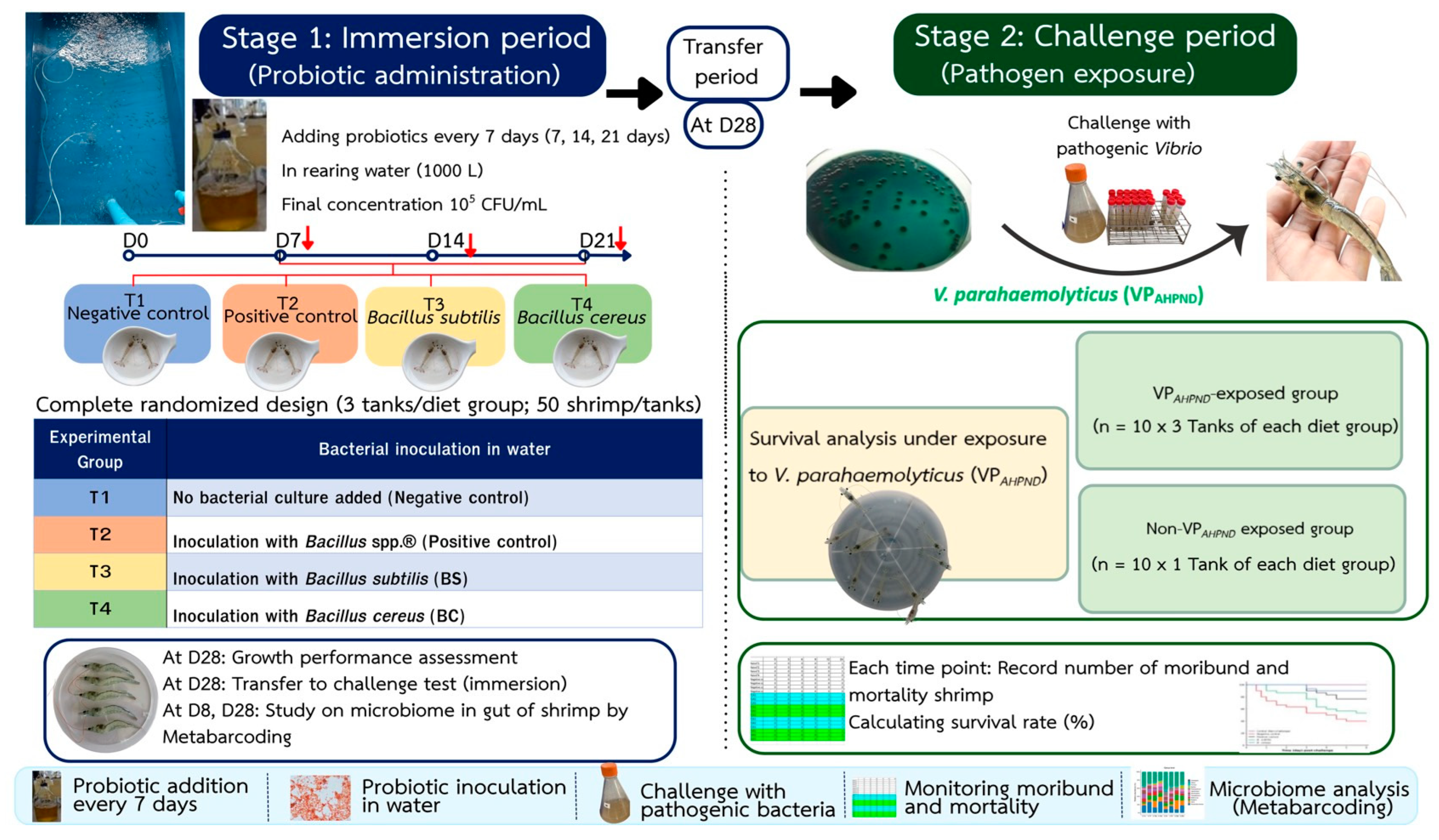
Water quality was maintained within suitable ranges for shrimp culture throughout the trial. Sediment was siphoned daily from every tank before feeding. Water exchange was performed when the ammonia concentration exceeded 0.5 mg-N/L: approximately 30% during the early phase, and 50% thereafter. In practice, water was exchanged on days 9, 13, 18, and 24 of the experiment (Supplementary Materials, Table S2 and Figure S1). Probiotics were applied on days 7, 14, and 21; no water exchange was performed on bacterial-application days. Temperature, pH, alkalinity, ammonia, and nitrite were maintained within suitable ranges for shrimp culture throughout the trial (temperature 27–28 °C, pH 7.5–8.5, alkalinity ≥ 150 mg/L, calcium ≥ 80 mg/L, magnesium ≥ 700 mg/L, ammonia and nitrite ≤ 0.5 mg-N/L). Shrimp were fed three times daily (06:00, 12:00, and 18:00) at 5% of body weight per day. The experiment lasted continuously for 28 days. The overall experimental design is shown in Figure 1. The figure layout was assembled by the authors using Canva (Canva Pty Ltd., Sydney, Australia); all photographs were produced by the authors.

2.4. Addition of Bacillus spp. to the Shrimp Rearing Water

The commercial Bacillus spp.® product, B. subtilis (BS) and the B. cereus (BC) group strain were added to the shrimp rearing water according to each treatment every 7 days (1 L per tank), giving an estimated final bacterial concentration of approximately 1 × 10⁵ CFU/mL in the rearing water for a total of three applications on days 7, 14 and 21 of the rearing trial. This target concentration is consistent with the range previously applied for water-based Bacillus probiotics in shrimp culture [27]. The seven-day application interval was selected to reflect the periodic probiotic-application schedule commonly practiced by Thai shrimp farmers, who typically re-apply probiotics at weekly intervals. Shrimp were reared until the end of the trial on day 28.

2.5. Growth Performance and Feed Utilization

Growth performance was evaluated at the end of the 28 days experimental period. Ten shrimp were randomly sampled from each tank and individually weighed to determine final body weight. For growth performance and feed conversion outcomes, tank-level mean values and feed records were used as the experimental units, resulting in =3 replicate tanks per treatment. The following parameters were calculated according to standard aquaculture growth equations [28]:
Weight gain (g) = Final weight (g) − Initial weight (g)
Specific growth rate (SGR, %/day) = [(ln Final weight − ln Initial weight)/Experimental days] × 100
Average daily gain (ADG, g/day) = (Final weight − Initial weight)/Experimental days
Feed conversion ratio (FCR) = Total feed intake (g)/Total weight gain (g)

2.6. Vibrio parahaemolyticus (VPAHPND) Challenge

The Vibrio parahaemolyticus (VPAHPND) challenge was conducted at the end of the rearing trial. On day 28, ten shrimps from each rearing tank were transferred to separate challenge tanks containing 10 L of seawater at 15 ppt, with three replicate tanks per treatment. Each treatment included a corresponding non-challenged control group to which no bacterial suspension was added. For the challenged groups, 10 mL of VPAHPND suspension (1 × 10⁹ CFU/mL) was added to each tank to obtain a final bacterial concentration of 1 × 10⁶ CFU/mL. After 6 h of immersion exposure, the water was completely exchanged. During the challenge period, no further probiotic (Bacillus) was added to the water, and all groups were fed the same commercial pelleted feed as during the rearing period. Shrimp survival was recorded for 6 days post-challenge. Survival rate (%) was analyzed using the Kaplan–Meier method, and survival curves among treatments were compared using the log-rank test.

2.7. Statistical Analysis

Growth performance and feed-conversion data are presented as mean ± SD (=3 replicate tanks). These data were analyzed by one-way ANOVA, and differences among means were compared using Duncan’s multiple range test at p < 0.05. Because the B. cereus group strain had a numerically lower initial weight, final body weight was additionally analyzed by analysis of covariance (ANCOVA) with initial weight as a covariate; the assumption of homogeneity of regression slopes was verified prior to analysis.
Post-challenge survival was analyzed using the Kaplan–Meier method, and survival curves among treatments were compared using the log-rank test. Pairwise comparisons between groups were then performed, and the resulting p-values were adjusted for multiple comparisons using the Benjamini–Hochberg (BH) procedure to control the false discovery rate; groups sharing the same letter did not differ significantly (adjusted p ≥ 0.05). All statistical analyses were performed in RStudio (version 4.6.1), with statistical significance set at p < 0.05.

2.8. Gut Microbiota Analysis by 16S rRNA Amplicon Sequencing

2.8.1. Sample Collection

On days 8 and 28 after the bacterial applications, the foregut and midgut of the shrimp were collected. Four shrimp from each tank were randomly sampled, and the guts of the four shrimp from each tank were pooled into a single sample (n = 3 pools per treatment) for bacterial community analysis by 16S rRNA amplicon sequencing.

2.8.2. DNA Extraction and 16S Amplicon Sequencing

Total DNA was extracted from each gut sample that were performed with a QIAamp PowerFecal Pro DNA Kit (Qiagen, Germantown, MD, USA) according to the manufacturer’s instructions. After extraction, DNA quality was measured using a NanoDrop spectrophotometer (Thermo Fisher Scientific, Waltham, MA, USA) to ensure a concentration > 50 ng/µL and an OD260/280 ratio (purity) of 1.8–2.2. For 16S ribosomal RNA (rRNA) amplicon sequencing, libraries were constructed using polymerase chain reactions (PCR) with universal primers that amplify the V3–V4 hypervariable regions of the 16S rRNA genes: 341F (5′-ACTCCTACGGGAGGCAGCA-3′) and 805R (5′-GGACTACHVGGGTWTCTAAT-3′). Indexed adapters were added to the ends of the 16S rRNA amplicons to generate indexed libraries ready for next-generation sequencing. Qualified libraries were sequenced on an Illumina NovaSeq 6000 platform (Illumina, San Diego, CA, USA) to generate paired-end reads. Raw image data were processed by base calling to produce FASTQ files containing nucleotide sequences and their corresponding quality scores.

2.8.3. Bioinformatics Analysis and Statistics

Raw paired-end sequencing reads were processed using a standardized amplicon sequence variant (ASV)-based workflow (v4.1.0). Adapter sequences and low-quality bases were removed using Trimmomatic (v0.33), followed by PCR primer trimming using Cutadapt (v1.9.1). Quality-filtered reads were subsequently processed using the DADA2 package (v1.20.0) in R, including quality filtering, error-rate learning, dereplication, denoising, paired-end merging, and chimera removal to generate high-confidence non-chimeric ASVs. Taxonomic classification was performed using a combined BLASTn (v2.9.0)- and Bayesian-based approach against the SILVA reference database (release 138.1). Microbial community composition was characterized at the phylum and genus levels. Downstream microbial community analyses were conducted using the phyloseq package in R. Alpha diversity (Chao1 and Shannon) was analyzed by a two-way linear model with Treatment and Time as fixed effects and their interaction (Alpha diversity~Treatment × Time), followed by Type III ANOVA. Heteroscedasticity-consistent (HC3) standard errors were used to make the inference robust to heterogeneity of variance among groups. Beta diversity was calculated using Bray-Curtis dissimilarity and visualized by PCoA. Differences in community composition among groups were tested by permutational multivariate analysis of variance (PERMANOVA; 999 permutations) using the vegan package in R. Differential analysis between groups was performed using LEfSe (LDA effect size (Default threshold: >4) and p-value (Default threshold: <0.05). A p < 0.05 or p < 0.01 was considered statistically significant in all analyses. Raw data and supporting figures for the bioinformatics and statistical analyses are available in the Supplementary Materials.
During the preparation of this manuscript, the authors used Claude Opus 4.8 (Anthropic, San Francisco, CA, USA) solely to assist with minor language editing and grammatical improvements. All content, including the ideas, concepts, results, and discussion, was generated entirely by the authors. Following AI-assisted language editing, the manuscript was also reviewed by a professional language editor. The authors have reviewed and edited the output and take full responsibility for the content of this publication.

3. Results

3.1. Effects of Probiotic Immersion on Growth Performance and Feed Utilization

The initial mean weight of shrimp was 9.36 ± 0.88 to 10.10 ± 0.46 g; no significant differences were observed among treatments (p > 0.05). The final weight after 28 days was higher in the B. cereus-treated group (16.68 ± 0.89 g) and lower in the B. subtilis-treated group (15.01 ± 1.41 g). Final weight was similar among groups (p > 0.05). Survival rate during the 28-day rearing period was 100% in all treatment groups.
Clear differences were observed in growth parameters. The B. cereus-treated group showed the highest weight gain (7.32 ± 0.72 g), specific growth rate (SGR, 2.07 ± 0.24%/day), and average daily gain (ADG, 0.261 ± 0.026 g/day), which were significantly higher than those of the control (5.56 ± 0.56 g; SGR 1.58 ± 0.13%/day; ADG 0.199 ± 0.020 g/day) and the B. subtilis-treated group (4.91 ± 1.50 g; SGR 1.41 ± 0.38%/day; ADG 0.175 ± 0.054 g/day) (p < 0.05). The commercial Bacillus spp.® product group showed intermediate values that did not differ significantly from the other groups. The B. cereus-treated group also had the lowest feed conversion ratio (FCR, 1.36 ± 0.02); however, FCR did not differ significantly among groups (p > 0.05). Because WG, SGR, and ADG are derived directly from initial and final weights, and the B. cereus group strain had a numerically lower initial weight (9.36 g vs. 9.95–10.10 g), final body weight was additionally analyzed by ANCOVA with initial weight as a covariate. The homogeneity-of-slopes assumption held (interaction p = 0.78). After adjustment, final weight did not differ significantly among treatments (F3,7 = 2.58, p = 0.14; B. cereus vs. control, p = 0.11), consistent with the non-significant difference in unadjusted final weight (p = 0.15). The higher WG, SGR, and ADG values in the B. cereus group strain are therefore partly attributable to its lower starting weight and should be interpreted as numerical trends rather than definitive growth-promoting effects (Table 1).
In summary, the B. cereus group strain showed numerically higher WG, SGR, and ADG under comparable feed intake; however, these differences were not supported after adjusting for initial weight (ANCOVA), and final body weight and FCR did not differ among groups. These growth results should therefore be regarded as preliminary trends.
The commercial Bacillus spp.® product produced intermediate effects, whereas B. subtilis supplementation did not differ from the control in any of the measured parameters.

3.2. Survival Rate of P. vannamei Challenged with V. parahaemolyticus (VPAHPND)

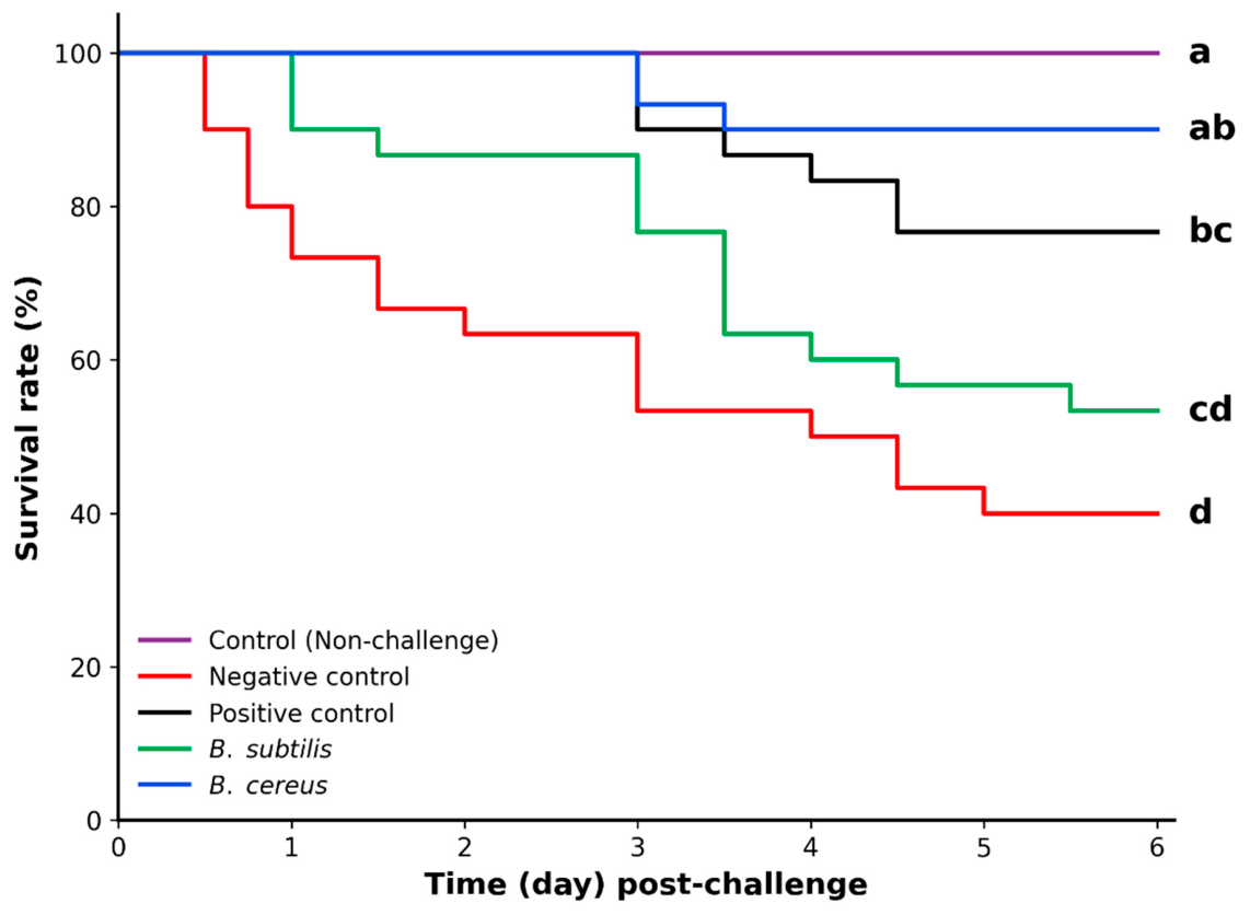
Survival of P. vannamei following the VPAHPND challenge was analyzed by the Kaplan–Meier method (Figure 2). In the non-challenged control, survival remained at 100% throughout the 6-day observation period, confirming that mortality in the challenged groups was attributable to the pathogen rather than to handling or rearing conditions. Among the challenged groups, the B. cereus group strain showed the highest survival (approximately 90%), followed by the commercial Bacillus spp.® product (positive control, ~76%) and the B. subtilis group (~53%), whereas the group without bacterial addition (negative control) showed the lowest survival (~40%).
The log-rank test indicated significant differences in survival among treatments (p < 0.05). After pairwise comparison with Benjamini–Hochberg adjustment, the survival of the B. cereus group strain did not differ significantly from that of the non-challenged control, whereas the negative control had significantly lower survival than the B. cereus group strain and the positive control. These results indicate that water-based supplementation with the B. cereus group strain conferred the strongest protection against VPAHPND infection, comparable to the commercial product and significantly better than the untreated control.

3.3. Gut Microbial Community Analysis

3.3.1. Next-Generation Sequencing Data and ASV Analysis

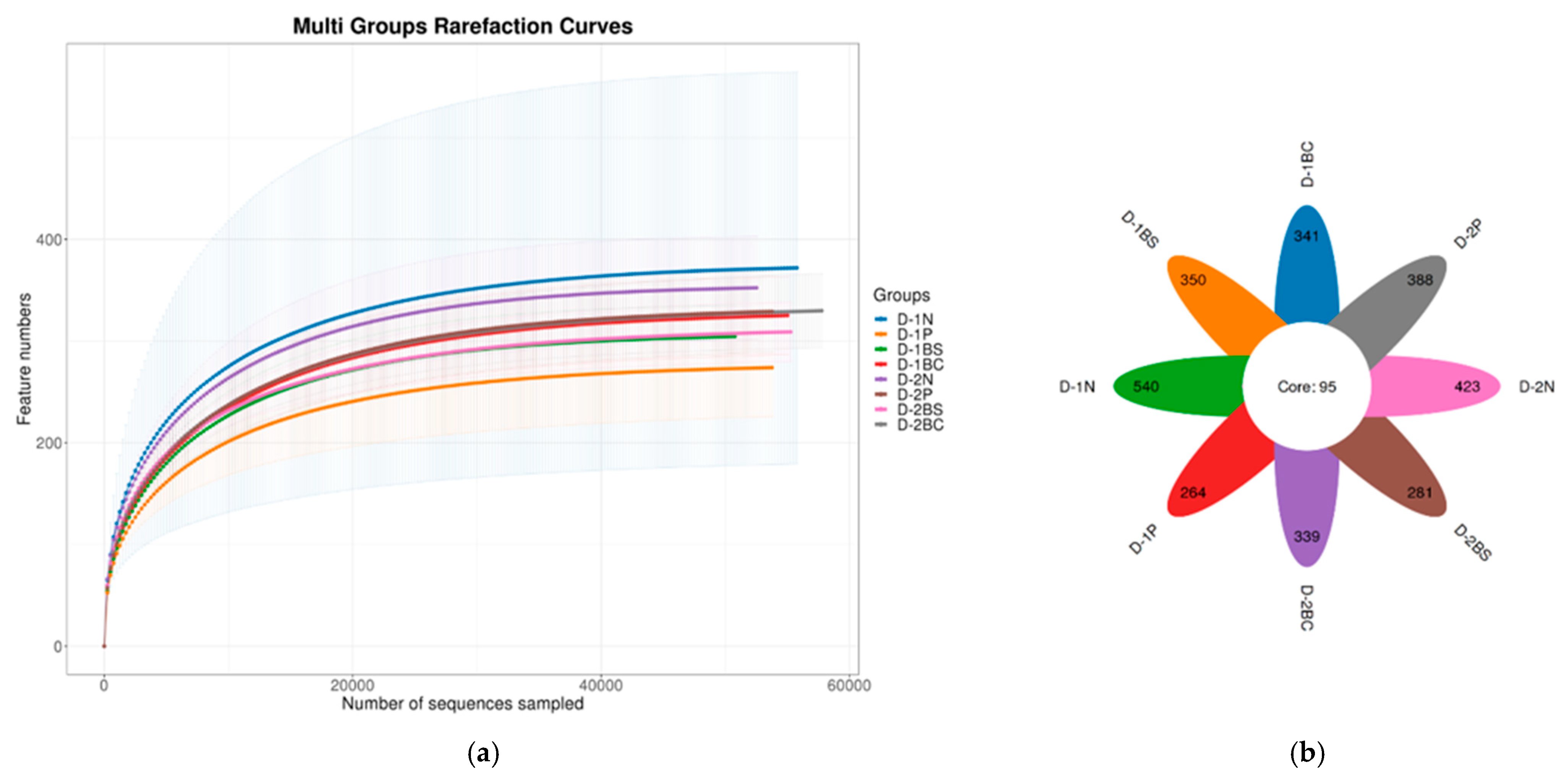
After DNA extraction and sequencing of the V3–V4 region of the 16S rRNA gene in all samples, the high-throughput sequencing data were statistically analyzed. After quality control, 1,620,837 reads and 4110 ASVs were obtained, classified into 23 phyla, 46 classes, 122 orders, 242 families, and 506 genera. The rarefaction curves of all experimental groups approached a near-plateau at approximately 40,000 sequences (Figure 3a), indicating that the sequencing depth was sufficient to capture the microbial diversity of the shrimp gut in all groups.
The amplicon sequence variants (ASVs) of the gut microbial communities of P. vannamei following probiotic administration via water inoculation on day 8 (D-1) and day 28 (D-2) were visualized using a flower plot (Figure 3b). The number of cores ASVs shared among all groups (95 ASVs) was relatively small compared with the number of group-specific ASVs, indicating that a large proportion of ASVs were unique to individual groups. Among the groups, the negative control without bacterial inoculation showed the highest number of group-specific ASVs at both time points (D-1N and D-2N). It should be noted, however, that the number of unique ASVs reflects the presence of low-abundance and rare taxa and is therefore distinct from the overall alpha diversity indices (Chao1 and Shannon), which showed no significant differences among groups (see Section 3.3.2). As this comparison is descriptive and not based on statistical testing, these patterns should be interpreted with caution. Nevertheless, they may tentatively suggest that the addition of Bacillus was associated with a more structured community containing fewer group-specific taxa, in contrast to the more dispersed community observed in the control group.

3.3.2. Alpha and Beta Diversities

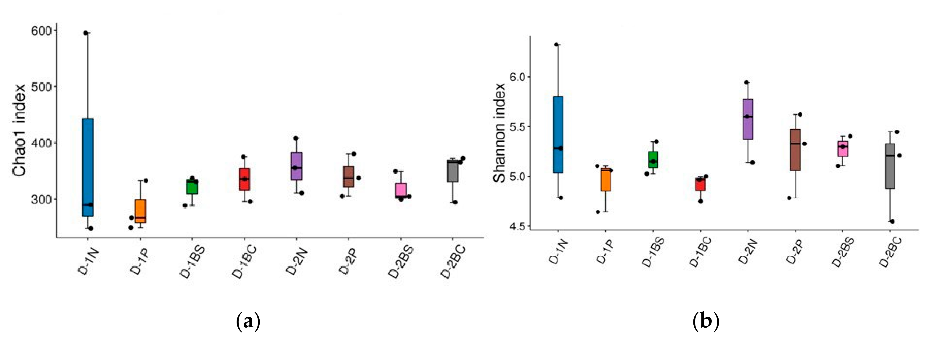
Alpha diversity was evaluated using the Chao1 richness (Figure 4a) and Shannon diversity indices (Figure 4b). A linear model including treatment, sampling time, and their interaction (Treatment × Time) was used to assess the effects of the experimental factors. For Chao1 richness, neither treatment (F3,16 = 0.623, p = 0.610), sampling time (F1,16 = 0.045, p = 0.834), nor the Treatment × Time interaction (F3,16 = 0.510, p = 0.681) had a significant effect. Similarly, Shannon diversity was not significantly affected by treatment (F3,16 = 1.354, p = 0.292), sampling time (F1,16 = 0.224, p = 0.642), or the Treatment × Time interaction (F3,16 = 0.106, p = 0.955). These results indicate that probiotic inoculum in the rearing water, the type of added bacteria, and the duration of administration had no significant effect on the overall alpha diversity of the shrimp gut microbial community, in terms of both species richness (Chao1) and diversity including evenness (Shannon), and that the treatment effects did not vary between sampling times. Given the limited biological replication (=3 pooled samples per treatment × sampling-time combination), these findings should be interpreted with appropriate caution.
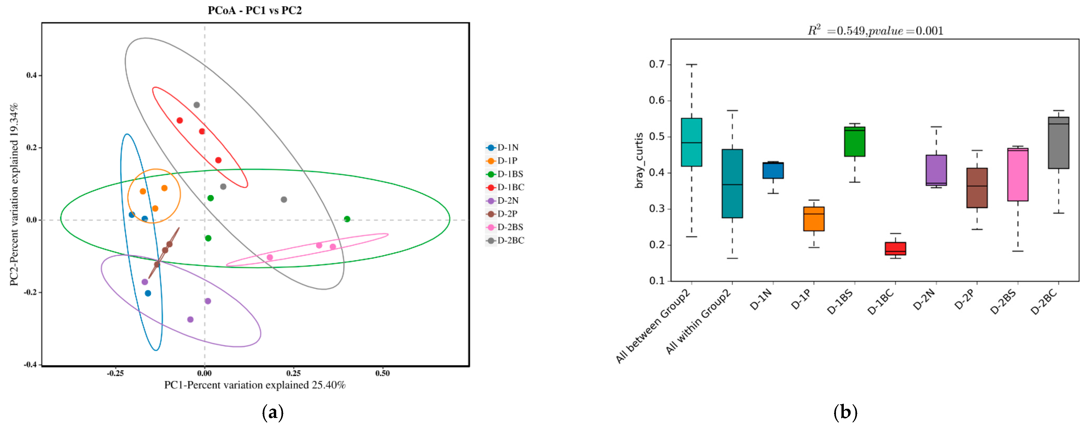
The gut microbial community structure of P. vannamei among the experimental groups on day 8 (D-1) and day 28 (D-2) was assessed by Principal Coordinates Analysis (PCoA) based on Bray-Curtis dissimilarity. The microbial communities of most experimental groups overlapped; however, the B. cereus group on day 8 (D-1BC) and the B. subtilis group on day 28 (D-2BS) tended to cluster separately from the other groups and showed high within-group similarity (Figure 5a). Permutational multivariate analysis of variance (PERMANOVA) confirmed that the gut microbial community structure differed significantly among the groups (pseudo-F7 = 2.78, R2 = 0.55, p = 0.001, 999 permutations), with between-group dissimilarity being greater than within-group dissimilarity, consistent with the pattern observed in the PCoA (Figure 5b). In contrast to the alpha-diversity results, which showed no significant differences among groups (p > 0.05), these findings indicate that probiotic inoculation into the rearing water did not alter the overall diversity of the gut microbial community but was associated with differences in its species composition and structure among groups.

3.3.3. Bacterial Community Structure Between Treatments

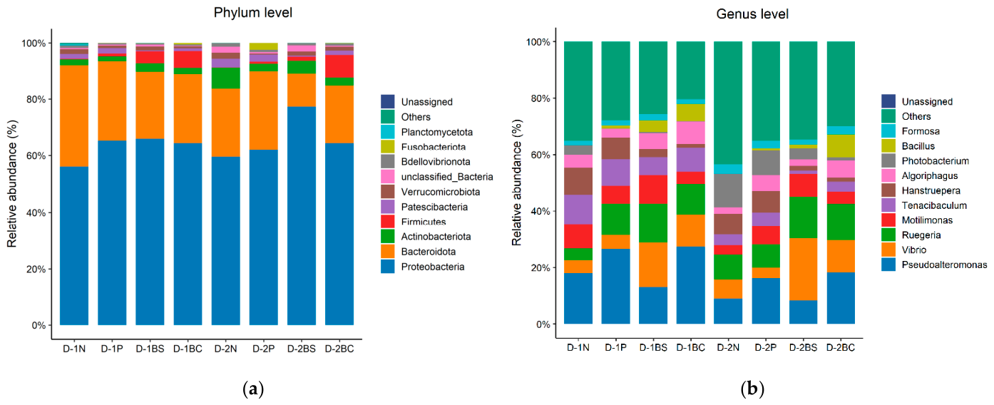
The bacterial community structure of all samples was visualized at the phylum and genus level (Figure 6). The bacterial composition at the phylum level was visualized in bar plots of the relative abundance (Figure 6a). The gut microbial communities of all experimental groups were predominantly composed of Proteobacteria (approximately 55–77%), followed by Bacteroidota, while other phyla, including Actinobacteriota, Firmicutes, Patescibacteria, Verrucomicrobiota, and Bdellovibrionota, were present in lower proportions. The B. subtilis-treated group on day 28 (D-2BS) showed the highest proportion of Proteobacteria (approximately 77%). Regarding the phylum Firmicutes, to which the genus Bacillus belongs, a slight increase in its proportion was observed in some of the inoculated groups, such as D-1BC. Overall, however, the phylum-level composition was similar among the groups.
The bacterial composition at the genus level was visualized in bar plots of the relative abundance of the top 10 taxa (Figure 6b). The gut microbial communities showed clearer differences among groups than at the phylum level. The predominant genera included Pseudoalteromonas, Vibrio, Ruegeria, Motilimonas, Tenacibaculum, Hanstruepera, and Bacillus. Pseudoalteromonas was present in high proportions in several groups, particularly in D-1P and D-1BC (approximately 27%). Vibrio was found in relatively high proportions in the groups sampled on day 8 (D-1P, D-1BS, and D-1BC) and tended to decrease in the groups sampled on day 28 (the D-2 groups).
The genus Bacillus was most notably represented in the B. cereus-treated group on day 8 (D-1BC), consistent with the bacterial inoculation into the rearing water, whereas the other groups showed relatively low proportions of Bacillus. In addition, the groups sampled on day 28 (D-2N, D-2P, D-2BS, and D-2BC) exhibited a higher proportion of the “Others” category (low-abundance genera not among the dominant taxa) than the groups sampled on day 8, indicating that the composition of minor genera became more diverse as the experimental period progressed.

3.3.4. Differential Analysis Between Treatments

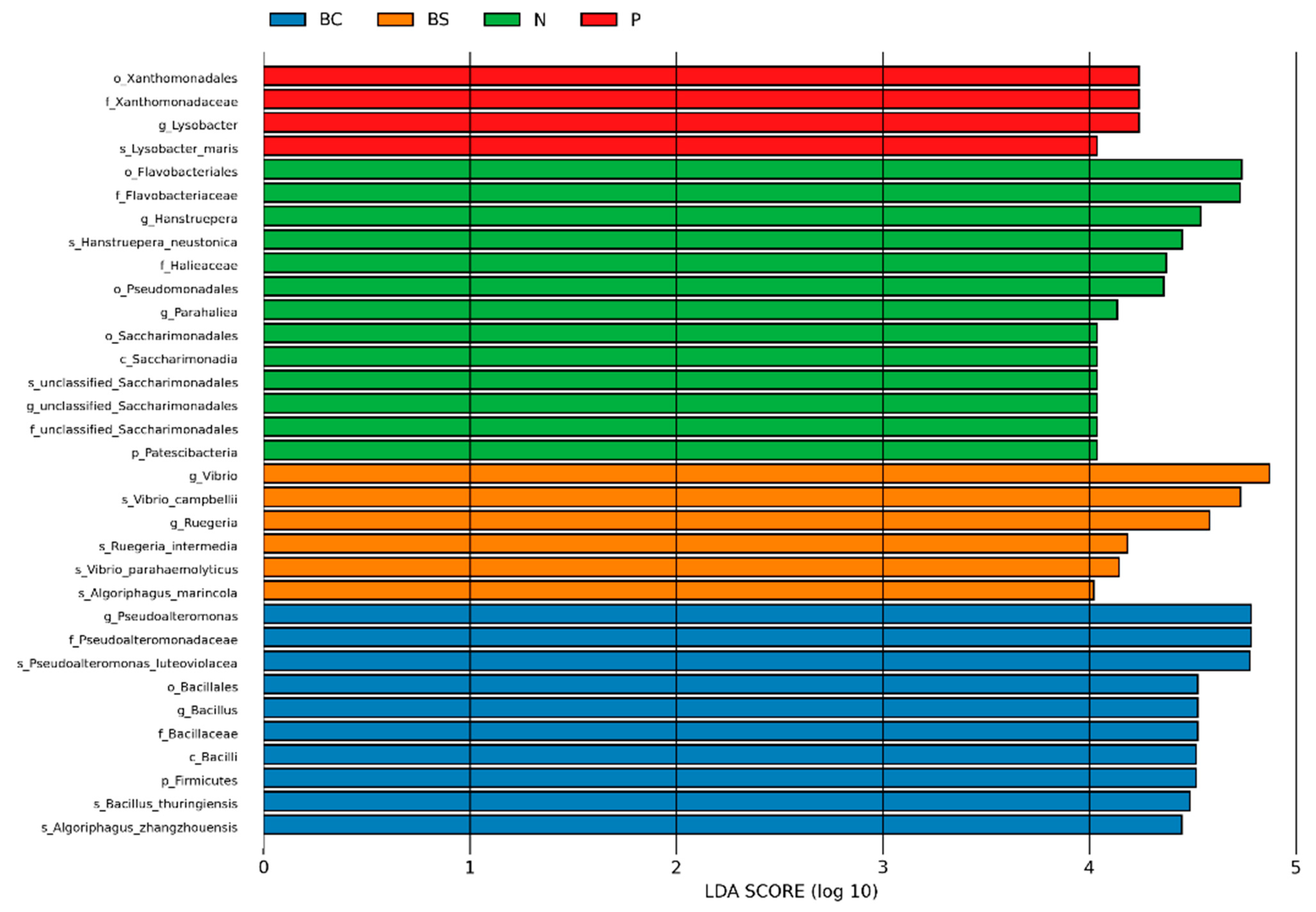
To identify taxa differentially enriched among treatments, LEfSe analysis was performed with samples pooled across sampling times (LDA score > 4.0, p < 0.05; Figure 7). Each treatment group was characterized by a distinct set of enriched taxa. The B. cereus group (BC) was enriched in Pseudoalteromonas (P. luteoviolacea), Bacillus (including B. thuringiensis), and Algoriphagus zhangzhouensis. The B. subtilis group (BS) was enriched in Vibrio, including V. campbellii and the AHPND-associated V. parahaemolyticus, together with Ruegeria. The negative control (N) was characterized by Flavobacteriaceae, Hanstruepera, and Parahaliea, and the positive control (P) by Lysobacter (L. maris).
Notably, the potentially pathogenic genus Vibrio, including V. parahaemolyticus, was a significant biomarker of the B. subtilis group but not of the B. cereus group, whereas the B. cereus group was instead enriched in Bacillus and Pseudoalteromonas, the latter of which includes strains with reported antagonistic activity against Vibrio. These differential-abundance patterns extend beyond Bacillus alone and indicate that the treatments were associated with broader shifts in the gut microbial community.

4. Discussion

The use of probiotics to promote shrimp health has become increasingly popular, as probiotics can improve water quality while enhancing growth and disease resistance. In aquaculture practice, probiotics are commonly applied in two ways: direct addition to the rearing water and dietary supplementation, both of which have been reported to benefit shrimp [28]. However, data on the water-based (immersion) application of probiotics in relation to intestinal bacterial abundance and disease resistance in shrimp remain more limited than those on dietary supplementation. The present study therefore contributes new knowledge and demonstrates the efficacy of applying probiotic bacteria in water on shrimp health.
The present study investigated the effects of B. subtilis and a B. cereus group strain added to the rearing water on the growth, disease resistance, and gut microbial composition of white shrimp (P. vannamei). The results showed that shrimp reared with the B. cereus group strain had numerically higher SGR and ADG than the control; however, these differences were not statistically robust after adjusting for a lower initial weight in this group by ANCOVA (p = 0.14), and final body weight and FCR did not differ significantly among groups. Together with the relatively short 28-day rearing period, this indicates that the growth results should be regarded as short-term, proof-of-concept trends rather than definitive growth-promoting effects, and longer-term trials with balanced initial weights are required for confirmation
The numerically higher growth values observed in the B. cereus group strain are directionally in line with previous reports in which B. cereus improved growth, including its application in a biofloc system for shrimp [13] and studies in juvenile coho salmon (Oncorhynchus kisutch) [29] and Pengze crucian carp (Carassius auratus var. Pengze) [30]. In those studies, such growth effects were attributed to enhanced nutrient absorption and increased activity of digestive enzymes (amylase, lipase, protease, esterase, β-galactosidase, and leucine-aminopeptidase) [31,32]. However, digestive-enzyme activities and nutrient absorption were not measured in the present study; these mechanisms are therefore discussed only as hypotheses derived from previous work, and, unlike those studies, the growth differences observed here were not statistically robust after adjusting for initial weight.
The shrimp reared in water supplemented with the B. cereus group strain showed the highest survival rate after the VPAHPND challenge. The better survival was associated with the highest relative abundance of the genus Bacillus in the gut, suggesting that an increased gut presence of Bacillus may be an important part of probiotic protection [33,34]. It should be noted, however, that because 16S rRNA analysis resolves taxa at the genus level, this increased abundance cannot be attributed with certainty to the administered strain, and strain-specific confirmation would be required to establish gut colonization by the inoculated strain.
Comparable protective effects have been reported previously: Ref. [35] demonstrated that the B. cereus sensu stricto strain P64 associated with the internal and external surfaces of white shrimp and protected animals from infection by V. parahaemolyticus, and Ref. [36] reported that shrimp fed a mixture of B. cereus and B. subtilis had higher survival after infection with Vibrio spp. Several mechanisms may hypothetically contribute to such protection, although none was directly assessed in the present study. One possibility is competitive exclusion, whereby probiotic strains that establish early and at sufficient levels compete with pathogenic bacteria for intestinal attachment sites and nutrients, which may restrict Vibrio growth in the gut [33]. In addition, Bacillus has been reported to inhibit pathogens through antimicrobial compounds such as bacteriocins, antimicrobial peptides, and lipopeptides [32]; for example, B. pumilus H2 produces an anti-Vibrio substance structurally identical to amicoumacin A, which inhibits up to 29 Vibrio strains [37]. It has also been suggested that improved shrimp survival following V. parahaemolyticus challenge may relate to antimicrobial peptides produced by probiotics [38]. Furthermore, previous studies have reported that Bacillus can stimulate the innate immune system of shrimp by enhancing phagocytic activity and the expression of prophenoloxidase (proPO) and lysozyme genes [39]. However, antimicrobial activity, immune parameters, and pathogen abundance were not measured in the present study; these mechanisms are therefore presented as hypotheses supported by previous work rather than as processes demonstrated by our data, and they remain to be confirmed in future studies.
The intestinal microbiota of P. vannamei is essential in nutrient metabolism, immune modulation, and disease resistance, and its composition is strongly shaped by diet and culture environment [39,40]. In the present study, although the Shannon and Chao1 indices did not differ significantly among groups, β-diversity analysis (PERMANOVA) showed significant differences in gut microbial community structure among groups. This indicates that adding the B. cereus group strain to the rearing water every 7 days did not change the overall diversity of the gut microbiota but was associated with differences in its community composition. A comparable pattern was reported by [13], who found that adding B. cereus in a biofloc system altered the gut microbial composition, whereas biofloc alone or biofloc without continued probiotic input had no significant effect, suggesting that the effect may be frequency- and context-dependent. Although the present study applied the strain at a single frequency (every 7 days) and therefore cannot directly assess frequency dependence, the observed community shift is consistent with the notion that periodic supplementation helps maintain probiotic populations and their associated metabolites within the gut and rearing environment [41,42].
Proteobacteria, Bacteroidota, and Actinobacteriota were the dominant phyla in all treatment groups, consistent with reports for the white-shrimp gut [13,27]. These phyla have significant roles in nutrient digestion and community balance. The similarity in phylum-level composition across all groups indicates that the core microbiota of the shrimp gut is under host-selective filtering, which maintains a relatively stable community structure even with the addition of different probiotics. The commercial Bacillus spp.® product and the B. cereus group strain resulted in the detection of the genus Bacillus in the shrimp gut, which was otherwise nearly absent in the control group. This higher relative abundance of Bacillus in the inoculated groups is consistent with the water-to-gut transmission concept reported previously [43]; however, because the water microbiome was not analyzed in parallel, our data cannot directly demonstrate this route.
This study also showed that the B. cereus group strain was associated with a higher and more consistent relative abundance of the genus Bacillus in the shrimp gut than B. subtilis and the commercial Bacillus spp.® product, which persisted 7 days after the final application (D-2, day 28). LEfSe analysis identified the genus Bacillus as a prominent biomarker of the B. cereus group strain. This higher intestinal abundance may be related to the strain having been isolated from the culture environment itself (native), potentially allowing better adaptation to intestinal and pond conditions than allochthonous or commercial strains [44,45,46], together with the spore-forming and intestinal-germination characteristics of the genus Bacillus [47]. A comparable observation was reported by [48], who found that a B. cereus strain persisted in the intestine of post-larval shrimp. It should be noted, however, that because 16S rRNA analysis resolves taxa at the genus level, these findings cannot confirm colonization by the administered strain, which would require strain-specific tracking.

5. Conclusions

This study evaluated two native Bacillus strains, B. subtilis and a B. cereus group strain isolated from shrimp pond sediment and applied through the rearing water, on the growth, disease resistance, and gut microbiota of P. vannamei. Water-based supplementation with the B. cereus group strains every 7 days was associated with the highest survival rate after V. parahaemolyticus challenge and the highest relative abundance of the genus Bacillus in the shrimp gut, whereas B. subtilis and the commercial product showed weaker or less consistent effects. Numerically higher SGR and ADG were also observed in this group; however, these growth differences were not robust after adjusting for a lower initial weight (ANCOVA) and should be regarded as preliminary. Although alpha diversity was unaffected, the gut microbial community structure differed significantly among groups (beta diversity, PERMANOVA). These results indicate that direct addition of beneficial bacteria to the rearing water can increase the relative abundance of Bacillus in the shrimp gut and modulate the gut microbial community, offering a potential alternative to dietary supplementation for microbiome modulation. Nevertheless, this study should be interpreted as a proof-of-concept: because the B. cereus group includes potentially toxigenic and antimicrobial-resistant strains, a comprehensive strain-level safety assessment is an indispensable prerequisite before this approach can be recommended for practical use.

Supplementary Materials

The following supporting information can be downloaded at https://www.mdpi.com/article/10.3390/bacteria5040060/s1, Supplementary files: Supplementary material file-Table S1 Characterization of Bacillus, Table S2 Water quality and Figure S1 Concentrations of inorganic nitrogen compounds. All samples, statistical analysis, Read-length distribution, and quality statistics for all sequencing samples, including sample metadata and summary metrics.

Author Contributions

Research design and drafting of the manuscript, M.S.; development of analytical methods and experimental methodology, M.K.; collection and analysis of experimental data, analysis of results, T.T., K.S., K.K. and C.C.; supervision of the experimental data and interpretation of the final results, J.W. All authors have read and agreed to the published version of the manuscript.

Funding

This research was funded by Burapha University through the Thailand Science Research and Innovation (TSRI) Fund under the Fundamental Fund scheme, fiscal year B.E. 2568 (2025) (Grant No. 1.4/2568).

Institutional Review Board Statement

The following protocols were conducted in accordance with the Burapha University Biosafety Committee (Approval No. IBC 049/2568, 10 May 2025). All animal procedures were performed in accordance with the Ethical Principles and Guidelines for the Use of Animals of the National Research Council of Thailand. The study protocol was approved by Burapha University Institutional Animal Care and Use Committee (Approval No. IACUC 023/2568, 20 May 2025).

Informed Consent Statement

Not applicable.

Data Availability Statement

The original contributions presented in this study are included in the article. Further inquiries can be directed to the corresponding author. The 16S rRNA gene sequencing data generated in this study have been deposited in the NCBI Sequence Read Archive (SRA) under BioProject accession number (PRJNA1513981).

Acknowledgments

During the preparation of this manuscript, the authors used Claude Opus 4.8 (Anthropic) solely to assist with minor language editing and grammatical improvements. All content, including the ideas, concepts, results, and discussion, was generated entirely by the authors. Following AI-assisted language editing, the manuscript was also reviewed by a professional language editor. The authors have reviewed and edited the output and take full responsibility for the content of this publication.

Conflicts of Interest

The authors declare no conflicts of interest.

References

  1. FAO. The State of World Fisheries and Aquaculture; FAO: Rome, Italy, 2024. [Google Scholar]
  2. Yu, Y.-B.; Choi, J.-H.; Kang, J.-C.; Kim, H.J.; Kim, J.-H. Shrimp bacterial and parasitic disease listed in the OIE: A review. Microb. Pathog. 2022, 166, 105545. [Google Scholar] [CrossRef] [Scilit]
  3. Tran, L.; Nunan, L.; Redman, R.M.; Mohney, L.L.; Pantoja, C.R.; Fitzsimmons, K.; Lightner, D.V. Determination of the infectious nature of the agent of acute hepatopancreatic necrosis syndrome affecting penaeid shrimp. Dis. Aquat. Org. 2013, 105, 45–55. [Google Scholar] [CrossRef] [Scilit]
  4. Kumar, V.; Roy, S.; Behera, B.K.; Bossier, P.; Das, B.K. Acute Hepatopancreatic Necrosis Disease (AHPND): Virulence, Pathogenesis and Mitigation Strategies in Shrimp Aquaculture. Toxins 2021, 13, 524. [Google Scholar] [CrossRef] [Scilit]
  5. Milijasevic, M.; Veskovic-Moracanin, S.; Babic Milijasevic, J.; Petrovic, J.; Nastasijevic, I. Antimicrobial Resistance in Aquaculture: Risk Mitigation within the One Health Context. Foods 2024, 13, 2448. [Google Scholar] [CrossRef] [Scilit]
  6. Okocha, R.; Olatoye, O.; Adedeji, O. Food safety impacts of antimicrobial use and their residues in aquaculture. Public Health Rev. 2018, 39, 21. [Google Scholar] [CrossRef] [Scilit]
  7. Chauhan, A.; Singh, R. Probiotics in aquaculture: A promising emerging alternative approach. Symbiosis 2019, 77, 99–113. [Google Scholar] [CrossRef] [Scilit]
  8. Gadhiya, A.; Katariya, S.; Khapandi, K.; Chhatrodiya, D. Probiotics as a sustainable alternative to antibiotics in aquaculture: A review of the current state of knowledge. Microbe 2025, 8, 100426. [Google Scholar] [CrossRef] [Scilit]
  9. Gatesoupe, F.J. The use of probiotics in aquaculture. Aquaculture 1999, 180, 147–165. [Google Scholar] [CrossRef] [Scilit]
  10. Soltani, M.; Ghosh, K.; Hoseinifar, S.H.; Kumar, V.; Lymbery, A.J.; Roy, S.; Ringø, E. Genus Bacillus, promising probiotics in aquaculture: Aquatic animal origin, bio-active components, bioremediation and efficacy in fish and shellfish. Rev. Fish. Sci. Aquac. 2019, 27, 331–379. [Google Scholar] [CrossRef] [Scilit]
  11. Sukkasem, K.; Thiankham, S.; Srisomwong, K.; Kantha, P.; Kitikiew, S. Effects of probiotics Lacticaseibacillus paracasei and Bacillus amyloliquefaciens on water quality and inhibition of Vibrio vulnificus and Vibrio alginolyticus in white shrimp (Litopenaeus vannamei) culture. Int. J. Agric. Technol. 2025, 21, 1161–1176. [Google Scholar] [CrossRef] [Scilit]
  12. Esquén, B.D.A.; Ancajima, D.T.; Cadena, D.A.; Dominguez-Mendoza, L.; Leyva, S.; Somocurcio, Z.M.M.; Saldarriaga, Y.D.E.; Rampelotto, P.H.; Guzman, E.F.L.; Diringer, B.M. Bioaugmentation with native Bacillus strains enhance nitrate and nitrite removal and reshape microbiomes in low-salinity shrimp cultures: Elucidating genetic mechanisms. PLoS ONE 2026, 21, e0339620. [Google Scholar] [CrossRef] [Scilit]
  13. Ding, S.; Cai, W.; Xu, Y.; Jin, C.; Ma, X.; Rao, L.; Gao, Y.; Li, H.; Chu, Z. Impact of Bacillus cereus Supplementation in Feed and Biofloc Water on Growth Performance, Immune Responses, and Intestinal Microbiota of Pacific whiteleg shrimp (Litopenaeus vannamei). Fishes 2026, 11, 222. [Google Scholar] [CrossRef] [Scilit]
  14. Eissa, E.S.H.; Dowidar, H.A.; Al-Hoshani, N.; Baazaoui, N.; Alshammari, N.M.; Kari, Z.A.; Ibrahim, S.; Munir, M.B.; Farga, A.A.; Eissa, M.E.H.; et al. Dietary supplementation with fermented prebiotics and probiotics can increase growth, immunity, and histological alterations in Pacific whiteleg shrimp (Litopenaeus vannamei) challenged with Vibrio parahaemolyticus. Aquacult. Int. 2025, 33, 62. [Google Scholar] [CrossRef] [Scilit]
  15. Lomelí-Álvarez, M.F.; Escamilla-Montes, R.; Diarte-Plata, G.; Guo, X.; Fierro-Coronado, J.A.; Rubio-Luque, A.M.; Vega-Carranza, A.S.; González, A.L. Dietary and water probiotics enhance immunity, modulate microbiota, and increase survival of Penaeus vannamei challenged with V. parahaemolyticus. Braz. J. Microbiol. 2026, 57, 1. [Google Scholar] [CrossRef] [Scilit]
  16. Zokaeifar, H.; Babaei, N.; Saad, C.R.; Kamarudin, M.S.; Sijam, K.; Balcazar, J.L. Administration of Bacillus subtilis strains in the rearing water enhances the water quality, growth performance, immune response, and resistance against Vibrio harveyi infection in juvenile white shrimp, Litopenaeus vannamei. Fish Shellfish Immunol. 2014, 36, 68–74. [Google Scholar] [CrossRef] [Scilit]
  17. Nimrat, S.; Saksit, W.; Vuthiphandchai, V. Screening of bacteria isolated from black tiger shrimp (Penaeus monodon) intestine using as probiotics in aquaculture. RMUTP Sci. J. 2022, 16, 105–116. [Google Scholar]
  18. Torres-Maravilla, E.; Parra, M.; Maisey, K.; Vargas, R.A.; Cabezas-Cruz, A.; Gonzalez, A.; Tello, M.; Bermúdez-Humarán, L.G. Importance of Probiotics in Fish Aquaculture: Towards the Identification and Design of Novel Probiotics. Microorganisms 2024, 12, 626. [Google Scholar] [CrossRef] [Scilit]
  19. Kaushik, J.K.; Kumar, A.; Duary, R.K.; Mohanty, A.K.; Grover, S.; Batish, V.K. Functional and probiotic attributes of an indigenous isolate of Lactobacillus plantarum. PLoS ONE 2009, 4, e8099. [Google Scholar] [CrossRef] [Scilit]
  20. Mojgani, N.; Hussaini, F.; Vaseji, N. Characterization of indigenous Lactobacillus strains for probiotic properties. Jundishapur J. Microbiol. 2015, 8, e59798. [Google Scholar] [CrossRef] [Scilit]
  21. Mohammed, E.A.H.; Ahmed, A.E.M.; Kovács, B.; Pál, K. The significance of probiotics in aquaculture: A review of research trend and latest scientific findings. Antibiotics 2025, 14, 242. [Google Scholar] [CrossRef] [Scilit]
  22. Heyse, J.; Props, R.; Kongnuan, P.; De Schryver, P.; Rombaut, G.; Defoirdt, T.; Boon, N. Rearing water microbiomes in white leg shrimp (Litopenaeus vannamei) larviculture assemble stochastically and are influenced by the microbiomes of live feed products. Environ. Microbiol. 2021, 23, 281–298. [Google Scholar] [CrossRef] [Scilit]
  23. Md Zoqratt, M.Z.H.; Eng, W.W.H.; Thai, B.T.; Austin, C.M.; Gan, H.M. Microbiome analysis of Pacific white shrimp gut and rearing water from Malaysia and Vietnam: Implications for aquaculture research and management. PeerJ 2018, 6, e5826. [Google Scholar] [CrossRef] [Scilit]
  24. Vega-Carranza, A.S.; Escamilla-Montes, R.; Fierro-Coronado, J.A.; Diarte-Plata, G.; Guo, X.; García-Gutiérrez, C.; Luna-González, A. Investigating the effect of bacilli and lactic acid bacteria on water quality, growth, survival, immune response, and intestinal microbiota of cultured. Litopenaeus Vannamei Anim. 2014, 14, 2676. [Google Scholar]
  25. Noman, M.; Kazmi, S.S.U.H.; Saqib, H.S.A.; Fiaz, U.; Pastorino, P.; Barcelò, D.; Tayyab, M.; Liu, W.; Wang, Z.; Yaseen, Z.M. Harnessing probiotics and prebiotics as eco-friendly solution for cleaner shrimp aquaculture production: A state of the art scientific consensus. Sci. Total Environ. 2024, 915, 169921. [Google Scholar] [CrossRef] [Scilit]
  26. Tiewpair, P.; Kongtawee, O.; Roongkamnertwongsa, J. Comparison of the Virulence and Antibiotic Resistant of Vibrio parahaemolyticus Caused of Acute Hepatopancreatic Necrosis Disease (AHPND) from Different Pacific White Shrimp (Litopenaeus vannamei, Boone 1931) Cultured Area in Thailand; Technical Paper No. 5/2023; Aquatic Animal Health Research and Development Division, Department of Fisheries, Ministry of Agriculture and Cooperatives: Bangkok, Thailand, 2023. [Google Scholar]
  27. Kaewcharoen, W.; Srisapoome, P. Probiotic effects of Bacillus spp. from Pacific white shrimp (Litopenaeus vannamei) on water quality and shrimp growth, immune responses, and resistance to Vibrio parahaemolyticus (AHPND strains). Fish Shellfish Immunol. 2019, 94, 175–189. [Google Scholar] [CrossRef] [Scilit]
  28. Tong, Y.; Zhang, M.; Li, Q.; Hu, X.; Wei, Y.; Zhao, W.; Yu, X.; Peng, M.; Yang, C.; Chen, X.; et al. Effectiveness evaluation of five probiotics on shrimp and water environment. Aquac. Rep. 2025, 45, 103110. [Google Scholar] [CrossRef] [Scilit]
  29. Li, F.; Xie, Y.; Guo, M.; Liu, Y.; Tong, T.; Zhang, Q.; Kong, W. Effects of dietary Bacillus cereus supplementation on the growth performance, serum physiology and biochemistry, Nrf2, TLR/NF-κB signaling pathways, and intestinal health of juvenile coho salmon (Oncorhynchus kisutch). Aquac. Rep. 2024, 36, 102177. [Google Scholar] [CrossRef] [Scilit]
  30. Li, J.; Fang, P.; Yi, X.; Kumar, V.; Peng, M. Probiotics Bacillus cereus and B. subtilis reshape the intestinal microbiota of Pengze crucian carp (Carassius auratus var. Pengze) fed with high plant protein diets. Front. Nutr. 2022, 9, 1027641. [Google Scholar] [CrossRef] [Scilit]
  31. Amenyogbe, E.; Droepenu, E.K.; Ayisi, C.L.; Boamah, G.A.; Duker, R.Q.; Abarike, E.D.; Huang, J. Impact of probiotics, prebiotics, and synbiotics on digestive enzymes, oxidative stress, and antioxidant defense in fish farming: Current insights and future perspectives. Front. Mar. Sci. 2024, 11, 1368436. [Google Scholar] [CrossRef] [Scilit]
  32. Li, H.; Fan, S.; Gao, Y.; Cai, Y.; Chu, Z.; Wang, L. Evaluation of modulatory properties of Bacillus cereus isolated from the gut of Litopenaeus vannamai on growth, intestinal morphology, digestive enzyme activities, immune responses and disease resistance of Litopenaeus vannamai. Aquac. Res. 2020, 52, 1299–1310. [Google Scholar] [CrossRef] [Scilit]
  33. Knipe, H.; Temperton, B.; Lange, A.; Bass, D.; Tyler, C.R. Probiotics and competitive exclusion of pathogens in shrimp aquaculture. Rev. Aquacult. 2021, 13, 324–352. [Google Scholar] [CrossRef] [Scilit]
  34. Rohani, M.F.; Islam, S.M.; Hossain, M.K.; Ferdous, Z.; Siddik, M.A.; Nuruzzaman, M.; Padeniya, U.; Brown, C.; Shahjahan, M. Probiotics, prebiotics and synbiotics improved the functionality of aquafeed: Upgrading growth, reproduction, immunity and disease resistance in fish. Fish Shellfish Immunol. 2022, 120, 569–589. [Google Scholar] [CrossRef] [Scilit]
  35. Ramirez, M.; Domínguez-Borbor, C.; Salazar, L.; Debut, A.; Vizuete, K.; Sonnenholzner, S.; Alexis, F.; Rodríguez, J. The probiotics Vibrio diabolicus (Ili), Vibrio hepatarius (P62), and Bacillus cereus sensu stricto (P64) colonize internal and external surfaces of Penaeus vannamei shrimp larvae and protect it against Vibrio parahaemolyticus. Aquaculture 2022, 549, 737826. [Google Scholar] [CrossRef] [Scilit]
  36. Wongpracha, P.; Kanthawong, P.; Arun, J.; Jeyaraj, G.P.; Jongput, B.; Srijad, S.; Kitikiew, S. Effects of Bacillus probiotics, Bacillus subtilis and Bacillus cereus dietary additional to controlling Vibriosis infection of white shrimp (Litopenaeus vannamei). Int. J. Agric. Technol. 2024, 20, 427–440. [Google Scholar]
  37. Gao, X.-Y.; Liu, Y.; Miao, L.-L.; Li, E.W.; Hou, T.-T.; Liu, Z.-P. Mechanism of Anti-Vibrio Activity of Marine Probiotic Strain Bacillus Pumilus H2, and Characterization of the Active Substance. AMB Express 2017, 7, 23. [Google Scholar] [CrossRef] [Scilit]
  38. Puan, S.L.; Erriah, P.; Baharudin, M.M.A.; Yahaya, N.M.; Kamil, W.N.; Ali, M.S.M.; Ahmad, S.A.; Oslan, S.N.; Lim, S.; Sabri, S. Antimicrobial peptides from Bacillus spp. and strategies to enhance their yield. Appl. Microbiol. Biotechnol. 2023, 107, 5569–5593. [Google Scholar] [CrossRef] [Scilit]
  39. Maurus, G.; Joe, J.T.X.; Phạm, Q.H.; Lin, S.-C.; Lu, M.-W. Commercial probiotics enhance growth and immunity of white shrimp (Litopenaeus vannamei) and resistence to infection by Vibrio parahaemolyticus. J. World Aquac. Soc. 2025, 56, e70036. [Google Scholar] [CrossRef] [Scilit]
  40. Corey, C.H.; Bass, D.; Stentiford, G.D.; van der Giezen, M. Understanding the role of the shrimp gut microbiome in health and disease. J. Invertebr. Pathol. 2021, 186, 107387. [Google Scholar] [CrossRef] [Scilit]
  41. Xiong, J.B.; Sha, H.N.; Chen, J. Updated roles of the gut microbiota in exploring shrimp etiology, polymicrobial pathogens, and disease incidence. Zool. Res. 2024, 45, 910–923. [Google Scholar] [CrossRef] [Scilit]
  42. Lee, S.; Meslier, V.; Bidkhori, G.; Garcia-Guevara, F.; Etienne-Mesmin, L.; Clasen, F.; Park, J.; Plaza Onate, F.; Cai, H.; Le Chatelier, E. Transient colonizing microbes promote gut dysbiosis and functional impairment. npj Biofilms Microbiomes 2024, 10, 80. [Google Scholar] [CrossRef] [Scilit]
  43. Shen, X.; Jin, H.; Zhao, F.; Kwok, L.-Y.; Zhao, Z.; Sun, Z. Short-term probiotic supplementation affects the diversity, genetics, growth, and interactions of the native gut microbiome. iMeta 2024, 3, e253. [Google Scholar] [CrossRef] [Scilit]
  44. De Schryver, P.; Vadstein, O. Ecological theory as a foundation to control pathogenic invasion in aquaculture. ISME J. 2024, 8, 2360–2368. [Google Scholar]
  45. Ringø, E.; Harikrishnan, R.; Soltani, M.; Ghosh, K. The Effect of Gut Microbiota and Probiotics on Metabolism in Fish and Shrimp. Animals 2022, 12, 3016. [Google Scholar] [CrossRef] [Scilit]
  46. Ringø, E.; Van Doan, H.; Lee, S.H.; Soltani, M.; Hoseinifar, S.H.; Harikrishnan, R.; Song, S.K. Probiotics, lactic acid bacteria and bacilli: Interesting supplementation for aquaculture. J. Appl. Microbiol. 2020, 129, 116–136. [Google Scholar] [CrossRef] [Scilit]
  47. Setlow, P.; Wang, S.; Li, Y.Q. Germination of Spores of the Orders Bacillales and Clostridiales. Annu. Rev. Microbiol. 2017, 71, 459–477. [Google Scholar] [CrossRef] [Scilit]
  48. Vidal-Campello, J.; Pessôa, M.; Santos, F.; Mendes, P.; Mendes, M. Probiotics potential of Bacillus cereus against Vibrio spp. in post-larvae shrimps. Rev. Caatinga 2018, 31, 495–503. [Google Scholar] [CrossRef] [Scilit]
Figure 1. Overview of the experimental design. Red arrows indicate the days of probiotic addition to the rearing water (D7, D14 and D21) for T2, T3 and T4; T1 received no bacterial inoculum.
Bacteria 05 00060 g001
Figure 2. Kaplan–Meier survival curves of P. vannamei in each experimental group over 6 days following the challenge with V. parahaemolyticus (VPAHPND): non-challenged control (no bacterial addition and no pathogen exposure), negative control (no bacterial addition, challenged), commercial Bacillus spp.® product (positive control, challenged), B. subtilis (BS, challenged), and B. cereus group strain (BC, challenged). Survival curves were compared using the log-rank test, followed by pairwise comparisons with Benjamini–Hochberg (BH) adjustment. Different letters (a–d) indicate significant differences in survival among groups (adjusted p < 0.05); groups sharing a common letter do not differ significantly.
Bacteria 05 00060 g002
Figure 3. (a) Rarefaction curves showing the relationship between the number of sequences sampled and the number of features (ASVs) of the gut microbial communities of P. vannamei in each experimental group. Solid lines represent the mean values, and shaded areas indicate the range of variation within each group. (b) Flower plot showing the number of shared ASVs among all groups (core ASVs, central circle) and the number of group-specific ASVs (unique ASVs, petals) of the gut microbial communities of P. vannamei in each experimental group. D-1 and D-2 denote samples collected on day 8 and day 28 after the bacteria were inoculated into the shrimp rearing water, respectively (N = negative control, P = positive control [commercial Bacillus spp.® product], BS = B. subtilis, BC = B. cereus group strain).
Bacteria 05 00060 g003
Figure 4. Alpha diversity of the gut microbial communities of P. vannamei in each experimental group, shown as (a) the Chao1 richness index and (b) the Shannon diversity index. D-1 and D-2 denote samples collected on day 8 and day 28 after the bacteria were inoculated into the shrimp rearing water, respectively (N = negative control, P = positive control [commercial Bacillus spp.® product], BS = B. subtilis, BC = B. cereus group). Alpha diversity was analyzed using a two-way linear model with treatment and sampling time as factors (Treatment × Time); no significant differences were detected among treatments or sampling times (p > 0.05). The horizontal line within each box indicates the median, box edges represent the first and third quartiles, whiskers extend to the data range, and dots represent individual samples.
Bacteria 05 00060 g004
Figure 5. Beta diversity of the gut microbial communities of P. vannamei in each experimental group. (a) Principal Coordinates Analysis (PCoA) based on Bray-Curtis dissimilarity, with PC1 and PC2 explaining 25.40% and 19.34% of the total variation, respectively; each point represents an individual sample, and ellipses indicate the confidence intervals of each group. (b) Box plot of Bray-–Curtis dissimilarity comparing between-group and within-group dissimilarity; community structure differed significantly among groups by PERMANOVA (R2 = 0.55, p = 0.001, 999 permutations). D-1 and D-2 denote samples collected on day 8 and day 28 after the bacteria were inoculated into the shrimp rearing water, respectively (N = negative control, P = positive control [commercial Bacillus spp.® product], BS = B. subtilis, BC = B. cereus group strain).
Bacteria 05 00060 g005
Figure 6. Taxonomic composition of the gut microbial communities of P. vannamei in each experimental group, shown as stacked bar charts of relative abundance at (a) the phylum level and (b) the genus level. D-1 and D-2 denote samples collected on day 8 and day 28 after the bacteria were inoculated into the shrimp rearing water, respectively (N = negative control, P = positive control [commercial Bacillus spp.® product], BS = B. subtilis, BC = B. cereus group strain).
Bacteria 05 00060 g006
Figure 7. Differentially abundant taxa among treatment groups identified by LEfSe (linear discriminant analysis effect size). Taxa with an LDA score (log10) > 4.0 and p < 0.05 are shown, with samples pooled across sampling times for each treatment. Bars indicate taxa significantly enriched in each treatment group: BC (B. cereus group strain), BS (B. subtilis), N (negative control), and P (positive control, commercial Bacillus spp.® product). Prefixes denote taxonomic ranks (p, phylum; c, class; o, order; f, family; g, genus; s, species).
Bacteria 05 00060 g007
Table 1. Growth performance of P. vannamei reared in water supplemented with different probiotics at the end of the 28-day trial: negative control (no bacterial addition), commercial Bacillus spp.® product (positive control), Bacillus subtilis (BS), and Bacillus cereus (BC) group strain.
ParametersControl (Negative Control)Bacillus spp.®
(Positive Control)
Bacillus subtilis
(BS)
Bacillus cereus
(BC) Group Strain
Initial weight (g)9.95 ± 0.3010.10 ± 0.4610.10 ± 0.189.36 ± 0.88
Final weight (g)15.51 ± 0.6716.50 ± 0.2815.01 ± 1.4116.68 ± 0.89
Adjusted final weight (g) 115.4716.3714.8816.98
Weight gain (g)5.56 ± 0.56 ᵇ6.40 ± 0.18 ᵃᵇ4.91 ± 1.50 ᵇ7.32 ± 0.72 ᵃ
SGR (%/day)1.58 ± 0.13 ᵇ1.76 ± 0.10 ᵃᵇ1.41 ± 0.38 ᵇ2.07 ± 0.24 ᵃ
ADG (g/day)0.199 ± 0.020 ᵇ0.229 ± 0.006 ᵃᵇ0.175 ± 0.054 ᵇ0.261 ± 0.026 ᵃ
Survival (%)100100100100
FCR1.58 ± 0.051.41 ± 0.021.56 ± 0.161.36 ± 0.02
Values are presented as mean ± SD (=3 replicate tanks). Different superscript letters within the same row indicate significant differences in unadjusted values (one-way ANOVA followed by Duncan’s multiple range test, p < 0.05). SGR = specific growth rate; ADG = average daily gain; FCR = feed conversion ratio. 1 Adjusted final weight = estimated marginal mean from ANCOVA with initial weight as a covariate (evaluated at the common initial weight of 9.88 g; homogeneity-of-slopes assumption satisfied, interaction p = 0.78). After adjustment, no significant difference in final weight was detected among treatments (F3,7 = 2.58, p = 0.14); the differences in WG, SGR, and ADG therefore represent numerical trends and should be interpreted with caution (see Section 3.1).
Disclaimer/Publisher’s Note: The statements, opinions and data contained in all publications are solely those of the individual author(s) and contributor(s) and not of MDPI and/or the editor(s). MDPI and/or the editor(s) disclaim responsibility for any injury to people or property resulting from any ideas, methods, instructions or products referred to in the content.