Article

Comprehensive In Silico Structural and Functional Analysis of Human Gut Bacterial β-Glucuronidases Reveals Stability, Ligand Recognition, and Interaction Networks


1
Functional Polysaccharides Research Group, Instituto de Ciencias Aplicadas, Facultad de Ingeniería, Universidad Autónoma de Chile, Sede Talca, Talca 3460000, Chile
2
Department of Biotechnology, School of Bioengineering and Biosciences, Lovely Professional University, Phagwara 144411, Punjab, India
3
Indo-American Cancer Research Foundation (IACRF), Basavatarakam Indo-American Cancer Hospital & Research Institute (BIACH&RI), Hyderabad 500034, Telangana, India
*
Authors to whom correspondence should be addressed.
Bacteria 2026, 5(3), 39; https://doi.org/10.3390/bacteria5030039
Submission received: 19 April 2026 / Revised: 11 June 2026 / Accepted: 24 June 2026 / Published: 2 July 2026

Abstract

Carbohydrate-active enzymes (CAZymes) encoded by the human gut microbiome are central mediators of dietary glycan metabolism and host–microbe biochemical homeostasis. Among these, β-glucuronidases represent functionally pivotal hydrolases implicated in metabolism, intestinal physiology, and therapeutic modulation. The present study performs an integrative in silico structural and functional interrogation of β-glucuronidases derived from Acidobacterium capsulatum (3VNY), Bacteroides ovatus (6D8K), and Faecalibacterium prausnitzii (6ED2). An integrated computational framework encompassing physicochemical parameters profiling, hierarchical structural prediction, tertiary-structure validation, salt-bridge energetics, functional domain and motif annotation, protein–protein interaction reconstruction, ligand-binding thermodynamics via molecular docking, and residue-resolved non-covalent interaction network mapping using the Protein Contacts Atlas (PCA) was employed. Physicochemical analyses indicated that all enzymes are thermostable, intracellular, and hydrophilic, while secondary-structure organization revealed a functional balance between helix-mediated rigidity and coil-driven flexibility. Structural validation metrics identified 6ED2 as the most conformationally stable architecture, whereas 6D8K displayed enhanced functional complexity, including enriched motif composition, membrane-associated features, and superior ligand-binding affinity. Docking simulations highlighted castanospermine and calcium saccharate as the most favorable interacting ligands across enzyme variants. Importantly, PCA-based interaction analysis revealed distinct ligand-centered atomic contact networks, with immediate contact counts of 57 (3VNY), 32 (6D8K), and 41 (6ED2), providing residue-level insight into stabilization mechanisms and interaction topology beyond conventional docking metrics. Collectively, these findings establish a multidimensional computational framework linking structural stability, functional diversification, ligand recognition, and atomic interaction networks in gut microbial β-glucuronidases, thereby supporting future biochemical validation, microbiome-targeted therapeutics, and biotechnological or cosmeceutical applications.

1. Introduction

The human gut microbiota comprises millions of microorganisms residing in the gastrointestinal tract, forming a highly diverse and dynamic ecosystem shaped by constant competition and selective pressures [1]. This complex microbial consortium plays a crucial role in human health by facilitating the fermentation of indigestible dietary fibers into short-chain fatty acids, synthesizing essential vitamins, regulating bile acid metabolism, supporting immune system development, and modulating neurological signaling through the gut–brain axis [2,3]. Disruption of this balanced microbial community, commonly known as dysbiosis, can result from factors such as excessive antibiotic use, unhealthy lifestyle choices, and poor dietary habits, and has been increasingly associated with a wide spectrum of diseases [3,4]. These conditions include metabolic, inflammatory, autoimmune, and neuropsychiatric disorders, such as obesity, diabetes, inflammatory bowel disease, autism spectrum disorders, and cardiovascular diseases [5].
The gut microbiota provides a wide range of metabolic functions by processing carbohydrates, proteins, and lipids derived from diverse dietary sources using enzymes that are not encoded within the human genome [6]. To maintain a stable gut microbial ecosystem, it is essential to understand the carbon and energy sources that sustain microbial survival and proliferation. In this context, human-indigestible polysaccharides, collectively referred to as dietary fibers, including cellulose, hemicellulose, β-glucan, pectin, mucilage, gums, and lignin, serve as the primary energy substrates for gut microorganisms [5,7,8]. Carbohydrate-active enzymes (CAZymes), encompassing glycoside hydrolases, carbohydrate esterases, and polysaccharide lyases, are responsible for the breakdown of these complex dietary glycans [9]. Although approximately 17 CAZymes are encoded by the human genome, the majority of carbohydrate degradation is mediated by CAZymes produced by the resident gut microbiota [10]. These microbial enzymes catalyze the conversion of complex glycoconjugates, oligosaccharides, and polysaccharides into fermentable monosaccharides [11]. For example, gut microbes can metabolize dietary carbohydrates into bioavailable metabolites such as butyrate, thereby functioning as an efficient energy-harvesting system within the human host [10]. Owing to the diverse biological roles of carbohydrates, CAZymes have broad applications in health, nutrition, and biotechnology [12].
In this context, gut microbial CAZymes play a role in host–microbe metabolic interactions through the transformation of complex carbohydrate substrates. Among gut microbial CAZymes enzyme family, β-glucuronidase is important, as it is involved in the glucuronide metabolism, influencing host–microbe interactions [13]. Due to their structural diversity and physiological relevance, they are considered representative CAZymes for studying structure–function relationships in the human gut microbiome. At the molecular level, the functional efficiency of these enzymes is strongly dependent on protein stability, which is a critical determinant of catalytic performance and substrate interaction. Protein stability is a critical determinant of functional performance and is largely governed by non-covalent interactions, including hydrogen bonding, hydrophobic interactions, electrostatic forces, van der Waals interactions, and, in some cases, covalent bonds [14]. Stability represents a fundamental property that reflects protein structure, function, and overall biological fitness. Structural stability is particularly important for protein functionality under adverse conditions such as variations in temperature, pH, pressure, and the presence of salts or organic solvents. Therefore, elucidating structure–function relationships is essential for understanding protein behavior and functionality [15]. In silico-based structural and functional analyses offer valuable insights by enabling the prediction of previously unknown properties from amino acid sequences of CAZymes, thereby guiding and complementing laboratory-based experimental studies [16]. Ultimately, such computational approaches can be applied to gut microbiota and human health research to enhance understanding of disease mechanisms and metabolic pathways involving CAZymes.
In the present study, β-glucuronidase was selected as a representative CAZyme family produced by phylogenetically distinct human gut microbial candidates, enabling a comparative structure–function understanding along with evolutionarily diverse enzymes within an analytical framework. Unlike previous studies [17,18], the main objective of the present study focuses on the comparative analysis of β-glucuronidase structures affecting protein stability, substrate interaction, and functionality. Previous studies mainly emphasized the identification of gut microbial β-glucuronidases, their catalytic activity, adaptive functions within the intestinal microbiome, and inhibitor interactions associated with drug reactivation [17,18]. In contrast, the present study comparatively investigates experimentally resolved β-glucuronidase structures from phylogenetically distinct gut microbes to evaluate structural stability, physicochemical properties, and non-covalent interaction patterns within a unified computational framework. In this context, the novelty of the present approach is to provide a more comprehensive understanding of structure–function relationships in gut microbial CAZymes. Thus, β-glucuronidase was selected as a representative CAZyme produced by three different human gut microbial candidates, and comprehensive in silico structural and functional analyses were performed using computational biology tools.

2. Materials and Methods

2.1. Dataset Curation and Sequence Retrieval

CAZy (www.cazy.org, accessed on: 8 January 2024) database was used to gather information about the selected enzymes and organisms producing them. On the other hand, RCSB-PDB database is a globally recognized open-access repository providing FASTA and PDB format for biological macromolecules, including proteins, DNA, and RNA. In the present study, RCSB-PDB database (www.rcsb.org, accessed on: 8 January 2024) was used to retrieve the protein sequences and structures of the selected protein, i.e., β-glucuronidase.

2.2. Physicochemical Characters Profiling

The physicochemical properties, which provide insights into the binding affinity and structural integrity of proteins [19,20], were analyzed for the retrieved protein sequences using the ExPASy ProtParam tool [19] (http://web.expasy.org/protparam/, accessed on 8 January 2026). Parameters like molecular weight, theoretical pI, amino acid composition, instability index, aliphatic index, and GRAVY were computed to evaluate protein structural stability and functional properties.

2.3. Hierarchical Structural Characterization

2.3.1. Primary Compositional Architecture

A web-based tool, GPMAW, provides brief information about physicochemical parameters, amino acid composition and activity of different proteases like trypsin, chymotrypsin and many more on the primary structure of the query protein (https://www.alphalyse.com/customer-support/gpmaw-bioinformatics-tool/start-gpmaw-lite/, accessed on: 8 January 2024). The study was also supported using another tool (http://web.expasy.org/protparam/, accessed on 22 May 2026) for the analysis of amino acid composition of the selected protein sequences. The representation of amino acid composition was done using a heatmap tool (http://heatmapper.ca/expression/, accessed on: 8 January 2026).

2.3.2. Secondary Conformational Topology

This step is considered to be the first step towards the tertiary structure prediction and also gives us some information regarding protein relationships, activity, and functions [21]. A web-based server, SOPMA (Self-Optimized Prediction Method with Alignment), is used for the prediction of secondary arrangement of the retrieved protein sequences in terms of α-helices, β turn, extended strand and random coil percentages (https://npsa-prabi.ibcp.fr/cgi-bin/npsa_automat.pl?page=npsa_sopma.html, accessed on: 19 April 2025) [22].

2.3.3. Tertiary Structure Prediction, Validation, and Salt-Bridge Energetics

The side chains of the various amino acids interact to form a stable three-dimensional shape known as tertiary structure. Its analysis is used for getting information regarding protein stability and function [23]. A web-based tool, ESBRI, was used for the detection of the pair of amino acids forming salt bridges along with their mean distance present in the tertiary structure of the query protein (http://bioinformatica.isa.cnr.it/ESBRI/input.html, accessed on: 8 January 2025) [24]. An in silico tool, SWISS-MODEL QMEAN (QMEANDisCo) (Qualitative Model Energy Analysis), is used to perform homology 3D modeling of the retrieved protein sequences (https://swissmodel.22asy.org/qmean/, accessed on: 8 January 2024). Structure validation is performed by using SAVES v6.0 server [25] in which ERRAT and Verify 3D functions are used for evaluations (http://servicesn.mbi.ucla.edu/SAVES/, accessed on: 20 April 2025). Ramachandran plot for analyzing the geometry of the tertiary structure of proteins is done using the MolProbity tool v4.5 (http://molprobity.biochem.duke.edu/index.php, accessed on: 8 January 2026) [26].

2.4. Functional Annotation and Interaction Network Reconstructions

InterPro (www.ebi.ac.uk/interpro/, accessed on: 21 April 2025) was used for protein functional characterization on the basis of functional sites, families and domain [27,28]. Motif finder server (http://www.genome.jp/tools/motif/, accessed on: 8 January 2024) was used to determine the functional motifs as well as the family of the query of sequence [29]. Protein sequences were studied using STRING v11.0 database to understand their interaction with closely related proteins (https://string-db.org/, accessed on: 24 January 2026) [30].

2.5. Ligand-Binding Thermodynamics and Molecular Docking Analysis

Molecular docking is a computational approach which intends to anticipate the best conformation between two molecules [31]. PubChem (https://pubchem.ncbi.nlm.nih.gov/, accessed on: 8 January 2024) was used to download the 3-D structures of ligand in .sdf format. The format of these files was converted into .pdb format using Discover Studio Visualizer 4.0 software [32]. Molecular docking was executed using the AutoDock 4.2 software, which is a user-friendly docking software that facilitates docking a wide variety of ligands with several protein targets using binding energy (docking score in terms of Kcal/mole) and inhibition constant (Ki) [33]. Moreover, the study was also supported by CBdock 2 [34]. Pymol2.4 was used to visualize each of the docked protein ligand complexes. Then, these docked protein complexes were uploaded in PDBSUM Ligplot+ to generate the Ligplot for determining and characterizing the docked protein–ligand interactions, hydrogen bonding and hydrophobic interactions [35].

2.6. Residue-Resolved Non-Covalent Contact Network Mapping

Protein Content Atlas (PCA) (http://pca.mbgroup.bio/index.html, accessed on: 15 January 2026) is an online tool used for analyzing protein structure using residue–residue interaction network. This interactive database contains non-covalent interaction of ~110,000 PDB crystal structures [36]. The aim of the present analysis extends beyond the computation of network analysis. To enable detailed analysis of structural interactions, the present approach provides interactive visualizations adapted to different hierarchical levels of biological organization, including secondary structural elements, individual subunits, interfacial regions, and complete macromolecular assemblies. PCA can be applied to analyze contact patterns within single proteins or protein complexes, as well as interactions involving proteins with nucleic acids, small molecules, or other ligands. In the present study, PCA was employed to examine and visualize non-covalent interactions between carbohydrate-active enzyme (CAZyme) β-glucuronidase from human gut bacteria, PDB ID: 3VNY and the ligand propane-1,2,3-triol (GOL502); PDB ID: 6D8K and the ligand 6-chloranyl-3-[(2-hexyl-2,3-dihydro-1,3-thiazol-4-yl)methyl]quinazolin-4-one (Q25); and PDB ID: 6ED2 and the ligand propane-1,2,3-triol (GOL701) in chain A.

3. Results and Discussions

3.1. Dataset Integrity and Comparative Enzyme Selection

To ensure analytical robustness and structural reliability, experimentally resolved β-glucuronidase structures from representative human gut bacterial taxa were systematically curated from the RCSB Protein Data Bank (RCSB-PDB). Selection criteria prioritized high-resolution crystallographic entries, taxonomic diversity within the gut microbiome, and functional relevance within the carbohydrate-active enzyme (CAZyme) repertoire. Accordingly, three structurally characterized β-glucuronidases were selected: Acidobacterium capsulatum ATCC 51196 (PDB ID: 3VNY) [37], Bacteroides ovatus (PDB ID: 6D8K) [38], and Faecalibacterium prausnitzii A2-165 (PDB ID: 6ED2).
Regarding the selection of dataset, although A. capsulatum ATCC 51196 is primarily isolated from soil, its inclusion in the present study was based on the availability of the GH79 β-glucuronidase crystal structure and its relevance for comparative evolutionary analysis [39]. Since GH79 β-glucuronidases are conserved across diverse microbial habitats, this enzyme served as an important reference for evaluating conserved and divergent structural features related to protein stability, ligand interaction, and functionality in comparison with gut-associated β-glucuronidases. Moreover, it is from a phylogenetically distance lineage. Incorporation of this with two regular lineages strengthens the comparative structural–function analysis. The present study primarily focused on GH79 and GH2-related β-glucuronidases due to the availability of experimentally stable structures and their suitability for comparative structural analyses. GH1 family β-glucuronidases were not included because they possess comparatively distinct catalytic folds, substrate-recognition patterns, and broader glycosidase activities, which could introduce additional structural heterogeneity in the current investigation. For each enzyme, both primary sequence information (FASTA format) and three-dimensional structural coordinates (PDB format) were retrieved to enable integrated physicochemical, structural, functional, and interaction-based computational analyses. This curated dataset establishes a comparative structural framework spanning phylogenetically distinct yet metabolically relevant gut microbes, thereby facilitating integrative evaluation of structure–function relationships, ligand-binding behavior, and stability determinants among β-glucuronidase CAZymes.

3.2. Physicochemical Determinants of Stability and Solvent Interaction

Physicochemical characterization is one of the important parts of analysis, as it gives a brief idea about the nature, quality, and stability of the protein. Here, selected β-glucuronidase enzymes were characterized on the basis of different parameters like number of amino acids, molecular weight (MW), theoretical pI, aliphatic index (AI), instability index (II), and grand average of hydropathicity (GRAVY) using the ExPASyProtParam [22] tool (Table 1). II tells us about the protein stability, where a value greater than 40 is considered unstable and lower than 40 is considered stable [40]. All the three β-glucuronidase enzymes have an instability index below 40 (26.73 for 3VNY, 38.38 for 6D8K and 34.99 for 6ED2). However, based on a lower instability index value, among all three, 3VNY is more stable compared to others. AI indicated the thermal stability of a protein, where the high value indicates more thermal stability [41]. In this study, 6ED2, 3VNY and 6D8K show aliphatic values of 74.52, 73.36 and 77.25, respectively, indicating good thermostability of all three β-glucuronidase enzymes. GRAVY is used to define the hydrophobicity of a protein, where a negative value indicates hydrophilic nature and positive value indicates hydrophobic nature [42]. Results showed that all proteins have a negative GRAVY value (−0.381 for 6ED2, −0.265 for 3VNY, and −0.496 for 6D8K), indicating the hydrophilic character of the proteins. The hydrophilic nature may support their functional adaptation within the environment of the human gastrointestinal tract, where efficient interaction with soluble carbohydrate substrates and glucuronide conjugates is essential for catalytic activity. Structural stability together with solvent accessibility are important functional requirements for gut microbial CAZymes, as these enzymes remain active under fluctuating pH conditions, varying metabolite concentrations, and continuous substrate exposure within the intestinal ecosystem.
β-glucuronidase from Bacteroides ovatus (6D8K) has better interaction with water compared to others. The alkalinity of the protein can be predicted from the theoretical pI value; a value more than 7 indicates alkaline nature, whereas a value less than 7 indicates acidic nature of the protein [43]. 3NVY and 6ED2 have a pI value of <7 (6.24 for both), indicating its slight acidic nature, whereas 6D8K has a pI value of >7 (8.33), which indicates alkaline nature.
The human gastric environment is acidic, with pH values typically ranging from 1.5 to 3.5 under fasting conditions, while food intake transiently elevates the pH to approximately 3–7 [44]. Hence, proteins with both acidic and alkaline characteristics may remain functionally stable depending on their microbial niche and local microenvironment. Therefore, the observed differences in pI values may reflect adaptive structural variations among phylogenetically distinct gut microorganisms rather than direct suitability to gastric conditions. Hence, according to physiochemical parameters, both 3NVY and 6ED2 can be better candidates compared to 6D8K.

3.3. Conformational Architecture Across Structural Hierarchies

3.3.1. Primary Composition Trends

The analysis of primary structure provides insights into the amino acid composition and polypeptide chain formation [45]. Amino acid composition in the protein structure has been represented using the heatmap tool [46], as shown in Figure 1. It revealed the presence of leucine and glycine in higher amounts among top five contributing amino acids in all three protein sequences. Comparison of top five amino acids with respect to their percentage in three different proteins is shown in Figure 1A. Leucine was present in the highest amount in 6D8K (7.4%) and 6ED2 (9.0%). In the case of 3VNY, alanine (13.9%) was present in the highest amount, whereas leucine was only 9.2%. The presence of aliphatic amino acids (alanine) in the primary structure of the protein indicates the thermal as well as structural stability [47]. In all three of them, cysteine was present in the lowest amount (0.2% in 3VNY, 0.7% in 6D8K, 0.8% in 6ED2), proclaiming all proteins to be intracellular in nature, as also stated before by Sarkar et al. [48]. In 3VNY and 6ED2, the amount of negatively charged residues (Asp + Glu) is higher than that of positively charged amino acid residues (Arg + Lys), whereas for 6D8K, it is opposite (Figure 1B).

3.3.2. Secondary Structure: Flexibility vs. Rigidity

Secondary structural organization of the selected β-glucuronidases was predicted using the SOPMA consensus-based algorithm [22], enabling quantitative estimation of α-helices, β-strands, β-turns, extended strands, and random coils across the three enzyme architectures. Comparative profiling revealed α-helical enrichment in the 3VNY structure (29.71%), exceeding that of 6ED2 (25.52%) and 6D8K (24.29%), a structural feature frequently associated with enhanced conformational rigidity and thermostability in enzymatic proteins [48]. In parallel, all three β-glucuronidases exhibited substantial random-coil content, 43.42% in 6ED2, 47.34% in 3VNY, and 42.38% in 6D8K, indicating the presence of structurally flexible regions that can facilitate substrate accommodation, catalytic behavior, and adaptive conformational transitions during enzyme function. The proportion of random coils is widely recognized as a determinant of protein flexibility and functional mobility [49], whereas ordered secondary elements such as α-helices contribute to structural stabilization and maintenance of active-site geometry, which are essential for enzymatic bioactivity. Collectively, the coexistence of helix-driven rigidity and coil-mediated flexibility reveals a balanced structural architecture characteristic of catalytically competent enzymes. The flexibility may facilitate substrate accommodation and catalytic adaptability, whereas ordered α-helical regions help preserve active-site integrity and structural stability [50] necessary for sustained enzymatic activity within the intestinal environment.

3.3.3. Tertiary Structure Validation and Salt-Bridge Stabilization

Tertiary structure analysis, homology-based 3D modeling, and structure validation were performed. Salt bridges are the hydrogen bonds between negatively charged amino acids (Glu, Asp) and positively charged amino acids (Arg, His) with an atomic distance of less than 7 Å, which ensures structural stability, as it is a key factor in stabilizing and destabilizing protein structures [24,51]. The salt-bridge interaction was defined as interactions between a side-chain carboxyl oxygen atom of negatively charged amino acids and side-chain nitrogen atom of positively charged amino acids. Except for the Lys-Glu salt bridge in 3VNY and His-Asp salt bridge in 6D8K, all other types of salt bridges are present in each one of the entry sequences, with an average mean distance ranging from 2.91 Å to 3.8 Å. Asp-Arg salt bridges are the dominating salt bridges in all three of them. According to the present study, the number of salt bridges is higher in 6D8K compared to others, so it can be confirmed as having better structural stability. In this context, it can be explained that salt-bridge interactions may also contribute to the functional resilience of gut microbial CAZymes by stabilizing tertiary conformations under variable intestinal pH and ionic conditions in gut ecosystems.
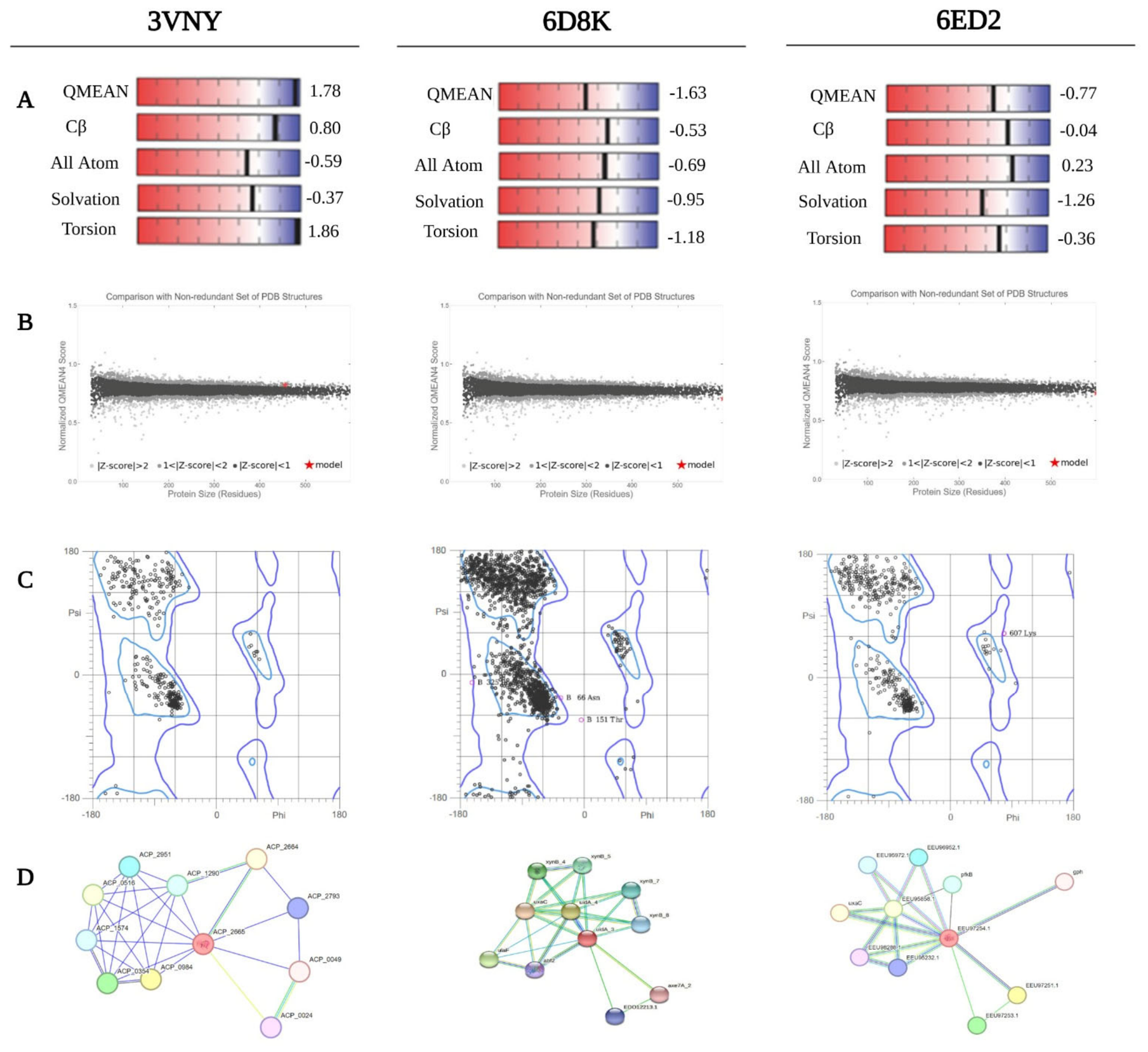
The SWISS-MODEL QMEAN tool was utilized to estimate the global model quality of the selected proteins by comparing their structures with the non-redundant PDB structure. Homology modeling of all three sequences is done using the SWISS-MODEL tool and 3D model for sequences 3VNY, 6D8K and 6ED2, as shown in Figure 2A. The expected QMEAN score should be within a range of 0–1 [52] and the Z-average value of a protein should be <1 [53]. Global model quality is estimated by comparing targeted protein structures with non-redundant PDB structures and prediction results are shown in Figure 2. GMQE score (ideally in the range 0–1, where 1 is a better score) is 0.93, 0.81 and 0.91, whereas QMEAN Z-score (values around 0 mean good quality structure, whereas −4.0 or below means low quality structure) is 1.78, −1.63 and −0.77 for 3VNY, 6D8K and 6ED2 respectively, as seen in Table 1. Local model quality has significance in structure interpretation and results show local model credibility with the amino acid sequence of each of the proteins, viz. 3VNY, 6ED2 and 6D8K, as seen in Figure 2B.
SAVES v6.0 server was used to verify the quality and stability of X-ray crystallographic structures of proteins, helpful for protein structure validation, in which functions like ERRAT and Verify3D are used for evaluations. ERRAT is a web-based server that helps to evaluate the model quality by analyzing the error in the crystallographic structure using statistical non-bonded atomic interaction [45]. Using ERRAT, the quality of each model is characterized by a single score based on the percentage (95%) of the structure that meets the confidence level of accuracy. In the present study, 6ED2, 3VNY and 6D8K showed ERRAT error values < 95: 94.683, 94.956, and 90.905 respectively.
The MolProbity [26] tool is used to judge the stereochemical quality of protein by analyzing various postscript plots of overall protein structure geometry and residue-by-residue geometry in the form of Ramachandran plot. It provides a graphical representation of the backbone torsion angles (φ and ψ) of amino acid residues, illustrating the conformational space corresponding to allowed secondary structure regions within distinct quadrants [54]. In quadrant 1, rare left-handed α-helices conformations are only allowed. In Quadrant 2, most favorable conformations of atoms are present. It displays the sterically permitted conformities for β-strands. Quadrant 3 is the second biggest region where right-handed alpha helices are placed. Quadrant 4 is the region where conformation with disfavored regions lie. In this study, Ramachandran plots of the three proteins 3VNY, 6D8K and 6ED2 showed that 98.92%, 92.94% and 97.61% (Figure 2C) amino acid residues are found in the most favorable region respectively, revealing the satisfactory quality of the selected model [48,55]. Verify 3D provides the percentage of residues with ≥0.2 3D-1D score (Table 1). Moreover, three of these models were reliable, as they had no or very few residues in disallowed regions.

3.4. Functional Diversification and Interaction Topology

TMHMM was used to analyze the presence of transmembrane helices to understand the location of the protein within the cell. In the present study, 3VNY and 6ED2 were found to have no transmembrane helix, confirming the cytoplasmic location; however, 6D8K contains one transmembrane helix, confirming a possible location near the cell membrane. InterPro is used for functional analysis of the proteins by grouping them into families and by predicting the domain position and important sites in the protein [27]. In the present study, 3VNY was found to have one glycosidase domain (37–367), one (trans)glycosidase domain (53–358), and one glycosyl hydrolase family 79 C-terminal beta domain (381–471). Present analyses also confirm that 3VNY belongs to the glycosyl hydrolase family, which includes enzymes with beta-glucuronidase activity, involved in the hydrolysis of β-glucuronosyl and 4-O-methyl-beta-glucuronosyl residues of arabinogalactan-protein. They are also capable of catalyzing (trans)glycosylation reactions, transferring glucuronic acid residues to various monosaccharide acceptors, including glucose, galactose, and xylose (https://www.ebi.ac.uk/interpro/result/InterProScan/iprscan5-R20260117-151535-0132-69302353-p2m/internal-1768662932929-1-1/, accessed on 24 January 2026). 6D8K also belongs to the same family as 3VNY. It was found to have a galactose-binding domain-like (28–198), glycosyl hydrolases family 2, sugar-binding domain (78–197 and 199–290), glycosidases (292–595) and immunoglobulin-like (Ig-like) fold. These are involved in biological processes related to carbohydrate metabolic processes (https://www.ebi.ac.uk/interpro/result/InterProScan/iprscan5-R20260117-153017-0552-12297732-p1m/internal-1768663813350-795-1/, accessed on 24 January 2026). Like the other two, 6ED2 contains a galactose-binding domain-like (25–207), glycosyl hydrolases family 2, sugar-binding domain (42–206), glycosidases (300–628) overlapped with (Trans)glycosidases domain in the same region, and beta-galactosidase/glucuronidase domain (212–299). Beta-galactosidase/glucuronidase domain superfamily has an immunoglobulin-like β-sandwich fold composed of seven strands in two sheets [48,56], classified in CAZy (CArbohydrate-Active EnZymes) hydrolases, the glycosidic bond between two or more carbohydrates, or between a carbohydrate and a non-carbohydrate moiety (https://www.ebi.ac.uk/interpro/result/InterProScan/iprscan5-R20260117-161018-0445-20371639-p1m/internal-1768666209895-1791-1/, accessed on 24 January 2026). Motifs were analyzed among all of the three proteins, where the highest number of motifs is found in 6D8K, i.e., 7; however, only 4 and 2 motifs are found in 6ED2 and 3VNY respectively as shown in Table 2. STRING v11.0 database is used to predict interactions of query sequences with closely related proteins [30]. As a result, we got three interactomes for each protein query, as shown in Figure 2D. Moreover, the conserved sequence patterns aligned in the β-glucuronidase sequences were visualized using WebLogo to identify conserved amino acid residues and motif regions (Figure S1).

3.5. Ligand Recognition Specificity and Binding Energetics

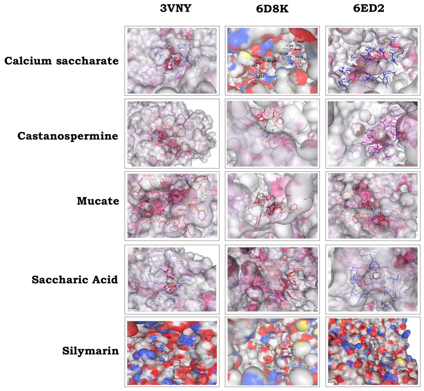
Among the structure-based approaches, molecular docking is the most used one. AutoDock 4.2, along with CBDock2, employs a rapid grid-based energy evaluation approach combined with an efficient search algorithm to explore ligand torsional flexibility, providing an automated method for predicting interactions between ligands and bio-macromolecular targets [35]. During the protein–ligand docking process, small molecules form cavities through hydrogen bonding, which helps to determine whether the binding site remains present throughout the docking [57,58]. It is a computational simulation that places the structure of the small molecule (ligand) in different orientations and conformations within the biomacromolecule (usually protein) active site, aiming to find the optimal binding mode through the calculation of the ligand–receptor binding energy [59]. In the present study, molecular docking of bacterial β-glucuronidase enzyme was predicted against five small molecules: calcium saccharate, castanospermine, mucate, saccharic acid, and silymarin. Accurate estimation of binding affinity, governed by specific recognition mechanisms between β-glucuronidase and its ligands, is critical for understanding structure–function relationships and guiding in silico evaluation of carbohydrate-active enzymes from the human gut microbiome. Such insights are essential for elucidating the potential functional roles of β-glucuronidase in the metabolism of dietary carbohydrates, xenobiotics, and host-derived conjugates within the human gut environment. The results of molecular docking are presented in Figure 3. Moreover, the amino acids involved in the docking interaction are presented in Figure S2. In the case of 3VNY, calcium saccharate exhibited the most favorable binding energy (−4.45 Kcal/mol) among other ligands, suggesting a stronger and more stable interaction with the enzyme’s active or binding site, whereas castanospermine showed the weakest interaction, with a binding energy of −0.75 kcal/mol, indicating limited binding compatibility in this structural context. On the other hand, for 6Ed2, castanospermine demonstrated the highest binding affinity (−4.7 kcal/mol), highlighting a better structural complementarity, while saccharic acid displayed the lowest binding energy (−2.7 kcal/mol), suggesting comparatively weaker interactions. 6D8K showed a similar kind of result, where castanospermine showed the strongest binding (−4.73 kcal/mol), whereas silymarin exhibited the least favorable interaction (−1.29 kcal/mol) (Table 3). These variations in binding energies across different β-glucuronidase structures emphasize the influence of structural differences and residue composition on ligand recognition and stabilization. The observed ligand-specific binding (Figure 3) preferences suggest that structural heterogeneity among gut bacterial β-glucuronidases may rule differential substrate or inhibitor interactions, potentially influencing enzyme functionality within the human gut environment. Such ligand recognition variability may represent an adaptive functional feature enabling efficient utilization of structurally diverse dietary glycans and xenobiotic compounds [60]. Therefore, the interplay between structural stability, active-site organization, and ligand-binding specificity is likely critical for maintaining metabolic versatility and microbial survival in the competitive gut environment.

3.6. Atomic-Level Non-Covalent Interaction Landscapes

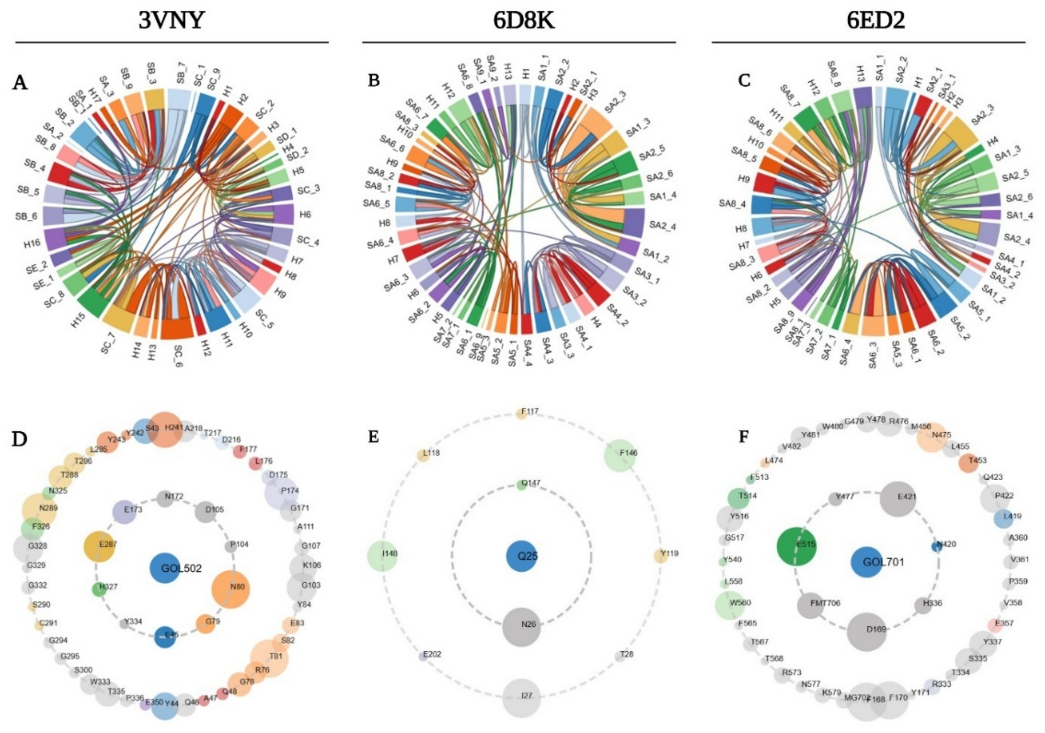
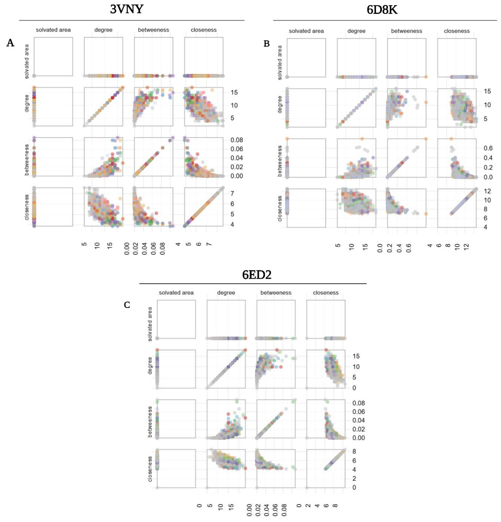
In the present study, Protein Contacts Atlas was used to visualize the protein non-covalent contacts [32]. The present analysis was performed for human gut bacterial β-glucuronidase enzyme, PDB ID: 3VNY with ligand propane-1,2,3-triol (GOL502); PDB ID: 6D8K with ligand 6-chloranyl-3-[(2-hexyl-2,3-dihydro-1,3-thiazol-4-yl)methyl]quinazolin-4-one (Q25); and PDB ID: 6ED2 with ligand propane-1,2,3-triol (GOL701) in chain A. Within the interaction network generated by PCA, nodes correspond to proteins or nucleic acids, while edges represent interaction interfaces connecting different chain subunits. Chord diagrams were used to visualize protein–protein contacts at the secondary structure level (Figure 4). In plots, each arc denotes an individual secondary structural element, and the connecting chords illustrate interaction frequencies between them. An interactive asteroid plot is demonstrated in Figure 4, where the inner ring indicates the first shell of immediate atomic contacts, whereas the outer ring indicates the second shell of extended atomic contacts. In addition, the size of the circle is proportional to the total number of contacts the residue is involved in with any of the residues in the ring inward to it. The asteroid plots were employed to portray the local atomic environment surrounding selected ligands, providing a ligand-centered view of interactions. The analysis revealed that ligand GOL502 forms 57 atomic contacts with its immediate neighboring residues in 3VNY, whereas the ligand Q25 showed total atomic residues of 32 as immediate contacts in 6D8K, and ligand GOL701 exhibited 41 total atomic contacts with its immediate neighboring residues in the case of 6ED2. In addition, scatter plots for 3VNY, 6D8K and 6ED2 (Figure 5) were generated to provide quantitative, residue-level interaction insights. Circular markers represent residues from the first chain, whereas square markers correspond to residues from the second chain.

4. Conclusions

This study presents a comprehensive in silico comparative structural and functional analysis of β-glucuronidase enzymes derived from three representative human gut bacterial species, Acidobacterium capsulatum ATCC 51196 (3VNY), Bacteroides ovatus (6D8K), and Faecalibacterium prausnitzii A2-165 (6ED2). Through an integrated bioinformatics framework, the work systematically evaluated physicochemical stability, secondary and tertiary structural organization, salt-bridge energetics, tertiary-structure validation, functional domain architecture, motif diversity, protein–protein interaction topology, ligand-binding energetics, and residue-resolved non-covalent interaction landscapes. Physicochemical characterization confirmed that all three enzymes exhibit thermostable, intracellular, and hydrophilic properties, while secondary-structure analysis demonstrated a coordinated balance between structural rigidity and functional flexibility essential for catalytic competence. Despite these shared characteristics, notable differences were observed among the enzymes. Structural validation analyses indicated that 6ED2 exhibited better overall structural stability, whereas 6D8K displayed a greater diversity of conserved motifs, a more complex interaction network, and enhanced ligand-binding capacity. Tertiary-structure validation using QMEAN, ERRAT, Verify3D, and Ramachandran plot analyses confirmed the high quality and reliability of all models, further supporting the observed differences in structural stability and functional specialization among the enzymes. Comparative analysis further suggested that although the catalytic framework of β-glucuronidases is highly conserved, differences in active-site microenvironments contribute to distinct ligand-recognition patterns. Molecular docking further revealed ligand-specific interaction preferences, particularly toward castanospermine and calcium saccharate, underscoring the influence of structural heterogeneity on catalytic recognition. Among the studied enzymes, 6D8K consistently exhibited the strongest binding affinities toward several ligands, especially castanospermine, suggesting greater substrate-recognition flexibility and a more favorable catalytic environment. In contrast, 3VNY and 6ED2 displayed distinct interaction profiles, reflecting species-specific variations in catalytic-center organization and ligand stabilization mechanisms. Crucially, PCA-based non-covalent interaction mapping provided an additional mechanistic layer by revealing distinct ligand-centered atomic contact networks and residue-level stabilization patterns across enzyme variants, thereby extending structural interpretation beyond conventional docking or validation metrics. These findings provide additional evidence that structural variations of the enzymes influence ligand stabilization and functional behavior. Overall, the integration of structural bioinformatics, interaction-network analytics, and ligand-binding thermodynamics provides a systems-level understanding of gut microbial β-glucuronidases. Collectively, the integration of physicochemical characterization, structural validation, docking analyses, and residue-level interaction mapping provides direct computational evidence supporting the functional diversification of gut microbial β-glucuronidases. The observed differences in structural stability, motif composition, catalytic-site environment, and ligand-binding behavior likely contribute to variations in substrate specificity and metabolic adaptability among gut microorganisms. The findings establish a robust computational foundation for future experimental validation, microbiome-informed therapeutic targeting, and translational biotechnological or cosmeceutical exploitation of CAZyme β-glucuronidases.

Supplementary Materials

The following supporting information can be downloaded at: https://www.mdpi.com/article/10.3390/bacteria5030039/s1. Figure S1: Representation of conserved sequence patterns aligned in β-glucuronidase amino acid sequences generated using WebLogo tool, where conserved motifs are indicated by (*) along with corresponding motif numbers according to Table 2. Figure S2: Molecular docking analysis of β-glucuronidase enzyme (3VNY, 6ED2 and 6D8K) and five ligands (Calcium Saccharate, Castanospermine, Mucate, Saccharic Acid, Silymarin) indicating catalytic pocket and specific amino acid (AA) residues involved in each docking.

Author Contributions

Conceptualization, A.B. and S.V.; methodology, A.S., L.G. and S.S.; formal analysis, A.S., L.G., A.B. and S.S.; investigation, A.S., L.G. and S.S.; data curation, A.S., L.G. and S.S.; writing—original draft preparation, A.S., L.G., S.S., A.B. and S.V.; writing—review and editing, A.S., L.G., S.S., A.B. and S.V.; supervision, A.B. and S.V. All authors have read and agreed to the published version of the manuscript.

Funding

This research received no external funding.

Institutional Review Board Statement

Not applicable.

Informed Consent Statement

Not applicable.

Data Availability Statement

The raw data supporting the conclusions of this article will be made available by the authors on request.

Acknowledgments

A.B. and S.V. acknowledge ANID Fondecyt Regular 1231917 by Govt. of Chile. S.S. acknowledges IUP24-12.

Conflicts of Interest

The authors declare no conflicts of interest.

References

  1. Coyte, K.Z.; Rakoff-Nahoum, S. Understanding Competition and Cooperation within the Mammalian Gut Microbiome. Curr. Biol. 2019, 29, R538–R544. [Google Scholar] [CrossRef] [PubMed]
  2. Van de Wouw, M.; Schellekens, H.; Dinan, T.G.; Cryan, J.F. Microbiota-gut-brain axis: Modulator of host metabolism and appetite. J. Nutr. 2017, 147, 727–745. [Google Scholar] [CrossRef] [PubMed]
  3. Kharb, A.; Zhu, X. Unlocking therapeutic impacts of the gut microbiota with computational tools. Curr. Opin. Biotechnol. 2026, 97, 103431. [Google Scholar] [CrossRef] [PubMed]
  4. Shayista, H.; Prasad, M.N.; Raj, S.N.; Prasad, A.; Satish, S.; Ranjini, H.K.; Manju, K.; Ravikumara Chouhan, R.S.; Khohlova, O.Y.; Perianova, O.V.; et al. Impact of macrolide antibiotics on gut microbiota diversity with age-specific implications and scientific insights. Med. Microecol. 2025, 24, 100122. [Google Scholar] [CrossRef]
  5. Ye, L.; Das, P.; Li, P.; Ji, B.; Nielsen, J. Carbohydrate active enzymes are affected by diet transition from milk to solid food in infant gut microbiota. FEMS Microbiol. Ecol. 2019, 95, 159. [Google Scholar] [CrossRef] [PubMed]
  6. Vernocchi, P.; Del Chierico, F.; Putignani, L. Gut Microbiota Metabolism and Interaction with Food Components. Int. J. Mol. Sci. 2020, 21, 3688. [Google Scholar] [CrossRef] [PubMed]
  7. Zhang, T.; Yang, Y.; Liang, Y.; Jiao, X.; Zhao, C. Beneficial effect of intestinal fermentation of natural polysaccharides. Nutrients 2018, 10, 1055. [Google Scholar] [CrossRef] [PubMed]
  8. Lyu, Y.; Wen, L.; Zhong, Y.; Yin, D.; Yang, M.; Wen, Y.; Li, Y.; Hu, J. Effects of Segatella copri-accessible polysaccharides on gut microbiota and intestinal immune function of CTX-induced immunocompromised mice. Food Biosci. 2026, 76, 108291. [Google Scholar] [CrossRef]
  9. Ameri, R.; García, J.L.; Derenfed, A.B.; Pradel, N.; Neifar, S.; Mhiri, S.; Mezghanni, M.; Jaouadi, N.Z.; Barriuso, J.; Bejar, S. Genome sequence and Carbohydrate Active Enzymes (CAZymes) repertoire of the thermophilic Caldicoprobacter algeriensis TH7C1T. Microb. Cell Fact. 2022, 21, 91. [Google Scholar] [CrossRef] [PubMed]
  10. Bhattacharya, T.; Ghosh, T.S.; Mande, S.S. Global Profiling of Carbohydrate Active Enzymes in Human Gut Microbiome. PLoS ONE 2015, 10, e0142038. [Google Scholar] [CrossRef] [PubMed]
  11. El Kaoutari, A.; Armougom, F.; Gordon, J.I.; Raoult, D.; Henrissat, B. The abundance and variety of carbohydrate-active enzymes in the human gut microbiota. Nat. Rev. Microbiol. 2013, 11, 497–504. [Google Scholar] [CrossRef] [PubMed]
  12. Garron, M.L.; Henrissat, B. The continuing expansion of CAZymes and their families. Curr. Opin. Chem. Biol. 2019, 53, 82–87. [Google Scholar] [CrossRef] [PubMed]
  13. Shen, J.; Xu, S.; Zhao, Q.; Zhang, J.; Zhang, H. β-Glucuronidase at the Microbiota—Host Interface: Dual Regulatory Roles and Precision Modulation by Natural Products. Molecules 2026, 31, 601. [Google Scholar] [CrossRef] [PubMed]
  14. Rehman, I.; Kerndt, C.C.; Botelho, S. Biochemistry, tertiary protein structure. In StatPearls; StatPearls Publishing: Treasure Island, FL, USA, 2024. Available online: https://www.ncbi.nlm.nih.gov/books/NBK470269/ (accessed on 24 January 2026).
  15. Suplatov, D.; Panin, N.; Kirilin, E.; Shcherbakova, T.; Kudryavtsev, P.; Švedas, V. Computational Design of a pH Stable Enzyme: Understanding Molecular Mechanism of Penicillin Acylase’s Adaptation to Alkaline Conditions. PLoS ONE 2014, 9, e100643. [Google Scholar] [CrossRef] [PubMed]
  16. Pal, S.; Sengupta, K. In silico analysis of phylogeny, structure, and function of arsenite oxidase from unculturable microbiome of arsenic contaminated soil. J. Genet Eng. Biotechnol. 2021, 19, 47. [Google Scholar] [CrossRef] [PubMed]
  17. Gloux, K.; Berteau, O.; El Oumami, H.; Beguet, F.; Leclerc, M.; Dore, J. A metagenomic β-glucuronidase uncovers a core adaptive function of the human intestinal microbiome. Proc. Natl. Acad. Sci. USA 2011, 108, 4539–4546. [Google Scholar] [CrossRef]
  18. Biernat, K.A.; Pellock, S.J.; Bhatt, A.P.; Bivins, M.M.; Walton, W.G.; Tran, B.N.T.; Redinbo, M.R. Structure, function, and inhibition of drug reactivating human gut microbial β-glucuronidases. Sci. Rep. 2019, 9, 825. [Google Scholar] [CrossRef] [PubMed]
  19. Gasteiger, E.; Hoogland, C.; Gattiker, A.; Duvaud, S.; Wilkins, M.R.; Appel, R.D.; Bairoch, A. Protein Identification and Analysis Tools on the ExPASy Server. In The Proteomics Protocols Handbook; Walker, J.M., Ed.; Humana Press: Totowa, NJ, USA, 2005; pp. 571–607. [Google Scholar] [CrossRef]
  20. Bozek, K.; Lengauer, T.; Sierra, S.; Kaiser, R.; Domingues, F.S. Analysis of Physicochemical and Structural Properties Determining HIV-1 Coreceptor Usage. PLoS Comput. Biol. 2013, 9, e1002977. [Google Scholar] [CrossRef] [PubMed]
  21. Ma, Y.; Liu, Y.; Cheng, J. Protein Secondary Structure Prediction Based on Data Partition and Semi-Random Subspace Method. Sci. Rep. 2018, 8, 9856. [Google Scholar] [CrossRef] [PubMed]
  22. Geourjon, C.; Deléage, G. SOPMA: Significant improvements in protein secondary structure prediction by consensus prediction from multiple alignments. Bioinformatics 1995, 11, 681–684. [Google Scholar] [CrossRef] [PubMed]
  23. Dutta, B.; Deska, J.; Bandopadhyay, R.; Shamekh, S. In silico characterization of bacterial chitinase: Illuminating its relationship with archaeal and eukaryotic cousins. J. Genet Eng. Biotechnol. 2021, 19, 19. [Google Scholar] [CrossRef] [PubMed]
  24. Costantini, S.; Colonna, G.; Facchiano, A.M. ESBRI: A web server for evaluating salt bridges in proteins. Bioinformation 2008, 3, 137–138. [Google Scholar] [CrossRef] [PubMed]
  25. Colovos, C.; Yeates, T.O. Verification of protein structures: Patterns of nonbonded atomic interactions. Protein Sci. 1993, 2, 1511–1519. [Google Scholar] [CrossRef] [PubMed]
  26. Williams, C.J.; Headd, J.J.; Moriarty, N.W.; Prisant, M.G.; Videau, L.L.; Deis, L.N.; Verma, V.; Keedy, D.A.; Hintze, B.J.; Chen, V.B.; et al. MolProbity: More and better reference data for improved all-atom structure validation. Protein Sci. 2018, 27, 293–315. [Google Scholar] [CrossRef] [PubMed]
  27. Hunter, S.; Apweiler, R.; Attwood, T.; Bairoch, A.; Bateman, A.; Binns, D.; Bork, P.; Das, U.; Daugherty, L.; Duquenne, L.; et al. InterPro: The integrative protein signature database. Nucleic Acids Res. 2009, 37, D211–D215. [Google Scholar] [CrossRef] [PubMed]
  28. Saleem, A.; Rajput, S. Insights from the in silico structural, functional and phylogenetic characterization of canine lysyl oxidase protein. J. Genet Eng. Biotechnol. 2020, 18, 20. [Google Scholar] [CrossRef] [PubMed]
  29. Pramanik, K.; Ghosh, P.K.; Ray, S.; Sarkar, A.; Mitra, S.; Maiti, T.K. An in silico structural, functional and phylogenetic analysis with three dimensional protein modeling of alkaline phosphatase enzyme of Pseudomonas aeruginosa. J. Genet Eng. Biotechnol. 2017, 15, 527–537. [Google Scholar] [CrossRef] [PubMed]
  30. Szklarczyk, D.; Gable, A.L.; Lyon, D.; Junge, A.; Wyder, S.; Huerta-Cepas, J.; Simonovic, M.; Doncheva, N.T.; Morris, J.H.; Bork, P.; et al. STRING v11: Protein–protein association networks with increased coverage, supporting functional discovery in genome-wide experimental datasets. Nucleic Acids Res. 2019, 47, D607–D613. [Google Scholar] [CrossRef] [PubMed]
  31. Maia, R.T.; Amador, V.C. Molecular Docking for Detoxifying Enzyme Studies. In Molecular Docking; InTech: Rijeka, Croatia, 2018. [Google Scholar] [CrossRef][Green Version]
  32. Sharma, S.; Kumar, P.; Chandra, R. Applications of BIOVIA Materials Studio, LAMMPS, and GROMACS in Various Fields of Science and Engineering. In Molecular Dynamics Simulation of Nanocomposites Using BIOVIA Materials Studio, Lammps and Gromacs; Sharma, S., Ed.; Elsevier: Amsterdam, The Netherlands, 2019; pp. 329–341. [Google Scholar] [CrossRef]
  33. Morris, G.M.; Huey, R.; Lindstrom, W.; Sanner, M.F.; Belew, R.K.; Goodsell, D.S.; Olson, A.J. AutoDock4 and AutoDockTools4: Automated docking with selective receptor flexibility. J. Comput. Chem. 2009, 30, 2785–2791. [Google Scholar] [CrossRef] [PubMed]
  34. Liu, Y.; Yang, X.; Gan, J.; Chen, S.; Xiao, Z.X.; Cao, Y. CB-Dock2: Improved protein–ligand blind docking by integrating cavity detection, docking and homologous template fitting. Nucleic Acids Res. 2011, 50, W159–W164. [Google Scholar] [CrossRef] [PubMed]
  35. Rizvi, S.M.D.; Shakil, S.; Haneef, M. A simple click by click protocol to perform docking: AutoDock 4.2 made easy for non-bioinformaticians. EXCLI J. 2013, 12, 831–857. [Google Scholar] [PubMed]
  36. Kayikci, M.; Venkatakrishnan, A.J.; Scott-Brown, J.; Ravarani, C.N.J.; Flock, T.; Babu, M.M. Visualization and analysis of non-covalent contacts using the Protein Contacts Atlas. Nat. Struct. Mol. Biol. 2018, 25, 185–194. [Google Scholar] [CrossRef] [PubMed]
  37. Michikawa, M.; Ichinose, H.; Momma, M.; Biely, P.; Jongkees, S.; Yoshida, M.; Kaneko, S. Structural and biochemical characterization of glycoside hydrolase family 79 β-glucuronidase from Acidobacterium capsulatum. J. Biol. Chem. 2012, 287, 14069–14077. [Google Scholar] [CrossRef] [PubMed]
  38. Pellock, S.J.; Walton, W.G.; Biernat, K.A.; Torres-Rivera, D.; Creekmore, B.C.; Xu, Y.; Redinbo, M.R. Three structurally and functionally distinct β-glucuronidases from the human gut microbe Bacteroides uniformis. J. Biol. Chem. 2018, 293, 18559–18573. [Google Scholar] [CrossRef] [PubMed]
  39. Ward, N.L.; Challacombe, J.F.; Janssen, P.H.; Henrissat, B.; Coutinho, P.M.; Wu, M.; Kuske, C.R. Three genomes from the phylum Acidobacteria provide insight into the lifestyles of these microorganisms in soils. Appl. Environ. Microbiol. 2009, 75, 2046–2056. [Google Scholar] [CrossRef] [PubMed]
  40. Gangadhar, C.G.; Rohit, B.K.; Basappa, B.K. In silico characterization of beta-galactosidase using computational tools. J. Bioinform. Seq. Anal. 2016, 8, 1–11. [Google Scholar] [CrossRef]
  41. Panda, S.; Chandra, G. Physicochemical characterization and functional analysis of some snake venom toxin proteins and related non-toxin proteins of other chordates. Bioinformation 2012, 8, 891–896. [Google Scholar] [CrossRef] [PubMed]
  42. Chang, K.Y.; Yang, J.R. Analysis and Prediction of Highly Effective Antiviral Peptides Based on Random Forests. PLoS ONE 2013, 8, e70166. [Google Scholar] [CrossRef] [PubMed]
  43. Hasan, R.; Rony, M.N.H.; Ahmed, R. In silico characterization and structural modeling of bacterial metalloprotease of family M4. J. Genet Eng. Biotechnol. 2021, 19, 25. [Google Scholar] [CrossRef] [PubMed]
  44. Brinck, J.E.; Sinha, A.K.; Laursen, M.F.; Dragsted, L.O.; Raes, J.; Uribe, R.V.; Licht, T.R. Intestinal pH: A major driver of human gut microbiota composition and metabolism. Nat. Rev. Gastroenterol. Hepatol. 2025, 22, 639–656. [Google Scholar] [CrossRef] [PubMed]
  45. Flores-Castañón, N.; Sarkar, S.; Banerjee, A. Structural, functional, and molecular docking analyses of microbial cutinase enzymes against polyurethane monomers. JHM Lett. 2022, 3, 100063. [Google Scholar] [CrossRef]
  46. Babicki, S.; Arndt, D.; Marcu, A.; Liang, Y.; Grant, J.R.; Maciejewski, A.; Wishart, D.S. Heatmapper: Web-enabled heat mapping for all. Nucleic Acids Res. 2016, 44, W147–W153. [Google Scholar] [CrossRef] [PubMed]
  47. Chauhan, R.; Prabhakaran, S.; Taj, G.; Guru, S.K.; Pandey, D. Structural and functional characterization of an amino acid transporter in Amaranthus hypochondriacus using bioinformatics tools. J. Adv. Biol. Biotechnol. 2024, 27, 280–288. [Google Scholar] [CrossRef]
  48. Sarkar, S.; Banerjee, A.; Chakraborty, N.; Soren, K.; Chakraborty, P.; Bandopadhyay, R. Structural-functional analyses of textile dye degrading azoreductase, laccase and peroxidase: A comparative in silico study. Electron J. Biotechnol. 2020, 43, 48–54. [Google Scholar] [CrossRef]
  49. Craveur, P.; Joseph, A.P.; Esque, J.; Narwani, T.J.; Noel, F.; Shinada, N.; Goguet, M.; Leonard, S.; Poulain, P.; Bertrand, O.; et al. Protein flexibility in the light of structural alphabets. Front. Mol. Biosci. 2015, 2, 20. [Google Scholar] [CrossRef] [PubMed]
  50. Villegas, V.; Viguera, A.R.; Avilés, F.X.; Serrano, L. Stabilization of proteins by rational design of α-helix stability using helix/coil transition theory. Fold. Des. 1996, 1, 29–34. [Google Scholar] [CrossRef]
  51. Kumar, S.; Nussinov, R. Salt bridge stability in monomeric proteins. J. Mol. Biol. 1999, 293, 1241–1255. [Google Scholar] [CrossRef] [PubMed]
  52. Berman, H.M.; Westbrook, J.; Feng, Z.; Gilliland, G.; Bhat, T.N.; Weissig, H.; Bourne, P.E. The protein data bank. Nucleic Acids Res. 2000, 28, 235–242. [Google Scholar] [CrossRef] [PubMed]
  53. Benkert, P.; Biasini, M.; Schwede, T. Toward the estimation of the absolute quality of individual protein structure models. Bioinformatics 2011, 27, 343–350. [Google Scholar] [CrossRef] [PubMed]
  54. Laskowski, R.A.; MacArthur, M.W.; Moss, D.S.; Thornton, J.M. PROCHECK: A program to check the stereochemical quality of protein structures. J. Appl. Crystallogr. 1993, 26, 283 91. [Google Scholar] [CrossRef]
  55. Pitman, M.R.; Menz, R.I. Methods for Protein Homology Modelling. In Applied Mycology and Biotechnology; Elsevier: Amsterdam, The Netherlands, 2006; pp. 37–59. [Google Scholar] [CrossRef]
  56. Jacobson, R.H.; Zhang, X.J.; DuBose, R.F.; Matthews, B.W. Three-dimensional structure of β-galactosidase from E. coli. Nature 1994, 369, 761–766. [Google Scholar] [CrossRef] [PubMed]
  57. Bulusu, G.; Desiraju, G.R. Strong and weak hydrogen bonds in protein–ligand recognition. J. Indian Inst. Sci. 2020, 100, 31–41. [Google Scholar] [CrossRef]
  58. Gaur, P.; Polipalli, Y.; Bandhu, X.; Chakravarty, S.; Banerjee, S.; Meza, C.; Suravajhala, R.; Sarkar, S.; Banerjee, A. Unveiling the potential of bacterial keratinase with antiacne drugs for cosmeceutical applications: An in silico evaluation. J. Umm Al-Qura Univ. Appl. Sci. 2025, 1–24. [Google Scholar] [CrossRef]
  59. Jereva, D.; Alov, P.; Tsakovska, I.; Angelova, M.; Atanassova, V.; Vassilev, P.; Ikonomov, N.; Atanassov, K.; Pajeva, I.; Pencheva, T. Application of InterCriteria Analysis to Assess the Performance of Scoring Functions in Molecular Docking Software Packages. Mathematics 2022, 10, 2549. [Google Scholar] [CrossRef]
  60. de Kivit, S.; Kraneveld, A.D.; Garssen, J.; Willemsen, L.E. Glycan recognition at the interface of the intestinal immune system: Target for immune modulation via dietary components. Eur. J. Pharmacol. 2011, 668, S124–S132. [Google Scholar] [CrossRef] [PubMed]
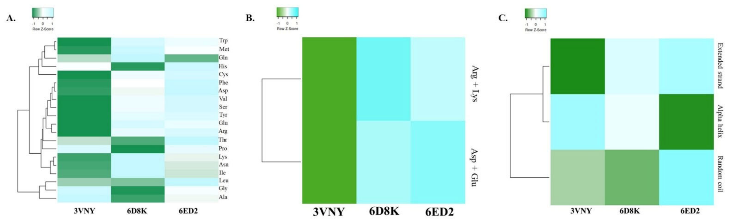
Figure 1. (A) Amino acid composition of all three β-glucuronidase enzyme sequences produced by three different bacteria. (B) Variance in the composition of negatively charged (Asp + Glu) and positively charged (Arg + Lys) amino acid residues in the primary structure of β-glucuronidases. (C) Comparison of secondary structure among three proteins.
Bacteria 05 00039 g001
Figure 2. (A) Evaluation of Z-score and (B) QMEAN score of local quality estimate and comparison with a non-redundant set of PDB structure. (C) Ramachandran plot for residues of β-glucuronidase enzyme predicted by MolProbity. (D) Interactome of the targeted protein predicted by STRING.
Bacteria 05 00039 g002
Figure 3. Molecular docking analysis of β-glucuronidase enzyme (3VNY, 6ED2 and 6D8K) and five ligands (Calcium Saccharate, Castanospermine, Mucate, Saccharic Acid, Silymarin).
Bacteria 05 00039 g003
Figure 4. Non-covalent interaction analysis of CAZyme β-glucuronidase using the protein contact atlas. Chord plot of (A) 3VNY, (B) 6D8K, (C) 6ED2 representing residue–residue interaction across the protein structure. Asteroid plot of (D) 3VNY, (E) 6D8K, (F) 6ED2, highlighting significant contact points within the protein.
Bacteria 05 00039 g004
Figure 5. Scatter plot analysis from the protein contact atlas, visualizing non-covalent interaction for (A) 3VNY, (B) 6D8K, (C) 6ED2, where each plot highlights residue–residue contact points within the protein structure, showing their respective interaction patterns.
Bacteria 05 00039 g005
Table 1. Comparative physicochemical parameters as well as quality assessment scores of the three different CAZymes under study.
BacteriaPDB IDStructural AnalysisQuality Assessment Scores
Number of Amino AcidsMW (KDa)Theoretical pIAIIIGRAVYQMEAN Z-ScoreERRAT Quality Factor3D-1D Score (%)AA in FR of Ramachandran Plot (%)
Faecalbacterium prausnitzii A2-1656ED263171.8986.2474.5234.99−0.381−0.7794.68398.4891.7
Acidobacterium capsulatum ATCC 511963VNY48852.2846.2473.3626.73−0.2651.7894.95697.6491.4
Bacteroides ovatus KLE16566D8K59769.2648.3377.2538.38−0.496−1.6390.90597.4684.4
MW—Molecular Weight; II—Instability Index; AI—Aliphatic Index; GRAVY—Grand Average Hydropathy; FR—Favorable Region.
Table 2. Motif search results for three query sequences via Motif search tool.
ProteinNo. of MotifsPfamPositionDescription
3VNY2Glyco_hydro_79_n74–310PF03662, Glycosyl hydrolase family 79, N-terminal domain
Glyco_hydro_79_c381–470PF16862, Glycosyl hydrolase family 79 C-terminal beta domains
6D8K7Glyco_hydro_2_C292–550PF02836, Glycosyl hydrolases family 2, TIM barrel domain
Glyco_hydro_2_N35–197PF02837, Glycosyl hydrolases family 2, sugar binding domain
Glyco_hydro_2199–290PF00703, Glycosyl hydrolases family 2
BetaGal_dom4_586–167PF13364, Beta-galactosidase jelly roll domain
Acetyltransf_1279–365PF00583, Acetyltransferase (GNAT) family
TT1725281–349PF18324, Hypothetical protein TT1725
Acetyltransf_3272–365PF13302, Acetyltransferase (GNAT) domain
6ED24 Glyco_hydro_2_C300–621PF02836, Glycosyl hydrolases family 2, TIM barrel domain
Glyco_hydro_2_N42–206PF02837, Glycosyl hydrolases family 2, sugar binding domain
Glyco_hydro_2221–298PF00703, Glycosyl hydrolases family 2
BetaGal_dom4_580–160PF13364, Beta-galactosidase jelly roll domain
Table 3. Binding Energy of β-glucuronidase enzyme (3VNY, 6ED2 and 6D8K) and five ligands (Calcium Saccharate, Castanospermine, Mucate, Saccharic Acid, Silymarin).
LigandBinding Energy (Kcal/mol)
3VNY6ED26D8K
Calcium Saccharate−4.45−4.5−3.34
Castanospermine−0.75−4.7−4.73
Mucate−0.97−3.6−2.96
Saccharic Acid−0.99−2.7−2.06
Silymarin−3.65−3.6−1.29
Disclaimer/Publisher’s Note: The statements, opinions and data contained in all publications are solely those of the individual author(s) and contributor(s) and not of MDPI and/or the editor(s). MDPI and/or the editor(s) disclaim responsibility for any injury to people or property resulting from any ideas, methods, instructions or products referred to in the content.