Review

Advanced Sequencing Approaches for the Subgingival Microbiome: Technology Selection, Quality Control, and Best Practices in Periodontal Research


by
Hadeel Mazin Akram
* and
Saif Sehaam Saliem
College of Dentistry, University of Baghdad, Baghdad 10071, Iraq
*
Author to whom correspondence should be addressed.
Bacteria 2026, 5(1), 11; https://doi.org/10.3390/bacteria5010011
Submission received: 22 October 2025 / Revised: 8 January 2026 / Accepted: 6 February 2026 / Published: 9 February 2026
(This article belongs to the Special Issue Bacterial Molecular Biology: Stress Responses and Adaptation)

Abstract

Sequencing technologies have reshaped the study of the subgingival microbiome, but selecting the appropriate method remains challenging because of differences in resolution, cost, host DNA contamination, and computational complexity. This review compares 16S rRNA sequencing, full-length 16S, shotgun metagenomics, and metatranscriptomics with respect to taxonomic resolution, functional output, sample requirements, and analytical limitations. Key practical issues, including low microbial biomass, contamination control, and the choice of appropriate bioinformatic tools, are emphasized to help researchers avoid common pitfalls. A decision-making framework is provided to link study goals to suitable sequencing methods while outlining realistic budget and sample-handling constraints. The review concludes with recommendations for integrating sequencing with complementary techniques to improve the accuracy, reproducibility, and clinical relevance of periodontal microbiome studies.

1. Introduction

Subgingival plaque is low-biomass, host-DNA-rich, and structurally organized, so method choice is rarely neutral; it shifts which organisms appear and which conclusions are defensible [1]. This narrative review synthesizes current evidence on sequencing technologies for subgingival microbiome analysis and offers practical guidance for choosing methods based on research goals, sample features, and available resources.
Marker-gene (16S) surveys still work well for large cohorts and diversity patterns, but they limit taxonomic resolution and infer function indirectly [2]. Shotgun metagenomics resolves species/strains and genes, although human DNA can dominate libraries and skew the cost–benefit equation [3]. Full-length 16S (PacBio HiFi) can close some taxonomic gaps at a lower cost than shotgun sequencing, yet it still lacks direct functional readouts [4]. A question-first design is advisable: select the least complex assay that can answer the clinical or mechanistic question, and pair it with rigorous contamination controls and compositional statistics.
The subgingival microbiome is a highly complex and dynamic ecosystem of bacteria, archaea, fungi, and viruses that colonize the space between the teeth and the gingiva [1,5,6]. Dysbiosis within this community contributes to periodontal disease progression [1,7], and periodontitis has been linked to various systemic diseases, including cardiovascular disease, diabetes, and rheumatoid arthritis [8,9,10,11].
Recent developments in sequencing techniques have revolutionized our knowledge of periodontal pathogenesis. 16S rRNA sequencing-based large-scale studies have discovered core microbiomes and disease-associated taxa in a variety of populations [12,13], whereas shotgun metagenomics has elucidated the functional potentials and strain-level diversity responsible for pathogenicity [14,15]. The use of metatranscriptomic strategies is today offering novel windows into active microbial processes in response to disease and treatment [16,17]. It is also important to recognize that different sequencing platforms detect different biological domains: 16S identifies bacteria and archaea, whereas shotgun metagenomics and metatranscriptomics are required to study fungi and viruses [18].
Despite these advances, the rapid expansion of sequencing platforms has made it difficult to select the most appropriate method for periodontal research, particularly in low-biomass, host-DNA-rich environments such as subgingival plaque. Prior reviews outline the general features of different approaches but offer limited guidance on selecting methods for specific research questions. This review addresses that gap by linking common periodontal research aims such as identifying dysbiotic communities, comparing strain-level diversity of Porphyromonas gingivalis, detecting antibiotic-resistance or virulence pathways, assessing short-term functional shifts following therapy, or examining host–microbiome interactions, to the sequencing strategies most suited to answering them. The aim is to outline the strengths and limitations of each method and provide practical decision-making guidance based on research objectives, biological constraints, and available resources.

2. Materials and Methods

2.1. Review Design and Scope

This narrative review synthesizes current evidence on advanced sequencing technologies for subgingival microbiome analysis. Unlike systematic reviews that follow rigid inclusion criteria, narrative reviews allow for broader synthesis of diverse study types and methodological approaches, making them particularly suitable for rapidly evolving technical fields where standardized protocols are still emerging [19].

2.1.1. Literature Search Strategy

A thorough search was conducted in various databases, including PubMed/MEDLINE, Web of Science, and Google Scholar, to identify relevant articles. The search strategy included using MeSH/THES and free-text terms about the oral and periodontal microbiome, sequencing technologies, and the primary methodological steps. Key search terms were “subgingival microbiome”, “periodontal microbiome”, “oral microbiome” and “dental plaque microbiome”. These were joined with technology name keywords such as ”16S rRNA sequencing,” “shotgun metagenomics,” “metatranscriptomics,” “long-read sequencing,” “PacBio” and “Oxford Nanopore.” In addition, to collect methodological literature, “sample collection,” “contamination control,” “host DNA depletion,” compositional analysis”, and STORMS guidelines were used. Finally, disease-specific clinical entities such as “periodontitis,” “gingivitis” or “peri-implantitis” were employed to narrow down the search results based on relevant diseases.

2.1.2. Study Selection and Data Synthesis

The search initially identified 642 records. After title/abstract screening, 211 articles remained. Following full-text assessment, 120 studies were included in the final synthesis (Figure 1). Studies were selected for inclusion if they involved human subjects with periodontal health or disease and analyzed samples from subgingival plaque, supragingival plaque, or saliva. The research must have utilized a high-throughput sequencing approach, such as 16S rRNA, shotgun metagenomics, metatranscriptomics, or long-read sequencing. Original research articles, methodological studies, comparative analyses, and high-quality reviews published in English were considered. Conversely, studies were excluded if they were conducted exclusively on non-human subjects or in vitro models, relied solely on culture-based techniques or quantitative PCR without high-throughput sequencing, or analyzed non-oral sample types. Case reports, editorials, and non-peer-reviewed conference abstracts were also excluded, as were studies with insufficient methodological detail to allow for a comprehensive quality assessment. This review considered studies published between January 2010 and October 2025.
This study summarized aspects of study design, population characteristics, sample collection and storage, sequencing technology and bioinformatics pipeline, quality control measures, statistical considerations, and major findings with respect to the performance of the technology irrespective of cost or practical consideration for each eligible paper. The findings of this review were thematically analyzed, rather than a meta-analysis with an emphasis towards technology selection and best practice methodologies in periodontal research. Sample size, negative controls, bioinformatics transparency, and adherence to reporting guidelines such as STORMS and MIMARKS were all considered measures of the quality of microbiome studies [20,21].

3. Overview of Sequencing Technologies for Subgingival Microbiome Research

Each sequencing approach differs in the types of organisms it can detect in subgingival plaque [22]. Standard 16S rRNA sequencing captures bacteria and archaea but does not provide information on fungi or viruses. Shotgun metagenomics covers all major microbial domains, including bacteria, archaea, fungi, and viruses, and allows microbial profiles to be interpreted alongside host DNA when needed [16]. Long-read platforms such as PacBio and Oxford Nanopore can also detect multiple domains and produce more complete assemblies, though their higher cost limits their routine use. Metatranscriptomics extends beyond organism identification by measuring actively expressed microbial and host genes, making it particularly useful for examining functional interactions between the microbiome and the host [22].
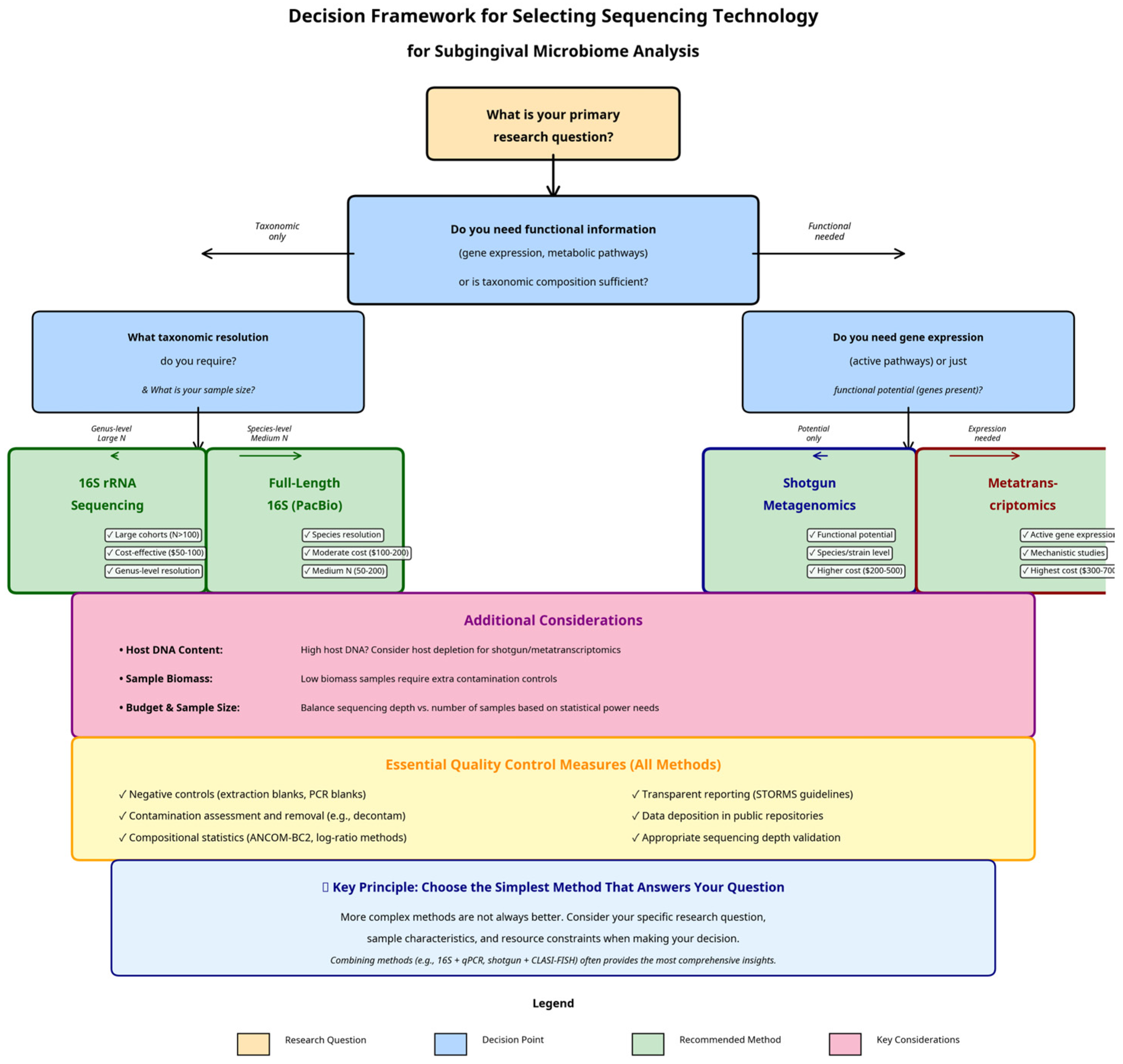
Figure 2 summarizes a practical decision-making framework for selecting the most appropriate sequencing approach based on research objectives, sample constraints, and required resolution.

3.1. 16S rRNA Gene Sequencing

The use of 16S rRNA gene sequencing is a “marker-gene” approach that has traditionally served as the principal tool for identifying taxa within microbial communities. In essence, 16S rRNA gene sequencing involves the PCR-amplification and subsequent sequencing of a defined region(s) of the 16S ribosomal RNA gene. Since each bacterial and archeal organism will contain the 16S ribosomal RNA gene, the gene’s variability allows investigators to determine whether two organisms are related to one another at various taxonomic levels. 16S rRNA gene sequencing is so widely applied today because of its relatively low cost and ability to support large-scale epidemiological studies using thousands of individual samples [12,23]. A second factor contributing to the popularity of 16S rRNA gene sequencing is the presence of well-established protocols and standardized bioinformatic pipelines, e.g., QIIME 2 and DADA2, which provide the investigator with reproducible and comparable results among multiple studies [24,25].
Despite its widespread application, several important limitations of the 16S rRNA gene sequencing approach exist. The most significant limitation of 16S rRNA gene sequencing is its limited taxonomic resolution. While 16S rRNA gene sequencing may be able to classify microbes down to the genus level consistently, the classification of microbes to the species level often proves difficult, and identification at the strain level is virtually impossible [26]. These limitations in resolution represent a major problem in periodontal research since different species and strains that belong to the same genus can have very different degrees of pathogenicity. Additionally, while 16S rRNA gene sequencing provides no direct information regarding the function of an organism, there are tools available (e.g., PICRUSt2), that allow researchers to predict function based on the taxonomic profile generated from 16S rRNA gene sequencing data. However, these predicted functions are based on the reference genomes of the organisms included in the database and should be viewed with caution; ideally, validation of the predicted functions would be conducted using shotgun metagenomic data when possible [2,27]. Finally, the selection of primers and the specificity of the hypervariable region targeted can significantly affect the representation of the microbial community, thereby potentially introducing bias into the results of 16S rRNA gene sequencing experiments [28,29].

3.2. Shotgun Metagenomics

Whole-genome shotgun (WGS) metagenomics offers a much higher-resolution alternative to 16S rRNA sequencing. Instead of targeting a single marker gene, this approach sequences all genomic DNA in a sample, including that from bacteria, archaea, viruses, and the host. This untargeted approach provides a comprehensive view of the microbial community, allowing for accurate species- and even strain-level taxonomic identification [14,30]. The most significant advantage of WGS is its ability to provide direct information about the functional potential of the microbiome. By analyzing the full complement of genes present, researchers can identify metabolic pathways, virulence factors, and antibiotic resistance genes, offering deep insights into what the microbial community is capable of doing [31,32].
WGS metagenomics has a number of limitations, primarily in terms of analyzing subgingival plaque. Contamination with host (human) DNA represents the greatest limitation to WGS metagenomics. For example, human DNA can comprise greater than 90% of the total DNA within a subgingival sample. Consequently, the majority of the cost and reads generated by sequencing the host genome as opposed to the microbial genome [3,33]. Although many host DNA depletion strategies have been developed, each may also introduce bias [34]. Furthermore, the cost per sample will be significantly higher than 16S sequencing, which could hinder large-scale analyses. Additionally, the study of WGS data is much more computationally intensive and requires the use of specialized bioinformatic tools and computing resources to assemble, bin, and functionally annotate [35,36].

3.3. Metatranscriptomics: Unveiling the Active Microbiome

Although metagenomics assesses the functional capacity of a microbial community (i.e., the genes it contains), meta-transcriptomics measures the functional expression of a microbial community through the sequencing of all of the RNA transcripts present within a sample. As such, this methodology permits investigators to determine which genes are currently being transcribed (expressed) by the microbiome at a particular point in time, thereby offering a more real-time and responsive representation of the microbial functions occurring in response to environmental stimuli, e.g., the transition between periodontal health and disease [16,37]. Therefore, metatranscriptomics represents an exceptional tool for use in mechanistic investigations directed toward understanding the molecular mechanisms responsible for the progression of disease or the microbial response to treatment [38,39]. An example of the utility of metatranscriptomics was demonstrated through a meta-analysis of metatranscriptomic data, in which specific virulence factor gene expression from P. gingivalis and T. forsythia was found to be upregulated during periodontitis, thus providing direct evidence of their involvement in the diseased state [16].
A primary limitation of metatranscriptomics is that RNA is inherently unstable and degrades faster than DNA, requiring strict adherence to sampling and preservation protocols, e.g., immediate flash-freezing in liquid nitrogen, which may be difficult to accomplish in a clinical environment [40,41]. Additionally, virtually all of the RNA within a bacterial cell is composed of ribosomal RNA (rRNA), whereas mRNA is relatively rare in bacteria. Therefore, an effective rRNA depletion step is crucial to ensure that sequencing efforts are focused on the functionally informative mRNA transcripts [42,43]. The cost of metatranscriptomics is also significantly higher than both 16S and WGS approaches, and the data analysis is even more complex, requiring specialized pipelines to map transcripts to genomes and quantify gene expression levels [44,45].

3.4. A Comparative Framework for Technology Selection

Choosing the right technology requires a careful consideration of the trade-offs between taxonomic resolution, functional information, cost, and the specific challenges of the research question. A summary of these trade-offs is presented in Table 1 below.
For large-scale epidemiological studies aiming to characterize broad shifts in microbial diversity across hundreds of subjects, 16S rRNA sequencing remains the most practical and cost-effective choice [12,46]. When the research question requires precise species- or strain-level identification, or when understanding the functional potential of the community is critical (e.g., investigating antibiotic resistance), the higher resolution of shotgun metagenomics is necessary [14,32]. Metatranscriptomics should be reserved for studies where the primary goal is to understand the active functional processes driving a specific phenotype, such as the active inflammatory response in periodontitis, despite its higher cost and technical complexity [16,39]. Ultimately, an integrated approach, where initial large-scale 16S surveys are followed by deeper WGS or metatranscriptomic analysis on a subset of informative samples, may provide the most comprehensive insights into the role of the subgingival microbiome in periodontal health and disease.

4. Methodological Considerations in Subgingival Microbiome Research

The quality and accuracy of the information regarding the subgingival microbiome depend greatly on the researcher’s methodological decisions in all aspects of the research process (sample collection through final data analysis). In addition, the specific environment of the subgingival crevice, characterized by low levels of microorganisms, high levels of host DNA, and a defined biofilm structure, increases the influence of those decision-making processes [47,48].

4.1. Dental Plaque Collection Methods

Sampling of subgingival plaque using various sampling techniques is a critical first step in generating an accurate microbiome profile. Each sampling technique samples different microorganisms (or microbial niches) within the subgingival (periodontal) environment and has its own degree of potential for introducing both bias and/or contamination [49]. Therefore, two of the most common tools used to collect subgingival plaque (curettes and paper points), have their own unique advantages/disadvantages depending on the objectives of the study.

4.1.1. Curette Sampling

Curettes are considered the gold standard method for obtaining subgingival plaque from periodontal pockets in both clinical practice and research environments [50]. The process of using a curette is simple; a clean Gracey or universal curette is inserted at the base of the pocket, and the curette is then scraped along the root surface to dislodge and physically remove the biofilm [51]. This method has many benefits, it will allow the collection of the total biofilm from the entire pocket, thus allowing the collection of both biofilm-attached and planktonic free-floating organisms, therefore creating an overall view of the microbial population present within the pocket. Because curette sampling disrupts the biofilm in a manner that is most likely to capture all microorganisms, including tightly adhered ones, it generally collects a greater amount of microbial biomass than do other methods of sample collection, and in addition, tends to collect a greater number of different microorganisms [52]. As a result, there is a greater likelihood that the species being sampled will be intact, and thus less likely to be damaged by the physical forces used during the sample collection process, thereby reducing the potential for sampling errors due to physical damage of the microorganisms being studied [53]. Although there are disadvantages associated with the use of curettes for the collection of periodontal pathogens, namely; it is an invasive method which requires a great deal of clinical experience to perform effectively and may cause discomfort to the patient; in addition, there is a high degree of variability in the quality of the sample depending on how well the sample area is isolated, and there is a possibility of contamination of the sample from supra-gingival plaque and/or saliva if the sample area is not properly isolated before the collection of the sample. Regardless of these limitations, when comparing results obtained using curette samples with those obtained using other methods of sample collection, researchers have consistently found that the curette sample contains a greater number of different microorganisms, i.e., greater species richness, and a greater diversity of the species present, i.e., greater Shannon diversity [54]. For example, Beyer et al. (2017) reported that they found greater relative abundance values of the periodontal genus Corynebacterium, Prevotella, Selenomonas, Actinomyces, and Treponema in the curette samples, versus those samples collected with paper points. These findings indicate that the curette is highly effective in the collection of the anaerobic community commonly found in diseased sites [54].

4.1.2. Paper Point Sampling

Paper points are a good substitute to curettes for studying periodontal disease because they are easy to use and do not involve as much invasion into the periodontal pocket as does a curette.
The process begins with the insertion of an absorbent paper point into the gingival sulcus, then the paper point is left there for a short time, usually 10–30 s, allowing the paper point to soak up gingival crevicular fluid and any bacteria loosely attached to the gingiva. Paper point sampling is easy to perform, has a low learning curve, is less painful for the patient, and allows for fast and standardized sampling that is ideal for large-scale studies and clinical monitoring [55,56]. One of the limitations of paper point sampling is that it tends to sample only those bacteria that are loose on the surface of the biofilm, rather than capturing all of the bacteria tightly embedded within the biofilm. Consequently, many researchers report a lower bacterial diversity than what is found when using a curette [54], and in addition, the amount of biomass obtained is generally lower than that obtained by using a curette. This can create problems when trying to sequence the community because the amount of DNA needed for the sequencing is typically higher than the amount produced by the paper point sampling [53]. Additionally, many studies have identified that background DNA contamination from the paper points used to collect the DNA can be a major problem. For example, Exiguobacterium, Enterococcus, and Pseudomonas were identified as contaminants on unused, sterile paper points [54,57]. Therefore, including unused paper points as negative controls is an important quality control step to consider when collecting DNA samples with paper points.

4.2. Sample Processing and Storage

The proper processing and storing of the collected samples are very important to preserve the quality of the microbial community profile from the time of sampling until the time that it is analyzed. It is best to process the samples immediately at the time of collection. However, if immediate processing is not feasible, samples should be stored on ice and processed within several hours to limit changes in the microbial community composition [53]. Long-term storage requires that the samples be kept at −80 degrees Celsius when analyzing DNA (DNA-based) studies. When conducting metatranscriptome studies, the analysis of unstable RNA molecules used in these studies requires samples to be immediately frozen in liquid nitrogen and then stored at −80 degrees Celsius to prevent RNA degradation [40]. Preservation media (e.g., TE buffer, PBS, etc.) have been shown to aid in preserving the quality of the sample; however, the most suitable medium will likely depend on the method of analysis and application downstream [55]. Lastly, it is imperative to limit the number of times samples are thawed, because each cycle of freezing and thawing results in the destruction of cells, resulting in an alteration of the microbial community structure. Creating aliquots of the samples at the time of collection is a cost-effective and efficient means of limiting the number of thaw cycles [53].

4.3. Quality Control and Contamination Management

Subgingival plaque contains very low microbial biomass, which makes contamination a critical concern at nearly every step of sampling, extraction, and sequencing. Contaminants may arise from sampling instruments, paper points, clinical environments, extraction kits, or laboratory reagents, and their influence is often magnified during PCR amplification [48,58]. Because of this, the use of appropriate negative controls is essential. Unused sterile curettes or paper points, along with reagent-only extraction blanks, should be processed in parallel with clinical samples to identify non-biological sequences introduced during collection or laboratory handling. These controls allow investigators to detect and remove contaminant taxa from the dataset during downstream bioinformatic filtering [59,60].
Beyond negative controls, several safeguards should be incorporated into routine workflows to minimize false signals. Physical separation of pre-PCR and post-PCR areas reduces the risk of aerosolized contamination, and clean, UV-treated workspaces with filtered pipette tips help maintain a controlled environment [60]. Sequencing a defined mock community provides an additional layer of validation by revealing extraction or amplification bias, while host DNA depletion strategies, although sometimes necessary, should be tested carefully, and any loss in microbial yield documented. Computational approaches such as removing taxa enriched in negative controls or using dedicated tools like Decontam, further improve data reliability by distinguishing true low-abundance organisms from contaminants [60].
Standardization of procedures also plays an important role in ensuring data integrity. Detailed and consistently applied Standard Operating Procedures (SOPs) minimize batch effects and increase confidence that observed differences between study groups reflect biological variation rather than technical inconsistencies [61]. Transparency in reporting is equally important. The STORMS (Strengthening the Organization and Reporting of Microbiome Studies) checklist [20], provides a structured framework for documenting sample collection, storage, extraction, sequencing, and analysis. Adhering to such guidelines enhances reproducibility and improves the clarity and reliability of microbiome research. Collectively, rigorous contamination control, standardized workflows, and clear reporting practices strengthen the validity of subgingival microbiome studies and reduce the risk of misinterpreting technical artifacts as biological findings.

4.4. 16S rRNA Amplicon Sequencing in Periodontal Research

16S rRNA amplicon sequencing is the most common methodology for studying microbial communities in periodontology, as it is cost-effective, has established methods, and is validated for oral microbiome studies. At approximately $50 per sample, it enables large cohort studies with hundreds of participants, necessary for discovering biomarkers and understanding microbial communities at the population level [62]. The selection of specific areas within the 16S rRNA gene for primer targeting significantly influences taxonomy resolution and microbial community profiles. Studies show that V1–V3 regions provide better taxonomic resolution for oral taxa than V3–V4 regions, particularly for identifying similar species within Streptococcus, Prevotella, and Fusobacterium genera [28]. Nagai et al. (2024) analyzed several primer pairs across oral samples and found V1–V2 regions most effective for oral microbiome analysis, achieving 95% accuracy for genus-level and 78% accuracy for species-level identifications [29]. While V3–V4 regions remain popular due to compatibility with Illumina sequencing and extensive use in past studies, V1–V2 regions are considered optimal for oral microbiome analysis. The validation of primers against oral-specific databases like HOMD is crucial, as studies show significant gaps in primer coverage for oral taxa, including uncultivated species that may influence periodontal health and disease [63].
Studies utilizing the 16S rRNA gene have contributed to our basic understanding of the role of the oral microbiome in the development of periodontal diseases and responses to treatments. The most notable example of such a study was the study of Arredondo et al. (2023); these investigators collected subgingival samples from 1020 individuals from four countries and observed both common and population-specific microbiological patterns of periodontitis [23]. These investigators found that a core set of 15 bacterial genera was present in each population and consistently associated with periodontal disease; however, they also observed significant differences in the community structures of the subgingival microbiota between the four populations studied.
Researchers have investigated whether microbial community compositions are linked to systemic health. Miyauchi et al. (2025) showed that periodontitis patients exhibit persistent alterations in oral and gut microbiomes, resulting in distinct serum metabolome profiles indicating systemic metabolic effects [64]. Modern 16S rRNA analysis has evolved from operational taxonomic unit (OTU) clustering to amplicon sequence variant (ASV) approaches, providing single-nucleotide resolution. Processing reads with QIIME 2 using DADA2 for ASV inference is now recommended, with all non-default parameters reported [24,25]. Downstream differential abundance testing should use methods like ANCOM-BC2 that account for compositional constraints and allow covariate adjustment and repeated measures [65]. Recent improvements have addressed oral microbiome analysis challenges, including oral-specific taxonomic classifiers trained on HOMD sequences, improving species-level assignments for oral taxa by 15–20% compared to generic classifiers [66]. The integration of phylogenetic information through tools like PICRUSt2 enables functional prediction from 16S data, though these predictions should be validated with shotgun metagenomics when possible [27].

4.5. Full-Length 16S Sequencing with Long-Read Technologies

Full-length 16S sequencing using PacBio HiFi or Oxford Nanopore technologies represents a valuable middle ground between short-read 16S and shotgun metagenomics. By sequencing the entire ~1500 base pair 16S rRNA gene rather than short hypervariable regions, this approach achieves significantly improved taxonomic resolution while maintaining the simplicity and lower cost of marker-gene surveys [4]. A recent study comparing different methods of benchmarking indicated that 16S rDNA sequencing from start to finish (full-length) could achieve an accurate species assignment for approximately 95% of species in an oral sample, while sequencing the V3–V4 region could only accurately assign a species about 65% of the time [67]. Therefore, an increased species-assignment accuracy is most beneficial when used in clinical settings in which species identification will be directly used in making decisions on treatment.
The full-length 16S rDNA sequencing method enables species-level resolution without costly shotgun metagenomic analysis. Recent applications include strain-level epidemiology studies tracking pathogens through treatment and identifying transmission patterns in family cohorts [67]. A key application is in peri-implantitis studies, where species-level resolution helps understand relationships between implant-associated and periodontal microbiomes. Long-read 16S studies show that while overall community structure is similar between peri-implant and periodontal sites, specific strains of Porphyromonas gingivalis and Aggregatibacter actinomycetemcomitans show distinct associations with implant surfaces versus natural teeth [68]. The cost-effectiveness of full-length 16S depends on research questions and sample size. For studies needing species-level resolution in moderate sample sizes (50–200 samples), full-length 16S provides better value than shotgun metagenomics. However, for large epidemiological studies or when functional information is needed, alternative approaches may be more appropriate [45]. The method lacks functional information and cannot detect horizontal gene transfer events or novel metabolic capabilities. Higher cost per sample compared to short-read 16S limits its application in resource-poor environments or large datasets. When functional gene content is necessary, when budget constraints favor short-read methods for large samples (N > 500), or when rapid clinical results are needed, this application should be avoided [69].

4.6. Shotgun Metagenomics: Comprehensive Genomic Analysis

Shotgun metagenomics allows researchers to evaluate all the genetic material in microbial populations; this is a great way to assess the taxonomic and functional characteristics of microbes at the level of species/strain. Shotgun metagenomics has changed how we understand the disease process in periodontal diseases by providing a clear understanding of the functions involved in the disease process [16]. The major issue with subgingival plaque samples is the amount of human DNA present within these samples. Since it makes up approximately 70–90% of the total DNA, it can be very costly for shotgun sequencing [68]. Because there are only 1–3 million reads of microbial DNA from a sample when you sequence a sample at a normal read depth of 10–20 million reads, it requires either a much higher sequencing depth than normal (which increases the cost of sequencing by 3–5 times), or a host depletion protocol [30] to determine what is occurring in the sample effectively.
Recent cost-effectiveness analyses have shown that shallow shotgun metagenomics (1–5 million microbial reads) can still outperform 16S for detecting virulence genes and antibiotic resistance markers but may lack sufficient depth for comprehensive functional profiling or strain-level analysis [69]. The decision should be based on whether gene presence or absence is adequate or if quantitative functional profiling is required. Host depletion strategies require careful validation. A comprehensive study by Farina et al. (2019) compared multiple approaches in diabetic and non-diabetic periodontitis patients using whole metagenomic shotgun sequencing and found that saponin-based depletion reduced host DNA by 85% but also reduced recovery of Actinomyces species by 40%, while enzymatic approaches showed more selective depletion but were less efficient overall [14].
Shotgun metagenomics has provided insights into periodontal pathogenesis. The study by Manzoor et al. (2024) used shotgun metagenomic sequencing of saliva samples from 120 participants to identify microbial and functional biomarkers of early periodontal disease, revealing 19 bacterial species and 4 functional pathways associated with gingivitis and achieving 90.7% accuracy in predictive models [31]. Geographic differences in periodontal microbiome composition are supported by a multi-country study analyzing 80 subgingival samples from healthy individuals and periodontitis patients across 4 countries, using shotgun metagenomics. While common pathogenic functions were preserved among populations, there was notable variability in antibiotic-resistant gene profiles and metabolic function, suggesting population-specific treatment strategies are needed [15]. Shotgun metagenomics produces high-resolution functional profiles of microbial communities through pathway analysis. The HUMAN3 pipeline generates quantitative measures of metabolic pathway abundance and activity [35]. Functional analysis by Oh et al. (2023) evaluated five body habitats of periodontitis patients and showed distinct metabolic signatures, where subgingival sites had increased amino acid degradation pathways for host-derived proteins and supragingival sites showed increased carbohydrate metabolic activity [70]. Shotgun metagenomics enables strain-level analysis impossible with 16S approaches. Studies show that different strains of the same species can have varying pathogenic potential. Analysis of P. gingivalis strains reveals that those with specific fimA genotypes are more strongly associated with severe periodontitis and poor treatment outcomes [71,72].
The main challenges associated with shotgun metagenomics are its higher cost and computational complexity. Current costs range from $200–500 per sample, depending on sequencing depth, making it prohibitive for very large studies. The large amount of data generated requires significant computational resources and bioinformatics expertise [36]. Assembly-based approaches can be challenging for complex communities like those in subgingival plaque, where closely related strains may be difficult to resolve, though recent advances in long-read sequencing and hybrid assembly approaches are beginning to address these limitations [73]. This approach should be avoided for simple diversity surveys, when budget constraints limit sequencing depth below effective thresholds (≤1 M microbial reads), when samples have extremely low microbial biomass requiring prohibitively deep sequencing, or when rapid results are needed for clinical decision-making [74].

4.7. Long-Read and Hybrid Assembly Approaches

Long-read sequencing technologies (PacBio HiFi and Oxford Nanopore) are superior for assembling large genomic regions that comprise oral communities and for generating high-quality Metagenome-Assembled Genomes (MAGs). Additionally, they are highly beneficial when attempting to understand the genomic bases of periodontal pathogenesis and Antibiotic Resistance [75]. One major advantage of long-read sequencing is its ability to provide higher contig lengths and strain-resolution by crossing through repetitive regions and Mobile Genetic Elements that are often fragmented in short-read assemblages [76]. As such, these sequencing techniques will continue to be highly advantageous for dissecting oral microbiomes due to the prevalence of horizontal gene transfer and the importance of mobile genetic elements in both virulence and antibiotic resistance [77].
Several recent studies comparing the performance of different sequencing techniques for the assembly of genomic data from oral samples have demonstrated that PacBio HiFi sequencing can produce complete or near-complete genomes for 60–80% of abundant species present in an oral sample, whereas short-read sequencing techniques can only produce complete or near-complete genomes for 20–30% of those same species [78]. Furthermore, the improved assembly quality produced by long-read sequencing allows for greater accuracy when annotating virulence factors, antibiotic resistance genes, and metabolic pathways. Although hybrid workflows using long-read scaffolding with short-read polishing are a cost-effective option for more complex oral communities, they achieve assembly quality equivalent to long-read only methods but at 60–70% of the cost by utilizing the accuracy of short reads for error correction while using long reads to resolve large complex regions of the genome [79].
The long-read sequencing technologies have greatly enhanced our understanding of the genomic diversity of periodontal pathogens. For example, a recent study that analyzed the pan-genome of P. gingivalis isolated from periodontitis patients found that there was a vast amount of genomic diversity in P. gingivalis, with approximately 40% of the genes found in each isolate being accessory rather than core. the discovery of such genomic diversity in P. gingivalis has significant implications for vaccine development and therapeutic targeting [80,81].
Another area where long-read sequencing has greatly advanced our knowledge of the oral biofilm is in the study of mobile genetic elements (MGEs). Recent studies have extensively analyzed integrative conjugative elements (ICEs) in samples collected from individuals suffering from periodontal disease and have discovered a complex network of gene transfer among ICEs that contribute to antibiotic resistance and the dissemination of virulence factors; most of these ICEs would have gone undetected or were misassembled if analyzed using short-read sequencing approaches [82,83].
Although adaptive sampling technology for Oxford nanopore sequencing allows for the potential to enrich microbial DNA in real-time during sequencing, it has limited effectiveness for host depletion in practice. In fact, previous studies have shown that enrichment factors for microbial DNA in oral samples are generally less than 2-Fold, indicating that traditional host depletion methods remain the best method for preparing oral samples for sequencing [84]. Rapid pathogen detection in clinical settings using real-time analysis capabilities of Oxford nanopore sequencing has also shown great promise. For instance, recent proof-of-concept studies have demonstrated the ability to identify pathogens in as little as 4–6 h after sample collection; however, the cost-effectiveness of this method is currently not sufficient to support widespread adoption in routine clinical use [85,86].
While long-read sequencing has proven to be a powerful tool for genomic research, it does have several limitations. One of the primary limitations of long-read sequencing is raw read accuracy. While raw read accuracy is significantly improved over short-read sequencing platforms, it can still be as low as 1–5% in homopolymer regions commonly found in bacterial genomes, which can negatively affect variant calling and strain-level analysis [87].
Cost remains a significant barrier, with long-read sequencing typically costing 2–5 times more than equivalent short-read approaches, though costs are declining rapidly, and the superior assembly quality may justify the additional expense for specific research questions [88]. This approach should be avoided when standard diversity metrics are sufficient, when budget constraints are primary concerns, when rapid turnaround times are required for clinical applications, or when the research question does not require high-quality genome assemblies [89].

4.8. Metatranscriptomics: Capturing Active Microbial Processes

Meta-transcriptomics has allowed researchers to directly measure microbial gene expression in relation to periodontal diseases and, therefore, identify which pathways are actually active as opposed to simply being present within the disease state. Meta-transcriptomics has dramatically altered the way we understand the mechanisms of periodontal disease by identifying the functional processes responsible for the progression of the disease [22]. Ovsepian et al. (2024), utilized a meta-analysis strategy on four oral meta-transcriptomic studies that were geographically and technologically diverse to analyze 54 sub-gingival plaque samples from both healthy subjects and patients with periodontitis [16]. Their comprehensive analyses revealed that fifty gene families related to the functions of transmembrane transport and secretion, amino acid metabolism, production of surface proteins and flagellum, energy metabolism, and DNA supercoiling had significantly higher levels of expression in the diseased subjects compared to their healthy counterparts. Additionally, their analyses demonstrated that the four most important bacterial virulence factor genes were transcriptionally activated at significantly higher levels in the diseased subjects. These included the TonB-dependent receptor of P. gingivalis, the surface antigen BspA of T. forsythia, and adhesins A (PsaA) and type I glyceraldehyde-3-phosphate dehydrogenase (GAPDH) of Streptococcus species. Moreover, the majority of the genes that were found to be differentially expressed between the two groups were not detectable in individual studies, thereby demonstrating the utility of using meta-analysis approaches [16,90].
More recently, longitudinal studies have allowed researchers to investigate the time-course dynamics of microbial gene expression during disease progression. Specifically, Duran-Pinedo et al. (2025) conducted an extensive longitudinal analysis of host-microbiome metatranscriptomes in periodontitis patients for eighteen months while delaying treatment to allow them to follow the progression of the disease [17]. Their analyses identified three stages of microbial activity, which occurred in sequential order. In stage one, there was increased expression of genes that coded for adhesion and biofilm formation. Following this increase in adhesion and biofilm formation genes, in stage two, they observed an increase in the expression of genes that code for virulence factors and host tissue degradation enzymes. Overall, meta-transcriptomics has been very useful for studying treatment responses.
A detailed study by Duran-Pinedo et al. (2023) assessed the effects of scaling and root planing on gene expression by both host and microbiome in subgingival plaque samples and found that while microbial community composition changed gradually over 3 months, functional gene expression showed rapid changes within 1 week of treatment [38].
Earlier work by Duran-Pinedo and colleagues has established metatranscriptomics as a powerful tool for understanding oral dysbiosis. Their 2015 study was among the first to comprehensively profile the metatranscriptome of the oral microbiome during periodontitis progression, revealing functional signatures of oral dysbiosis that were not apparent from metagenomic data alone [39]. Subsequent work by Yost et al. (2015) used microbial metatranscriptome analysis to identify functional signatures of oral dysbiosis during periodontitis progression, demonstrating that the shift from health to disease involves coordinated changes in microbial gene expression across multiple species [39]. Ram-Mohan and Meyer (2020) performed comparative metatranscriptomics of periodontitis and identified a common polymicrobial shift in metabolic function, along with novel putative disease-associated non-coding RNAs that may play regulatory roles in the disease process [91].
More recent applications have extended metatranscriptomics to related oral conditions. Belstrøm et al. (2021) demonstrated that periodontitis is associated with species-specific gene expression patterns of the oral microbiota, with different bacterial species showing distinct transcriptional responses to the disease environment [92]. Duran-Pinedo et al. (2015) also characterized the small RNA transcriptome of the oral microbiome during periodontitis progression, revealing a complex landscape of regulatory RNAs that may coordinate microbial community behavior [93]. Most recently, Joshi et al. (2025) used integrative microbiome and metatranscriptome-based analyses to reveal diagnostic biomarkers for peri-implantitis, demonstrating the clinical utility of this approach for related oral inflammatory conditions [94].
A major challenge to the technical application of metatranscriptomics relates to RNA degradation and ribosomal RNA (rRNA) abundance within bacteria. Preservation of RNA can be achieved by either immediate flash freezing or using a specific type of RNA stabilizing solution; however, both methods are potentially difficult to apply in clinical settings. rRNA depletion is also a key consideration when conducting metatranscriptomics, as there has been evidence that kits specific to bacteria are superior to those that provide general applications, especially when evaluating oral samples. Additionally, metatranscriptomics is a relatively expensive technique compared to DNA-based techniques, and analyzing the resulting data is a much more complex process and will typically require the use of specialized software to map and quantify transcripts [44,45]. Despite the complexity and expense associated with metatranscriptomics, this technique provides unique information that is unavailable through the use of DNA sequencing alone and, therefore, makes it a valuable tool for studying mechanisms associated with periodontal disease.

4.9. Complementary Spatial Techniques

Although high-throughput sequencing technologies like 16S rRNA gene sequencing and shotgun metagenomics provide an overall view of the diversity and function of the microbial community in the subgingival biofilm, these approaches homogenize spatially organized biofilm communities and therefore do not account for the spatial distribution of the microbial populations or for the way that spatial relationships between microbial populations influence the functional properties of the biofilm. Spatial organization is a key component of the structure of the subgingival biofilm ecosystem and influences how functional interactions between the microbial populations occur and how the potential for disease exists [80,95]. A few recent studies on spatial imaging and single-cell techniques have started to address this gap in our knowledge base by generating additional data related to the physical structure of the biofilm in addition to bulk sequence data.
One of the most promising tools to study the structure of the subgingival biofilm at the micron level is Combinatorial Labeling and Spectral Imaging Fluorescence In Situ Hybridization (CLASI-FISH), which allows researchers to label and image the spatial location of different bacteria within intact biofilm structures at the same time [96,97]. Although CLASI-FISH is not a sequencing method, it provides spatial information that complements sequencing-derived community profiles [98]. When used alongside approaches such as qPCR, culture-based assays, or functional imaging, it helps confirm whether taxa identified through sequencing occupy specific niches or interact in meaningful ways [99]. These combined strategies often produce a more complete and biologically grounded interpretation of subgingival microbial communities than sequencing alone [100].
Understanding the spatial organization of the subgingival biofilm also has important implications for developing effective treatments for periodontitis. Subpopulations of the biofilm may respond differently to antimicrobial agents and spatial heterogeneity in metabolic activity may create areas of the biofilm that are resistant to treatment due to the existence of protected niches [101]. Debridement therapies, such as scaling and root planning, target disrupting the biofilm structure and understanding the spatial organization of the biofilm can lead to the development of more effective debridement treatments [102,103,104]. Several recent clinical studies have integrated both spatial and functional data to improve the efficacy of periodontal treatments, including the use of adjunctive antimicrobial agents and new therapeutic approaches [105,106,107,108].

4.10. Bioinformatics and Statistical Considerations

The large amounts of data produced by studies of the human microbiota require new, specialized statistical tools to analyze them. The use of the proper statistical tool can greatly influence the conclusions drawn from research on the human microbiota, and therefore, it is important to choose an analytical tool suited for analyzing this type of data [109].
Statistical tools such as t-tests, ANOVA, and regression are typically used when examining differences in the relative abundance of bacteria among different groups; however, they often result in spurious findings because of the compositional nature of the data. This means that the total proportion of all bacterial species present in each sample must equal 100%, creating artificial negative correlations between some species and making traditional statistical methods unsuitable for analyzing data from the human microbiota.
Tools specifically designed for analyzing data from the human microbiota, such as the log-ratio transformation (and other similar techniques), and tools such as ANCOM-BC2, have been developed to test for differential abundance while addressing both multiple comparisons and compositional effects [110].
Recent advancements in the development of analytical tools for the human microbiota include those capable of handling the high frequency of zeros observed in the data collected from the human microbiota, which is especially common in oral samples, where many types of bacteria may be present in only a small number of samples. A commonly used technique for analyzing compositional data is the CLR (centered log-ratio) transformation; however, there are several ways to handle zero values in CLR transformed data, and the selection of the method depends on the study design. Some examples of how to handle zero values include multiplicative replacement, Bayesian methods for zero imputation, and simply adding a pseudo-count to replace the value with something non-zero. However, using a pseudo-count is not recommended as it can introduce bias into the data [111].
Machine learning tools for analyzing data from the human microbiota are also subject to the same types of limitations as statistical tools. Machine learning algorithms require careful consideration to avoid batch effects, study-specific biases, and the possibility of over-fitting to technical artifacts instead of biologically meaningful patterns. The cross-validation strategy used for evaluating the performance of machine learning algorithms should also consider the hierarchical structure of the data, as well as any confounding variables [112].
Advancements in the area of interpretable machine learning have allowed researchers to go beyond simply determining whether a model is accurate at classifying disease based on the composition of the human microbiota and provide information about the specific microbial features driving the predictions. For example, tools such as SHAP (SHapley Additive exPlanations) allow researchers to determine which taxa or functional categories in the data are contributing most to the prediction of disease [113].
Another area of interest in the field of the human microbiota is co-occurrence network analysis, which involves constructing a network based on the presence/absence of different microbial species in samples, and then identifying which species appear to interact with each other. However, care must be taken when interpreting these analyses, as correlation does not necessarily indicate causation, and many apparent interactions may actually be a reflection of the fact that the species prefer to occupy the same environment rather than indicating a direct interaction between the two [114].
Recent methodological advancements include the development of methods that can differentiate between direct and indirect interactions between species, as well as processes that consider the compositional nature of the data when constructing networks [115]. As a result, researchers are moving away from simply using correlation-based networks and toward more mechanistic models of the structure of microbial communities.

4.11. Data Management, Reproducibility, and Reporting Standards

The sharing of raw and processed microbiome data along with accompanying documentation is necessary to ensure reproducibility in microbiome research; specifically, there are many challenges associated with the data intensive nature of microbiome data analysis, which can make it difficult to reproduce results from one study to another [116]. To this end, raw sequencing data must be deposited into publicly accessible databases (e.g., SRA or ENA) with sufficient metadata to describe how samples were collected and processed, as well as to provide any clinical or environmental information that could affect the microbial community being studied. Additionally, sample-specific metadata should follow established community standards (e.g., MIMARKS) for describing marker gene sequences [21].
Processed data should be provided in a similar format and include output files from each step of the analytical process (i.e., taxonomy tables, functional profiles, etc.) as well as results of statistical analysis (including any error values). A common set of file formats (such as BIOM for taxonomy data) used throughout the field of microbiome research will increase the ability to share and combine data between multiple studies [117]. Bioinformatics pipelines should be documented, including details on the software versions and parameter settings used to run the pipeline, to allow others to reproduce the same results.
Version control of code using tools like GitHub and clear documentation of all steps taken in an analysis to allow for easy reproduction of results will help to address issues related to transparency and reproducibility. All parameters used to run the bioinformatics pipeline should be clearly documented, including the software version(s), reference database(s) version, and any non-default parameters [118]. Additionally, containerizing the workflow (using tools like Docker or Singularity) will ensure that the analysis can be reproduced on any computing platform since all software dependencies will be packaged together within the container [119].
Reporting on the quality of the sequencing data and the result of the analysis is also important. This includes providing metrics on the quality of the sequencing (e.g., sequencing depth, contamination levels detected through negative controls), and metrics regarding the repeatability of the analysis (e.g., technical replicate concordance). Using mock communities with a known microbial community composition and spike-in controls will validate the entire analytical workflow from sample preparation through bioinformatics analysis [120]. Validation of results using independent datasets or different analytical approaches (e.g., validating 16S rRNA gene sequencing results with qPCR for selected taxa) will strengthen confidence in the results of an analysis and will assist in identifying any potential artifacts introduced during the analytical process [121]. The STORMS (Strengthening the Organization and Reporting of Microbiome Studies) checklist is a useful resource for authors to consult when preparing manuscripts for submission to peer-reviewed journals since it addresses all aspects of conducting and reporting microbiome research [20].

4.12. Future Perspectives and Emerging Technologies

Oral microbiome research continues to grow quickly, with new technologies emerging that will increase our knowledge about the mechanisms that contribute to both periodontal health and disease. With single cell sequencing we can now study microbial communities with greater detail than ever before, identifying subpopulations and rare cells that define community behavior [122]. Single-cell studies of early oral biofilm populations show that there is much cell-to-cell variability in gene expression among what appears to be genetically identical cells, which suggests that phenotypic heterogeneity is an important factor in determining the function of biofilms and their ability to develop antibiotic resistance. Spatial transcriptomics methods are also being developed that will allow us to map where genes are expressed in an intact biofilm structure and how this relates to biofilm function [123]. Understanding how biofilm structure affects disease pathology and treatment responses at the molecular level is essential for advancing our understanding of periodontal disease.
Machine learning (including deep learning and ensemble methods) is becoming a more common approach to identifying complex patterns in large multi-omic datasets and predicting treatment outcomes from microbiome profiles [124]. This may lead to the development of personalized treatment plans based on unique characteristics of an individual’s microbiome, rather than using a “one size fits all” treatment plan. Multi-omic machine learning methods that integrate microbiome data with additional clinical and molecular data types (such as host genomics, proteomics, and metabolomics) will likely provide a broader understanding of the mechanisms underlying periodontal disease [125], potentially uncovering novel relationships between the host and microbiome that are not apparent when studying them separately.
As our understanding of the functions of the oral microbiome continues to expand, so too does the development of therapeutics that target the microbiome, such as probiotics, prebiotics, and precision antimicrobial agents [4]. Sequencing technologies will be critical tools for developing and monitoring these therapeutics throughout their preclinical and clinical trial phases. Clinical trials are already underway that involve using sequencing data to inform the selection of specific treatments tailored to an individual’s unique microbiome profile [126]. As sequencing costs continue to decline and turnaround times shorten, it could eventually become feasible to routinely include microbiome profiling in the diagnostic process for periodontal patients, thus allowing for truly personalized and precision-based treatment planning.

4.13. Implications Summary

In practical terms, the choice of sequencing platform influences not only taxonomic resolution but also the type of biological questions that can be addressed. 16S rRNA data are generally adequate for community-level shifts, whereas shotgun and metatranscriptomic approaches are necessary for understanding virulence, resistance, and metabolic pathways. Long-read sequencing further supports strain-level epidemiology. For clinicians and researchers, these distinctions help determine when a simpler method is sufficient and when deeper sequencing is justified.

4.14. Limitation of the Review

This review is narrative rather than systematic, so selection bias cannot be fully excluded. Cost estimates are approximate and vary by platform and region. Rapidly evolving sequencing technologies may cause certain details to become outdated within short timeframes. Functional interpretation of metagenomic and metatranscriptomic studies also varies between pipelines, which limits direct comparability.

5. Conclusions

Selecting an appropriate sequencing method for subgingival microbiome research depends on the research question, required taxonomic resolution, sample biomass, and available resources. 16S rRNA sequencing remains suitable for diversity surveys and large cohorts, whereas shotgun metagenomics is preferred for strain-level resolution and functional profiling. Full-length 16S provides a practical middle option when species-level identification is needed, but budgets are limited. Metatranscriptomics is best used when the goal is to quantify active microbial functions or host–microbiome responses but requires strict RNA handling protocols. Regardless of the platform chosen, researchers must incorporate contamination controls, document all analytical steps, and use compositional statistics to ensure accuracy and reproducibility. Integrating sequencing with complementary molecular approaches will become increasingly important as periodontal microbiome research moves toward clinical translation and personalized treatment strategies.

Funding

This research received no external funding.

Institutional Review Board Statement

Not applicable. This study is a narrative review and did not require ethical approval as it does not involve human participants, patient data, or animal research.

Informed Consent Statement

Not applicable. No new human data were collected for this review.

Data Availability Statement

No new experimental data were generated. The illustrative figures created for this review are available from the corresponding author upon reasonable request.

Conflicts of Interest

The authors declare no conflicts of interest.

Abbreviations

ANCOM-BC2—Analysis of Compositions of Microbiomes with Bias Correction 2; ARG—Antibiotic Resistance Gene; ASV—Amplicon Sequence Variant; CLASI-FISH—Combinatorial Labeling and Spectral Imaging Fluorescence In Situ Hybridization; CLR—Centered Log-Ratio; DADA2—Divisive Amplicon Denoising Algorithm 2; DNA—Deoxyribonucleic Acid; ENA—European Nucleotide Archive; FAIR—Findable, Accessible, Interoperable, Reusable; FISH—Fluorescence In Situ Hybridization; GAPDH—Glyceraldehyde-3-Phosphate Dehydrogenase; HiFi—High Fidelity; HOMD—Human Oral Microbiome Database; HUMAnN3—HMP Unified Metabolic Analysis Network 3; ICE—Integrative Conjugative Element; MAG—Metagenome-Assembled Genome; MIMARKS—Minimum Information about a Marker Gene Sequence; mRNA—Messenger RNA; OTU—Operational Taxonomic Unit; PBS—Phosphate-Buffered Saline; PCR—Polymerase Chain Reaction; PERMANOVA—Permutational Multivariate Analysis of Variance; qPCR—Quantitative Polymerase Chain Reaction; QIIME 2—Quantitative Insights Into Microbial Ecology 2; RIN—RNA Integrity Number; RNA—Ribonucleic Acid; rRNA—Ribosomal RNA; SHAP—SHapley Additive exPlanations; SOP—Standard Operating Procedure; SRA—Sequence Read Archive; STORMS—Strengthening The Organization and Reporting of Microbiome Studies; TE—Tris-EDTA buffer.

References

  1. Abdulkareem, A.A.; Al-Taweel, F.B.; Al-Sharqi, A.J.; Gul, S.S.; Sha, A.; Chapple, I.L. Current concepts in the pathogenesis of periodontitis: From symbiosis to dysbiosis. J. Oral Microbiol. 2023, 15, 2197779. [Google Scholar] [CrossRef]
  2. Sevigny, J.L.; Rothenheber, D.; Diaz, K.S.; Zhang, Y.; Agustsson, K.; Bergeron, R.D.; Thomas, W.K. Marker genes as predictors of shared genomic function. BMC Genom. 2019, 20, 268. [Google Scholar] [CrossRef] [PubMed]
  3. Marotz, C.A.; Sanders, J.G.; Zuniga, C.; Zaramela, L.S.; Knight, R.; Zengler, K. Improving saliva shotgun metagenomics by chemical host DNA depletion. Microbiome 2018, 6, 42. [Google Scholar] [CrossRef] [PubMed]
  4. Buetas, E.; Jordán-López, M.; López-Roldán, A.; D’Auria, G.; Martínez-Priego, L.; De Marco, G.; Carda-Diéguez, M.; Mira, A. Full-length 16S rRNA gene sequencing by PacBio improves taxonomic resolution in human microbiome samples. BMC Genom. 2024, 25, 310. [Google Scholar] [CrossRef]
  5. Deo, P.N.; Deshmukh, R. Oral microbiome: Unveiling the fundamentals. J. Oral Maxillofac. Pathol. 2019, 23, 122–128. [Google Scholar] [CrossRef]
  6. Ali, A.M.; Mohammed, A.N.; Al-Waeli, H.A.; Al-Ani, H.A. Association of salivary Interleukin-6 levels in smokers with periodontitis. J. Baghdad Coll. Dent. 2024, 36, 39–49. [Google Scholar] [CrossRef]
  7. Abusleme, L.; Dupuy, A.K.; Dutzan, N.; Silva, N.; Burleson, J.A.; Strausbaugh, L.D.; Gamonal, J.; Diaz, P.I. The subgingival microbiome in health and periodontitis and its relationship with community biomass and inflammation. ISME J. 2013, 7, 1016–1025. [Google Scholar] [CrossRef]
  8. Al-Taweel, F.B.; Saliem, S.S.; Abd, O.H.; Whawell, S.A. Assessment of serum interleukin-1β and interleukin-6 levels in patients with chronic periodontitis and coronary heart disease. Eur. J. Gen. Dent. 2021, 10, 78–83. [Google Scholar] [CrossRef]
  9. Akram, H.M.; Najm, A.A.; Ali, S. Periodontal health status for post-menopausal women with and without osteoporosis (clinical and radiographical study). J. Pharm. Sci. Res. 2018, 10, 1552–1555. [Google Scholar]
  10. Albuslutan, A.A.; Saliem, S.S.; Shibli, J. Impact of oral hygiene instructions on cytokines in smokers and vapers with gingivitis. J. Baghdad Coll. Dent. 2025, 37, 78–89. [Google Scholar] [CrossRef]
  11. Na, H.S.; Jung, N.Y.; Song, Y.; Kim, S.Y.; Kim, H.J.; Lee, J.Y.; Chung, J. A distinctive subgingival microbiome in patients with periodontitis and Alzheimer’s disease compared with cognitively unimpaired periodontitis patients. J. Clin. Periodontol. 2024, 51, 43–53. [Google Scholar] [CrossRef] [PubMed]
  12. Soueidan, A.; Idiri, K.; Becchina, C.; Esparbès, P.; Legrand, A.; Le Bastard, Q.; Montassier, E. Pooled analysis of oral microbiome profiles defines robust signatures associated with periodontitis. mSystems 2024, 9, e00930-24. [Google Scholar] [CrossRef]
  13. Del Pinto, R.; Ferri, C.; Giannoni, M.; Cominelli, F.; Pizarro, T.T.; Pietropaoli, D. Meta-analysis of oral microbiome reveals sex-based diversity in biofilms during periodontitis. JCI Insight 2024, 9, e171311. [Google Scholar] [CrossRef] [PubMed]
  14. Farina, R.; Severi, M.; Carrieri, A.; Miotto, E.; Sabbioni, S.; Trombelli, L.; Scapoli, C. Whole metagenomic shotgun sequencing of the subgingival microbiome of diabetics and non-diabetics with different periodontal conditions. Arch. Oral Biol. 2019, 104, 13–23. [Google Scholar] [CrossRef] [PubMed]
  15. Alexandre, A.; Gerard, À.; Sergio, I.; Wim, T.; Isabelle, L.; Maria José, C.; Lorena, I.; Enrique, H.; Gerardo, M.A.; Carolina, M.; et al. Geographic Influence on Subgingival Microbiota in Health and Periodontitis: A Multinational Shotgun Metagenomic Study. J. Periodontal Res. 2025, 60, 910–922. [Google Scholar] [CrossRef]
  16. Ovsepian, A.; Kardaras, F.S.; Skoulakis, A.; Hatzigeorgiou, A.G. Microbial signatures in human periodontal disease: A metatranscriptome meta-analysis. Front. Microbiol. 2024, 15, 1383404. [Google Scholar] [CrossRef]
  17. Duran-Pinedo, A.; Solbiati, J.O.; Teles, F.; Yanping, Z.; Frias-Lopez, J. Longitudinal host-microbiome dynamics of metatranscription identify hallmarks of progression in periodontitis. Microbiome 2025, 13, 119. [Google Scholar] [CrossRef]
  18. Madison, J.D.; LaBumbard, B.C.; Woodhams, D.C. Shotgun metagenomics captures more microbial diversity than targeted 16S rRNA gene sequencing for field specimens and preserved museum specimens. PLoS ONE 2023, 18, e0291540. [Google Scholar] [CrossRef]
  19. Green, B.N.; Johnson, C.D.; Adams, A. Writing narrative literature reviews for peer-reviewed journals: Secrets of the trade. J. Chiropr. Med. 2006, 5, 101–117. [Google Scholar] [CrossRef]
  20. Mirzayi, C.; Renson, A.; Genomic Standards Consortium; Massive Analysis and Quality Control Society; Zohra, F. Reporting guidelines for human microbiome research: The STORMS checklist. Nat. Med. 2021, 27, 1885–1892. [Google Scholar] [CrossRef]
  21. Yilmaz, P.; Kottmann, R.; Field, D.; Knight, R.; Cole, J.R.; Amaral-Zettler, L.; Gilbert, J.A.; Karsch-Mizrachi, I.; Johnston, A.; Cochrane, G. Minimum information about a marker gene sequence (MIMARKS) and minimum information about any (x) sequence (MIxS) specifications. Nat. Biotechnol. 2011, 29, 415–420. [Google Scholar] [CrossRef]
  22. Huang, Y.; Zhao, X.; Cui, L.; Huang, S. Metagenomic and metatranscriptomic insight into oral biofilms in periodontitis and related systemic diseases. Front. Microbiol. 2021, 12, 728585. [Google Scholar] [CrossRef] [PubMed]
  23. Arredondo, A.; Àlvarez, G.; Isabal, S.; Teughels, W.; Laleman, I.; Contreras, M.; Isbej, L.; Huapaya, E.; Mendoza, G.; Mor, C. Comparative 16S rRNA gene sequencing study of subgingival microbiota of healthy subjects and patients with periodontitis from four different countries. J. Clin. Periodontol. 2023, 50, 1176–1187. [Google Scholar] [CrossRef]
  24. Bolyen, E.; Rideout, J.R.; Dillon, M.R.; Bokulich, N.A.; Abnet, C.C.; Al-Ghalith, G.A.; Alexander, H.; Alm, E.J.; Arumugam, M.; Asnicar, F. Reproducible, interactive, scalable and extensible microbiome data science using QIIME 2. Nat. Biotechnol. 2019, 37, 852–857. [Google Scholar] [CrossRef]
  25. Callahan, B.J.; McMurdie, P.J.; Rosen, M.J.; Han, A.W.; Johnson, A.J.A.; Holmes, S.P. DADA2: High-resolution sample inference from Illumina amplicon data. Nat. Methods 2016, 13, 581–583. [Google Scholar] [CrossRef] [PubMed]
  26. Johnson, J.S.; Spakowicz, D.J.; Hong, B.-Y.; Petersen, L.M.; Demkowicz, P.; Chen, L.; Leopold, S.R.; Hanson, B.M.; Agresta, H.O.; Gerstein, M. Evaluation of 16S rRNA gene sequencing for species and strain-level microbiome analysis. Nat. Commun. 2019, 10, 5029. [Google Scholar] [CrossRef] [PubMed]
  27. Douglas, G.M.; Maffei, V.J.; Zaneveld, J.R.; Yurgel, S.N.; Brown, J.R.; Taylor, C.M.; Huttenhower, C.; Langille, M.G. PICRUSt2 for prediction of metagenome functions. Nat. Biotechnol. 2020, 38, 685–688. [Google Scholar] [CrossRef]
  28. Na, H.S.; Song, Y.; Yu, Y.; Chung, J. Comparative analysis of primers used for 16S rRNA gene sequencing in oral microbiome studies. Methods Protoc. 2023, 6, 71. [Google Scholar] [CrossRef]
  29. Nagai, T.; Shiba, T.; Komatsu, K.; Watanabe, T.; Nemoto, T.; Maekawa, S.; Kobayashi, R.; Matsumura, S.; Ohsugi, Y.; Katagiri, S. Optimal 16S rRNA gene amplicon sequencing analysis for oral microbiota to avoid the potential bias introduced by trimming length, primer, and database. Microbiol. Spectr. 2024, 12, e03512-23. [Google Scholar] [CrossRef]
  30. Jovel, J.; Patterson, J.; Wang, W.; Hotte, N.; O’Keefe, S.; Mitchel, T.; Perry, T.; Kao, D.; Mason, A.L.; Madsen, K.L. Characterization of the gut microbiome using 16S or shotgun metagenomics. Front. Microbiol. 2016, 7, 459. [Google Scholar] [CrossRef]
  31. Manzoor, M.; Leskelä, J.; Pietiäinen, M.; Martinez-Majander, N.; Könönen, E.; Niiranen, T.; Lahti, L.; Sinisalo, J.; Putaala, J.; Pussinen, P.J. Shotgun metagenomic analysis of the oral microbiome in gingivitis: A nested case-control study. J. Oral Microbiol. 2024, 16, 2330867. [Google Scholar] [CrossRef]
  32. Gager, Y.; Koppe, J.; Vogl, I.; Gabert, J.; Jentsch, H. Antibiotic resistance genes in the subgingival microbiome and implications for periodontitis therapy. J. Periodontol. 2023, 94, 1295–1301. [Google Scholar] [CrossRef]
  33. Pereira-Marques, J.; Hout, A.; Ferreira, R.M.; Weber, M.; Pinto-Ribeiro, I.; Van Doorn, L.-J.; Knetsch, C.W.; Figueiredo, C. Impact of host DNA and sequencing depth on the taxonomic resolution of whole metagenome sequencing for microbiome analysis. Front. Microbiol. 2019, 10, 1277. [Google Scholar] [CrossRef]
  34. Marquet, M.; Zöllkau, J.; Pastuschek, J.; Viehweger, A.; Schleußner, E.; Makarewicz, O.; Pletz, M.W.; Ehricht, R.; Brandt, C. Evaluation of microbiome enrichment and host DNA depletion in human vaginal samples using Oxford Nanopore’s adaptive sequencing. Sci. Rep. 2022, 12, 4000. [Google Scholar] [CrossRef]
  35. Beghini, F.; McIver, L.J.; Blanco-Míguez, A.; Dubois, L.; Asnicar, F.; Maharjan, S.; Mailyan, A.; Manghi, P.; Scholz, M.; Thomas, A.M. Integrating taxonomic, functional, and strain-level profiling of diverse microbial communities with bioBakery 3. eLife 2021, 10, e65088. [Google Scholar] [CrossRef]
  36. Boulund, F.; Pereira, M.B.; Jonsson, V.; Kristiansson, E. Computational and statistical considerations in the analysis of metagenomic data. In Metagenomics; Elsevier: Amsterdam, The Netherlands, 2025; pp. 83–104. [Google Scholar]
  37. Duran-Pinedo, A.E. Metatranscriptomic analyses of the oral microbiome. Periodontology 2000 2021, 85, 28–45. [Google Scholar] [CrossRef]
  38. Duran-Pinedo, A.E.; Solbiati, J.; Teles, F.; Frias-Lopez, J. Subgingival host–microbiome metatranscriptomic changes following scaling and root planing in grade II/III periodontitis. J. Clin. Periodontol. 2023, 50, 316–330. [Google Scholar] [CrossRef] [PubMed]
  39. Yost, S.; Duran-Pinedo, A.E.; Teles, R.; Krishnan, K.; Frias-Lopez, J. Functional signatures of oral dysbiosis during periodontitis progression revealed by microbial metatranscriptome analysis. Genome Med. 2015, 7, 27. [Google Scholar] [CrossRef]
  40. Klassert, T.E.; Fiebig, J.; Zubiria-Barrera, C.; Wurschi, L.; Neubert, R.; Döding, A.; Schulze-Späte, U.; Slevogt, H. Evaluation of a nucleic acid preservation protocol for microbiome studies involving samples from the oral cavity. J. Microbiol. Methods 2025, 237, 107248. [Google Scholar] [CrossRef] [PubMed]
  41. Jensen, M.; Wippler, J.; Kleiner, M. Evaluation of RNA later as a field-compatible preservation method for metaproteomic analyses of bacterium-animal symbioses. Microbiol. Spectr. 2021, 9, e01429-21. [Google Scholar] [CrossRef] [PubMed]
  42. Ojala, T.; Häkkinen, A.-E.; Kankuri, E.; Kankainen, M. Current concepts, advances, and challenges in deciphering the human microbiota with metatranscriptomics. Trends Genet. 2023, 39, 686–702. [Google Scholar] [CrossRef]
  43. Wahl, A.; Huptas, C.; Neuhaus, K. Comparison of rRNA depletion methods for efficient bacterial mRNA sequencing. Sci. Rep. 2022, 12, 5765. [Google Scholar] [CrossRef] [PubMed]
  44. Shakya, M.; Lo, C.-C.; Chain, P.S. Advances and challenges in metatranscriptomic analysis. Front. Genet. 2019, 10, 904. [Google Scholar] [CrossRef]
  45. Franzosa, E.A.; McIver, L.J.; Rahnavard, G.; Thompson, L.R.; Schirmer, M.; Weingart, G.; Lipson, K.S.; Knight, R.; Caporaso, J.G.; Segata, N. Species-level functional profiling of metagenomes and metatranscriptomes. Nat. Methods 2018, 15, 962–968. [Google Scholar] [CrossRef]
  46. Szafranski, S.P.; Wos-Oxley, M.L.; Vilchez-Vargas, R.; Jáuregui, R.; Plumeier, I.; Klawonn, F.; Tomasch, J.; Meisinger, C.; Kühnisch, J.; Sztajer, H. High-resolution taxonomic profiling of the subgingival microbiome for biomarker discovery and periodontitis diagnosis. Appl. Environ. Microbiol. 2015, 81, 1047–1058. [Google Scholar] [CrossRef] [PubMed]
  47. Santigli, E.; Trajanoski, S.; Eberhard, K.; Klug, B. Sampling modification effects in the subgingival microbiome profile of healthy children. Front. Microbiol. 2017, 7, 2142. [Google Scholar] [CrossRef][Green Version]
  48. Eisenhofer, R.; Minich, J.J.; Marotz, C.; Cooper, A.; Knight, R.; Weyrich, L.S. Contamination in low microbial biomass microbiome studies: Issues and recommendations. Trends Microbiol. 2019, 27, 105–117. [Google Scholar] [CrossRef] [PubMed]
  49. Lu, H.; Zou, P.; Zhang, Y.; Zhang, Q.; Chen, Z.; Chen, F. The sampling strategy of oral microbiome. iMeta 2022, 1, e23. [Google Scholar] [CrossRef]
  50. Bollen, C.; Quirynen, M. Specimen collection in dental plaque and oral microbiology. Rev. Belg. De Med. Dent. 1994, 49, 44–51. [Google Scholar]
  51. Veschetti, L.; Paiella, S.; Carelli, M.; Zotti, F.; Secchettin, E.; Malleo, G.; Signoretto, C.; Zulianello, G.; Nocini, R.; Crovetto, A. Dental plaque microbiota sequence counts for microbial profiling and resistance genes detection. Appl. Microbiol. Biotechnol. 2024, 108, 319. [Google Scholar] [CrossRef]
  52. Jervøe-Storm, P.M.; AlAhdab, H.; Koltzscher, M.; Fimmers, R.; Jepsen, S. Comparison of curet and paper point sampling of subgingival bacteria as analyzed by real-time polymerase chain reaction. J. Periodontol. 2007, 78, 909–917. [Google Scholar] [CrossRef] [PubMed]
  53. Luo, T.; Srinivasan, U.; Ramadugu, K.; Shedden, K.A.; Neiswanger, K.; Trumble, E.; Li, J.J.; McNeil, D.W.; Crout, R.J.; Weyant, R.J. Effects of specimen collection methodologies and storage conditions on the short-term stability of oral microbiome taxonomy. Appl. Environ. Microbiol. 2016, 82, 5519–5529. [Google Scholar] [CrossRef]
  54. Beyer, K.; Brandt, B.; Buijs, M.; Brun, J.; Crielaard, W.; Bolstad, A.; Zaura, E. Comparison of paperpoint and curette sampling of subgingival microbiome composition as analyzed by 16S rRNA gene amplicon sequencing. J. Oral Microbiol. 2017, 9, 1325260. [Google Scholar] [CrossRef][Green Version]
  55. Kim, H.J.; Park, D.H.; Han, S.H.; Kim, S.-Y. Optimal storage time and temperature of human oral samples to minimize microbiome changes: A scoping review. Jpn. Dent. Sci. Rev. 2024, 60, 220–231. [Google Scholar] [CrossRef] [PubMed]
  56. Hartroth, B.; Seyfahrt, I.; Conrads, G. Sampling of periodontal pathogens by paper points: Evaluation of basic parameters. Oral Microbiol. Immunol. 1999, 14, 326–330. [Google Scholar] [CrossRef]
  57. Janson, T.M.; Gager, Y.; Hatz, C.R.; Köhler, A.-K.; Gartenmann, S.J.; Schmidlin, P.R. Microbial sampling using interdental brushes and paper points around teeth and implants: A pilot study for comparison. Diagnostics 2023, 13, 1054. [Google Scholar] [CrossRef]
  58. Díaz, S.; Escobar, J.S.; Avila, F.W. Identification and removal of potential contaminants in 16S rRNA gene sequence data sets from low-microbial-biomass samples: An example from mosquito tissues. mSphere 2021, 6, e00506-21. [Google Scholar] [CrossRef] [PubMed]
  59. Villette, R.; Autaa, G.; Hind, S.; Holm, J.B.; Moreno-Sabater, A.; Larsen, M. Refinement of 16S rRNA gene analysis for low biomass biospecimens. Sci. Rep. 2021, 11, 10741. [Google Scholar] [CrossRef]
  60. Davis, N.M.; Proctor, D.M.; Holmes, S.P.; Relman, D.A.; Callahan, B.J. Simple statistical identification and removal of contaminant sequences in marker-gene and metagenomics data. Microbiome 2018, 6, 226. [Google Scholar] [CrossRef]
  61. Knight, R.; Vrbanac, A.; Taylor, B.C.; Aksenov, A.; Callewaert, C.; Debelius, J.; Gonzalez, A.; Kosciolek, T.; McCall, L.I.; McDonald, D. Best practices for analysing microbiomes. Nat. Rev. Microbiol. 2018, 16, 410–422. [Google Scholar] [CrossRef]
  62. Zheng, W.; Tsompana, M.; Ruscitto, A.; Sharma, A.; Genco, R.; Sun, Y.; Buck, M.J. An accurate and efficient experimental approach for characterization of the complex oral microbiota. Microbiome 2015, 3, 48. [Google Scholar] [CrossRef]
  63. Chen, T.; Yu, W.-H.; Izard, J.; Baranova, O.V.; Lakshmanan, A.; Dewhirst, F.E. The Human Oral Microbiome Database: A web accessible resource for investigating oral microbe taxonomic and genomic information. Database 2010, 2010, baq013. [Google Scholar] [CrossRef]
  64. Miyauchi, E.; Yamazaki, K.; Tsuboi, Y.; Nakajima, T.; Ono, S.; Mizuno, K.; Takahashi, N.; Imamura, K.; Morita, H.; Miura, N. Patients with periodontitis exhibit persistent dysbiosis of the gut microbiota and distinct serum metabolome. J. Oral Microbiol. 2025, 17, 2499284. [Google Scholar] [CrossRef]
  65. Lin, H.; Peddada, S.D. Analysis of compositions of microbiomes with bias correction. Nat. Commun. 2020, 11, 3514. [Google Scholar] [CrossRef]
  66. Escapa, I.F.; Chen, T.; Huang, Y.; Gajare, P.; Dewhirst, F.E.; Lemon, K.P. New insights into human nostril microbiome from the expanded human oral microbiome database (eHOMD): A resource for the microbiome of the human aerodigestive tract. mSystems 2018, 3, e00187-18. [Google Scholar] [CrossRef] [PubMed]
  67. Campodónico, V.L.; Ruelle, J.; Fitzgerald, A.; Bergman, Y.; Osborne, B.; Bourdas, D.; Lu, J.; Carroll, K.C.; Simner, P.J. Evaluation of long-read 16S rRNA next-generation sequencing for identification of bacterial isolates in a clinical diagnostic laboratory. J. Clin. Microbiol. 2025, 63, e01670-24. [Google Scholar] [CrossRef]
  68. McArdle, A.J.; Kaforou, M. Sensitivity of shotgun metagenomics to host DNA: Abundance estimates depend on bioinformatic tools and contamination is the main issue. Access Microbiol. 2020, 2, e000104. [Google Scholar] [CrossRef]
  69. Hillmann, B.; Al-Ghalith, G.A.; Shields-Cutler, R.R.; Zhu, Q.; Gohl, D.M.; Beckman, K.B.; Knight, R.; Knights, D. Evaluating the information content of shallow shotgun metagenomics. mSystems 2018, 3, e00069-18. [Google Scholar] [CrossRef] [PubMed]
  70. Oh, S.; Lee, H.-J.; Park, K.U. Metagenomic characterization of the microbiomes in five different body habitats of otherwise healthy individuals with periodontal disease. Front. Cell. Infect. Microbiol. 2023, 13, 1257816. [Google Scholar] [CrossRef] [PubMed]
  71. Enersen, M.; Nakano, K.; Amano, A. Porphyromonas gingivalis fimbriae. J. Oral Microbiol. 2013, 5, 20265. [Google Scholar] [CrossRef]
  72. Alameer, M.I.; Abdulbaqi, H.R.; Martin, D. Antibacterial effect of green tea against Porphyromonas gingivalis: A literature review. J. Baghdad Coll. Dent. 2024, 36, 70–78. [Google Scholar] [CrossRef]
  73. Eisenhofer, R.; Nesme, J.; Santos-Bay, L.; Koziol, A.; Sørensen, S.J.; Alberdi, A.; Aizpurua, O. A comparison of short-read, HiFi long-read, and hybrid strategies for genome-resolved metagenomics. Microbiol. Spectr. 2024, 12, e03590-23. [Google Scholar] [CrossRef]
  74. Marais, G.; Valley-Omar, Z.; Marais, S.; McMullen, K.; Bateman, K.; van der Westhuizen, D.; Maseko, M.; Hardie, D.; Brink, A. Clinical metagenomics in a resource-limited setting. J. Infect. 2023, 87, 604–606. [Google Scholar] [CrossRef] [PubMed]
  75. Shaw, J.; Marin, M.G.; Li, H. High-resolution metagenome assembly for modern long reads with myloasm. bioRxiv 2025, 674543. [Google Scholar] [CrossRef] [PubMed]
  76. Nurk, S.; Meleshko, D.; Korobeynikov, A.; Pevzner, P.A. metaSPAdes: A new versatile metagenomic assembler. Genome Res. 2017, 27, 824–834. [Google Scholar] [CrossRef]
  77. Cuscó, A.; Pérez, D.; Viñes, J.; Fàbregas, N.; Francino, O. Novel canine high-quality metagenome-assembled genomes, prophages and host-associated plasmids provided by long-read metagenomics together with Hi-C proximity ligation. Microb. Genom. 2022, 8, 000802. [Google Scholar] [CrossRef]
  78. Moss, E.L.; Maghini, D.G.; Bhatt, A.S. Complete, closed bacterial genomes from microbiomes using nanopore sequencing. Nat. Biotechnol. 2020, 38, 701–707. [Google Scholar] [CrossRef]
  79. Wick, R.R.; Judd, L.M.; Gorrie, C.L.; Holt, K.E. Unicycler: Resolving bacterial genome assemblies from short and long sequencing reads. PLoS Comput. Biol. 2017, 13, e1005595. [Google Scholar] [CrossRef]
  80. Lamont, R.J.; Koo, H.; Hajishengallis, G. The oral microbiota: Dynamic communities and host interactions. Nat. Rev. Microbiol. 2018, 16, 745–759. [Google Scholar] [CrossRef]
  81. Stocke, K.; Lamont, G.; Tan, J.; Scott, D.A. Delineation of global, absolutely essential and conditionally essential pangenomes of Porphyromonas gingivalis. Sci. Rep. 2024, 14, 22247. [Google Scholar] [CrossRef]
  82. Dewhirst, F.E.; Chen, T.; Izard, J.; Paster, B.J.; Tanner, A.C.; Yu, W.-H.; Lakshmanan, A.; Wade, W.G. The human oral microbiome. J. Bacteriol. 2010, 192, 5002–5017. [Google Scholar] [CrossRef]
  83. Michaelis, C.; Grohmann, E. Horizontal gene transfer of antibiotic resistance genes in biofilms. Antibiotics 2023, 12, 328. [Google Scholar] [CrossRef]
  84. Kovaka, S.; Fan, Y.; Ni, B.; Timp, W.; Schatz, M.C. Targeted nanopore sequencing by real-time mapping of raw electrical signal with UNCALLED. Nat. Biotechnol. 2021, 39, 431–441. [Google Scholar] [CrossRef]
  85. Charalampous, T.; Kay, G.L.; Richardson, H.; Aydin, A.; Baldan, R.; Jeanes, C.; Rae, D.; Grundy, S.; Turner, D.J.; Wain, J. Nanopore metagenomics enables rapid clinical diagnosis of bacterial lower respiratory infection. Nat. Biotechnol. 2019, 37, 783–792. [Google Scholar] [CrossRef]
  86. Miao, J.; Han, N.; Qiang, Y.; Zhang, T.; Li, X.; Zhang, W. 16SPIP: A Comprehensive Analysis Pipeline for Rapid Pathogen Detection in Clinical Samples Based on Nanopore Sequencing. BMC Bioinform. 2023, 18, 568. [Google Scholar]
  87. Wenger, A.M.; Peluso, P.; Rowell, W.J.; Chang, P.-C.; Hall, R.J.; Concepcion, G.T.; Ebler, J.; Fungtammasan, A.; Kolesnikov, A.; Olson, N.D. Accurate circular consensus long-read sequencing improves variant detection and assembly of a human genome. Nat. Biotechnol. 2019, 37, 1155–1162. [Google Scholar] [CrossRef] [PubMed]
  88. Iyer, S.V.; Goodwin, S.; McCombie, W.R. Leveraging the power of long reads for targeted sequencing. Genome Res. 2024, 34, 1701–1718. [Google Scholar] [CrossRef]
  89. Sarashetti, P.; Lipovac, J.; Tomas, F.; Šikić, M.; Liu, J. Evaluating data requirements for high-quality haplotype-resolved genomes for creating robust pangenome references. Genome Biol. 2024, 25, 312. [Google Scholar] [CrossRef] [PubMed]
  90. Kareem, H.H.; Al-Ghurabi, B.H.; Albadri, C. Molecular detection of Porphyromonas gingivalis in COVID-19 Patients. J. Baghdad Coll. Dent. 2022, 34, 52–61. [Google Scholar] [CrossRef]
  91. Ram-Mohan, N.; Meyer, M.M. Comparative metatranscriptomics of periodontitis supports a common polymicrobial shift in metabolic function and identifies novel putative disease-associated ncRNAs. Front. Microbiol. 2020, 11, 482. [Google Scholar] [CrossRef]
  92. Belstrøm, D.; Constancias, F.; Drautz-Moses, D.I.; Schuster, S.C.; Veleba, M.; Mahé, F.; Givskov, M. Periodontitis associates with species-specific gene expression of the oral microbiota. npj Biofilms Microbiomes 2021, 7, 76. [Google Scholar] [CrossRef] [PubMed]
  93. Duran-Pinedo, A.E.; Yost, S.; Frias-Lopez, J. Small RNA transcriptome of the oral microbiome during periodontitis progression. Appl. Environ. Microbiol. 2015, 81, 6688–6699. [Google Scholar] [CrossRef] [PubMed]
  94. Joshi, A.A.; Szafrański, S.P.; Steglich, M.; Yang, I.; Qu, T.; Schaefer-Dreyer, P.; Xiao, X.; Behrens, W.; Grischke, J.; Häussler, S. Integrative microbiome-and metatranscriptome-based analyses reveal diagnostic biomarkers for peri-implantitis. npj Biofilms Microbiomes 2025, 11, 175. [Google Scholar] [CrossRef] [PubMed]
  95. Valm, A.M. The structure of dental plaque microbial communities in the transition from health to dental caries and periodontal disease. J. Mol. Biol. 2019, 431, 2957–2969. [Google Scholar] [CrossRef]
  96. Valm, A.M.; Welch, J.L.M.; Rieken, C.W.; Hasegawa, Y.; Sogin, M.L.; Oldenbourg, R.; Dewhirst, F.E.; Borisy, G.G. Systems-level analysis of microbial community organization through combinatorial labeling and spectral imaging. Proc. Natl. Acad. Sci. USA 2011, 108, 4152–4157. [Google Scholar] [CrossRef]
  97. Mark Welch, J.L.; Rossetti, B.J.; Rieken, C.W.; Dewhirst, F.E.; Borisy, G.G. Biogeography of a human oral microbiome at the micron scale. Proc. Natl. Acad. Sci. USA 2016, 113, E791–E800. [Google Scholar] [CrossRef]
  98. Mark Welch, J.L.; Dewhirst, F.E.; Borisy, G.G. Biogeography of the oral microbiome: The site-specialist hypothesis. Annu. Rev. Microbiol. 2019, 73, 335–358. [Google Scholar] [CrossRef]
  99. Olsen, I. Organization of supragingival plaque at the micron scale. J. Oral Microbiol. 2018, 10, 1438722. [Google Scholar] [CrossRef]
  100. Diaz, P.I. Microbial diversity and interactions in subgingival biofilm communities. Periodontal Dis. 2011, 15, 17–40. [Google Scholar]
  101. Bisht, K.; Wakeman, C.A. Discovery and therapeutic targeting of differentiated biofilm subpopulations. Front. Microbiol. 2019, 10, 1908. [Google Scholar] [CrossRef]
  102. Seidel, M.; Borenius, H.; Schorr, S.; Christofzik, D.; Graetz, C. Results of an experimental study of subgingival cleaning effectiveness in the furcation area. BMC Oral Health 2021, 21, 381. [Google Scholar] [CrossRef]
  103. Bastendorf, K.-D.; Strafela-Bastendorf, N.; Lussi, A. Mechanical removal of the biofilm: Is the curette still the gold standard? Monogr. Oral Sci. 2020, 29, 105–118. [Google Scholar]
  104. Ciantar, M. Time to shift: From scaling and root planing to root surface debridement. Prim. Dent. J. 2014, 3, 38–42. [Google Scholar] [CrossRef]
  105. Zúñiga-Loor, D.; Parise-Vasco, J.M.; Montesinos-Guevara, C. Mechanical debridement combined with Amoxicillin and metronidazole compared with mechanical debridement alone for the treatment of chronic periodontitis: An overview of systematic reviews. Dent. Med. Probl. 2024, 61, 439–446. [Google Scholar] [CrossRef] [PubMed]
  106. Mohammed, S.A.; Akram, H.M. Evaluating the Efficacy of Resveratrol-Containing Mouthwash as an Adjunct Treatment for Periodontitis: A Randomized Clinical Trial. Eur. J. Dent. 2025, 19, 354–365. [Google Scholar] [CrossRef] [PubMed]
  107. Salih, T.M.; Mahmood, M.S. An Evaluation of the Effectiveness of Coenzyme Q10 Gel in Management of Patients with Chronic Periodontitis (II inter group comparison). J. Baghdad Coll. Dent. 2016, 28, 127–132. [Google Scholar] [CrossRef]
  108. Fadhil, R. The association of crevicular albumin level with the severity of periodontal destruction in chronic periodontitis patients after initial periodontal treatment. J. Baghdad Coll. Dent. 2014, 26, 134–137. [Google Scholar] [CrossRef]
  109. Gloor, G.B.; Macklaim, J.M.; Pawlowsky-Glahn, V.; Egozcue, J.J. Microbiome datasets are compositional: And this is not optional. Front. Microbiol. 2017, 8, 2224. [Google Scholar] [CrossRef] [PubMed]
  110. Morton, J.T.; Marotz, C.; Washburne, A.; Silverman, J.; Zaramela, L.S.; Edlund, A.; Zengler, K.; Knight, R. Establishing microbial composition measurement standards with reference frames. Nat. Commun. 2019, 10, 2719. [Google Scholar] [CrossRef]
  111. Martín-Fernández, J.A.; Barceló-Vidal, C.; Pawlowsky-Glahn, V. Dealing with zeros and missing values in compositional data sets using nonparametric imputation. Math. Geol. 2003, 35, 253–278. [Google Scholar] [CrossRef]
  112. Pasolli, E.; Truong, D.T.; Malik, F.; Waldron, L.; Segata, N. Machine learning meta-analysis of large metagenomic datasets: Tools and biological insights. PLoS Comput. Biol. 2016, 12, e1004977. [Google Scholar] [CrossRef]
  113. Lundberg, S.M.; Lee, S.-I. A unified approach to interpreting model predictions. Adv. Neural Inf. Process. Syst. 2017, 30. [Google Scholar]
  114. Faust, K.; Raes, J. Microbial interactions: From networks to models. Nat. Rev. Microbiol. 2012, 10, 538–550. [Google Scholar] [CrossRef]
  115. Kurtz, Z.D.; Müller, C.L.; Miraldi, E.R.; Littman, D.R.; Blaser, M.J.; Bonneau, R.A. Sparse and compositionally robust inference of microbial ecological networks. PLoS Comput. Biol. 2015, 11, e1004226. [Google Scholar] [CrossRef]
  116. Huttenhower, C.; Finn, R.D.; McHardy, A.C. Challenges and opportunities in sharing microbiome data and analyses. Nat. Microbiol. 2023, 8, 1960–1970. [Google Scholar] [CrossRef]
  117. McDonald, D.; Clemente, J.C.; Kuczynski, J.; Rideout, J.R.; Stombaugh, J.; Wendel, D.; Wilke, A.; Huse, S.; Hufnagle, J.; Meyer, F. The Biological Observation Matrix (BIOM) format or: How I learned to stop worrying and love the ome-ome. Gigascience 2012, 1, 7. [Google Scholar] [CrossRef]
  118. Grüning, B.; Dale, R.; Sjödin, A.; Chapman, B.A.; Rowe, J.; Tomkins-Tinch, C.H.; Valieris, R.; Köster, J.; Bioconda Team. Bioconda: Sustainable and comprehensive software distribution for the life sciences. Nat. Methods 2018, 15, 475–476. [Google Scholar] [CrossRef]
  119. Schanzel, B.; Leznik, M.; Volpert, S.; Domaschka, J.; Wesner, S. Unified Container Environments for Scientific Cluster Scenarios. Univ. Tübingen 2019. [Google Scholar] [CrossRef]
  120. Schloss, P.D. Identifying and overcoming threats to reproducibility, replicability, robustness, and generalizability in microbiome research. mBio 2018, 9, e00525-18. [Google Scholar] [CrossRef] [PubMed]
  121. Sinha, R.; Abu-Ali, G.; Vogtmann, E.; Fodor, A.A.; Ren, B.; Amir, A.; Schwager, E.; Crabtree, J.; Ma, S.; Microbiome Quality Control Project Consortium; et al. Assessment of variation in microbial community amplicon sequencing by the Microbiome Quality Control (MBQC) project consortium. Nat. Biotechnol. 2017, 35, 1077–1086. [Google Scholar] [CrossRef] [PubMed]
  122. Lloréns-Rico, V.; Simcock, J.A.; Huys, G.R.; Raes, J. Single-cell approaches in human microbiome research. Cell 2022, 185, 2725–2738. [Google Scholar] [CrossRef]
  123. Rao, A.; Barkley, D.; França, G.S.; Yanai, I. Exploring tissue architecture using spatial transcriptomics. Nature 2021, 596, 211–220. [Google Scholar] [CrossRef] [PubMed]
  124. Hernández Medina, R.; Kutuzova, S.; Nielsen, K.N.; Johansen, J.; Hansen, L.H.; Nielsen, M.; Rasmussen, S. Machine learning and deep learning applications in microbiome research. ISME Commun. 2022, 2, 98. [Google Scholar] [CrossRef] [PubMed]
  125. Subramanian, I.; Verma, S.; Kumar, S.; Jere, A.; Anamika, K. Multi-omics data integration, interpretation, and its application. Bioinform. Biol. Insights 2020, 14, 1177932219899051. [Google Scholar] [CrossRef] [PubMed]
  126. Santonocito, S.; Ferlito, S.; Polizzi, A.; Ronsivalle, V.; Sclafani, R.; Valletta, A.; Lo Giudice, A.; Cavalcanti, R.; Spagnuolo, G.; Isola, G. Therapeutic and metagenomic potential of the biomolecular therapies against periodontitis and the oral microbiome: Current evidence and future perspectives. Int. J. Mol. Sci. 2022, 23, 13708. [Google Scholar] [CrossRef]
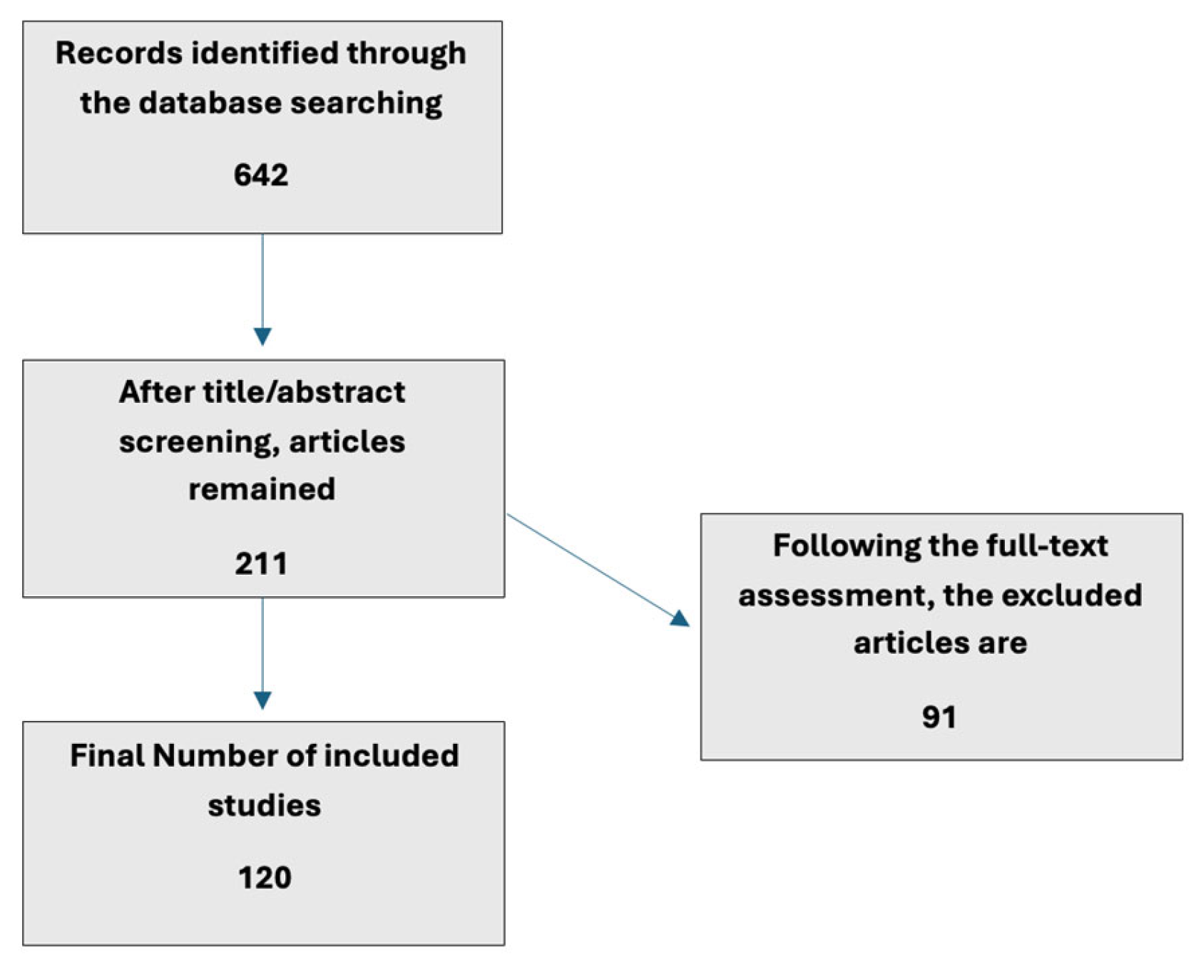
Figure 1. Literature search and study selection.
Bacteria 05 00011 g001
Figure 2. Decision-making framework for selecting sequencing technology for subgingival microbiome analysis.
Bacteria 05 00011 g002
Table 1. Key Differences Between Major Microbiome Sequencing Approaches.
Feature16S rRNA SequencingShotgun Metagenomics (WGS)Metatranscriptomics
Primary outputTaxonomic profile (who is there?)Taxonomic profile and functional potential (what can they do?)Functional activity (what are they doing?)
Taxonomic resolutionGenus (species with long-reads)Species, StrainSpecies
Functional informationInferred (e.g., PICRUSt2)Direct (gene presence)Direct (gene expression)
Host DNA issueMinimal (bacterial-specific primers)Major (requires depletion/deep sequencing)Major (requires depletion/deep sequencing)
Cost per SampleLowHighVery High
BioinformaticsStandardized, moderate complexityComplex, high computational needVery complex, specialized expertise
Best forLarge cohorts, diversity surveys, biomarker discoveryStrain-level analysis, functional potential, antibiotic resistanceMechanistic studies, treatment response, identifying active pathways
Approximate cost per sample (typical range50–100 $200–500 $400–1000 $
Disclaimer/Publisher’s Note: The statements, opinions and data contained in all publications are solely those of the individual author(s) and contributor(s) and not of MDPI and/or the editor(s). MDPI and/or the editor(s) disclaim responsibility for any injury to people or property resulting from any ideas, methods, instructions or products referred to in the content.