Article

Characterization and Biological Activity of Magnesium Nanoparticles Synthesized from Escherichia coli Metabolites Against Multidrug-Resistant Bacteria

1
Department of Biological Sciences, Faculty of Science, Beirut Arab University, Beirut P.O. Box 11-5020, Lebanon
2
Department of Nutrition and Dietetics, Faculty of Health Sciences, Beirut Arab University, Tarik El Jedidah, Riad El Solh, Beirut P.O. Box 11-5020, Lebanon
3
Molecular Biology Unit, Department of Zoology, Faculty of Science, Alexandria University, Alexandria 21568, Egypt
4
Botany and Microbiology Department, Faculty of Science, Alexandria University, Alexandria 21568, Egypt
5
Physics Department, Faculty of Science, University of Tabuk, Tabuk 47512, Saudi Arabia
*
Authors to whom correspondence should be addressed.
Bacteria 2025, 4(3), 48; https://doi.org/10.3390/bacteria4030048
Submission received: 8 August 2025 / Revised: 26 August 2025 / Accepted: 5 September 2025 / Published: 10 September 2025

Abstract

(1) Background: This study evaluated the efficacy of magnesium nanoparticles (MgNPs) synthesized through a green method utilizing bacterial metabolites (BMs) produced by Escherichia coli. (2) Methods: BMs were tested for total phenolic content by high-performance liquid chromatography. MgNPs were characterized by X-ray diffraction, transmission electron microscopy, Fourier transform infrared spectroscopy, photoluminescence, and ultraviolet–visible spectroscopy. MgNPs and BMs were tested for antibacterial and antibiofilm potentials against multidrug-resistant clinical isolates by agar well diffusion, minimum inhibitory and bactericidal concentration assays, time–kill test, and inhibition of biofilm formation and destruction of pre-formed biofilm assays. Furthermore, they were tested for antioxidant potential by 2,2-diphenyl-1-picryhydrazyl radical scavenging assay. (3) Results: BMs included carbohydrates, reducing sugars, and phenols (gallic acid and catechin) with a total phenolic content of 0.024 mg GAE/g. MgNPs showed a pure crystalline structure with a spherical shape, 17.8 nm in size, and a 4.19 eV energy gap. Bacteria included Streptococcus pneumonia, Enterococcus faecium, Klebsiella pneumonia, and Salmonella Typhimurium. The antibacterial results showed inhibition zones ranging between 7.2 and 10.4 mm, a bactericidal effect of MgNPs, a bacteriostatic effect of BMs, and growth inhibition after 3 h. The antibiofilm results demonstrated significant inhibition of biofilm formation (inhibition percentages of 64.931% for MgNPs and 71.407% for BMs). However, the assays revealed modest biofilm destruction (eradication percentages of 48.667% for MgNPs and 37.730% for BMs). Antioxidant capacity revealed notable scavenging activity of MgNPs (scavenging activity of 41.482%) and weak activity of BMs (scavenging activity of 16.460%). (4) Conclusions: These findings support the application of MgNPs in biomedical fields.

1. Introduction

Bacterial infections are a major clinical complication that can lead to prolonged hospital stays [1]. They can cause chronic infections leading to high mortality rates. For centuries, antibiotics have been widely used for the treatment of bacterial infections due to their powerful outcomes and cost-effectiveness. However, the widespread use of antibiotics has led to an increase in multidrug resistance [2]. In addition, the spread of biofilms, which are usually more resistant to antibiotics than bacteria or fungi, is also a problem, especially in nosocomial and colon infections [1].
Another widespread problem that is known to be detrimental to human health is oxidative stress and free radicals. Many studies have shown that free radicals contribute to several pathologies, including cancer and respiratory diseases. To treat oxidative stress, treatment therapies like radiation have been applied. In addition, some antioxidant agents have been studied as a therapy for free radicals. Ascorbic acid has been widely studied and has shown promising antioxidant effects. However, the need for new therapeutic agents has become urgent. Some compounds, including polyphenols and flavonoids, are recommended for free radical treatment [3,4].
As a way to treat bacterial and biofilm infections and oxidative stress exerted by free radicals, nanoparticles (NPs) are being widely used. There are different physicochemical approaches to the synthesis of NPs. However, green avenues have gained a lot of attention due to their reliability and sustainability [5]. In addition, these approaches are eco-friendly and cost-effective and have been used for the synthesis of different nanomaterials [5,6,7]. There are many biological resources for the green synthesis of NPs. Biological approaches, in contrast to physicochemical approaches, are economical and harmless, use ecologically compatible solvents, and produce purer materials [8,9].
Bacteria are characterized by the secretion of extracellular enzymes, known as bacterial metabolites (BMs), that work as reducing agents for metals. In addition, they are considered inexpensive, as their reactions can be controlled by manipulating the pH, temperature, and concentration of metal ions, thus producing NPs [10]. Several bacteria, especially Klebsiella, Bacillus, Pseudomonas, and Lactobacillus, have been shown to be capable of synthesizing NPs with unique characteristics, including composition and size [8,11,12]. They are considered a great source of reducing agents (enzymes, proteins, polysaccharides, amino acids, and vitamins) [8]. It is worth mentioning that Escherichia coli (E. coli) is a Gram-negative, non-spore-forming bacterium, and most of its strains are non-pathogenic. It is known to produce many metabolites that are powerful in the synthesis of metal NPs, as reported by Altaee et al., due to their redox potential [13]. E. coli-derived metabolites have many advantages. They furnish strong reducing and capping biomolecules (enzymes, peptides, and proteins) that enable smaller particles and enhanced activity. Numerous E. coli biosynthesis processes involve approximately 20 nm sized NPs. This small size is linked to higher antibacterial efficacy. In addition, E. coli proteins and enzymes act as native capping layers that improve colloidal stability and endow reactive functional groups. Furthermore, the fast growth and genetic tractability of E. coli allow metabolite output to be tuned, which increases NP yield and tolerance to metal precursors, demonstrating scalable control not readily available in plant extracts. It is worth mentioning that supernatant-based E. coli syntheses simplify downstream purification and enable rapid, room-temperature production [14,15,16].
The spread of metal-based NPs synthesized by green techniques, especially silver, gold, and zinc, represents a great breakthrough in the domain of nanotechnology due to the production of high-quality products. Recently, magnesium NPs (MgNPs) and their derivatives have served as effective antimicrobial and antioxidant agents due to their high ionic content, small size (less than 20 nm), large surface area, stability, and photocatalytic characteristics [5]. In addition, their tolerance to high temperatures has enabled their use in many biomedical fields [5,6].
MgNPs have been shown to inhibit Gram-positive, Gram-negative, and spore-forming bacteria [1]. In addition, it was previously reported that Mg-doped NPs exhibited significant antibacterial actions, reaching 99.9% inhibition, especially against E. coli and Staphylococcus aureus (S. aureus) [17]. Previous studies reported the antibacterial action of MgNPs biosynthesized from bacteria against Citrobacter, Clostridium, and Streptococcus [18]. Antibacterial mechanisms include the binding of NPs to the bacterial surface, rupturing the cell wall, hampering biochemical pathways and the synthesis of nucleic acids, inhibiting enzymatic activity, generating reactive oxygen species (ROS), and destroying cell organelles. These actions collectively lead to cellular damage and ultimately result in cell death [6,19]. However, the exact action of MgNPs against many clinical and environmental pathogens is still not very clear, and there is no exact dose to apply, as they inhibit each bacterium differently [1].
For the antioxidant potential, MgNPs showed promising antioxidant activity reaching 90% against 2,2-diphenyl-1-picryhydrazyl (DPPH) as reported by Amrulloh et al. [20] The main antioxidant mechanism relies on scavenging free radicals and ROS. For example, a previous study reported by Bhardwaj et al. showed that MgNPs act by transferring a single electron and hydrogen, which prevents DNA damage from oxidative stress [21]. In addition, many studies report a significant antioxidant potential of Mg-based NPs, which is comparable to standards such as ascorbic acid [22,23]. These findings suggest that Mg-derived NPs can possess antioxidant properties, making them potential candidates for various biomedical and environmental applications. However, their efficacy may vary depending on the synthesis method and the source of the extract used.
To our knowledge, no studies have focused on the biosynthesis of MgNPs from the BMs of E. coli. While green synthesis of MgNPs using plants or fungi has been described, these approaches rely on phytochemicals or fungal enzymes that differ significantly from BMs. Reports on microbial synthesis of MgNPs remain extremely limited, and none have demonstrated the role of E. coli secondary metabolites as reducing and stabilizing agents. In this regard, this study aims to greenly biosynthesize MgNPs from the BMs of E. coli, characterize them, test their antibacterial and antibiofilm potentials against clinical bacterial isolates including Klebsiella pneumonia (K. pneumonia), Streptococcus pneumonia (S. pneumonia), Enterococcus faecium (E. faecium), and Salmonella Typhimurium (S. typhimurium), and detect their antioxidant activity against DPPH. Our study, therefore, provides a new biosynthetic route for MgNPs and demonstrates how E. coli metabolites can yield NPs with antibacterial, antibiofilm, and antioxidant activities, offering a distinct contribution beyond earlier plant- or fungal-based approaches.

2. Materials and Methods

2.1. Microorganisms and Culture Conditions

E. coli, isolated from a wastewater sample, was used in this study. The isolate was identified according to Mezher et al. [24]. A nutrient broth (NB; TM Media, TM 1054, El Achour, Algeria) medium was inoculated with E. coli to reach a turbidity of 0.5 McFarland. The bacterial suspension was incubated aerobically at 37 °C for 24 h under static conditions. Following incubation, the cell-free supernatant (CFS) was collected by centrifugation at 6000 rpm for 25 min, then filtered with a Whatman paper (1 mm; WhatmanTM, Cat No 1001110). The filtrate containing BMs was used in the synthesis of MgNPs as a reducing agent [25].

2.2. Phytochemical Analysis of BMs

Different tests were performed to detect the phytochemical composition of the BMs, as follows.

2.2.1. Carbohydrates

The test for carbohydrates was performed by mixing 1 mL of the BMs with 2–3 drops of 1% alfa-naphthol (LABCHEM, LC-108901, Zelienople, PA, USA) and 2 mL of concentrated sulfuric acid (H2SO4; K50052513, CAS-N0-7664-93-9, Merck Millipore, Darmstadt, Germany) in a tube. Carbohydrates were detected by the formation of purple rings on the sides of the tube [26].

2.2.2. Reducing Sugars

The test was performed by mixing 0.5 mL of the BMs with 1 mL of water and 5–8 drops of Fehling A (BFCLAB, Bengaluru, India) and Fehling B (BFCLAB, India), with heating for 5 min. Reducing sugars were detected by the formation of a brick-red precipitate [27].

2.2.3. Glycosides

The test was performed by mixing 1 mL of the BMs with 1 mL of glacial acetic acid (UniClean, Houston, TX, USA), a drop of 5% ferric chloride (FeCl3; BASF, Ludwigshafen, Germany), and 1 mL of concentrated H2SO4. The glycosides were detected by a brown ring at the interface, along with violet and greenish rings below the brown ring [28].

2.2.4. Terpenoids

The test was performed by mixing 1 mL of the BMs with 2 mL of chloroform (Merck, Darmstadt, Germany) and 3 mL of concentrated H2SO4. Terpenoids were detected by the appearance of a red-brown color [29].

2.2.5. Phenols

The test was performed by mixing 5 mL of the BMs with 1 mL of 1% potassium ferricyanide (C6FeK4N6; EASTCHEM, Changzhou, China) and 1 mL of 1% ferric chloride (FeCl3). The phenols were detected by the formation of a green-blue color [30].

2.3. Total Phenolic Content

To quantitatively confirm the presence of phenols in the BMs, the Folin–Ciocalteu assay was applied to determine the total phenolic content (TPC) [31]. In brief, the test was performed by mixing 200 µL of the BMs with 800 µL of 7.5% (w/v) sodium carbonate (Na2CO3; Sigma Aldrich, Steinheim, Germany) and 1000 µL of diluted Folin–Ciocalteu reagent (1/10 v/v; Sigma Aldrich, Steinheim, Germany). The solution was incubated for 10 min at 60 °C, followed by 10 min at 4 °C. An ultraviolet–visible (UV–Vis) spectrophotometer (GENESYS 10 UV, Thermo Electron Corporation, Waltham, MA, USA) was used to measure the absorbance at 750 nm. Gallic acid (Sigma Aldrich, Steinheim, Germany) was used as a standard to draw a calibration curve and determine the TPC, which is expressed as milligrams of gallic acid equivalents (GAE) per gram (mg GAE/g).

2.4. High-Performance Liquid Chromatography (HPLC)

HPLC was employed to identify and quantify phenolic compounds in the BMs. The analysis utilized an Agilent 1100 Series HPLC system, equipped with a Zorbax column oven, an autosampler, and a diode array detector (Barcelona, Spain). Separation of the phenolics was carried out on a C18 column (250 mm × 4.6 mm, 5 μm particle size; Thermofisher, Waltham, MA, USA). To aid in identification, standard phenolic compounds including rutin, catechin, hydroxybenzoic acid, protocatechuic acid, ellagic acid, chlorogenic acid, p-coumaric acid, and gallic acid (Sigma Aldrich, Steinheim, Germany) were used. The mobile phase consisted of acidified nano-pure water (adjusted to pH 2.3 with hydrochloric acid (HCl; Sigma Aldrich, Steinheim, Germany), labeled as solvent A and HPLC-grade methanol (solvent B; Sigma Aldrich, Steinheim, Germany). The elution began with isocratic conditions (85% A and 15% B) from 0 to 5 min, followed by a gradient transition from 85% A and 15% B to 100% B and 0% A between 5 and 30 min. This was maintained under isocratic conditions from 30 to 35 min. A 10 μL sample volume was injected at a flow rate of 1 mL/min. Phenolic identification was achieved by comparing the retention times of the sample peaks with those of the standards, and concentrations were calculated using calibration curves generated from multiple standard concentrations [32].

2.5. Synthesis of MgNPs

The BMs were mixed at equal volumes with 5 mM of magnesium nitrate (Mg(NO3)2; Drug House (P) Ltd., RE3615, New Delhi, India). The solutions were incubated in a shaker incubator at 120 rpm in dark conditions at 37 °C for 24 h. A color change was observed in the reaction mixture due to the reduction reaction. After incubation, the solutions were centrifuged at 13,000 rpm for 10 min to purify the MgNPs. Sterile distilled water was used to wash the pellets (MgNPs) three times. They were further centrifuged at the same speed. Finally, the NPs were dried in an oven (ESCO, Singapore) at 40 °C for 24 h. The obtained MgNPs were stored in Eppendorf tubes (2 mL) covered with paraffin under dry conditions at room temperature for further use. For biomedical applications, stock aqueous solutions of the biosynthesized MgNPs were prepared at a concentration of 1 mg/mL. Then, half-fold dilutions were used to prepare solutions of concentrations ranging between 0.03125 and 1 mg/mL.

2.6. Characterization of the Biosynthesized MgNPs

The characterization of the biosynthesized MgNPs was applied according to Darwich et al. [33], as follows.

2.6.1. X-Ray Diffraction (XRD)

The crystallographic structure of the MgNPs was determined by XRD. A Bruker D8 Advance (Bruker, Ettlingen, Germany) equipped with Cu-Kα radiation of wavelength 1.54060 A was used to analyze the XRD patterns. The data were collected at 2θ range of 20°–80° at 0.02° per second. The XRD profiles were optimized using the MAUD software (version 2.992).

2.6.2. Transmission Electron Microscopy (TEM)

The average particle size of the MgNPs was determined by TEM. The test was performed using a transmission electron microscope (JEOL, Tokyo, Japan) operating at 80 kV with a resolution of 0.1 nm. The ImageJ software (version 1.54 g) was used to analyze the TEM images taken at a 100 nm magnification.

2.6.3. Fourier Transform Infrared Spectroscopy (FTIR)

The functional groups of the MgNPs were identified at room temperature using a Fourier transform infrared spectrophotometer (FTIR-8400S, Nicolet iS5, ThermoScientific, Berlin, Germany) within a 4000–400 cm−1 spectral range.

2.6.4. Photoluminescence (PL)

A fluorescence spectrophotometer (JASCO-FP-8600, JASCO, Pfungstadt, Germany) with a Xenon (Xe) laser was used determine the PL spectra at a 420 nm excitation wavelength and within a 400–700 nm range.

2.6.5. Ultraviolet–Visible Spectroscopy (UV–Vis)

A UV–Vis spectrophotometer (V-670; Jasco, Tokyo, Japan) was used to evaluate the optical properties of the MgNPs at room temperature and within a 200–700 nm range.

2.7. Antibacterial Activity of the Biosynthesized MgNPs

2.7.1. Isolation, Identification, and Preparation of the Bacteria

The antibacterial activity of the biosynthesized MgNPs and BMs was examined against four clinical bacterial isolates obtained from Hamoud Hospital University Medical Center (HHUMC, Sidon, Lebanon). The bacterial isolates were identified using the VITEK 2 Automated System (bioMérieux Inc., Providence, RI, USA), by preparing a bacterial suspension in sodium chloride (NaCl; Sigma Aldrich, Steinheim, Germany), adjusting it to 0.5 McFarland, and inserting it into VITEK cards for biochemical tests.

2.7.2. Antibiogram Assay

Antibiotic resistance patterns of the bacterial isolates were determined using the agar disk diffusion method according to the Clinical and Laboratory Standards Institute [34]. Four bacterial cultures were prepared to 0.5 McFarland Standard (1.5 × 10⁸ CFU/mL) in sterile distilled water prior to the assay, and 100 µL was inoculated onto Mueller–Hinton agar (MHA; TM Media, TM 1054, El Achour, Algeria) medium. The antibiotic concentrations in the applied discs were as follows: Cefamandole (30 μg/disc), Moxifloxacin (5 μg/disc), Ceftizoxime (30 μg/disc), Flumequine (30 μg/disc), Neomycin (30 μg/disc), Cefazoline (30 μg/disc), Chloramphenicol (30 μg/disc), Cefradine (30 μg/disc), Clindamycin (2 μg/disc), Cefadroxil (30 μg/disc), Lincomycin (2 μg/disc), Sulfamethoxazole (200 μg/disc), Ampicillin (10 μg/disc), and Minocycline (30 μg/disc), representing different families of antibiotics that were tested on all isolated strains. The plates were incubated for 24 h at 37 °C. Following the incubation period, the zones of inhibition (ZOIs) were measured using the ImageJ software and compared to CLSI, 2024 [34].

2.7.3. Agar Well Diffusion Assay

The antibacterial potential of the MgNPs and BMs was assayed by agar well diffusion. In brief, the test was performed by spreading 100 µL of the prepared bacterial suspensions on MHA plates, leaving them to dry for 5 min, and then punching the plate with a cork-borer of size 6 mm to create wells. After that, 100 µL of the prepared MgNPs solutions (concentrations ranging between 0.03125 and 1 mg/mL) and BMs (concentrations ranging between 3.125 and 100%) was added to the wells. Distilled water was used as a control. After diffusion for 1 h at 4 °C, the plates were incubated for 24 h at 37 °C. The ZOIs were measured using the ImageJ software, and diameters greater than 7 mm were considered significant [33].

2.7.4. Minimum Inhibitory Concentration (MIC) and Minimum Bactericidal Concentration (MBC) Assay

According to CLSI guidelines, the MIC of the MgNPs and BMs against clinical isolates was determined using the broth dilution assay in 96-well microtiter plates [34]. The test was performed by adding 90 µL Mueller–Hinton broth (MHB; TM Media, TM 1054, El Achour, Algeria) and 10 µL of the 0.5 McFarland suspensions to the wells of microtiter plates. Then, 100 µL of the serially-diluted MgNP and BM solutions was added to the wells. The plates were stored for 24 h at 37 °C. Cultures without treatment were used as a control. After incubation, the optical density (O.D.) of bacterial growth was measured at 595 nm using an enzyme-linked immunosorbent assay (ELISA; Infetik Mpr-H200BC, Jinan, China) reader. The MIC was defined as the lowest concentration of the MgNPs and BMs that inhibited the visible growth of bacteria. For detecting the MBCs, a loopful was taken from the clear wells and spread on MHA plates. The MBC was defined as the lowest concentration that killed 99% of bacteria (no clear growth of colonies on the agar) [24].

2.7.5. Time–Kill Test

The time–kill test was performed to detect the time needed by the biosynthesized MgNPs and BMs to inhibit bacterial growth. The test was conducted by adding 90 µL of MHB and 10 µL of the 0.5 McFarland bacterial suspensions to the wells of 96-well microtiter plates. Then, 100 µL of the MICs × 2 of the MgNP and BM solutions was added to the wells. The plates were incubated at 37 °C and the O.D. was measured at 595 nm in the interval 0–24 h using an ELISA reader [33].

2.8. Antibiofilm Assays

The inhibition of biofilm formation assay was performed to test the ability of the biosynthesized MgNPs and BMs to inhibit the formation of bacterial biofilms. The test was performed in 96-well microtiter plates where 90 µL of MHB was added to 10 µL of the prepared bacterial suspensions. The plates were cultivated at 37 °C for 3 h to allow the biofilms to attach. After incubation, 100 µL of the prepared MgNPs at concentrations ranging between 0.03125 and 4 mg/mL (starting at MIC × 2) and BMs (at concentration of 100%) was added to the wells. The plates were again incubated at 37 °C for 24 h. Cultures with no treatment were used as a control. After incubation, the plates were washed three times with sterile distilled water and dried in an oven at 40 °C for 15 min. To visualize the biofilms, the wells were stained with 1% crystal violet (CV; Alpha Chemika, CV506, Mumbai, India) and incubated for 15 min at room temperature. The plates were then washed five times with sterile distilled water and the biofilms were observed as purple rings on the walls of the wells. Finally, 100 µL of absolute ethanol (200/190, HAKINMHAN) was added to de-stain the wells and the O.D. was measured at 595 nm using an ELISA reader to quantify the biofilms [24]. The biofilms were quantified according to the following Equation (1):
Percentage   of   biofilm   inhibition   ( % ) = O . D . ( n e g a t i v e   c o n t r o l ) O . D . ( t r e a t e d   s a m p l e ) O . D . ( n e g a t i v e   c o n t r o l ) × 100
A similar protocol to the inhibition of biofilm formation was applied to detect the ability of the biosynthesized MgNPs and BMs to eradicate pre-formed biofilms. The only difference was the incubation of the bacteria in MHB for 30 h to form the biofilms before treatment. The staining and quantification techniques were identical to the inhibition of biofilm formation [24].

2.9. DPPH Radical Scavenging Assay

The DPPH radical scavenging assay was used to evaluate the antioxidant activity of the biosynthesized MgNPs and BMs. To assess the MgNPs, 1 mL of NP solution at concentrations ranging from 0.0625 to 1 mg/mL was mixed with 1 mL of a 0.3 mM DPPH (ANCUS, Scotland, UK) solution prepared in methanol. A mixture of DPPH and methanol (200/190, HAKINMHAN) served as the negative control, while ascorbic acid (AZCOR-C, India) was used as the positive control. For the BMs, 50 µL of the extract was added to 1.45 mL of 0.06 mM DPPH solution. Trolox (Sigma Aldrich, Steinheim, Germany) acted as the positive control and methanol as the negative control in this test, with Trolox also serving as the standard antioxidant [35]. All samples were kept in the dark for 30 min, followed by absorbance measurement at 517 nm using a spectrophotometer [33,36]. The percentage of DPPH radical inhibition was calculated using Equation (2):
DPPH   radical - scavenging   activity   ( % ) = O . D . ( n e g a t i v e   c o n t r o l ) O . D . ( t r e a t e d   s a m p l e ) O . D . ( n e g a t i v e   c o n t r o l ) × 100

2.10. Statistical Analysis

To ensure statistical confidence, each experiment was run three times. The experimental outcomes’ data values were documented as the mean ± standard error of the mean. A t-test was used to evaluate the significance, with p < 0.05 considered statistically significant. Statistical significance tests (t-tests) were performed in Excel software (64-bit edition) and graphs were drawn in Origin software (64-bit edition).

3. Results and Discussion

3.1. Characterization of the Bacterial Metabolites (BMs)

3.1.1. BMs Produced by E. coli

Upon testing BMs commonly known to be produced by bacteria, the tests revealed the presence of carbohydrates, reducing sugars, and surprisingly phenols. The results are presented in Table 1. E. coli is known to produce carbohydrates and reducing sugars, but not glycosides, terpenoids, and phenols. In addition, it is not naturally specialized in phenolic compound production. However, its metabolism includes the production of some aromatic structures like amino acids and tyrosine which have a phenol group, but free phenol is not known to be produced as a metabolic product in many E. coli strains. To produce phenols, Thompson et al. reported that E. coli should be engineered [37]. This result could be attributed to the difference in the structural conformation of bacterial species from place to place in response to varying sources and environmental conditions as shown by Justice et al. [38]. In addition, it has been reported that Gram-negative bacteria can produce phenols, especially Clostridium, Bacillus, and Pseudomonas species. They produce phenolic compounds as by-products of fermentation [39,40].

3.1.2. Total Phenolic Content (TPC)

For the confirmation of the presence of phenols in the BMs, the TPC was determined. A calibration curve was constructed using gallic acid calibration standards at concentrations ranging between 0 and 0.12 mg/mL. The TPC of the BMs was calculated using the calibration curve equation and reported in mg GAE/g dry mass of the BMs. The BMs showed a TPC of 0.024 ± 0.009 mg GAE/g. This confirms the presence of phenols in the BMs. The TPC is an important parameter in extracts because it determines their scavenging abilities [41]. It is worth mentioning that no studies have shown the TPC of the BMs of E. coli and the specific data on TPC in bacterial extracts are limited. However, the TPC has been quantified in some other bacteria. For example, a previous study by Rani et al. reported a TPC of 55 mg GAE/100mg in Streptomyces cellulosae (S. cellulosae) metabolic extract [42]. In addition, the TPC is directly correlated with the scavenging activity and influences the antimicrobial potential [43].

3.1.3. Characterization of E. coli Metabolites by HPLC

HPLC analysis was conducted to identify the BMs. Phenolic compounds were identified based on their retention times, and their concentrations were estimated from the peak areas (Figure S2). The coefficient of determination (R2) of the gallic acid calibration curve (y = 35.622x; Figure S2a) was 0.9985 and that of catechin calibration curve (y = 5.8214x; Figure S2b) was 0.9989 at concentrations ranging from 0 to 12 mg/L. This suggests excellent linearity in the studied range of concentrations. The HPLC results revealed that among the tested polyphenols, gallic acid (0.12 ± 0.01 mg/L) and catechin (0.27 ± 0.01 mg/L) were present in the BMs. The chromatograms of the mentioned polyphenols are presented in Figure S1. Although the natural production of gallic acid and catechin is associated with plants, these compounds can be produced by bacteria, especially Pseudomonas and Bacillus species, and some engineered Escherichia species [44,45,46]. For example, Alberto et al. reported in a previous study the production and metabolism of gallic acid and catechin by Lactobacillus hilgardii (L. hilgardii) [47]. Another study by Xia et al. also reported the production of these phenols by Akkermansia muciniphila (A. muciniphilia) [48]. However, the mentioned extracts contain, in addition to the identified gallic acid and catechin, other phenolic compounds. This could be explained by the difference in the extraction techniques and the varying composition and metabolic reactions [49]. In addition, it is worth mentioning that it is hard to find the exact composition of secondary metabolites since HPLC provides no information regarding the chemical composition of the metabolites of an extract [50]. It is also notable that the detected gallic acid and catechin play multiple roles in the biosynthesis and activity of the MgNPs. These phenolic compounds can act as reducing agents, donating electrons to convert Mg2+ ions into metallic MgNPs [51]. They may also serve as capping and stabilizing agents, binding to the NP surface through hydroxyl and carbonyl groups, which prevents aggregation and enhances colloidal stability. Additionally, their inherent bioactive properties, such as antioxidant activity and potential interference with bacterial cell walls, can contribute to the observed antibacterial, antibiofilm, and antioxidant effects of the MgNPs [52]. Overall, the HPLC analysis revealed the presence of two polyphenols that play essential roles in biomedical applications, especially antibacterial applications.

3.2. Synthesis and Characterization of the MgNPs

3.2.1. Synthesis

The formation of MgNPs was first detected by the change of the clear light-yellow solution to turbid yellowish, as presented in Figure 1.

3.2.2. Characterization

X-Ray Diffraction (XRD)
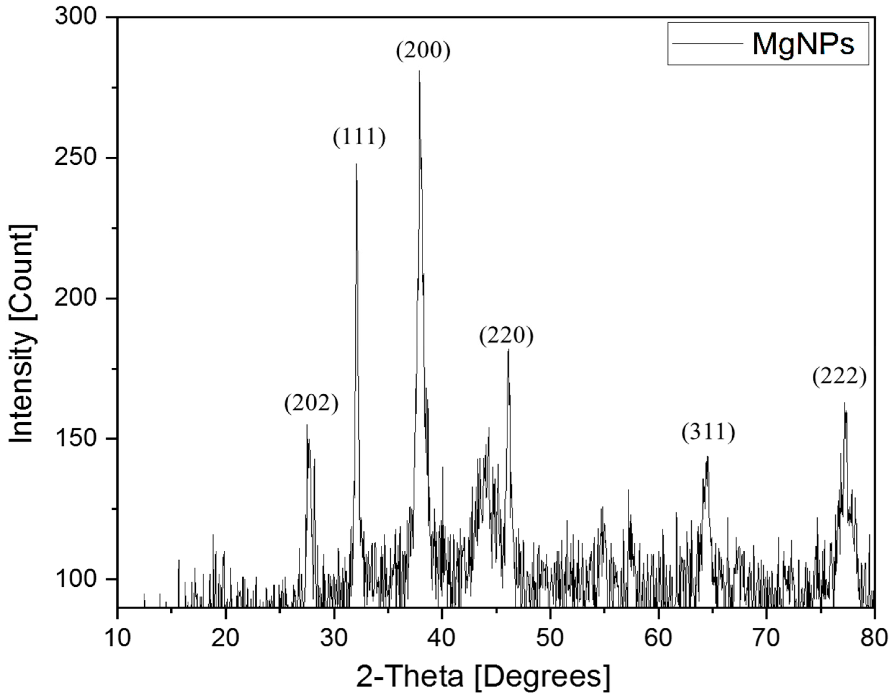
XRD is a technique used to detect the crystallographic structure of NPs [53]. It relies on the measurement of various structural properties and atomic arrangements of the crystalline phases [54]. Figure 2 represents the XRD patterns of the MgNPs, revealing the presence of six diffraction peaks. The peaks were observed at 27.2°, 32.8°, 38.6°, 46.9°, 64.9°, and 78.5°. The peak at 27.2° (202) has not been observed previously in bacterially synthesized NPs. It most likely corresponds to a minor crystalline impurity formed during synthesis or drying. This explanation could be due to the high reactivity of the MgNPs. In addition, the small peaks observed could also be explained by the presence of some impurities due to the use of capping agents during the synthesis process. However, the peaks at 32.8°, 38.6°, 46.9°, 64.9°, and 78.5° correspond to (111), (200), (220), (311), and (222), respectively. These peaks confirm the crystalline structure of the MgNPs and indicate the cubic phase of Mg. Previous studies have shown that the presence of such peaks and the crystallinity of the NPs explain the small size of the NPs, which ranges between 20 and 27 nm [11]. It has been established that MgNPs are highly prone to oxidation, and characteristic Mg-oxide (MgO) peaks often appear in XRD patterns of MgNPs. However, several studies have reported that biosynthesized MgNPs can retain metallic Mg [55], which is similar to our case where the BMs acted not only as reducing agents but also as stabilizing and capping agents, which contributed to preserving metallic Mg in the NP core. Similar findings have been reported in green synthesis studies where stabilizing biomolecules provided partial protection against complete oxidation [56]. Furthermore, the characteristic peaks at 38.6° and 64.9° are indexed to metallic Mg, in agreement with earlier reports [57].
To confirm the size of the biosynthesized MgNPs, the crystalline size was calculated from the XRD data using the Scherrer Equation (3) as follows:
D = K λ β c o s θ
where D is the average crystalline size, K is Scherrer’s constant, λ is the X-ray wavelength, β is the line broadening at full width at half maximum (FWHM), and θ is the Bragg’s angle. After performing the calculations, the average crystalline size of the MgNPs obtained was approximately 17.41 nm.
In addition, the degree of crystallinity was obtained from the following Equation (4):
Crystallinity = A r e a   o f   c r y s t a l l i n e   p e a k s A r e a   o f   a l l   p e a k s × 100
where the average area of crystalline peaks is 291.16 nm2 and the average area of all peaks is 465.39 nm2. After performing the calculations, the crystallinity percentage of the biosynthesized MgNPs obtained was approximately 63.8%. This result is consistent with the size of the MgNPs obtained, since there are some impurities in the samples as mentioned previously. Our result is similar to a previous study performed by Jiang et al. who showed that a crystallinity percentage ranging between 60 and 65% corresponds to a size of 39–41 nm of NPs [58].
Our results confirm those of previous studies where similar peaks were detected, e.g., that of Hassan et al., who synthesized MgO-NPs from Rhizopus oryzae and detected the same five peaks as obtained in our present study [7]. In addition, Abdel-Maksud et al. synthesized MgNPs from Lactobacillus gasseri and revealed the same peaks, thus indicating the crystallinity of the NPs. Moreover, the peaks present at 46.9° (200) and 64.9° (222) indicate the presence of Mg hydroxide (Mg(OH)2) [11]. Overall, the XRD patterns indicate the purity of the synthesized MgNPs and reveal their crystalline structure.
Transmission Electron Microscopy (TEM)
TEM is a useful technique applied for the quantitative determination of particle size, shape, and distribution [59]. The TEM micrographs of the MgNPs, presented in Figure 3b, are annealed at a 100 nm scale and show that the majority of the NPs have a spherical morphology and are slightly agglomerated. In addition, the particle sizes are not uniform. This could be attributed to the capping and reducing agents present in the BMs. This result is similar to that obtained by Darwich et al., who synthesized yttrium NPs from pine needle and showed agglomeration due to the reducing agents present in the pine extract used [33]. After measuring the particle dimensions using the ImageJ software, the average size of the particles was obtained by fitting the particle size histogram to the log-normal function [60] and was approximately 17.8 nm as shown in Figure 3a. When compared to other studies targeting the synthesis of MgNPs, we notice that our MgNPs are smaller in size. For example, Amrulloh et al. showed that the particle size of MgO-NPs biosynthesized from Moringa oleifera (M. oleifera) extract ranged between 60 and 100 nm [20]. In addition, Abbas et al. reported the synthesis of MgO-NPs and their size ranged between 30 and 100 nm [61]. This could explain the advantage of the synthesis of NPs from bacterial metabolites. Furthermore, Patil et al. synthesized plasmonic NPs from bacterial metabolites and showed that their size exceeded 50 nm [9]. This could be explained by the difference in the BMs and the reactions with the metal nitrate. Our results were similar to a previous study performed by Hassan et al., who synthesized MgO-NPs from the fungus R. oryzae and showed a particle size of 20 nm [7]. Overall, the TEM results of the MgNPs confirm their nano-size, shape, and morphology.
Fourier Transform Infrared Spectroscopy (FTIR)
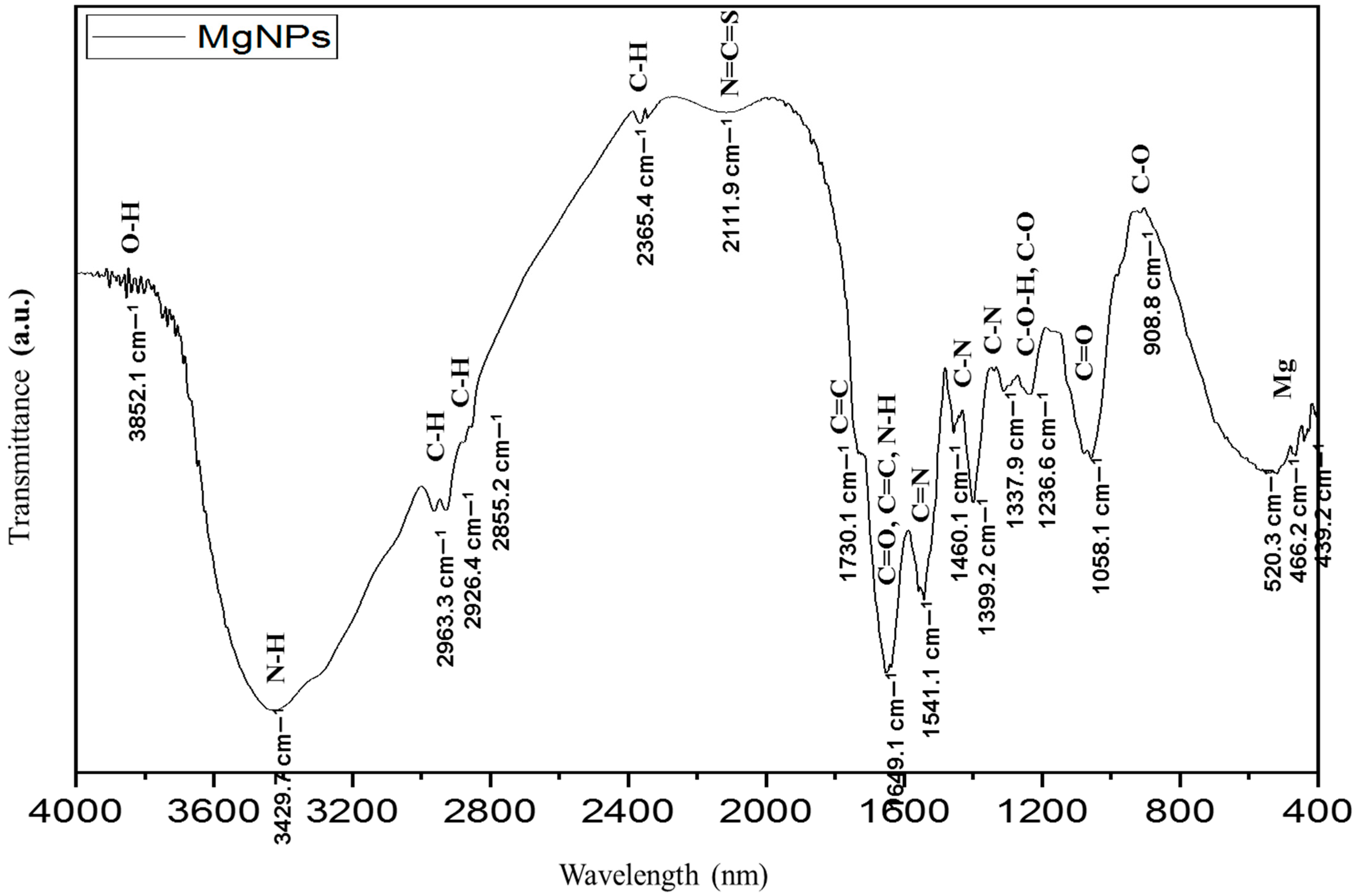
FTIR is a helpful technique in the identification of unknown materials and determining the quantity and quality of components in a sample [62]. FTIR was performed to determine the functional groups present in the biosynthesized MgNPs. The results, presented in Figure 4, revealed the formation of various vibrational bands. The peaks observed at 3852.1 and 3429.7 cm−1 correspond to the O-H stretching group. The O-H group is explained by the presence of alcoholic or phenolic stretches and many bioactive metabolites [5,63]. The peak at 3429.7 cm−1 corresponds to N-H stretch. This group explains the presence of amide or amine groups in the bacterial protein membrane [11]. The peaks observed at 2963.3, 2926.4, 2855.2, and 2365.4 cm−1 correspond to C-H stretching, which indicates the presence of alkanes [64,65]. In addition, the peak observed at 2111.9 could correspond to N=C=S. The peaks observed at 1739.1, 1649.1, and 1541.1 cm−1 correspond to C=C, C=O, and C=N stretches, respectively. These explain the presence of aromatic rings in the nucleic acids of the nucleus, amides, and polysaccharides [11]. The peak observed at 1337.9 cm−1 corresponds to C-N stretching, explaining the presence of primary amines and aliphatic amino acids [11]. The peaks observed at 1307.8, 1236.6, 1058.1, and 908.8 cm−1 correspond to C-O-H, C-O, and C=O stretches. These stretches indicate the presence of carbonyl groups involved in the reduction of Mg2+ to MgNPs [17,64]. In addition, the presence of C=O suggests the presence of aldehyde, ester, and carboxylic acid groups [10]. The C-O stretching also confirms the presence of a link between the formed MgNPs and OH/CO groups [66]. The peaks observed in the range of 620.3–430.2 cm−1 are attributed to the functional group of metal bonds (Mg), thus showing Mg stretching [65,66].
Photoluminescence (PL)
PL investigation was performed to study the intrinsic and extrinsic properties of the biosynthesized nanostructures. The test was carried out using fluorescence emission spectroscopy at room temperature by applying an excitation wavelength of 420 nm and within a visible range of 400–700 nm. The PL spectra were deconvoluted by the Voigt function. The MgNPs’ visible luminescence is caused by the recombination of electron–hole pairs between the d-band and the sp-conduction region above the Fermi level [67]. The electron–hole recombination processes are facilitated by defect states within the NPs. Specifically, interstitial Mg defects are the most probable contributors. These intrinsic lattice defects create localized states within the bandgap that act as recombination centers, enabling radiative transitions that result in PL. Interstitial Mg defects may also contribute to emission features by creating donor-like levels close to the conduction band. In addition, the BMs involved in biosynthesis may play a role in stabilizing or even enhancing these defect states. BMs can adsorb onto the NP surface and influence nucleation and growth, potentially introducing or preserving surface vacancies and unsaturated coordination sites. Such biomolecule–NP interactions have been reported to alter defect-related optical behavior in biologically synthesized NPs [68]. The PL results, presented in Figure 5, showed two peaks. A prominent peak with high intensity was observed at 457.9 nm. This peak corresponds to the blue emission when compared to the previous literature [69,70]. The other peak was observed at 490.2 nm, which also corresponds to the blue emission. It was reported that blue emissions range between 454 and 490 nm [69,71]. The visible emissions are affected by the synthesis conditions, size of NPs, and their morphology. The blue emissions are attributed to the recombination of electrons in the Mg vacancies with the hole in the valence band [72,73]. To explain the mechanism, the visible luminescence results from the excitation of electrons from the d-bands into states above the Fermi level. Additionally, after undergoing relaxation through electron–phonon scattering, which leads to energy loss, an electron from the sp-band recombines with a hole, resulting in photoluminescence via radiative emission [74]. It is worth mentioning that the observed PL in the NPs arises mainly from defect-related states rather than quantum confinement or doping. The PL is most reasonably attributed to the presence of intrinsic defects, such as oxygen vacancies and surface states, which are well documented to produce luminescence in Mg nanostructures [55]. In addition, the BMs used during biosynthesis may also contribute to the PL behavior. The metabolites can adsorb onto the NP surface, modify surface defect density, and even introduce new functional groups that interact with electronic states. Such surface modifications have been reported to enhance or shift PL emission in biosynthesized NPs [51].
Ultraviolet–Visible Spectroscopy (UV–Vis)
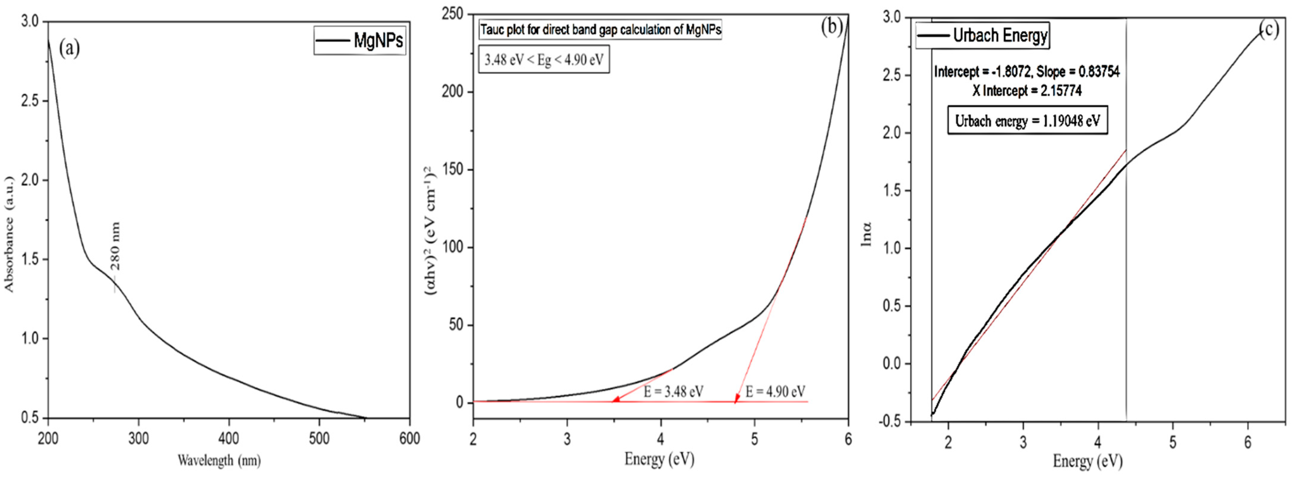
UV–Vis spectroscopy was performed to analyze the chemical properties of the tested MgNPs. The formation of MgNPs was confirmed by UV–Vis spectroscopy in a range of 200–700 nm. The UV–Vis results are presented in Figure 6a. The absorption peak was detected at 280 nm. A similar peak was previously detected by Dolati et al., who synthesized copper oxide NPs from the metabolites of Bacillus coagulans [11]. This peak is evidence of the reduction of magnesium nitrate Mg(NO3)2 to MgNPs [5]. The observed absorption peak corresponds well to the surface plasmon resonance typically reported for metallic MgNPs synthesized under reducing conditions [75]. The number of electrons released during the reduction of NO3 to NO2 determines the amount of color obtained. This reduction is responsible for the transformation of Mg2+ to Mg0 [76]. In addition, the observed phenomena explain the excitation of metals during the formation of MgNPs [76,77]. During the biosynthesis of MgNPs, a redox reaction occurs. Mg(NO3)2 contains Mg2+ ions that need to be reduced to metallic Mg (Mg0) to form NPs. During this reduction, NO3 ions act as electron acceptors and are partially reduced to NO2. The number of electrons transferred from the reducing agents (BMs) to NO3 determines the extent of reduction, which is visually observable as a change in color of the reaction mixture. In essence, the color change reflects the degree to which Mg2+ ions have been reduced to metallic MgNPs, indicating the progress of the reduction reaction [68]. It was previously reported that the formation of a single peak shows small-sized NPs, and the peak observed at 270–280 nm indicates a fast photocatalytic interaction [77]. The observed UV results indicate perfect states of the synthesized MgNPs due to the absence of variations in the peaks and absorption intensities.
The direct bandgap energy (Eg) of the biosynthesized MgNPs was determined from the absorbance through the relation between the absorption coefficient (α) and the photon energy (hv) according to the following Equation (5) [33]:
(αhv)n = B (hvEg)
where B is a constant and n is an exponent with a value of 2 for the direct transition of the bandgap.
The absorption coefficient (α) was calculated using the measured absorbance (A) according to the following Equation (6):
α = 2.303 A t
where t is the path length.
The direct bandgap was determined using Tauc’s plot (Figure 6b), where the linear region of the (αhν)2 versus photon energy (hν) curve was extrapolated to estimate the bandgap energy. The MgNPs displayed an average bandgap of 4.19 eV. This value aligns with findings from earlier studies involving the synthesis of silver NPs using plant-based extracts [78,79]. However, none of the previous studies reporting the synthesis of MgNPs from the BMs mentioned the band gap energy. It is known that bulk metallic Mg typically exhibits overlapping conduction and valence bands. Nanoscale materials often display properties distinct from their bulk counterparts due to reduced dimensionality, high surface-to-volume ratio, and surface electronic states. In the case of our NPs, several factors may contribute to the appearance of distinct band-like features. At the nanoscale, surface plasmon resonance and localized electronic states can alter the density of states near the Fermi level, giving rise to transitions that resemble bandgap-like behavior [80]. In addition, the biosynthesis process using BMs may result in partial capping or surface modification, which could introduce additional energy levels or trap states that influence optical absorption and emission. Finally, intrinsic defects or vacancies on the NP surface can create localized states that facilitate radiative recombination and PL [81]. Therefore, the distinct optical bands and fluorescence observed in our NPs are most likely the result of size- and surface-induced effects at the nanoscale, along with defect-related electronic states, rather than intrinsic bulk metallic behavior.
The Urbach energy (Eu) refers to the width of the band tails of the localized states. It is calculated by the slope (lnα) of the linear portion against photon energy according to the following Equation (7) [33]:
α = α 0 exp h ν E u
where α 0 is a constant. The results of the variation in l n α as a function of h ν are presented in Figure 6c. The Eu values of the MgNPs are obtained from the reciprocal of the slope of the linear part at the tail region. The Eu value obtained is 1.19 eV. This is similar to previous studies reporting the action of Mg doping on copper oxide NP and Mg composites [17,69,70,71]

3.3. Antibacterial Activity of the Biosynthesized MgNPs and BMs Against the Clinical Bacterial Isolates

3.3.1. Identification of the Clinical Bacteria Strains

The bacterial isolates were identified by VITEK. The VITEK assay is a rapid technique for the identification of microorganisms, especially bacteria. It relies on a large number of biochemical tests to identify bacteria at the species level [82]. The VITEK results were obtained for the four bacterial isolates as follows: Gram-positive bacteria included S. pneumonia and E. faecium, and Gram-negative bacteria included K. pneumonia and S. typhimurium. The levels of identification were classified as excellent (96–99%), very good (93–95%), good (89–92%), and acceptable (85–88%) [24]. The results and the levels of identification are presented in Table 2.

3.3.2. Antibiogram Assay

The antibiogram results, presented in Table 3 and Figure S3, revealed that the bacterial isolates were resistant to most of the tested antibiotics. S. pneumonia was sensitive to Moxifloxacin, Ceftizoxime, Flumequine, and Sulfamethoxazole only. E. faecium showed sensitivity to Moxifloxacin only. K. pneumonia was sensitive to Moxifloxacin, Flumequine, and Sulfamethoxazole only. S. typhimurium showed susceptibility to Moxifloxacin and Sulfamethoxazole only. This means that the tested bacteria show increased multidrug resistance. Previous reports showed high emergence of the mentioned bacteria, and they are associated with high mortality rates [83,84,85,86]. In addition, the tested antibiotics belong to the broad-spectrum family, meaning that they can inhibit a wide range of bacteria by different mechanisms. The mechanisms include inhibition of DNA synthesis, inhibition of protein synthesis by targeting ribosomal subunits, inhibition of cell wall synthesis, and inhibition of folic acid metabolism [87]. However, bacteria resist these antibiotics by specific mechanisms, including limiting the uptake of the antibiotic, modifying the antibiotic target, and inactivating the antibiotic and its active efflux [88]. This alarming issue raises the need for new antimicrobial agents that can treat the multidrug resistance of bacteria.

3.3.3. Agar Well Diffusion

An agar well diffusion assay was performed to detect the biological activity of the biosynthesized MgNPs and BMs against the bacterial isolates. The results, presented in Table 4 and Figure S4, revealed significant ZOIs (>7 mm) starting at a concentration of 0.0625 mg/mL of the MgNPs. The best inhibitory activity (largest ZOI) was observed against E. faecium and S. typhimurium with zones reaching 10.100 ± 0.047 mm and 10.433 ± 0.098 mm, respectively, at the highest NP concentration (1 mg/mL). A significant effect was also observed against S. pneumonia and K. pneumonia, thus showing that the MgNPs have a broad effect and can inhibit Gram-positive and Gram-negative bacteria similarly. The antibacterial potential of the MgNPs depends on many factors. The bacterial morphology, synthesis techniques, and the size, surface area, and chemical composition of the NPs play vital roles in the antibacterial field [20]. The results obtained in this study are similar to those obtained by Rotti et al. who tested the effect of greenly synthesized Mg-oxide NPs against E. coli and S. aureus and showed inhibition at low NP concentrations [18]. In addition, Hassan et al. reported the antibacterial effect of Mg-oxide NPs greenly synthesized from R. oryzae, showing a better effect against Gram-negative bacteria and attributing that to the ability of the small-size NPs to penetrate the thin layer of peptidoglycan of Gram-negative bacteria [7]. The main mechanisms that affect the efficiency of the MgNPs against bacteria include the production of ROS which cause cell damage, interaction with the cell wall, and Mg2+ efflux, altering internal metabolic reactions by disrupting protein structures. In addition, the small size of the NPs, their large surface area, and their surface charge enhance their antibacterial effect [7]. For the BMs, they showed ZOIs at the highest concentration only (100%) against all isolates—with ZOIs ranging between 7.233 ± 0.027 mm and 8.033 ± 0.072 mm. The better effect of the MgNPs compared to the BMs is attributed to the difference in their composition and the low polyphenol content of the BMs reported by the HPLC results. The different functional groups of the MgNPs reported by the FTIR results can interact with the internal structures of the bacterial cells, leading to cell lysis [11]. The results were significant with p-values ˂ 0.05 as shown in Table 4.

3.3.4. MIC and MBC of the Biosynthesized MgNPs and BMs

MIC and MBC assays were performed to detect the bacteriostatic and bactericidal potential of the biosynthesized MgNPs and BMs. The MIC results, presented in Table 5 and Figure 7, revealed that the biosynthesized MgNPs had a bactericidal effect against all the bacterial isolates with MBCs 0.125 mg/mL against E. faecium and S. typhimurium, 0.25 mg/mL against S. pneumonia, and 0.5 mg/mL against K. pneumonia. The effect was similar against both Gram-positive and Gram-negative bacteria. Previous studies reported that NPs exert a better inhibitory effect against Gram-positive bacteria than Gram-negative bacteria due to the presence of a protective cell wall in Gram-negative bacteria [89]. Darwich et al., who tested the effect of silver NPs on different bacteria, revealed a better effect against Gram-positive bacteria [90]. In addition, Adnan et al. showed that copper NPs exert a better effect against Gram-positive bacteria [17]. On the other hand, other studies showed a better effect against Gram-negative bacteria [7,61]. For example, Rabaa et al. tested the effect of composite MgNPs and revealed a better effect against Gram-positive bacteria [71]. The difference in effect between Gram-positive and Gram-negative bacteria is attributed to the difference in bacterial structure [7]. Gram-positive bacteria have a thick layer of peptidoglycan, while Gram-negative bacteria have a thinner layer of peptidoglycan with lipopolysaccharides [7]. Thus, the successive interaction of the NPs with the Gram-negative bacterial cells is attributed to the negative charge of lipopolysaccharides and the positive charge of the NPs [7]. This indicates that the biosynthesized MgNPs in the present work have a broader effect, thus making them promising in the antimicrobial field. The bactericidal effect of the MgNPs is attributed to their small size (17.8 nm) as obtained from the TEM results; purity and crystalline structure as obtained from the XRD data; and different functional groups as detected from the FTIR results, especially the Mg stretching; as well as the low bandgap (4.19 eV) obtained from the UV–Vis results [70,71,90]. The bactericidal activity of the MgNPs arises from a combination of their surface chemistry, lattice vibrations, and optical properties. Surface functional groups introduced by the BMs enhance adhesion to bacterial membranes, facilitating disruption of membrane integrity. The Mg stretching modes observed in FTIR indicate a reactive surface capable of promoting the formation of ROS, which damage bacterial cell walls, proteins, and DNA. Additionally, the relatively low bandgap allows photoexcitation under ambient light, generating electron–hole pairs that further contribute to ROS formation [68,91]. All the factors mentioned enhance the antibacterial potential of NPs. To confirm the bactericidal effect of the MgNPs, the MBC/MIC ratio was determined—showing a value of 2 for all bacterial isolates. It has been reported that a ratio ˂ 4 reveals a bactericidal effect of an NP [17]. This confirms the bactericidal effect of our MgNPs. The BMs, on the other hand, showed a bacteriostatic effect against all of the bacterial isolates and were effective only at the highest concentration (100%). The bacteriostatic effect of the BMs could be explained by the polyphenols detected from the HPLC results (gallic acid and catechin). Gallic acid is shown to have an excellent antibacterial effect, especially against Gram-negative bacteria [92]. In addition, catechin is shown to have significant antibacterial action due to its ability to damage the bacterial cell membrane [93]. The results showed statistical significance with p-values ˂ 0.05, as presented in Table S1.

3.3.5. Time–Kill Results of the Biosynthesized MgNPs and BMs

The time–kill results, presented in Table 6, revealed the time needed by the biosynthesized MgNPs and BMs to inhibit bacterial growth at MICs. The MgNP concentrations used included 0.125 mg/mL against S. pneumonia, 0.25 mg/mL against K. pneumonia, and 0.0625 mg/mL against E. faecium and S. typhimurium. The BM concentration was 100% against all isolates. The results showed that the MgNPs inhibited the growth of E. faecium after 1 h of incubation, K. pneumonia after 2 h of incubation, and S. pneumonia and S. typhimurium after 3 h of incubation. Similarly, the BMs inhibited the growth of S. pneumonia and E. faecium after 2 h of incubation and K. pneumonia and S. typhimurium after 3 h of incubation. The time ranging between 1 and 3 h corresponds to the stage of attachment of the bacterial isolates to start colonizing [90]. This means that the MgNPs and the BMs inhibited this attachment. The main mechanism of attachment inhibition relies on the limitation of nutrient uptake by the bacterial cells after interacting with the MgNP components, especially Mg2+ [90]. Similarly, the interaction of the BM components prevents adaptation of the bacterial cells due to the change in their conformation. This leads to cell lysis after altering the metabolic reactions [90]. It is worth mentioning that the 3 h timeframe indicates the inhibition time of bacterial growth but cannot explain the bacteriostatic or bactericidal activity of the MgNPs and BMs. All results were significant with p-values ˂ 0.05, as presented in Table S2.

3.4. Antibiofilm Results of the Biosynthesized MgNPs and BMs

Inhibition of biofilm formation is detected as significant at percentages exceeding 10% and negative percentages indicate enhancement of biofilm formation [24,94]. To test the antibiofilm potential of the MgNPs and BMs against the bacterial biofilms, concentrations starting at MIC × 2 of each sample were applied, since biofilms are more resistant than bacterial isolates [95]. The results are presented in Table 7. Regarding the inhibition of biofilm formation, the MgNPs significantly inhibited the formation of all bacterial biofilms starting at the lowest concentrations. The inhibitory percentage against S. pneumonia, E. faecium, K. pneumonia, and S. typhimurium reached 57.676 ± 5.766, 38.149 ± 9.024, 64.931 ± 5.548, and 53.497 ± 1.426%, respectively. The BMs also inhibited the formation of all biofilms at the highest concentration (100%) with inhibitory percentages of 33.554 ± 0.271, 27.051 ± 4.515, 71.407 ± 2.671, and 20.867 ± 4.757 against S. pneumonia, E. faecalis, K. pneumonia, and S. typhimurium, respectively. Similarly, the MgNPs were able to destroy pre-formed biofilms starting at a concentration of 0.5 mg/mL, in which the best destruction percentages reached 48.667 ± 6.730, 13.176 ± 2.205, 48.493 ± 5.283, and 7.074 ± 4.77% against S. pneumonia, E. faecium, K. pneumonia, and S. typhimurium, respectively. However, the BMs showed destructive effects against S. pneumonia and S. typhimurium only, with destruction percentages of 37.730 ± 8.099 and 25.442 ± 3.231%, respectively. The biofilm growth was inhibited in a concentration-dependent manner. Our results are similar to those revealed by Abdel-Aziz et al., who showed an inhibitory effect of Mg-oxide NPs against biofilms with inhibition percentages reaching 78% [96]. Due to their resistance, bacterial biofilms secrete exopolysaccharides which increase their attachment. The main mechanism of inhibition exerted by the MgNPs relies on the inhibition of these exopolysaccharides by specific interactions, which in turn prevents the attachment of the biofilms [24]. It is sometimes observed that BMs inhibit biofilm formation more effectively than MgNPs. This can be explained by the presence of specific biomolecules in the BMs, such as proteins, enzymes, secondary metabolites, and phenols, which can directly interfere with bacterial adhesion, quorum sensing, or extracellular polymeric substance (EPS) synthesis. In contrast, MgNPs, while capable of generating ROS and physically interacting with cells, may have more generalized effects and lower specificity against biofilm matrix components. This suggests that the BMs themselves contain bioactive compounds with targeted antibiofilm activity, which could potentially be harnessed independently or in combination with MgNPs for enhanced biofilm control. Similar findings have been reported in the previous literature. For example, Iravani et al. reviewed that metabolite-capped NPs may retain or enhance some activity of the original metabolites, but their biofilm penetration may differ [52]. Sagar et al. showed that BMs can inhibit quorum sensing and biofilm formation in Pseudomonas aeruginosa (P. aeruginosa) [97], and Singh et al. demonstrated that extracellular BMs can disrupt EPS production, reducing biofilm stability [98]. Most of the results were significant with p-values ˂ 0.05, as shown in Table 7.

3.5. DPPH Radical Scavenging of the Biosynthesized MgNPs and BMs

The radical scavenging assay was performed to detect the ability of the MgNPs and BMs to exert an antioxidant activity against DPPH. The results, presented in Table 8, reveal a significant antioxidant activity of the MgNPs and BMs. The MgNPs showed significant antioxidant activity reaching 41.842 ± 0.460%, which is similar to ascorbic acid (49.074 ± 0.145%). The similar results between the MgNPs and ascorbic acid explain the importance of the MgNPs in the antioxidant domain. The antioxidant activity of the MgNPs depends on several mechanisms. The MgNPs act as reducing agents and hydrogen donors, enhancing the antioxidant activity [90]. In addition, the antioxidant action is directly proportional to the concentration of the MgNPs. The results are similar to those reported by Amrulloh et al., who showed the significant activity of Mg-oxide NPs against DPPH and explained this action by the ability of the NPs to donate hydrogen and perform oxidation–reduction reactions, which increases the antioxidant capacity [20]. All results were significant with a p-value ˂ 0.05, as shown in Table 7. To determine the scavenging activity of the BMs, a calibration curve was constructed using Trolox as a standard at concentrations ranging between 0 and 0.14 mg/mL. The antioxidant activity of the BMs was calculated using the calibration curve equation and reported as 29.680 ± 0.35%. The scavenging activity of the BMs is significant and can be explained by the TPC which has a direct relation with the scavenging potential [43]. The two parameters are linearly correlated [99]. Overall, the DPPH activity correlated with the TPC indicates the potential of the BMs in the biomedical applications. It is worth mentioning that the MgNPs possessed measurable antioxidant potential. Importantly, the antioxidant effect of MgNPs is likely enhanced by the phenolic metabolites (gallic acid and catechin) capping the NPs, suggesting that the activity arises from both the metallic core and the surface biomolecules. For example, Girma et al. demonstrated that bioinspired Mg-oxide NPs exhibited free radical scavenging activity ranging from 60.8% to 93.9% in the DPPH assay, depending on the concentration. In comparison, ascorbic acid showed scavenging activity between 82% and 96%, indicating that Mg-oxide NPs possess antioxidant properties comparable to ascorbic acid [22]. In addition, Al-Harbi et al. reported that Mg-oxide NPs showed considerable free radical scavenging activity, suggesting moderate antioxidant activity [23]. All results were significant with a p-value ˂ 0.05, as presented in Table 8.

4. Conclusions

This study investigated the biosynthesis of MgNPs from the BMs of E. coli, their characterization, and their potential in some biomedical applications, including antibacterial and antibiofilm activities against clinical bacterial isolates and antioxidant activity against DPPH. The XRD results showed a pure crystalline structure with spherical shape and six prominent peaks ranging between 27.2 and 78.5°. TEM revealed a size of 17.8 nm for the MgNPs. FTIR revealed the presence of Mg stretching. PL showed two peaks in the blue emission, and UV–Vis showed an energy gap of 4.19 eV. Among the biomedical applications, after testing for multidrug resistance and observing increased resistance of the bacterial isolates to different antibiotics, the MgNPs demonstrated significant antibacterial actions at low concentrations against both Gram-positive and Gram-negative bacteria. In addition, they exhibited significant antibiofilm actions in inhibiting the formation of biofilms and destroying pre-formed biofilms. Similarly, they exerted significant antioxidant potential comparable to ascorbic acid. The mentioned results open an avenue to apply the biosynthesized MgNPs in various biomedical domains as alternative antimicrobial and antioxidant agents.

Supplementary Materials

The following supporting information can be downloaded at https://www.mdpi.com/article/10.3390/bacteria4030048/s1, Figure S1: Representative chromatograms of the HPLC analysis showing (a) gallic acid and (b) catechin; HPLC: high-performance liquid chromatography; Figure S2: Calibration curve of (a) gallic acid and (b) catechin; Figure S3: Antibiogram results showing the bacterial susceptibility to the different antibiotics; Figure S4: Agar well diffusion results showing the ZOIs of the MgNPs (1–5: half-fold concentrations ranging between 0.0625 and 1 mg/mL) and BMs (1–5: half-fold concentrations ranging between 3.125 and 100%) against the bacterial isolates (MgNPs: magnesium nanoparticles; BMs: bacterial metabolites; ZOIs: zones of inhibition); Table S1: MIC results showing the O.D. values of bacterial growth and statistical significance (vs. negative control) of the MgNPs and BMs; Table S2: Time–kill results showing the O.D. values of bacterial growth and statistical significance (vs. growth control) of the MgNPs and BMs.

Author Contributions

Conceptualization, M.I.K.; methodology, M.M., S.K. and M.I.K.; software, M.M.; validation, M.I.K.; formal analysis, M.M., M.I.K., D.E.B. and T.A.H.; investigation, M.M. and M.I.K.; data curation, M.M., M.I.K., D.E.B. and T.A.H.; writing–original draft preparation, M.M.; writing–review and editing, S.K., M.I.K., D.E.B. and T.A.H.; supervision, M.I.K., D.E.B. and T.A.H. All authors have read and agreed to the published version of the manuscript.

Funding

This research received no external funding.

Institutional Review Board Statement

Not applicable.

Informed Consent Statement

Not applicable.

Data Availability Statement

The data presented in this study are available on request from the corresponding author.

Conflicts of Interest

The authors declare no conflicts of interest.

Abbreviations

The following abbreviations are used in this manuscript:
BMsBacterial metabolites
CFSCell-free supernatant
CLSIClinical and Laboratory Institute
CVCrystal violet
DNADeoxyribonucleic acid
DPPH2,2-diphenyl-1-picryhydrazyl
ELISAEnzyme-linked immunosorbent assay
EPSExtracellular polymeric substances
FTIRFourier transform infrared spectroscopy
GAEGallic acid equivalents
HHUMCHamoud Hospital University Medical Center
HPLCHigh-performance liquid chromatography
MBCMinimum bactericidal concentration
MgNPsMagnesium nanoparticles
MgO-NPsMagnesium oxide nanoparticles
MHAMueller–Hinton agar
MHBMueller–Hinton broth
MICMinimum inhibitory concentration
NANutrient agar
NBNutrient broth
NPsNanoparticles
O.D.Optical density
PLPhotoluminescence
SEMStandard error of the mean
TEMTransmission electron microscopy
TPCTotal phenolic content
UV–VisUltraviolet–visible spectroscopy
XRDX-ray diffraction
ZOIsZones of inhibition

References

  1. Nguyen, N.-Y.T.; Grelling, N.; Wetteland, C.L.; Rosario, R.; Liu, H. Antimicrobial Activities and Mechanisms of Magnesium Oxide Nanoparticles (nMgO) against Pathogenic Bacteria, Yeasts, and Biofilms. Sci. Rep. 2018, 8, 16260. [Google Scholar] [CrossRef]
  2. Wang, L.; Hu, C.; Shao, L. The antimicrobial activity of nanoparticles: Present situation and prospects for the future. Int. J. Nanomed. 2017, 12, 1227–1249. [Google Scholar] [CrossRef]
  3. Pizzino, G.; Irrera, N.; Cucinotta, M.; Pallio, G.; Mannino, F.; Arcoraci, V.; Squadrito, F.; Altavilla, D.; Bitto, A. Oxidative Stress: Harms and Benefits for Human Health. Oxidative Med. Cell. Longev. 2017, 2017, 8416763. [Google Scholar] [CrossRef]
  4. Yoshikawa, T. You Oxidative Stress and Bio-Regulation. Int. J. Mol. Sci. 2024, 25, 3360. [Google Scholar] [CrossRef]
  5. Younis, I.Y.; El-Hawary, S.S.; Eldahshan, O.A.; Abdel-Aziz, M.M.; Ali, Z.Y. Green synthesis of magnesium nanoparticles mediated from Rosa floribunda charisma extract and its antioxidant, antiaging and antibiofilm activities. Sci. Rep. 2021, 11, 16868. [Google Scholar] [CrossRef]
  6. Singh, A.; Gautam, P.K.; Verma, A.; Singh, V.; Shivapriya, P.M.; Shivalkar, S.; Sahoo, A.K.; Samanta, S.K. Green synthesis of metallic nanoparticles as effective alternatives to treat antibiotics resistant bacterial infections: A review. Biotechnol. Rep. 2020, 25, e00427. [Google Scholar] [CrossRef]
  7. Hassan, S.E.D.; Fouda, A.; Saied, E.; Farag, M.M.; Eid, A.M.; Barghoth, M.G.; Awad, M.A.; Hamza, M.F.; Awad, M.F. Rhizopus oryzae-Mediated Green Synthesis of Magnesium Oxide Nanoparticles (MgO-NPs): A Promising Tool for Antimicrobial, Mosquitocidal Action, and Tanning Effluent Treatment. J. Fungi 2021, 7, 372. [Google Scholar] [CrossRef]
  8. Dolati, M.; Tafvizi, F.; Salehipour, M.; Movahed, T.K.; Jafari, P. Biogenic copper oxide nanoparticles from Bacillus coagulans induced reactive oxygen species generation and apoptotic and anti-metastatic activities in breast cancer cells. Sci. Rep. 2023, 13, 3256. [Google Scholar] [CrossRef]
  9. Patil, S.; Sastry, M.; Bharde, A. Size and Shape Directed Novel Green Synthesis of Plasmonic Nanoparticles Using Bacterial Metabolites and Their Anticancer Effects. Front. Microbiol. 2022, 13, 866849. [Google Scholar] [CrossRef]
  10. Saleh, M.N.; Khoman, S. Alwan Bio-synthesis of silver nanoparticles from bacteria Klebsiella pneumonia: Their characterization and antibacterial studies. J. Phys. Conf. Ser. 2020, 1664, 012115. [Google Scholar] [CrossRef]
  11. Abdel-Maksoud, G.; Abdel-Nasser, M.; Hassan, S.E.-D.; Eid, A.; Abdel-Nasser, A.; Fouda, A. Green synthesis of magnesium oxide nanoparticles using probiotic strain Lactobacillus gasseri and their activity against fungal strains isolated from historical manuscripts. Egypt. J. Chem. 2023, 66, 179–189. [Google Scholar] [CrossRef]
  12. Kumari, R.; Barsainya, M.; Singh, D.P. Biogenic synthesis of silver nanoparticle by using secondary metabolites from Pseudomonas aeruginosa DM1 and its anti-algal effect on Chlorella vulgaris and Chlorella pyrenoidosa. Environ. Sci. Pollut. Res. 2017, 24, 4645–4654. [Google Scholar] [CrossRef]
  13. Altaee, N.; Kadhim, M.J.; Hameed, I.H. Characterization of Metabolites Produced by E. Coli and Analysis of Its Chemical Compounds Using GC-MS. Int. J. Curr. Pharm. Rev. Res. 2016, 7, 13–19. [Google Scholar]
  14. Kulkarni, D.; Sherkar, R.; Shirsathe, C.; Sonwane, R.; Varpe, N.; Shelke, S.; More, M.P.; Pardeshi, S.R.; Dhaneshwar, G.; Junnuthula, V.; et al. Biofabrication of nanoparticles: Sources, synthesis, and biomedical applications. Front. Bioeng. Biotechnol. 2023, 11, 1159193. [Google Scholar] [CrossRef]
  15. Divya, K.; Kurian, L.C.; Vijayan, S.; Manakulam Shaikmoideen, J. Green synthesis of silver nanoparticles by Escherichia coli: Analysis of antibacterial activity. J. Water Environ. Nanotechnol. 2016, 1, 63–74. [Google Scholar]
  16. Yuan, Q.; Bomma, M.; Xiao, Z. Enhanced Silver Nanoparticle Synthesis by Escherichia Coli Transformed with Candida Albicans Metallothionein Gene. Materials 2019, 12, 4180. [Google Scholar] [CrossRef]
  17. Adnan, R.M.; Mezher, M.; Abdallah, A.M.; Awad, R.; Khalil, M.I. Synthesis, Characterization, and Antibacterial Activity of Mg-Doped CuO Nanoparticles. Molecules 2022, 28, 103. [Google Scholar] [CrossRef]
  18. Rotti, R.B. Green synthesis of MgO nanoparticles and its antibacterial properties. Front. Chem. 2023, 11, 1143614. [Google Scholar] [CrossRef]
  19. Rodríguez-Hernández, A.-P.; Vega-Jiménez, A.L.; Vázquez-Olmos, A.R.; Ortega-Maldonado, M.; Ximenez-Fyvie, L.-A. Antibacterial Properties In Vitro of Magnesium Oxide Nanoparticles for Dental Applications. Nanomaterials 2023, 13, 502. [Google Scholar] [CrossRef]
  20. Amrulloh, H.; Fatiqin, A.; Simanjuntak, W.; Afriyani, H.; Annissa, A. Antioxidant and Antibacterial Activities of Magnesium Oxide Nanoparticles Prepared using Aqueous Extract of Moringa Oleifera Bark as Green Agents. J. Multidiscip. Appl. Nat. Sci. 2021, 1, 44–53. [Google Scholar] [CrossRef]
  21. Bhardwaj, K.; Dhanjal, D.S.; Sharma, A.; Nepovimova, E.; Kalia, A.; Thakur, S.; Bhardwaj, S.; Chopra, C.; Singh, R.; Verma, R.; et al. Conifer-Derived Metallic Nanoparticles: Green Synthesis and Biological Applications. Int. J. Mol. Sci. 2020, 21, 9028. [Google Scholar] [CrossRef]
  22. Girma, W.M.; Aragie, M.S.; Berehe, B.A.; Assen, A.H. Biogenic synthesis and characterization of MgO nanoparticles using Verbascum sinaiticum: Antibacterial, free radical, and reactive oxygen species scavenging activities. Mater. Adv. 2025, 6, 4003–4015. [Google Scholar] [CrossRef]
  23. Al-Harbi, L.M.; Ezzeldien, M.; Elhenawy, A.A.; Said, A.H. Assessment of the bioactivity of bioinspired magnesium oxide nanoparticles from the Azadirachta indica extract. Front. Bioeng. Biotechnol. 2024, 12, 1480694. [Google Scholar] [CrossRef]
  24. Mezher, M.; Hajj, R.E.; Khalil, M. Investigating the antimicrobial activity of essential oils against pathogens isolated from sewage sludge of southern Lebanese villages. Germs 2022, 12, 488. [Google Scholar] [CrossRef]
  25. Bassam, M.; Mezher, M.; Khalil, M. Optimization of the bio-synthesis of magnesium nanoparticles from staphylococcus haemolyticus: A pilot study. BAU J.—Sci. Technol. 2023, 5, 4. [Google Scholar] [CrossRef]
  26. Katoch, R. Carbohydrate Estimations. In Analytical Techniques in Biochemistry and Molecular Biology; Springer: New York, NY, USA, 2011; pp. 67–76. [Google Scholar] [CrossRef]
  27. Hernández-López, A.; Félix, D.A.S.; Sierra, Z.Z.; Bravo, I.G.; Dinkova, T.D.; Avila-Alejandre, A.X. Quantification of Reducing Sugars Based on the Qualitative Technique of Benedict. ACS Omega 2022, 5, 32403–32410. [Google Scholar] [CrossRef]
  28. Godlewska, K.; Pacyga, P.; Najda, A.; Michalak, I. Investigation of Chemical Constituents and Antioxidant Activity of Biologically Active Plant-Derived Natural Products. Molecules 2023, 28, 5572. [Google Scholar] [CrossRef]
  29. Das, B.K.; Al-Amin, M.M.; Russel, S.M.; Kabir, S.; Bhattacherjee, R.; Hannan, J.M.A. Phytochemical Screening and Evaluation of Analgesic Activity of Oroxylum indicum. Indian J. Pharm. Sci. 2014, 76, 571. [Google Scholar]
  30. Tongco, J.V.V.; Aguda, R.M.; Razal, R.A. Proximate analysis, phytochemical screening, and total phenolic and flavonoid content of Philippine bamboo Schizostachyum lumampao. J. Chem. Pharm. Res. 2014, 6, 709–713. [Google Scholar]
  31. Singleton, V.L.; Orthofer, R.; Lamuela-Raventós, R.M. Analysis of total phenols and other oxidation substrates and antioxidants by means of folin-ciocalteu reagent. In Methods in Enzymology 299; Academic Press: Cambridge, MA, USA, 1999; pp. 152–178. [Google Scholar] [CrossRef]
  32. Vizzotto, M.; Cisneros-Zevallos, L.; Byrne, D.H.; Ramming, D.W.; Large, W.R.O. Variation Found in the Phytochemical and Antioxidant Activity of Peach and Plum Germplasm. J. Am. Soc. Hortic. Sci. 2007, 132, 334–340. [Google Scholar] [CrossRef]
  33. Darwich, N.A.; Mezher, M.; Abdallah, A.M.; El-Sayed, A.F.; El Hajj, R.; Hamdalla, T.A.; Khalil, M.I. Green Synthesis of Yttrium Derivatives Nanoparticles Using Pine Needle Leaf Extract: Characterization, Docking, Antibacterial, and Antioxidant Potencies. Processes 2024, 12, 1713. [Google Scholar] [CrossRef]
  34. Available online: https://www.scribd.com/document/845816256/CLSI-2024-Compressed-1 (accessed on 4 September 2025).
  35. Brand-Williams, W.; Cuvelier, M.E.; Berset, C. Use of a free radical method to evaluate antioxidant activity. LWT—Food Sci. Technol. 1995, 28, 25–30. [Google Scholar] [CrossRef]
  36. Baliyan, S.; Mukherjee, R.; Priyadarshini, A.; Vibhuti, A.; Gupta, A.; Pandey, R.P.; Chang, C.M. Determination of Antioxidants by DPPH Radical Scavenging Activity and Quantitative Phytochemical Analysis of Ficus religiosa. Molecules 2022, 27, 1326. [Google Scholar] [CrossRef]
  37. Thompson, B.; Machas, M.; Abed, O.; Nielsen, D.R. Applying a ‘Metabolic Funnel’ for Phenol Production in Escherichia coli. Fermentation 2021, 7, 216. [Google Scholar] [CrossRef]
  38. Justice, S.S.; Hunstad, D.A.; Cegelski, L.; Hultgren, S.J. Morphological plasticity as a bacterial survival strategy. Nat. Rev. Microbiol. 2008, 6, 162–168. [Google Scholar] [CrossRef]
  39. Plamada, D.; Nemes, A.S.; Teleky, B.E.; Pascuta, M.S.; Odocheanu, R.; Mitrea, L.; Calinoiu, L.F.; Szabo, K.; Vodnar, D.C. Microbial Production of Aromatic Phenolic Compounds. In Microbial Production of Food Bioactive Compounds; Karaca, A.C., Jafari, S.M., Harzevili, F.D., Eds.; Springer International Publishing: Cham, Switzerland, 2022; pp. 1–24. [Google Scholar] [CrossRef]
  40. Saito, Y.; Sato, T.; Nomoto, K.; Tsuji, H. Identification of phenol- and p-cresol-producing intestinal bacteria by using media supplemented with tyrosine and its metabolites. FEMS Microbiol. Ecol. 2018, 94, fiy125. [Google Scholar] [CrossRef]
  41. Siddiqui, N.; Rauf, A.; Latif, A.; Mahmood, Z. Spectrophotometric determination of the total phenolic content, spectral and fluorescence study of the herbal Unani drug Gul-e-Zoofa (Nepeta bracteata Benth). J. Taibah Univ. Med. Sci. 2017, 12, 360–363. [Google Scholar] [CrossRef]
  42. Rani, R.; Arora, S.; Kaur, J.; Manhas, R.K. Phenolic compounds as antioxidants and chemopreventive drugs from Streptomyces cellulosae strain TES17 isolated from rhizosphere of Camellia sinensis. BMC Complement. Altern. Med. 2018, 18, 82. [Google Scholar] [CrossRef]
  43. Ispiryan, A.; Atkociuniene, V.; Makstutiene, N.; Sarkinas, A.; Salaseviciene, A.; Urbonaviciene, D.; Viskelis, J.; Pakeltiene, R.; Raudone, L. Correlation between Antimicrobial Activity Values and Total Phenolic Content/Antioxidant Activity in Rubus idaeus L. Plants 2024, 13, 504. [Google Scholar] [CrossRef]
  44. Sarıözlü, N.Y.; Kıvanç, M. Isolation of gallic acid-producing microorganisms and their use in the production of gallic acid from gall nuts and sumac. Afr. J. Biotechnol. 2009, 8, 1110–1115. [Google Scholar]
  45. Balderas-Hernández, V.E.; Treviño-Quintanilla, L.G.; Hernández-Chávez, G.; Martinez, A.; Bolívar, F.; Gosset, G. Catechol biosynthesis from glucose in Escherichia coli anthranilate-overproducer strains by heterologous expression of anthranilate 1,2-dioxygenase from Pseudomonas aeruginosa PAO1. Microb. Cell Fact. 2014, 13, 136. [Google Scholar] [CrossRef]
  46. Marhuenda-Muñoz, M.; Laveriano-Santos, E.P.; Tresserra-Rimbau, A.; Lamuela-Raventós, R.M.; Martínez-Huélamo, M.; Vallverdú-Queralt, A. Microbial Phenolic Metabolites: Which Molecules Actually Have an Effect on Human Health? Nutrients 2019, 11, 2725. [Google Scholar] [CrossRef]
  47. Alberto, M.R.; Gómez-Cordovés, C.; Manca de Nadra, M.C. Metabolism of Gallic Acid and Catechin by Lactobacillus hilgardii from Wine. J. Agric. Food Chem. 2004, 52, 6465–6469. [Google Scholar] [CrossRef]
  48. Xia, Y.; Zhang, X.; Jiang, M.; Zhang, H.; Wang, Y.; Zhang, Y.; Seviour, R.; Kong, Y. In vitro co-metabolism of epigallocatechin-3-gallate (EGCG) by the mucin-degrading bacterium Akkermansia muciniphila. PLoS ONE 2021, 16, e0260757. [Google Scholar] [CrossRef]
  49. Nag, M.; Kar, A.; Chanda, J.; Mukherjee, P.K. RP-HPLC analysis of methanol extract of Viscum articulatum. J. Ayurveda Integr. Med. 2020, 11, 277–280. [Google Scholar] [CrossRef]
  50. Higgs, R.E.; Zahn, J.A.; Gygi, J.D.; Hilton, M.D. Rapid Method To Estimate the Presence of Secondary Metabolites in Microbial Extracts. Appl. Environ. Microbiol. 2001, 67, 371–376. [Google Scholar] [CrossRef]
  51. Vanlalveni, C.; Lallianrawna, S.; Biswas, A.; Selvaraj, M.; Changmai, B.; Rokhum, S.L. Green synthesis of silver nanoparticles using plant extracts and their antimicrobial activities: A review of recent literature. RSC Adv. 2021, 11, 2804–2837. [Google Scholar] [CrossRef]
  52. Iravani, S. Bacteria in Nanoparticle Synthesis: Current Status and Future Prospects. Int. Sch. Res. Not. 2014, 2014, 359316. [Google Scholar] [CrossRef]
  53. Saeed, S.; Iqbal, A.; Ashraf, M.A. Bacterial-mediated synthesis of silver nanoparticles and their significant effect against pathogens. Environ. Sci. Pollut. Res. 2020, 27, 37347–37356. [Google Scholar] [CrossRef]
  54. Sharma, R.; Bisen, D.P.; Shukla, U.; Sharma, B.G. X-ray diffraction: A powerful method of characterizing nanomaterials. Recent Res. Sci. Technol. 2012, 4, 77–79. [Google Scholar]
  55. Saberi, A.; Baltatu, M.S.; Vizureanu, P. Recent Advances in Magnesium–Magnesium Oxide Nanoparticle Composites for Biomedical Applications. Bioengineering 2024, 11, 508. [Google Scholar] [CrossRef]
  56. Raliya, R.; Tarafdar, J.C.; Singh, S.K.; Gautam, R.; Choudhary, K.; Maurino, V.G.; Saharan, V. MgO Nanoparticles Biosynthesis and Its Effect on Chlorophyll Contents in the Leaves of Clusterbean (Cyamopsis tetragonoloba L.). Adv. Sci. Eng. Med. 2014, 6, 538–545. [Google Scholar] [CrossRef]
  57. Lomonosov, V.; Yang, J.; Fan, Y.; Hofmann, S.; Ringe, E. Stability of Plasmonic Mg-MgO Core–Shell Nanoparticles in Gas-Phase Oxidative Environments. Nano Lett. 2024, 24, 7084–7090. [Google Scholar] [CrossRef]
  58. Jiang, J.; Oberdörster, G.; Elder, A.; Gelein, R.; Mercer, P.; Biswas, P. Does nanoparticle activity depend upon size and crystal phase? Nanotoxicology 2008, 2, 33–42. [Google Scholar] [CrossRef]
  59. Anjum, D.H. Characterization of nanomaterials with transmission electron microscopy. IOP Conf. Ser. Mater. Sci. Eng. 2016, 146, 012001. [Google Scholar] [CrossRef]
  60. Paswan, S.K.; Kumari, S.; Kar, M.; Singh, A.; Pathak, H.; Borah, J.P.; Kumar, L. Optimization of structure-property relationships in nickel ferrite nanoparticles annealed at different temperature. J. Phys. Chem. Solids 2021, 151, 109928. [Google Scholar] [CrossRef]
  61. Abbas, Z.; Hassan, M.A.; Huang, W.; Yu, H.; Xu, M.; Chang, X.; Fang, X.; Liu, L. Influence of Magnesium Oxide (MgO) Nanoparticles on Maize (Zea mays L.). Agronomy 2024, 14, 617. [Google Scholar] [CrossRef]
  62. Coates, J. Interpretation of Infrared Spectra, A Practical Approach. In Encyclopedia of Analytical Chemistry, 1st ed.; Meyers, R.A., Ed.; Wiley: Hoboken, NJ, USA, 2000. [Google Scholar] [CrossRef]
  63. Nisa, F.Y.; Rahman, M.A.; Saha, S.; Ahmed, A.A.; Rafi, M.K.J.; Sultana, F.; Majid, M.; Choudhury, T.R. Unraveling Tamarindus indica Pulp-Derived Green Magnesium Oxide Nanoparticles for Cardioprotective Potential against Doxorubicin-Induced Cardiomyopathy: A Comprehensive Biochemical and Gene Expression Study. ACS Omega 2023, 8, 45626–45644. [Google Scholar] [CrossRef]
  64. Koley, R.; Mishra, D.; Mondal, N.K. Magnesium oxide nanoparticles alleviate arsenic toxicity, reduce oxidative stress and arsenic accumulation in rice (Oryza sativa L.). Environ. Sci. Pollut. Res. Int. 2023, 30, 117932–117951. [Google Scholar] [CrossRef]
  65. Vega-Jiménez, A.L.; González-Alva, P.; Rodríguez-Hernández, A.P.; Vázquez-Olmos, A.R.; Paz-Díaz, B. Oxide nanoparticles based in magnesium as a potential dental tool to inhibit bacterial activity and promote osteoblast viability. Dent. Mater. J. 2023, 43, 11–19. [Google Scholar] [CrossRef]
  66. Naguib, G.H.; Abd El-Aziz, G.S.; Kayal, R.A.; Mira, A.I.; Hajjaj, M.S.; Mously, H.A.; Hamed, M.T. Cytotoxic effects of dose dependent inorganic magnesium oxide nanoparticles on the reproductive organs of rats. Ann. Med. 2023, 55, 2258917. [Google Scholar] [CrossRef]
  67. Verma, A.; Mehata, M.S. Controllable synthesis of silver nanoparticles using Neem leaves and their antimicrobial activity. J. Radiat. Res. Appl. Sci. 2016, 9, 109–115. [Google Scholar] [CrossRef]
  68. Iravani, S. Green synthesis of metal nanoparticles using plants. Green Chem. 2011, 13, 2638–2650. [Google Scholar] [CrossRef]
  69. Rabaa, M.; Mezher, M.; Aridi, A.; Naoufal, D.; Khalil, M.I.; Awad, R.; Abdeen, W. Influence of Lanthanum Doping on the Photocatalytic and Antibacterial Capacities of Mg0.33Ni0.33Co0.33Fe2O4 Nanoparticles. Catalysts 2023, 13, 693. [Google Scholar] [CrossRef]
  70. Adnan, R.; Abdallah, A.M.; Mezher, M.; Noun, M.; Khalil, M.; Awad, R. Impact of Mg-doping on the structural, optical, and magnetic properties of CuO nanoparticles and their antibiofilm activity. Phys. Scr. 2023, 98, 55935. [Google Scholar] [CrossRef]
  71. Rabaa, M.; Mezher, M.; Aridi, A.; Naoufal, D.; Khalil, M.; Awad, R. Improved Photocatalytic and Antibacterial Activity of Mg0.33 Ni0.33Co0.33GdxFe2−xO4 Nanoparticles Synthesized via the Co-precipitation Method. ChemistrySelect 2023, 8, e202301951. [Google Scholar] [CrossRef]
  72. Alaizeri, Z.M.; Alhadlaq, H.A.; Aldawood, S.; Akhtar, M.J.; Amer, M.S.; Ahamed, M. Facile Synthesis, Characterization, Photocatalytic Activity, and Cytotoxicity of Ag-Doped MgO Nanoparticles. Nanomaterials 2021, 11, 2915. [Google Scholar] [CrossRef]
  73. Karthikeyan, C.; Sisubalan, N.; Sridevi, M.; Varaprasad, K.; Basha, M.H.G.; Shucai, W.; Sadiku, R. Biocidal chitosan-magnesium oxide nanoparticles via a green precipitation process. J. Hazard. Mater. 2021, 411, 124884. [Google Scholar] [CrossRef]
  74. Vasireddy, R.; Paul, R.; Mitra, A.K. Green Synthesis of Silver Nanoparticles and the Study of Optical Properties. Nanomater. Nanotechnol. 2012, 2, 8. [Google Scholar] [CrossRef]
  75. Ringe, E. Shapes, Plasmonic Properties, and Reactivity of Magnesium Nanoparticles. J. Phys. Chem. C 2020, 124, 15665–15679. [Google Scholar] [CrossRef]
  76. Ullah, H.; Ullah, I.; Rehman, G.; Hamayun, M.; Ali, S.; Rahman, A.; Lee, I.J. Magnesium and Zinc Oxide Nanoparticles from Datura alba Improve Cognitive Impairment and Blood Brain Barrier Leakage. Molecules 2022, 27, 4753. [Google Scholar] [CrossRef]
  77. Pathania, D.; Kumar, S.; Thakur, P.; Chaudhary, V.; Kaushik, A.; Varma, R.S.; Furukawa, H.; Sharma, M.; Khosla, A. Essential oil-mediated biocompatible magnesium nanoparticles with enhanced antibacterial, antifungal, and photocatalytic efficacies. Sci. Rep. 2022, 12, 11431. [Google Scholar] [CrossRef]
  78. Malik, M.A.; Batterjee, M.G.; Kamli, M.R.; Alzahrani, K.A.; Danish, E.Y.; Nabi, A. Polyphenol-Capped Biogenic Synthesis of Noble Metallic Silver Nanoparticles for Antifungal Activity against Candida auris. J. Fungi 2022, 8, 639. [Google Scholar] [CrossRef]
  79. Ali, I.A.M.; Ahmed, A.B.; Al-Ahmed, H.I. Green synthesis and characterization of silver nanoparticles for reducing the damage to sperm parameters in diabetic compared to metformin. Sci. Rep. 2023, 13, 2256. [Google Scholar] [CrossRef]
  80. Link, S.; El-Sayed, M.A. Size and Temperature Dependence of the Plasmon Absorption of Colloidal Gold Nanoparticles. J. Phys. Chem. B 1999, 103, 4212–4217. [Google Scholar] [CrossRef]
  81. Kamat, P.V. Photophysical, Photochemical and Photocatalytic Aspects of Metal Nanoparticles. J. Phys. Chem. B 2022, 106, 7729–7744. [Google Scholar] [CrossRef]
  82. Ling, T.K.W.; Liu, Z.K.; Cheng, A.F.B. Evaluation of the VITEK 2 System for Rapid Direct Identification and Susceptibility Testing of Gram-Negative Bacilli from Positive Blood Cultures. J. Clin. Microbiol. 2003, 41, 4705–4707. [Google Scholar] [CrossRef]
  83. Wang, X.; Biswas, S.; Paudyal, N.; Pan, H.; Li, X.; Fang, W.; Yue, M. Antibiotic Resistance in Salmonella Typhimurium Isolates Recovered From the Food Chain Through National Antimicrobial Resistance Monitoring System Between 1996 and 2016. Front. Microbiol. 2019, 10, 985. [Google Scholar] [CrossRef]
  84. Li, Y.; Kumar, S.; Zhang, L.; Wu, H.; Wu, H. Characteristics of antibiotic resistance mechanisms and genes of Klebsiella pneumoniae. Open Med. 2023, 18, 20230707. [Google Scholar] [CrossRef]
  85. Cillóniz, C.; Garcia-Vidal, C.; Ceccato, A.; Torres, A. Antimicrobial Resistance Among Streptococcus pneumoniae. In Antimicrobial Resistance in the 21st Century; Fong, I.W., Shlaes, D., Drlica, K., Eds.; Springer International Publishing: Cham, Switzerland, 2018; pp. 13–38. [Google Scholar] [CrossRef]
  86. Hollenbeck, B.L.; Rice, L.B. Intrinsic and acquired resistance mechanisms in enterococcus. Virulence 2012, 3, 421–569. [Google Scholar] [CrossRef]
  87. Halawa, E.M.; Fadel, M.; Al-Rabia, M.W.; Behairy, A.; Nouh, N.A.; Abdo, M.; Olga, R.; Fericean, L.; Atwa, A.M.; El-Nablaway, M.; et al. Antibiotic action and resistance: Updated review of mechanisms, spread, influencing factors, and alternative approaches for combating resistance. Front. Pharmacol. 2024, 14, 1305294. [Google Scholar] [CrossRef]
  88. Reygaert, W.C. An overview of the antimicrobial resistance mechanisms of bacteria. AIMS Microbiol. 2018, 4, 482–501. [Google Scholar] [CrossRef]
  89. Silhavy, T.J.; Kahne, D.; Walker, S. Cell Envelope. Cold Spring Harb. Perspect. Biol. 2010, 2, a000414. [Google Scholar] [CrossRef]
  90. Darwich, N.A.; Mezher, M.; Abdallah, A.M.; El-Sayed, A.F.; El Hajj, R.; Hamdalla, T.A.; Khalil, M.I. Biosynthesis; Characterization; and Antibacterial, Antioxidant, and Docking Potentials of Doped Silver Nanoparticles Synthesized from Pine Needle Leaf Extract. Processes 2024, 12, 2590. [Google Scholar] [CrossRef]
  91. El-Khawaga, A.M.; Ayman, M.; Hafez, O.; Shalaby, R.E. Photocatalytic, antimicrobial and antibiofilm activities of MgFe2O4 magnetic nanoparticles. Sci. Rep. 2024, 14, 12877. [Google Scholar] [CrossRef]
  92. Tian, Q.; Wei, S.; Su, H.; Zheng, S.; Xu, S.; Liu, M.; Bo, R.; Li, J. Bactericidal activity of gallic acid against multi-drug resistance Escherichia coli. Microb. Pathog. 2022, 173, 105824. [Google Scholar] [CrossRef]
  93. Mita, S.R.; Muhtar, N.I.; Kusuma, S.A.; Sriwidodo, S.; Hendrawan, R.P. Catechins as Antimicrobial Agents and Their Contribution to Cosmetics. Cosmetics 2025, 12, 11. [Google Scholar] [CrossRef]
  94. Famuyide, I.M.; Aro, A.O.; Fasina, F.O.; Eloff, J.N.; McGaw, L.J. Antibacterial and antibiofilm activity of acetone leaf extracts of nine under-investigated south African Eugenia and Syzygium (Myrtaceae) species and their selectivity indices. BMC Complement. Altern. Med. 2019, 19, 141. [Google Scholar] [CrossRef]
  95. Shree, P.; Singh, C.K.; Sodhi, K.K.; Surya, J.N.; Singh, D.K. Biofilms: Understanding the structure and contribution towards bacterial resistance in antibiotics. Med. Microecol. 2023, 16, 100084. [Google Scholar] [CrossRef]
  96. Abdel-Aziz, M.M.; Emam, T.M.; Elsherbiny, E.A. Bioactivity of magnesium oxide nanoparticles synthesized from cell filtrate of endobacterium Burkholderia rinojensis against Fusarium oxysporum. Mater. Sci. Eng. C 2020, 109, 110617. [Google Scholar] [CrossRef]
  97. Sagar, P.K.; Sharma, P.; Singh, R. Inhibition of Quorum Sensing Regulated Virulence Factors and Biofilm Formation by Eucalyptus globulus against Multidrug-Resistant Pseudomonas aeruginosa. J. Pharmacopunct. 2022, 25, 37–45. [Google Scholar] [CrossRef] [PubMed]
  98. Singh, S.; Datta, S.; Narayanan, K.B.; Rajnish, K.N. Bacterial exo-polysaccharides in biofilms: Role in antimicrobial resistance and treatments. J. Genet. Eng. Biotechnol. 2021, 19, 140. [Google Scholar] [CrossRef] [PubMed]
  99. Kalemba, M.R.K.; Makhuvele, R.; Njobeh, P.B. Phytochemical screening, antioxidant activity of selected methanolic plant extracts and their detoxification capabilities against AFB1 toxicity. Heliyon 2024, 10, e24435. [Google Scholar] [CrossRef] [PubMed]
Figure 1. Color change of the solution from (a) clear yellow to (b) turbid yellow indicating the formation of the NPs.
Bacteria 04 00048 g001
Figure 2. XRD patterns of the MgNPs biosynthesized from the metabolites of E. coli.
Bacteria 04 00048 g002
Figure 3. (a) Particle size distribution and (b) TEM images of the MgNPs.
Bacteria 04 00048 g003
Figure 4. FTIR patterns of the MgNPs biosynthesized from the metabolites of E. coli.
Bacteria 04 00048 g004
Figure 5. PL data of the MgNPs biosynthesized from the bacterial metabolites of E. coli.
Bacteria 04 00048 g005
Figure 6. (a) UV–Vis spectra, (b) band gap energy, and (c) Urbach energy of the MgNPs biosynthesized from the metabolites of E. coli.
Bacteria 04 00048 g006
Figure 7. MBC results of the MgNPs and BMs against the bacterial isolates (MBC: minimum bactericidal concentration; MgNPs: magnesium nanoparticles; BMs: bacterial metabolites).
Bacteria 04 00048 g007
Table 1. Phytochemical compounds present in the bacterial metabolites.
Phytochemical CompoundPresent/Absent
Carbohydrates+ 1
Reducing sugars+
Glycosides2
Terpenoids
Phenols+
1 Present. 2 Absent.
Table 2. Levels of identification of the clinical bacterial isolates.
Bacterial IsolatesLevel of Identification (%)
Gram-positiveS. pneumonia97 (excellent)
E. faecium93 (very good)
Gram-negativeK. pneumonia96 (excellent)
S. typhimurium95 (very good)
Table 3. Antibiogram of the bacterial isolates to different antibiotics.
AntibioticBacterial Isolates and 1 ZOIs ± 2 SEM (mm)
S. pneumoniaE. faeciumK. pneumoniaS. typhimurium
Cefamandole0.000 ± 0.000
5 (R)
14.333 ± 0.272
4 (I)
0.000 ± 0.000
(R)
14.666 ± 0.720
(I)
Moxifloxacin33.000 ± 0.942
3 (S)
22.666 ± 0.272
(S)
22.000 ± 0.471
(S)
20.333 ± 0.272
(S)
Ceftizoxime22.000 ± 1.247
(S)
15.333 ± 0.544
(I)
0.000 ± 0.000
(R)
8.666 ± 0.272
(R)
Cefpodoxime0.000 ± 0.000
(R)
8.666 ± 0.272
(R)
11.66 ± 0.272
(I)
14.666 ± 0.272
(I)
Flumequine29.666 ± 0.720
(S)
16.000 ± 0.000
(I)
24.666 ± 0.272
(S)
12.666 ± 0.272
(I)
Neomycin3.333 ± 0.720
(R)
17.333 ± 0.720
(I)
16.333 ± 0.272
(I)
0.000 ± 0.000
(R)
Cephazolin0.000 ± 0.000
(R)
0.000 ± 0.000
(R)
0.000 ± 0.000
(R)
0.000 ± 0.000
(R)
Chloramphenicol0.000 ± 0.000
(R)
0.000 ± 0.000
(R)
12.333 ± 0.272
(I)
0.000 ± 0.000
(R)
Cefradine0.000 ± 0.000
(R)
10.666 ± 0.272
(I)
0.000 ± 0.000
(R)
0.000 ± 0.000
(R)
Clindamycin0.000 ± 0.000
(R)
0.000 ± 0.000
(R)
8.000 ± 0.000
(R)
0.000 ± 0.000
(R)
Cefadroxil0.000 ± 0.000
(R)
0.000 ± 0.000
(R)
0.000 ± 0.000
(R)
0.000 ± 0.000
(R)
Lincomycin0.000 ± 0.000
(R)
16.666 ± 0.272
(I)
0.000 ± 0.000
(R)
9.333 ± 0.272
(R)
Sulfamethoxazole32.333 ± 1.186
(S)
0.000 ± 0.000
(R)
22.333 ± 0.272
(S)
22.000 ± 0.471
(S)
Ampicillin0.000 ± 0.000
(R)
12.666 ± 0.272
(I)
7.666 ± 0.272
(I)
18.666 ± 0.272
(I)
Minocycline0.000 ± 0.000
(R)
0.000 ± 0.000
(R)
0.000 ± 0.000
(R)
0.000 ± 0.000
(R)
1 Zones of inhibition. 2 Standard error of the mean. 3 Susceptible. 4 Intermediate. 5 Resistant.
Table 4. ZOIs of the MgNPs and BMs against the bacterial isolates.
SampleConcentration3 ZOI ± 4 SEM (mm)
S. pneumoniaE. faeciumK. pneumoniaS. typhimurium
1 MgNPs
(mg/mL)
0.03125
p-value
0.000 ± 0.000
***
0.000 ± 0.000
***
0.000 ± 0.000
***
0.000 ± 0.000
***
0.0625
p-value
0.000 ± 0.000
***
7.333 ± 0.072
***
0.000 ± 0.000
***
7.466 ± 0.072
***
0.125
p-value
7.200 ± 0.047
***
7.933 ± 0.027
***
0.000 ± 0.000
***
8.133 ± 0.027
***
0.25
p-value
7.466 ± 0.027
***
8.833 ± 0.027
***
7.133 ± 0.072
***
8.866 ± 0.027
***
0.5
p-value
7.666 ± 0.027
***
9.433 ± 0.054
***
7.733 ± 0.098
***
9.833 ± 0.054
***
1
p-value
8.166 ± 0.072
***
10.100 ± 0.047
***
8.633 ± 0.072
***
10.433 ± 0.098
***
2 BMs
(%)
3.125
p-value
0.000 ± 0.000
***
0.000 ± 0.000
***
0.000 ± 0.000
***
0.000 ± 0.000
***
6.25
p-value
0.000 ± 0.000
***
0.000 ± 0.000
***
0.000 ± 0.000
***
0.000 ± 0.000
***
12.5
p-value
0.000 ± 0.000
***
0.000 ± 0.000
***
0.000 ± 0.000
***
0.000 ± 0.000
***
25
p-value
0.000 ± 0.000
***
0.000 ± 0.000
***
0.000 ± 0.000
***
0.000 ± 0.000
***
50
p-value
0.000 ± 0.000
***
0.000 ± 0.000
***
0.000 ± 0.000
***
0.000 ± 0.000
***
100
p-value
7.366 ± 0.072
***
7.233 ± 0.027
***
7.633 ± 0.072
***
8.033 ± 0.072
***
1 Magnesium nanoparticles. 2 Bacterial metabolites. 3 Zones of inhibition. 4 Standard error of the mean; *** p-value ˂ 0.001.
Table 5. MIC and MBC of the MgNPs and BMs against the bacterial isolates.
SampleConcentrationBacterial Isolates
S. pneumoniaE. faeciumK. pneumoniaS. typhimurium
1 MgNPs (mg/mL)3 MIC0.1250.06250.250.0625
4 MBC0.250.1250.50.125
MBC/MIC2222
2 BMs (%)MIC100100100100
MBC5 NDNDNDND
MBC/MICNDNDNDND
1 Magnesium nanoparticles. 2 Bacterial metabolites. 3 Minimum inhibitory concentration. 4 Minimum bactericidal concentration. 5 ND: not determined.
Table 6. Time of inhibition of the bacterial isolates by the MgNPs and BMs.
SampleBacterial Isolates and Time of Inhibition (h)
S. pneumoniaE. faeciumK. pneumoniaS. typhimurium
1 MgNPs3123
2 BMs2233
1 Magnesium nanoparticles. 2 Bacterial metabolites.
Table 7. Percentages of inhibition of biofilm formation and destruction of pre-formed biofilms by the MgNPs and BMs.
TestBacterial
Isolates
Concentration
1 MgNPs (mg/mL)2 BMs (%)
0.25
p-Value
0.5
p-Value
1
p-Value
2
p-Value
4
p-Value
100
p-Value
Percentage of inhibition (%) ± 3 SEM S. pneumonia47.242 ± 4.495
*
49.405 ± 5.254
*
51.816 ± 4.922
*
55.227 ± 5.385
*
57.676 ± 5.766
*
33.554 ± 0.271
***
E. faecium16.942 ± 7.653
4 NS
22.923 ± 6.944
NS
27.403 ± 8.543
NS
30.666 ± 7.890
NS
38.149 ± 9.024
NS
27.051 ± 4.515
0.039
*
K. pneumonia44.691 ± 3.782
*
53.942 ± 4.142
**
56.214 ± 4.045
**
60.433 ± 3.877
**
64.931 ± 5.548
*
71.407 ± 2.671
**
S. typhimurium3.349 ± 6.537
*
40.694 ± 3.333
**
45.760 ± 1.449
***
49.160 ± 2.546
**
53.497 ± 1.426
***
20.867 ± 4.757
NS
Percentage of destruction (%) ± SEMS. pneumonia−7.779 ± 28.399
NS
11.846 ± 15.696
NS
25.484 ± 17.597
NS
44.167 ± 8.716
NS
48.667 ± 6.730
*
37.730 ± 8.09
NS
E. faecium−42.121 ± 17.503
NS
−34.060 ± 18.556
NS
−7.474 ± 3.309
NS
7.651 ± 5.250
NS
13.176 ± 2.205
*
−69.158 ± 11.656
*
K. pneumonia25.264 ± 1.126
**
30.778 ± 2.832
*
34.158 ± 2.590
**
45.740 ± 6.067
*
48.493 ± 5.283
*
−13.443 ± 4.468
NS
S. typhimurium−32.495 ± 12.207
NS
−15.446 ± 4.458
NS
−14.069 ± 4.859
NS
−7.449 ± 5.482
NS
7.074 ± 4.77
NS
25.442 ± 3.231
**
1 Magnesium nanoparticles. 2 Bacterial metabolites. 3 Standard error of the mean. 4 Not significant; * p-value ˂ 0.05, ** p-value ˂ 0.01, *** p-value ˂ 0.001.
Table 8. Antioxidant activity of the MgNPs, BMs, and ascorbic acid against DPPH.
SampleConcentrationPercentage of 3 DPPH Scavenging ± 4 SEM (%)
1 MgNPs (mg/mL)0
p-value
0.000 ± 0.000
***
0.0625
p-value
17.412 ± 0.125
***
0.125
p-value
21.183 ± 0.104
***
0.25
p-value
29.303 ± 0.243
***
0.5
p-value
32.821 ± 0.121
***
1
p-value
41.842 ± 0.460
***
Ascorbic acid (mg/mL)0
p-value
0.000 ± 0.000
***
0.0625
p-value
35.823 ± 0.132
***
0.125
p-value
40.467 ± 0.150
***
0.25
p-value
43.522 ± 0.055
***
0.5
p-value
43.994 ± 0.083
***
1
p-value
49.074 ± 0.145
***
2 BMs (%)100
p-value
29.680 ± 0.35
***
1 Magnesium nanoparticles. 2 Bacterial metabolites. 3 2,2-diphenyl-1-picrylhydrazyl. 4 Standard error of the mean; *** p-value ˂ 0.001.
Disclaimer/Publisher’s Note: The statements, opinions and data contained in all publications are solely those of the individual author(s) and contributor(s) and not of MDPI and/or the editor(s). MDPI and/or the editor(s) disclaim responsibility for any injury to people or property resulting from any ideas, methods, instructions or products referred to in the content.