
Enterococcus faecium Isolates Present in Human Breast Milk Might Be Carriers of Multi-Antibiotic Resistance Genes


Abstract
:1. Introduction
2. Results
2.1. Identification of Bacterial Isolates
2.2. Assessment of Antibiotic Resistance
2.3. Verifying the Presence of Transferable Genes
2.4. Gene Expression Analysis
3. Discussion
3.1. Identification of Bacterial Isolates
3.2. Identification of Antibiotic Resistance
3.3. Verifying the Presence of Transferable Genes
4. Materials and Methods
4.1. Chemicals
4.2. Isolation of Bacterial Isolates
4.3. Assessment of Haemolytic Properties and Catalase Activity Testing
4.4. Antibiotic Susceptibility Testing
4.5. Extraction of Genomic DNA, Sequencing and Identification
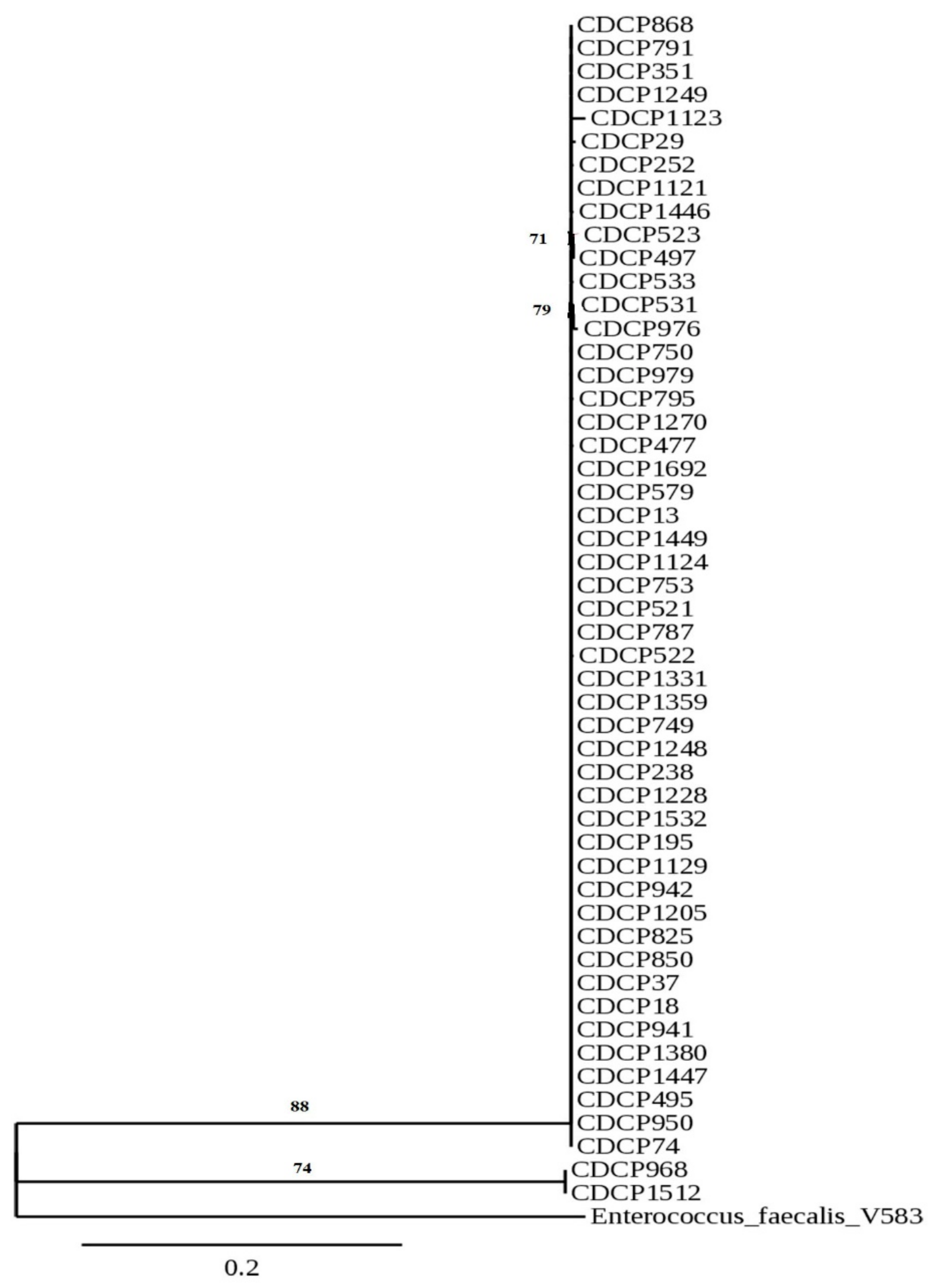
4.6. Phylogenetic Analysis
4.7. Extraction of Plasmid DNA and Detection of Antibiotic Resistance Genes with PCR
4.8. Determination of Gene Expression
5. Conclusions
Author Contributions
Funding
Institutional Review Board Statement
Informed Consent Statement
Data Availability Statement
Acknowledgments
Conflicts of Interest
References
- ISO. Milk and Milk Products—Determination of the Minimal Inhibitory Concentration (MIC) of Antibiotics Applicable to Bifidobacteria and Non-Enterococcal Lactic Acid Bacteria (LAB); ISO: Geneva, Switzerland, 2012; Volume 9936. [Google Scholar]
- EFSA (European Food Safety Authority). Guidance on the assessment of bacterial susceptibility to antimicrobials of human and veterinary importance. EFSA J. 2012, 10, 1–10. [Google Scholar] [CrossRef]
- Hanchi, H.; Mottawea, W.; Sebei, K.; Hammami, R. The genus Enterococcus: Between probiotic potential and safety concerns-an update. Front. Microbiol. 2018, 9, 1–16. [Google Scholar] [CrossRef] [PubMed]
- Dias, J.A.R.; Abe, H.A.; Sousa, N.C.; Silva, R.D.F.; Cordeiro, C.A.M.; Gomes, G.F.E.; Ready, J.S.; Mouriño, J.L.P.; Martins, M.L.; Carneiro, P.C.F.; et al. Enterococcus faecium as potential probiotic for ornamental neotropical cichlid fish, Pterophyllum scalare (Schultze, 1823). Aquac. Int. 2019, 27, 463–474. [Google Scholar] [CrossRef]
- Da Costa Sousa, N.; Silva do Couto, M.V.; Andrade Abe, H.; Guimarães Paixão, P.E.; Martins Cordeiro, C.A.; Monteiro Lopes, E.; Ready, J.S.; Fernandes Alves Jesus, G.; Laterça Martins, M.; Pereira Mouriño, J.L.; et al. Effects of an Enterococcus faecium -based probiotic on growth performance and health of Pirarucu, Arapaima gigas. Aquac. Res. 2019, 50, 1–9. [Google Scholar] [CrossRef]
- Hu, C.; Xing, W.; Liu, X.; Zhang, X.; Li, K.; Liu, J.; Deng, B.; Deng, J.; Li, Y.; Tan, C. Effects of dietary supplementation of probiotic Enterococcus faecium on growth performance and gut microbiota in weaned piglets. AMB Express 2019, 9, 1–2. [Google Scholar] [CrossRef]
- Chen, Y.J.; Min, B.J.; Cho, J.H.; Kwon, O.S.; Son, K.S.; Kim, I.H.; Kim, S.J. Effects of dietary Enterococcus faecium SF68 on growth performance, nutrient digestibility, blood characteristics and faecal noxious gas content in finishing pigs. Asian-Australas. J. Anim. Sci. 2006, 19, 406–411. [Google Scholar] [CrossRef]
- Pollmann, M.; Nordhoff, M.; Pospischil, A.; Tedin, K.; Wieler, L.H. Effects of a Probiotic Strain of Natural Chlamydia Infection in Swine. Infect. Immun. 2005, 73, 4346–4353. [Google Scholar] [CrossRef] [PubMed] [Green Version]
- Khalkhali, S.; Mojgani, N. In vitro and in vivo safety analysis of Enterococcus faecium 2C isolated from human breast milk. Microb. Pathog. 2018, 116, 73–77. [Google Scholar] [CrossRef]
- Aşgın, N.; Taşkın, E. Is there any association between antibiotic resistance and virulence genes in Enterococcus isolates? Med. Sci. Discov. 2019, 6, 310–315. [Google Scholar] [CrossRef]
- Pärnänen, K.; Karkman, A.; Hultman, J.; Lyra, C.; Bengtsson-Palme, J.; Larsson, D.G.J.; Rautava, S.; Isolauri, E.; Salminen, S.; Kumar, H.; et al. Maternal gut and breast milk microbiota affect infant gut antibiotic resistome and mobile genetic elements. Nat. Commun. 2018, 9, 1–11. [Google Scholar] [CrossRef] [Green Version]
- Fernández, L.; Rodríguez, J.M. Human Milk Microbiota: Origin and Potential Uses. Nestle Nutr. Inst. Workshop Ser. 2020, 94, 75–85. [Google Scholar] [CrossRef] [PubMed]
- Fijan, S. Microorganisms with claimed probiotic properties: An overview of recent literature. Int. J. Environ. Res. Public Health 2014, 11, 4745–4767. [Google Scholar] [CrossRef] [PubMed]
- Zimmermann, P.; Curtis, N. Breast milk microbiota: A complex microbiome with multiple impacts and conditioning factors. J. Infect. 2020, 81, 17–47. [Google Scholar] [CrossRef] [PubMed]
- Becker, K.; Heilmann, C.; Peters, G. Coagulase-Negative Staphylococci. Clin. Microbiol. Rev. 2014, 27, 870–926. [Google Scholar] [CrossRef] [Green Version]
- Patterson, M.J. Streptococcus. In Medical Microbiology; Baron, S., Ed.; University of Texas Medical Branch at Galveston: Galveston, TX, USA, 2020; pp. 1–25. [Google Scholar]
- Ramsey, M.; Hartke, A.; Huycke, M. The Physiology and Metabolism of Enterococci. In Enterococci: From Commensals to Leading Causes of Drug Resistant Infection; Clewell, D., Gilmore, M., Ike, Y., Eds.; Massachusetts Eye and Ear Infirmary: Boston, MA, USA, 2014; pp. 1–55. [Google Scholar]
- Golob, M.; Pate, M.; Kušar, D.; Dermota, U.; Avberšek, J.; Papić, B.; Zdovc, I.; Bondi, M. Antimicrobial Resistance and Virulence Genes in Enterococcus faecium and Enterococcus faecalis from Humans and Retail Red Meat. Biomed Res. Int. 2019, 2019, 14–16. [Google Scholar] [CrossRef] [Green Version]
- Chakraborty, A.; Pal, N.K.; Sarkar, S.; Gupta, M. Sen Antibiotic resistance pattern of Enterococci isolates from nosocomial infections in a tertiary care hospital in Eastern India. J. Nat. Sci. Biol. Med. 2015, 6, 394–397. [Google Scholar] [CrossRef] [Green Version]
- Jabbari, S.M.; Shiadeh; Pormohammad, A.; Hashemi, A.; Lak, P. Global prevalence of antibiotic resistance in blood-isolated Enterococcus faecalis and Enterococcus faecium: A systematic review and meta-analysis. Infect. Drug Resist. 2019, 12, 2713–2725. [Google Scholar] [CrossRef] [Green Version]
- Hollenbeck, B.L.; Rice, L.B. Intrinsic and acquired resistance mechanisms in enterococcus. Virulence 2012, 3, 421–433. [Google Scholar] [CrossRef] [Green Version]
- Hryniewicz, W.; Ozorowski, T. Szpitalna Lista Antybiotyków Propozycja Kierowana do Szpitali; Narodowy Program Ochrony Antybiotyków: Warszawa, Poland, 2011. [Google Scholar]
- Gawryszewska, I.; Żabicka, D.; Bojarska, K.; Malinowska, K.; Hryniewicz, W.; Sadowy, E. Invasive enterococcal infections in Poland: The current epidemiological situation. Eur. J. Clin. Microbiol. Infect. Dis. 2016, 35, 847–856. [Google Scholar] [CrossRef] [Green Version]
- Miller, W.R.; Munita, J.M.; Arias, C.A. Mechanisms of antibiotic resistance in enterococci. Expert Rev. Anti. Infect. Ther. 2014, 12, 1221–1236. [Google Scholar] [CrossRef]
- Trivedi, K.; Cupakova, S.; Karpiskova, R. Virulence factors and antibiotic resistance in enterococci isolated from food-stuffs. Vet. Med. 2011, 56, 352–357. [Google Scholar] [CrossRef]
- Sanlibaba, P.; Senturk, E. Prevalence, characterization and antibiotic resistance of enterococci from traditional cheeses in Turkey. Int. J. Food Prop. 2018, 21, 1955–1963. [Google Scholar] [CrossRef] [Green Version]
- Kamuś, M. Leczenie Trądziku Pospolitego—Charakterystyka Schorzenia, Przegląd Preparatów OTC Oraz Rx do Stosowania Miejscowego; Wielkopolska Okręgowa Izba Aptekarska: Poznań, Poland, 2021. [Google Scholar]
- Asadollahi, P.; Razavi, S.; Asadollahi, K.; Pourshafie, M.R.; Talebi, M. Rise of antibiotic resistance in clinical enterococcal isolates during 2001–2016 in Iran: A review. New Microbes New Infect. 2018, 26, 92–99. [Google Scholar] [CrossRef] [PubMed]
- Stefaniuk, E.; Suchocka, U.; Bosacka, K.; Hryniewicz, W. Etiology and antibiotic susceptibility of bacterial pathogens responsible for community-acquired urinary tract infections in Poland. Eur. J. Clin. Microbiol. Infect. Dis. 2016, 35, 1363–1369. [Google Scholar] [CrossRef] [PubMed] [Green Version]
- Granados-Chinchilla, F.; Rodríguez, C. Tetracyclines in Food and Feedingstuffs: From Regulation to Analytical Methods, Bacterial Resistance, and Environmental and Health Implications. J. Anal. Methods Chem. 2017, 2017, 1315497. [Google Scholar] [CrossRef]
- Marin Garrido, A.; Gálvez, A.; Pérez Pulido, R. Antimicrobial Resistance in Enterococci. J. Infect. Dis. Ther. 2014, 2. [Google Scholar] [CrossRef] [Green Version]
- Šeputiene, V.; Bogdaite, A.; Ružauskas, M.; Sužiedeliene, E. Antibiotic resistance genes and virulence factors in Enterococcus faecium and Enterococcus faecalis from diseased farm animals: Pigs, cattle and poultry. Pol. J. Vet. Sci. 2012, 15, 431–438. [Google Scholar] [CrossRef] [Green Version]
- Coque, T.M.; Singh, K.V.; Weinstock, G.M.; Murray, B.E. Characterization of dihydrofolate reductase genes from trimethoprim- susceptible and trimethoprim-resistant strains of Enterococcus faecalis. Antimicrob. Agents Chemother. 1999, 43, 141–147. [Google Scholar] [CrossRef] [Green Version]
- Woegerbauer, M.; Zeinzinger, J.; Springer, B.; Hufnagl, P.; Indra, A.; Korschineck, I.; Hofrichter, J.; Kopacka, I.; Fuchs, R.; Steinwider, J.; et al. Prevalence of the aminoglycoside phosphotransferase genes aph(3’)-IIIa and aph(3’)-IIa in Escherichia coli, Enterococcus faecalis, Enterococcus faecium, Pseudomonas aeruginosa, Salmonella enterica subsp. Enterica and Staphylococcus aureus isolates in Aust. J. Med. Microbiol. 2014, 63, 210–217. [Google Scholar] [CrossRef] [Green Version]
- Ahmed, M.O.; Baptiste, K.E. Vancomycin-Resistant Enterococci: A Review of Antimicrobial Resistance Mechanisms and Perspectives of Human and Animal Health. Microb. Drug Resist. 2018, 24, 590–606. [Google Scholar] [CrossRef] [Green Version]
- Yu, J.; Gao, W.; Qing, M.; Sun, Z.; Wang, W.; Liu, W.; Pan, L. Identification and characterization of lactic acid bacteria isolated from traditional pickles in Sichuan, China. J. Gen. Appl. Microbiol 2012, 58, 163–172. [Google Scholar] [CrossRef] [PubMed] [Green Version]
- Dereeper, A.; Guignon, V.; Blanc, G.; Audic, S.; Buffet, S.; Chevenet, F.; Dufayard, J.F.; Guindon, S.; Lefort, V.; Lescot, M.; et al. Phylogeny.fr: Robust phylogenetic analysis for the non-specialist. Nucleic Acids Res. 2008, 36, 465–469. [Google Scholar] [CrossRef] [PubMed]
- Wajda, Ł.; Wyderka, M.; Polak, Z.; Duda-Chodak, A.; Makarewicz, M. Examination of novel Aureobasidium pullulans isolates dominating apple microflora and assessing their potential for apple juice spoilage. World J. Microbiol. Biotechnol. 2018, 34, 11. [Google Scholar] [CrossRef] [PubMed] [Green Version]
- Salem, I.; Ramser, A.; Isham, N.; Ghannoum, M.A. The gut microbiome as a major regulator of the gut-skin axis. Front. Microbiol. 2018, 9, 1–14. [Google Scholar] [CrossRef] [Green Version]
- Urbaniak, C.; Burton, J.P.; Reid, G. Breast, milk and microbes: A complex relationship that does not end with lactation. Women’s Health 2012, 8, 385–398. [Google Scholar] [CrossRef] [Green Version]
- Ouoba, L.I.I.; Lei, V.; Jensen, L.B. Resistance of potential probiotic lactic acid bacteria and bifidobacteria of African and European origin to antimicrobials: Determination and transferability of the resistance genes to other bacteria. Int. J. Food Microbiol. 2008, 121, 217–224. [Google Scholar] [CrossRef]
- Agersø, Y.; Jensen, L.B.; Givskov, M.; Roberts, M.C. The identification of a tetracycline resistance gene tet(M), on a Tn916-like transposon, in the Bacillus cereus group. FEMS Microbiol. Lett. 2002, 214, 251–256. [Google Scholar] [CrossRef]

| Isolate Number | Accession Number | MIC Values (µg/mL) | |||||||||||||||
|---|---|---|---|---|---|---|---|---|---|---|---|---|---|---|---|---|---|
| Gent | Kan | Strep | Tet | Ery | Clin | Chlor | Amp | Neo | Van | Q/D | Lin | Trim | Cip | Rif | Tyl | ||
| CDCP13 | MT814617 | > | > | > | > | > | > | > | 0.5 S | 64 | 2 S | 1 | 2 | > | > | 32 | 2 S |
| CDCP18 | MT883426 | > | > | > | > | > | > | > | 1 S | 128 | 2 S | 1 | 2 | > | > | 64 | 1 S |
| CDCP29 | MT814618 | > | > | > | > | 8 R | > | 64 R | 8 R | > | 2 S | > | 2 | > | 128 | > | 1 S |
| CDCP37 | MT814619 | > | > | > | > | > | > | 64 R | 16 R | > | 4 S | > | > | > | > | > | 2 S |
| CDCP74 | MT882797 | 64 R | 512 S | 256 R | > | 8 R | > | 8 S | 0.5 S | > | 2 S | > | 2 | 32 | 128 | 32 | 2 S |
| CDCP195 | MT882798 | 128 R | 256 S | 256 R | > | > | > | 8 S | 0.5 S | > | 2 S | > | 2 | 16 | > | 32 | 2 S |
| CDCP238 | MT814620 | 128 R | 512 S | > | > | 8 R | 4 S | 8 S | 0.5 S | > | 2 S | 8 | 2 | > | 128 | 32 | 2 S |
| CDCP252 | MT882787 | 128 R | 256 S | > | > | > | > | 8 S | 1 S | > | 2 S | 8 | 2 | 64 | 64 | > | 1 S |
| CDCP351 | MT814240 | 256 R | 512 S | > | 64 R | > | 4 S | 8 S | 1 S | > | 2 S | 2 | 2 | 2 | 128 | 32 | 1 S |
| CDCP477 | MT882695 | 64 R | 8 S | 64 S | 8 R | > | 0.125 S | 8 S | 1 S | 256 | 2 S | 1 | 2 | > | 128 | 32 | 2 S |
| CDCP495 | MT883429 | 32 S | 256 S | 64 S | 8 R | > | 0.25 S | 4 S | 0.5 S | 64 | 1 S | 1 | 2 | > | > | 32 | 1 S |
| CDCP497 | MT882696 | 64 R | 8 S | 128 S | 0.5 S | > | 0.125 S | 16 S | 0.S5 | 64 | 1 S | 1 | 2 | 4 | 128 | 32 | 2 S |
| CDCP521 | MT882697 | 32 S | 4 S | 32 S | 4 S | > | 0.125 S | 0.5 S | 0.5 | 64 | 1 S | 1 | 2 | 4 | 128 | 32 | 2 S |
| CDCP522 | MT882698 | 64 R | 1024 S | 64 S | 4 S | 8 R | 0.125 S | 16 S | 1 | 64 | 1 S | 1 | 2 | 16 | 128 | 32 | 2 S |
| CDCP523 | MT882699 | 64 R | 8 S | 64 S | 4 S | 8 R | 0.125 S | 4 S | 0.5 | 64 | 1 S | 1 | 2 | 64 | 128 | 64 | 2 S |
| CDCP531 | MT882700 | 16 S | 8 S | 64 S | 4 S | > | 0.125 S | 4 S | 0.5 | 64 | 1 S | 1 | 2 | 2 | 128 | 32 | 2 S |
| CDCP533 | MT882701 | 32 S | 256 S | 128 S | 4 S | > | 0.125 S | 8 S | 1 | 32 | 8 R | > | 4 | 128 | 128 | 32 | 2 S |
| CDCP579 | MT883425 | 64 R | 256 S | 128 S | 4 S | 4 S | 0.25 S | 4 S | 1 | 64 | 1 S | 0.5 | 2 | > | 128 | 32 | 2 S |
| CDCP749 | MT882799 | 32 S | 128 S | 64 S | 2 S | 8 R | 0.125 S | 2 S | 1 | 32 | 0.5 S | 0.5 | 2 | 4 | 128 | 32 | 1 S |
| CDCP750 | MT882800 | 32 S | 128 S | 64 S | 2 S | 8 R | 0.25 S | 4 S | 1 | 64 | 0.5 S | 0.5 | 2 | > | 128 | 16 | 2 S |
| CDCP753 | MT882801 | 64 R | 512 S | 64 S | 2 S | > | 0.5 S | 8 S | 1 | 64 | 0.5 S | 0.5 | 2 | > | 128 | 32 | 1 S |
| CDCP787 | MT882802 | 32 S | 128 S | 32 S | 2 S | > | 0.25 S | 4 S | 2 | 64 | 0.5 S | 0.5 | 2 | > | 128 | 32 | 2 S |
| CDCP791 | MT882803 | 64 R | 128 S | 64 S | 16 R | > | 0.125 S | 4 S | 1 | 32 | 0.5 S | 0.5 | 2 | > | > | 32 | 1 S |
| CDCP795 | MT882804 | 32 S | 512 S | 64 S | 16 R | 8 R | 0.125 S | 4 S | 1 | 64 | 0.5 S | 0.5 | 2 | > | > | 32 | 1 S |
| CDCP825 | MT882805 | 8 S | 32 S | 32 S | 8 R | 4 S | 0.25 S | 16 S | > | 64 | 16 R | 0.5 | 2 | > | 16 | 0.125 | 1 S |
| CDCP850 | MT882788 | 64 R | 256 S | 64 S | 4 S | > | 0.25 S | 8 S | 1 | 64 | 1 S | 1 | 4 | > | 128 | 32 | 1 S |
| CDCP868 | MT882789 | 64 R | 256 S | 64 S | 4 S | > | 0.25 S | 8 S | 1 | 64 | 1 S | 2 | 4 | > | > | 32 | 1 S |
| CDCP941 | MT882790 | 32 S | 256 S | 128 S | 4 S | > | 0.25 S | 4 S | 0.5 | 256 | 1 S | 1 | 2 | > | 128 | 32 | 1 S |
| CDCP942 | MT882791 | 32 S | 256 S | 64 S | 4 S | > | 0.25 S | 4 S | 0.5 | 128 | 1 S | 1 | 2 | > | 128 | 16 | 2 S |
| CDCP950 | MT882792 | 32 S | 128 S | 256 R | 4 S | > | 0.5 S | 16 S | 0.5 | 64 | 1 S | 0.5 | 2 | > | 128 | 16 | 1 S |
| CDCP968 | MT882793 | 16 S | 128 S | 32 S | 2 S | > | 0.25 S | 4 S | 0.5 | > | 1 S | 1 | 2 | 16 | 128 | 16 | 2 S |
| CDCP976 | MT882794 | 32 S | 256 S | 128 S | 2 S | > | 0.25 S | 4 S | 0.5 | 64 | 1 S | 0.5 | 2 | > | 128 | 32 | 2 S |
| CDCP979 | MT882795 | 32 S | 256 S | 64 S | 2 S | > | 0.25 S | 4 S | 0.5 | 64 | 0.5 S | 0.25 | 1 | > | 128 | 32 | 2 S |
| CDCP1121 | MT814241 | 32 S | 128 S | 64 S | 16 R | > | 0.25 S | 4 S | 1 | 32 | > | 1 | 2 | > | 128 | 32 | 2 S |
| CDCP1123 | MT882796 | 128 R | 1024 S | 256 R | 4 S | > | 1 S | 16 S | 2 | 256 | 1 S | 2 | 4 | > | > | 32 | 2 S |
| CDCP1124 | MT814242 | 256 R | > | > | 32 R | > | 8 R | 16 S | 0.5 | > | 2 S | 4 | 2 | 64 | > | 8 | > |
| CDCP1129 | MT883427 | 32 S | 64 S | 64 S | > | > | 0.5 S | 4 S | 1 | 32 | > | 1 | 2 | 16 | > | 16 | > |
| CDCP1205 | MT814243 | 32 S | 128 S | 64 S | 32 R | > | 0.25 S | 8 S | 1 | 32 | 1 S | 1 | 2 | > | > | 32 | 1 S |
| CDCP1228 | MT814244 | 64 R | 256 S | 128 S | > | > | 0.25 S | 8 S | 1 | 32 | > | 1 | 2 | > | > | 64 | 1 S |
| CDCP1248 | MT814245 | 32 S | 512 S | 64 S | 64 R | > | 0.5 S | 4 S | 1 | 32 | > | 1 | 1 | > | > | 64 | 4 S |
| CDCP1249 | MT814246 | 32 S | 128 S | 128 S | 16 R | > | 0.25 S | 4 S | 1 | 64 | 1 S | 1 | 2 | > | 128 | 32 | 2 S |
| CDCP1270 | MT814247 | 32 S | 128 S | 128 S | 64 R | > | 0.125 S | 4 S | 1 | 64 | > | 1 | 1 | 64 | 128 | 32 | 2 S |
| CDCP1331 | MT814248 | 64 R | 1024 S | 128 S | 32 R | > | 0.5 S | 8 S | 1 | 128 | > | 2 | 4 | > | > | > | > |
| CDCP1359 | MT814249 | 32 S | 1024 S | 128 S | 32 R | > | 0.25 S | 4 S | 1 | 64 | 2 S | 1 | 2 | > | 128 | 64 | > |
| CDCP1380 | MT814250 | 256 R | 1024 S | 256 R | 64 R | > | 2 S | 4 S | 1 | 256 | 4 S | 8 | 16 | > | 128 | 64 | > |
| CDCP1446 | MT814251 | 32 S | 128 S | 128 S | > | > | 0.5 S | 8 S | 1 | 64 | > | 1 | 2 | > | > | 16 | > |
| CDCP1447 | MT814621 | 32 S | 128 S | 64 S | 64 R | > | 0.5 S | 4 S | 1 | 64 | > | 1 | 2 | > | > | 16 | 2 S |
| CDCP1449 | MT814252 | 64 R | 128 S | 64 S | 64 R | > | 0.5 S | 4 S | 1 | 64 | > | 1 | 2 | 64 | > | 16 | 1 S |
| CDCP1512 | MT883428 | 64 R | 128 S | 64 S | > | > | 0.125 S | 4 S | 1 | 64 | > | 1 | 2 | > | > | 16 | > |
| CDCP1532 | MT814253 | 128 R | 512 S | > | > | > | > | 16 S | 1 | 256 | 1 S | > | 4 | 4 | 128 | 8 | 2 S |
| CDCP1692 | MT814254 | 128 R | 512 S | > | 2 S | > | 1 S | 8 S | 1 | > | 1 S | 1 | 2 | > | 128 | 64 | 2 S |
| Cut-off values | 32 | 1024 | 128 | 4 | 4 | 4 | 16 | 2 | ns | 4 | ns | ns | ns | ns | ns | 4 | |
| Percentage of resistance strains | 54.9 | 11.8 | 31.4 | 56.9 | 96.1 | 19.6 | 7.8 | 5.9 | 19.6 | 21.6 | 11.8 | 2.0 | 66.7 | 37.3 | 7.8 | 13.7 | |
| Antibiotic | Gent | Kan | Strep | Tet | Ery | Clin | Chlor | Amp | Van | Tyl | Neo | Q/D | Trim | Lin | Cip | Rif | ||||||||||||||||||
|---|---|---|---|---|---|---|---|---|---|---|---|---|---|---|---|---|---|---|---|---|---|---|---|---|---|---|---|---|---|---|---|---|---|---|
| Isolate Number | aac(6′)-aph(2″) | aph3′-IIIa | aph2′-IIIa | ant(2″) | ant(6) | ant(2) | ant(2″)-Ia | aadA | strA | strB | tetM | tetK | tetL | tetO | tetS | ErmB | ErmB1 | lunA | lunB | catA4 | catII | mecA | BlaZ | ErmG | vanH | ErmA | ErmT | Aph(3′)-IIIa | ermB | dfrA14 | clcD | gyrA | parC | marA |
| CDCP238 | - | - | - | - | + | - | - | - | - | - | + | - | - | - | + | - | - | - | - | - | - | - | - | - | - | - | - | + | - | - | - | - | - | - |
| CDCP351 | - | - | - | - | - | - | - | - | - | - | - | - | - | + | - | + | - | - | - | - | - | - | - | - | - | - | - | + | - | - | - | - | - | - |
| CDCP477 | - | - | - | - | - | - | - | - | - | - | - | - | - | - | - | + | - | - | - | - | - | - | - | - | - | - | - | - | - | - | - | - | - | - |
| CDCP495 | - | - | - | - | - | - | - | - | - | - | - | - | - | - | - | + | - | - | - | - | - | - | - | - | - | - | - | - | - | - | - | - | - | - |
| CDCP521 | - | - | - | - | - | - | - | - | - | - | - | - | - | - | - | + | - | - | - | - | - | - | - | - | - | - | - | - | - | - | - | - | - | - |
| CDCP532 | - | - | - | - | - | - | - | - | - | - | - | - | - | - | - | + | - | - | - | - | - | - | - | - | - | - | - | - | - | - | - | - | - | - |
| CDCP531 | - | - | - | - | - | - | - | - | - | - | - | - | - | - | - | + | - | - | - | - | - | - | - | - | - | - | - | - | - | - | - | - | - | - |
| CDCP533 | - | - | - | - | - | - | - | - | - | - | - | - | - | - | - | + | - | - | - | - | - | - | - | - | - | - | - | - | - | - | - | - | - | - |
| CDCP750 | - | - | - | - | - | - | - | - | - | - | - | - | - | - | - | - | - | - | - | - | - | - | - | - | - | - | - | - | - | + | - | - | - | - |
| CDCP753 | - | - | - | - | - | - | - | - | - | - | - | - | - | - | - | + | - | - | - | - | - | - | - | - | - | - | - | - | - | + | - | - | - | - |
| CDCP787 | - | - | - | - | - | - | - | - | - | - | - | - | - | - | - | + | - | - | - | - | - | - | - | - | - | - | - | - | - | + | - | - | - | - |
| CDCP791 | - | - | - | - | - | - | - | - | - | - | - | - | - | - | - | - | - | - | - | - | - | - | - | - | - | - | - | - | - | + | - | - | - | - |
| CDCP795 | - | - | - | - | - | - | - | - | - | - | - | - | - | - | - | - | - | - | - | - | - | - | - | - | - | - | - | - | - | + | - | - | - | - |
| CDCP825 | - | - | - | - | - | - | - | - | - | - | - | - | - | - | - | - | - | - | - | - | - | - | - | - | - | - | - | - | - | + | - | - | - | - |
| CDCP850 | - | - | - | - | - | - | - | - | - | - | - | - | - | - | - | - | - | - | - | - | - | - | - | - | - | - | - | - | - | + | - | - | - | - |
| CDCP868 | - | - | - | - | - | - | - | - | - | - | - | - | - | - | - | - | - | - | - | - | - | - | - | - | - | - | - | - | - | + | - | - | - | - |
| CDCP941 | - | - | - | - | - | - | - | - | - | - | - | - | - | - | - | - | - | - | - | - | - | - | - | - | - | - | - | - | - | + | - | - | - | - |
| CDCP950 | - | - | - | - | - | - | - | - | - | - | - | - | - | - | - | - | - | - | - | - | - | - | - | - | - | - | - | - | - | + | - | - | - | - |
| CDCP979 | - | - | - | - | - | - | - | - | - | - | - | - | - | - | - | - | - | - | - | - | - | - | - | - | - | - | - | - | - | + | - | - | - | - |
| CDCP1208 | - | - | - | - | - | - | - | - | - | - | - | - | - | + | - | - | - | - | - | - | - | - | - | - | - | - | - | - | - | - | - | - | - | - |
| CDCP1228 | - | - | - | - | - | - | - | - | - | - | - | - | - | + | - | - | - | - | - | - | - | - | - | - | - | - | - | - | - | + | - | - | - | - |
| CDCP1248 | - | - | - | - | - | - | - | - | - | - | - | - | - | - | - | - | - | - | - | - | - | - | - | - | - | - | - | - | - | + | - | - | - | - |
| CDCP1249 | - | - | - | - | - | - | - | - | - | - | - | - | - | + | - | - | - | - | - | - | - | - | - | - | - | - | - | - | - | - | - | - | - | - |
| CDCP1331 | - | - | - | - | - | - | - | - | - | - | - | - | - | + | - | - | - | - | - | - | - | - | - | - | - | - | - | - | - | - | - | - | - | + |
| CDCP1359 | - | - | - | - | - | - | - | - | - | - | - | - | - | - | - | - | - | - | - | - | - | - | - | - | - | - | - | - | - | + | - | - | - | - |
| CDCP1380 | - | - | - | - | - | - | - | - | - | - | - | - | - | - | - | - | - | - | - | - | - | - | - | - | - | - | - | - | - | + | - | - | - | - |
| CDCP1446 | - | - | - | - | - | - | - | - | - | - | - | - | - | - | - | - | - | - | - | - | - | - | - | - | - | - | - | - | - | + | - | - | - | - |
| CDCP1447 | - | - | - | - | - | - | - | - | - | - | - | - | - | + | - | - | - | - | - | - | - | - | - | - | - | - | - | - | - | - | - | - | - | - |
| CDCP1449 | - | - | - | - | - | - | - | - | - | - | - | - | - | + | - | - | - | - | - | - | - | - | - | - | - | - | - | - | - | + | - | - | - | - |
| CDCP1512 | - | - | - | - | - | - | - | - | - | - | + | - | - | - | - | + | - | - | - | - | - | - | - | - | - | - | - | - | - | - | - | - | - | - |
| CDCP1532 | - | - | - | - | - | - | - | - | - | - | - | - | - | - | - | - | - | - | - | - | - | - | - | - | - | - | - | + | - | + | - | - | - | - |
| CDCP1692 | - | - | - | - | - | - | - | - | - | - | - | - | - | - | - | - | - | - | - | - | - | - | - | - | - | - | - | + | - | + | - | - | - | - |
| Antibiotic (Gene) | Isolate No. | ΔCt for the Target | ΔCt for the Reference | Relative Ratio |
|---|---|---|---|---|
| Tetracycline (tetK) | CDCP351 | 0 | 0 | 0 |
| CDCP1228 | 6.74 × 10⁻⁴ | 4.52 × 10⁻⁹ | 1.49 × 10⁵ | |
| CDCP1331 | 2.03 × 10⁻⁷ | 1.11 × 10⁻⁹ | 1.82 × 10² | |
| CDCP1449 | 0 | 0 | 0 | |
| Erythromycin (ErmB) | CDCP351 | 1.14 × 10⁻² | 1.48 × 10⁻⁷ | 7.69 × 10⁴ |
| CDCP753 | 9.72 × 10⁻³ | 2.06 × 10⁻⁷ | 4.72 × 10⁴ | |
| CDCP787 | 1.68 × 10⁻³ | 3.02 × 10⁻⁷ | 5.58 × 10³ | |
| Neomycin (Aph(3′)-IIIa) | CDCP351 | 3.69 × 10⁻² | 1.27 × 10⁻⁴ | 2.90 × 10² |
| CDCP1532 | 1.72 × 10⁻² | 4.48 × 10⁻⁴ | 3.83 × 101 | |
| CDCP1692 | 1.88 | 3.14 × 10⁻⁴ | 5.98 × 10³ | |
| Rifampicin (marA) | CDCP1331 | 1.65 × 10⁻² | 3.92 × 10⁻⁶ | 4.21 × 10³ |
| Trimethoprim (dfrA14) | CDCP753 | 5.08 × 10⁻³ | 3.94 × 10⁻⁷ | 1.29 × 10⁴ |
| CDCP787 | 3.77 × 10⁻³ | 1.35 × 10⁻⁷ | 2.80 × 10⁴ | |
| CDCP1228 | 2.14 × 10⁻³ | 7.91 × 10⁻⁷ | 2.70 × 10³ | |
| CDCP1449 | 5.07 × 10⁻³ | 0 | 0 | |
| CDCP1532 | 4.74 × 10⁻² | 9.92 × 10⁻⁵ | 4.78 × 10² | |
| CDCP1692 | 1.79 × 10⁻² | 4.30 × 10⁻⁴ | 4.17 × 101 |
| Antibiotic | Gene Name | Primer Sequences (5′-3′) | Primer Name | Tm of Primers (°C) | Annealing Temperature (°C) | Final Mg2+ Concentration (mM) for the Regular PCR | Product Size (bp) | Reference |
|---|---|---|---|---|---|---|---|---|
| Kanamicin | aph3′ IIIa | GCCGATGTGGATTGCGAAAA | Aph3F | 59.83 | 55 | 4 | 292 | [41] |
| GCTTGATCCCCAGTAAGTCA | Aph3R | 56.92 | ||||||
| Aph(2″)-IIIa | TCGCTTGGTGAGGGCTTTAG | Aph2F | 60.04 | 55 | 4 | 402 | Current study | |
| CTGATCCTCCACAGCTTCCG | Aph2R | 60.18 | ||||||
| ant(2″)-I | CAGATGAGCGAAATCTGCCG | Ant2F | 59.42 | 54 | 4 | 226 | Current study | |
| CAAGCAGGTTCGCAGTCAAG | Ant2R | 59.76 | ||||||
| Tetracyclin | tetM | CTTGTTCGAGTTCCAATGC | tetMF | 54.74 | 55 | 4 | 401 | [42] |
| GGTGAACATCATAGACACGC | tetMR | 56.62 | ||||||
| tetK | TTAGGTGAAGGGTTAGGTCC | tetKF | 55.84 | 56 | 4 | 697 | [42] | |
| GCAAACTCATTCCAGAAGCA | tetKR | 56.62 | ||||||
| CGATAGGAACAGCAGTATATGGAA | tetK2F * | 59.76 | 60 * | 3 | 164 * | Current study | ||
| AGATCCTACTCCTTGTACTAACCT | tetK2R * | 59.13 | ||||||
| tet(L) | CATTTGGTCTTATTGGATCG | tetLF | 52.04 | 52 | 4 | 456 | [42] | |
| ATTACACTTCCGATTTCGG | tetLR | 52.77 | ||||||
| tetO | GCATTCTGGCTCACGTTGAC | tetOF | 59.83 | 56 | 4 | 985 | Current study | |
| TGCGGCAACAGTATTTCGTTC | tetOR | 59.8 | ||||||
| ATTAACTTAGGCATTCTGGCTCA | tetO2F * | 59.76 | 60 * | 3 | 176 * | Current study | ||
| GATGTCACTGCTGTCTGGAT | tetO2R * | 59.13 | ||||||
| tetS | GATAAGGCAGAGCCTGGTGAG | tetSF | 60.2 | 55 | 4 | 414 | Current study | |
| AGCCCAGAAAGGATTTGGAGG | tetSR | 59.99 | ||||||
| Chloramphenikol | Cat(A4) | CAATGCACCTTTAGCCAGACCG | catA4F | 62.08 | 60 | 4 | 310 | Current study |
| AGGCTAGATCGTCGCCGTATTG | catA4R | 62.27 | ||||||
| Cat(II) | TTCTCTGCACTGTCCTGCCG | catIIF | 62.44 | 60 | 4 | 311 | Current study | |
| AACCGTGCTGCATGAAAGCC | catIIR | 62.15 | ||||||
| Gentamicin | aac(6′)-aph(2″) | CCTCGTGTAATTCATGTTCTGGC | GentF | 59.11 | 58 | 4 | 675 | [41] |
| ACAGAGCCTTGGGAAGATGAA | GentR | 57.75 | ||||||
| Erythromicin | erm(B)-1 | GCATTTAACGACGAAACTGGC | ermB1F | 58.76 | 55 | 4 | 247 | Current study |
| ATAGATGTCAGACGCACGGC | ermB1R | 60.25 | ||||||
| ACTACTTAGGATGATGTCGTGGAA | EryF * | 60.93 | 60 | 5 | 188 | Current study | ||
| CCCTGAACAATTGGTGGCATA | EryR * | 60.40 | ||||||
| Klindamicin | lnu(A) | TTGGTTAGATGGTGGCTGGG | lnuAF | 59.67 | 60 | 4 | 253 | Current study |
| ACCTTCTGGGTTTGCTTGGG | lnuAR | 60.47 | ||||||
| lnu(B) | TGACGTAGCTCCGTACTTGATG | lnuBF | 59.9 | 57 | 4 | 166 | Current study | |
| AAGCATAGCCTTCGTATCAGG | lnuBR | 57.61 | ||||||
| Ampicillin | mecA | CAGGTACTGCTATCCACCCTC | mecAF | 59.31 | 55 | 4 | 770 | Current study |
| TTCTGCAGTACCGGATTTGCC | mecAR | 60.95 | ||||||
| Bla(Z) | AACAGTTCACATGCCAAAGAG | BlaZF | 57.00 | 57 | 4 | 485 | Current study | |
| AAAGTCTTGCCGAAAGCAGC | BlaZR | 59.69 | ||||||
| Cyprofloksacin | gyrA | TTCCATTCGGATACGCGGAG | gyrAF | 59.97 | 60 | 2.75 | 432 | Current study |
| CCACGCAAAATATGAGCCCG | gyrAR | 59.97 | ||||||
| parC | CCCTTGAACATGAACGTCCT | parCF | 57.81 | 60 | 4 | 177 | Current study | |
| GAGATAGGCGATCAGCAAGC | parCR | 58.57 | ||||||
| Streptomicin | ant(6) | ACTGGCTTAATCAATTTGGG | Ant6F | 53.79 | 59 | 1.5 | 597 | Current study |
| GCCTTTCCGCCACCTCACCG | Ant6R | 56.11 | ||||||
| Ant(2) | ACACAACGCAGGTCACATTG | Ant2F | 59.34 | 56 | 4 | 421 | Current study | |
| ACTGGTGGTACTTCATCGGC | Ant2R | 59.75 | ||||||
| Ant(2”)-Ia | CAGATGAGCGAAATCTGCCG | Ant2IaF | 59.42 | 56 | 4 | 226 | Current study | |
| CAAGCAGGTTCGCAGTCAAG | Ant2IaR | 59.76 | ||||||
| aadA | AGGTAGTTGGCGTCATCGAG | aadAF | 59.54 | 55 | 4 | 724 | Current study | |
| TCGCCTTTCACGTAGTGGAC | aadAR | 60.04 | ||||||
| Str(A) | CTTGGTGATAACGGCAATTC | straF | 55.06 | 55 | 4 | 549 | [41] | |
| CCAATCGCAGATAGAAGGC | straR | 55.87 | ||||||
| Str(B) | ATCGTCAAGGGATTGAAACC | strbF | 55.76 | 56 | 4 | 509 | [41] | |
| GGATCGTAGAACATATTGGC | strbR | 53.68 | ||||||
| ant(6) | AGGGACATAGTTCCGACTGAT | StrepF * | 60.93 | 60 * | 3 | 198 * | Current study | |
| AACCTTCCACGACATCATCC | StrepR * | 60.40 | ||||||
| Vankomicin | EmrG | TGAAATAGGTGCAGGGAAAGG | EmrGF | 59.09 | 58 | 4 | 330 | Current study |
| AGCAATGCTAGTGATCTGTTTG | EmrGR | 58.19 | ||||||
| vanHa | TCGGAATCCAACGCCAAATC | vanHaF | 60.05 | 60 | 4 | 428 | Current study | |
| CTTCGGCTGCGACTATAAGC | vanHaR | 59.98 | ||||||
| Tylosine | ErmT | GGGAAAGGTCATTTCTCGTTTG | ErmTF | 58.99 | 57 | 4 | 252 | Current study |
| ACTTTCTGTAGCTGTGCTTTC | ErmTR | 57.62 | ||||||
| ErmA | TCGTTGAGAAGGGATTTGCG | ErmAF | 59.7 | 59 | 1.5 | 273 | Current study | |
| TCAAAGCCTGTCGGAATTGG | ErmAR | 59.62 | ||||||
| Dalfopristin | ermB | GGCATTTAACGACGAAACTGGC | ermBDF | 60.98 | 60 | 4 | 322 | Current study |
| TGAGTGTGCAAGAGCAACCC | ermBDR | 60.82 | ||||||
| Trimethoprim | dfrA14 | TGGTTGCGGTCCAGACATAC | dfrA14F * | 60.04 | 60 * | 2.75 | 261 | Current study |
| ATTTCTCCGCCACCAGACAC | dfrA14R * | 60.32 | ||||||
| Linesolid | clcD | TGCGTTGTTTGCTTTAAGTCCG | CfrBF | 60.54 | 60 | 4 | 490 | Current study |
| ACCGCAAGCAGCGTCTATATC | CfrBR | 60.6 | ||||||
| Rifampicin | marA | ACAACCTGGAATCGCCACTG | marAF * | 60.61 | 60 * | 5 | 270 * | Current study |
| TCATCCGGTATTTATGCGGCG | marAR * | 60.94 | ||||||
| Neomycin | Aph(3′)-IIIa | AAGATACGGAAGGAATGTCTCC | NeoF | 57.33 | 57 | 4 | 600 | Current study |
| TGTCATACCACTTGTCCGCC | NeoR | 60.04 | ||||||
| CATCAGGCTCTTTCACTCCAT | NeoF * | 59.57 | 60 * | 3 | 200 * | Current study | ||
| CAAGTTCCTCTTCGGGCTT | NeoR * | 59.18 | ||||||
| Positive control | 16S rRNA | CCTGCAATCCGAACTGAGA | 16SEF * | 58.96 | 60 * | 3 | 105 * | Current study |
| CCTTATGACCTGGGCTACAC | 16SER * | 59.20 |
Publisher’s Note: MDPI stays neutral with regard to jurisdictional claims in published maps and institutional affiliations. |
© 2022 by the authors. Licensee MDPI, Basel, Switzerland. This article is an open access article distributed under the terms and conditions of the Creative Commons Attribution (CC BY) license (https://creativecommons.org/licenses/by/4.0/).
Share and Cite
Wajda, Ł.; Ostrowski, A.; Błasiak, E.; Godowska, P. Enterococcus faecium Isolates Present in Human Breast Milk Might Be Carriers of Multi-Antibiotic Resistance Genes. Bacteria 2022, 1, 66-87. https://doi.org/10.3390/bacteria1020007
Wajda Ł, Ostrowski A, Błasiak E, Godowska P. Enterococcus faecium Isolates Present in Human Breast Milk Might Be Carriers of Multi-Antibiotic Resistance Genes. Bacteria. 2022; 1(2):66-87. https://doi.org/10.3390/bacteria1020007
Chicago/Turabian StyleWajda, Łukasz, Adam Ostrowski, Ewelina Błasiak, and Patrycja Godowska. 2022. "Enterococcus faecium Isolates Present in Human Breast Milk Might Be Carriers of Multi-Antibiotic Resistance Genes" Bacteria 1, no. 2: 66-87. https://doi.org/10.3390/bacteria1020007
APA StyleWajda, Ł., Ostrowski, A., Błasiak, E., & Godowska, P. (2022). Enterococcus faecium Isolates Present in Human Breast Milk Might Be Carriers of Multi-Antibiotic Resistance Genes. Bacteria, 1(2), 66-87. https://doi.org/10.3390/bacteria1020007