Article

Chromatic Bacteria v.2-A Himar1 Transposon-Based Delivery Vector to Extend the Host Range of a Toolbox to Fluorescently Tag Bacteria

by
Christian Stocks
1,2,
Rudolf O. Schlechter
1,2,3,4 and
Mitja N. P. Remus-Emsermann
1,2,3,4,*
1
School of Biological Sciences, Faculty of Science, University of Canterbury, Christchurch 8140, New Zealand
2
Bioprotection Aotearoa, Lincoln University, Lincoln 7647, New Zealand
3
Biomolecular Interaction Centre, Faculty of Science, University of Canterbury, Christchurch 8140, New Zealand
4
Department of Biology, Chemistry, and Pharmacy, Institute for Microbiology, Freie Universität, 14195 Berlin, Germany
*
Author to whom correspondence should be addressed.
Bacteria 2022, 1(1), 56-65; https://doi.org/10.3390/bacteria1010006
Submission received: 8 December 2021 / Revised: 7 February 2022 / Accepted: 10 February 2022 / Published: 15 February 2022

Abstract

:
A recent publication described the construction and utility of a comprehensive “Chromatic Bacteria” toolbox containing a set of genetic tools that allows for fluorescently tagging a variety of Proteobacteria. In an effort to expand the range of bacteria taggable with the Chromatic Bacteria toolbox, a series of Himar1 transposon vectors was constructed to mediate insertion of fluorescent protein and antibiotic resistant genes. The Himar1 transposon was chosen as it is known to function in a wide range of bacterial species. To test the suitability of the new Himar1 Chromatic Bacteria plasmid derivatives, conjugations were attempted on recently isolated non-model organisms. Although we were unsuccessful in delivering the plasmids into Gram-positive bacterial isolates, we successfully modified previously recalcitrant isolates to the first set of the Chromatic Bacteria toolbox, such as Sphingomonas sp. Leaf357 and Acidovorax sp. Leaf84. This manuscript reports on the currently available plasmids and transposition success in different bacteria.

1. Introduction

Recently, we described the construction of a comprehensive “Chromatic Bacteria” toolbox containing a set of plasmids, denoted the pMRE series, to fluorescently tag a wide range of bacterial isolates [1]. These plasmid tools were well received by the community and, at the time of writing this article, have been ordered more than 400 times from the non-profit plasmid distribution platform addgene.org in the last 3 years alone (https://www.addgene.org/browse/article/28196767/, accessed on 7 December 2021). Fluorescently tagging bacteria using genetic manipulation is a state-of-the-art technology to study and track bacterial behaviour and physiology in situ and in vitro, such as on plant leaf surfaces and other environments [2,3,4,5]. By fluorescently tagging bacteria, it is possible to study them at the micrometer resolution. This is in stark contrast with the meta-omic research which has driven microbiology and microbial ecology during the last decade. Fluorescent tagging allows the study of bacteria at single cell resolution, which gives insight into biofilm formation, bacteria–bacteria and bacteria–host interactions [1].
The original Chromatic Bacteria toolbox employs three different vectors for fluorescent tagging, paired with one of eight fluorescent proteins and additionally one of four combinations of antibiotic resistant cassettes. The three different vectors are based on (i) a broad-host plasmid, (ii) a Tn5 transposon delivery plasmid, and (iii) a Tn7 transposon delivery plasmid. We have determined the host-range of the Chromatic Bacteria toolbox by extensive plasmid conjugation experiments. As a result, it was shown that, even though wide, the host range of the toolset is limited to Proteobacteria [1].
To further expand the host range of the Chromatic toolbox, we incorporated a Himar1 transposon-based delivery system. The Himar1 transposable element belongs to the mariner family of transposons. Mariner transposons can be found throughout eukaryotic and prokaryotic organisms [6,7]. The Himar1 transposon gene was discovered in the horn fly, Haematobia irritans, and has been mutated to construct a hyperactive version [8]. The gene encodes for a transposase protein that functions through a cut and paste mechanism and cleaves sequences between thymine adenine (AT) dinucleotide sites. Unlike other transposases such as the Tn5 transposase, the Himar1 transposase requires no cofactors to be provided in trans to initialise or mediate gene transposition by the host organism [8]. These low requirements for transposition make Himar1 ideal for random insertion mutagenesis and gene delivery into organisms that do not provide such cofactors. The simplicity in mechanism and broad target range of Himar1 shares similarities with the well characterised Mu transposition complex which has been shown to work in Gram-negative and Gram-positive species [9,10]. The Himar1 transposase has been shown to function in Gram-negative bacteria, such as Pseudomonas fluorescens and Flavobacterium johnsoniae [11,12], and Gram-positive bacteria, including Staphylococcus aureus, Clostridium perfringens, Streptococcus mutans, and Mycobacterium smegmatis [13,14,15,16]. In this study we describe the construction and utility of Himar1-based suicide vectors that allow the fluorescent tagging of bacteria.

2. Results

2.1. Construction of pMRE-Himar Series

We constructed a total of 23 plasmid vectors carrying different combinations of fluorescent proteins ranging from cyan to near-infrared fluorescence and antibiotic resistances including chloramphenicol, chloramphenicol and gentamicin; chloramphenicol and kanamycin; and chloramphenicol, kanamycin, and erythromycin (Figure 1, Table 1). For fluorescent protein emissions and excitation spectra, please refer to Schlechter et al. (2018) [1].

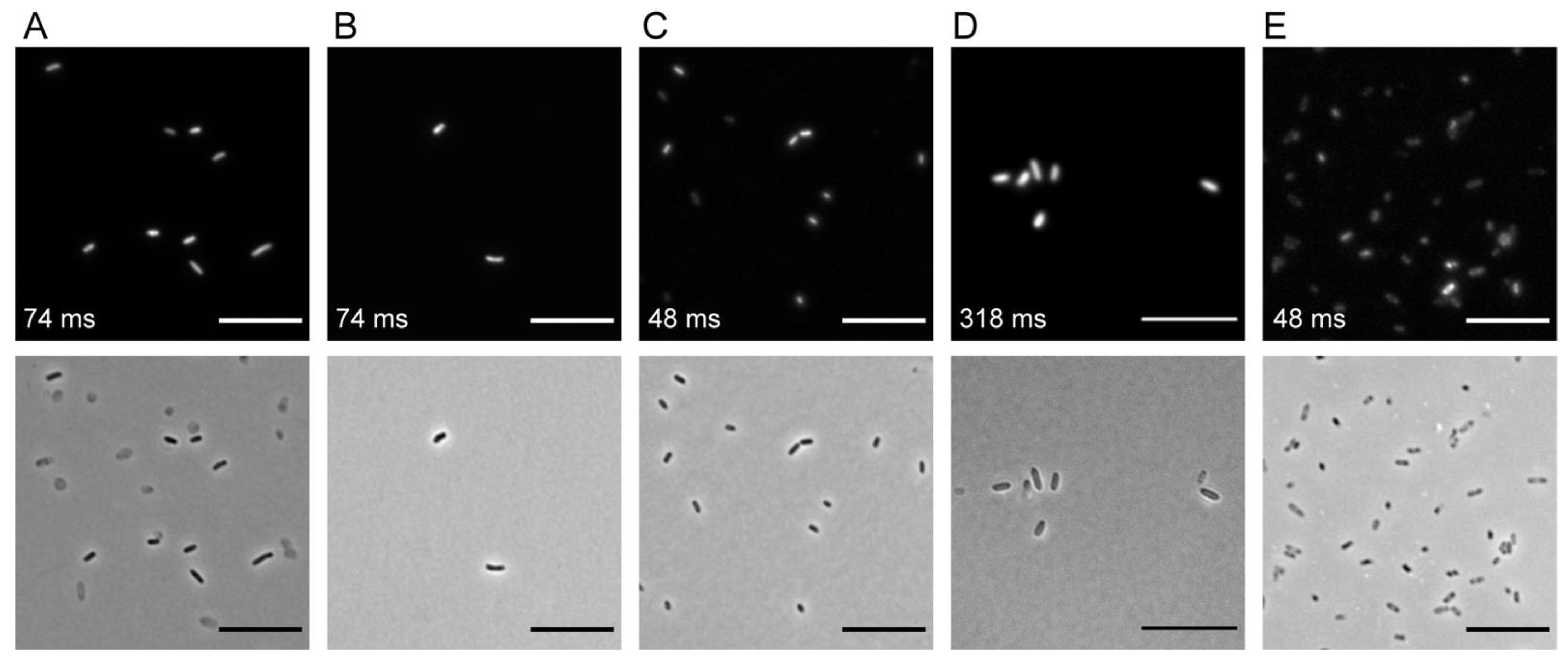
2.2. Transposon Delivery by Conjugation

Using E. coli ST18 as a plasmid donor strain, conjugations were performed into the recipient strains listed in Table 2. It was possible to obtain transposon insertion mutants for four Proteobacterial strains as indicated in Table 2; all mutants exhibited detectable fluorescent protein emission as determined using macroscopical observations at the single-colony level (data not shown) and at the single-cell resolution, as determined using widefield epifluorescence microscopy (Figure 2). The insertion sites for several independent insertion events were determined using arbitrary PCR and sequencing. For Sphingomonas sp. Leaf357 and Acidovorax sp. Leaf84, most of the transposons were inserted in either intergenic regions or hypothetical protein-coding genes (Table 3). We were not able to obtain insertion mutants for strains belonging to the Actinobacteria or Bacteroidetes phyla listed in Table 2 using conjugation.

3. Discussion

A range of new plasmids was constructed for the Chromatic Bacteria toolbox based on the R6K origin of replication plasmid backbone containing the Himar1 transposase. Additionally, a series of plasmids containing the ErmR gene was constructed. A full list of plasmids constructed is included in Table 1. The previous and novel antibiotic resistance gene combinations make the plasmids a versatile tool for bacterial genetic manipulation that accounts for different antibiotic resistances that naturally occur in the recipient organism. Even though we constructed these additional new plasmids in E. coli S17-1 [1,17], we made use of E. coli ST18 as a donor strain for conjugation experiments [18]. E. coli ST18 is an S17-1 derivative that lacks the hemA gene. Mutation in hemA results in a strong auxotrophy and dependency of E. coli ST18 on exogenously provided 5-aminolevulinic acid. Thereby, there is no need to counterselect against the conjugation donor after conjugation by using minimal media or intrinsic antibiotic resistances of the host. The transposon mutants can be selected on their respective optimal complex medium with the addition of an antibiotic selecting for the transposon. Since the vector contains the R6K origin of replication, it only replicates if the pir gene is present and expressed in the host, i.e., the plasmids are suicide vectors that are not able to replicate in their recipient. The successful use of ErmR as a selective marker has been previously reported in clinically relevant Gram-positive pathogens, such as Staphylococcus aureus [23] and Clostridium perfringens [14], and lactic acid bacteria, such as Streptococcus mutans UA159 [13]. Hence, plasmids containing ErmR were added as an alternative antibiotic resistance gene, for strains in which the original genes may not be expressed or function correctly.
Using the newly developed vectors, we were able to deliver Himar transposons randomly into bacterial recipients, similarly to the previously described pMRE-Tn5-transposon series [1]. In comparison to our pMRE-Tn5-transposon plasmids, we were able to genetically modify additional recipients, such as Acidovorax sp. Leaf84, and Sphingomonas sp. Leaf357, while retaining the ability to deliver Himar transposons into S. melonis Fr1 and P. eucalypti 299R. The enhanced spectrum of successful transposition can likely be accredited to the properties of the Himar1 transposase that does not require additional host cofactors to function [8]. However, we were not successful in conjugating the plasmids and inserting the transposons into the genomes of non-model Gram-positive bacterial strains including Aeromicrobium sp. Leaf245, Agreia sp. Leaf 335, Arthrobacter sp. Leaf145, Microbacterium sp. Leaf320, Microbacterium sp. Leaf 347, Plantibacter sp. Leaf1, Rathayibacter sp. Leaf296, Rhodococcus sp. Leaf225, or Williamsia sp. Leaf354. In the future, we plan to use alternative techniques such as electroporation to deliver Himar transposons into non-model Gram-positive bacteria. However, establishing successful transformation protocols for non-model bacteria requires a high degree of optimisation, and was, therefore, not a part of this study. In the future, it would be interesting to benchmark this activity against other transposition systems, such as the Phage Mu transposition complex which has successfully been used to generate insertion libraries in Gram-negative and Gram-positive bacteria [23,24]. During such tests, it may be beneficial to further explore the Tn5 transposome system [25], which works using a similarly to the Phage Mu transposition complex by using transposase proteins that are pre-attached to a linear insertion sequence. Although the Tn5 mediated insertion using the expression of transposon genes previously tested by Schlechter et al. (2018) [1] was unsuccessful in tagging Gram-positive bacteria, the use of a transposome system may be more efficient [26,27].
To map the insertion site of the transposons, arbitrary PCR can be used as described above [1]. Alternatively, the R6K origin of replication is present in the transposon which allows, next to the above-described arbitrary PCR, determination of the insertion site of the transposon into the recipient’s genome. To that end, the genome of the recipient can be isolated, digested with a rare restriction enzyme that does not cut in the transposon sequence, such as KpnI, and then re-ligated and transformed into E. coli S17-1 or another λpir factor expressing E. coli cloning host [28]. Due to the R6K origin of replication contained and the antibiotic resistances located in the transposon, this will result in functional plasmids. These plasmids can then be sequenced using a sequencing primer (Table 4) to determine the sequence of the insertions site. This process is significantly more time intensive and less cost effective than the arbitrary PCR described above, but might be advantageous in cases where the arbitrary PCR does not yield results of sufficient quality.
As described in Schlechter et al. (2018), the here-constructed plasmids allow for convenient fluorescent tagging of environmental bacteria and cover a different host range compared to the previously described vectors. Fluorescent protein tags are the prerequisite for many experimental studies that follow different populations simultaneously, identify focal populations in complex environments, or to follow the behaviour of individual cells [4,29,30,31,32]. Currently, many delivery systems, including the first versions of the Chromatic bacteria, function almost exclusively in Proteobacteria; the Himar transposons have been shown to have a wider range of activity. Thereby the here-introduced plasmids can serve as a one fits many solutions for tagging Proteobacteria.

4. Materials and Methods

4.1. Strains and Media

All strains, respective growth media, and growth temperature used in this study are listed in Table 2. Lysogeny broth (LB, HiMedia, Kuwait City, Kuwait), nutrient broth (NB; HiMedia), Reasoner’s 2A media (R2A, HiMedia), and Reasoner’s 2A agar (R2A agar, HiMedia) were prepared according to manufacturer’s instructions. Media were supplemented with 1.5% Oxoid™ bacteriological agar (Agar No. 1, Thermo Fisher, Waltham, MA, USA) where needed. To support growth of E. coli ST18, media was supplemented with 50 mg L−1 5-aminolevulinic acid (5-ala, Sigma, St. Louis, MO, USA). When appropriate, media were supplemented with antibiotics with the following concentrations: 100 mg L−1 ampicillin, 20 mg L−1 chloramphenicol, 10 mg L−1 colistin, 100 mg L−1 erythromycin, 15 mg L−1 gentamicin, and/or 50 mg L−1 kanamycin.

4.2. Plasmid Construction

The Himar transposase, antibiotic resistance, and fluorescent protein genes were retrieved from the plasmids pHimarEm1, pMRE13X, pMRE14X, and pMRE15X to construct the Himar-based transposon delivery plasmid series [1,12]. Plasmids were constructed as previously described [1]. The here-described plasmids are denoted pMRE-Himar-1XY series, where X can either be 3, 4, 5, 7, or 9, representing the different antibiotic resistance gene combinations of chloramphenicol; gentamicin and chloramphenicol; kanamycin and chloramphenicol; erythromycin and chloramphenicol, or erythromycin, kanamycin, and chloramphenicol, respectively, and where Y can be 0, 1, 2, 3, 4, 5, 6, or 7 representing blue, cyan, green, yellow, orange, red, near-infrared, or green fluorescent protein (FP) gene, respectively (mTagBFP2, mTurquoise2, sGFP2, mOrange2, sYFP2, mScarlet-I, mCardinal, or mClover3, respectively).
Each plasmid was constructed by amplifying three fragments: the plasmid backbone, a unique antibiotic resistance gene (X fragment), and a unique FP gene upstream of a chloramphenicol resistance gene (Y fragment). All PCRs were performed using Phusion High-Fidelity Polymerase (Thermo Scientific). Primers used for cloning are listed in Table 4. For PCR mixes containing primers with overhangs, a touchdown PCR protocol was performed starting with an annealing temperature 10 °C above the lowest tm of the primer pair. This was reduced by 1 °C for ten cycles before running the PCR with the annealing temperature set to tm. After amplification, all PCR reactions were DpnI treated to digest the methylated plasmid template DNA before the PCR fragments were purified with the DNA Clean & Concentrator Kit (Zymo Research, Irvine, California). To construct pMRE-Himar-13X, 14X and 15X, the plasmid backbone was amplified from pHimarEm1 using pHimarEm1+XmaI_FWD and pHimarEm1_XbaI_RVS2. Antibiotic resistance genes and fluorescent protein genes were amplified from pMRE-13X, pMRE-14X, or pMRE15X. The X fragments were amplified using primers pMRE-HimarEm1+XbaI_overhang_FW2 and pntpII_1_REV. The Y fragments were amplified using primers pntpII_1_FWD and pMRE-HimarEm1+XmaI_overhang_RV. For construction of the pMRE-Himar-17X and pMRE-Himar-19X series, pHimarEm1 was amplified using primers pHimarEm1+Xmal_FWD and pHimarEm1_EmR+XbaI_RV. Using primers pMRE-HimarEm1_EmR+XbaI_overh_FWD and pntpII_1_REV, antibiotic resistance genes were amplified from pMRE-13X to create the pMRE-Himar-17X series, or from pMRE-15X to create pMRE-Himar-19X series. The FP fragment was amplified using the primers pntpII_1_FWD and pMRE-HimarEm1_EmR+XbaI_overh_RV from pMRE-15X plasmids.
Gibson assembly was performed as previously described [1]. Briefly, the fragments were mixed at a 1:3 backbone: insert molar ratio with between 20–100 ng of backbone fragment being used. No more than 5 µL DNA solution was added to a 15 µL Gibson assembly mix. Where appropriate, water was added to top up the reaction volume to 20 µL. The Gibson assembly mix was incubated at 50 °C for 20 min. Chemically competent E. coli S17-1 cells were then transformed using 10 µL of the Gibson assembly mix [33]. After the plasmids were confirmed by Sanger sequencing, they were cloned into chemically competent E. coli ST18 [33].

4.3. Transposon Delivery Using Conjugation

Two parental matings were performed to deliver the pHimar1Em-based plasmids into a range of bacterial strains, following the protocol described by Schlechter et al. (2019) [34]. In variation to this protocol, conjugations were performed using the auxotrophic E. coli ST18 as a plasmid donor strain [18]. To perform the conjugations, recipient strains (Table 2) were grown in 50 mL of suitable media for up to three days depending on their growth rate. Single colonies of E. coli ST18 donor strains were produced and used to inoculate overnight cultures of LB supplemented with appropriate antibiotics and 5-aminolevulinic acid (5-ala). From these overnight cultures, 2 mL was inoculated into 100 mL fresh LB and grown until the donor cultures reached an OD600nm of 0.5. Then, both donor and recipient cells were collected by centrifugation and resuspended in phosphate buffered saline (PBS) to an OD600nm of 1. Approximately 5 mL recipient and donor cells were combined in a 1:1 ratio, then centrifuged and resuspended in 200 µL of PBS. The suspension was spotted onto a 0.44 µm S-pak Membrane filter (Millipore) which was placed on LB agar plates supplemented with 5-ala. The bacterial mixes were incubated at 30 °C overnight. Bacteria were recovered from the filter by vigorous vortexing in 10 mL PBS in 50 mL falcon tubes. Subsequently, the filter was dismissed, and the mixes were concentrated by centrifugation and resuspension in 1 mL PBS, before 10 µL, 100 µL, and the remaining volume were plated on media without 5-ala and appropriate antibiotics to select for transconjugants. Transposon insertion mutant colonies appeared up to five days after conjugations were performed. To obtain a pure culture of the transposon insertion mutants, colonies were restreaked at least three times.

4.4. Screening for Fluorescent Colonies and Fluorescence Microscopy

For convenient and quick assessment of colony level fluorescence, a blue light gel reader was used for fluorescent proteins with emission wavelengths between 500 and 680 nm. Fluorescence microscopy was performed on a Zeiss AxioImager.M1 fluorescent widefield microscope equipped with Zeiss filter sets 38HE, 43HE, 46HE, and 47HE, (BP 470/40-FT 495-BP 525/50, BP 550/25-FT 570-BP 605/70, BP 500/25-FT 515-BP 535/30, and BP 436/25-FT 455-BP 480/40, respectively), an Axiocam 506, and the software Zeiss Zen 2.3. Single-cell fluorescence was analysed as described previously [35]. In short, bacteria were mounted on an agarose slab (~1 mm thick, 1% agarose in milliQ water) and samples were analysed using a Zeiss AxioImager.M1 at 1000× magnification. Images were processed using ImageJ/Fiji [36].

4.5. Arbitrary PCR

The transposon insertion of fluorescently (FP)-tagged bacterial strains was mapped using arbitrary PCR [37]. Briefly, a first PCR was performed using a mix of random primers Arb1, Arb2, and Arb3 containing an adapter oligo at the 5’-end and a primer targeting the transposon insertion (Table 4). Amplification was performed in a total volume of 25 µL containing: 15.05 µL ddH2O, 5 µL Phusion GC buffer, 1.25 µL 10 mM dNTP mix, 1.25 µL 10 µM arbitrary primers, 0.5 µL 10 µM ARB-RB-PCR1, 0.75 µL DMSO, 0.2 µL Phusion, and 1 µL DNA template. Cycling conditions were as follows: initial denaturation of 95 °C for 5 min; six cycles of 30 s at 95 °C for denaturation, 30 s at 30 °C for annealing, and 1.5 min at 72 °C for extension; then, 30 cycles of 30 s at 95 °C for denaturation, 30 s at 45 °C for annealing, 2 min at 72 °C for extension, and 5 min at 72 °C for a final extension. Then, PCR products were purified using DNA Clean & ConcentratorTM-5 (Zymo Research) following the manufacturer’s recommendations and eluted in 30 µL. A second PCR was performed using primers PCR2-AP and ARB-RB-PCR2. Amplification was performed in a total volume of 25 µL, containing: 12.8 µL ddH2O, 5 µL Phusion GC buffer, 1.25 µL 10 mM dNTP mix, 1 µL 10 µM arbitrary primers, 1 µL 10 µM ARB-RB-PCR1, 0.75 µL DMSO, 0.2 µL Phusion, and 3 µL of ten-fold dilution of purified PCR fragments. Cycling conditions were as follows: initial denaturation of 95 °C for 3 min; 30 cycles of 30 s at 95 °C for denaturation, 30 s at 52 °C for annealing, 2 min at 72 °C for extension, and 5 min at 72 °C for a final extension. PCR products were then purified and concentrated using the Zymo Research DNA Clean & ConcentratorTM-5 following the manufacturer’s recommendations and sequenced using Sanger’s sequencing (Macrogen, Seoul, Korea). The sequencing results were mapped against the genome of the corresponding bacterial strain using Blast [38] and a local database of draft genome sequences of the strains used in this study using the software Geneious Prime (version 2020.1.2, Biomatters, Auckland, New Zealand).

5. Conclusions

The here-described plasmids extend the previously constructed Chromatic bacteria toolbox host range and utility. All plasmids and sequences will be made available through the non-profit service addgene.org upon peer reviewed publication (Addgene plasmids numbers to be determined). The extended Chromatic bacteria vector series can often be used as a one-stop solution for fluorescently marking bacteria to enable a hassle-free solution for groups that do not want to try and establish novel genetic systems for newly isolated bacterial strains.

Author Contributions

Conceptualization, R.O.S. and M.N.P.R.-E.; methodology, C.S., R.O.S., and M.N.P.R.-E.; formal analysis, C.S.; investigation, C.S.; writing—original draft preparation, C.S.; writing—review and editing, C.S., R.O.S., and M.N.P.R.-E.; visualization, C.S. and R.O.S.; supervision, R.O.S. and M.N.P.R.-E.; project administration, M.N.P.R.-E.; funding acquisition, M.N.P.R.-E. All authors have read and agreed to the published version of the manuscript.

Funding

This project was supported by the New Zealand Tertiary Education Commission CoRE grant to the Bio-Protection Research Centre and a Marsden Fast-Start grant to M.N.P.R-E (UOC1704) and additional funding by Callaghan Innovation is acknowledged. C.S. is supported by a UC Masters scholarship and a Bio-Protection Research Centre summer scholarship. R.O.S. is supported by a New Zealand International Doctoral Research Scholarship (NZIDRS) and a University of Canterbury Doctoral Scholarship.

Institutional Review Board Statement

Not applicable.

Informed Consent Statement

Not applicable.

Data Availability Statement

Plasmids will be made available at https://www.addgene.org/Mitja_Remus-Emsermann/, accessed on 7 December 2021.

Acknowledgments

The authors thank Mark Silby and Lucy McCully (University of Massachusetts, Dartmouth) for the kind gift of pHimarEm1. The publication of this article was funded by Freie Universität Berlin.

Conflicts of Interest

The authors declare no conflict of interest. The funders had no role in the design of the study; in the collection, analyses, or interpretation of data; in the writing of the manuscript; or in the decision to publish the results.

References

  1. Schlechter, R.O.; Jun, H.; Bernach, M.; Oso, S.; Boyd, E.; Muñoz-Lintz, D.A.; Dobson, R.C.J.; Remus, D.M.; Remus-Emsermann, M.N.P. Chromatic bacteria—A broad host-range plasmid and chromosomal insertion toolbox for fluorescent protein expression in bacteria. Front. Microbiol. 2018, 9, 3052. [Google Scholar] [CrossRef] [Green Version]
  2. Miebach, M.; Schlechter, R.; Clemens, J.; Jameson, P.E.; Remus-Emsermann, M.N.P. Litterbox—A gnotobiotic zeolite-clay system to investigate Arabidopsis-microbe interactions. Microorganisms 2020, 8, 464. [Google Scholar] [CrossRef] [Green Version]
  3. Bernach, M.; Soffe, R.; Remus-Emsermann, M.N.P.; Nock, V. Micropatterning of hybrid polydimethylsiloxane for replica leaves. Jpn. J. Appl. Phys. 2019, 58, SDDK01. [Google Scholar] [CrossRef]
  4. Remus-Emsermann, M.N.P.; Schlechter, R.O. Phyllosphere microbiology: At the interface between microbial individuals and the plant host. New Phytol. 2018, 218, 1327–1333. [Google Scholar] [CrossRef] [PubMed]
  5. Nadell, C.D.; Drescher, K.; Foster, K.R. Spatial structure, cooperation and competition in biofilms. Nat. Rev. Microbiol. 2016, 14, 589–600. [Google Scholar] [CrossRef] [PubMed]
  6. Tellier, M.; Bouuaert, C.C.; Chalmers, R. Mariner and the ITm superfamily of transposons. Microbiol. Spectr. 2015, 3, MDNA3–MDNA0033-2014. [Google Scholar] [CrossRef] [Green Version]
  7. Lampe, D.J. Bacterial genetic methods to explore the biology of mariner transposons. Genetica 2010, 138, 499–508. [Google Scholar] [CrossRef]
  8. Lampe, D.J.; Churchill, M.E.; Robertson, H.M. A purified mariner transposase is sufficient to mediate transposition in vitro. EMBO J. 1996, 15, 5470–5479. [Google Scholar] [CrossRef]
  9. Lamberg, A.; Nieminen, S.; Qiao, M.; Savilahti, H. Efficient insertion mutagenesis strategy for bacterial genomes involving electroporation of in vitro-assembled DNA transposition complexes of bacteriophage Mu. Appl. Environ. Microbiol. 2002, 68, 705–712. [Google Scholar] [CrossRef] [Green Version]
  10. Pajunen, M.I.; Pulliainen, A.T.; Finne, J.; Savilahti, H. Generation of transposon insertion mutant libraries for Gram-positive bacteria by electroporation of phage Mu DNA transposition complexes. Microbiology 2005, 151, 1209–1218. [Google Scholar] [CrossRef] [Green Version]
  11. McCully, L.M.; Bitzer, A.S.; Seaton, S.C.; Smith, L.M.; Silby, M.W. Interspecies social spreading: Interaction between two sessile soil bacteria leads to emergence of surface motility. mSphere 2019, 4, e00696-18. [Google Scholar] [CrossRef] [Green Version]
  12. Braun, T.F.; Khubbar, M.K.; Saffarini, D.A.; McBride, M.J. Flavobacterium johnsoniae gliding motility genes identified by mariner mutagenesis. J. Bacteriol. 2005, 187, 6943–6952. [Google Scholar] [CrossRef] [PubMed] [Green Version]
  13. Nilsson, M.; Christiansen, N.; Høiby, N.; Twetman, S.; Givskov, M.; Tolker-Nielsen, T. A mariner transposon vector adapted for mutagenesis in oral streptococci. Microbiologyopen 2014, 3, 333–340. [Google Scholar] [CrossRef] [PubMed]
  14. Liu, H.; Bouillaut, L.; Sonenshein, A.L.; Melville, S.B. Use of a mariner-based transposon mutagenesis system to isolate Clostridium perfringens mutants deficient in gliding motility. J. Bacteriol. 2013, 195, 629–636. [Google Scholar] [CrossRef] [Green Version]
  15. Li, M.; Rigby, K.; Lai, Y.; Nair, V.; Peschel, A.; Schittek, B.; Otto, M. Staphylococcus aureus mutant screen reveals interaction of the human antimicrobial peptide dermcidin with membrane phospholipids. Antimicrob. Agents Chemother. 2009, 53, 4200–4210. [Google Scholar] [CrossRef] [Green Version]
  16. Rubin, E.J.; Akerley, B.J.; Novik, V.N.; Lampe, D.J.; Husson, R.N.; Mekalanos, J.J. In vivo transposition of mariner-based elements in enteric bacteria and mycobacteria. Proc. Natl. Acad. Sci. USA 1999, 96, 1645–1650. [Google Scholar] [CrossRef] [Green Version]
  17. Simon, R.; Priefer, U.; Pühler, A. A broad host range mobilization system for in vivo genetic engineering: Transposon mutagenesis in Gram negative bacteria. Biotechnology 1983, 1, 784–791. [Google Scholar] [CrossRef]
  18. Thoma, S.; Schobert, M. An improved Escherichia coli donor strain for diparental mating. FEMS Microbiol. Lett. 2009, 294, 127–132. [Google Scholar] [CrossRef] [Green Version]
  19. Bai, Y.; Müller, D.B.; Srinivas, G.; Garrido-Oter, R.; Potthoff, E.; Rott, M.; Dombrowski, N.; Münch, P.C.; Spaepen, S.; Remus-Emsermann, M.; et al. Functional overlap of the Arabidopsis leaf and root microbiota. Nature 2015, 528, 364–369. [Google Scholar] [CrossRef]
  20. Innerebner, G.; Knief, C.; Vorholt, J.A. Protection of Arabidopsis thaliana against leaf-pathogenic Pseudomonas syringae by Sphingomonas strains in a controlled model system. Appl. Environ. Microbiol. 2011, 77, 3202–3210. [Google Scholar] [CrossRef] [Green Version]
  21. Remus-Emsermann, M.N.P.; Kim, E.B.; Marco, M.L.; Tecon, R.; Leveau, J.H.J. Draft genome sequence of the phyllosphere model bacterium Pantoea agglomerans 299R. Genome Announc. 2013, 1, e00036-13. [Google Scholar] [CrossRef] [PubMed] [Green Version]
  22. Feil, H.; Feil, W.S.; Chain, P.; Larimer, F.; DiBartolo, G.; Copeland, A.; Lykidis, A.; Trong, S.; Nolan, M.; Goltsman, E.; et al. Comparison of the complete genome sequences of Pseudomonas syringae pv. syringae B728a and pv. tomato DC3000. Proc. Natl. Acad. Sci. USA 2005, 102, 11064–11069. [Google Scholar] [CrossRef] [PubMed] [Green Version]
  23. Tu Quoc, P.H.; Genevaux, P.; Pajunen, M.; Savilahti, H.; Georgopoulos, C.; Schrenzel, J.; Kelley, W.L. Isolation and characterization of biofilm formation-defective mutants of Staphylococcus aureus. Infect. Immun. 2007, 75, 1079–1088. [Google Scholar] [CrossRef] [Green Version]
  24. Laasik, E.; Ojarand, M.; Pajunen, M. Novel mutants of Erwinia carotovora subsp. carotovora defective in the production of plant cell wall degrading enzymes generated by Mu transpososome-mediated insertion mutagenesis. FEMS Microbiol. Lett. 2005, 243, 93–99. [Google Scholar] [PubMed]
  25. Goryshin, I.Y.; Jendrisak, J.; Hoffman, L.M.; Meis, R.; Reznikoff, W.S. Insertional transposon mutagenesis by electroporation of released Tn5 transposition complexes. Nat. Biotechnol. 2000, 18, 97–100. [Google Scholar] [CrossRef]
  26. Fernandes, P.J.; Powell, J.A.C.; Archer, J.A.C. Construction of Rhodococcus random mutagenesis libraries using Tn5 transposition complexes. Microbiology 2001, 147, 2529–2536. [Google Scholar] [CrossRef] [Green Version]
  27. Vidal, J.E.; Chen, J.; Li, J.; McClane, B.A. Use of an EZ-Tn5-based random mutagenesis system to identify a novel toxin regulatory locus in Clostridium perfringens strain 13. PLoS ONE 2009, 4, e6232. [Google Scholar] [CrossRef] [Green Version]
  28. Metcalf, W.W.; Jiang, W.; Wanner, B.L. Use of the rep technique for allele replacement to construct new Escherichia coli hosts for maintenance of R6K gamma origin plasmids at different copy numbers. Gene 1994, 138, 1–7. [Google Scholar] [CrossRef]
  29. Wang, P.; Robert, L.; Pelletier, J.; Dang, W.L.; Taddei, F.; Wright, A.; Jun, S. Robust growth of Escherichia coli. Curr. Biol. 2010, 20, 1099–1103. [Google Scholar] [CrossRef] [Green Version]
  30. Diard, M.; Garcia, V.; Maier, L.; Remus-Emsermann, M.N.P.; Regoes, R.R.; Ackermann, M.; Hardt, W.-D. Stabilization of cooperative virulence by the expression of an avirulent phenotype. Nature 2013, 494, 353–356. [Google Scholar] [CrossRef]
  31. Ledermann, R.; Bartsch, I.; Remus-Emsermann, M.N.; Vorholt, J.A.; Fischer, H.-M. Stable fluorescent and enzymatic tagging of Bradyrhizobium diazoefficiens to analyze host-plant infection and colonization. Mol. Plant. Microbe Interact. 2015, 28, 959–967. [Google Scholar] [CrossRef] [PubMed] [Green Version]
  32. Schlechter, R.O.; Kear, E.J.; Remus, D.M.; Remus-Emsermann, M.N.P. Fluorescent protein expression as a proxy for bacterial fitness in a high-throughput assay. Appl. Environ. Microbiol. 2021, 87, e0098221. [Google Scholar] [CrossRef]
  33. Sambrook, J.; Fritsch, E.F.; Maniatis, T. Molecular Cloning: A Laboratory Manual; Cold Spring Harbor Laboratory Press: Cold Spring Harbor, NY, USA, 1989. [Google Scholar]
  34. Schlechter, R.; Remus-Emsermann, M. Delivering “chromatic bacteria” fluorescent protein tags to proteobacteria using conjugation. Bio-Protocol 2019, 9, e3199. [Google Scholar] [CrossRef] [PubMed]
  35. Remus-Emsermann, M.N.P.; Gisler, P.; Drissner, D. MiniTn7-transposon delivery vectors for inducible or constitutive fluorescent protein expression in Enterobacteriaceae. FEMS Microbiol. Lett. 2016, 363, fnw178. [Google Scholar] [CrossRef] [PubMed]
  36. Schindelin, J.; Arganda-Carreras, I.; Frise, E.; Kaynig, V.; Longair, M.; Pietzsch, T.; Preibisch, S.; Rueden, C.; Saalfeld, S.; Schmid, B.; et al. Fiji: An open-source platform for biological-image analysis. Nat. Methods 2012, 9, 676–682. [Google Scholar] [CrossRef] [PubMed] [Green Version]
  37. Das, S.; Noe, J.C.; Paik, S.; Kitten, T. An improved arbitrary primed PCR method for rapid characterization of transposon insertion sites. J. Microbiol. Methods 2005, 63, 89–94. [Google Scholar] [CrossRef] [PubMed]
  38. Altschul, S.F.; Gish, W.; Miller, W.; Myers, E.W.; Lipman, D.J. Basic local alignment search tool. J. Mol. Biol. 1990, 215, 403–410. [Google Scholar] [CrossRef]
Figure 1. Overview of cloning procedures. (A) Construction of pMRE-Himar-13Y to pMRE-Himar-15Y. The pHimarEm1 backbone was amplified using PCR and Gibson assembly primers without the erythromycin resistance gene. Plasmids from the original Chromatic Bacteria series were PCR amplified in two separate reactions using Gibson assembly primers targeting the chloramphenicol resistance gene and the promoter driving the respective fluorescent protein gene expression and, where applicable, a secondary antibiotic resistance gene. These three fragments were joined using isothermal assembly. (B) Construction of pMRE-Himar-17Y and pMRE-Himar-19Y plasmids. The procedure was similar as described in (A), but, in this case, the erythromycin resistance gene encoded on the pHimarEm1 plasmid (highlighted in blue) was included when amplifying the backbone.
Bacteria 01 00006 g001
Figure 2. Fluorescence of recipients after stable integration of constitutively expressed fluorescent protein genes at the single cell resolution. Representative images of fluorescently tagged bacterial strains. In all cases, fluorescence (upper panel) and phase contrast images (lower panel) are included. (A) Acidovorax sp. Leaf84::MRE-Himar-145. (B) Sphingomonas sp. Leaf357::MRE-Himar-145. (C) Sphingomonas melonis FR1::MRE-Himar-156; (D) Pantoea eucalypti 299R::MRE-Himar-145; (E) Escherichia coli ST18 (pMRE-Himar-156). Exposure times are given in the respective fluorescence images. Scale bar = 10 µm.
Bacteria 01 00006 g002
Table 1. Plasmids constructed in this work.
NameFluorescent Protein GeneSelectable Marker(s)
pMRE-Himar-131mTurquoise2CmR
pMRE-Himar-133sYFP2CmR
pMRE-Himar-134mOrange2CmR
pMRE-Himar-135mScarlet-ICmR
pMRE-Himar-136mCardinalCmR
pMRE-Himar-137mClover3CmR
pMRE-Himar-140mTagBFP2CmR, GmR
pMRE-Himar-141mTurquoise2CmR, GmR
pMRE-Himar-142sGFP2CmR, GmR
pMRE-Himar-143sYFP2CmR, GmR
pMRE-Himar-144mOrange2CmR, GmR
pMRE-Himar-145mScarlet-ICmR, GmR
pMRE-Himar-146mCardinalCmR, GmR
pMRE-Himar-147mClover3CmR, GmR
pMRE-Himar-151mTurquoise2CmR, KmR
pMRE-Himar-153sYFP2CmR, KmR
pMRE-Himar-155mScarlet-ICmR, KmR
pMRE-Himar-157mClover3CmR, KmR
pMRE-Himar-171mTurquoise2CmR, EmR
pMRE-Himar-174mOrange2CmR, EmR
pMRE-Himar-175mScarlet-ICmR, EmR
pMRE-Himar-176mCardinalCmR, EmR
pMRE-Himar-177mClover3CmR, EmR
pMRE-Himar-191mTurquoise2CmR, KmR, EmR
pMRE-Himar-192sGFP2CmR, KmR, EmR
pMRE-Himar-193sYFP2CmR, KmR, EmR
pMRE-Himar-194mOrange2CmR, KmR, EmR
pMRE-Himar-195mScarlet-ICmR, KmR, EmR
pMRE-Himar-196mCardinalCmR, KmR, EmR
pMRE-Himar-197mClover3CmR, KmR, EmR
Table 2. Bacterial strains used in this work.
StrainFeatures, NotesGrowth
Medium
Growth
Temperature
Transconjugants (Yes/No)Source
Escherichia coli S17-1Cloning host for R6K replicon plasmids; :RP4-2 pro thi hsdR+ Tpr Smr Tc::Mu-Kan::Tn7/λpirLB37 °Cn.a.[17]
Escherichia coli ST18Conjugation donor; Genotype: S17-1 λpirΔhemALB, 5-ala37 °Cn.a.[18]
Aeromicrobium sp. Leaf245Transposon recipient (Actinobacteria)NB30 °Cno[19]
Agreia sp. Leaf335Transposon recipient (Actinobacteria)NB30 °Cno[19]
Arthrobacter sp. Leaf145Transposon recipient (Actinobacteria)NB30 °Cno[19]
Microbacterium sp. Leaf320Transposon recipient (Actinobacteria)R2A30 °Cno[19]
Microbacterium sp. Leaf347Transposon recipient (Actinobacteria)R2A30 °Cno[19]
Plantibacter sp. Leaf1Transposon recipient (Actinobacteria)NB30 °Cno[19]
Rathayibacter sp. Leaf296Transposon recipient (Actinobacteria)NA30 °Cno[19]
Rhodococcus sp. Leaf225Transposon recipient (Actinobacteria)NA30 °Cno[19]
Williamsia sp. Leaf354Transposon recipient (Actinobacteria)NB30 °Cno[19]
Acidovorax sp. Leaf84Transposon recipient (Proteobacteria)R2A30 °Cyes[19]
Sphingomonas melonis FR1Transposon recipient (Proteobacteria)NB30 °Cyes[20]
Pantoea eucalypti 299RTransposon recipient (Proteobacteria)LB30 °Cyes[21]
Pseudomonas syringae B728aTransposon recipient (Proteobacteria)LB30 °Cno[22]
Sphingomonas sp. Leaf17Transposon recipient (Proteobacteria)NB30 °Cno[19]
Sphingomonas sp. Leaf34Transposon recipient (Proteobacteria)R2A30 °Cno[19]
Sphingomonas sp. Leaf357Transposon recipient (Proteobacteria)R2A30 °Cyes[19]
Pedobacter sp. Leaf194Transposon recipient (Bacteroidetes)R2A30 °Cno[19]
n.a.: Not applicable.
Table 3. Transposon insertion sites.
Strain NameTransposon Insertion Flanking Region SequenceRegion Hit
Sphingomonas sp. Leaf357::
MRE-Himar-145/1
AGGGGCTCGCAGTCGATTTACCGGTTCGCATGATCGTAACCGCACAGGGGAAGGAAACATGGGCTCTCTTCCGCCAGCGCGGTGGGATGTACCCTGAGBeta-hexosaminidase CDS
Sphingomonas sp. Leaf357::
MRE-Himar-145/2
TGTTATAACCCGGGGCCCAGAAGCGCGCGAGGTAGTCTTTGAATGGATACATGGGCAGATATGCGATAACGCCGTCGAGCTTCCGGTTGGCGACGTCTCAGTCCGCGTCCATGACGACCCCGAGCGTGTHypothetical protein
Sphingomonas sp. Leaf357::
MRE-Himar-145/4
CTCGGCGCGCAGGCCAATCTGTGGGCCGAATATATCGTGACGCCCACCGAATCCCAACATGCGCTGTTCCCGCGCGTCGACGCGCTGGCCGAGATCGCCTGHypothetical protein
Acidovorax sp. Leaf84::
MRE-Himar-145/2
AGTCAACATCGAAAAGCTCGGAGACTATGTGAATCGCTATGGCGTCAATAGCTTTTTCGACGCATCCGATGATGCCCATCIntergenic region
Acidovorax sp. Leaf84::
MRE-Himar-145/5
ATCTGATCTTCAGACAGTCTGTCGGTAGCTCCCTCGCGCCTTGCAGAGCAGATGATGTGTTCCCCTTGAAAACGCCCTTGACATCATGCACCTCGACGHypothetical protein
Acidovorax sp. Leaf84::
MRE-Himar-145/6
TAATCGGTGGATGGTAAATAGATAGGAAATTTATCACTGTGTTTCATAACAGGTTGIntergenic region
Table 4. Primers used in this study.
NameSequence (5′ to 3′) 1Tm (°C)
pHimarEm1+XmaI_FWDcccgggCAATTCGAGGGGTATCGCTCT67
pHimarEm1_XbaI_RVS2tctagaGCACGAGGAAATTGCGCAAAAA67
pMRE-HimarEm1+XbaI_overhang_FW2gcaatttcctcgtgctctagaATATAAACTGCCAGGAATTGGG62
pntpII_1_REVGCCATGTAAGCCCACTGCAAGCTAC73
pntpII_1_FWDGTAGCTTGCAGTGGGCTTACATGGC73
pMRE-HimarEm1+XmaI_overhang_RVgatacccctcgaattgcccgggCTGGCGGCCGCAAGCTCC74
pMRE-HimarEm1_EmR+XbaI_overhang_RVtttcatccttcgtagtctagaCATAAACTGCCAGGAATTGGGGAT67
pHimarEm1_EmR+XbaI_RVtctagaCTACGAAGGATGAAATTTTTCAGGG63
ARB-RB-PCR1CTGGGGTAATGACTCTCTAGC59
ARB-RB-PCR2CTGAGTAGGACAAATCCGCCG62
PCR2 AP-PCRGGCCACGCGTCGACTAGTCA66
Arb1GGCCACGCGTCGACTAGTCANNNNNNNNNNGCTCGn.a.
Arb2GGCCACGCGTCGACTAGTCANNNNNNNNNNGACTCn.a.
Arb3GGCCACGCGTCGACTAGTCANNNNNNNNNNGATACn.a.
Sequencing primerCTGGTTCCGCGCACATTTC61
1 Primer sequences in majuscules indicate base complementarity to the PCR template, and minuscules indicate overhanging regions with complementarity to adjacent fragments. n.a.: not applicable.
Publisher’s Note: MDPI stays neutral with regard to jurisdictional claims in published maps and institutional affiliations.

