Article

Advanced Glycation End Products Induce Microglial Activation and Impair Neurodevelopment in Human iPSC-Derived Brain Organoids


1
College of Veterinary Medicine, Western University of Health Sciences, Pomona, CA 91766-1854, USA
2
Graduate College of Biomedical Sciences, Western University of Health Sciences, Pomona, CA 91766-1854, USA
3
College of Dental Medicine, Western University of Health Sciences, Pomona, CA 91766-1854, USA
*
Author to whom correspondence should be addressed.
Organoids 2026, 5(2), 12; https://doi.org/10.3390/organoids5020012
Submission received: 4 February 2026 / Revised: 11 April 2026 / Accepted: 17 April 2026 / Published: 20 April 2026

Abstract

Advanced Glycation End Products (AGEs) are reactive compounds formed through the non-enzymatic glycation of proteins, lipids, or nucleic acids due to exposure to reducing sugars. They accumulate through endogenous metabolic dysregulation and exogenous dietary intake, particularly high-fat and high-sugar foods prepared at high temperatures. The interaction between AGEs and their receptor, RAGE (receptor for Advanced Glycation End Products), has been implicated in a range of pathological conditions, including diabetes and metabolic syndrome. However, the impact of AGEs accumulation on neurodevelopment remains poorly understood. In this study, we investigated the effects of AGEs on human-induced pluripotent stem cell (iPSC)-derived cerebral organoids comprising neurons, astrocytes, and microglia. Our findings reveal that AGEs induce RAGE expression, leading to microglial activation, increased deposition of amyloid-beta (Aβ) aggregates, and impaired neurodevelopment. Additionally, elevated levels of AGE-modified proteins, along with altered microglial polarization, were observed in cerebral organoids modeling Western Pacific Amyotrophic Lateral Sclerosis and Parkinsonism–Dementia Complex (ALS-PDC). These findings demonstrate AGEs as active drivers of neurodevelopmental disruption and establish a mechanistic link between metabolic stress and increased susceptibility to neurodegenerative disease.

1. Introduction

Advanced Glycation End Products (AGEs) encompass a diverse range of irreversible molecular adducts formed via non-enzymatic glycation and glyoxidation reactions involving proteins, lipids, and nucleic acids in the presence of reducing sugars [1,2]. AGEs arise from both endogenous processes, including the Maillard reaction, the polyol pathway, and glyoxidation, as well as from exogenous sources such as red meat and high-fat and high-sugar foods—particularly when cooked at high temperatures through frying, grilling, roasting, or baking.
AGEs exert harmful effects on tissues through both receptor-mediated and non-receptor-mediated mechanisms [1]. In receptor-mediated pathways, AGEs interact with the receptor for advanced glycation end products (RAGE), activating intracellular signaling cascades that stimulate NF-κB and increase the production of pro-inflammatory cytokines such as TNF-α and IL-1 [1]. RAGE, a member of the immunoglobulin superfamily, plays a critical role in immune and inflammatory processes and is encoded within the major histocompatibility complex (MHC) Class III region. More than 28 ligands have been identified for RAGE, including S100 proteins, high-mobility group box 1 (HMGB1), and amyloid-beta (Aβ) [3,4]. Studies have shown that RAGE deletion in knockout mice provides protection against cardiovascular and Alzheimer’s disease pathology [5], whereas transgenic expression of RAGE in microglia accelerates neuroinflammation and Aβ accumulation, leading to faster cognitive decline in Alzheimer’s disease models [6]. However, the specific effects of AGE-induced RAGE signaling in microglia and their contribution to neurodevelopmental and neurodegenerative diseases remain poorly understood.
Microglia, the resident immune cells of the central nervous system (CNS), play critical roles in neuronal development, extracellular matrix remodeling, and synaptic pruning [7]. They are the primary immunocompetent population in the brain and exhibit long-term self-renewal potential [8]. In response to developmental or pathological cues, microglia modify their morphology and function to maintain homeostasis or drive immune defense [9]. Activated microglia can adopt pro-inflammatory phenotypes associated with the release of IL-1, IL-6, and TNF-α and activation of the NOD-like receptor family pyrin domain-containing 3 (NLRP3) inflammasome, a state strongly linked to progressive neurodegeneration [10,11]. In contrast, homeostatic microglia promote neurogenesis, protein clearance, and neuronal repair through the release of regulatory cytokines such as IL-4, IL-10, and TGF-β [12,13,14], and support matrix integrity, tissue repair, and neuronal regeneration [15,16,17].
Despite their central importance, the role of AGEs in altering microglial heterogeneity and homeostasis has been minimally explored [18]. Advances in stem cell technology have enabled the generation of microglia from human iPSCs [19], providing new opportunities to model their behavior in human-specific contexts. Using iPSC-derived cerebral organoids containing neurons, astrocytes, and microglia, our study investigates how AGE-induced RAGE activation disrupts microglial function and impairs neurodevelopment. We show that AGEs exposure delays neuronal maturation and increases populations of reactive microglia associated with elevated RAGE expression.
We further examined the involvement of AGEs in ALS-PDC, an environmentally mediated neurodegenerative disease that displays clinical and pathological features of ALS, Parkinson’s disease, and Alzheimer’s disease [20,21]. ALS-PDC was first identified among the Chamorro people of Guam and is linked to the traditional consumption of cycad seeds [22]. The principal genotoxic compounds in cycad are methylazoxymethanol (MAM) and β-methylamino-L-alanine (BMAA), which induce DNA alkylation and persistent neurodevelopmental abnormalities in animal models [23,24] and disrupt amino acid and nitrogen metabolism [25,26]. In our study, cerebral organoids generated from iPSCs derived from an ALS-PDC patient exhibited elevated AGE-modified protein accumulation and increased reactive astrocyte and microglia populations. Transcriptomic analysis identified dysregulated AGE-RAGE signaling as a convergent pathway following BMAA exposure, suggesting that aberrant AGE accumulation and altered microglial polarization contribute to ALS-PDC pathogenesis. These findings demonstrate that iPSC-derived neuronal networks and 3D cerebral organoids serve as a powerful model to dissect environmental neurotoxicity and microglia-mediated neurodegenerative mechanisms and support future development of microglia-targeted therapeutic interventions [27,28].

2. Experimental Procedures

2.1. Differentiation of Neuron–Astrocyte–Microglia Cerebral Organoids and Neuronal Networks from ALS-PDC-Affected and -Unaffected iPSCs

Gender- and age-matched ALS-PDC-affected and -unaffected lymphoblastoid cell lines (LCLs) were generously provided by Drs. Teepu Siddique and Glen Kisby. These lymphoid cells were reprogrammed into patient-specific iPSCs as previously described [29,30]. Neural rosettes containing neuronal and microglial progenitors were derived from iPSCs of an ALS-PDC-affected patient and a matched unaffected control using protocols from our prior publications [30,31].
For the generation of cerebral organoids, neural rosettes were dissociated by incubating with STEMdiff™ Neural Rosette Selection Reagent (Stem Cell Technologies Inc., Vancouver, BC, Canada, Catalog #5832) for 30 min at 37 °C. The isolated rosettes were then transferred into individual wells of a low-adhesion 24-well suspension culture plate. Organoids were cultured in neuronal maintenance medium (NMM) supplemented with 100 ng/mL interleukin-34 (IL-34) (BioLegend, San Diego, CA, USA, Catalog #577906), 5 ng/mL macrophage colony-stimulating factor (M-CSF) (BioLegend, San Diego, CA, USA, Catalog #574804), and 50 ng/mL transforming growth factor-beta (TGF-β) (Life Technologies Inc., Carlsbad, CA, USA, Catalog #PIRP8600) for one month.
To generate 2D monolayer neuronal cultures, neural rosettes were treated with Gentle Cell Dissociation Reagent for 10 min at 37 °C and seeded onto Matrigel-coated coverslips at a density of approximately 1 × 10⁴ cells per well. After two weeks of culture in NMM supplemented with IL-34 (100 ng/mL), M-CSF (5 ng/mL), and TGF-β (50 ng/mL), the progenitors differentiated into mature neurons, astrocytes, and microglia.

2.2. Exposure AGEs, BMAA to Neuronal Networks

For AGEs treatment, neuronal rosettes were seeded in 24-well plates at a density of 50,000 cells/cm2, followed by culture in neuronal maintenance medium with supplements as described above. The neuronal cultures were treated with 100 µM and 200 µM concentrations of AGE-BSA (glucose-modified) from Cayman Chemical, Ann Arbor, MI, USA, Catalog #22968), which is a glycation product formed by incubating bovine serum albumin (BSA) with glucose. It acts as a standard for studying Advanced Glycation End Products (AGEs) in diseases such as diabetes and aging. For two weeks during the neuronal development and three days for the mature neuronal networks study. For BMAA exposure, varying concentrations of BMAA (Sigma, St. Louis, MO, USA, Catalog # B-107) were used to test for the toxicity (1, 10, 100, and 1000 µM). 100 µM of BMAA was selected for further exposure experiments. Both AGE and BMAA were reintroduced into the wells whenever the culture medium was replaced.

2.3. Cerebral Organoid and 2D Neuronal Network Processing and Immunohistochemistry

Both cerebral organoids and 2D neuronal networks were fixed in 4% paraformaldehyde (PFA). Organoids were fixed for 24 h at room temperature and then transferred to a 30% sucrose solution for cryoprotection. Frozen organoids were embedded in OCT compound and sectioned into 10 μm thick slices using a cryostat (Leica CM1950, Nußloch, Germany). For immunocytochemistry, the sections were washed three times with PBS, permeabilized with 0.1% Triton X-100 (Sigma-Aldrich, St. Louis, MO, USA) for 15 min at room temperature, blocked with 2% BSA, and incubated overnight at 4 °C with primary antibodies against the following markers: MAP2 (mature neurons; Santa Cruz Biotechnology, Santa Cruz, CA, USA, #SC20172), GFAP (astrocytes; Thermo Fisher, Waltham, MA, USA, #MA5-12023), TMEM119 and Iba1 (microglia; Abcam, Cambridge, UK, #ab185333 and #ab15690), beta-amyloid (1–42; Abcam, Cambridge, UK, #ab10148), and RAGE (Santa Cruz Biotechnology, Santa Cruz, CA, USA, #SC-365154). On the following day, sections were washed three times with PBST (1 × PBS containing 0.1% Tween-20) and twice with PBS, followed by incubation with fluorescent secondary antibodies (Life Technologies, Carlsbad, CA, USA, #A-11029 and #A-11010) for 2 h at room temperature. After additional washes, the slices were mounted with Fluoromount-G containing DAPI and cover-slipped. Fluorescent images were captured using a Zeiss LSM 880 confocal microscope with Airyscan, 5 Series (Oberkochen, Germany). Data were collected from three independent biological replicate images per condition and analyzed using ImageJ (version 14.5r, 2025). Statistical significance was determined using ANOVA and Student’s t-test (p < 0.05). Correlations were assessed using Pearson correlation coefficients. All statistical analyses were performed with GraphPad Prism version 8 (GraphPad Software, Inc. Boston, MA, USA).

2.4. RNA-seq and Data Analysis

Total RNA was extracted from ALS-PDC-unaffected organoids and those exposed to BMAA using TRIzol Reagent (Fisher Scientific, Waltham, MA, USA) following the manufacturer’s instructions. RNA-seq library construction, sequencing, and downstream analysis were performed at the City of Hope Integrative Genomics Core using standard protocols [20,32]. For comparisons between ALS-PDC-unaffected and treated samples (n = 3 per group), differential gene expression analysis was conducted using DESeq2 [33]. Raw counts were used to calculate p-values, and false discovery rate (FDR) values were adjusted using the Benjamini–Hochberg method. DEGs were defined as those with a fold change > 2.0, an unadjusted p-value < 0.05, and an FDR < 0.25. Gene Ontology (GO) and KEGG pathway enrichment analyses were conducted using the goseq package [34], with gene symbol-to-GO mapping defined for the human genome reference hg19. Enrichment results were visualized using histograms plotting –log10 (p-value) for enriched genes [35]. Detailed protocols are described in Hong et al. (2023) [30]. RNA-seq data from this study have been deposited in the NCBI under BioProject #: PRJNA75348.

2.5. Western Blot Analysis

Western blot analysis was performed using standard procedures. Triplicate healthy control and ALS-PDC samples were lysed, and total protein was quantified. Equal amounts of healthy control and ALS-PDC protein were loaded onto SDS-PAGE gels for separation, followed by blocking, incubation with AGE antibodies, and signal detection. Band intensities were quantified using imaging software (e.g., ImageJ) and normalized to β-actin as a loading control to determine relative protein expression.

3. Results

3.1. AGEs Exposure Delays Neurodevelopment and Increases Amyloid-β Accumulation in Human iPSC-Derived Cerebral Organoids

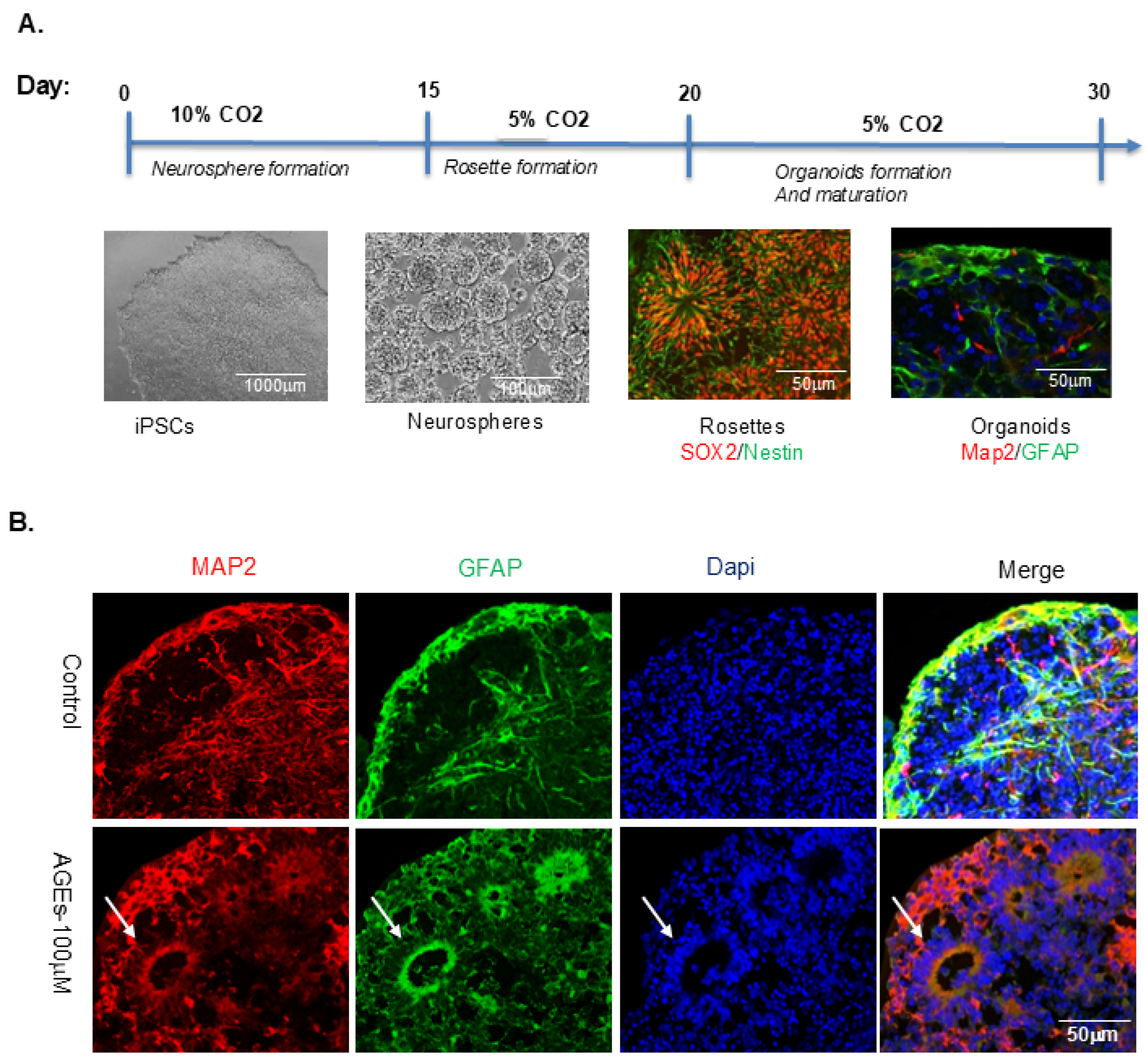
To evaluate the neurodevelopmental toxicity of AGEs, we utilized human iPSC-derived cerebral organoids. Neural rosettes containing neuronal and microglial progenitors were generated from lymphoid cell line (LCL)-derived iPSCs under 10% CO2 conditions, as previously described [30,31]. These rosettes were cultured in neuronal maintenance medium (NMM) supplemented with IL-34, macrophage colony-stimulating factor (M-CSF), and TGF-β in low-adhesion suspension plates for one month (Figure 1A). AGEs treatment (100 µM) was administered throughout the culture period, with untreated organoids serving as controls. The concentration was selected based on previous findings indicating that AGEs above 0.5 μg/mL induced IL-6 release without cytotoxicity [36], along with our preliminary data identifying 100 µM as the minimal effective dose that disrupts neuronal network formation.
After treatment, organoids were fixed, sectioned (10 µm), and immunostained for MAP2 and GFAP. AGEs-exposed organoids showed impaired neural differentiation, characterized by persistent rosette structures and reduced numbers of mature neurons (Figure 1B).
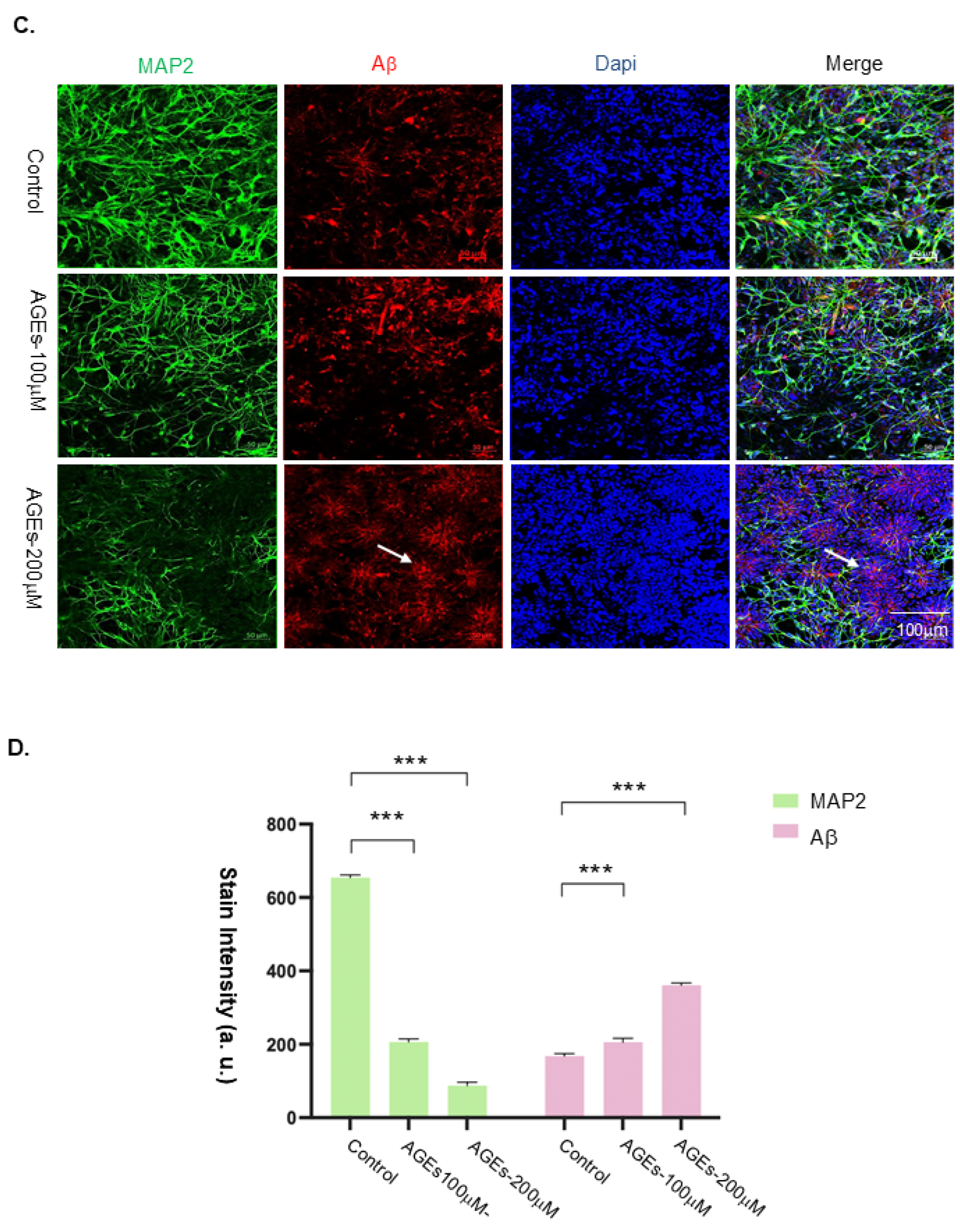
To further validate these findings, neural rosettes were plated on matrigel-coated six-well plates to promote neuronal network expansion in 2D culture and were treated with 100 µM or 200 µM AGEs, or left untreated (control). Immunostaining for MAP2 and Aβ demonstrated a dose-dependent reduction in neuronal population (Figure 1C). Aβ accumulation was significantly increased in neuronal progenitors within rosette-like structures (Figure 1C,D), suggesting that AGE-induced Aβ deposition disrupts neuronal differentiation during early neuronal development.

3.2. AGEs-Induced RAGE Expression and Microglia Activation in Brain Organoids and 2D Neuronal Networks

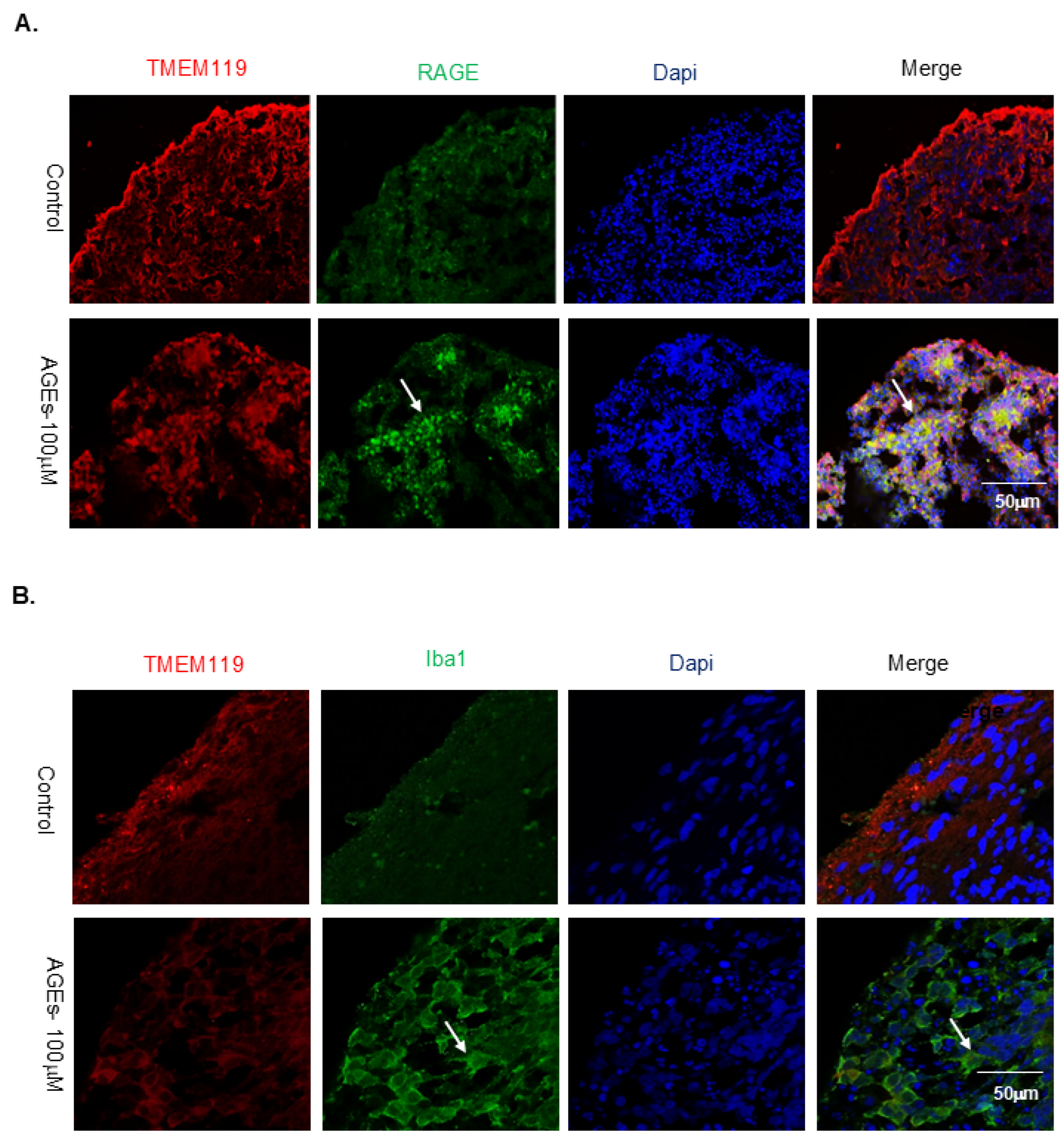
To investigate the mechanisms by which AGEs impair neurodevelopment, we examined the expression of RAGE and microglial activation in AGE-treated and control brain organoids. Immunostaining for RAGE and microglial markers TMEM119 and Iba1 demonstrated that AGEs exposure markedly increased RAGE expression, particularly within neuronal rosette regions (Figure 2A). Additionally, AGEs-treated organoids exhibited increased Iba1 signal intensity and pronounced morphological alterations indicative of microglial activation (Figure 2B).
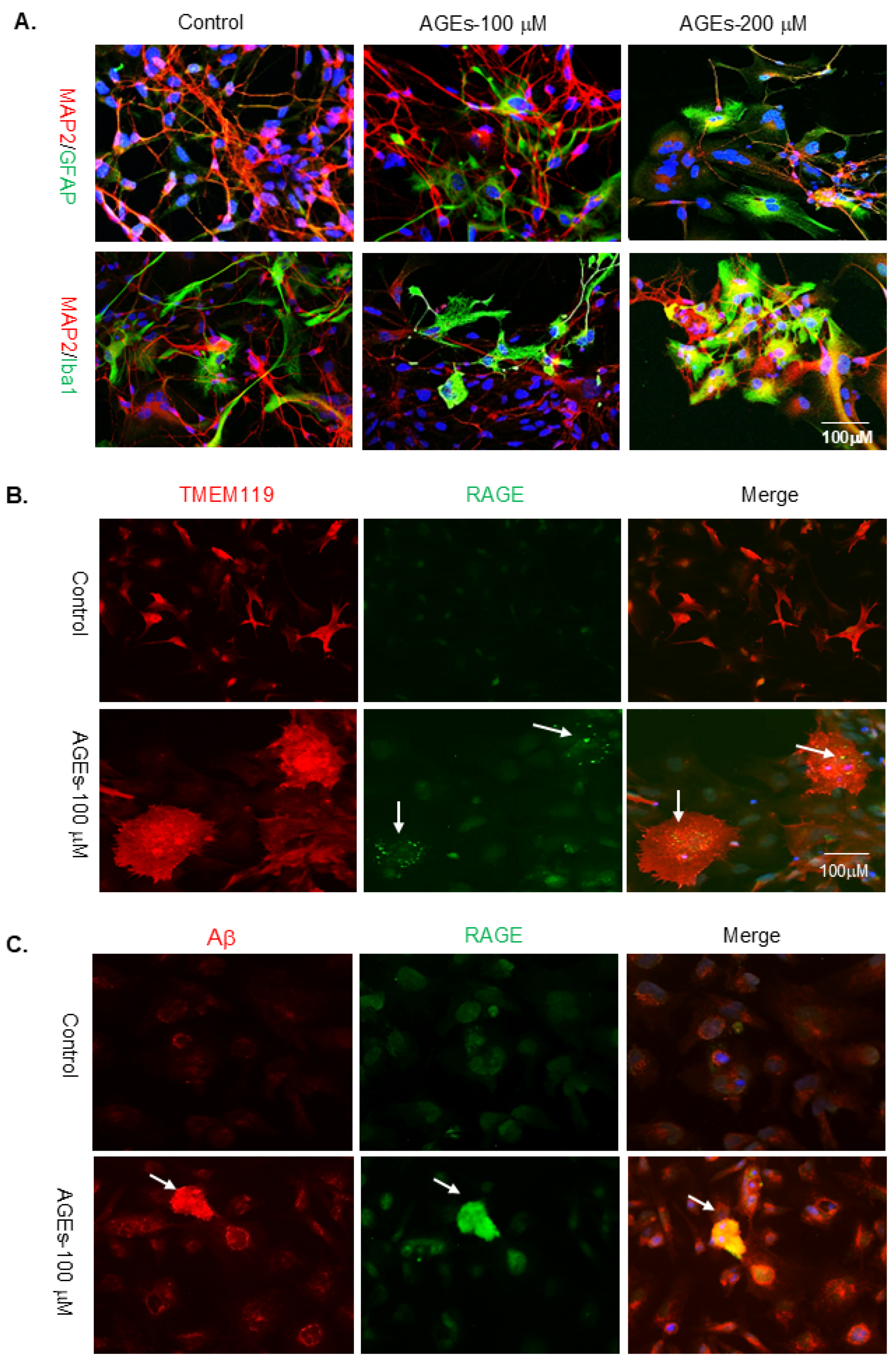
To further characterize microglial morphology in response to AGEs exposure, we utilized neuron–astrocyte–microglia 2D cultures and exposed them to 100 µM or 200 µM AGEs for three days. Immunostaining with MAP2/GFAP and MAP2/Iba1 revealed significant morphological changes in both astrocytes and microglia. At 100 µM AGEs, microglia transitioned from elongated, ramified morphologies to amoeboid, activated forms. At 200 µM AGEs, amoeboid microglia further increased in number and were accompanied by a notable reduction in mature neuronal populations (Figure 3A), indicating heightened neuroinflammatory responses at higher AGE levels. Because Aβ is a well-established ligand for RAGE, we next assessed RAGE co-localization with TMEM119+ microglia and Aβ deposits. Confocal imaging revealed RAGE expression in amoeboid microglia and clear co-localization with Aβ, as shown by yellow merged fluorescence (Figure 3B,C). Together, these findings demonstrate that AGEs upregulate RAGE, promote microglial activation, and enhance Aβ accumulation, collectively contributing to neurodevelopmental disruption in iPSC-derived cerebral organoids.

3.3. ALS-PDC-Affected Cerebral Organoids Exhibit Reduced Neuronal Populations and Elevated Reactive Astrocytes and Microglia

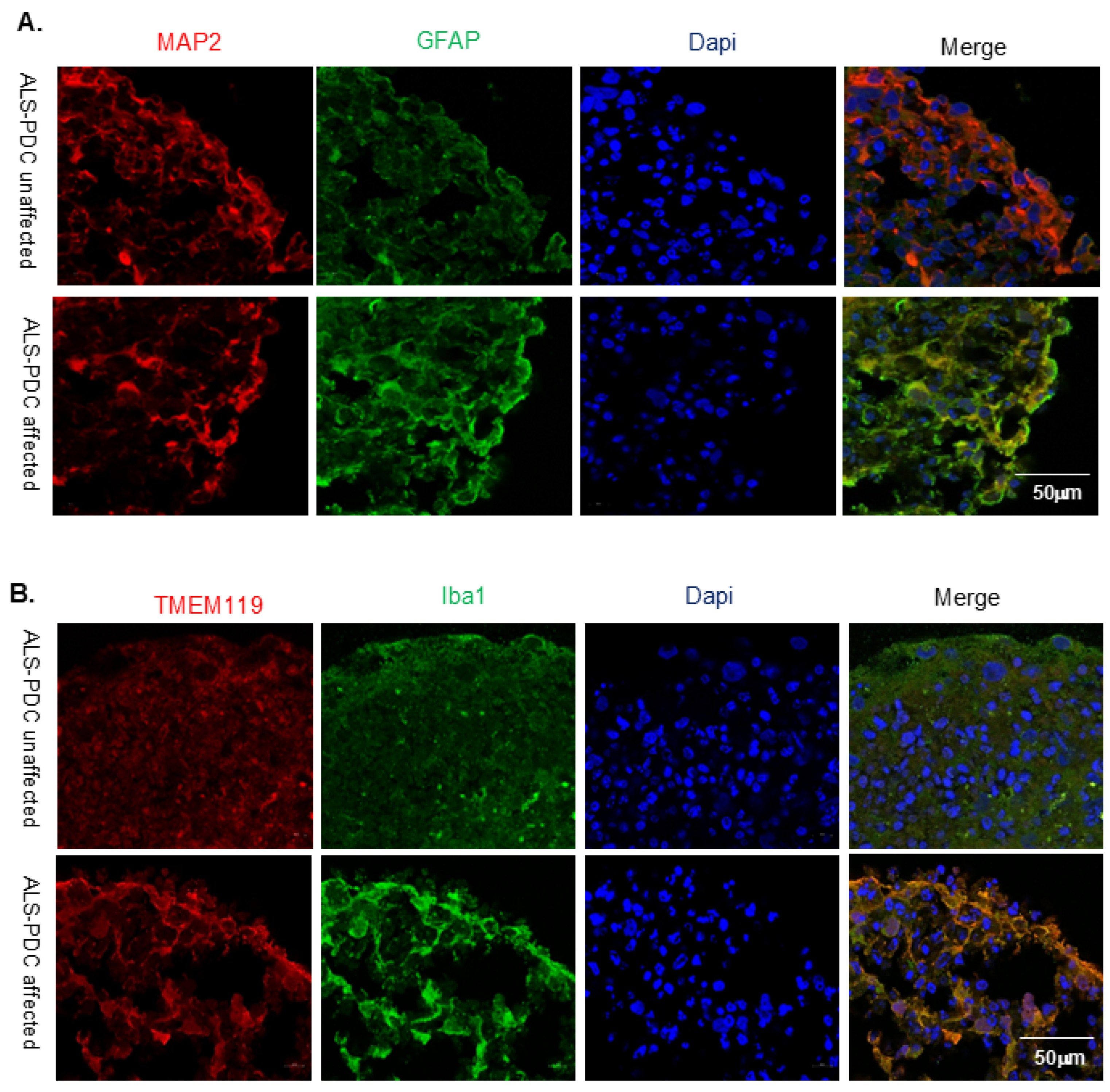
Amyotrophic lateral sclerosis–Parkinsonism-dementia complex (ALS-PDC) is a unique neurodegenerative disorder associated with early-life exposure to cycad-derived environmental neurotoxins and characterized by progressive age-related cognitive decline [37,38]. To model disease pathology, ALS-PDC-affected and unaffected cerebral organoids were generated from iPSCs derived from patient LCLs. Immunostaining with MAP2/GFAP and TMEM119/Iba1 revealed that ALS-PDC-affected organoids displayed a reduced neuronal population accompanied by markedly increased reactive astrocyte and microglia populations (Figure 4A,B).
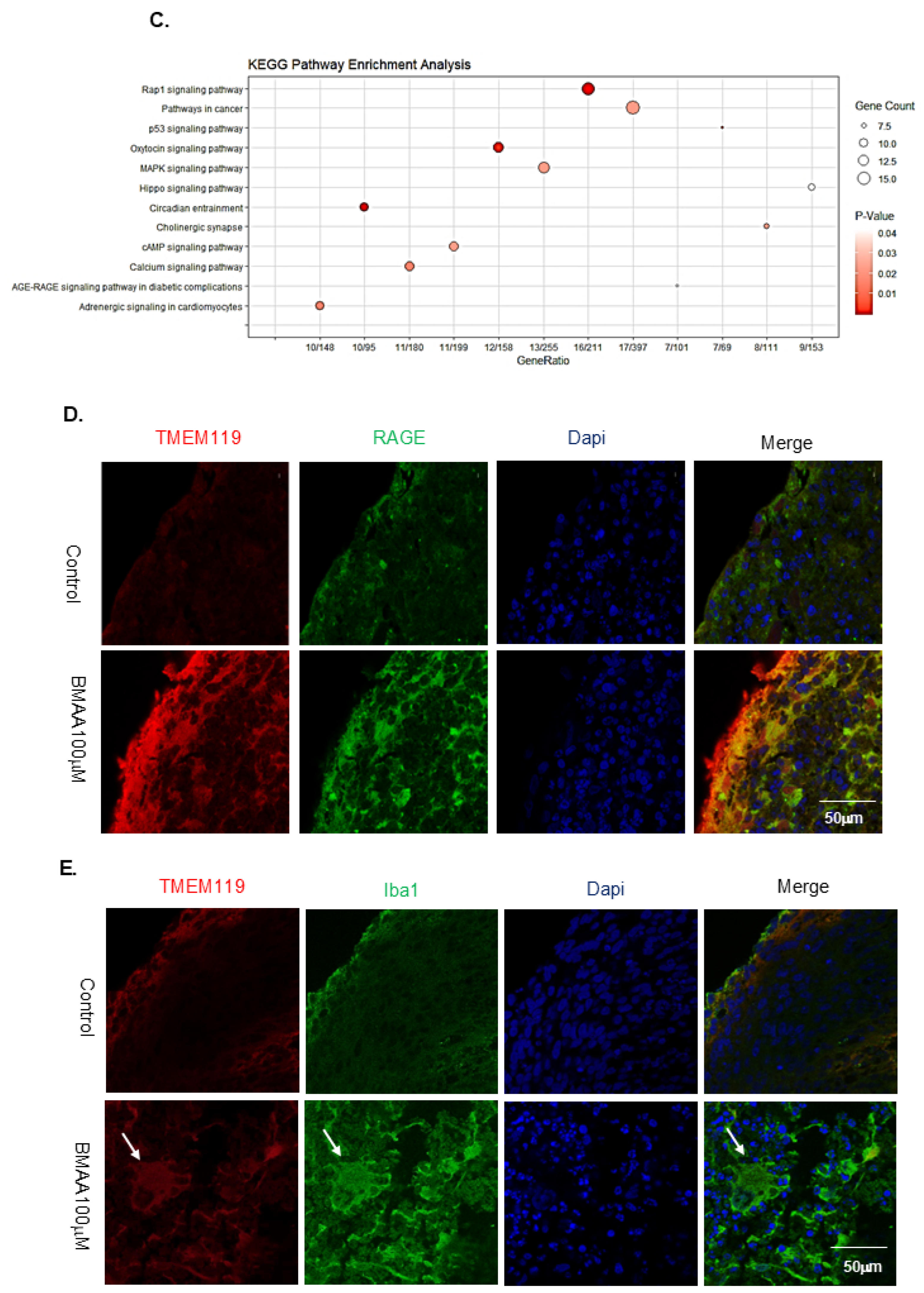
Our previous RNA-seq analysis of three-month-old ALS-PDC-affected and unaffected cerebral organoids revealed robust differentially expressed genes (DEGs), among which pyridoxine 5′-phosphate oxidase (PNPO) was one of the most significantly downregulated in ALS-PDC-affected organoids [30]. PNPO is a key enzyme in vitamin B6 metabolism and is known to suppress the formation of AGEs [39]. To explore potential mechanistic links, healthy cerebral organoids were exposed to well-characterized cycad neurotoxins β-N-methylamino-L-alanine (BMAA). KEGG pathway analysis demonstrated that the AGE–RAGE signaling pathway was among the top 12 differentially enriched pathways upregulated in response to BMAA exposure (Figure 4C). Consistently, immunostaining of BMAA-treated organoids showed enhanced RAGE expression and TMEM119 signal, together with an increase in the size and abundance of Iba1-positive microglia, indicating BMAA-induced microglial activation (Figure 4E,F).
Figure 2. AGEs Exposure Increased RAGE Expression and Promoted Microglial Activation in Cerebral Organoids and Neuronal Networks. (A) Cerebral organoids were generated from rosettes containing neuronal and microglial progenitors and treated with or without AGEs (100 µM). Immunostaining with TMEM119 and RAGE antibodies showed that AGE exposure increased RAGE expression, particularly in neuronal rosettes as shown in arrows. (B) Organoids were stained with TMEM119 (red) and Iba1 (green) antibodies, indicating that AGE exposure promoted microglial activation, as evidenced by increased Iba1 expression and morphological changes as shown in arrows.
Organoids 05 00012 g002
Figure 3. AGE exposure promoted microglial activation and increased RAGE and Aβ expression in 2D neuronal networks. (A) Morphological alterations in astrocytes and microglia were observed in mature neuronal networks following AGE exposure, indicating microglia activation. (B) Immunostaining of mature neuronal networks with TMEM119 and RAGE antibodies demonstrated that morphological changes in microglia correlated with elevated RAGE expression as pointed in arrows. (C) Immunostaining with RAGE (green) and Aβ (red) antibodies showed co-localization in AGE-treated neuronal networks, suggesting that Aβ accumulation was associated with increased RAGE expression. Images were acquired using a Zeiss LSM 880 confocal microscope with a 20× objective. Scale bars = 50 μm.
Organoids 05 00012 g003
Figure 4. ALS-PDC-affected cerebral organoids showed reduced neurons and increased reactive astrocytes and microglia. (A) Cerebral organoids were generated from iPSCs of a 57-year-old female Chamorro patient with ALS-PDC and from an age- and gender-matched healthy control (WT). Immunostaining with MAP2 9red) and GFAP(green) revealed a greater number of reactive astrocytes in ALS-PDC-affected organoids. (B) Organoids from ALS-PDC-affected and unaffected iPSCs were stained with TMEM119 (red) and Iba1 (green), showing more reactive microglia in ALS-PDC samples. (C) KEGG pathway enrichment analysis of DEGs from BMAA-exposed organoids based on RNA-seq data. (D,E) Organoids exposed to BMAA showed increased expression of RAGE (green) and Iba1 (green), suggesting activation of microglia in response to environmental toxins. Images were acquired using a Zeiss LSM 880 confocal microscope with a 20× objective. Scale bars = 50 μm.
Organoids 05 00012 g004aOrganoids 05 00012 g004b

3.4. ALS-PDC-Affected Cerebral Organoids Accumulate Elevated Levels of AGE-Modified Proteins and Aβ Plaques

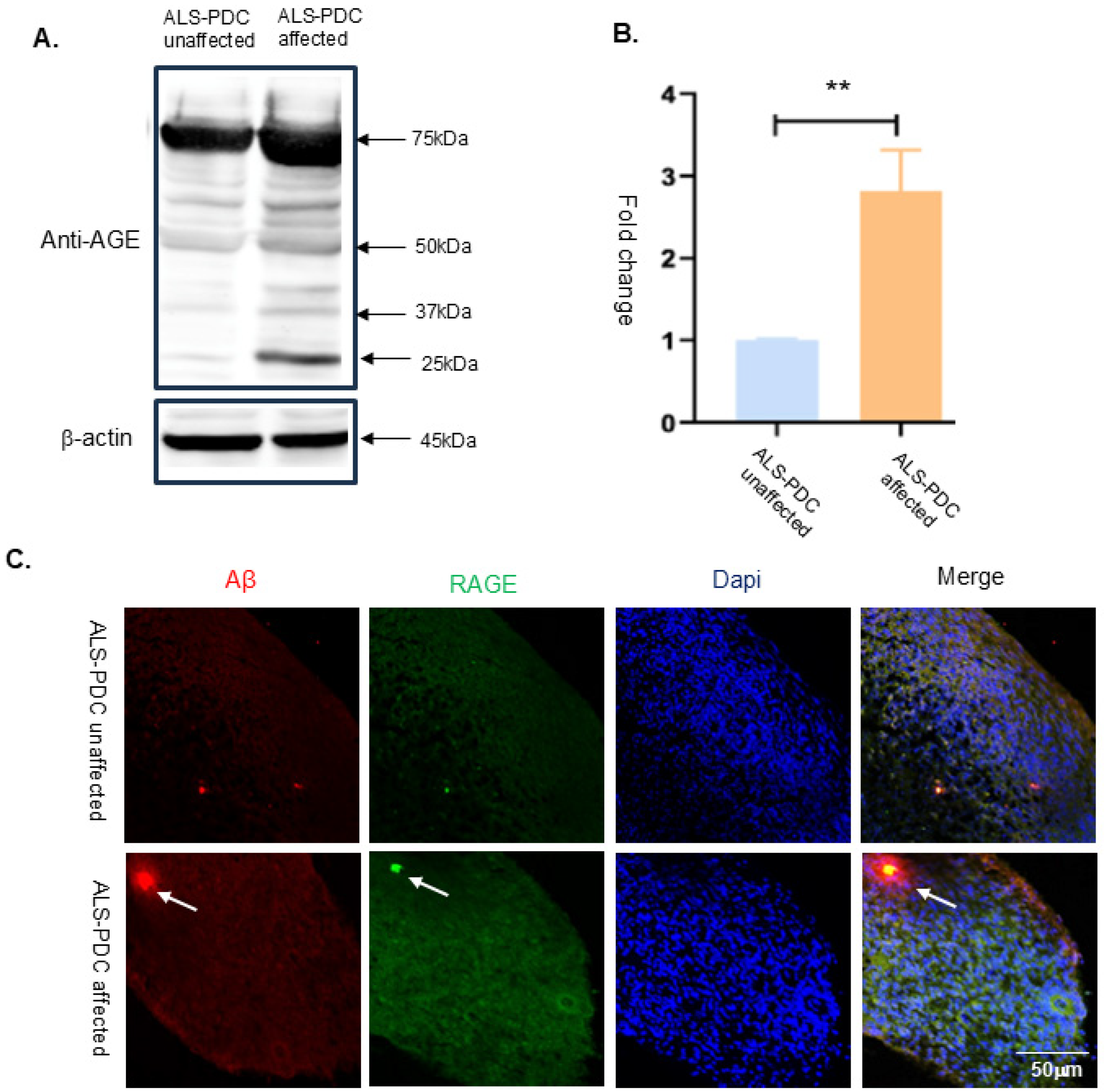
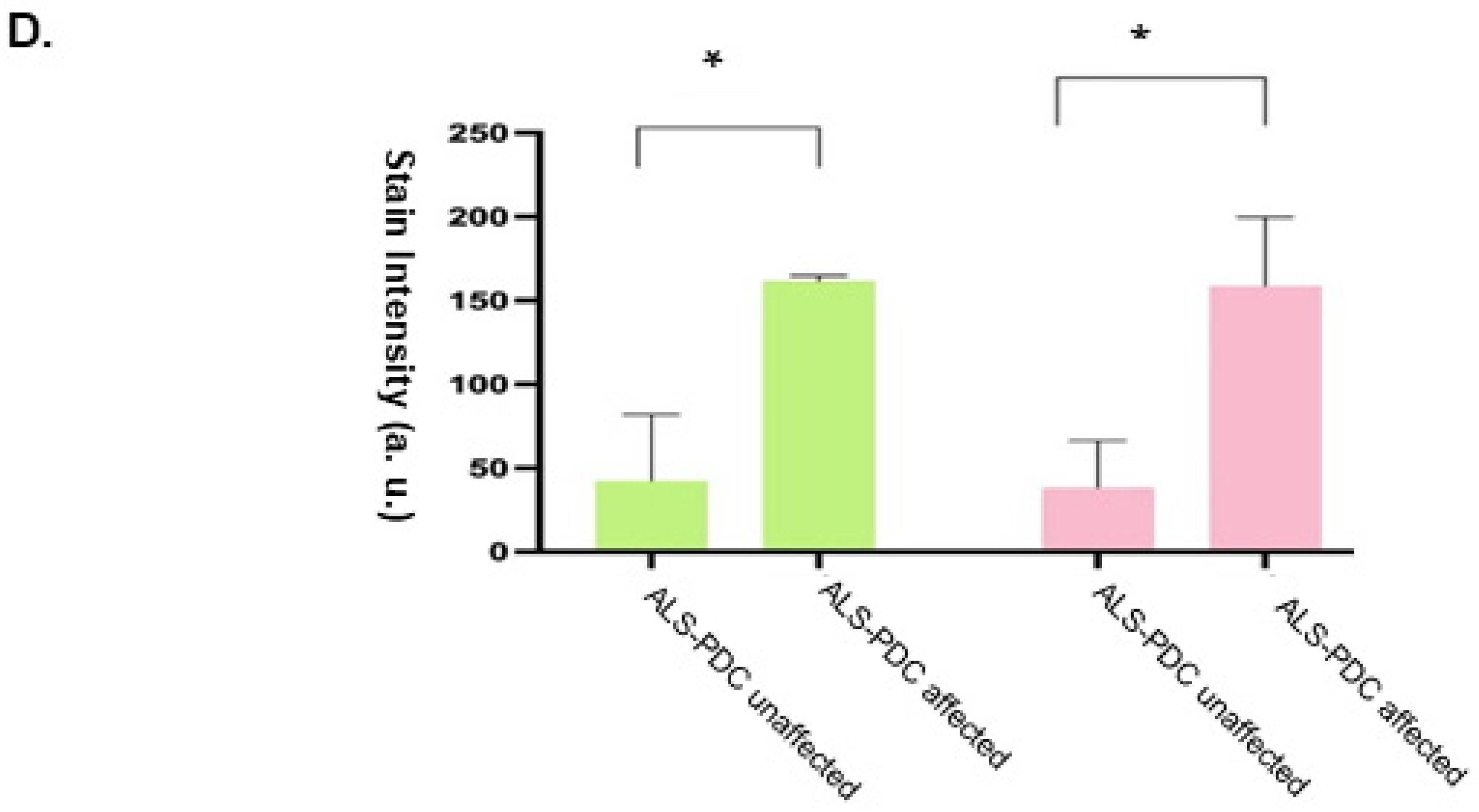
Building on these findings, we performed Western blot analysis using an anti-AGE antibody to detect AGE-modified proteins. The results showed that ALS-PDC-affected cerebral organoids exhibited significantly higher levels of AGE-conjugated proteins compared to unaffected organoids (Figure 5A,B). Additionally, immunostaining of three-month-old organoid sections with RAGE and Aβ antibodies revealed a substantial increase in RAGE immunoreactivity and enhanced Aβ deposition in ALS-PDC-affected organoids relative to controls (Figure 5C,D). These findings indicate that developmental exposure to cycad-derived neurotoxins such as BMAA may impair metabolic pathways, leading to excessive AGEs accumulation and RAGE-mediated neuroinflammation. Together, this AGE–RAGE–Aβ axis likely contributes to the progressive neurodegenerative pathology characteristic of ALS-PDC and may represent a shared mechanism with other age-related cognitive disorders.
Figure 5. ALS-PDC-affected organoids accumulated more AGE-modified proteins and Aβ plaques. (A) Western blot analysis of ALS-PDC-affected and -unaffected cerebral organoids showed higher levels of AGE-modified proteins in affected samples. (B) Quantification of AGE-conjugated protein levels from data was analyzed using Student’s t-test in GraphPad Prism version 8, ** indicated p-value less than or equal to 0.01 (C) Immunostaining with RAGE and Aβ antibodies revealed increased accumulation of both proteins in ALS-PDC-affected organoids. (D) Quantification of RAGE and Aβ expression from three representative images. All values were compared to the ALS-PDC-unaffected condition. Statistical significance was determined using Student’s t-test, * indicated p-value less than or equal to 0.05.
Organoids 05 00012 g005aOrganoids 05 00012 g005b

4. Discussion

AGEs are a group of chemical compounds formed through non-enzymatic glycation reactions and can originate from both exogenous and endogenous sources [2]. While glycation reactions naturally occur at low levels in the body, their accumulation is greatly accelerated during aging and in multiple neurodegenerative conditions. Additionally, dietary intake of highly processed or heat-treated foods can introduce substantial amounts of AGEs into the body. In this study, we demonstrate that AGEs upregulate the expression of RAGE, leading to the activation of pro-inflammatory microglia, enhanced Aβ deposition, and impaired neurodevelopment.
To further explore the correlation between AGE-modified proteins and neuronal dysfunction, we utilized ALS-PDC, an environmentally mediated neurodegenerative disease associated with metabolic dysfunction, as a cellular model [21,23]. ALS-PDC is characterized by the accumulation of Aβ plaques and neurofibrillary tangles [24]. Developmental exposure to cycad-derived genotoxins is thought to play a critical role in disease onset. This neurodegenerative condition manifests later in life following early-life exposure to phytogenotoxins in cycad seeds, historically used for food or medicinal purposes. Our KEGG pathway analysis indicated that exposure to the cycad neurotoxin BMAA significantly activated the AGE–RAGE signaling axis. Western blot analysis showed elevated levels of AGE-conjugated proteins in ALS-PDC-affected cerebral organoids. Immunostaining with RAGE, Iba1, and Aβ antibodies revealed increased microglial reactivity and greater Aβ accumulation in ALS-PDC organoids compared to controls. Aβ plaques are widely implicated in disrupting synaptic function and provoking neuroinflammation [4]. These findings support the conclusion that AGE–RAGE signaling contributes to Aβ aggregation and may play a pivotal role in ALS-PDC pathogenesis.
In summary, our results demonstrate that AGEs enhance RAGE expression, drive microglial activation, intensify Aβ aggregation, and impair neuronal development. We hypothesize that the AGE–RAGE axis contributes to neurodevelopmental deficits via two coordinated mechanisms: (1) promoting Aβ aggregation that suppresses neurogenesis, and (2) stimulating inflammatory microglial activation [3,30]. This inflammatory polarization disrupts microglial homeostasis and depletes supportive, neuroprotective microglial subtypes essential for maintaining neurogenesis [40], thereby exacerbating neuronal impairment. In addition, AGEs exposure could contribute to other mechanisms, including oxidative stress, mitochondrial dysfunction,
The precise molecular role of Aβ in inhibiting neurodevelopment warrants further investigation. Future studies using RAGE-knockout iPSC lines will help elucidate downstream signaling pathways involved in AGE-induced microglial activation and its contribution to neurodegeneration.

5. Conclusions

Human iPSC-derived brain organoids containing neurons, astrocytes, and microglia provide a physiologically relevant platform to examine the impact of AGEs on human neurodevelopment. Our findings demonstrate that AGEs activate microglia through RAGE-dependent signaling, promote Aβ accumulation, and impair neuronal maturation. In organoids modeling Western Pacific amyotrophic lateral sclerosis and Parkinsonism–dementia complex (ALS-PDC), elevated AGE-modified proteins and altered microglial polarization identify AGEs as active pathological mediators linking metabolic stress to heightened vulnerability to neurodegenerative disease.

Author Contributions

Y.H.: Conceptualization, methodology, supervision, and revision of the manuscript. R.K.: Methodology, data curation, and writing—original draft preparation. G.S.: Methodology and data curation. J.L.: Methodology and validation. Q.Q.L.: Supervision, and writing—reviewing and editing. All authors have read and agreed to the published version of the manuscript.

Funding

This research received Western University of Health Sciences internal funding and a Boehringer Ingelheim Fellowship for student research.

Institutional Review Board Statement

Not applicable for studies not involving humans or animals.

Informed Consent Statement

Not applicable for studies not involving humans.

Data Availability Statement

RNA-seq data from this study have been deposited in the NCBI under BioProject #: PRJNA75348.

Conflicts of Interest

The authors declare no conflicts of interest. The funders had no role in the design of the study; in the collection, analyses, or interpretation of data; in the writing of the manuscript; or in the decision to publish the results.

References

  1. Prasad, K.; Mishra, M. AGE-RAGE Stress, Stressors, and Antistressors in Health and Disease. Int. J. Angiol. 2018, 27, 1–12. [Google Scholar] [CrossRef] [Scilit]
  2. Zgutka, K.; Tkacz, M.; Tomasiak, P.; Tarnowski, M. A Role for Advanced Glycation End Products in Molecular Ageing. Int. J. Mol. Sci. 2023, 24, 9881. [Google Scholar] [CrossRef] [Scilit]
  3. Schmidt, A.M.; Yan, S.D.; Yan, S.F.; Stern, D.M. The multiligand receptor RAGE as a progression factor amplifying immune and inflammatory responses. J. Clin. Investig. 2001, 108, 949–955. [Google Scholar] [CrossRef]
  4. Yan, R. Physiological Functions of BACE1 and BACE2. Front. Mol. Neurosci. 2017, 10, 97. [Google Scholar] [CrossRef] [Scilit]
  5. Fang, F.; Yu, Q.; Arancio, O.; Chen, D.; Gore, S.S.; Yan, S.S.; Yan, S.F. RAGE mediates Abeta accumulation in a mouse model of Alzheimer’s disease. Hum. Mol. Genet. 2018, 27, 1002–1014. [Google Scholar] [CrossRef] [Scilit] [PubMed]
  6. Hemonnot, A.L.; Hua, J.; Ulmann, L.; Hirbec, H. Microglia in Alzheimer Disease: Well-Known Targets and New Opportunities. Front. Aging Neurosci. 2019, 11, 233. [Google Scholar] [CrossRef] [Scilit] [PubMed]
  7. Blevins, H.M.; Xu, Y.; Biby, S.; Zhang, S. The NLRP3 Inflammasome Pathway: A Review of Mechanisms and Inhibitors for the Treatment of Inflammatory Diseases. Front. Aging Neurosci. 2022, 14, 879021. [Google Scholar] [CrossRef] [Scilit]
  8. Harry, G.J. Microglia during development and aging. Pharmacol. Ther. 2013, 139, 313–326. [Google Scholar] [CrossRef] [Scilit]
  9. Nayak, D.; Roth, T.L.; McGavern, D.B. Microglia development and function. Annu. Rev. Immunol. 2014, 32, 367–402. [Google Scholar] [CrossRef] [Scilit]
  10. Hickman, S.; Izzy, S.; Sen, P.; Morsett, L.; El Khoury, J. Microglia in neurodegeneration. Nat. Neurosci. 2018, 21, 1359–1369. [Google Scholar] [CrossRef] [Scilit]
  11. Mathys, H.; Adaikkan, C.; Gao, F.; Young, J.Z.; Manet, E.; Hemberg, M.; De Jager, P.L.; Ransohoff, R.M.; Regev, A.; Tsai, L.H. Temporal Tracking of Microglia Activation in Neurodegeneration at Single-Cell Resolution. Cell Rep. 2017, 21, 366–380. [Google Scholar] [CrossRef] [Scilit]
  12. Noh, M.Y.; Lim, S.M.; Oh, K.W.; Cho, K.A.; Park, J.; Kim, K.S.; Lee, S.J.; Kwon, M.S.; Kim, S.H. Mesenchymal Stem Cells Modulate the Functional Properties of Microglia via TGF-beta Secretion. Stem Cells Transl. Med. 2016, 5, 1538–1549. [Google Scholar] [CrossRef] [Scilit] [PubMed]
  13. Wang, Y.; Szretter, K.J.; Vermi, W.; Gilfillan, S.; Rossini, C.; Cella, M.; Barrow, A.D.; Diamond, M.S.; Colonna, M. IL-34 is a tissue-restricted ligand of CSF1R. Nat. Immunol. 2012, 13, 753–760. [Google Scholar] [CrossRef] [Scilit] [PubMed]
  14. Wendimu, M.Y.; Hooks, S.B. Microglia Phenotypes in Aging and Neurodegenerative Diseases. Cells 2022, 11, 2091. [Google Scholar] [CrossRef] [Scilit] [PubMed]
  15. Moehle, M.S.; West, A.B. M1 and M2 immune activation in Parkinson’s Disease: Foe and ally? Neuroscience 2015, 302, 59–73. [Google Scholar] [CrossRef] [Scilit]
  16. Paolicelli, R.C.; Bolasco, G.; Pagani, F.; Maggi, L.; Scianni, M.; Panzanelli, P.; Giustetto, M.; Ferreira, T.A.; Guiducci, E.; Dumas, L.; et al. Synaptic pruning by microglia is necessary for normal brain development. Science 2011, 333, 1456–1458. [Google Scholar] [CrossRef] [Scilit]
  17. Tang, Y.; Le, W. Differential Roles of M1 and M2 Microglia in Neurodegenerative Diseases. Mol. Neurobiol. 2016, 53, 1181–1194. [Google Scholar] [CrossRef] [Scilit]
  18. Reddy, V.P.; Aryal, P.; Darkwah, E.K. Advanced Glycation End Products in Health and Disease. Microorganisms 2022, 10, 1848. [Google Scholar] [CrossRef] [Scilit]
  19. Muffat, J.; Li, Y.; Yuan, B.; Mitalipova, M.; Omer, A.; Corcoran, S.; Bakiasi, G.; Tsai, L.H.; Aubourg, P.; Ransohoff, R.M.; et al. Efficient derivation of microglia-like cells from human pluripotent stem cells. Nat. Med. 2016, 22, 1358–1367. [Google Scholar] [CrossRef] [Scilit]
  20. Miklossy, J.; Steele, J.C.; Yu, S.; McCall, S.; Sandberg, G.; McGeer, E.G.; McGeer, P.L. Enduring involvement of multiple pathologies in ALS/PDC of Guam. Acta Neuropathol. 2008, 116, 625–637. [Google Scholar] [CrossRef] [Scilit]
  21. Spencer, P.S.; Palmer, V.; Kisby, G. The ALS/PDC syndrome of Guam and the cycad hypothesis. Neurology 2009, 72, 474–476. [Google Scholar] [CrossRef] [Scilit]
  22. Zhang, Z.X.; Anderson, D.W.; Mantel, N.; Roman, G.C. Motor neuron disease on Guam: Geographic and familial occurrence. Acta Neurol. Scand. 1996, 94, 51–59. [Google Scholar] [CrossRef] [Scilit] [PubMed]
  23. Kisby, G.E.; Ellison, M.; Spencer, P.S. Content of the neurotoxins cycasin and BMAA in cycad flour prepared by Guam Chamorros. Neurology 1992, 42, 1336–1340. [Google Scholar] [CrossRef] [Scilit] [PubMed]
  24. Kisby, G.E.; Spencer, P.S. Genotoxic Damage During Brain Development Presages Prototypical Neurodegenerative Disease. Front. Neurosci. 2021, 15, 752153. [Google Scholar] [CrossRef] [Scilit] [PubMed]
  25. Delcourt, N.; Claudepierre, T.; Maignien, T.; Arnich, N.; Mattei, C. Cellular and Molecular Aspects of the beta-N-Methylamino-l-alanine (BMAA) Mode of Action within the Neurodegenerative Pathway. Toxins 2017, 10, 6. [Google Scholar] [CrossRef] [Scilit]
  26. Engskog, M.K.; Ersson, L.; Haglof, J.; Arvidsson, T.; Pettersson, C.; Brittebo, E. β-N-Methylamino-L-alanine (BMAA) perturbs alanine, aspartate and glutamate metabolism pathways in human neuroblastoma cells as determined by metabolic profiling. Amino Acids 2017, 49, 905–919. [Google Scholar] [CrossRef] [Scilit]
  27. Dutta, D.; Heo, I.; Clevers, H. Disease Modeling in Stem Cell-Derived 3D Organoid Systems. Trends Mol. Med. 2017, 23, 393–410. [Google Scholar] [CrossRef] [Scilit]
  28. Fujimori, K.; Tezuka, T.; Ishiura, H.; Mitsui, J.; Doi, K.; Yoshimura, J.; Tada, H.; Matsumoto, T.; Isoda, M.; Hashimoto, R.; et al. Modeling neurological diseases with induced pluripotent cells reprogrammed from immortalized lymphoblastoid cell lines. Mol. Brain 2016, 9, 88. [Google Scholar] [CrossRef] [Scilit]
  29. Barrett, R.; Ornelas, L.; Yeager, N.; Mandefro, B.; Sahabian, A.; Lenaeus, L.; Targan, S.R.; Svendsen, C.N.; Sareen, D. Reliable generation of induced pluripotent stem cells from human lymphoblastoid cell lines. Stem Cells Transl. Med. 2014, 3, 1429–1434. [Google Scholar] [CrossRef] [Scilit]
  30. Hong, Y.; Dong, X.; Chang, L.; Xie, C.; Chang, M.; Aguilar, J.S.; Lin, J.; Lin, J.; Li, Q.Q. Microglia-containing cerebral organoids derived from induced pluripotent stem cells for the study of neurological diseases. iScience 2023, 26, 106267. [Google Scholar] [CrossRef] [Scilit]
  31. Begum, A.N.; Guoynes, C.; Cho, J.; Hao, J.; Lutfy, K.; Hong, Y. Rapid generation of sub-type, region-specific neurons and neural networks from human pluripotent stem cell-derived neurospheres. Stem Cell Res. 2015, 15, 731–741. [Google Scholar] [CrossRef] [Scilit] [PubMed]
  32. Anders, S.; Pyl, P.T.; Huber, W. HTSeq—A Python framework to work with high-throughput sequencing data. Bioinformatics 2015, 31, 166–169. [Google Scholar] [CrossRef] [Scilit] [PubMed]
  33. Love, M.I.; Huber, W.; Anders, S. Moderated estimation of fold change and dispersion for RNA-seq data with DESeq2. Genome Biol. 2014, 15, 550. [Google Scholar] [CrossRef] [Scilit] [PubMed]
  34. Ashburner, M.; Ball, C.A.; Blake, J.A. Gene ontology: Tool for the unification of biology. The Gene Ontology Consortium. Nat. Genet. 2000, 25, 25–29. [Google Scholar] [CrossRef] [Scilit]
  35. Young, M.D.; Wakefield, M.J.; Smyth, G.K.; Oshlack, A. Gene ontology analysis for RNA-seq: Accounting for selection bias. Genome Biol. 2010, 11, R14. [Google Scholar] [CrossRef] [Scilit]
  36. Shen, C.Y.; Wu, C.H.; Lu, C.H.; Kuo, Y.M.; Li, K.J.; Hsieh, S.C.; Yu, C.L. AGEs suppressed cytokines via MAPK-ERK and NF-κB pathways. Molecules 2019, 24, 2461. [Google Scholar] [CrossRef] [Scilit]
  37. Kisby, G.E.; Olivas, A.; Standley, M.; Lu, X.; Pattee, P.; O’Malley, J.; Li, X.; Muniz, J.; Nagalla, S.R. Genotoxicants target distinct molecular networks in neonatal neurons. Environ. Health Perspect. 2006, 114, 1703–1712. [Google Scholar] [CrossRef] [Scilit]
  38. Kisby, G.E.; Standley, M.; Lu, X.; O’Malley, J.; Lin, B.; Muniz, J.; Luo, N.L.; Pattee, P.; Back, S.A.; Nagalla, S.R. Molecular networks perturbed in a developmental animal model of brain. Neurobiol. Dis. 2005, 19, 108–118. [Google Scholar] [CrossRef] [Scilit]
  39. Ramis, R.; Ortega-Castro, J.; Caballero, C.; Casasnovas, R.; Cerrillo, A.; Vilanova, B.; Adrover, M.; Frau, J. How Does Pyridoxamine Inhibit the Formation of Advanced Glycation End Products? Antioxidants 2019, 8, 344. [Google Scholar] [CrossRef] [Scilit]
  40. Wee, Y.S.; Weis, J.J.; Gahring, L.C.; Rogers, S.W.; Weis, J.H. Age-related onset of obesity corresponds with metabolic dysregulation. PLoS ONE 2015, 10, e0123218. [Google Scholar] [CrossRef] [Scilit]
Figure 1. AGEs Treatment Inhibits Neurodevelopment and Increases Aβ Accumulation. (A) Schematic figure illustrates the procedure for generating organoids from human iPSCs. The neural rosettes used for subsequent organoid differentiation were immunostained with progenitor-stage markers SOX2 and Nestin. (B) Cerebral organoids generated from rosettes were treated with AGEs (100 µM) and untreated as a control. The organoids were immunostained for MAP2 and GFAP, revealing that AGEs treatment inhibited the differentiation of rosettes into mature neurons. (C) Neuronal networks were derived from iPSC-generated rosettes and exposed to 100 µM and 200 µM AGEs, with untreated cultures serving as controls. Immunostaining with antibodies against MAP2 and Aβ showed that AGEs-treated cultures exhibited more undifferentiated rosette-like structures (indicated by arrows). Images were captured using a Zeiss LSM 880 confocal microscope with a 10× objective. Scale bar = 100 μm. (D) Quantification of MAP2-positive neurons and Aβ accumulation in control and AGE-treated cultures demonstrated that AGE exposure significantly reduced neuronal populations and increased Aβ deposition. Statistical analysis was performed using Student’s t-test in GraphPad Prism version 8, with p-values indicated in the figure. *** p < 0.001.
Organoids 05 00012 g001aOrganoids 05 00012 g001b
Disclaimer/Publisher’s Note: The statements, opinions and data contained in all publications are solely those of the individual author(s) and contributor(s) and not of MDPI and/or the editor(s). MDPI and/or the editor(s) disclaim responsibility for any injury to people or property resulting from any ideas, methods, instructions or products referred to in the content.