Proceeding Paper

RhsP2 Protein as a New Antibacterial Toxin Targeting RNA †

by
Tamara Nami Haj Marza
Department of Bioinformatics, Syrian Virtual University, Damascus P.O. Box 35329, Syria
Presented at the 4th International Electronic Conference on Antibiotics, 21–23 May 2025; Available online: https://sciforum.net/event/ECA2025.
Med. Sci. Forum 2025, 35(1), 3; https://doi.org/10.3390/msf2025035003
Published: 24 July 2025
(This article belongs to the Proceedings of The 4th International Electronic Conference on Antibiotics)

Abstract

Many bacteria, such as Pseudomonas aeruginosa, have encoded many toxins like RhsP2 that target non-coding RNAs (ncRNAs) in a similar mechanism to ART components; bacterial RNA loses its function of amino acid translation. A virtual screening approach was used to investigate RhsP2, which targets 16s rRNAs and then disrupts the translation of bacterial amino acids to proteins. Rifamycin is the bioreference as it forms a stable complex with the bacterial RNA in its active sites. Using different docking software can determine the best predicted conformations between RhsP2/16S and rRNA, and analyzing the docking score for both Affinity Binding and the root mean square deviation (RMSD) of particle coordinates helps choose the most appropriate drugs by using tools such as bioinformatics platforms and databases.

1. Introduction

The latest national annual report of the English Surveillance Program for Antimicrobial Utilization and Resistance, ESPAUR-2022, showed that the estimated total number of severe antibiotic-resistant infections in England rose by 2.2% in 2021 compared to 2020. This is equivalent to 148 severe antibiotic-resistant infections every day in 2021 [1]. A global review of antimicrobial resistance (AMR) and its future impact estimated that there would be 10 million global AMR deaths annually in the year 2050 if we do not preserve the effectiveness of current antibiotics or develop new ones [2]. Therefore, investigating and understanding resistance to newer antibiotics is an important area of pharmaceutical development. Here, two points need to be considered:
(I) Proper target selection, particularly targets that are not prone to rapid resistance. Other important considerations include the conservative structure of the target protein across bacterial species and the lack of structural homology with similar functions in the mammalian host to avoid toxicity.
(II) Improvement of chemical libraries of previously used drugs to overcome the limitations of species diversity, especially in Gram-negative organisms [3].
The newly synthesized compounds released to the market are almost all derivatives of existing classes that target the bacterial cell wall or DNA replication; consequently, there is often pre-existing cross-resistance within the microbiome. However, transcription appears to be an underutilized target. Figure 1 shows antibiotic targets and resistance mechanisms. So far, there are only two antibiotics targeting bacterial RNA polymerase (RNAP) on the market: the Rifamycin series and fidaxomicin/lipiarmycin [4].
As noted above, the prime requirement for discovering effective antibiotic drugs is to develop novel ways of thinking and identifying things that were previously missed with the aid of modern artificial intelligence programs.
In recent decades, virtual screen software has been successfully used to detect novel bio-activities, as it is known that the number of known compounds has reached tens of millions, and most major pharmaceutical companies have become very interested in the methods of simulation and virtual docking “in silico”; with the increased progression of algorithms by the experts of bioinformatics, these methods are gaining acceptance in terms of accuracy, sophistication, and sensitivity [6].
Similarly, this review, which introduces the basic principles and software of molecular docking, summarizes the applications of this method in hundreds of discovered marine drugs, proposes designs using the virtual screening and docking method, and finally, evaluates the obtained results in order to provide a stronger theoretical basis for clinical practices [7].
The aim of this work: Using computational docking and modeling, the interactions between two proteins, namely RhsP2toxin/16S rRNAtarget, were investigated in detail in order to identify residues belonging to the active sites of the studied toxin (RhsP2), which targets 16 subunit ribosomal Ribonucleic Acid (16S rRNA), which can represent a novel type of antibacterial bio-drugs.

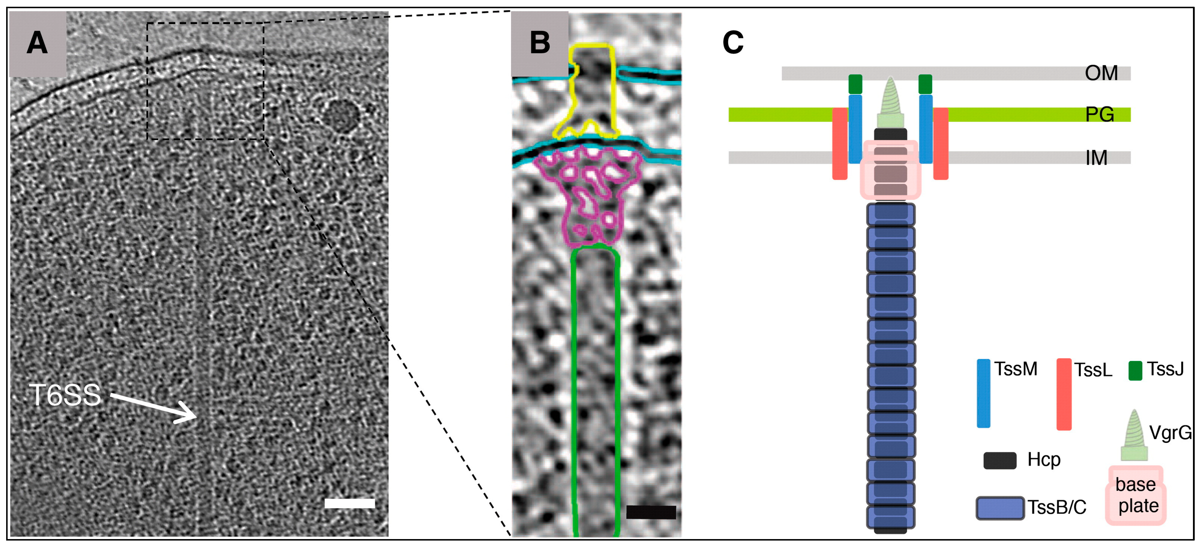
1.1. H2T6SS System (Type Six Secretion System Encodes loci Named H2)

RhsP2 is a toxin secreted by Pseudomonas aeruginosa organisms and is considered one of the groups of cytoplasmic enzymes affecting RNA through the mechanism of ADP-ribosyltransferase(ART). This substance enters the bacterial cell through a T6SS-type VI injection system (Figure 2) that delivers the toxin to the vicinity of other bacterial cells. The H2-T6SS genetic organization is conserved across Pseudomonas aeruginosa isolates, and the core H2-T6SS genes contain a specific set of four genes encoded as PAAR +Hcp protein (Hcp2) + VgrG protein (VgrG14) + Rhs family element [8].

1.2. Mechanism of Action of RhsP2

Preliminary experiments proposed RhsP2 acts as one of the ADP Ribosyltransferase toxins (ART toxin) that modifies two hydroxyl groups of RNA by transferring the Adenosine DiPhosphate ribosyl(ADPr) moiety from NAD+ residues and to target ncRNA in the form of a mono- or poly-ADP-ribose chain; due to its negative charge and large spatial form, it inhibits RNA function in the translation of amino acids to proteins.

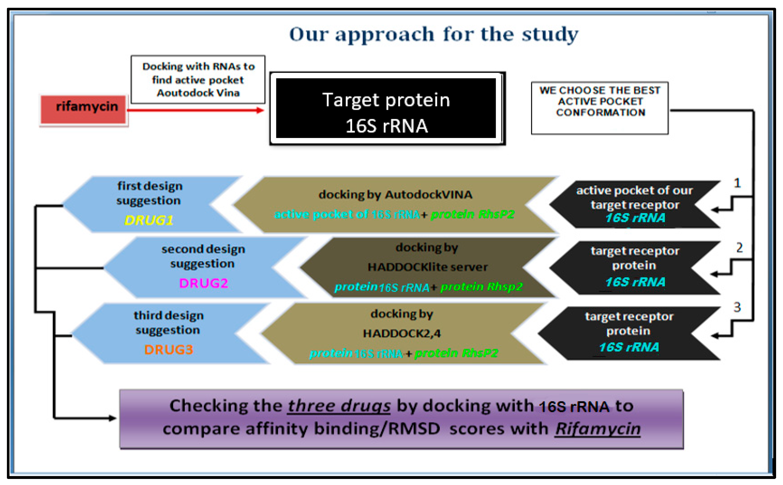
2. Methodology (Rigid Receptor + Flexible Ligand)

An overview of the research methodology approach is shown in the flowchart in Figure 3.

2.1. Part I:Preparing Target Protein, RhsP2toxin, and Bioreference in Order to Generate Protein–Protein Interaction (PPI)

2.1.1. Prepare Target Protein

16S rRNA is the RNA component of the 30S subunit of a prokaryotic ribosome. The genes coding for it are referred to as the 16S rRNA gene and are used for phylogenetic studies, as it is highly conserved between different species of bacteria and archaea. Because of the difference between prokaryotic 16S and human 18S rRNA, the drug can be selectively used to kill bacterial cells.
The chosen model of the conservative domain of “Trichonephila clavate”was modeled and saved in an“AF-16S rRNA Trichonephila clavate.pdb” format and visualized by the pyMOL tool (Figure 4).

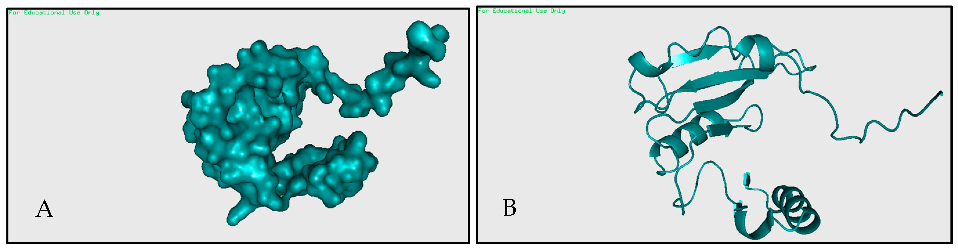
2.1.2. Prepare RhsP2 Toxin

RhsP2 is a protein belonging to the Rhs family of proteins (PDB ID: 7RT7) [10]. Organism(s): Pseudomonas aeruginosa UCBPP-PA14.
RhsP2 is found in a complex of multiple chains (Figure 5), consisting of two molecule types: RhsP2 and RhsI2, as shown by the crystal structure of the RhsP2 C-terminal toxin domain in complex with its immunity protein RhsI2. The simple chain P2 (which is the chain recommended in the article as a toxin with a mechanism of ADP ribosylation) was saved in .pdb format (Figure 6). It consists of 150 aa, with 7 B Sheets + 6 a Helix + loops.

2.1.3. Determine Bioreference and Target Protein Active Site

There are two main classes of antibiotics associated with 16S rRNA: amino glycosides and tetracyclines. Usually, antibiotics that target the 16S rRNA lead to the inhibition of translation initiation or the impairment of translation elongation [11].
From the original article, Rifamycin has an affinity for16S rRNA [12] and has activity and interaction similar to RhsP2, and hence can be used as a reference for two purposes: to determine the active pocket of the target protein 16S rRNA by AutoDockVINA (Figure 7) and evaluate the final proposed drugs.

2.2. Part II: Protein–Protein Interaction (PPI) Docking

  • The active pocket of 16S rRNA was displayed by the pyMOL TOOL version 2.5.8, which was used in the second docking with RhsP2; because VINA is not programmed for protein–protein docking, RhsP2 is considered as a ligand instead of a protein when using AutoDock VINA. After the docking results are obtained, extract the active sites of RhsP2 via pyMOL. This resulted in the first suggested drug design (DRUG1) (stochastic shape-based method).
  • Using Online Docking Programs, HADDOCKlite and HADDOCK 2,4 Server, for Protein-Protein Docking to Suggest New Pharmacophore Models (DRUG2 and DRUG3).

2.3. Part III: Evaluate Pharmacophors (DRUG1,2,3)

After extracting the pharmacophores (drug design 1, 2, 3…), their quality and credibility should be checked by comparing them to the bioreference Rifamycin using AutoDock VINA against the target receptor 16S rRNA. The comparison should compare the Affinity Binding Energy/RMSD criterion to the reference. Those recognized as having the best conformation structures can be considered as a prodrug resembling RhsP2 residues.

3. Results and Discussion

The first drug design, DRUG1, was generated using the stochastic method; after docking 16S rRNA/DRUG1 usingAutoDock VINA to evaluate the results, it showed good conformation with the target protein 16s rRNA (Figure 8).
The second drug, DRUG2, was designed using the protein–protein interaction method via the HADDOCKlite server; after docking 16S rRNA/DRUG2 by AutoDock VINA in order to evaluate results, it showed good conformation with the target protein 16S rRNA (Figure 9).
The third drug, DRUG3, was designed using the protein–protein interaction method viaHADDOCK2.4. DRUG3, after docking 16S rRNA/DRUG3 viaAutoDock VINA in order to evaluate results, showed good conformation with the target protein 16S rRNA (Figure 10).
In order to decide whether the proposed models successfully model the biological interaction with the target protein, they must be compared using docking software and compared with a standard reference, Rifamycin, which has shown good interactions and effective binding to the active pocket of the target protein.
AutoDock VINA has nine modes for each input procedure, and gives the Affinity Binding Energy and RMSD scores, as shown in Table 1. In order to check the quality and credibility of the suggested pharmacophores, they are compared to Rifamycin to determine if they have a good biological structure and compatibility with the target RNA.
The resulting graph lines in Figure 11 show the convergence in the docking scores of the binding energy and RMSD between DRUG2 and 3. DRUG2 was generated by the docking process via HADDOCKlite protein–protein interaction, as its binding energy average was −5.86 vs. −7.14 (kcal/mol) for Rifamycin and had mean RMSD values of 4.6/8.86 vs. 7.63/11.45 for Rifamycin. DRUG3 was generated by HADDOCK 2.4 protein–protein interaction. DRUG1, obtained using the stochastic structure method, had a reasonable binding affinity but high variability in the RMSD score.
The PyMOL tool displays the conformations of the nine models for the three predicted drugs as shown in Figure 12.

4. Conclusions

This paper focuses on molecular docking methods and their applications in drug discovery and design based on the structure.
  • The first part provides information on the studied protein secreted by the bacterium P. aeruginosa RhsP2 as a newly discovered protein of the Rhs family. Rifamycin was used in this study to compare the accuracy of docking programs. The second part describes in detail the docking procedures, which were used in order to dock the protein–protein complexes, and the software and criteria used in the assessment. The third part presents the docking results obtained from the individual programs, AutoDock VINA, HADDOCKlite, and HADDOCK 2.4, and compares them in terms of the binding energy and RMSD.
  • These results show that the recently developed HADDOCKlite server obtained similar drugs that were the closest to the Rifamycin reference in terms of their interaction with the active pocket of the 16S rRNA target protein, followed by HADDOCK 2.4 server and the stochastic method using AutoDock VINA, which showed a deviation in predicting the binding energies of the docking complex.
  • The results were reasonable as a preliminary prediction with one target 16S rRNA, which was selected as a special bacterial ribosome that is different from the human 18S rRNA ribosome; thus, the toxin RhsP2 has a good interaction with 16S rRNA, suggesting that it could be an inhibitor of 16S rRNA by binding to the active pocket (similar to the action of Rifamycin) and inhibiting its function in translating amino acids into proteins.
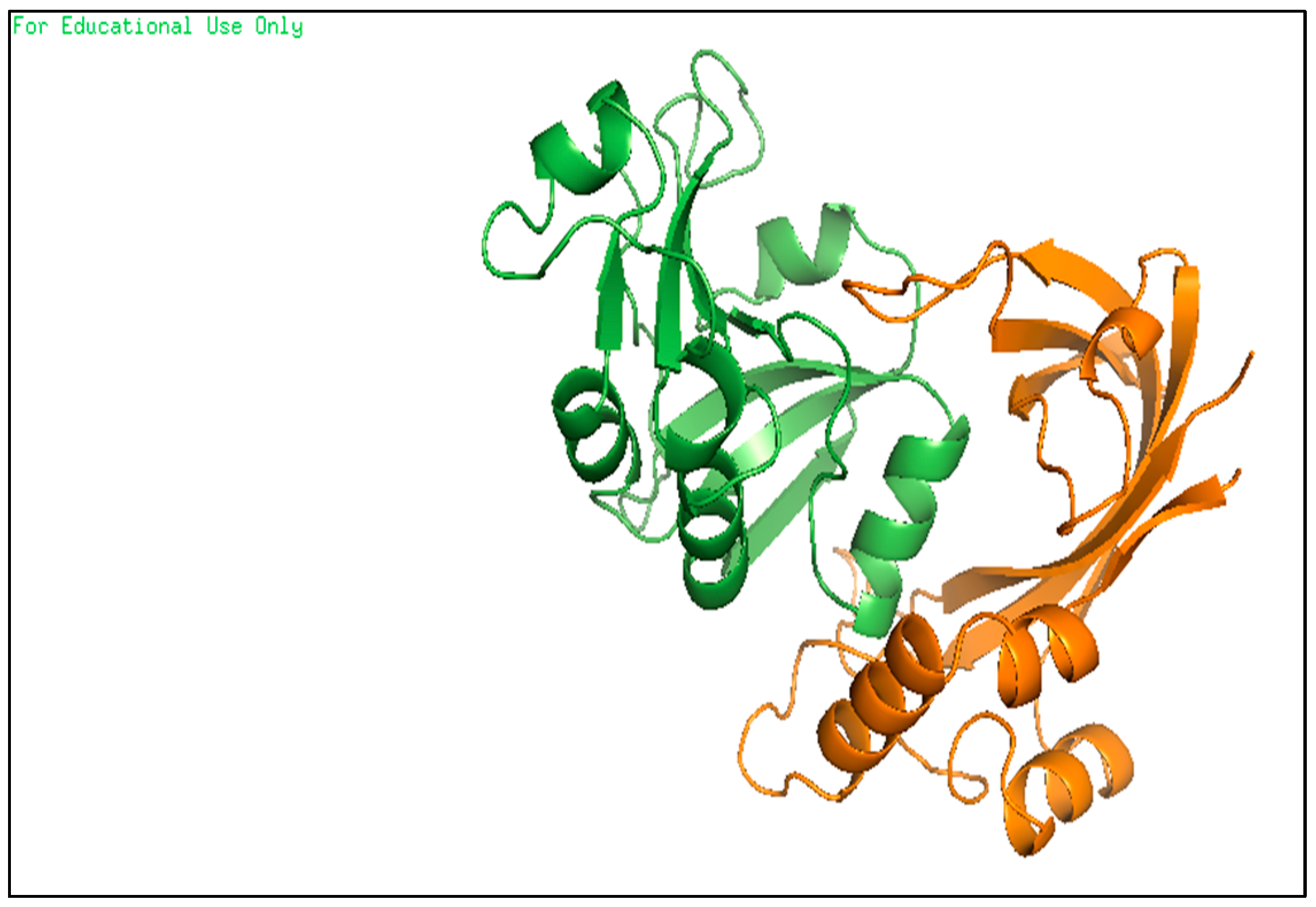
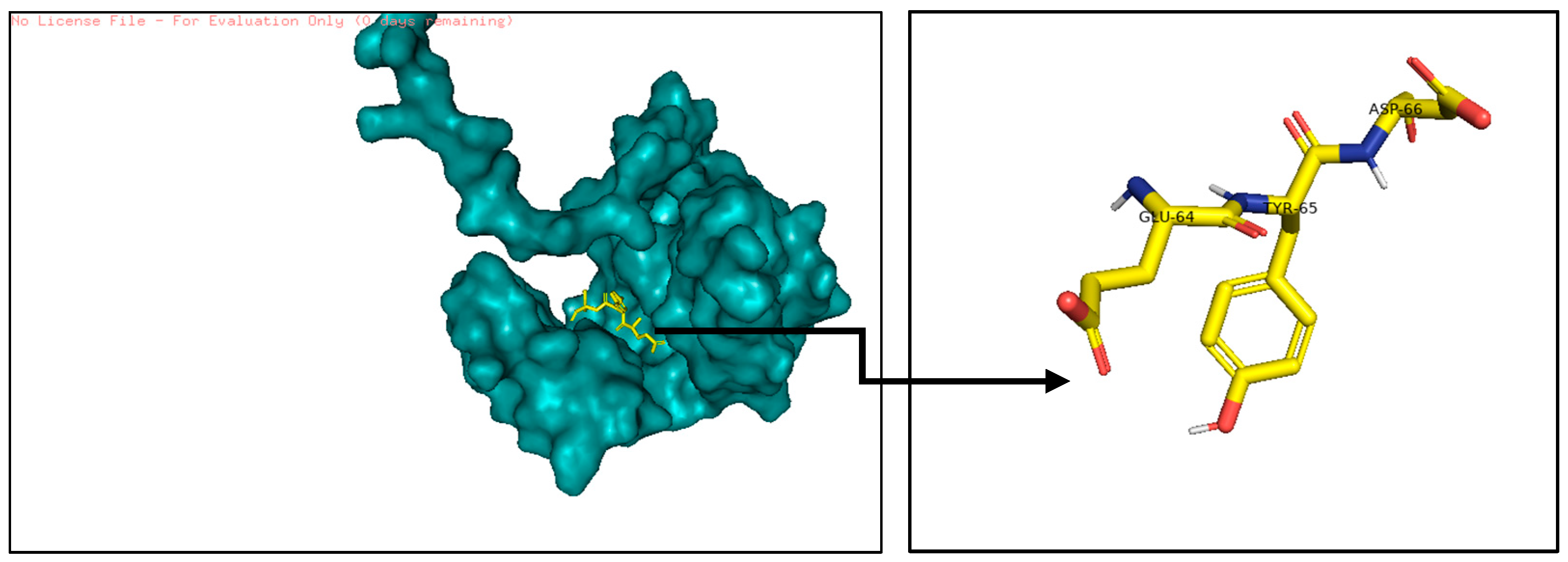
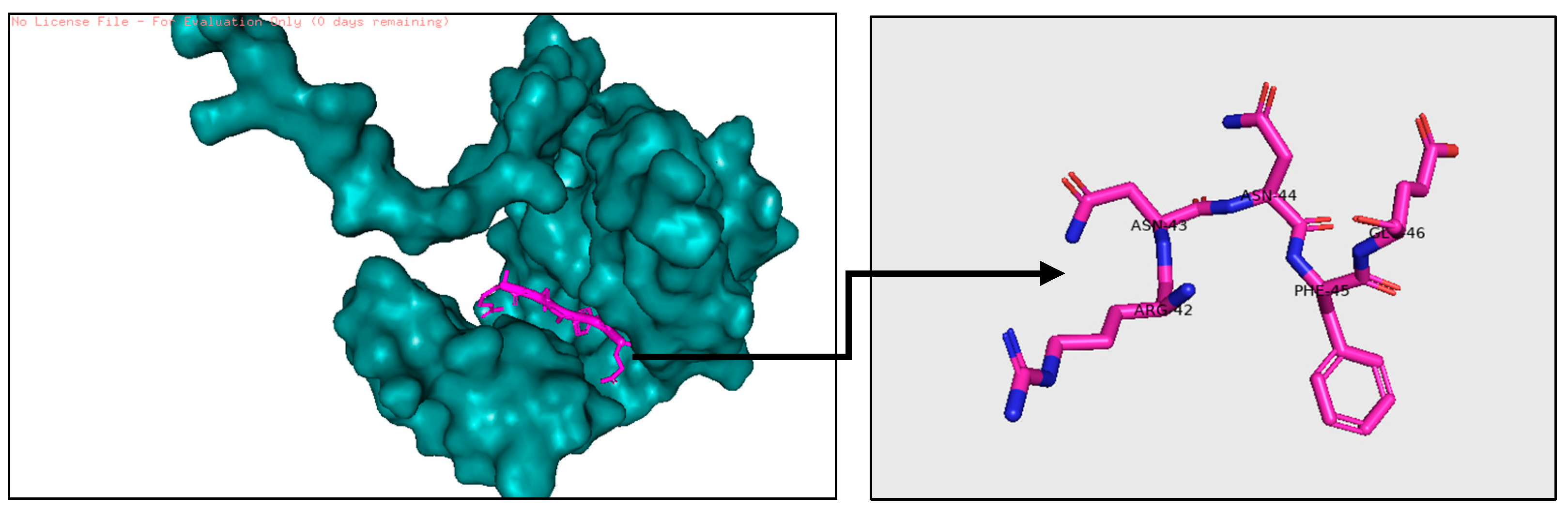
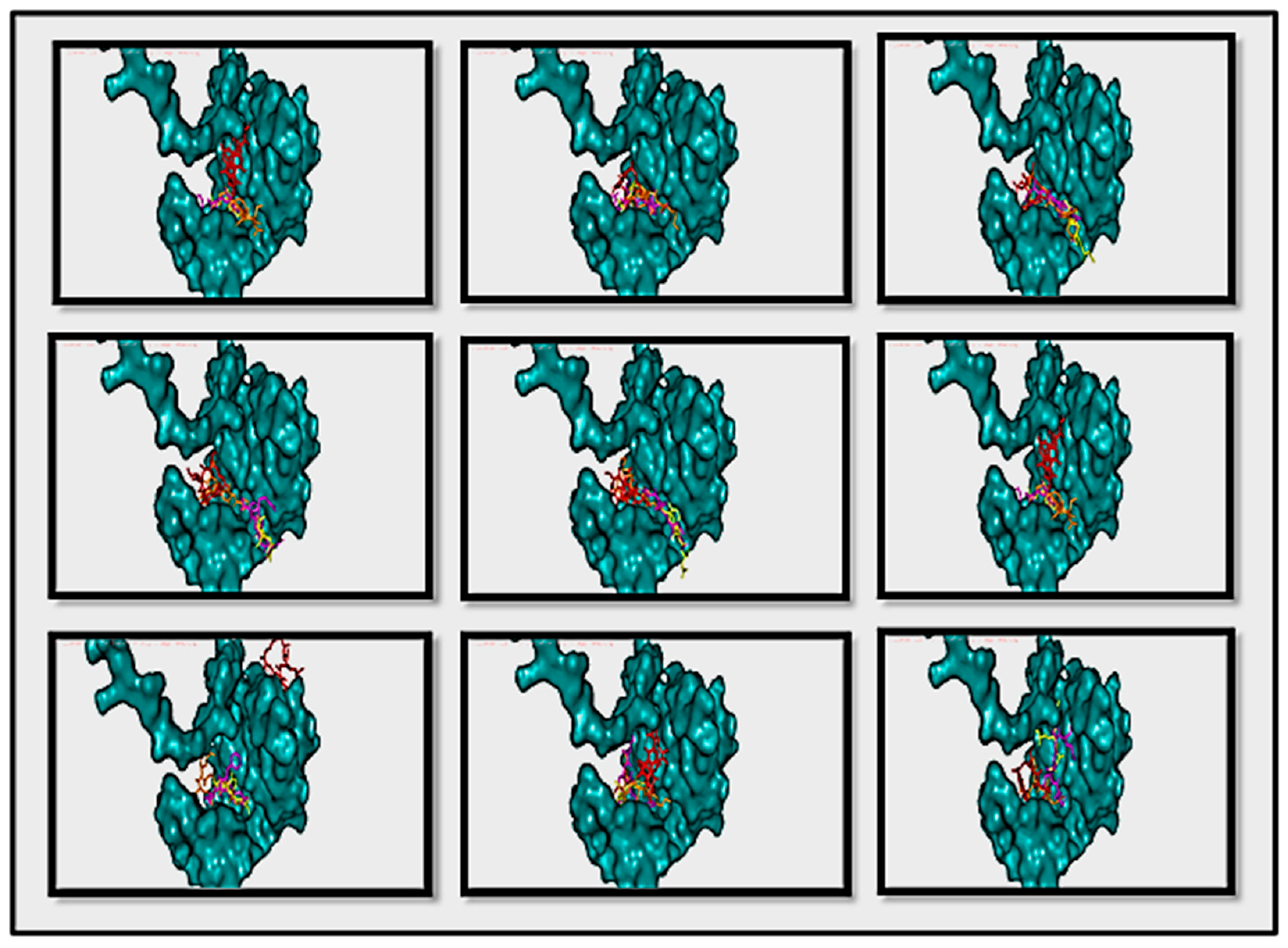
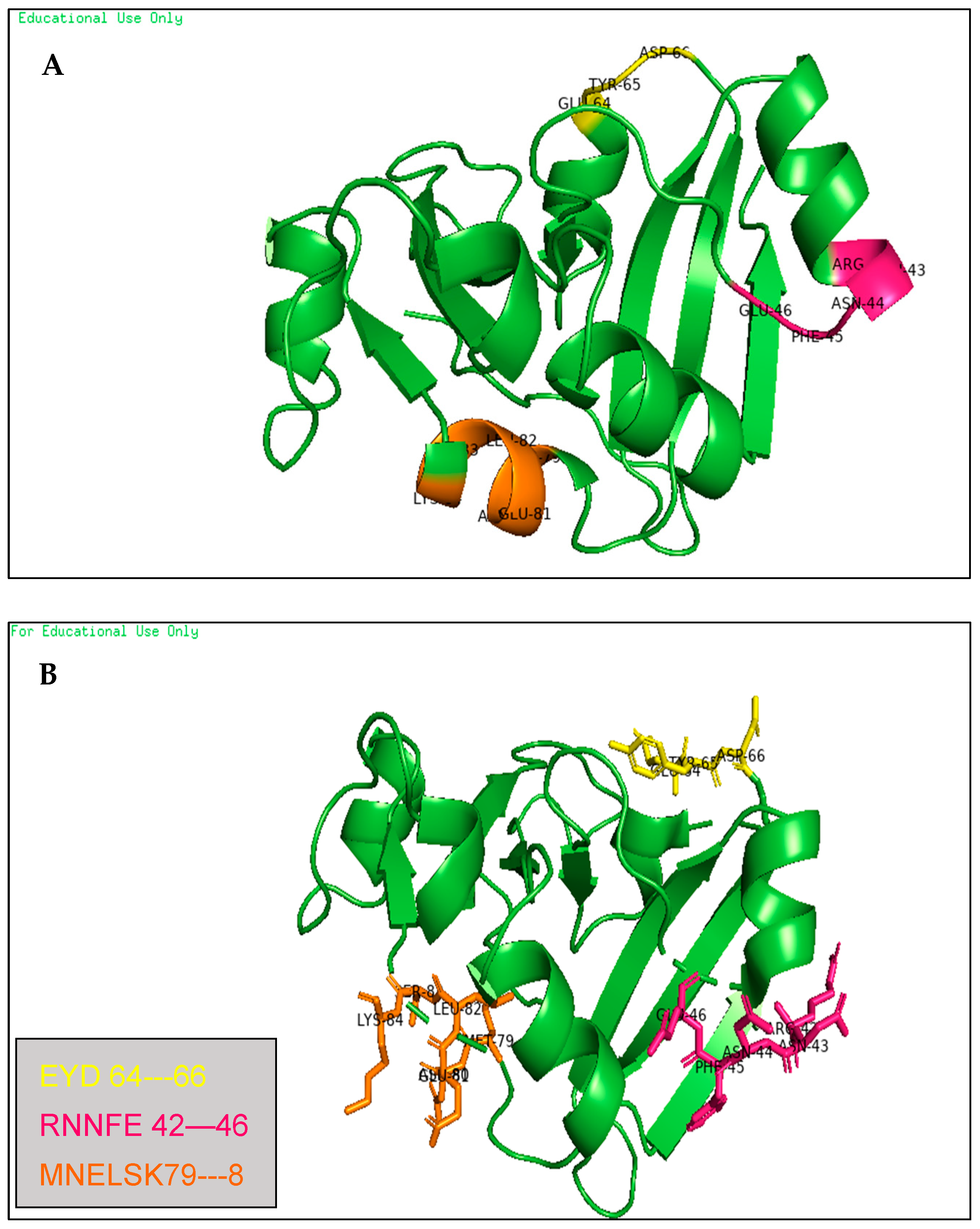
  • These binding sites were predicted by molecular docking methods, with at least three sites, as shown in Figure 13 in two ways of pyMOL displaying; carton with a colored parts which refer to the three predicted drugs (A) and, carton with a sticky colored parts which refer to the three predicted drugs (B).
(DRUG1 = GLU-64,TYR-65,ASP-66),
(DRUG2 = ARG-42,ASN-43,ASN-44,PHE-54,GLU-46),
(DRUG3 = MET-79,ASN-80,GLU-81,LEU-82,SER-83,LYS-84).

5. Future Work

  • Finish calculation for 18S rRNA: Due to the lack of confirmed studies on the selectivity of the RhsP2 complex with human RNA proteins, it is necessary to re-conduct the experimental studies, which requires repeating the docking processes with other human protein targets, comparing the new binding scores with the previous RNA bacterial target ones, and determining the effectiveness and toxicity of the studied toxin.
  • After first docking with RNA as a rigid receptor target, it has become increasingly clear that side-chain flexibility plays a crucial role in ligand–protein complexes. These changes allow the receptor to alter its binding site according to the orientation of the ligand; therefore, it is necessary to re-conduct flexible docking using Monte Carlo or molecular dynamics docking methods.

Funding

This research received no external funding.

Institutional Review Board Statement

Not applicable.

Informed Consent Statement

Not applicable.

Data Availability Statement

The data presented in this study are openly available at ref. [1].

Conflicts of Interest

The authors declare no conflict of interest.

Abbreviations

RhsP2Toxin secreted by Pseudomonas aeruginosa organisms belonging to the group of cytoplasmic enzymes affecting RNA through the mechanism of ADP-ribosyltransferase (from the group of ARTs)
ARTsADP-RibosylTransferases
ADPrAdenosine DiPhosphate ribosyl
ncRNAsNon-coding RNAs
H2T6SSType Six Secretion System Encodes Loci Named H2
AMRAntimicrobial Resistance
RNAPRNA Polymerase
PPIProtein–Protein Interaction
RMSDRoot Mean Square Deviation of Particle Coordinates
16S rRNA16 Subunit Ribosomal Ribonucleic Acid

References

  1. New Data Shows 148 Severe Antibiotic-Resistant Infections a Day in 2021. Available online: https://www.gov.uk/government/news/new-data-shows-148-severe-antibiotic-resistant-infections-a-day-in-2021 (accessed on 21 November 2022).
  2. ESPAUR Report 2022—The Latest Findings on Antimicrobial Resistance; UK Health Security Agency: London, UK, 21 November 2022. Available online: https://ukhsa.blog.gov.uk/2022/11/21/espaur-report-2022/ (accessed on 21 November 2022).
  3. Silver, L.L. Challenges of Antibacterial Discovery. Clin. Microbiol. Rev. 2011, 24, 71–109. [Google Scholar] [CrossRef] [PubMed]
  4. Ma, C.; Yang, X.; Lewis, P.J. Bacterial transcription as a target for antibacterial drug development. Microbiol. Mol. Biol. Rev. 2016, 80, 139–160. [Google Scholar] [CrossRef] [PubMed]
  5. Wright, G.D. Q&A: Antibiotic resistance: Where does it come from and what can we do about it? BMC Biol. 2010, 8, 1–6. [Google Scholar] [CrossRef] [PubMed]
  6. Sahoo, R.N.; Pattanaik, S.; Pattnaik, G.; Mallick, S.; Mohapatra, R. Review on the use of Molecular Docking as the First Line Tool in Drug Discovery and Development. Indian J. Pharm. Sci. 2022, 84, 1334–1337. [Google Scholar] [CrossRef]
  7. Chen, G.; Seukep, A.J.; Guo, M. Recent advances in molecular docking for the research and discovery of potential marine drugs. Mar. Drugs 2020, 18, 545. [Google Scholar] [CrossRef] [PubMed]
  8. Jones, C.; Hachani, A.; Manoli, E.; Filloux, A. An rhs gene linked to the second type VI secretion cluster is a feature of the Pseudomonas aeruginosa strain PA14. J. Bacteriol. 2014, 196, 800–810. [Google Scholar] [CrossRef] [PubMed]
  9. Zoued, A.; Brunet, Y.R.; Durand, E.; Aschtgen, M.S.; Logger, L.; Douzi, B.; Journet, L.; Cambillau, C.; Cascales, E. Architecture and assembly of the Type VI secretion system. Biochim. Et Biophys. Acta Mol. Cell Res. 2014, 1843, 1664–1673. [Google Scholar] [CrossRef] [PubMed]
  10. Bullen, N.P.; Prehna, G.; Whitney, J.C. Crystal Structure of the RhsP2 C-Terminal Toxin Domain in Complex with Its Immunity Protein, RhsI2; Canadian Institutes of Health Research (CIHR); Natural Sciences and Engineering Research Council (NSERC): Ottawa, ON, Canada, 2022. [Google Scholar] [CrossRef]
  11. Popova, K.B.; Otcheva, L.A.; Traykovska, M.; Penchovsky, R. RNA as a potent target for antibacterial drug discovery. Biomed. J. Sci. Tech. Res. 2018, 10, 7752–7754. [Google Scholar] [CrossRef]
  12. Bullen, N.P.; Sychantha, D.; Thang, S.S.; Culviner, P.H.; Rudzite, M.; Ahmad, S.; Shah, V.S.; Filloux, A.; Prehna, G.; Whitney, J.C. An ADP-ribosyltransferase toxin kills bacterial cells by modifying structured non-coding RNAs. Mol. Cell 2022, 82, 3484–3498.e11. [Google Scholar] [CrossRef] [PubMed]
Figure 1. Antibiotic targets and mechanism of resistance (adopted from [5]).
Msf 35 00003 g001
Figure 2. An overview of the T6SS. (A) Electron cryo-tomograph of Vibrio cholerae cells producing the T6SS. The arrow points to the long cytoplasmic tubular structure corresponding to the T6SS sheath. A magnification of the upper part of the tomograph is shown in panel (B), emphasizing the presence of distinct complexes (green, tail sheath; purple, putative base plate; yellow, putative membrane complex). (C) A schematic representation of the T6SS based on the electron cryo-tomographs and on genetic, biochemical, microscopy, and structural data (IM, inner membrane; PG, peptidoglycan layer; OM, outer membrane). Scale bar is 50 nm (adopted from [9]).
Msf 35 00003 g002
Figure 3. An overview of the methodology.
Msf 35 00003 g003
Figure 4. 16S rRNA ((A) surface mod; (B) carton mod, displayed by pyMOL). Note that RNAs are upstream in the translation pathway (protein synthesis). Inhibiting 1 RNA (ribosome) could prevent ~1000 proteins. It functions as a target for the RhsP2toxin (which binds to 16S rRNA molecules instead of mRNA with hydrogen bonds).
Msf 35 00003 g004
Figure 5. RhsP2 with its immunity protein RhsI2, downloaded from RCSD PDB in pdb format. Displayed in pyMOL. Green = RhsP2; orange = RhsI2.
Msf 35 00003 g005
Figure 6. RhsP2-chain A downloaded from RCSB PDB, displayed by pyMOL.
Msf 35 00003 g006
Figure 7. Docking Rifamycin against the target protein 16S rRNA to fix the active pocket, displayed by pyMOL.
Msf 35 00003 g007
Figure 8. DRUG1 (yellow) interacts with 16S rRNA (blue) in its selective active pocket.
Msf 35 00003 g008
Figure 9. DRUG2 (magenta) interacts with 16S rRNA (blue) in its selective active pocket.
Msf 35 00003 g009
Figure 10. DRUG3 (orange) interacts with 16S rRNA (blue) in its selective active pocket.
Msf 35 00003 g010
Figure 11. Average score of RMSD and Affinity Binding for drugs and Rifamycin in all modes.
Msf 35 00003 g011
Figure 12. Nine binding modes for Rifamycin, DRUG1, DRUG2, and DRUG3 by AutoDock VINA with the target protein 16S rRNA, displayed by pyMOL tool.
Msf 35 00003 g012
Figure 13. RhsP2 toxin with the suggested active binding sites as drugs 1, 2, and 3 (displayed by pyMOL). (A) carton with a colored parts which refer to the three predicted drugs; (B) carton with a sticky colored parts which refer to the three predicted drugs.
Msf 35 00003 g013
Table 1. Affinity Binding and RMSD criterion of three DRUGS (DRUG1: yellow, DRUG2: magenta, DRUG3: orange) compared to Rifamycin (red).
RifamycinDRUG1DRUG2DRUG3
ModeAffinity (kcal/mol)rmsd l.brmsd u.bMode(Kcal/mol) Affinityrmsd l.brmsd u.bMode(Kcal/mol) Affinityrmsd l.brmsd u.bMode(Kcal/mol) Affinityrmsd l.brmsd u.b
1−7.6001−6.4001−6.6001−600
2−7.42.8018.552−5.813.49815.8782−63.3336.8512−5.73.94510.73
3−7.413.28816.7553−5.79.98414.5663−5.93.7787.6353−5.43.94910.854
4−7.32.3897.8564−5.41.622.6364−5.93.95711.0924−5.34.8358.465
5−7.35.498.715−5.42.4467.4185−5.77.36711.7315−5.24.447.674
6−6.92.7357.8546−5.38.99513.8756−5.75.31411.9746−5.14.3677.817
7−6.83.1846.6227−5.312.98916.887−5.77.45511.2387−5.15.3979.726
8−6.811.2216.2258−5.213.79117.2858−5.76.5911.0538−54.51111.925
9−6.827.56730.5349−5.17.27510.8469−5.63.6668.229−55.5039.228
Disclaimer/Publisher’s Note: The statements, opinions and data contained in all publications are solely those of the individual author(s) and contributor(s) and not of MDPI and/or the editor(s). MDPI and/or the editor(s) disclaim responsibility for any injury to people or property resulting from any ideas, methods, instructions or products referred to in the content.

