Abstract
This study aims to apply a kinematic analysis to characterize and compare a normal canine gait with a canine gait with cranial cruciate ligament (CrRL) rupture with free and open-source software. Two dogs walked ten times. A bidimensional kinematic analysis was performed. Spatiotemporal analysis showed significant differences between dogs. The dog with CrCL rupture obtained higher results for all parameters except stance and step time. Also, the stifle angle did not verify differences in absolute angle, but the signal showed differences in patterns between normal and abnormal gait. This study supports that software assisting clinicians’ diagnosis with CrCl ruptures.
1. Introduction
CrCL rupture is one of the most common causes of dog lameness [1,2]. The etiopathogenesis of CrCL is not fully known, but regardless of the cause, it results in stifle joint instability and progressive degenerative joint disease [3]. Furthermore, this type of condition causes lameness, an increase in cranial tibial translation, and internal rotation and abduction of the tibia [4,5]. Gait analysis has evolved [6], it is well-established [7] and it is a way to perform clinical diagnosis for CrCl rupture [8]. Subjective analysis has allowed the identification of gait patterns, but the results, and thus the applications, are limited and different agreements between observers can be obtained [6,8,9]. Alternatively, objective gait analysis has emerged, allowing clinicians accurately study the canine gait cycle [6]. Several methods and instruments can be applied in gait analysis, such as kinetic and kinematic methods, and they are associated with instruments, such as force platforms and motion analysis systems, respectively [7,9]. The kinematic gait analysis is one the most applied, and it helps effectively in quantitative evaluation, measuring segments and joints and identifying gait asymmetry [8]. However, the current methods are usually expensive and require extensive data processing, so this study aims at an exploratory study which consists of applying a kinematic analysis to characterize and compare a normal canine gait with a canine gait with CrCL with free and open-source software. As a result, we could share with veterinaries a simple and useful tool for their daily work to follow and analyze their recovery of the canine gait.
2. Materials and Methods
2.1. Experimental Design Overview
The experiment conducted in the present study consisted of a comparison between a normal and an abnormal canine gait. Each dog performed ten trials with a natural speed in a developed outdoor space of two meters near the Egas Moniz Veterinary University Clinic. We used one high-speed camera and analyzed one step to determine the spatiotemporal parameters and the stifle angle using the motion analysis system Kinovea®.
2.2. Subjects
The study subjects were two dogs, one healthy Labrador Retriever and one Bouvier Bernouis with CrCL rupture, selected by convenience. The dogs’ owners were informed of the objective and authorized the data collection. The CrCL rupture diagnosis was assessed by a cranial “drawer” and tibial compression test and conducted by an experienced veterinary orthopedic surgeon. The diagnosis was confirmed by Lateral radiography of the femur-tibio-patellar joint taken doing the tibial compression test and without tibial compression.
2.3. Measurement Protocol and Data Analysis
A veterinarian analyzed the subjects and placed three landmarks defining the stifle angle (Figure 1). The dogs performed a walk of three minutes before the analysis. Dogs started walking toward the outdoor calibrated zone of two meters with a natural but uncontrol speed, and each dog walked ten times. One high-speed camera (Casio Exilim® ZR200, Casio, Tokyo, Japan) collecting at 120 Hz was used during the bidimensional kinematic analysis. It was placed perpendicular to the right of the dog’s line. Data processing for the high-speed was conducted using the Kinovea® software (version 0.9.5) to define the stance phase time, swing phase time, step time, step length and step speed, and tracking the ankle, stifle and grand trochanter landmarks defining the stifle angle. Step frequency () and step speed () were calculated. Here, represents the step frequency; is the step time; is the step speed and SL is the step length. Their results were organized using Microsoft Excel Office, and kinematic data processing was performed using the Spyder package in Python 3.7. Data from the high-speed camera were smoothed using the spectral power analysis [10], and a cut-off frequency of 9 Hz was applied.
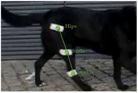
Figure 1.
Segment model used to define the stifle angle.
2.4. Statistical Analysis
All walks (n = 20) performed by dogs were analyzed. Descriptive statistics were determined to describe the spatiotemporal data and stifle angle, with means and standard deviation values. Normality was calculated using the Shapiro–Wilk test (p ≤ 0.05). The independent samples t-test was used to compare the proposed parameters, and when the normality was not verified, the Mann–Whitney test was applied [8].
3. Results
Table 1 provides the results of the descriptive and comparison statistical analysis for the spatiotemporal data and stifle angle parameters.

Table 1.
Spatiotemporal data and stifle angle parameters descriptive and comparative analysis.
Figure 2 represents the stifle angular displacement in sagittal plane to a canine gait with CrCL (Figure 2a) and a normal canine gait (Figure 2b).
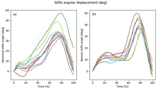
Figure 2.
(a) Signal pattern to a canine gait with CrCL; (b) Signal pattern to normal canine gait; Each color line means each walk.
4. Discussion
In this study, we performed a bidimensional kinematic analysis to compare spatiotemporal parameters and the stifle angle between the normal and CrCL dog. When we analyzed Table 1, as expected, CrCL influenced canine gait by changing some parameters. All results were higher in normal canines except for the stance and step phase time following the literature [11] (p. 837). Specifically regarding the temporal parameters, our results were higher than Jenkins et al. (stride, stance, and swing phases were 0.575, 0.355 and 0.221 s, respectively) [12] (p. 81). However, they did not report the step speed, which is a factor that can change the temporal step parameters. Additionally, we verified significant differences in the stance phase time, stance and swing phase in percentage and step length, frequency, and speed (see Table 1). These results were divergent with Møller et al., which did not verify differences in the spatiotemporal parameters analyzed. However, they used a lower sample frequency (50 Hz) and a non-motion analysis system (Media Player Classic Home Cinema for Windows v.10.0) [8] (p. 66). Regarding the stifle angle (see Table 1), the amplitude did not verify differences between gaits and obtain angular displacement lower than Agostinho et al. (stifle angle: 52.48 ± 4.04 and 62.37 ± 6.53 deg), but they used a higher speed of 2.1 and 2.2 m/s, while we obtained a step speed around 1 m/s [9]. Lastly, regarding the stifle angle signal (Figure 2), when compared with the literature, we verified a similar but inverse signal [9,11], and signal differences between dogs, i.e., the gait to CrCL dog started flexion early (around 20% time gait phase). Our results followed the literature and showed that our proposal can be a useful diagnostic tool for veterinarians in their daily routine.
5. Conclusions
This study allowed us to characterize the normal and CrCL rupture canine gait with free and open-source software obtaining detailed information relevant to clinicians. Furthermore, this information should be confirmed with a higher sample and be suggested to veterinarians for inclusion in the follow-up after surgery to ensure that the dog restored all stifle joint range of motion.
Author Contributions
Conceptualization, P.M.-O. and A.M.L.; methodology, P.M.-O.; software, P.M.-O.; validation, P.M.-O. and A.M.L.; formal analysis, P.M.-O. and A.M.L.; investigation, P.M.-O.; resources, P.M.-O. and A.M.L.; data curation, P.M.-O.; writing—original draft preparation, P.M.-O. and A.M.L.; writing—review and editing, P.M.-O. and A.M.L.; visualization, P.M.-O. and A.M.L.; supervision, P.M.-O. and A.M.L.; project administration, P.M.-O. and A.M.L. All authors have read and agreed to the published version of the manuscript.
Funding
This research received no external funding.
Institutional Review Board Statement
Not applicable.
Informed Consent Statement
Not applicable.
Data Availability Statement
Not applicable.
Conflicts of Interest
The authors declare no conflict of interest.
References
- Kowaleski, M.; Boudrieau, R.; Pozzi, A. Stifle Joint. In Veterinary Surgery: Small Animal Expert Consult; Elsevier: St. Louis, MO, USA, 2018; pp. 1071–1167. ISBN 978-0-323-32065-8. [Google Scholar]
- Barnhart, M.; Bufkin, B.; Litsky, A. Biomechanical Comparison of Four Methods of Fixation of a Polymeric Cranial Cruciate Ligament in the Canine Femur and Tibia. Vet. Comp. Orthop. Traumatol. 2019, 32, 112–116. [Google Scholar] [CrossRef] [PubMed]
- Niebauer, G.W.; Restucci, B. Etiopathogenesis of Canine Cruciate Ligament Disease: A Scoping Review. Animals 2023, 13, 187. [Google Scholar] [CrossRef] [PubMed]
- Tinga, S.; Kim, S.E.; Banks, S.A.; Jones, S.C.; Park, B.H.; Pozzi, A.; Lewis, D.D. Femorotibial Kinematics in Dogs with Cranial Cruciate Ligament Insufficiency: A Three-Dimensional in-Vivo Fluoroscopic Analysis during Walking. BMC Vet. Res. 2018, 14, 85. [Google Scholar] [CrossRef] [PubMed]
- Tashman, S.; Anderst, W.; Kolowich, P.; Havstad, S.; Arnoczky, S. Kinematics of the ACL-Deficient Canine Knee during Gait: Serial Changes over Two Years. J. Orthop. Res. 2004, 22, 931–941. [Google Scholar] [CrossRef] [PubMed]
- Gillette, R.L.; Angle, T.C. Recent Developments in Canine Locomotor Analysis: A Review. Vet. J. 2008, 178, 165–176. [Google Scholar] [CrossRef] [PubMed]
- Pálya, Z.; Rácz, K.; Nagymáté, G.; Kiss, R.M. Development of a Detailed Canine Gait Analysis Method for Evaluating Har-nesses: A Pilot Study. PLoS ONE 2022, 17, e0264299. [Google Scholar] [CrossRef] [PubMed]
- Møller, J.H.; Vitger, A.D.; Poulsen, H.H.; Miles, J.E. Simple Video-Based Spatiotemporal Gait Analysis Is Not Better than Subjective Visual Assessment of Lameness in Dogs. VCOT Open 2021, 4, e65–e71. [Google Scholar] [CrossRef]
- Agostinho, F.S.; Rahal, S.C.; Miqueleto, N.S.M.L.; Verdugo, M.R.; Inamassu, L.R.; El-Warrak, A.O. Kinematic Analysis of Labrador Retrievers and Rottweilers Trotting on a Treadmill. Vet. Comp. Orthop. Traumatol. 2011, 24, 185–191. [Google Scholar] [CrossRef] [PubMed]
- Winter, D.A. Biomechanics and Motor Control of Human Movement, 3rd ed.; John Wiley & Sons: Hoboken, NJ, USA, 2005; ISBN 978-0-471-44989-8. [Google Scholar]
- DeCamp, C.E. Kinetic and Kinematic Gait Analysis and the Assessment of Lameness in the Dog. Vet. Clin. N. Am. Small Anim. Pract. 1997, 27, 825–840. [Google Scholar] [CrossRef] [PubMed]
- Jenkins, G.J.; Hakim, C.H.; Yang, N.N.; Yao, G.; Duan, D. Automatic Characterization of Stride Parameters in Canines with a Single Wearable Inertial Sensor. PLoS ONE 2018, 13, e0198893. [Google Scholar] [CrossRef] [PubMed]
Disclaimer/Publisher’s Note: The statements, opinions and data contained in all publications are solely those of the individual author(s) and contributor(s) and not of MDPI and/or the editor(s). MDPI and/or the editor(s) disclaim responsibility for any injury to people or property resulting from any ideas, methods, instructions or products referred to in the content. |
© 2023 by the authors. Licensee MDPI, Basel, Switzerland. This article is an open access article distributed under the terms and conditions of the Creative Commons Attribution (CC BY) license (https://creativecommons.org/licenses/by/4.0/).