Proceeding Paper

Effects of RF Currents on Cytokines Production in Human Keratinocytes †

by
María Luisa Hernández-Bule
1,*,
Elena Toledano-Macías
1,
María Antonia Martínez-Pascual
1,
Alejandro Úbeda
1 and
Montserrat Fernández-Guarino
2
1
Bioelectromagnetic Lab, Instituto Ramón y Cajal de Investigación Sanitaria (IRYCIS), Hospital Ramón y Cajal, Crta. Colmenar Viejo, km. 9.100, 28034 Madrid, Spain
2
Dermatology Service, Instituto Ramón y Cajal de Investigación Sanitaria (IRYCIS), Hospital Ramón y Cajal, Crta. Colmenar Viejo, km. 9.100, 28034 Madrid, Spain
*
Author to whom correspondence should be addressed.
Presented at the 2nd International Electronic Conference on Biomedicines, 1–31 March 2023; Available online: https://ecb2023.sciforum.net/.
Med. Sci. Forum 2023, 21(1), 29; https://doi.org/10.3390/ECB2023-14096
Published: 1 March 2023
(This article belongs to the Proceedings of The 2nd International Electronic Conference on Biomedicines)

Abstract

:
Wound healing consists of a sequence of coordinated phases: inflammation, proliferation, and remodeling. In skin lesions, neutrophils and keratinocytes are the main cell types participating in the inflammatory phase, during which the release of mediators intervening in the regulation of the subsequent regenerative phases takes place. These mediators are involved in tissue regeneration through induction of transendothelial migration, enzyme secretion, cell adhesion, and T-Cell activation and cytotoxicity, as well as neutrophil accumulation at the wound site. Among these mediators, the keratinocyte-synthesized chemokines RANTES, MCP-1, MIP-1, and IL-8 stand out. Although therapies applying electromagnetic fields or electric currents have been shown to have anti-inflammatory effects in a variety of experimental models and in patients through reduced production of proinflammatory cytokines such as IFN-Υ and increased production of IL-10, the knowledge on the biological basis of these effects is still limited. Previous studies by our group have shown that subthermal treatment with radiofrequency (RF) currents used in capacitive-resistive electric transfer (CRET) therapy promotes the proliferation and migration of various cell types, such as human ADSC (stem cells), fibroblasts or keratinocytes, involved in skin regeneration. This study investigates the effects of in vitro treatment with CRET currents on cytokine production by HaCat human keratinocytes. The results reveal that, compared to sham-exposed controls, RF stimulation induces decreased production of IL-8 and RANTES and increased MCP-1, without significantly affecting other chemokines such as MIP-1. Taken together, our results indicate that due to the RF effects on the production of chemokines involved in the modulation of the inflammatory phase of wound regeneration, CRET therapy could be effective in the treatment of skin wounds.

1. Introduction

Inflammation is an early critical phase of wound regeneration, and results from the interaction between cells of the immune system, endothelial cells, fibroblasts, and keratinocytes. The latter, whose proliferation and migration are crucial for the inflammatory process, synthesize cytokines involved in the regulation of inflammation in skin diseases, as well as chemotaxis-inducing cytokines. Keratinocytes also synthesize a set of proteins relevant to the inflammatory process, since they induce transendothelial migration and leukocyte accumulation. These are the monocyte inflammatory protein (MIP-1), the monocyte chemoattractant protein (MCP-1), and IL-8 [1]. In fact, since severe or prolonged inflammatory processes lead to defective and delayed tissue regeneration, it is foreseeable that inhibition of proinflammatory cytokine synthesis has relevant clinical applications.
Although various modalities of anti-inflammatory physical therapies for tissue regeneration have been assayed recently, the molecular mechanisms underlying the response of acute or chronic inflammatory processes to the applied physical stimuli are generally poorly studied and insufficiently characterized. For instance, it has been reported that in patients with Crohn’s disease, treatment with low-frequency pulsed fields can increase the production of anti-inflammatory IL-10 and decrease that of proinflammatory INF-Υ [2]. Also, subthermal treatment in mice with ultrahigh frequency signals has been shown to reduce inflammation and neutrophil production of reactive oxygen species (ROS) [3].
Another anti-inflammatory physical strategy [4], based on the non-invasive application of radiofrequency currents at 448 kHz, is the capacitive resistive electrical transfer (CRET). Although the biomechanisms involved in the potential anti-inflammatory CRET effects are not yet sufficiently investigated, previous studies by our group have revealed that intermittent, subthermal CRET stimulation promotes the proliferation of various types of human cells, including stem cells [5], keratinocytes, and fibroblasts, as well as migration of human fibroblasts [6]. The objective of the present work is to investigate whether subthermal CRET stimulation is also capable of affecting human keratinocyte cytokine expression.

2. Methods

2.1. Cell Culture

Human epidermal keratinocyte cell line HaCaT was purchased from CLS Cell Lines Service (300493, Heidelberg, Germany). Cells were maintained in high glucose D-MEM (Biowhittaker, Lonza, Verviers, Belgium) supplemented with inactivated fetal bovine serum, 10% (Gibco, MA, USA), 1% glutamine, and 1% penicillin-streptomycin (Gibco). The cells were subcultured once a week and in each experiment, they were seeded on 60 mm Petri dishes (Nunc, Roskilde, Denmark). Depending on the objective to be analyzed, a total of 8 or 10 Petri dishes were used per experimental repetition.

2.2. Electric Treatment

The procedure for RF exposure has been described in detail elsewhere [5,7]. Briefly, pairs of sterile stainless steel electrodes designed ad hoc for in vitro stimulation were inserted in all Petri dishes and connected in series 4 days after seeding. The intermittent stimulation pattern consisted of 5-min pulses of 448 kHz, sine wave current delivered at subthermal densities of 100 µA/mm2, separated by 4-h interpulse lapses, and administered for a total of 48 h. A signal generator (Indiba Activ HCR 902, INDIBA®, Barcelona, Spain) energized the electrodes of dishes for electrical stimulation, while the remaining plates were sham-exposed simultaneously inside an identical, separate CO2 incubator.

2.3. Cell Proliferation Assay

To study cell proliferation, the XTT assay (Roche, Switzerland) was used. Cell proliferation assay was used according to the manufacturer’s protocol. The cells were seeded at a density of 5500 cells/cm2 and incubated for 3 days. Cells were treated for 48 h with CRET or sham-exposed treatment and subsequently incubated for 3 h with XTT tetrazolium salt in an atmosphere of 37 °C and 6.5% CO2. Metabolically active cells reduced XTT to colored formazan compounds that were quantified with a microplate reader (TECAN, Männedorf, Switzerland) at a wavelength of 492 nm.

2.4. ELISA Assay

IL-8/CXCL8, CCL2/MCP-1, CCL3/MIP-1 alpha, and CCL5/RANTES human cytokines were analyzed using the ELISA technique (R&D Systems, Abingdon, UK). HaCaT cells (6800 cells/cm2) were seeded and maintained in high glucose D-MEM medium supplemented with 10% inactivated fetal bovine serum (Gibco), 1% glutamine, and 1% penicillin-streptomycin (Gibco) at 37 °C during 4 days. Next, cultures were exposed to CRET for 48 h, while the remaining plates were sham-exposed for the same time. At the end of each experimental replicate, cells were mechanically detached, centrifuged, and lysed in a lysis standard buffer. Protein concentration was determined using a Pierce BCA protein assay (Thermo Fisher Scientific, Inc. Rockford, IL, USA). The ELISA technique was carried out following the manufacturer’s recommended protocol.

2.5. Statistical Analysis

All procedures and analyses were conducted in blind conditions for treatment. At least three independent replicates were conducted per experiment, cell type, or treatment. Two-tailed unpaired Student’s t-test was applied using GraphPad Prism 6.01 software (GraphPad Software, San Diego, CA, USA). Differences p < 0.05 were considered significant statistically.

3. Results and Discussion

3.1. Cell Proliferation

The results of the XTT assay showed that compared to controls, a 48-h intermittent treatment at a subthermal current density of 100 μA/mm2 significantly increase keratinocyte proliferation (10% over controls; p < 0.05) (Figure 1), which confirms and reinforces previously reported CRET effects on keratinocyte proliferation [6]. Added to the existing body of data on CRET promotion of adipose-derived human stem cells (ADSC) and human dermal fibroblast proliferation [5,6], these results confirm a block of evidence sustaining the potential applications of CRET in tissue regeneration.

3.2. Cytokine Expression

Electromagnetic fields of various frequency ranges have been reported to induce significant changes in the modulation of cytokine expression in keratinocytes of the HaCaT line [8,9]. The present results show that also subthermal treatment with RF CRET signal affects cytokine expression in HaCaT cells. Namely, 48-h electrical stimulation induced a statistically significant overexpression (18.56% over controls) of MCP-1 in the culture medium, whereas cytokines IL-8 and RANTES were underexpressed (11.11% and 13.56% over controls, respectively), although the differences with respect to controls did not reach levels of statistical significance (Table 1). No detectable levels of MIP-1 expression were found in the HaCaT cultures, either treated or controls.
Cytokine MCP-1 is synthesized in large amounts by resident cells (endothelial cells and keratinocytes located at the wound edge) as well as by macrophages involved in inflammatory processes. During wound regeneration, MCP-1 also intervenes in different phases of the process attracting cells such as monocytes and lymphocytes, capable of synthesizing cell-growth regulatory and promoting factors. This cytokine is also involved in angiogenesis, by promoting endothelial cell migration, and in wound regeneration, by favoring mast cell migration. Mast cells, in turn, synthesize high levels of IL-4 which stimulates fibroblast proliferation and has anti-inflammatory effects [1]. Thus, the increase in MCP-1 production in CRET-treated HaCaT could decrease inflammation through the promotion of mast cell migration and its mediation for IL-4 expression. Therefore, the overexpression of the anti-inflammatory cytokine MCP-1, together with the aforementioned effects of electrostimulation on the migration and proliferation of fibroblasts and keratinocytes, reinforces the evidence of the potential action of CRET in wound healing promotion.
IL-8 and RANTES are proinflammatory chemokines that act as neutrophil chemotactic factors at the inflammation site. IL-8 regulates the synthesis of adhesion molecules, amplifies local inflammation, and stimulates angiogenesis [1], while RANTES promotes T-cell proliferation or apoptosis, and the release of various proinflammatory cytokines [10]. The herein-reported underexpression of these two cytokines is consistent with a potential local anti-inflammatory effect induced by CRET electrical stimulation. These results are also consistent with previous observations of HaCaT proliferation promotion and inhibition of the synthesis of proinflammatory cytokines such as IL-8 and RANTES, after a 48-h exposure to low-frequency electromagnetic fields [11].

4. Conclusions

Together with previously reported data on the cellular response to CRET subthermal stimulation, the effects on keratinocyte proliferation and cytokine expression obtained in this preliminary study reinforce the existing evidence on the potential anti-inflammatory and skin regenerative action of CRET electrical stimulation. Additional research on the response of skin cells to CRET is necessary in order to identify and characterize potential applications of this therapy for anti-inflammatory treatments.

Author Contributions

Conceptualization, M.L.H.-B. and M.A.M.-P.; methodology, M.L.H.-B. and M.A.M.-P.; software, E.T.-M.; validation, M.L.H.-B.; formal analysis, M.L.H.-B.; investigation, M.L.H.-B., E.T.-M., M.F.-G., M.A.M.-P. and A.Ú.; resources, M.L.H.-B. and M.A.M.-P.; data curation, M.L.H.-B. and E.T.-M.; writing—original draft preparation, M.L.H.-B.; writing—review and editing, M.L.H.-B., E.T.-M., M.F.-G., M.A.M.-P. and A.Ú.; visualization, M.L.H.-B., E.T.-M., M.F.-G., M.A.M.-P. and A.Ú.; supervision, M.L.H.-B.; project administration, M.L.H.-B. and A.Ú.; funding acquisition, A.Ú. and M.L.H.-B. All authors have read and agreed to the published version of the manuscript.

Funding

This work was financially supported by Fundación para la Investigación Biomédica del Hospital Ramón y Cajal, through Project FiBio-HRC No. 2015/0050.

Institutional Review Board Statement

Not applicable.

Informed Consent Statement

Not applicable.

Data Availability Statement

The data presented in this study are available on request from the corresponding author. The data are not publicly available due to privacy restrictions.

Acknowledgments

We thank Srta. Silvia Sacristán for her technical assistance during the experiments.

Conflicts of Interest

The authors declare no conflict of interest.

References

  1. Gillitzer, R.; Goebeler, M. Chemokines in Cutaneous Wound Healing. J. Leukoc. Biol. 2001, 69, 513–521. [Google Scholar] [CrossRef] [PubMed]
  2. Kaszuba-Zwoiñska, J.; Cie, I.; Mach, T. Magnetic Field Anti-Inflammatory Effects in Crohn’s Disease Depends Upon Viability and Cytokine Profile of the Immune Competent Cells. J. Physiol. Pharmacol. 2008, 59, 177–187. [Google Scholar] [PubMed]
  3. Lushnikov, K.V.; Shumilina, Y.V.; Yakushina, V.S.; Gapeev, A.B.; Sadov, V.B. Effects of Low-Intensity Ultrahigh Frequency Electromagnetic Radiation on inflammatory processes. Bull. Exp. Biol. Med. 2004, 137, 412–415. [Google Scholar] [CrossRef] [PubMed]
  4. Carralero-Martínez, A.; Muñoz Pérez, M.A.; Kauffmann, S.; Blanco-Ratto, L.; Ramírez-García, I. Efficacy of Capacitive Resistive Monopolar Radiofrequency in the Physiotherapeutic Treatment of Chronic Pelvic Pain Syndrome: A Randomized Controlled Trial. Neurourol. Urodyn. 2022, 41, 962–972. [Google Scholar] [CrossRef] [PubMed]
  5. Hernández-Bule, M.L.; Paíno, C.L.; Trillo, M.Á.; Úbeda, A. Electric Stimulation at 448 KHz Promotes Proliferation of Human Mesenchymal Stem Cells. Cell. Physiol. Biochem. 2014, 34, 1741–1755. [Google Scholar] [CrossRef] [PubMed]
  6. Hernández-Bule, M.L.; Toledano-Macías, E.; Naranjo, A.; de Andrés-Zamora, M.; Úbeda, A. In Vitro Stimulation with Radiofrequency Currents Promotes Proliferation and Migration in Human Keratinocytes and Fibroblasts. Electromagn. Biol. Med. 2021, 40, 338–352. [Google Scholar] [CrossRef] [PubMed]
  7. Hernández-Bule, M.L.; Trillo, M.A.; Cid, M.A.; Leal, J.; Ubeda, A. In Vitro Exposure to 0.57-MHz Electric Currents Exerts Cytostatic Effects in HepG2 Human Hepatocarcinoma Cells. Int. J. Oncol. 2007, 30, 583–592. [Google Scholar] [CrossRef] [PubMed]
  8. Szabo, I.; Rojavin, M.A.; Rogers, T.J.; Ziskin, M.C. Reactions of Keratinocytes to in Vitro Millimeter Wave Exposure. Bioelectromagnetics 2001, 22, 358–364. [Google Scholar] [CrossRef] [PubMed]
  9. Patruno, A.; Ferrone, A.; Costantini, E.; Franceschelli, S.; Pesce, M.; Speranza, L.; Amerio, P.; D’Angelo, C.; Felaco, M.; Grilli, A.; et al. Extremely Low-Frequency Electromagnetic Fields Accelerates Wound Healing Modulating MMP-9 and Inflammatory Cytokines. Cell Prolif. 2018, 51, e12432. [Google Scholar] [CrossRef] [PubMed]
  10. Appay, V.; Rowland-Jones, S.L. RANTES: A Versatile and Controversial Chemokine. Trends Immunol. 2001, 22, 83–87. [Google Scholar] [CrossRef] [PubMed]
  11. Vianale, G.; Reale, M.; Amerio, P.; Stefanachi, M.; Di Luzio, S.; Muraro, R. Extremely Low Frequency Electromagnetic Field Enhances Human Keratinocyte Cell Growth and Decreases Proinflammatory Chemokine Production. Br. J. Dermatol. 2008, 158, 1189–1196. [Google Scholar] [CrossRef] [PubMed]
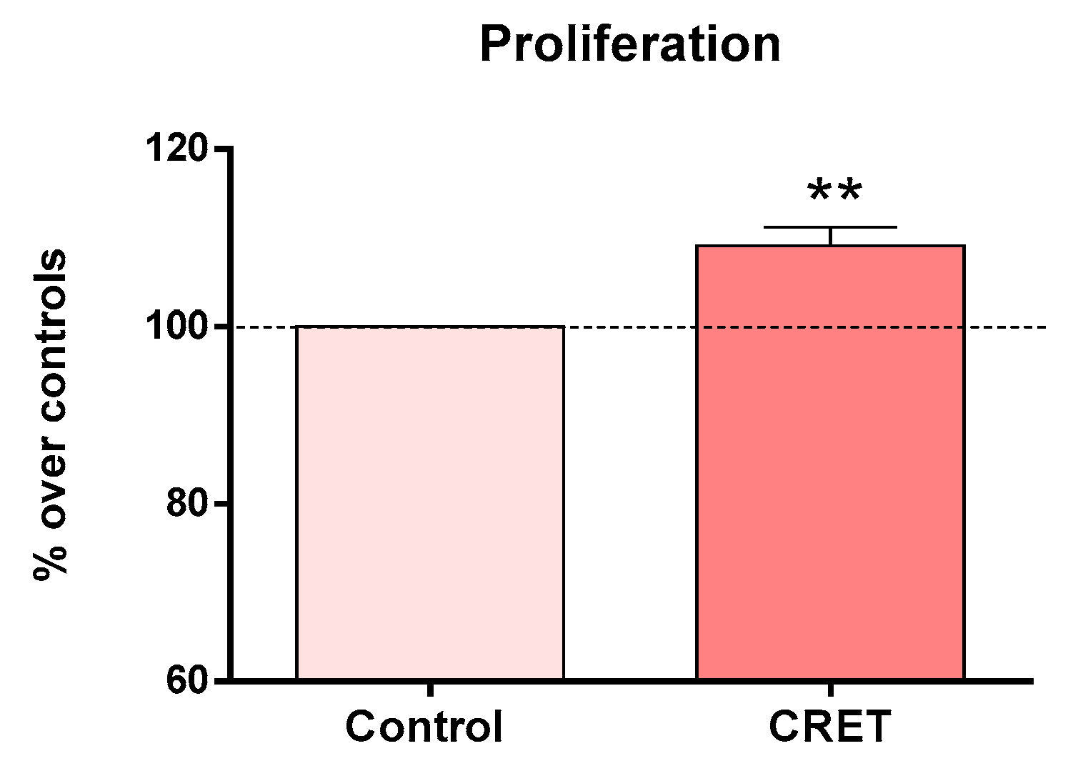
Figure 1. Cell proliferation after 48 h of intermittent CRET exposure at 100 µA/mm2. Data are means ± SEM normalized over the corresponding sham-exposed controls (dash line 100%). Six experimental replicates. **: 0.001 ≤ p < 0.01. Student’s t-test.
Msf 21 00029 g001
Table 1. ELISA assay for MCP-1, IL-8 and RANTES expression after 48 h of intermittent CRET treatment at 100 µA/mm2 current density. Percent over sham-exposed controls. Means ± SEM of at least 3 experimental replicates per cytokine. Student’s t-test.
MCP-1IL-8RANTES
CRET118.564 ± 3.483 *88.884 ± 14.55586.439 ± 12.395
Control100.0 ± 0.00003100.0 ± 0.00012100.0 ± 0.0001
* p = 0.033.
Disclaimer/Publisher’s Note: The statements, opinions and data contained in all publications are solely those of the individual author(s) and contributor(s) and not of MDPI and/or the editor(s). MDPI and/or the editor(s) disclaim responsibility for any injury to people or property resulting from any ideas, methods, instructions or products referred to in the content.