Next Article in Journal
Occupational Hazards Associated with the Quality and Training Needs of Public Health Inspectors in Greece
Previous Article in Journal
Introduction to the ESPAUR Webinar and Report 2021–2022: Key Findings and Stakeholder Engagement
 
 
msf-logo
Article Menu

Article Menu

Proceeding Paper

Standard Operating Procedure for the Analysis of Microplastics in Larval Fish Diets †

1
Department of Management, Economics and Industrial Engineering, Politecnico di Milano, Via Lambruschini 4b, 20156 Milan, Italy
2
Metabolism of Cities Living Lab, San Diego State University, 5500 Campanile Dr, San Diego, CA 98182, USA
Presented at the International One Health Conference 2022, Catania, Italy, 27–28 September 2022.
Med. Sci. Forum 2023, 19(1), 1; https://doi.org/10.3390/msf2023019001
Published: 9 May 2023
(This article belongs to the Proceedings of International One Health Conference)

Abstract

:
Microplastics (MPs) pollution has increasingly exposed the pelagic biota to physical harm. The small size of micro-particles makes them more suitable for passive ingestion by a wide variety of organisms with serious effects on growth rates, respiration and vital functions, bioaccumulation of pollutants, and, ultimately, species survival. Nevertheless, our knowledge of plastic intake in nursery habitats is still very limited. When encounters with MPs occur at the larval stage, it is suggested that fish can develop altered feeding behaviors with cascading effects on the entire food web. This study provides a step-by-step protocol to identify and enumerate polymer particles found in fish diets. The procedure is intended for the analysis of larval and juvenile fish populations with a developed digestive tract. It includes guiding questions for research design, a list of supplies and reagents to extract and mount the fish diets on microscope slides for semi-permanent conservation, the protocol for microscopic and statistical analysis, and the interpretation of the results. We suggest that the gut content could be used to assess (i) the bioavailability of polymers in water systems, (ii) the incidence of an encounter between larval fish and MPs, and (iii) the possible alternations in fish’ feeding behaviors as soon as they leave their parental stage.

1. Introduction

Plastics have been documented in every marine and freshwater environment. Microplastics (MPs) generate from the breakdown of larger plastic debris into infinitesimal particles that can be ingested by a wide variety of organisms [1]. Increasing attention has also been drawn to MPs that originate from land and make their way into the water as microbeads for abrasive uses, employed in cosmetics or dental hygiene products, and filaments from synthetic clothing [2,3].
Plastic pollution jeopardizes the health and subsistence of the pelagic biota resulting in physical harm, such as entanglement and ingestion or chemical contamination [1].
Encounters between organisms and MPs account for a greater risk of biological incidents. In fact, due to their small size, MPs can promptly enter the food web bearing negative consequences across all trophic levels [4,5,6,7]. The most documented effects of MPs ingestion include reduced feeding up to starvation due to saturation of the digestive tract [7], decreased energy reserves [8], hepatic stress, and increased liver toxicity caused by bioaccumulation and translocation of pollutants via polymer particles [9,10].
While passive ingestion of MPs by marine organisms is receiving increasing attention [11,12,13,14,15,16,17], there is limited information on plastic intake in freshwater systems [18,19,20,21], especially when assessing the feeding behaviors of larval and early-life-stage fish. Previous studies indicate that fish larvae can feed on plastic in their nursery habitats with detrimental consequences for their survival, growth, and reproductive rates. It has been suggested that the bioavailability of polymer particles could alter their ecological functions (e.g., metabolism) as soon as they leave their parental stage of yolk sacs to develop feeding behaviors [22,23].
The least understood are the mechanisms of translocation of polymers throughout the food web. The occurrence of unanticipated changes in the natural feeding environment could substantiate the incidence of cascade effects across all levels of the food chain, with increased concerns for species survival when such effects fall below the threshold level [24]. It has been demonstrated that MPs can accumulate in prey fields where micro-preys are readily available and abundant, thereby impacting larvae’s food supply [23]. When encounters between fish larvae and microdebris occur, altering feeding behaviors, it is suggested that plastic could make its way to the top of the chain transferring from one trophic level to the next via food supply (e.g., the discovery of MPs in higher level trophic organisms) [25,26].
The central aim of this paper is to produce a reproducible, representative, and accurate Standard Operating Procedure (SOP) for MP analysis. The procedure explained hereinafter is intended for the analysis of larval and juvenile fish populations with a developed digestive tract. It includes guiding questions for research design, a list of supplies and reagents to extract and mount the fish diets on the microscope slides for semi-permanent conservation, a protocol for microscopic analysis, and an interpretation of the results. The remained of the paper is structured as follows: Section 2 presents the step-by-step protocol for MP analysis. Following, Section 3 presents an application of the SOP. Finally, conclusions and final remarks are presented in Section 4.

2. Methodology

2.1. Experimental Design

Prior to the MP analysis, it is imperative to define the study’s research questions, the methodology to be employed, and related impacts on the results, including limitations, possible uncertainty, and assumptions, in line with the principle of experimental design [27]. In general, an MP’s assessment consists of three broad steps: sample pool selection, microscopic analysis, and statistical analysis and interpretation of results. Table 1 identifies a list of questions to guide the design of the analysis in its three sections.

2.2. Preparation of the Samples and Equipment

One of the processes that the CR aimed to improve was related to the delivery of thousands of histopathology reports. A project about machine learning was a solution implemented in one of the DMAIC cycles performed by the CR. Histopathology reports are primary data. After collection, when samples are returned to the lab, they can be conserved in vials filled with ethanol solution. Vial labels should indicate vial ID, sample species, sample stage, sampling site, sampling depth, sampling date, and storing solution.
For this analysis, the following supplies and reagents have been used:
-
Dissecting microscope with 80× magnification
-
Microscope slides, 25 mm × 75 mm
-
Cover slips, 22 mm × 22 mm
-
Microscope slide box
-
Vials, 20 mL and 3.7 mL
-
Vial box
-
Fine tipped probes
-
Minuten pins
-
Tweezers
-
Eyedropper
-
Ethanol 99%, 100 mL
-
Permount™ Mounting Media, 100 mL
-
Glycerin-Alcohol 50:50, 100 mL

2.3. Larvae Fish Dissection

(i) For microscopic examination of individual samples, we place one slide at a time under the dissecting microscope and, using an eyedropper, pour a small amount (2 drops recommended) of the Glycerin–Alcohol solution. (ii) One sample was then gently removed from the selected vial using tweezers and placed on the slide following the direction of the slide. It is recommended to handle the sample from its tail to avoid unintended damage to the digestive tract. (iii) An additional small amount (2 drops recommended) of the Glycerin–Alcohol solution was added to facilitate dissecting operations. (iv) To prepare the sample for diet removal, we focused the microscope at 10× and adjusted the lenses and lights as needed to have a clear view of the entire sample. (v) Using fine-tipped probes, we gently incised the sample to separate the digestive tract from the rest of the body, avoiding tearing the stomach apart during the process and adding Glycerin–Alcohol if needed as the sample dried out. (vi) When the digestive tract had been entirely excised, the body freed of mouth, esophagus, stomach, and intestine was returned to vials filled with an ethanol solution for conservation.

2.4. Diets Removal and Slide Preparation

(i) To remove the fish diets, we focused the microscope at 40× and adjusted the lenses and lights as needed. (ii) Following, we added an additional small amount (2 drops recommended) of the Glycerin–Alcohol solution to facilitate diet removal operations. (iii) Using minuten pins, we carefully opened the stomach and peeled off the content. (iv) For slide preparation, we gently covered the guts and their content with a slipcover making sure that it was evenly distanced from the slide corners and all air bubbles were removed. (v) Using an eyedropper, a small amount of Permount™ was poured on the cover slip, sealing one side at a time, making sure that there was no air infiltration when sealing one side. (vi) Mounted slides should then be stored in a safe space, and 24–48 h (depending on the drying conditions of the lab) are required prior to handling. Each slide should carry a label indicating slide ID, sample species, sample stage, sampling site, sampling depth, and sampling date as indicated on the relative vial.

2.5. Counting Particles

For MPs analysis, the mounted slides were placed under the dissecting microscope and focused at 80× magnification, adjusting lenses and lights as needed. Microscopes with greater magnification capacity can also be used for further analysis. MPs particles were categorized by shape and color. Based on their visual features, we classified MPs as beads, fibers, fragments, films, and foams using the methods described by [3] (Table 2). The dominant color was identified using the methods described by [16], using a 5- color scale (blue, black, red, orange, and transparent). Subsequently, the software Image-Pro Premier 9.3 was used to record the size of identified particles (e.g., length of fibers and area of beads, fragments, films, and foam). After the analysis, mounted slides were conserved in the microscope slide box.

3. SOP Application

3.1. Scope and Objectives

Using the protocol presented above, three sample pools were analyzed to assess the incidence of encounters between MPs and fish larvae. For this study, three freshwater environments with different characteristics were chosen. Sampling site 1 (S1) represents a connecting channel heavily used for tourism purposes and commercial and recreational fishing. The second site (S2) is a distributor channel connecting smaller lakes and wetlands and serves ecological restoration and recreation purposes. Finally, the third site (S3) represents an urban river subjected to anthropogenic influences from adjacent urban areas that account for the discharge of municipal wastewaters and sewage, runoff of chemical and hazardous materials from industrial activities, fishing, and intensive shipping operations. The choice of these sites serves the purpose of exploring possible effects of the external environment and sampling conditions (e.g., location and site) on the bioavailability of MPs in waterways.

3.2. Sample Selection

For this analysis, we sampled 218 larval fish native to the study area. For S1, we sampled 20 rainbow smelts (Osmerus mordax) and 28 burbots (Lota lota); for S2, we sampled 30 yellow perches (Perca flavescens), 30 rainbow smelts (Osmerus mordax), and 28 burbots (Lota lota); finally, for S3, we sampled 20 cyprinidae, 30 rainbow smelts (Osmerus mordax), and 33 yellow perches (Perca flavescens).

3.3. MPs Analysis

MPs were found at all sampling sites, confirming the results of similar studies [28,29,30,31,32]. In our analysis, 117 fish had ingested MPs, of which 24 fish (41.4%) from S1, 47 fish (53.4%) from S2, and 46 fish (55.4%) from S3. A detailed description of our findings is reported in Table 3.
Like previous studies [28], we found that fiber was the most prevalent particle type, with an average length of 691.1 nm to 2106.9 nm. Second, we found fragments, followed by foams and beads, with an average area of 2092.0 nm2 to 52,942.9 nm2. The most dominant colors for identified particles were blue, black, and red; while orange and transparent were found in at least one species for each sampling pool. A possible explanation for this pattern is provided by [16], that observed that fish are more likely to selectively ingest blue particles due to association with micro-preys (e.g., blue-pigmented copepods) and avoid orange particles). Concerning species-specific patterns, we observed that yellow perches reported the highest ingestion rates for S2 and S3, confirming the trends observed by [33,34]. No substantial differences were observed among the three sampling pools, and plastic pollution was ubiquitous at all sites. Therefore, a detailed analysis of possible sources and pathways of MP pollution could yield more insightful results on how specific features of the feeding environment affect larval fish’s feeding behaviors.

4. Conclusions

The SOP presented in this study provides a reproducible, step-by-step methodology to assess MPs accumulation in larval fish by analyzing the types, size, and color of micro debris found in the fish digestive tract. The findings of this study establish a foundation for further research related to feeding behaviors and the long-term impacts of plastic debris. In particular, future studies could compare the results of this analysis with MPs accumulations in water samples to understand if the likelihood of MPs ingestion is statistically correlated to selective changes in feeding behaviors or depends on the concentration of particles in the fish’s feeding environment (higher particle density). Some limitations of this approach can be associated with the depth and accuracy of the microscopic analysis. For example, this MPs analysis can be reinforced using spectroscopic analyses (e.g., Fourier transform infrared (FTIR) and Raman spectroscopy) [28,35] or thermal analyses [36], which provide a further level of verification by detecting the polymer composition of microparticles.

Funding

This research received no external funding.

Institutional Review Board Statement

Not applicable.

Informed Consent Statement

Not applicable.

Data Availability Statement

Data available upon request.

Conflicts of Interest

The authors declare no conflict of interest.

References

  1. Cózar, A.; Echevarría, F.; González-Gordillo, J.I.; Irigoien, X.; Úbeda, B.; Hernández-León, S.; Palma, A.T.; Navarro, S.; García-de-Lomas, J.; Ruiz, A.; et al. Plastic debris in the open ocean. Proc. Natl. Acad. Sci. USA 2014, 111, 10239–10244. [Google Scholar] [CrossRef]
  2. Fendall, L.S.; Sewell, M.A. Contributing to marine pollution by washing your face: Microplastics in facial cleansers. Mar. Pollut. Bull. 2009, 58, 1225–1228. [Google Scholar] [CrossRef]
  3. Free, C.M.; Jensen, O.P.; Mason, S.A.; Eriksen, M.; Williamson, N.J.; Boldgiv, B. High-levels of microplastic pollution in a large, remote, mountain lake. Mar. Pollut. Bull. 2014, 85, 156–163. [Google Scholar] [CrossRef] [PubMed]
  4. Betts, K. Why Small Plastic Particles May Pose a Big Problem in the Oceans. Environ. Sci. Technol. 2008, 42, 8995. [Google Scholar] [CrossRef]
  5. Boerger, C.M.; Lattin, G.L.; Moore, S.L.; Moore, C.J. Plastic ingestion by planktivorous fishes in the North Pacific Central Gyre. Mar. Pollut. Bull. 2010, 60, 2275–2278. [Google Scholar] [CrossRef]
  6. Davison, P.; Asch, R.G. Plastic ingestion by mesopelagic fishes in the North Pacific Subtropical Gyre. Mar. Ecol. Prog. Ser. 2011, 432, 173–180. [Google Scholar] [CrossRef]
  7. Wright, S.L.; Thompson, R.C.; Galloway, T.S. The physical impacts of microplastics on marine organisms: A review. Environ. Pollut. 2013, 178, 483–492. [Google Scholar] [CrossRef] [PubMed]
  8. Wright, S.L.; Rowe, D.; Thompson, R.C.; Galloway, T.S. Microplastic ingestion decreases energy reserves in marine worms. Curr. Biol. 2013, 23, R1031–R1033. [Google Scholar] [CrossRef]
  9. Browne, M.A.; Niven, S.J.; Galloway, T.S.; Rowland, S.J.; Thompson, R.C. Microplastic moves pollutants and additives to worms, reducing functions linked to health and biodiversity. Curr. Biol. 2013, 23, 2388–2392. [Google Scholar] [CrossRef]
  10. Rochman, C.M.; Hoh, E.; Kurobe, T.; Teh, S.J. Ingested plastic transfers hazardous chemicals to fish and induces hepatic stress. Sci. Rep. 2013, 3, 3263. [Google Scholar] [CrossRef]
  11. Hämer, J.; Gutow, L.; Köhler, A.; Saborowski, R. Fate of microplastics in the marine isopod Idotea emarginata. Environ. Sci. Technol. 2014, 48, 13451–13458. [Google Scholar] [CrossRef]
  12. Mazurais, D.; Ernande, B.; Quazuguel, P.; Severe, A.; Huelvan, C.; Madec, L.; Mouchel, O.; Soudant, P.; Robbens, J.; Huvet, A.; et al. Evaluation of the impact of polyethylene microbeads ingestion in European sea bass (Dicentrarchus labrax) larvae. Mar. Environ. Res. 2015, 112, 78–85. [Google Scholar] [CrossRef]
  13. Bellas, J.; Martínez-Armental, J.; Martínez-Cámara, A.; Besada, V.; Martínez-Gómez, C. Ingestion of microplastics by demersal fish from the Spanish Atlantic and Mediterranean coasts. Mar. Pollut. Bull. 2016, 109, 55–60. [Google Scholar] [CrossRef]
  14. Rummel, C.D.; Löder, M.G.; Fricke, N.F.; Lang, T.; Griebeler, E.M.; Janke, M.; Gerdts, G. Plastic ingestion by pelagic and demersal fish from the North Sea and Baltic Sea. Mar. Pollut. Bull. 2016, 102, 134–141. [Google Scholar] [CrossRef] [PubMed]
  15. Tanaka, K.; Takada, H. Microplastic fragments and microbeads in digestive tracts of planktivorous fish from urban coastal waters. Sci. Rep. 2016, 6, 34351. [Google Scholar] [CrossRef] [PubMed]
  16. Ory, N.C.; Sobral, P.; Ferreira, J.L.; Thiel, M. Amberstripe scad Decapterus muroadsi (Carangidae) fish ingest blue microplastics resembling their copepod prey along the coast of Rapa Nui (Easter Island) in the South Pacific subtropical gyre. Sci. Total Environ. 2017, 586, 430–437. [Google Scholar] [CrossRef] [PubMed]
  17. Zhou, Q.; Zhang, H.; Fu, C.; Zhou, Y.; Dai, Z.; Li, Y.; Chen, T.; Luo, Y. The distribution and morphology of microplastics in coastal soils adjacent to the Bohai Sea and the Yellow Sea. Geoderma 2018, 322, 201–208. [Google Scholar] [CrossRef]
  18. Biginagwa, F.J.; Mayoma, B.S.; Shashoua, Y.; Syberg, K.; Khan, F.R. First evidence of microplastics in the African Great Lakes: Recovery from Lake Victoria Nile perch and Nile tilapia. J. Great Lakes Res. 2016, 42, 146–149. [Google Scholar] [CrossRef]
  19. Silva-Cavalcanti, J.S.; Silva, J.D.B.; de França, E.J.; de Araújo, M.C.B.; Gusmão, F. Microplastics ingestion by a common tropical freshwater fishing resource. Environ. Pollut. 2017, 221, 218–226. [Google Scholar] [CrossRef]
  20. Jabeen, K.; Su, L.; Li, J.; Yang, D.; Tong, C.; Mu, J.; Shi, H. Microplastics and mesoplastics in fish from coastal and fresh waters of China. Environ. Pollut. 2017, 221, 141–149. [Google Scholar] [CrossRef]
  21. DeSantiago, R. Foraging Strategy May Predict Anthropogenic Debris Consumption in Wetland Fishes; NCUR: Edmond, OK, USA, 2018. [Google Scholar]
  22. Lönnstedt, O.M.; Eklöv, P. Environmentally relevant concentrations of microplastic particles influence larval fish ecology. Science 2016, 352, 1213–1216. [Google Scholar] [CrossRef] [PubMed]
  23. Steer, M.; Cole, M.; Thompson, R.C.; Lindeque, P.K. Microplastic ingestion in fish larvae in the western English Channel. Environ. Pollut. 2017, 226, 250–259. [Google Scholar] [CrossRef] [PubMed]
  24. Au, S.Y.; Lee, C.M.; Weinstein, J.E.; van den Hurk, P.; Klaine, S.J. Trophic transfer of microplastics in aquatic ecosystems: Identifying critical research needs. Integr. Environ. Assess. Manag. 2017, 13, 505–509. [Google Scholar] [CrossRef] [PubMed]
  25. Fry, D.M.; Fefer, S.I.; Sileo, L. Ingestion of plastic debris by Laysan albatrosses and wedge-tailed shearwaters in the Hawaiian Islands. Mar. Pollut. Bull. 1987, 18, 339–343. [Google Scholar] [CrossRef]
  26. Eriksson, C.; Burton, H. Origins and biological accumulation of small plastic particles in fur seals from Macquarie Island. AMBIO A J. Hum. Environ. 2003, 32, 380–385. [Google Scholar] [CrossRef]
  27. EPA (Environmental Protection Agency). Microplastics Expert Workshop Report. Trash Free Waters Dialogue Meeting. EPA Office of Wetlands, Oceans and Watersheds. Available online: https://www.epa.gov/sites/default/files/2018-03/documents/microplastics_expert_workshop_report_final_12-4-17.pdf (accessed on 8 August 2022).
  28. Baldwin, A.K.; Corsi, S.R.; Mason, S.A. Plastic debris in 29 Great Lakes tributaries: Relations to watershed attributes and hydrology. Environ. Sci. Technol. 2016, 50, 10377–10385. [Google Scholar] [CrossRef]
  29. Driedger, A.G.; Dürr, H.H.; Mitchell, K.; Van Cappellen, P. Plastic debris in the Laurentian Great Lakes: A review. J. Great Lakes Res. 2015, 41, 9–19. [Google Scholar] [CrossRef]
  30. Eriksen, M.; Mason, S.; Wilson, S.; Box, C.; Zellers, A.; Edwards, W.; Farley, A.; Amato, S. Microplastic pollution in the surface waters of the Laurentian Great Lakes. Mar. Pollut. Bull. 2013, 77, 177–182. [Google Scholar] [CrossRef]
  31. Zbyszewski, M.; Corcoran, P.L. Distribution and degradation of fresh water plastic particles along the beaches of Lake Huron, Canada. Water Air Soil Pollut. 2011, 220, 365–372. [Google Scholar] [CrossRef]
  32. Zbyszewski, M.; Corcoran, P.L.; Hockin, A. Comparison of the distribution and degradation of plastic debris along shorelines of the Great Lakes, North America. J. Great Lakes Res. 2014, 40, 288–299. [Google Scholar] [CrossRef]
  33. Munno, K.; Helm, P.A.; Rochman, C.; George, T.; Jackson, D.A. Microplastic contamination in Great Lakes fish. Conserv. Biol. 2022, 36, e13794. [Google Scholar] [CrossRef]
  34. Lu, X.; Deng, D.F.; Huang, F.; Casu, F.; Kraco, E.; Newton, R.J.; Mendoza, L.M.R. Chronic exposure to high-density polyethylene microplastic through feeding alters the nutrient metabolism of juvenile yellow perch (Perca flavescens). Anim. Nutr. 2022, 9, 143–158. [Google Scholar] [CrossRef] [PubMed]
  35. Wesch, C.; Barthel, A.K.; Braun, U.; Klein, R.; Paulus, M. No microplastics in benthic eelpout (Zoarces viviparus): An urgent need for spectroscopic analyses in microplastic detection. Environ. Res. 2016, 148, 36–38. [Google Scholar] [CrossRef] [PubMed]
  36. Shabaka, S.H.; Ghobashy, M.; Marey, R.S. Identification of marine microplastics in Eastern Harbor, Mediterranean Coast of Egypt, using differential scanning calorimetry. Mar. Pollut. Bull. 2019, 142, 494–503. [Google Scholar] [CrossRef] [PubMed]
Table 1. Guiding questions for sample selection and analysis (adapted from [27]).
1. Sample Pools Selection
How many samples are needed?
Which sample type is relevant?
Should samples be collected within or across species? If selection occurs across species, what does this mean for interpretation of the results?
Do sample’s natural habitats differ? How does this affect interpretation of the results?
What are the external factors/conditions to be considered when choosing samples?
What are the limitations of the sample pool(s) used?
2. Microscopic Analysis
Which polymer types are accounted for?
Which finding types (e.g., number of particles, particle color/size/weight, or particle visual coverage) are accounted for?
How are MPs sorted from other organic content in the fish diets? ● What are the limitations of the methods used?
3. Statistical Analysis and Interpretation of Results
Which polymer types are accounted for?
Which statistical tests are needed?
Which graph/chart types are used to explain findings?
Which types of findings are significant to the study?
Which statistical errors are accounted for?
What are the limitations of the analyses used
Table 2. Abacus of polymer particles by physical characteristics (adapted from [3]).
ParticleCharacteristicsImages
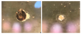
BeadHard, RoundedMsf 19 00001 i001
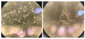
FiberThin, fibrous, linearMsf 19 00001 i002
FragmentHard, jaggedMsf 19 00001 i003
FilmThin, flimsyMsf 19 00001 i004
FoamLightweight, sponge
like
Msf 19 00001 i005
Table 3. Summary of MPs analysis for the three sampling sites.
Site SpeciesSamples
(#)
Ingestion
Rate (%)
Particle
Count (#)
Particle
Types
Particle Size (Mean)Particle Color
Length
(Fiber)
Area
(Bead,
Fragment,
Foam)
S1Rainbow
smelt
2045%10Bead (1),
Fragment (1)
fiber (8)
912.6 nm22,855.4
nm2
Orange,
black, blue, red
Burbot2853.6%29Fragment (7), fiber (22)1424.0
nm
15,685.6
nm2
Red,
black,
blue, transparent
S2Yellow
perch
3080%58Fragment (3),
foam (3),
fiber (52)
1208.3
nm
52,942.9
nm2
Red,
orange,
black,
blue,
transparent
Rainbow
smelt
3056.7%34Fiber
(34)
1610.2-Red, blue,
black, orange
Burbot2817.9%6Fragment (2),
fiber (4)
2106.9
nm
5576.4
nm2
Red,
black, blue
S3Cyprinidae2025%5Fiber (5)691.1 nm-Black, blue, red
Rainbow
smelt
3060%35Fragment
(2), fiber
(33)
1367.0
nm
4100.2
nm2
Red, blue,
black,
orange
Yellow
perch
3372.7%52Fragment (1),
fiber (51)
1405.5
nm
2092.0
nm2
Blue,
black, red,
transparent
Disclaimer/Publisher’s Note: The statements, opinions and data contained in all publications are solely those of the individual author(s) and contributor(s) and not of MDPI and/or the editor(s). MDPI and/or the editor(s) disclaim responsibility for any injury to people or property resulting from any ideas, methods, instructions or products referred to in the content.