Proceeding Paper

The Promising Shadow of Nanohybrid Liposomal Cerasomes towards the Treatment of Diabetes Mellitus †

1
Department of Pharmaceutical Sciences, Dibrugarh University, Assam 786004, India
2
Department of Pharmaceutics, College of Pharmacy, Najran University, Narjan 11001, Saudi Arabia
*
Author to whom correspondence should be addressed.
Presented at the 2nd International Electronic Conference on Healthcare, 17 February–3 March 2022. Available online: https://iech2022.sciforum.net/.
Med. Sci. Forum 2022, 10(1), 5; https://doi.org/10.3390/IECH2022-12295
Published: 17 February 2022
(This article belongs to the Proceedings of The 2nd International Electronic Conference on Healthcare)

Abstract

:
Nanocarriers are used globally in diverse fields to encapsulate drug toovercome various problems associated with different drug delivery systems such asstability, solubility, improvement in functional activities, homogeneity, and protection from physical and chemical hazard destructions. In the field of biomedicine, this approachis considered to be of particular importance as it can easily overcome the issues related to biocompatibility, bioavailability, toxicity profile, and therapeutic side effects. Lipid-based delivery systems have evolved as a newer approach in drug delivery. Advancement, development, and design modification of the lipid-based drug delivery systems have given rise to hybrid structures containing both inorganic and organic parts called cerasomes. Cerasomes are a modified form of liposomes that can overcome the limitations related to lipid nanoparticles. However, these cerasomes are also effective in treating diabetes mellitus, which is a metabolic disorder that has affecteda vast population worldwide for the last few decades. The development of cerasomes for the treatment of diabetes has provided a new window for researchers to overcome the different problems connected to the current therapies. This article provides a thorough assessment of the hybrid liposomal cerasomes literature and how it can be effectively employed in the treatment of diabetes.

1. Introduction

Diabetes mellitus (DM) is a chronic metabolic disorder characterized by hyperglycemia or high blood glucose levels with abnormal metabolism of carbohydrates, fats, and proteins due to an absolute or relative lack of insulin secretion or insulin resistance by peripheral tissuessuch as adipose tissue, liver, and skeletal muscle [1]. It is also associated with hyperaminoacidemia and hyperlipidemia. Long-term suffering from diabetes mellitus may result in the development of the critical complications of retinopathy with potential blindness; nephropathy, whichmay lead to renal failure; and neuropathy, which may cause autonomic dysfunction, including sexual dysfunction. People suffering from diabetes mellitus are also at risk of cardiovascular, vascular, peripheral, and cerebrovascular diseases [2]. The global prevalence rate of diabetes mellitus is expected to increase up to 9.9% (522 million) by the year 2030 from 8.3% (366 million) in 2011, as estimated from recent studies and surveys. The highest increase will take place in regions in developing economies such as India and China. The United States of America also has many patients who are suffering from diabetes mellitus. It was estimated that about 4million more men than women suffered from diabetes mellitus (185 million men and 181 million women) in 2011 [3,4]. Traditional treatments for diabetes mellitus, such as insulin sensitization and secretion, have undesirable side effects, resulting in patient noncompliance and treatment discontinuation. Nanotechnology has facilitated the development of novel glucose monitoring and insulin delivery systems that can improve diabetics’ quality of life [5]. Other therapies, such as βcell regeneration and gene therapy, are presently employed to control diabetes in addition to insulin and oral hypoglycemic medications. Due to the limitations of pharmacological treatment and the superiority of NPs in drug administration and imaging, researchers are greatly interested in nanocarriers in treating and managing diabetes mellitus [6,7]. Liposomes, polymer-based nanoparticles, and inorganic nanoparticles are the most frequently used nanoparticle-based drug delivery technologies in diabetes control. A liposome is a flexible supramolecular assembly with applications in biophysics, chemistry, colloid science, biochemistry, and biology [7,8]. These include drug delivery vehicles in medicine, adjuvants in vaccination, signal enhancers and carriers in medical diagnosis and analytical biochemistry, solubilizers, and support matrix for diverse compounds in cosmetics. Due to their unique features, liposomes have gained substantial attention for controlled or targeted medication and diagnostic release [8]. Despite all the effort, liposomes lack morphological stability. Liposome compositions may have a short shelf life due to chemical and physical instability. Cerasomes are a type of organic-inorganic forming lipid (CFL) whose vesicular size can be controlled using traditional monodispersed liposome preparation methods [9]. The advantages of cerasomes as a new drug delivery system are due to a liposomal bilayer structure that reduces the overall rigidity and density of centrosomes compared to silica NPs, which is expected to enhance their stability. Cerasomes have various applications, including gene carriers, medication delivery systems, various biomedical applications, and biological energy transfer [10]. A cerasome is a nanohybrid structure composed of organic and inorganic molecules. The nanohybrid was formulated by combining sol-gel processes with self-assembly of molecularly tailored lipidic organoalkoxysilanes in aqueous conditions to construct a liposomal bilayer structure coated with a lipid bilayer structureon its surface, an atomic layer of inorganic polyorganosiloxane networks. This silicon technique solves the general difficulties connected with current liposome technology in a straightforward and widely applicable manner. The thickness of both the organic and inorganic layers of a cerasome is determined by the cerasome’s molecular structure. Cerasomes have witnessed a significant increase in a wide variety of biological applications, including medication administration, diagnostics, and treatment of diabetes, cancer, and associated disorders. Due to their low toxicity, adjustable size and form, biocompatibility, and good stability in physiological conditions, these silica nanohybrids are of enormous interest [11,12]. Additionally, the extensive siloxane network on the surface of the keratome enables convenient and facile loading of a wide variety of medicinal compounds, imaging moieties, and/or surface functionalization via targeted ligands [10,12]. However, these cerasomes are beneficial in treating diabetes mellitus, a metabolic condition that has been afflicting a large proportion of the world’s population for several decades. The development of cerasomes for the treatment of diabetes has opened up new avenues for researchers to address various issues associated with current medications. Encapsulation of insulin into cerasomes in one step for a repeatable and injectable cerasomal insulin formulation. The addition ofDPPC to cerasomes produced a wide range of insulin release profiles, and mixed cerasomes had excellent storage stability when the DPPC(dipalmitoylphosphatidylcholine) level was less than 50%. Subcutaneous delivery of insulin-loaded cerasomes reduced blood glucose levels in a rat model of type I diabetes, and the impact was composition dependent. Cerasomes increased glucose tolerance from 6 h (free insulin) to over 16 h (insulin-loaded cerasomes). Moreover, compared to insulin-loaded liposomes, insulin-loaded cerasomes had a more extended and more consistent glucose-lowering profile. These findings show that cerasomes can deliver insulin and other proteins with short half-lives [13]. This article reviews the literature on hybrid liposomal cerasomes and discusses how they can effectively treat diabetes. Additionally, the advantages of nanohybrid carriers and the impediments to their development for diabetes control revealed in this paper may assist the broader scientific community by inspiring additional research in this field.

2. Physicochemical Properties of Cerasomes

A cerasome is a nanohybrid structure composed of organic and inorganic molecules. The thickness of a cerasome’s organic and inorganic layers is determined by its molecular structure due to their low toxicity, adaptability in size and shape, biocompatibility, and stability under physiological conditions. Additionally, the cerasome’s broad siloxane network permits the loading of a wide variety of medicinal chemicals, imaging moieties, and/or surface functionalization via specific ligands. Cerasomes’ morphological structure can be analyzed using several microscopic techniques, including transmission electron microscopy (TEM), scanning electron microscopy (SEM), and atomic microscopy (AFM). The interior structure of multilamellar vesicles (MLVs) with a bilayer thickness of approximately 5 nm was revealed. Fourier transform infrared (FT-IR) spectroscopy was used to detect the siloxane linkages on thecerasome surface. Stretching bands associated with the SiOSi and SiOH groups were found at approximately 1100 and 920 cm−1.In cerasomes in the dry state, the former peak intensity was significantly more prominent than the latter. As a result, it is hypothesized that cerasomes are composed of an inorganic silicate framework with a high degree of polymerization.
The lipid oligomers in cerasome were determined using MALDI-TOF-MS spectra. With increasing incubation time, the degree of polymerization of the siloxane network increases. Cerasome and silica nanoparticles (NPs) had zeta potentials of 24.2 1.0 and 26.3 2.1 mV in water, respectively, showing that cerasomes had similar surface characteristics to silica NPs. Under neutral and basic circumstances, the cerasome exhibited a significant negative charge, indicating deprotonation of the silanol groups on the cerasome surface. In a pH range less than 12.0, the cerasome’s zeta-potential grew linearly with pH, reaching +70 mV at pH 6. Freshly prepared cerasomes were stable in an alkaline environment but susceptible to acid. As the inorganic siloxane network on the surface of freshly prepared cerasomes was not well developed, the cerasomes’ resistance to an acidic solution was insufficient soon after preparation. Notably, the cerasomes prepared after 24 h demonstrated extraordinary morphological resilience to both acidic and alkaline conditions. Even after 24 h in acidic solution, the cerasome particle size remained nearly unchanged. However, the same acidic or alkaline treatment may destroy traditional liposomes [14,15,16].

3. Cerasomes in the Drug Delivery System

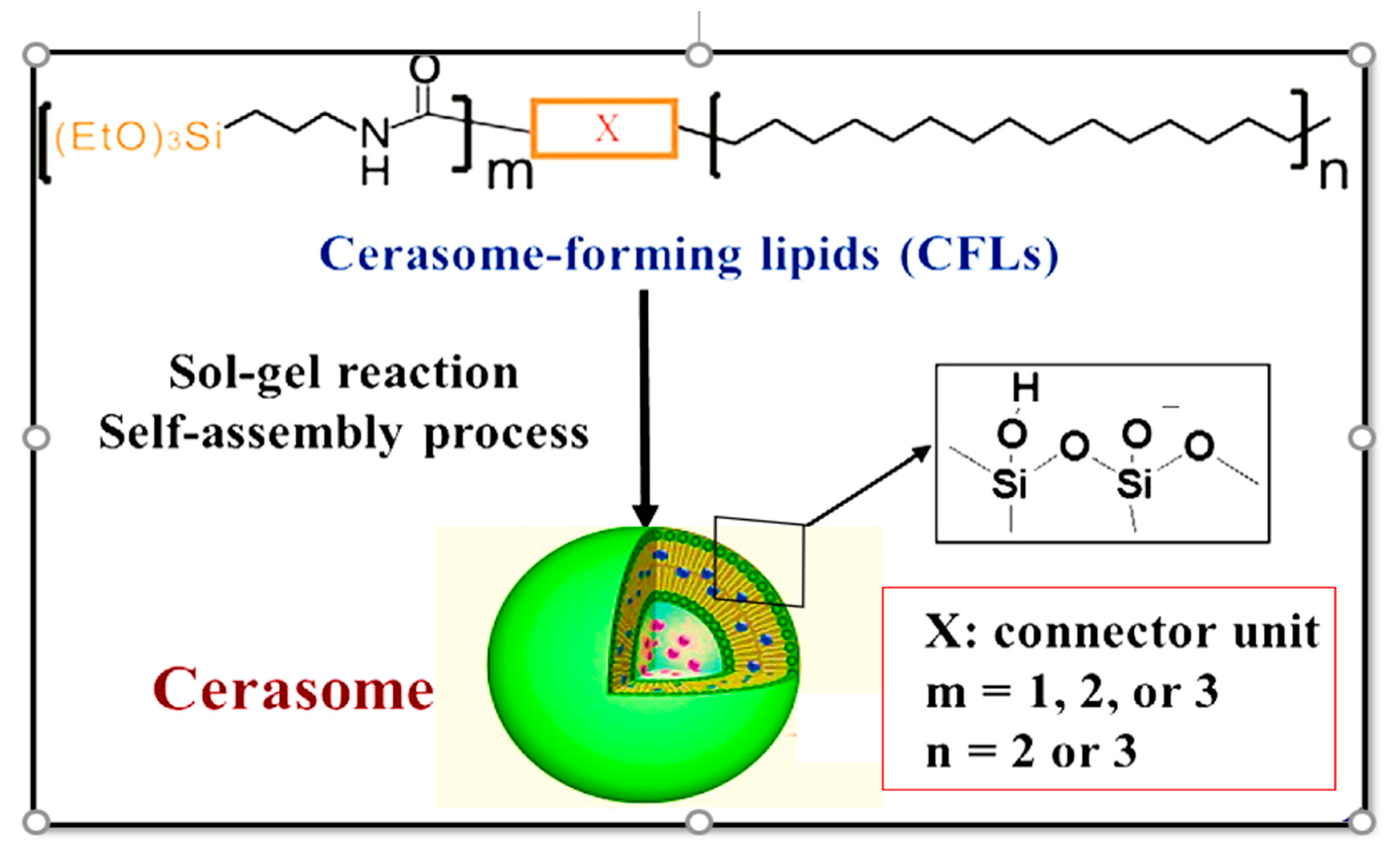
3.1. Design and Formulation of Cerasomes

Cerasomes are vesicles with a spherical structure and a lipid bilayer of around 4nm.The technique of fabricating cerasomes is analogous to liposomes and there are two methods involved in the preparation of cerasomes. The first method involves the dispersion of cerasome forming lipids (CFLs) into an aqueous solution followed by subjecting the mixture to a vortex shaker, resulting in the formation of a liposome using a sol-gel process (Figure 1). Further, the liposomal surface is subjected to a polyorganosilxane, resulting in the formation of cerasomes. Hence, cerasomes are obtained by the direct dispersion method. However, the ethanol sol injection method can also be used to prepare cerasome. In this method, an ethanolic solution of lipidic is incubated for a specific time period, resulting in the hydrolysis of CFLs. The resulting sol mixture is then injected into water followed by its incubation for an additional period of 24 h. Finally, the cerasomes are obtained, and this method is mainly taken up for those CFLs with poor solubility in water [14].

3.2. Characterization of Cerasomes Concerning Their Morphology

  • Morphology of the aggregate
Various microscopic measurements such as optical microscopy, transmission electron microscopy (TEM), atomic force microscopy (AFM), and scanning electron microscopy (SEM) are carried out to determine the aggregated structures of the cerasomes. As cerasomes exhibit a higher stability in terms of their morphology, they can easily be visualized by SEM. This property of cerasomes also allows precise TEM and AFM imaging. However, optical microscopy can visualize bigger micrometer-ranged cerasomes [14,15].
b.
Surface siloxane network
Cerasomes differ from liposomes because of an inorganic siloxane network at their surface. This inorganic unit provides a more excellent morphological stability to cerasomes. However, the presence of this unit can be analyzed using Fourier transform infrared spectroscopy. FTIR results indicate stretching bands around wavenumber 950 and 1100 cm−1 because of Si-OH and Si-O-Si groups, respectively [16].
c.
Phase separation and phase transition behavior
Differential scanning calorimetry (DSC) is used to evaluate the phase transition parameters of the cerasomes. The presence of the inorganic siloxane unit enhances the morphological stability of cerasomes because of which they do not change their integrity upon sonication conditions. Thus, the maximum temperature at the peak point(Tm) and the change in enthalpy (ΔH) are specific and indicative in case of cerasomes unlike the liposomes [15,17].

3.3. Cerasomes in Drug Delivery System

a.
Cerasomes as potent drug carriers
Cerasomes exhibit the property of encapsulation of hydrophilic, amphiphilic, and hydrophobic molecules. Researchers designed studies whereby a cationic cerasome was developed as a gene carrier. Further, a complex of cerasome and plasmid DNA exhibiting a viral size range of 70 nm was found to have a marked transfection performance such asserum compatibility, high activity, and less toxicity towards hepatic HepG2 and uterine HeLa cells. The complex was even efficient and strong at a stoichiometric nucleotide/lipid ratio. Thus, it was concluded that cerasomes can be utilized as a size-modulated carrier for diversified functional nucleic acids such as siRNAs and aptamers [18,19,20].
b.
Molecular devices for processing of information
Studies were designed that incorporateda cerasome and a lipid. The efficiency of the signal transduction was measured, which was found to be more efficient than the lipid peptide vesicle. Because of an elevated degree of the steroidal receptor phase separation, cerasomes possess a better signal transduction than the peptide lipid vesicle. As a result, a ternary complex of the receptor is formed, rendering cerasomes as molecular devices that can be used for processing information [21,22].

3.4. Cerasomes towards the Treatment of Diabetes

In the past few decades, it has been observed that a vast population across the globe suffers from diabetes, lowering the quality of human life. Insulin has evolved as an essential and principal therapy for treating different forms of diabetes mellitus. Due to its short half-life, high molecular weight, poor permeability, and lack of lipophilicity, there are difficulties associated with insulin delivery. However, research into the field was carried out and a hybrid form of liposome known as cerasome was developed. Methods such assol-gel and self-assembly strategy were incorporated for their formulation. The inorganic polyorganosiloxane surface of cerasomes is responsible for imparting a greater density and rigidity to these hybrid liposomes compared to silica nanoparticles. Thereby, it has held the attention as a novel delivery system for the release of drug at the target site. Studies and experiments were performed by loading insulin into the cerasomes. The release rate ofinsulin was modulated by the inclusion of dipalmitoylphosphatidylcholine (DPPC) into the cerasomes. A wide range of release profile alterations were observed upon varying the phospholipids’ molar ratios and the CFLs. The results of in vivo experiments suggested that cerasomes can be used as a potent nanocarrier that can enhance the rate of absorption of drugs post subcutaneous administration. Further, the in vitro study results indicated that insulin-loaded cerasomes exhibited marked stability upon long-term storage, more significant and sustained drug release, and minimal drug leakage compared to conventional liposomes. However, studies into the field were also carried out to determine the anti-diabetic potential of formulations incorporating resveratrol, which has high antioxidant properties. The in vivo results from STZ-induced rat models indicated that the cerasomal formulation lowered the blood glucose level in diabetic rats with elevated glucose levels. Further, a prolonged antioxidant property for some time of 24h was produced against oxidative stress. Hence, it was noted that cerasomes incorporating resveratrol can be effective against type 2 diabetes mellitus and oxidative stress associated with diabetes mellitus. Therefore, cerasomes can be used as effective and promising nanocarriers for peptide and protein drug delivery [23,24,25].

4. Upcoming Challenges and Future Prospects of Cerasomes in Diabetes Therapy

Due to their numerous excellent properties, liposomal hybrid cerasomes have made tremendous development as innovative drug delivery nanocarriers in biomedical medicine, particularly in diabetes diagnostics and treatment. Nonetheless, there are several limitations of the ongoing evolution of cerasomes. While drug carriers must be stable, the slow drug release rates from such stable cerasomes make it challenging to maintain an optimal concentration at the desired spot, limiting cancer treatment efficacy [3,5]. External stimuli (e.g., pH, temperature, and light) may be used to induce fast drug release from cerasomes to increase drug accumulation in tumor tissues and cells. Additionally, it is not easy to manage the size distribution of cerasomes using standard procedures, which substantially affects their pharmacokinetics in vivo. After preparing multilamellar cerasomes, the extrusion method may be utilized to formulate cerasomes with a uniform size distribution [6]. Furthermore, cerasomes facilitate targeting of specific sick cells within the disease location. Cerasomes, in particular, have the potential to function as intracellular delivery vehicles for proteins/peptides, antisense compounds, ribozymes, and DNA. Considering the several advantages of cerasomes, numerous prospective applications are possible. These applications include the use of cerasomes as diagnostic and therapeutic tools, sensors, information storage and processing systems, “smart” materials with regulated hydrophilicity/hydrophobicity, nanoscale robots, valves, and pistons, and others. The integration of chemically produced nanostructures with biomolecules to produce hybrid nanostructures with multifunctional properties is an exciting new direction for nanobiotechnology. Additionally, the majority of current research on cerasomes has been conducted in vitro, whereas in vivo investigations are far more challenging. Cerasomes’ long-term biocompatibility and pharmacokinetics in vivo should be thoroughly investigated [12,23]. To address the issues raised above, collaborative efforts and collaborations of researchers are required to accelerate the development of clinical applications of cerasomes as drug delivery vehicles.

5. Conclusions

Cerasomes are more stable than ordinary liposomes and have a lower stiffness and density than silica nanoparticle competitors. Cerasomes, on the other hand, are more biocompatible than silica nanoparticles. They typically have a high surface area, allowing for covalent and non-covalent surface functionalization with hydrophilic polymers, therapeutic moieties, and targeted ligands. Cerasomal medicines have fewer side effects and improved efficacy compared to their free counterparts. Cerasomes enable more effective drug delivery to disease locations due to their extended circulation residence time. Regardless of the advancements in the pharmaceutical industry, interesting future breakthroughs await us. Researchers predict that cerasomes will soon establish their full potential as a class of therapeutic agents, enabling considerable breakthroughs in treating diabetes. The development of cerasomes for the treatment of diabetes has provided a new window for researchers to overcome the different problems connected to current therapies.

Author Contributions

Conceptualization: K.P. and R.S.; Methodology: K.P. and R.S.; Manuscript review & editing: K.P., R.S., M.Z.A. and A.D. All authors have read and agreed to the published version of the manuscript.

Funding

This research received no external funding.

Institutional Review Board Statement

Not available.

Informed Consent Statement

Not available.

Data Availability Statement

Not available.

Conflicts of Interest

The authors declare no conflict of interest.

References

  1. Karamanou, M.; Protogerou, A.; Tsoucalas, G.; Androutsos, G.; Poulakou-Rebelakou, E. Milestones in the history of diabetes mellitus: The main contributors. World J. Diabetes 2016, 7, 1–7. [Google Scholar] [CrossRef] [PubMed]
  2. Simos, Y.V.; Spyrou, K.; Patila, M.; Karouta, N.; Stamatis, H.; Gournis, D.; Dounousi, E.; Peschos, D. Trends of nanotechnology in type 2 diabetes mellitus treatment. Asian J. Pharm. Sci. 2021, 16, 62–76. [Google Scholar] [CrossRef] [PubMed]
  3. Sudhakar, K.; Mishra, V.; Hemani, V.; Verma, A.; Jain, A.; Jain, S.; Charyulu, R.N. Reverse pharmacology of phytoconstituents of food and plant in the management of diabetes: Current status and perspectives. Trends Food Sci. Technol. 2021, 110, 594–610. [Google Scholar] [CrossRef]
  4. IDF Diabetes Atlas 9th Edition. Global Fact Sheet. 2019. Available online: https://diabetesatlas.org/upload/resources/material/20201028_180116_Global-factsheet-final.pdf (accessed on 13 July 2021).
  5. IDF Diabetes Atlas 9th Edition. Demographic and Geographic Outline. 2019. Available online: https://diabetesatlas.org/en/sections/demographic-and-geographic-outline.html (accessed on 13 July 2021).
  6. He, L.; Sabet, A.; Djedjos, S.; Miller, R.; Sun, X.; Hussain, M.A.; Radovick, S.; Wondisford, F.E. Metformin and Insulin suppress hepatic gluconeogenesis through phosphorylation of CREB binding protein. Cell 2009, 137, 635–646. [Google Scholar] [CrossRef]
  7. Lin, S.H.; Cheng, P.C.; Tu, S.T.; Hsu, S.R.; Cheng, Y.C.; Liu, Y.H. Effect of Metformin monotherapy on serum lipid profile in statin-naïve individuals with newly diagnosed type 2 diabetes mellitus: A cohort study. PeerJ 2018, 6, e4578. [Google Scholar] [CrossRef]
  8. Rena, G.; Hardie, D.G.; Pearson, E.R. The mechanisms of action of metformin. Diabetologia 2017, 60, 1577–1585. [Google Scholar] [CrossRef]
  9. Shurrab, N.T.; Arafa, E.-S.A. Metformin: A review of its therapeutic efficacy and adverse effects. Obes. Med. 2020, 17, 100186. [Google Scholar] [CrossRef]
  10. Chen, Y.; Shan, X.; Luo, C.; He, Z. Emerging nanoparticulate drug delivery systems of metformin. J. Pharm. Investig. 2020, 50, 219–230. [Google Scholar] [CrossRef]
  11. Proks, P.; Reimann, F.; Green, N.; Gribble, F.; Ashcroft, F. Sulfonylurea stimulation of insulin secretion. Diabetes 2002, 51 (Suppl. 3), S368–S376. [Google Scholar] [CrossRef]
  12. Sola, D.; Rossi, L.; Schianca, G.P.C.; Maffioli, P.; Bigliocca, M.; Mella, R.; Corlianò, F.; Fra, G.P.; Bartoli, E.; Derosa, G. Sulfonylureas and their use in clinical practice. Arch. Med. Sci. 2015, 11, 840–848. [Google Scholar] [CrossRef]
  13. Jin, Y.; Li, Y.; Pan, H.; Dai, Z. Liposomal nanohybrid cerasomes for controlled insulin release. RSC Adv. 2014, 4, 42808–42815. [Google Scholar] [CrossRef]
  14. Yue, X.; Dai, Z. Recent advances in liposomal nanohybrid cerasomes as promising drug nanocarriers. Adv. Colloid Interface Sci. 2013, 207, 32–42. [Google Scholar] [CrossRef] [PubMed]
  15. Kikuchi, J.; Yasuhara, K. Microscopy & micro/nano-imaging techniques: TEM. In Supramolecular Chemistry: From Molecules to Nanomaterials, 1st ed.; Steed, J.W., Gale, P.A., Eds.; John Wiley & Sons: Chichester, UK, 2012; Volume 1, pp. 231–250. [Google Scholar]
  16. Katagiri, K.; Hashizume, M.; Ariga, K.; Terashima, T.; Kikuchi, J. Preparation and characterization of a novel organic-inorganic nanohybrid “cerasome” formed with a liposomal membrane and silicate surface. Chem. Eur. J. 2007, 13, 5272–5281. [Google Scholar] [CrossRef] [PubMed]
  17. Murakami, Y.; Kikuchi, J. Supramolecular assemblies formed with synthetic peptide lipids. Functional models of biomembranes and enzymes. In Bioorganic Chemistry Frontiers, 1st ed.; Dugas, H., Ed.; Springer: Berlin, Germany, 1991; Volume 2, pp. 73–113. [Google Scholar]
  18. Matsui, K.; Sando, S.; Sera, T.; Aoyama, Y.; Sasaki, Y.; Komatsu, T.; Terashima, T.; Kikuchi, J. Cerasome as an infusible, cell-friendly, and serum-compatible transfection agent in a viral size. J. Am. Chem. Soc. 2006, 128, 3114–3115. [Google Scholar] [CrossRef] [PubMed]
  19. Sasaki, Y.; Matsui, K.; Aoyama, Y.; Kikuchi, J. Cerasome as an infusible and cell-friendly gene carrier: Synthesis of cerasome-forming lipids and transfection using cerasome. Nat. Protoc. 2006, 1, 1227–1234. [Google Scholar] [CrossRef] [PubMed]
  20. Matsui, K.; Sasaki, Y.; Komatsu, T.; Mukai, M.; Kikuchi, J.; Aoyama, Y. RNAi gene silencing using cerasome as a vial-size siRNA-carrier free from fusion and crosslinking. Bioorg. Med. Chem. Lett. 2007, 17, 3935–3938. [Google Scholar] [CrossRef] [PubMed]
  21. Sasaki, Y.; Yamada, M.; Terashima, T.; Wang, J.-F.; Hashizume, M.; Fan, S.-D.; Kikuchi, J. Construction of intermolecular communication system on “cerasome” as an organic-inorganic nanohybrid. Kobunshi Ronbunshu 2004, 61, 541–546. [Google Scholar] [CrossRef]
  22. Dai, Z.-F.; Tian, W.-J.; Yue, X.-L.; Zheng, Z.-Z.; Qi, J.-J.; Tamai, N.; Kikuchi, J. Efficient fluorescence resonance energy transfer in highly stable liposomal nanohybrid cerasome. Chem. Commun. 2009, 15, 2032–2034. [Google Scholar] [CrossRef]
  23. Yucel, C.; Karatoprak, G.S.; Aktas, Y. Nanoliposomal resveratrol as a novel approach to treatment of diabetes mellitus. J. Nanosci. Nanotechnol. 2018, 18, 3856–3864. [Google Scholar] [CrossRef]
  24. Das, R.J.; Baishya, K.; Pathak, K. Recent advancement of lipid drug conjugate as nanoparticulate drug delivery system. Int. Res. J. Pharm. 2013, 4, 73–78. [Google Scholar]
  25. Roumie, C.L.; Hung, A.M.; Greevy, R.A.; Grijalva, C.G.; Liu, X.; Murff, H.J.; Elasy, T.A.; Griffin, M.R. Comparative effectiveness of sulfonylurea and metformin monotherapy on cardiovascular events in type 2 diabetes mellitus: A cohort study. Ann. Intern. Med. 2012, 157, 601–610. [Google Scholar] [CrossRef] [PubMed]
Figure 1. Formation of cerasomes by sol-gel reaction(direct dispersion method).
Msf 10 00005 g001
Disclaimer/Publisher’s Note: The statements, opinions and data contained in all publications are solely those of the individual author(s) and contributor(s) and not of MDPI and/or the editor(s). MDPI and/or the editor(s) disclaim responsibility for any injury to people or property resulting from any ideas, methods, instructions or products referred to in the content.

